in vivo and in vitro analyses of an intron-encoded dna
TRANSCRIPT
Nucleic Acids Research, Vol. 18, No. 19 5659
In vivo and in vitro analyses of an intron-encoded DNAendonuclease from yeast mitochondria. Recognition site bysite-directed mutagenesis
B.Sargueil, D.Hatat, A.Delahodde and C.JacqLaboratoire de G6netique Moleculaire, CNRS UA 1302, Ecole Normale Superieure, 46 rue d'Ulm,75230 Paris Cedex 05, France
Received July 27, 1990; Revised and Accepted September 3, 1990
ABSTRACT
The pal 4 nuclease (termed ISce II) is encoded in thegroup I al 4 intron of the COX I gene of Saccharomycescerevisiae. It introduces a specific double-strand breakat the junction of the two exons A4-A5 and thusmediates the insertion of the intron into an intronlessstrain. To define the sequence recognized by pal 4 weintroduced 35 single mutations in its target sequenceand examined their cleavage properties either In vivoin E. coll (when different forms of the pal 4 proteinswere artificially produced) or in vitro with mitochondrialextracts of a mutant yeast strain blocked in the splicingof the al 4 intron. We also detected the pal 4 DNAendonuclease activity in extracts of the wild type strain.The results suggest that 6 to 9 noncontiguous basesIn the 17 base-pair region examined are necessary forpal 4 nuclease to bind and cleave its recognition site.We observed that the pal 4 nuclease specificity can besignificantly different with the different forms of theprotein thus explaining why only some forms are highlytoxic in E. coll. This study shows that pal 4 recognitionsite is a complex phenomenon and this might haveevolutionary implications on the transfer properties ofthe intron.
INTRODUCTION
The majority of group I and group II introns in organelle genescontain an intron open reading frame (ORF) which is, most ofthe time, in phase with the upstream exons, and which codesfor proteins involved in recombinational processes of RNA andDNA sequences. These different proteins are proposed to act asRNA maturases (1), DNA endonucleases (2, 3), DNArecombinases (4), and as reverse transcriptases (5). The differentactivities are mostly poorly understood. Concerning group Iintronic proteins, very good evidence of their RNA maturase orDNA endonuclease activities have been presented (6, 7, 8, 9,10). These two activities, which play a key role in two concertedevents of yeast-mitochondrial-genome structure and expression,are especially interesting to study.
DNA endonucleases (8, 9) are, at least in the case of groupI introns, responsible for intron insertion by specificallyintroducing a double strand cut in the vicinity of the exon junction.
In the two better documented cases, the mitochondrial DNAendonucleases recognize an 18 base-pair sequence overlappingthe two flanking exons (2, 11, 12). In contrast, a prokaryoticintron-encoded endonuclease from a mobile group I intron (13)seems to have a larger recognition sequence and it cleaves at adistance (up to 24 bp) from the insertion site (14).
RNA maturases (4, 7) are required for the splicing of one ortwo introns. RNA maturases are found in three out of the eightgroup I intron ORF in the yeast strain 777-3A. None of thesematurase-containing introns have been shown to be mobile (15)and at least in the case of the pbl 4 RNA maturase no DNAendonuclease activity has been found (11). This suggests that,at least in the wild-type cell, the two activities might be exclusiveand carried out by distinct proteins.
Interestingly, yeast mitochondrial DNA endonucleases andRNA maturases belong to the same family of group 1(16) intron-encoded proteins. In fact, two closely related intron-encodedproteins happens each to carry one of the two type of activities.They are coded in the bl 4 intron of cytochrome b gene and inthe al 4 intron of COXI gene from the Saccharomyces cerevisiaestrain 777-3A. pbl 4 is required for the in vivo splicing of thetwo introns bl 4 and al 4 and has thus a pleiotropic RNA maturaseactivity (17, 18, 19); the pal 4 protein has a specific DNAendonuclease activity and it promotes the insertion of the DNAintron into the genome of an intronless strain (12, 11). We haveshown, by making universal genetic code equivalents of themitochondrial genes, that the RNA maturase activity of pbl 4is present in a 250 amino acid long protein that can becytoplasmically translated and targeted to the yeast mitochondria.The DNA endonuclease activity can be observed with a 254amino acid long pal 4 protein expressed in E. coli (11). In therest of this work we will refer to the two short intronic productsmentioned above as the RNA maturase pbl 4 and as the DNAendonuclease pal 4 . However, it must be kept in mind that, invivo, these two proteins are translated as long chimeric proteinsencompassing the upstream exons. If these precursors arecertainly processed in the splicing mutants, nothing is knownabout the wild type situation because of the scarcity of thoseproteins (20).
In this study, we have extended the analysis of the in vivo andin vitro site recognition properties of the pal 4 protein. We havetaken advantage of the engineered pal 4 coding sequence
Downloaded from https://academic.oup.com/nar/article-abstract/18/19/5659/1104842by gueston 15 February 2018

5660 Nucleic Acids Research, Vol. 18, No. 19
expressed in E. coli to analyze, with a double plasmid system,the properties of mutated forms of the recognition sequence.Surprisingly, many single mutations within the 19 bp regionexamined do not affect cleavage by pal 4. These recognitionproperties are however slightly different between variants of theE. co/j-made protein. We have also compared their in vivoproperties to the in vitro properties of a crude mitochondrialextract of a mutant accumulating the pal 4 protein. These studies,which examine the recognition properties of an intronic DNAendonuclease that acts upon an exonic coding sequence, seemto indicate some relationships between the codon structure andthe DNA endonuclease specificity which might have evolutionaryimplications. Altogether this study shows that pal 4 siterecognition is a complex phenomenon similar to the one describedfor the HO nuclease (21).
MATERIALS AND METHODSStrains and plasmidsE. coli strain GY7511 (gal K, ilv, his, ( X cl 857ts, N7 N53,A Bam AHlj) (22), which produces cI857, was used for the invivo assays. RZ 1032 (HfrKL16 PO/45 lysA(6162), dutl, ungl,thil, relAl, Zdb-279: :TnlO supE44) (23) and HB2154 (K12,A(lac-pro), SupE, thi/F' proA+B + , lacP, lacZ DM15, mutl::TnlO) (24) were used for oligonucleotide-directed mutagenesis.
Yeast strains used for mitochondrial extracts were: CW02(a,ade2-l, trpl-1, leu2-3, 112 his3-ll, 15 ura3 CanR, (intronlesscytochrome b gene constructed by M. Labouesse) (18); 777-3A(a, adel, opl, (rho+)) (25); CO66 (constructed and kindly givenby P. Netter), which is isonuclear with 777-3A and is a doublemitochondrial mutant (box2-G2590 and 'box9 like' G192) (26);KM 91 is a diploid from the cross of 777-3A and KL14-4A/60/1(a, Hisl, trp2 (rho 0)).
Plasmids pUC A4-A5 and pGP al 4 (figure 1) were describedin Delahodde et al. (11).
Oligonucleotide mediated mutagenesis
The principle of this method has been described by Kunkel etal. (23). It was used to mutate the ACT codon at position 145to a CTT codon. The deoxyuridine-substituted template wasproduced in the RZ1032 strain and isolated following the CTABprocedure (27). Annealing of the phosphorylated oligonucleotide(5' GATATAGATTACTTAATAAAACTGG 3') to the single-stranded template, the elongation reactions and the ligationreactions were according Sambrook et al. (28). The reactionproducts were introduced into the HB 2154 strain and sevenclones randomly chosen were sequenced.
Construction of randomly mutagenized A4-A5 sitesOligonucleotides were synthesized on an automated DNAsynthesizer (Applied Biosystems 381 A). They contain the A4-A5target sequence plus terminal sequences to allow their insertioninto the EcoRI site of the pUC19. Four rounds of syndiesis weremade in which each base (A, G, T, C) was successivelyindividually degenerated. In order to have an average of one basesubstitution per molecule, the mixture of phosphoramidites wasas following: for A degeneracy 80%A, 7% of each other, forG degeneracy 70% G, 10% of each other, for C degeneracy 65%C, 12% of each other, for T degeneracy 70%T, 10% of eachother. Those percentages were calculated with respect to the totalamount of each nucleotide in the sequence. Five mutants which
could not be obtained by this random approach were completelysynthesized.
DNA sequencing
After mutagenesis DNA sequence was checked using chain-termination sequencing (29) according the sequenase protocol(USBC).
Endonuclease assays in E. coli
GY 7511 cotransformant clones containing pGPal 4 and pUC19A4-A5 were grown to stationnary phase at 28 °C. Denaturationof cI857 and al 4 production was induced by incubating the cellsfor 2 hours at 42°C (37° or 40°C has the same effect). PlasmidDNA was then isolated as described in Delahodde et al. (11).Plasmids were digested with Sspl and analyzed on a 0.9% agarosegel. For Southern blot analysis, Hybond N+ membrane(Amersham) was used to bind DNA that was transferred witha Vacugene apparatus (LKB). The probe (pUC19 A4A5) was32P labeled by random priming with the multiprime-DNA-labeling system (Amersham). Hybridization was performed asdescribed by Church et al. (30).
Mitochondrial extract preparation and endonuclease assay
Cells were grown in YEPGal (1 % yeast extract, 1 % peptone,2% galactose) and harvested during exponential growth.Mitochondria were isolated as described by Huspedth et al. (31)and further purified on a sucrose discontinuous gradient (20%,36% and 50% sucrose). Mitochondria were collected, lysed andthe endonuclease assay was performed according to Wenzlau etal. (12). The mit-t- strain extracts (results shown in Fig. 4) weredialyzed against Tris-HCl 50mM pH 7,5; NaCl lOOmM; EDTA2 mM; DTT 2mM; PMSF 0. lmM and 10% glycerol before theDNA endonuclease assay.
Pulsed-field electrophoresis
E. coli genomic DNA was prepared by the method of Caron (F.Caron personal communication) which is as follow . Cells (10ml) were harvested during exponential growth phase, washedtwice in TE buffer and suspended in 1 ml of buffer A (10%sucrose, 50mM Tris-HCl, pH 8) with 4 mg of lysozyme. Thecell suspension was then mixed with 2 ml of low melting agarose.These agarose plugs were incubated in 0.5M EDTA, pH 9, 1 %SDS, 1 % sarcosyl, and 1 mg/ml proteinase K overnight at 50cC.Before restriction, plugs were washed twice with TE buffer andthen once with the incubation buffer. Restriction digests with Nodwere done at 37°C overnight.
Pulsed-field electrophoreses were performed according to theCHEF (Contour-clamped Homogenous Electric field) principle(32). We used the LKB 2015 pulsaphor electrophoresis unit andthe electrophoretic conditions were the followings: 1.4% agarosegel in 0.25 x TBE buffer (22.5 mM Tris-borate, 0.5 mM EDTA),180 volts during 38 hours with a switching of 30 seconds at 10°C.
RESULTSIn vivo and in vitro cleavage of different mutated A4-A5recognition sites. Activity of the different forms of the E. colisynthetized pal 4 DNA endonuclease and activity of crudemitochondrial extracts
It was previously suggested (11, 12) that the recognition site ofthe pal 4 DNA endonuclease was in an 18 bp long sequenceoverlapping the A4-A5 splice junction. We constructed 35 single
Downloaded from https://academic.oup.com/nar/article-abstract/18/19/5659/1104842by gueston 15 February 2018

Nucleic Acids Research, Vol. 18, No. 19 5661
mutations within the A4-A5 recognition site and we introducedthem into a pUC vector to analyze, wether they are recognizedand cleaved when the pal 4 protein is produced in the same cell(see figure 1). By using this in vivo assay, three different formsof the pal 4 protein were examined, pal 4/A (258) correspondsto the translation, in E. coli, of the 3' terminal end (258 aminoacids) of the mitochondrial intron open reading frame in whichthe three codons UGA, AUA and CUN have been changed intotheir equivalents in the universal genetic code i.e. UGG, AUGand ACN, respectively (33). This pal 4/A protein has alreadybeen shown to be toxic to the bacteria if its gene is expressedand can only be stably maintained in E. coli in the presence oftightly blocked promoters. In the course of the construction ofthis gene (11) we observed that a much less toxic form of theprotein is obtained if the CUU codon (amino acid 145 whennumbered from the first amino acid of the intronic ORF) is notcorrected, that is if the protein contains a Leu at position 145instead of a Thr as suggested by the mitochondrial genetic code.We thus made pal 4/B which is a T to L (145) mutated formof the pal 4/A. We also introduced a second mutation (E toK, 122) in a shorter form (249 amino acids) of the pal 4/B proteinand this leads to the protein pal 4/C. This last mutation waschosen because it reveals, in vivo, an RNA maturase-like property(34) which might alter the DNA endonucleolytic activity.
These three forms of the pal 4 coding sequence were expressedin the presence of different mutated forms of the A4-A5 targetsequence and the cleavage efficiencies were compared to that ofthe wild-type sequence. Examples of such analyses are given inFigure 2. In most of the cases where the target site is cleaved,two stoichiometric homogeneous fragments appear at 2150 bpand 570 bp. This reveals the specific DNA endonuclease activityand indicates that all these DNA ends are fairly stable in E. coli.
Such an analysis has also been conducted, in vitro, on all 35mutated target sites with crude mitochondrial extracts of a strainin which RNA splicing of the intron al 4 is blocked because ofthe absence of the intron bl 4 (strain CW02 constructed by M.Labouesse; (35)). These in vitro results can be compared withthe previous in vivo approach where cleavage of the wild-typetarget sequence is similar. It appears that some mutations behavesimilarly in regard to the four different conditions (for instanceG+6) whereas some positions seem to be distinguished by thedifferent forms of the DNA endonuclease activity (for instanceT+5). Systematic analysis leads us to four specificity patternsrelated to the three forms of the pal 4 protein made in E. coliand to the mitochondrial extract. These results schematicallydistinguish three states in which the recognition site is cut likethe wild type sequence (long bars), the recognition site is partiallycut (middle bars) and the recognition site cut cannot be detected
Seal 2177
BstEII
PUC19A4A5 iacl
2720 bp
EcoRl 396
A4A5 recognit ion s i te
EcoRl 426
Hindi 115167
Clal5890 EcoRl 0
BamHI 4000
BstEII al4 endonuclease
51 C T T T
3' G fl fl fl
G f l f i G T f l T 3 '
flCTTCflTfi5-
^ P s t l1 100
dORI' P15A
2720 bp-»-2150 bp->-
1800 bp-*-
920 bp- * -570 bp- * -
UJ
toCDin
ud
U f
min
CL
UJ
inmin
o
oa
—
_
-10 -9 -6 -7 -6 -5 - 1 10
al4 Intron
Figure 1. Schematic approach to study, in vivo, the DNA endonucleolytic activity of the pal 4 intronic protein (I-Scell). Two compatible plasmids (A),one of themcontaining either the gene coding for the pal 4 DNA endonuclease, which was adapted to the 'universal' genetic code, and the other one the target sequence A4-A5,were cotransformed into the same E. coli strain. The pal 4 coding sequence was under the transcriptional control of the lambda PR promoter. It was repressed bythe thermosensitive CI 857 repressor, which was translated from both the same plasmid and strain GY7511. The mutated A4-A5 recognition sequence (Q was madefrom synthetic oligodeoxynucleotides and inserted into the EcoRl site of the pUC 19 polylinker sequence. The endonucleolytic activity of the E. co/i-made pal 4protein was estimated from Southern blot analyses (B) of the extracted DNA. Accurate characterization of the cleavage site was ensured by taking advantage ofthe unique BstE II site in the vicinity of the pal 4 cut site (Q.
Downloaded from https://academic.oup.com/nar/article-abstract/18/19/5659/1104842by gueston 15 February 2018

5662 Nucleic Acids Research, Vol. 18, No. 19
|pa!4/A (258)1
570
pal4/C (249,K122, L145)
iMitochondrial extract (CW02)|
2720 bp1800 bp
920 bp
Figure 2. A Southern blot analysis of DNA extracted from E. coli transformed with the two plasmids pUC A4-A5 and pGpal 4 (see legend of Fig. 1). 35 singlemutants in the 19 bp long target sequence were examined either in vivo (left) or in vitro (right) for their ability to be cleaved by the E. coli -made pal 4 protein(left) or by a mitochondrial extract of a yeast strain blocked in aI4 intron splicing (right). Results of only 11 mutants, named by their position relatively to the cleavagecut (see Fig. 1), are shown with the 258 amino acid long protein (pal 4/A 258) or its shorter mutated form (pal 4/C see text); the extracted DNA was cut withSsp I and hybridized with a pUC 19 probe. The same approach was used in the in vitro analysis (right) except that the DNA was cut with Sea I. The amount oftotal protein estimated by the BCA protein assay (Pierce Chemical Company) was 16 /jg/assay.
(small white bars). Considering the E. coli forms of the protein,it is clear that pal 4/A is the least specific in that only threepositions (—5; —2 and +4) are critical. At the opposite, pal 4/Ctolerates a few variable positions (about four out of eighteenpositions) and at least nine positions are stringent positions wherenone of the examined base changes was tolerated, pal 4/B behavesintermediately and, interestingly, exhibits a pattern somewhatsimilar to that of the mitochondrial extract (Figure 3,B and D).
It should be stressed that the three types of in vivo experiments(A,B,C) described above have been carried under rigorouslyidentical conditions (see Materials and Methods). Only the levelof the pal 4 protein could, a priori, vary in the cell due to eitherdifferential production or stability. This, however, is unlikelysince we observed an equivalent activity of the different formson the wild-type recognition sequence (Figure 2), and gelelectrophoresis analyses of the different transformants did notshow significant differences in the level of pal 4 (data not shown).
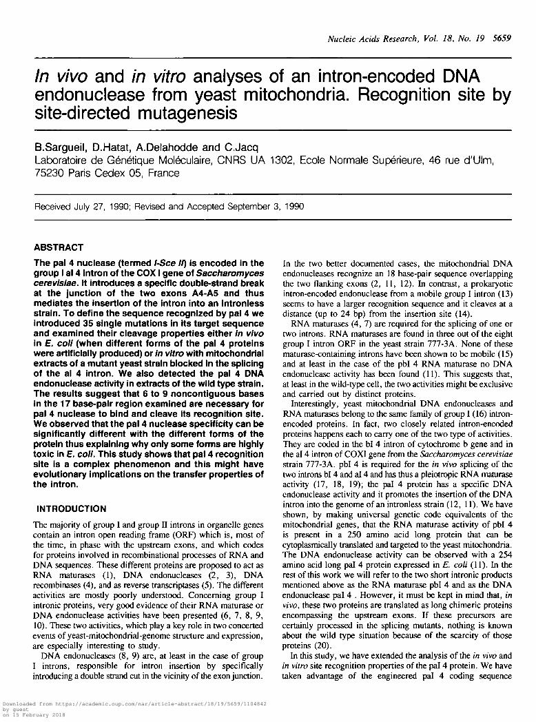
The yeast pal 4 DNA endonuclease activity can be observedin different mitochondrial genetic contextsWenzlau et al. (12) observed in lysates of mitochondria froma mutated strain an al 4-encoded, sequence-specific, DNAendonuclease activity which they did not detect in thecorresponding wild-type strain. Following a similar protocol, welooked for the presence of the same specific DNA endonucleaseactivity in four different genetic contexts (Figure 4). As expected,the strain CW02, which is devoid of the bl 4 RNA maturase andthus cannot splice the al 4 intron(therefore accumulating proteinproducts of that intron), exhibits the presence of an importantDNA endonuclease activity. The same specific activity is alsopresent in the double mutant C066, where two homologousintronic proteins, the bl 4 RNA maturase and the al 4 DNAendonuclease, accumulate. More surprisingly, we could observe(Figure 4) the al 4-specific DNA endonuclease activity in the
mitochondrial extracts of two wild-type strains: the haploid111-3k and the diploid KM91. This last observation is all themore interesting since we know that the al 4 intronic translationproduct responsible for the DNA endonuclease activity iscertainly, like the bl 4 translation product, in very low amountsin the wild type cell (20). This clearly suggests that themitochondrial protein has a very high specific activity or thatthe mitochondria contain an enhancer of the DNA endonucleaseactivity; the absence of this enhancer or the presence of aninhibitor in the strain examined by Wenzlau et al. (12) mightexplain the discrepancy between the two studies. It is difficultto analyze quantitatively the results of Figure 4 since we cannotestimate the concentration of the DNA endonuclease which hasnot yet been characterized (see discussion). We can only makea rough estimation of the endonuclease activity by consideringthe total amount of protein used in the different assays. Clearly,when the bl 4 RNA maturase accumulates in the same cell, itdoes not affect the DNA endonuclease activity (strain C066) andthe level of DNA endonuclease activity in the wild type cell islower than that of the mutant cell (CW02).
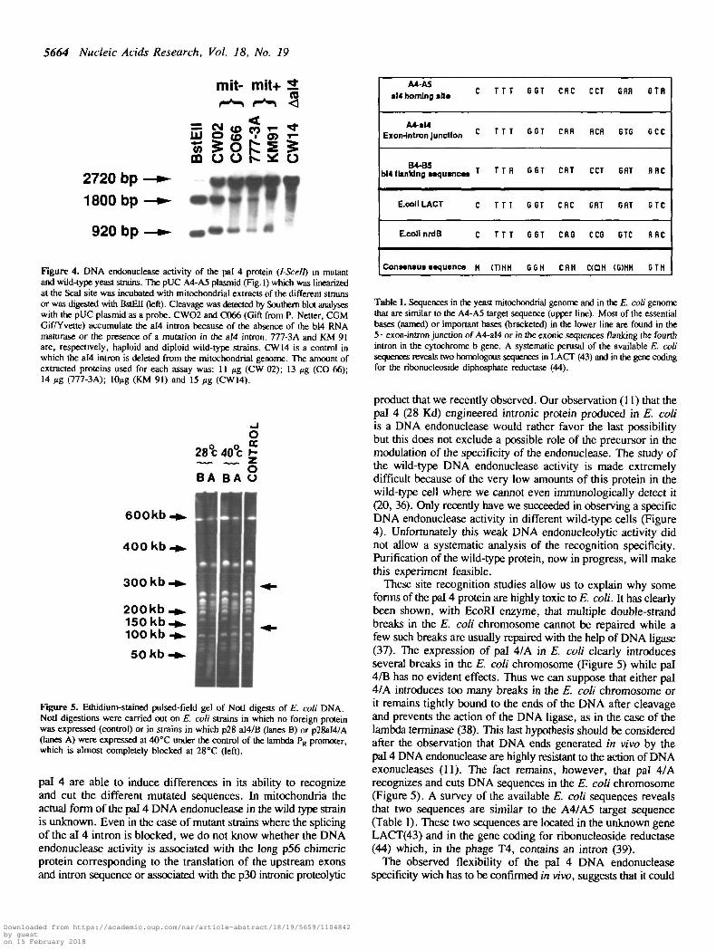
Effects of the pal 4 DNA endonuclease activity on E. coli
We have previously observed (11), that the production in E. coliof the wild type version (pal 4/A) of a putative mitochondrialal 4 intronic translational product is highly toxic to E. coli. Wenoticed that the mutated form pal 4/B and pal 4/C can beproduced without affecting bacterial growth. To further analyzethe effects of the pal 4 DNA endonuclease on E. coli, weexamined the structure of the E. coli chromosome after differentforms of the pal 4 protein were produced. The effects of the DNAendonuclease activity on the E. coli chromosome were analyzedby pulsed field gel electrophoresis (Figure 5) of Not I digests.In a control experiment, where a truncated form of the pal 4protein is produced, a stable pattern of the E. coli DNA can be
Downloaded from https://academic.oup.com/nar/article-abstract/18/19/5659/1104842by gueston 15 February 2018

p al4/A (258)
Nucleic Acids Research, Vol. 18, No. 19 5663
[Ch p al4/C (249, K122, L145)
rn|c
Cl T T Tl G G Tl CGl fl fl fll C C fll G
IIIfl fl C Cl Tl G fl fll G T fll T
|T Gl G G fll C T Tl C fl Tl fl-10-9 -8-7-6 -5 -1 -3 -2 -I
G flC T1 5
Tfl8 9 10 - 1 0 - 9 - 8 - 7 - 6 - 5 - 1 - 3 - 2 - 1 1 2 3 1 5 6 7 8 9 1 0
p al4/B (258, L145) - liltochondrial extract(CW 02)
- 1 0 - 9 - 8 - 7 - 6 - 5 - 1 - 3 - 2 - 1 1 2 3 1 3 6 7 8 9 10 . ( 0 - 9 - 8 - 7 - 6 - 5 - 1 - 3 - 2 - 1 I 2 3 1 5 6 7 8 9 10
Figure 3. DNA endonucleolytic properties of the different forms of the pal 4 intronic protein on different single mutants of its A4-A5 target sequence. Long darkbars correspond to single mutants that were cut like the wild-type A4-A5 target sequence. Half-length grey bars correspond to mutants that affect the cleavage efficiencyof the pal 4 DNA endonuclease. Short white bars correspond to single mutants that were not cleaved. All the indicated mutated sequences were from the upperstrand of the target sequence, pal 4/AJB/C are different forms of the E. co/i-made DNA endonuclease (see text) and the corresponding results have been obtainedfrom in vivo experiments. Mitochondrial extract properties have been examined in vitro as indicated in Fig. 2 and in Materials and Methods.
observed which is quite similar to the pattern obtained when thepal 4/B protein is produced. In contrast, the production of theprotein pal 4/A leads to dramatic effects on the E. colichromosome structure. When the E. coli cells are left at 40°Cfor 10 minutes, specific cleavage products clearly appear (arrowsin Fig. 5). Longer periods at 40°C lead to the completedegradation of the E. coli chromosome (data not shown).
DISCUSSION
In this work we have examined, in vivo or in vitro, the recognitionsite specificity of the intronic DNA endonuclease pal 4 (I-Scell)as a function of the different forms of the protein made in E.coli or from cellular extracts of a mitochondrial mutant.Surprisingly, we found that recognition of the pal 4 site is acomplex phenomenon. The different 17 bp forms of the minimalrecognition site do not have an equivalent role on the controlof the DNA endonucleolytic activity. By taking advantage of the35 single mutations that we constructed we observed that, in fact,many base changes do not affect cleavage in vivo or in vitro.It looks as if only a few base pairs scattered along the 17 bpminimal recognition site would be essential. Considering ourresults, a mean consensus sequence mainly based on the
properties of the yeast mitochondrial extract (figure 3), can bewritten as follows:
5 ' N . ( T ) N N . G G N . C A N . C ( C ) N . G N N . ( G ) T N 3 '
in which the indicated bases are important since at least one basechange in these positions severely affects the cleavage efficiency(bases between brackets correspond to less critical positions).Interestingly, the E. co/Z-made protein pal 4/B behaves ratherlike the DNA endonuclease from mitochondrial extracts whereasits 'wild type' form, pal 4/A, has a very much reduced specificity.In the case of pal 4/A only 5 positions in the target sequenceseem to be critical (G+4, A-2, C-3, G-5, G-6). The onlydifference between the two genes coding either for pal 4/A orpal 4/B is in the codon at position 145 which is either ACU orCUU. This indicates that when the CUU codon is translated asa Leu, which is at variance with the accepted view of themitochondrial genetic code (33), the properties of thecorresponding E. coli-made protein (pal 4/B) are rather similarto those of the mitochondrial protein accumulating in the yeastmutant CW02. This again (see (11)) raises some doubts on themeaning of the CUU codon in mitochondria.
Although it is tempting to speculate about the variations in thespecificity of the different proteins we will only stress the factthat only very limited variations in the structure of the protein
Downloaded from https://academic.oup.com/nar/article-abstract/18/19/5659/1104842by gueston 15 February 2018
5664 Nucleic Acids Research, Vol. 18, No. 19
tEll
ffi
mit-
CM (O
o o
mit+
O2720 bp1800 bp
920 bp
Figure 4. DNA endonuclease activity of the pal 4 protein (I-Scelt) in mutantand wild-type yeast strains. The pUC A4-A5 plasmid (Fig. 1) which was linearizedat the Seal site was incubated with mitochondrial extracts of the different strainsor was digested with BstETJ (left). Cleavage was detected by Southern Mot analyseswith the pUC plasmid as a probe. CWO2 and C066 (Gift from P. Netter, CGMGif/Yvette) accumulate the aI4 intron because of the absence of the bl4 RNAmaturase or the presence of a mutation in the aI4 intron. 777-3A and KM 91are, respectively, haploid and diploid wild-type strains. CW14 is a control inwhich the aI4 intron is deleted from the mitochondrial genome. The amount ofextracted proteins used for each assay was: 11 n% (CW 02); 13 fig (CO 66);14 us, (T77-3A); 10^g (KM 91) and 15 /*g (CW14).
28% 40°c £
BA BA 8
600kb-*•
400 kb-»*
300 kb-* -
200kb_•>.150 kb- * .100 kb -»•
50 kb-* -
Figure 5. Ethidium-stained pulsed-field gel of NotI digests of E. coli DNA.NotI digestions were carried out on E. coli strains in which no foreign proteinwas expressed (control) or in strains in which p28 a!4/B (lanes B) or p28aI4/A(lanes A) were expressed at 40°C under the control of the lambda PR promoter,which is almost completely blocked at 28°C (left)-
pal 4 are able to induce differences in its ability to recognizeand cut the different mutated sequences. In mitochondria theactual form of the pal 4 DNA endonuclease in the wild type strainis unknown. Even in the case of mutant strains where the splicingof the al 4 intron is blocked, we do not know whether the DNAendonuclease activity is associated with the long p56 chimericprotein corresponding to the translation of the upstream exonsand intron sequence or associated with the p30 intronic proteolytic
A4-A5al4 homing alta
A4-aMExon-lntron Junction
B4-B5b)4 flanking acquancaa
E.coll LACT
EcolinrdB
Conaansua ••quence
C
C
T
C
C
H
TT
TT
TT
TT
TT
(T)N
T
T
fl
T
T
H
GST
GGT
6GT
G 6T
G6T
GGN
CRC
CHR
CRT
CRC
CRG
CflH
CCT
RCR
CCT
CRT
CC6
C(OH
GRfl
6TG
GRT
GRT
GTC
(G)HH
GTfl
GCC
flflC
GTC
RRC
GTH
Table 1. Sequences in the yeast mitochondrial genome and in the E. coli genomethat are similar to the A4-A5 target sequence (upper line). Most of the essentialbases (named) or important bases (bracketed) in the lower line are found in the5 • exon-intron junction of A4-al4 or in the exonic sequences flanking the fourthintron in the cytochrome b gene. A systematic perusal of the available E. colisequences reveals two homologous sequences in LACT (43) and in the gene codingfor the ribonucleoside diphosphate reductase (44).
product that we recently observed. Our observation (11) that thepal 4 (28 Kd) engineered intronic protein produced in E. coliis a DNA endonuclease would rather favor the last possibilitybut this does not exclude a possible role of the precursor in themodulation of the specificity of the endonuclease. The study ofthe wild-type DNA endonuclease activity is made extremelydifficult because of the very low amounts of this protein in thewild-type cell where we cannot even immunologically detect it(20, 36). Only recently have we succeeded in observing a specificDNA endonuclease activity in different wild-type cells (Figure4). Unfortunately this weak DNA endonucleolytic activity didnot allow a systematic analysis of the recognition specificity.Purification of the wild-type protein, now in progress, will makethis experiment feasible.
These site recognition studies allow us to explain why someforms of the pal 4 protein are highly toxic to E. coli. It has clearlybeen shown, with EcoRI enzyme, that multiple double-strandbreaks in the E. coli chromosome cannot be repaired while afew such breaks are usually repaired with the help of DNA ligase(37). The expression of pal 4/A in E. coli clearly introducesseveral breaks in the E. coli chromosome (Figure 5) while pal4/B has no evident effects. Thus we can suppose that either pal4/A introduces too many breaks in the E. coli chromosome orit remains tightly bound to the ends of the DNA after cleavageand prevents the action of the DNA ligase, as in the case of thelambda terminase (38). This last hypothesis should be consideredafter the observation that DNA ends generated in vivo by thepal 4 DNA endonuclease are highly resistant to the action of DNAexonucleases (11). The fact remains, however, that pal 4/Arecognizes and cuts DNA sequences in the E. coli chromosome(Figure 5). A survey of the available E. coli sequences revealsthat two sequences are similar to the A4/A5 target sequence(Table 1). These two sequences are located in the unknown geneLACT(43) and in the gene coding for ribonucleoside reductase(44) which, in the phage T4, contains an intron (39).
The observed flexibility of the pal 4 DNA endonucleasespecificity wich has to be confirmed in vivo, suggests that it could
Downloaded from https://academic.oup.com/nar/article-abstract/18/19/5659/1104842by gueston 15 February 2018

Nucleic Acids Research, Vol. 18, No. 19 5665
provide a switch mechanism to control intron propagation.Variations in the specificity might depend on the presence of othercellular factors and this would imply that an intron might insertin locations different from its normal homing site, which suggestsa real transposition of the intron. Such an ectopic integration ofthe intron could explain the presence of homologous introns invarious genome locations (reviewed in (8, 40))
Moreover, the complexity of the recognition site was surprisingsince in the case of I-Scel, the first intronic DNA endonucleolyticactivity which has been studied in detail, there was a minimalsequence degeneracy that was tolerated within the 18 base pairlong recognition site (41). It should be noticed, however, thatthe pal 4 intronic DNA endonuclease recognizes and cuts asequence coding for a protein. In that respect, it is interestingto note (Figure 3) that sequence degeneracy is maximal at eachthird base codon position; conversely, the critical bases are alwaysfound in the first or second codon positions. If confirmed in thecase of the other intronic DNA endonucleases, the raison d-etreof such a property might be to favor the horizontal transfer ofintrons in sequences coding for homologous amino acid sequencesbut which differ in their third base positions. A survey of thedifferent known intron locations (reviewed in (8, 40)) supportsthis possibility.
More generally, the observed flexibility in the recognition ofA4-A5 exon sequences by the pal 4 DNA endonuclease isreminiscent of recent observations on the properties of themitochondrial transcriptional machinery, which can accommodateseveral different promoter sequences (42). On the other hand,by its relative complexity, the recognition sequence of the pal4 DNA endonuclease (l-Sce II) is similar to that of the HOnuclease which initiates mating-type interconversion in yeast (21).
ACKNOWLEDGEMENTS
We thank Kyle Tanner and Josette Banroques for critical readingof the manuscript, Pierre Netter for generous gift of yeastmutants, Javier Perea and Thierry Soussi for helpful discussions,Francine Creusot for initiating some of the experiments andFrancois Caron for his generous help in pulsed-fieldelectrophoresis experiments. B.S is a recipient of an MRESfellowship. This work was supported by grants from CNRS(URA 1302), by INSERM (no. 891007) and by University P.et M. Curie.
14. Chu, F. K., Maley, G., Pedersenlane, J., Wang, A. M. & Maley, F. (1990May) Proc. Nail. Acad. Sd. USA 87, 3574-3578.
15. MeunierB.,TianG. L., MacadrcC, Slonimski P.P., LazowskaJ. (1990)Structure , junction and biogenesis of energy transfer systems, E Quagliarello,S. Papa, F. Palmieri, C.Saccone eds Elsevier 169-174.
16. Michel F. (1984) Curr. Gen. 307-317.17. Labouesse M., Netter P. , Schroeder R. (1984) Eur. J. Biochem. 144, 85-93.18. Banroques J., Delahodde A. , Jacq C. (1986) Cell 46, 837-844.19. Banroques J., Perea J. , Jacq C. (1987) EMBO J. 6, 1085-1091.20. Jacq C , Banroques J. , Becam A.M., Slonimski P. Guiso N., Danchin A.
(1984) EMBOJ. 3, 1567-1572.21. NickoloffJ.A., Jeffrey D. S., Heffron F. (\990) Mol.Cel.BioL 1174-1179.22. Flamm E.L., Weisberg J. (1985)/. Mol. Biol. 183, 117-128.23. Kunkel T.A., Roberts J. D., Zakour R.A. (1987) Methods Enzymol. 154,
367-382.24. Carter P., BedouelleH. .Winter G. (1985) Nud. Adds Res. 13,4431-4443.25. Kotylak Z., Slonimski P. P. (1977) Mitochondria 1977 83-89.26. Schroeder R., Slonimski P. P. (1983) Mitochondria 203-209.27. DelSal G., Manfioletti G. , Schneider C. (1988) Nud. Adds. Res. 16,9878.28. Sambrook J., Fritsch E. F., Maniatis T. (1989) Molecular cloning : a
laboratory manual, 2nd ed.29. Sanger F., Nicklens S. , Coulson A.R. (1977) Proc. Natl. Acad. Sd. USA
74, 5463-5467.30. Church G.M., Gilbert W. (1984) Proc. NatL Acad. Sd. USA 81, 1991-1996.31. HuspedthM.,ShumardD.S.,TattiK.M., Grossman L.I. (1980) Biochem.
Biophys. Ada 610, 221-228.32. Chu et al. (1986) Sdence 234,33. Bonitz S.G., Berlani R. , Coruzzi G., Li M., Macino G., Nobrega F.G.,
Nobrega M.P., Thalenberg B.E., Tzagaloff A. (1980) Proc. Nail. Acad.Sd. USA 77, 3167-3170.
34. Dujardin G., Jacq C. , Slonimski P.P. (1982) Nature 298, 628-632.35. Labouesse M., Slonimski P. P. (1983) EMBOJ. 2, 269-276.36. Goguel V., Perea J. , Jacq C. (1989) Curr. Genet. 16, 241-246.37. Heitman T., Zinder N. D., Model P. (1989) Proc.Natl.Acad. Sd. USA 86,
2281-2285.38. Roberts D., Kleckner N. (1988) Proc. Natl. Acad. Sd. USA 85, 6037-6041.39. Gott J.M., Shub D. A., Belfort M. (1986) Cell 47, 81-87.40 Wolf K., Giudice L. Del (1988) Advances in Genetics 25, 185-308.41. Colleaux L., d'Auriol L. , Galibert F., Dujon B. (1988) Proc. Nail. Acad.
Sd. USA 85, 6022-6026.42. Fisher R.P., Parisi M. A., Clayton D.A. (1989) Genes Dev. 2202-2217.43. Buvinger W.E., Lampel K.A., Bojanowski RJ., Ritey M. (1984)7. Baaerioi
159, 618-623.44. Carlson J., Fuchs J.A., Messing J. (1984) Proc. Natl. Acad. Sci. 81,
4294-4297.
REFERENCES
l. , Slonimski P.P. (1980) C.R. Acad. Sc. Paris, sir.Jacq C , Lazowslca J.D290, 89-92.
2. Colleaux L., d'Auriol L. , Betermier M., Cottarel G., Jacquier A., GalibertF., Dujon B. (1986) Cell 44, 521 -533.
3. Zinn A.R., Butow R. A. (1985) Cell 40, 887-893.4. Kotylak Z., Lazowska J. , Hawthorne D.C., Slonimski P.P. (1985)
Achievments and perspectives in mitochondrial research 2, 1 —20.5. Michel F., Lang B. F. (1985) Nature 316, 641-643.6. Lambowitz A.M. (1989) Cell 56, 323-326.7. Scazzochio C. (1989) 77C 5, 168-172.8. Dujon B. (1989) Gene 82, 91 - 114.9. Belfort M. (1989 Jul) TIG 5, 209-213.
10. Perlman P.S., Butow R. A. (1989) Sdence 246, 1106-1109.11. Delahodde A., Goguel V. , Becam A.M., Creusot F., Perea J., Banroques
J., Jacq C. (1989) Cell 56, 431-441.12. WenzlauJ.M., Saldanha R. J., Butow R.A., Perlman P.S. (1989) Cell 56,
421-430.13. Quirk S.M., Bell-Pedersen D. , Belfort M. (1989) Cell 56, 455-465.
Downloaded from https://academic.oup.com/nar/article-abstract/18/19/5659/1104842by gueston 15 February 2018