in vivo anti-inflammatory activity and chemical ... · punica granatum l. is a plant widely used in...
TRANSCRIPT
76 Douaouri and Djebli
Int. J. Biosci. 2018
RESEARCH PAPER OPEN ACCESS
In vivo anti-inflammatory activity and chemical composition of
Algerian pomegranate (Punica granatum L.)
Nor El Houda Douaouri*, Noureddine Djebli
Laboratory of Pharmacognosy and Api-Phytotherapy, Department of Biology,
University of Abdelhamid Ibn Badis, Mostaganem, Algeria
Key words: Punica granatum, Peel extracts, Anti-inflammatory, Mice, Phenolic compounds
http://dx.doi.org/10.12692/ijb/12.2.76-90 Article published on February 10, 2018
Abstract
Punica granatum L. is a plant widely used in traditional Algerian medicine to treat digestive, inflammatory and
painful diseases. The objective of the present study was to determine the phenolic, flavonoids, anthocyanin,
hydrolyzable & condensed tannins, proanthocyanidin contents and to evaluate in vivo the anti-inflammatory
activity of the methanolic and aqueous extracts of the peel of Punica granatum fruit. Doses of 250 and 500
mg/kg of methanolic and aqueous extracts were administered orally in carrageenan-induced paw edema in mice,
using Diclofenac (50 mg/kg) as a standard drug. Increases in paw diameter were measured for 6 hours at a 1-
hour interval. After that, the mice were scarified and the inflamed paw tissue was removed and subjected to
histopathological study. The results of the methanolic (ME) and aqueous (AE) extracts showed a significant
inhibition (*** p <0.001) of the mouse paw edema in a dose-dependent manner after 6 hours of carrageenan
injection, compared to the control group. The percentages of edema inhibition of methanolic and aqueous
extracts at a dose of 500 mg/kg were 80.72% and 51.94%, respectively, after six hours. These results were
confirmed by the histological study, thus showing the presence of a less intense inflammatory infiltrate
compared to the control group where the inflammation was more pronounced thus proving that carrageenin did
induce an inflammatory reaction. This study revealed that peel extracts of Punica granatum have significant
anti-inflammatory activity, which could be explained by the presence of a large amount of phenolic compounds.
* Corresponding Author: Nor El Houda Douaouri [email protected]
International Journal of Biosciences | IJB |
ISSN: 2220-6655 (Print), 2222-5234 (Online)
http://www.innspub.net
Vol. 12, No. 2, p. 76-90, 2018

77 Douaouri and Djebli
Int. J. Biosci. 2018
Introduction
Inflammation is the response of an organism's
immune system to the damage caused to its cells and
vascularized tissues by microbial pathogens such as
viruses and bacteria, as well as by injurious chemicals
or physical insults (Mukhija and Sundriyal, 2013).
Inflammatory responses are essential for the
maintenance of normal tissue homeostasis (Ahmed,
2011). Although painful, inflammation is usually a
healing response, but in some instances,
inflammation proceeds to a chronic state, associated
with debilitating diseases such as arthritis, multiple
sclerosis, or even cancer (Weiss, 2002).
Inflammatory cells liberate a number of reactive
species at the site of inflammation leading to
exaggerated oxidative stress. On the other hand, a
number of reactive oxygen/nitrogen species can
initiate the intracellular signaling cascade that
enhances proinflammatory gene expression
(Anderson et al., 1994; Flohé et al., 1997). Thus,
inflammation and oxidative stress are closely related
pathophysiological events that are tightly linked with
one another. In fact, experimental data show the
simultaneous existence of low-grade chronic
inflammation and oxidative stress in many chronic
diseases like diabetic complications, cardiovascular
and neurodegenerative diseases, alcoholic liver
disease, and chronic kidney disease (Halliwell, 2006;
Hald et al., 2007; Onyango, 2008; Biswas, 2016).
In view of the considerable increase in the number of
inflammatory pathologies and the side effects of
synthetic anti-inflammatory drugs, many researchers
from all over the world are joining in the search for
compounds of vegetable origin which could alleviate
these negative aspects.
Medicinal plants have long been considered useful in
the development of new drugs and continue to play an
important role in drug discovery processes. These
plants are relatively inexpensive and available and
their uses depend on ancestral experience. The
majority of people in developing countries remain
dependent on traditional plants for health care.
Punica granatum L., a species belonging to the
Lythraceae family, is one of the most frequently used
medicinal plants in Algeria due to its lack of
knowledge of its therapeutic value. Pomegranate
peels are characterized by an interior network of
membranes comprising almost 26–30% of total fruit
weight and are characterized by substantial amounts
of phenolic compounds, including flavonoids
(anthocyanins, catechins, and other complex
flavonoids) and hydrolyzable tannins (punicalin,
pedunculagin, punicalagin, gallic and ellagic acid).
These compounds are concentrated in pomegranate
peel and juice, which account for 92% of the
antioxidant activity associated with the fruit (Negi et
al., 2003; Afaq et al., 2005; Zahin et al., 2010).
Several studies have demonstrated the anti-
inflammatory, antimicrobial, antihelminthic, and
antioxidant potential of the active ingredients of
pomegranate extracts, suggesting their preventive
and curative role in gastro-mucosal injuries and
cancer chemoprevention (Al-zoreky and
Nakahara,2003; Arun and Singh, 2012).
This study aims to evaluate the anti-inflammatory
activity of methanolic and aqueous extracts of the
fruit peel of Punica granatum L.
Materials and methods
Plant material
The fruit pomegranate (Punica granatum L.)
cultivar Doux de Messaad was collected from the
locality of Beni Tamou in the northern region of
Algeria at harvest maturity in the beginning of
October 2014, which is the normal ripening period for
the pomegranate. It has been identified and
authenticated at The National School of Agronomy
(ENSA), El Harrach (Algiers). The species exist in the
package number 35 at the herbarium of North Africa
and a voucher specimen was deposited in the
Department of Botany.
The fruits were harvested randomly from each of the
four orientations of the trees while avoiding the fruits
most exposed to the sun and also those that exist
squarely down the tree.

78 Douaouri and Djebli
Int. J. Biosci. 2018
After harvesting, the fruits are immediately and
carefully stored in a refrigerator at a temperature of 4
°C until the time of analysis.
Animals
Swiss albino mice (25-30 g) were collected from the
animal unit of Pasteur institute Algiers. The animals
were housed in standard cages, in groups of six, at
room temperature (25±3°C) and 12 h dark/light cycle,
with both food and water ad libitum.
Preparation of extracts
Methanolic extract (ME)
The peels were manually removed, sun-dried and
powdered. Approximately 400g powdered plant
material was extracted with methanol using a Soxhlet
extractor for 4 h. The extract was filtered through
Whatman-1 filter paper for removal of peel particles
and concentrated under vacuum at 40 °C. The marc
was dried to dry under the high for 2 days; it will be
used in the preparation of the aqueous extract.
Aqueous decoction of pomegranate peels (Hot- Water
Extract). The marc (50g) was put into 500 ml of
distilled water and boiled under reflux for 6 hours at a
temperature of 200°C. After stirring, the cooled
decoction was filtered through Whatman-1 filter
paper, preceded by passage through a muslin cloth to
avoid choking by vegetative debris. The separate marc
was subjected to a second extraction using 50 ml of
distilled water.
The collected supernatants were concentrated by a
Tel Star lyophilizer at a temperature of -55°C and
pressure of 0.10 bar.
Phytochemical analysis
A stock concentration of 1 % (W/ V) of each extract
obtained using methanol and water was prepared
using the respective solvent. Extracts were tested for
the presence of active principles: tannins, alkaloids,
phytosterols, triterpenoids, flavonoids, cardiac
glycosides, anthraquinone glycosides, saponins,
carbohydrates, proteins, amino acids and fixed oils &
fats following standard methods (Harborne, 1998;
Kokate, 2005; Roopalatha and Nair, 2013).
Polyphenols analysis
Total polyphénols content (TPC)
The Folin–Ciocalteu method was used for total
polyphenol concentration determination, based on
the optimized condition by Singleton and Rossi
(1965) with slight modifications. The dry extracts of
peel were diluted in a methanol-water mixture (6: 4
v/v). 300 μl of each extract is placed in a test tube
was mixed with 1.5 ml of 10-fold-diluted Folin–
Ciocalteu reagent and 1.2 ml of 7.5% sodium
carbonate. The mixture was allowed to stand for 90
min at room temperature before the absorbance was
measured by a PerkinElmer Lambda UV–Visible
spectrophotometer at 760 nm. Gallic acid was used as
a standard. The results were expressed as mg Gallic
acid equivalent per gram of dry extract).
Total flavonoids content (TFC)
The total flavonoids content in each extract was
determined spectrophotometrically according to the
method of Quettier-Deleu et al. (2000) in Orak et al.
(2012), using a method based on the formation of a
complex flavonoid-aluminum, having the absorbtivity
maximum at 430 nm. Quercetin was used to make the
calibration curve. One milliliter of each sample was
separately mixed with 1 ml of 2% aluminum chloride
methanolic solution. After incubation at room
temperature for 15 min, the absorbance of the
reaction mixture was measured at 430 nm with a
PerkinElmer Lambda UV–Visible and the flavonoids
content was expressed as mg Quercetin equivalent per
gram of dry extract.
Total anthocyanin content (TAC)
The TA was estimated by a pH differential method
using two buffer systems: potassium chloride buffer
pH 1.0 (25 mM) and sodium acetate buffer pH 4.5
(0.4 M) (Ozgen et al., 2008). Briefly, 0.4 ml of each
sample was mixed with 3.6 ml of corresponding
buffers and read against water as a blank at 510 and
700 nm.
Absorbance (A) was calculated as: A= (A510nm -
A700nm) pH1.0 – (A510nm -A700nm) pH4.5
The TAC of samples (mg cyanidin-3-glucoside/g of
extract) was calculated by the following equation:
TA= [A x MW x DF x 100] x 1/MA

79 Douaouri and Djebli
Int. J. Biosci. 2018
Where A: absorbance; MW: molecular weight (449.2
g/mol); DF: dilution factor (10); MA: molar
absorptivity coefficient of cyanidin-3-glucoside
(26.900).
Total hydrolyzable tannins content (HTC)
The content of hydrolyzable tannins was determined
by the method of Willis and Allen (1998) with slight
modifications. A mixture of one milliliter of each
Punica granatum extracts (prepared in distilled
water) and 5 ml of a 2.5 % aqueous solution of KIO3
are mixed with a for ten seconds. Absorbance is
obtained after incubation at ambient temperature and
was measured at 550 nm using a PerkinElmer
Lambda UV-Vis. Six concentrations of tannic acid
(TA) ranging from 0.5 to 2 mg/ml were prepared
under the same operating conditions as the samples.
The results were expressed as mg of tannic acid
equivalent per gram of dry weight.
Total condensed tannins content (CTC)
The content of condensed tannins in pomegranate
extracts was determined to utilize the modified
vanillin assay (Hagerman, 2002). Briefly, 1 ml of the
sample (prepared in methanol) was added to 5 ml of
the assay reagent (2.5 ml of the 1% vanillin solution
mixed with 2.5 ml of the HCl solution 8% (8 ml HCl
supplemented to 100 ml with methanol)), the mixture
was vigorously stirred. 5 ml of the 4% HCl solution
were added. The reaction mixture was kept in the
dark for 20 min and its optical density was measured
at 500 nm. Catechin was used as a standard, and the
results were expressed as mg of catechin equivalents
(mg CE/g).
Total proanthocyanidin content (TPrC)
Determination of Proanthocyanidins was based on
the procedure reported in Li et al. (2006) and Wang
et al. (2011). A quantity of 0.05 g of dried extracts was
dissolved in 5 ml methanol or the filtrates made up to
50 ml were used directly. A volume of 1 ml solution
was mixed with 3 ml of 4% vanillin–methanol
solution and 1.5 ml hydrochloric acid and the mixture
were allowed to stand for 15 min at room
temperature.
The absorbance at 500 nm was measured and the
proanthocyanidins were expressed as catechin
equivalents (mg CE/g of dry weight) using a catechin
(0-0.08 mg/ml) standard curve.
Acute toxicity test
It was done according to Organization for Economic
Co-operation and Development (OECD) guidelines
425 (2008). All extracts, namely methanolic and
aqueous peel extracts in 250 and 500 mg/kg doses
were administered to the mice orally (p.o.). All
animals were observed for a period of 24 h after drug
administration for behavioral changes, toxic
reactions, and mortality.
Anti-inflammatory activity
In this part of the experiment, the anti-inflammatory
activity of the pomegranate peel extracts was
investigated on carrageenan-induced inflammatory
paw edema (Winter et al., 1962).
The methanolic and aqueous extracts of pomegranate
peel was dissolved and dispersed in physiological
saline (0.09%) and administrated by orally for a
pretreated group of mice at 250 and 500mg/kg
dosage. Physiological saline (0.09%) was given to the
control group at the same volume as a vehicle. One
hour after administration, 50µl of 1% carrageen
solution was injected into the footpad of the hind
paws of each mouse in all groups. Prior to
carrageenan injection, the mice paw volume was
measured with a Digital Caliper. Increasing of
carrageenan-induced inflammatory paw volume was
measured at 1, 2 3, 4, 5 and 6 h over the injection. The
anti-inflammatory activity of P. granatum extracts
was compared with that of 50 mg/kg Diclofenac. The
percentage inhibition of the inflammation was
calculated from the formula: inhibition %= (D –
Dt)/D0*100.
Where D is the diameter of injected paw, D0 is the
average inflammation (hind paw edema) of the
control group of mice at a given time 0; and Dt is the
average of diameters of hind paw edema of the drug-
treated (i.e. extract or reference Diclofenac) mice at
the same time (Marzocco et al., 2004).

80 Douaouri and Djebli
Int. J. Biosci. 2018
Histological procedure
After the last measurement of the paw volume, all
animals were anesthetized with diethyl ether and the
paws were cut at lateral malleolus. Each sample was
fixed in 10% formaldehyde solution. The sections
were stained with hematoxylin and eosin (H&E) for
the evaluation of the histological changes.
Statistical Analysis
Data obtained from this study were analyzed using
Statistica software version 6.1 (StatSoft, Inc. USA,
2003). The results of phenolic contents are presented
as the mean and standard deviation (SD). All the data
of anti-inflammatory activity were expressed as mean
± S.E.M., and statistical analysis was carried out
using one-way analysis of variance (ANOVA),
followed by Dunnett's test.
Results and discussion
Phytochemical analysis
The phytochemical tests revealed that the various
extracts of the peel (methanolic, aqueous) of Punica
granatum are rich in secondary metabolites, in
particular alkaloids, flavonoids, tannins, glycosides,
carbohydrates, and saponins. The Ninhydrin, protein
(Biuret), phytosterols and fats and fixed oils tests
were negative in all extracts. The presence of these
different phytoconstituents may be responsible for
the therapeutic properties of the pomegranate.
Table 1. Phytochemical screening of Punica granatum peel extracts.
Compounds Test Methanolic extract (ME) Aqueous extract (AE)
Alkaloids Dragendorff’s + + + +
Flavonoids Lead acetate + + + + +
Tannin Gelatin + + + -
Ferric chloride + + + + + +
Steroids et triterpénoids Salkowski + + + + + +
Saponins Foam + + + + +
Cardiac Glycoside Keller-Killani ++ -
Hydroxyanthraquinone + +
Carbohydrates Fehling’s + +
Molish’s + + + +
Phytosterols Libermann-Burchard - -
Proteins Biuret - -
Amino acid Ninhydrin - -
Fixed Oils and Fats - -
+++: high presence; ++: moderate presence; +: low presence; - : absence.
The results of a phytochemical analysis are shown in
Table 1. These results are in agreement with the
results obtained by Hagir et al. (2016), which revealed
the presence of triterpenoids, steroids, flavonoids,
tannins, alkaloids, glycosides and saponins in the
various extracts of fruit peel of P. granatum, namely
the chloroform extract, methanolic extract and the
aqueous extract. Similar results were also reported
(Bhandary et al., 2012; Hegde et al., 2012; Uma et al.,
2012; Chebaibi and Filali, 2013; Moorthy et al., 2013;
Narasimha et al., 2015 ; Sajjad et al., 2015; Deore
Leena et al., 2016; Kesur et al., 2016).
Also, Ozcal and Dinc (1994) indicate that flavonoids
and tannins are the most abundant compounds in the
peel of this plant. However, the chemical composition
of the pomegranate and its products depends on the
cultivar, crop area and climate, fruit maturity stage,
cultural practices and production systems (Badenes et
al., 1998; Toor et al., 2006; Raffo et al., 2006;
Borochov-Neori et al., 2009; Zarei et al., 2011).
Polyphenols analysis
All studies of phenol contents are shown in Table 2.
The results showed that with methanol we obtained

81 Douaouri and Djebli
Int. J. Biosci. 2018
the highest value of total phenolic compounds
(227.92± 0.50 mg GAE/g; y = 0.0101x -0.028, r²=
0.9911) whereas the lowest content was obtained in
the aqueous extract (92.61±0.38 mg GAE/g). Our
results are significantly higher than those found by
Elfalleh et al. (2012) with 85.60 ± 4.87 and 53.65 ±
4.13 mg EAG/g for the methanolic and aqueous peel
extracts, respectively. Moreover, these results are
confirmed by those of Singh et al. (2002) who
reported that the maximum antioxidant compounds
yield was obtained with methanol compared to
acetone and water.
Table 2. Mean values of total Phenolic compounds of pomegranate peel extracts.
Samples
Total polyphenol
(TPC)
Total flavonoids
(TFC)
Total anthocyanins
(TAC)
Hydrolysable
tannin (HTC)
Condensed tannin
(CTC)
Total proanthocyanidin
( TPrC)
GAE mg/g dry weight QE mg/g dry
weight
CGE mg/g dry
weight
TAE mg/g dry
weight
CEmg/g dry weight EC mg / g of dry
weight
Methanolic
extract
227.92± 0.50 31.35±0.27 14.42±0.42 214.28±1.79 47.78±3.27 24.25± 0.24
Aquoeus extract 92.61±0.38 21.68±0.10 16.75±0.35 103.51±0.39 33.67±0.87 4.65± 0.07
Values are presented as means ± SD (n=3).
The total flavonoid content (Table 2) showed nearly
similar values of 31.35±0.27 and 21.68±0.10 mg EQ/g
(y = 0.038x + 0.018, r2 = 0.999) for the methanolic
and aqueous extracts of pomegranate peel,
respectively. Shiban et al. (2012) showed that the
composition of flavonoids in the methanol extract was
56.4 mg RE/ g. This is significantly higher than the
value obtained (31.35 ± 0.27 mg QE / g). As reported
by Souleman and Ibrahim (2016), aqueous extracts of
peels from different Egyptian cultivars have varying
flavonoid contents, ranging from 21.72 to 34.28 mg
RE/g. These results are comparable to ours.
Table 3. Effect of methanolic and aqueous extracts of P. granatum fruit rind on anti-inflammatory activity
(acute model).
Increase in paw thickness (mm)
Treatment Doses
(mg/kg)
Initial paw thickness
(mm)
1h 2h 3h 4h 5h 6h
Control 1.55±0.01 2.83±0.03 2.87±0.02 3.16±0.002 3.09±0.02 3.05±0.02 3.00± 0.03
Diclofenac® 50 1.55±0.01 2.79±0.04 2.61±0.01*** 2.36±0.01*** 2.26±0.01*** 2.05±0.02*** 1.82±0.02***
Methanolic extract 250 1.65±0.03 2.88±0.02 2.58±0.04*** 2.26±0.01*** 2.21±0.01*** 2.07±0.01*** 2.00±0.02***
500 1.74±0.02 2.85±0.04 2.90±0.01 2.29±0.02*** 2.19±0.01*** 2.10±0.01*** 2.06±0.02***
Aqueous extract 250 1.57±0.02 2.84±0.01 2.83±0.08 2.68±0.03*** 2.59±0.02*** 2.25±0.07*** 2.09±0.06***
500 1.47±0.01 2.65±0.02*** 2.69±0.01** 2.62±0.01*** 2.27±0.01*** 2.22±0.01 2.13±0.01***
Values are expressed in mean±SEM (n=6); **: P<0.001, ***: P<0.0001compared to the control (ANOVA,
Dunnett).
Proanthocyanidins are oligomeric and polymeric end
products of the flavonoid biosynthesis pathway. In
addition to this diversity, polyphenols can be
associated with various carbohydrates, organic acids
and with each other. The existence of
proanthocyanidins in common foods, including
cereals, fruits, nuts, and spices, affects their texture,
color, and taste.
However, they are increasingly recognized as having
beneficial effects on human health because of their
powerful antioxidant capacity and their protective
effects on human health; reduce the risk of chronic
diseases such as cardiovascular disease and cancer
(Dixon et al., 2005; Prior et al., 2005; Wissam et al.,
2012).

82 Douaouri and Djebli
Int. J. Biosci. 2018
In this study, a quantitative estimate of the total
proanthocyanidin content of the tested cultivar
showed that the methanolic extract contains the
highest value of proanthocyanidin then the aqueous
extract (24.25±0.24, 4.65±0.07 respectively) mg
CE/g, (the standard curve equation was y = 2.4552 x -
0.0012, r2 = 0.9798).
These values are higher than those reported for five
cultivars widely distributed in Egypt (0.085 to 0.339
mg CE/g for the methanolic extracts and 0.080 to
0.318 mg CE/g for the aqueous extracts) (Abdel-
Hady, 2013). On the other hand, Middha et al. (2013)
have also shown that the methanolic extract contains
a more or less important proanthocyanidin content;
14.09 ± 1.56 whereas it is 9.09 ± 0.86 mg CE/g for the
aqueous extract.
Fig. 1. Anti-inflammatory effect of methanolic and aqueous extracts of pomegranate peel (250 and 500mg/kg) in
carrageenan-induced paw edema in mice. Percent inhibition of paw edema after 6h of treatment.
Furthermore, total anthocyanins content was
16.75±0.35 and 14.42±0.42 mg CGE/g for methanolic
and aqueous extract, respectively. Indeed,
anthocyanins are the primary source for the attractive
red and red-purple colors of many fruits, including
pomegranate arils and peels, and also exhibit
considerable antioxidant activity (Seeram and Nair,
2002; Sehwartz et al., 2009).
As for the tannins, total hydrolyzable tannins content
was expressed as mg TAE/g of dry extract and varied
according to extraction solvent uses methanolic
extract (214.28±1.79) and aqueous extract
(103.51±0.39) mg TAE/g (the standard curve
equation was y = 0.2955 x+ 0.0784, r2= 0.9933).
While, the methanolic extract is characterized by the
highest content (47.78±3.27 mg CE/g) in condensed
tannins (the standard curve equation is y =0.353 x -
0.0011, r2= 0.9973).
These results clearly show that the hydrolyzable
tannin fraction in the pomegranate fruit peel is
greater than that of the condensed tannins. Saad et
al., (2013) also report this fluctuation between the
levels of hydrolyzable tannins and condensed tannins
in the pomegranate fruit peel of the various Tunisian
cultivars. They explain this by genetic and
environmental differences. On the other hand, they
justify this fluctuation by distinguishing the thickness
of the peel for each sample studied.
83 Douaouri and Djebli
Int. J. Biosci. 2018
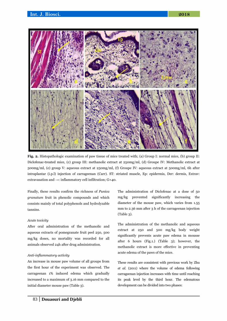
Fig. 2. Histopathologic examination of paw tissue of mice treated with; (a) Group I: normal mice, (b) group II:
Diclofenac-treated mice, (c) group III: methanolic extract at 250mg/ml, (d) Groupe IV: Methanolic extract at
500mg/ml, (e) group V: aqueous extract at 250mg/ml, (f) Groupe IV: aqueous extract at 500mg/ml, 6h after
intraplantar (i.p.l) injection of carrageenan (Carr). ST: striated muscle, Ep: epidermis, Der: dermis, Extrav:
extravasation and →: inflammatory cell infiltration; G×40.
Finally, these results confirm the richness of Punica
granatum fruit in phenolic compounds and which
consists mainly of total polyphenols and hydrolyzable
tannins.
Acute toxicity
After oral administration of the methanolic and
aqueous extracts of pomegranate fruit peel 250, 500
mg/kg doses, no mortality was recorded for all
animals observed 24h after drug administration.
Anti-inflammatory activity
An increase in mouse paw volume of all groups from
the first hour of the experiment was observed. The
carrageenan 1% induced edema which gradually
increased to a maximum of 3.16 mm compared to the
initial diameter mouse paw (Table 3).
The administration of Diclofenac at a dose of 50
mg/kg prevented significantly increasing the
diameter of the mouse paw, which varies from 1.55
mm to 2.36 mm after 3 h of the carrageenan injection
(Table 3).
The administration of the methanolic and aqueous
extract at 250 and 500 mg/kg body weight
significantly prevents acute paw edema in mousse
after 6 hours (Fig.1.) (Table 3); however, the
methanolic extract is more effective in preventing
acute edema of the paws of the mice.
These results are consistent with previous work by Zhu
et al. (2011) where the volume of edema following
carrageenan injection increases with time until reaching
its peak level by the third hour. The edematous
development can be divided into two phases:

84 Douaouri and Djebli
Int. J. Biosci. 2018
In the control group, from 0 to 3 hours, the edema
caused by the phlogistic agent increases progressively
and reaches a maximum at 3 hours (103.55%). From
3 to 6h: while the paw volume in the control group
remains relatively unchanged, the changes in paw
volume in the other experimental groups enter a
period of decline, with a decrease demarcating a
standard lot of others. Percentages of inhibition in
edema increased from 52.61 to 17.82% for the group
treated with diclofenac, respectively from the 3rd to
the 6th hour.
In the first phase, from 0 to 3h, it has been reported
that the vasodilation observed at the beginning of this
phase, called "early" phase, is due to the release of
histamine, serotonin and bradykinin mediators.
According to Alam et al. (2011), edema is caused by
secretion of 5-hydroxytryptamine (5-HT) during the
first hour, followed by kinins to increase vascular
permeability up to two and a half hours. As for the
bradykinins which are produced by the cascade of
plasma kinins at around 2:30, they would be
responsible for the increase of the vascular
permeability, thus causing an effusion of exudate into
the inflammatory focus. These hemodynamic changes
reached an increased level at the 3rd hour after
carrageenan injection and then began to decline
(Sharififar et al., 2012). While the late phase occurs in
1 hour and lasts 3 hours or more, it is characterized by
leukocyte infiltration and mediated only by
prostaglandins (Vinegar et al., 1969; Posadas et al.,
2004; Wang et al., 2010).
Administration of Diclofenac sodium (50mg/kg) in
the 2nd group showed a very significant decrease
(p<0.001) in the evolution of inflammation in the legs
of mice at the second hour, with a percentage of
inhibition of 68.52% which was lower than that found
in the negative control which was closed to 85%.
Inhibition of the volume of edema reached a
maximum threshold at the 6th hour after injection of
carrageenan with a percentage of 80.97% (Fig.1.).
NSAIDs act by inhibiting cyclooxygenase (COX),
which inhibits the synthesis of prostaglandins, which
gives this class of drugs its analgesic, antipyretic and
anti-inflammatory properties (Pereira-Leite et al.,
2016).
NSAIDs may be classified as non-selective (inhibiting
both COX-1 and COX-2) or selective COX-2 inhibitors
(celecoxib, etoricoxib, meloxicam and parecoxib).
However, most anti-inflammatory drugs are clinically
effective in the second phase of inflammation (Olajid
et al., 2000; Mehmood et al., 2016).
The evaluation of inhibition percent revealed that the
methanolic extract in the two doses (250 and 500
mg/kg p.c.), as well as the aqueous extract at 250
mg/kg, produced anti-inflammatory effects from the
second stage of development of edema, causing a very
significant reduction in carrageenan-induced paws
edema of mice. It is also noted that after 3 hours, the
methanolic extract at 250 and 500 mg/kg showed a
significant reduction in the volume of the paw of
64.37% and 69.72%, respectively, compared to the
group treated with Diclofenac (49.19%). However, the
aqueous extract at 500 mg/kg has anti-inflammatory
activity from the first stage of development of the
edema (early phase) compared to the negative
control. The anti-oedematous activity in the late
phase of the extracts could indicate that, like NSAIDs,
they would have an inhibitory effect on the synthesis
of prostaglandins, including the inhibition of COX.
This could explain the mode of action of the active
ingredients found in the peel extracts of the fruit of
Punica granatum.
Our results are consistent with those of Labib and El-
Ahmady (2015), which indicates that the anti-
inflammatory effect of methanol extract (200 mg/kg
b.w) similarly decreased in the group receiving the
reference product Indomethacin (20 mg/kg b.w) after
1 h. After 2 hours, the ME group (200mg/kg) showed
a significant decrease in edema volume of 53.3%,
which was considerably higher than that resulting
from the administration of Indomethacin (44, 86%).
On the other hand, these results are consistent with
several studies showing that the anti-inflammatory
activity of the extract can be explained in part by the
presence in the fruit of polyphenolic compounds such
as hydrolyzable tannins and flavonoids (Zarfeshany et
al., 2014).
85 Douaouri and Djebli
Int. J. Biosci. 2018
The latter are well-known components of anti-
inflammatory plants. Some flavonoids have shown
inhibitory action in various animal models of
inflammation. For example, some flavonoids have
shown to inhibit animal models of acute
inflammation: paw edema, ear edema and pleurisy
(Kim et al., 2017). The majority of the hydrolyzable
tannins present in the grenades; gallotannins, ellagic
acid, and gallagyl tannins, generally called
punicalagins, has shown an inhibitory effect on the
proliferation of human cancer cells and modulate
inflammatory subcellular signaling pathways due to
high antioxidant activity (Seeram et al., 2005).
Histopathology
The histopathological investigation was conducted by
hematoxylin and eosin staining. Biopsies of paws
were taken from the following groups: negative
control (saline, o.p), positive control (Diclofenac,
50mg/kg), the methanolic and aqueous extracts at
250 and 500mg/kg respectively 6h after i.p.l.
carrageenan injection (Fig.2.).
The paw tissues of the control mice (group I) showed
a polymorphic inflammation predominantly
lymphoplasmacytic (Fig.2a.). Inflammation has
spread to striated muscle tissue where there is a
granuloma that contains polymorphic cells.
Tissue samples from the legs of Diclofenac-treated
mice revealed a discrete inflammatory reaction
(Fig.2b.); of less intensity than the control (group I).
The evolution of the acute phase is quickly resolved
by Diclofenac action.
The inflammatory infiltrate is observed in the paw
tissues of the mice treated with the extracts at 250
mg/kg (Fig.2c & 2e.), but this infiltrate is less intense
than the control group. Whereas, it’s more intensive
than diclofenac group. However, treatment of mice at
500mg/kg exhibited a significant decrease in the
number of cellulars infiltrates (Fig.2d & 2f.).
In the present study, we showed that the
pomegranate peel extracts produced anti-
inflammatory effects in carrageenan-induced mousse
paw edema dose-dependently.
Our results confirmed previous findings that
pomegranate peel extracts exhibit a noticeable anti-
inflammatory effect in experimental models (Mo et
al., 2013; Mansouri et al. 2015).
Conclusion
Punica granatum showed anti-inflammatory power,
similar to those observed for non-steroidal anti-
inflammatory drugs, such as Diclofenac, inhibiting
edema and reducing the cellular infiltrates. It is also
suggested that the mechanism of action of Punica
granatum might be associated with the inhibition of
inflammatory mediators. However, further studies
are needed to isolate and characterize anti-
inflammatory bioactive compounds present in both
methanolic and aqueous extracts of Punica granatum
fruit peel.
References
Abdel-Hady NM. 2013. Quantitative diversity of
phenolic content in peels of some selected Egyptian
pomegranate cultivars correlated to antioxidant and
anticancer effects. Journal of Applied Sciences
Research 9(8), 4823-4830
Afaq F, Saleem M, Krueger CG, Reed JD,
Mukhta H. 2005. Anthocyanin and hydrolyzable
tannin-rich pomegranate fruit extract modulates
MAPK and NF‐κB pathways and inhibits skin
tumorigenesis in CD‐1 mice. International Journal of
Cancer 113(3), 423-433.
http://dx.doi.org/10.1002/ijc.20587
Ahmed AU. 2011. An overview of inflammation:
mechanism and consequences. Frontiers in Biology 6,
274-281.
Alam K, Pathak D, Ansari SH. 2011. Evaluation
of anti-inflammatory activity of Ammomum
subulatum fruit extract. International Journal of
Pharmaceutical Science Drug Research 3, 35-37.
Al-zoreky NS, Nakahara K. 2003.Antibacterial
activity of extracts from some edible plants commonly
consumed in Asia. International Journal of Food
Microbiology 80(3), 223–230.
https://doi.org/10.1016/S0168-1605(02)00169-1
86 Douaouri and Djebli
Int. J. Biosci. 2018
Anderson MT, Staal FJT, Gitler C, Herzenberg
LA, Herzenberg LA. 1994. Separation of oxidant-
initiated and redox-regulated steps in the NF-𝜅B
signal transduction pathway, Proceedings of the
National Academy of Sciences of the United States of
America 91(24), 11527–11531.
Arun N, Singh DP. 2012. Punica granatum: a
review on pharmacological and therapeutic
properties. International Journal of Pharmaceutical
Sciences and Research 3(15), 1240–1245.
www.dx.doi.org/10.13040/IJPSR.09758232.3(5).1240-45
Badenes ML, Martínez-Calvo J, Llácer G. 1998.
Analysis of apricot germplasm from the European
ecogeographical group. Euphytica 102(1), 93-99.
https://doi.org/10.1023/A:1018332312570
Bhandary SK, Kumari SN, Bhat VS, Sharmila
KP, Bekal MP. 2012. Preliminary phytochemical
screening of various extracts of Punica granatum
peel, whole fruit, and seeds. Nitte University Journal
of Health Science. 2(4), 35-38.
Biswas SK. 2016. Does the interdependence
between oxidative stress and inflammation explain
the antioxidant paradox?. Oxidative medicine and
cellular longevity 2016(2016), 9.
http://dx.doi.org/10.1155/2016/5698931
Borochov-Neori H, Judeinstein S, Tripler E,
Harari M, Greenberg A, Shomer I, Holland D.
2009. Seasonal and cultivar variations in antioxidant
and sensory Quality of pomegranate (Punica
granatum L.) fruit. Journal of Food Composition and
Analysis 22(3), 189-195.
https://doi.org/10.1016/j.jfca.2008.10.011
Chebaibi A, Filali FR. 2013. Bactericidal activity
and phytochemical screening of Moroccan
pomegranate (Punica granatum Linn.) peel aqueous
extracts. Journal of Medicinal Plants Research 7(14),
887-891.
https://doi.org/10.5897/JMPR12.988.
Deore Leena P, Bachhav Devidas G, Nikam
Vikas K, Heda Amol J. 2016. Study of
anthelmintic and antimicrobial activity of peel extract
of Punica granatum Linn. European Journal of
Pharmaceutical and medical research 3(4), 292-297.
Dixon RA, Xie D-Y, Sharma SB. 2005.
Proanthocyanidins– a final frontier in flavonoid
research. New Phytologist 165, 9-28.
Elfalleh W, Hannachi H, Tlili N, Yahia Y, Nasri
N, Ferchichi A. 2012. Total phenolic contents and
antioxidant activities of pomegranate peel, seed, leaf
and flower. Journal of Medicinal Plants Research
6(32), 4724-4730.
Flohé L, Brigelius-Flohé R, Saliou C, Traber
MG, Packer L. 1997. Redox regulation of NF-𝜅B
activation: Free Radical Biology and Medicine 22(6),
1115-1126.
https://doi.org/10.1016/S0891-5849(96)00501-1
Hagerman AE. 2002. Hydrolyzable tannin
structural chemistry. Tannin Handbook. Miami
University: Miami, FL, USA.
Hagir GAE, Alsheikh AA, Khadiga GAE. 2016.
Phytochemical screening and antibacterial activity of
Punica granatum fruit rind extracts. Global Journal
of Medicinal Plant Research 4(4), 9-15.
Hald A, Van Beek J, Lotharius J. 2007.
“Inflammation in Parkinson’s disease: causative or
epiphenomenal. In Inflammation in the Pathogenesis
of Chronic Diseases, Subcellular Biochemistry,
Springer, Dordrecht, The Netherlands 42, 249–279.
https://doi.org/10.1007/1-4020-5688-5_12
Halliwell B. 2006. Oxidative stress and
neurodegeneration: where are we now. Journal of
Neurochemistry 97, 1634–1658.
http://dx.doi.org/10.1111/j.1471-4159.2006.03907.x
Harborne JB. 1998. Phytochemical methods. 3rd.
India: Springer Pvt. Ltd., New Delhi, 66–68.
87 Douaouri and Djebli
Int. J. Biosci. 2018
Hegde CR, Madhuri M, Swaroop T, Nishitha
D, Arijit L, Bhattacharya S, Rohit KC. 2012.
Evaluation of antimicrobial properties, phytochemical
contents and antioxidant capacities of leaf extracts of
Punica granatum L. ISCA, Journal of Biological
Sciences 1(2), 32-37.
Kesur P, Gahlout M, Chauhan P, Prajapati HV.
2016. Evaluation of antimicrobial properties of peels
and juice extract of Punica granatum (Pomegranate).
International Journal of Research and Scientific
Innovation 3, 11-20.
Kim HP, Lim H, Kwon YS. 2017. Therapeutic
potential of medicinal plants and their constituents
on lung inflammatory disorders. Biomolecules and
Therapeutics 25(2), 91–104.
https://doi.org/10.4062/biomolther.2016.187
Kokate CK. 2005. A textbook of Practical
Pharmacognosy. 5thed. Vallabh Prakashan New Delhi
107-111.
Labib RM, El-Ahmady SH. 2015. Antinociceptive,
anti-gastric ulcerogenic and anti-inflammatory
activities of standardized Egyptian pomegranate peel
extract. Journal of Applied Pharmaceutical Science
5(1), 48–51.
Li Y, Guo C, Yang J, Wei J, Xu J, Cheng S.
2006. Evaluation of antioxidant properties of
pomegranate peel extract in comparison with
pomegranate pulp extract. Food Chemistry 96,
254−260.
Mansouri MT, Hemmati AA, Naghizadeh B,
Mard SA, Rezaie A, Ghorbanzadeh B. 2015. A
study of the mechanisms underlying the anti-
inflammatory effect of ellagic acid in carrageenan-
induced paw edema in rats. Indian Journal of
Pharmacology 47(3),292-298.
https://dx.doi.org/10.4103/0253-7613.157127
Marzocco S, Di Paola R, Serraino I,
Sorrentino R, Meli R, Mattaceraso G. 2004.
Effect of methylguanidine in carrageenan-induced
acute inflammation in the rats. European Journal of
Pharmacology 484(2-3), 341-350.
https://doi.org/10.1016/j.ejphar.2003.11.011
Mehmood A, Hamid I, Sharif A, Akhtar MF,
Akhtar B, Saleem A, Iqbal J, Shabbir M, Ali S.
2016. Evaluation of anti-inflammatory, analgesic and
antipyretic activities of aqueous and ethanolic
extracts of seeds of Buchanania Lanzan Spreng, in
animal models. Acta Poloniae Pharmaceutica – Drug
Research 73, 1601-1608.
Mertens-Talcott SU, Jilma-Stohlawetz P, Rios
J, Hingorani L, Derendorf H. 2006. Absorption,
metabolism and antioxidant effects of pomegranate
(Punica granatum L.) polyphenols after ingestion of a
standardized extract in healthy human volunteers.
Journal of Agricultural and Food Chemistry 54(23),
8956-8961.
Mo J, Panichayupakaranant P, Kaewnopparat
N, Nitiruangjaras A, Reanmongkol W. 2013.
Topical anti-inflammatory and analgesic activities of
standardized pomegranate rind extract in comparison
with its marker compound ellagic acid in vivo.
Journal of Ethnopharmacology 148(3), 901-908.
https://doi.org/10.1016/j.jep.2013.05.040
Moorthy K, Punitha T, Vinodhini R,
Sureshkumar BT, Vijayalakshmi P, Thajuddin
N. 2013. Antimicrobial activity and qualitative
phytochemical analysis of Punica granatum Linn.
(Pericarp) Journal of Medicinal Plants Research 7(9),
474- 479.
Mukhija M, Sundriyal A. 2013. Phytoconstituents
responsible for anti-inflammatory activity. Journal of
Natural Pharmaceuticals 4, 1-1.
Narasimha MK, Fazilath U, Soumya K,
Srinivas C. 2015. Antibacterial activity and
phytochemical screening of aqueous and methanolic
extracts of pomegranate (Punica granatum Linn.)
peel against bacterial wilt of tomato. International
Journal of Agriculture Innovations and Research
3(6), 1786-1792.
Negi PS, Jayaprakasha GK, Jena BS. 2003.
Antioxidant and antimutagenic activities of
pomegranate peel extracts. Food chemistry 80(3),
393-397.
https://doi.org/10.1016/S0308-8146(02)00279-0
88 Douaouri and Djebli
Int. J. Biosci. 2018
OECD. 2008. OECD Guideline for the testing of
chemicals No. 425: Acute Oral Toxicity– Up-and-
Down-Procedure (UDP), 8 pp. Paris, France.
Olajide OA, Awe SO, Makinde JM, Ekhelar AI,
Olusola A, Morebise O, Okpako DT. 2000.
Studies on the anti-inflammatory, antipyretic and
analgesic properties of Alstonia boonei stem bark.
Journal of Ethnopharmacology 71(1-2), 179-186.
https://doi.org/10.1016/S0378-8741(99)00200-7
Orak HH, Yagar H, Isbilir SS. 2012. Comparison
of antioxidant activities of juice, peel, seed of
pomegranate (Punica granatum L.) and inter-
relationships with total phenolic, Tannin,
anthocyanins and flavonoids contents. Food Science
and Biotechnology 21, 373-387.
https://doi.org/10.1007/s10068-012-0049-6
Onyango IG. 2008. Mitochondrial dysfunction and
oxidative stress in Parkinson’s disease.
Neurochemical research 33, 589-597.
https://doi.org/10.1007/s11064-007-9482-y
Ozkal, N, Dinc S. 1994. Evaluation of the
pomegranate (Punica granatum L.) peels from the
standpoint of pharmacy. Ankara University Eczacilik
Fak Derg 22, 21-29.
Ozgen M, Durgac C, Serce S, Kaya C. 2008.
Chemical and antioxidant properties of pomegranate
cultivars grown in the Mediterranean region of
Turkey. Food Chemistry 111(3), 703-706.
https://doi.org/10.1016/j.foodchem.2008.04.043
Pereira-Leite C, Nunes C, Jamal SK, Cuccovia
IM, Reis S. 2016. Nonsteroidal Anti‐Inflammatory
Therapy: A Journey Toward Safety. Medicinal
Research Reviews 37(4), 802–859.
http://dx.doi.org/10.1002/med.21424
Pérez-Vicente A, Gil-Izquierdo A, García-
Viguera C. 2002. In vitro gastrointestinal digestion
study of pomegranate juice phenolic compounds,
anthocyanins and vitamin C. Journal of Agricultural
and Food Chemistry 50, 2308-2312.
http://dx.doi.org/10.1021/jf0113833
Posadas I, Bucci M, Roviezzo F, Rossi A,
Parente L, Sautebin L, Cirino G. 2004.
Carrageenan‐induced mouse paw edema is biphasic,
age‐weight dependent and displays differential nitric
oxide cyclooxygenase‐2 expression. British Journal of
Pharmacology 142(2), 331-338.
http://dx.doi.org/10.1038/sj.bjp.0705650
Prior RL, Wu X, Schaich K. 2005. Standardized
methods for the determination of antioxidant capacity
and phenolics in foods and dietary supplements.
Journal of Agricultural and Food Chemistry 53(10),
4290-4302.
http://dx.doi.org/10.1021/jf0502698
Quettier-Deleu C, Gressier B, Vasseur J, Dine
T, Brunet C, Luyckx M, Cazin M, Cazin JC,
Bailleul F, Trotin F. 2000. Phenolic compounds
and antioxidant activities of buckwheat (Fagopyrum
esculentum Moench) hulls and flour. Journal of
Ethnopharmacology 72(1-2), 35-42.
https://doi.org/10.1016/S0378-8741(00)00196-3
Raffo A, La Malfa G, Fogliano V, Madani G,
Quaglia G. 2006. Seasonal variations in antioxidant
components of cherry tomatoes (Lycopersicon
esculentum cv. Naomi F1). Journal of Food
Composition and Analysis 19, 11-19.
Roopalatha, UC, Nair VM. 2013. Phytochemical
analysis of successive re extracts of the leaves of
Moringa Oleifera Lam. International Journal of
Pharmacy and Pharmaceutical Sciences 5(3), 629-634.
Saad H, Charrier-El Bouhtoury F, Pizzi A, Rode
K, Charrier B, Ayed N. 2012. Characterization of
pomegranate peels tannin extractives. Industrial Crops
and Products 40, 239-246.
https://doi.org/10.1016/j.indcrop.2012.02.038
Sajjad W, Sohail M, Ali B, Haq A, Din G, Hayat M,
Khan I, Ahmad M, Khan S. 2015. Antibacterial
activity of Punica granatum peel extract. Mycopath
13, 105-111.

89 Douaouri and Djebli
Int. J. Biosci. 2018
Schwartz E, Tzulker R, Glazer I, Bar-Ya’Akov
I, Wiesman Z, Tripler E, Bar-llan I, Fromm H,
Borochov-Neori H, Holland D, Amir R. 2009.
Environmental conditions affect the color, taste, and
antioxidant capacity of 11 pomegranate accessions’
fruits. Journal of Agricultural and Food Chemistry 57,
9197–9209
Seeram NP, Adams LS, Henning SM, Niu Y,
Zhang Y, Nair MG, Heber D. 2005. In vitro
antiproliferative, apoptotic and antioxidant activities
of punicalagin, ellagic acid, and a total pomegranate
tannin extract are enhanced in combination with
other polyphenols as found in pomegranate juice. The
Journal of Nutritional Biochemistry 16(6), 360-367.
https://doi.org/10.1016/j.jnutbio.2005.01.006
Seeram NP, Nair MG. 2002. Inhibition of lipid
peroxidation and structure capacity related studies of the
dietary constituent’s anthocyanins, anthocyanidins, and
catechins. Journal of Agricultural and Food Chemistry
50(19), 5308–5312.
http://dx.doi.org/10.1021/jf025671q
Sharififar F, Khazaeli P, Alli N, Talebian E,
Zarehshahi R, Amiri S. 2012. Study of
antinociceptive and anti-inflammatory activities of
certain Iranian medicinal plants. Journal of
International Ethnopharmacology 1(1), 19-24.
http://dx.doi.org/10.5455/jice.20120227104636
Sharma S, Chattopadhyay SK, Trivedi P,
Bawankule DU. 2010. Synthesis and anti-
inflammatory activity of derivatives of coumarino-
lignoid, cleomiscosin A, and its methyl ether.
European Journal of Medicinal Chemistry 45(11),
5150-5156.
Shiban MS, Al-Otaibi MM, Al-Zoreky NS. 2012.
Antioxidant activity of pomegranate (Punica
granatum L.) fruit peels. Food and Nutrition Sciences
3(7), 991.
Souleman AMA, Ibrahim GE. 2016. Evaluation of
Egyptian pomegranate cultivars for antioxidant
activity, phenolic and flavonoid contents. Egyptian
Pharmaceutical Journal 15(3), 143-9.
http://dx.doi.org/10.4103/1687-4315.197582
Singh RP, Murthy KNC, Jayaprakasha GK.
2002. Studies on the antioxidant activity of
pomegranate peel and seed extract using in vitro
models. Journal of Agricultural and Food Chemistry
50(1), 81–86.
http://dx.doi.org/10.1021/jf010865b
Singleton VL, Rossi JL. 1965. Colorimetry of total
phenolics with phosphomolybdic-phosphotungstic
acid reagents. American Journal of Enology and
Viticulture 16(3), 144–158.
Toor RK, Savage GP, Lister CE. 2006. Seasonal
Variations in the antioxidant composition of
greenhouse-grown Tomatoes. Journal of Food
Composition and Analysis 19(1), 1-10.
https://doi.org/10.1016/j.jfca.2004.11.008
Uma C, Gomathi D, Ravikumar G, Kalaiselvi
M, Palaniswamy M. 2012. Production and
properties of invertase from a Cladosporium
cladosporioides in SmF using pomegranate peel
waste as a substrate. Asian Pacific Journal of Tropical
Biomedicine 2(2), S605-S611.
https://doi.org/10.1016/S2221-1691(12)60282-2
Vinegar R, Schreiber W, Hugo R. 1969. Biphasic
development of carrageenin edema in rats. Journal of
Pharmacology and Experimental Therapeutics.
166(1), 96-103.
Wang R, Ding Y, Liu R, Xiang L, Du L. 2010.
Pomegranate, Bioactivities and Pharmacokinetics.
Fruits, Vegetables and Cereal Science and
Biotechnology 4(2), 77-87.
Wang Z, Pan Z, Ma H, Atungulu GG. 2011.
Extract of phenolics from pomegranate peels. The
Open Food Science Journal 5, 17-25.
http://dx.doi.org/10.2174/1874256401105010017
Weiss U, 2002. Inflammation. Nature 420, 845.
Willis RB, Allen PR. 1998. An improved method
for measuring hydrolyzable tannins using potassium
iodate. The Analyst 123, 435–439.
90 Douaouri and Djebli
Int. J. Biosci. 2018
Winter CA, Risley EA, Nuss GW. 1962.
Carrageenin-induced edema in hand paw of the rat as
an assay for anti-inflammatory drugs. Proceedings of
the society for Experimental Biology and Medicine
111(3), 544-547.
https://doi.org/10.3181/00379727-111-27849
Wissam Z, Ghada B, Wassim A, Warid K. 2012.
Effective extraction of polyphenols and
proanthocyanidins from pomegranate’s peel.
International Journal of Pharmacy and
Pharmaceutical Sciences 4(3), 675-82.
Zahin M, Aqil F, Ahmad I. 2010. Broad spectrum
antimutagenic activity of antioxidant active fraction
of Punica granatum L. peel extracts. Mutation
Research/Genetic Toxicology and Environmental
Mutagenesis. 703(2), 99–107.
https://doi.org/10.1016/j.mrgentox.2010.08.001
Zarei M, Azizi M, Bashir-Sadr Z. 2011.
Evaluation of physicochemical characteristics of
pomegranate (Punica granatum L.) fruit during
ripening. Fruits 66(2011), 121-129.
http://dx.doi.org/10.1051/fruits/2011021
Zarfeshany A, Asgary S, Javanmard SH. 2014.
Potent health effects of pomegranate. Advanced
Biomedical Research 3, 100.
http://dx.doi.org/10.4103/2277-9175.129371
Zhu ZZ, Ma KJ, Ran X, Zhang H, Zheng CJ,
Han T, Zhang QY, Qin LP. 2011. Analgesic, anti-
inflammatory and antipyretic activities of the
petroleum ether fraction from the ethanol extract of
Desmodium podocarpum. Journal of
Ethnopharmacology 133(3), 1126-1131.
https://doi.org/10.1016/j.jep.2010.11.042