in vivo antitumor potential of extracts from different
TRANSCRIPT
Applied Cancer ResearchPandey Applied Cancer Research (2017) 37:33 DOI 10.1186/s41241-017-0039-3
RESEARCH ARTICLE Open Access
In vivo antitumor potential of extracts fromdifferent parts of Bauhinia variegata linn.Against b16f10 melanoma tumour modelin c57bl/6 mice
Sonam Pandey1,2Abstract
Background: Melanoma is a metastatic type of skin cancer that is difficult to treat and the majority of efforts aredirected to the design of new drugs. Medicinal Plants have been the primary source of medicines since life onearth; more than 50% of existing cancer treatments is derived from plants. Bauhinia variegata is well-knownmedicinal plant used from the ancient era to till date for their medicinal values. Scientific literatures have notdocumented any evidence of the antitumour potential of Bauhinia variegata against B16F10 melanoma tumormodel in C57BL mice. The present investigation was undertaken to explore the antitumour activity of Leaf, stembark and flower extract of Bauhinia variegata against B16F10 melanoma tumour model in C57BL mice.
Methods: Hydro-methanolic extract prepared from the leaf, stem bark and flower of Bauhinia variegata wereassessed for their antitumor activity. The extracts at doses of 500 and 750 mg/kg b.wt. were given orally along withcyclophosphamide (chemotherapeutic drug) for 40 days for exploring antitumor activity against melanoma tumor(B16F10) in C57BL mice. Inhibition of tumor growth, increase in survival time of animal with treatment,histopathological studies and antioxidant parameter were determined.
Results: The Present investigation showed significant effect of the B. variegata L. in preventing melanoma tumor byB16F10 cell line in C57BL/6 mice. As compared with the tumour control group, the remarkable results especially inthe group which received B. variegata extract and cyclophosphamide together were obtained for all of themeasured parameters. Dose dependent response was observed in tumor volume, inhibition rate, life span time andantioxidant parameter of extracts. Combination treatment of cyclophosphamide and B. variegata extracts showedmore pronounced effect.
Conclusions: These findings suggest that B. variegata hydromethanolic extract may contain bioactive compoundsof potential therapeutic significance which are relatively safe from toxic effects, and can compromise the medicinaluse of this plant in folk medicine.
Keywords: Chemoprevention, Bauhinia variegata, Melanoma, B16F10 cell line, GSH
Correspondence: [email protected] State Biotechnology Mission, Gandhinagar, Gujarat, India2Research Department, Jawaharlal Nehru Cancer Hospital and ResearchCentre, Idgah Hills, Bhopal, Madhya Pradesh, India
© The Author(s). 2017 Open Access This article is distributed under the terms of the Creative Commons Attribution 4.0International License (http://creativecommons.org/licenses/by/4.0/), which permits unrestricted use, distribution, andreproduction in any medium, provided you give appropriate credit to the original author(s) and the source, provide a link tothe Creative Commons license, and indicate if changes were made. The Creative Commons Public Domain Dedication waiver(http://creativecommons.org/publicdomain/zero/1.0/) applies to the data made available in this article, unless otherwise stated.
Pandey Applied Cancer Research (2017) 37:33 Page 2 of 14
BackgroundCancer is a growing health problem in both developingand developed countries. Melanoma is the most aggressiveand deadly form of skin cancer. Patients with distant me-tastases have a five-year survival rate of 16% [1] and a me-dian survival of four to six months [2]. Until very recently,melanoma has been branded by the failure of chemother-apy and other therapeutic attempts. Currently, the maintreatments for cancer are chemotherapy, radiotherapy andsurgery. Chemotherapy is routinely used for cancer treat-ment. Since cancer cells lose many of the regulatory func-tions present in normal cells, they continue to dividewhen normal cells do not. This feature makes cancer cellssusceptible to chemotherapeutic drugs. Approximatelyfive decades of systemic drug discovery and developmenthave resulted in the establishment of a large collection ofuseful chemotherapeutic agents. Currently, some of theseplant-derived compounds are widely used for chemother-apy of cancerous patients. However, chemotherapeutictreatments are not devoid of their own intrinsic problems.Various kinds of toxicities may occur as a result of chemo-therapeutic treatments. For example, 5-fluorouracil, acommon chemotherapeutic agent, is known to cause mye-lotoxicity [3], cardiotoxicity [4] and has even been shownto act as a vasospastic agent in rare but documented cases[5]. Another widely used chemodrug, doxorubicin causescardiac toxicity [6–8], renal toxicity [9], and myelotoxicity.[10] Similarly, bleomycin a well-known chemotherapeuticagent, is known for its pulmonary toxicity [11–13]. Inaddition, bleomycin shows cutaneous toxicity [14]. Cyclo-phosphamide, a drug to treat many malignant conditions,has been shown to have bladder toxicity in the form ofhemorrhagic cystitis, immunosuppression, alopecia and athigh doses cardiotoxicity [15].A major problem associated with cancer chemotherapy
is the severe side effects resulting from normal tissuedamage. Consequently, agents which protect normal tis-sues against chemotherapy can increase the patient’s toler-ance to chemotherapy. Several chemicals have been foundto provide notable protection in experimental animals,but their clinical utility is limited by the drug toxicity onrepeated administration [16]. Therefore, there is a need tofind nontoxic and inexpensive drug/(s) for clinical chemoprotection. Recent studies have indicated that some of thecommonly used medicinal plants may be good source ofpotent but nontoxic chemoprotectors [17–20].Natural products discovered from medicinal plants have
played an important role in treatment of cancer, which is pro-jected to become the major cause of death worldwide. A widenumber of plant extracts are used against diseases in varioussystems of medicine such as Ayurveda, Unani and Siddha.Only a few of them have been scientifically explored. Plantderived natural products such as flavonoids, terpenes andalkaloids [21–23] and soon has received considerable
attention in recent years, due to their diverse pharmacologicalproperties including cytotoxic and cancer chemopreventiveeffects [24]. Plant based systems continue to play an essentialrole in healthcare and it has been estimated by WHO thatapproximately 80% of the world’s inhabitants rely mainly ontraditional medicine for their primary health care [25].The use of natural products in cancer therapy showed that
plants are a most important source of antitumor com-pounds, with new structures and mechanisms of action be-ing discovered [26]. Several plant-derived products induceapoptosis in neoplastic cells but not in normal cells [27–29].Bauhinia variegata (Family fabaceae, Genus Bauhinia)
is an herbaceous plant, found throughout India. The plantis known as Kachnara in Sanskrit and Hindi. All parts ofthe plant (leaves, flower buds, flower, stem, stem bark, seedsand roots) were used in traditional medicine. For usingvarious ailments like bronchitis, leprosy, and tumors. Thestem bark was used as astringent, tonic, anthelmintic andantidiabetic [30]. Infusion of the leaves was used as a laxa-tive and for piles. Dried buds were used in the treatment ofworm infestations, tumors, diarrhea, and piles. It is helpfulin managing skin discoloration [31–33].The phytochemical screening revealed that Bauhinia
variegata contained terpenoids, flavonoids, tannins, sapo-nins, reducing sugars, steroids and cardiac glycosides.Pharmacological studies showed that Bauhinia variegataexerted anticancer, antioxidant, hypolipidemic, antimicro-bial, anti-inflammatory, nephroprotective, hepatoprotective,antiulcer, immunomodulating, molluscicidal and woundhealing effects [34, 35]. Previous phytochemical studies onthe stems [36–38], flowers [34, 39], leave and seeds [40, 41]of this species have led to the isolation of several flavonoids.There are also a few reports of antitumor activity of B. var-
iegata ethanolic extract against Dalton’s ascetic lymphoma(DAL) in Swiss albino mice [42] and N-nitrosodiethylamineinduced experimental liver tumors in rats and human cancercell lines [43]. The present study evaluates the chemopreven-tive effect of the different parts and different doses of B.variegata on B16F10 melanoma tumor on C57BL mice.
MethodsAnimalsThe study was conducted on random bred, 6–7 weeks oldand 24–28 g b.wt. bearing, male C57BL/6 mice. Animalswere maintained under controlled conditions of temperatureand light (Light: dark, 10 h: 14 h.). They were providedstandard mice feed (procured from Hindustan Levers Ltd.)and water ad libitum.
ChemicalsReduced glutathione, Cyclophosphamide, 5,5′-dithiobis (2-nitrobenzoic acid) (DTNB), Sodium citrate and other chemi-cals were procured from Sigma Chemical Co (St Louis, MO).
Pandey Applied Cancer Research (2017) 37:33 Page 3 of 14
Identification and collection of plant materialAerial parts of B. variegata (Kachnar) like leaves, stem barkand floral bud were collected in the early stages of vegeta-tion from the Bhopal, and Tah-Niwas, District Mandla(M.P.), India, during the month of October, 2007. The iden-tification of the plant B. variegata L. (Kachnar) (family:Leguminose) was done by botanist Dr. S.S. Khan (VoucherSpecimen No: SP/101/LGOB/2007), Department of Botany,Safia Science College, Bhopal, Madhya Pradesh (India).
Preparation of B. variegata extractShade dried powdered plant material such as leaves, stembark and floral buds (50 g) were extracted by continuousmixing in 100 ml 50% methanol, and stem bark in 95%methanol, 24 h at room temperature. After filtration, metha-nol and water was evaporated at 60-70 °C temperature. Thepercentage yield of the crude extract was determined foreach parts of B. variegata and was for leaf 12%, stem work20% and floral buds 10%. The percentage extract yield wasestimated as dry weight/dry material weight × 100 [44]. Thedried powder was kept in air tight box.
Acute oral toxicity test (LD50)Experimental animalsAcute oral toxicity test was performed as per Organizationfor Economic Co-operation and Development (OECD)guidelines 423 [45]. Experiments were performed using 54male C57BL/6 mice were obtained from the AnimalHouse of the Jawaharlal Nehru Cancer Hospital andResearch Centre, Bhopal. The animals were randomlydivided into nine groups of 6 animals per group.
Administration doseFollowing the period of fasting, animals were weighedand extract was administered orally at a dose of 100,200, 400, 800, 1600, 3200, 6400 and 12,800 mg/kg. Afterthe administration of test substance, food for the micewas withheld for 2 h. Group I was given distilled water(10 ml/kg) as control [46].
Observation periodAnimals were observed individually after atleast once dur-ing the first 30 min, periodically during the first 24 h, withspecial attention given during the first 4 h, and daily there-after, for a total of 14 days. All the mice were observed atleast twice daily with the purpose of recording any symp-toms of toxicity, survival or behavioural changes [46].
Antitumor activity of B. variegata in subcutaneousmelanoma B16F10-bearing miceCell cultureB16F10 melanoma tumor cell line was purchased from theNational Centre for Cell Science (NCCS, Pune, India). Thecells were maintained in RPMI 1640 medium buffered with
2 g/l of HEPES and sodium bicarbonate, and supplementedwith dextrose, penicillin, streptomycin and 10% of fetalbovine serum. The cells were maintained in a humidifiedatmosphere containing 5% CO2 at 37 °C. When needed forexperiments the cells were harvested with trypsin: EDTA(0.05:0.03 [w/v]) solution, and then washed in phosphate-buffered saline (PBS, pH 7.4). For the animal experiments,the recovered cells were adjusted to 5 × 10⁵ cells/ml in PBSand then 200 μl of the suspension was injected subcutane-ously (S.C.) into dorsal side of C57BL/6 mice. After 8–10 days of injection, the tumor was found to develop into abudding state. When the tumor was developed to a palp-able level, two doses of the plant extracts (leaf, stem barkand floral bud) at 500 and 750 mg/kg b. wt. of mice weregiven orally and cyclophosphamide at 170 mg/kg wasinjected intraperitoneally (i.p.) every alternate day up to40 days. During the treatment, the size of implanted tumor[Fig. 4a] was regularly measured at given time interval, witha digital caliper and tumor volume was calculated [47].
Experimental designA total of 72 adult male C57BL mice aged 6–8 weekswere divided into 12 groups thus each group containing6 animals (Fig. 1).Group NC: receiving normal saline (10 mL/kg b. wt.)
treated as Normal Control.Group TC: (tumor-bearing mice) receiving normal sa-
line (10 mL/kg) treated as Tumor Control.Group CP: served as standard, which received cyclo-
phosphamide (CP) 170 mg/kg b.wt.(tumor-bearing mice).Group L1 and L2 (tumor bearing) Animals were re-
ceived 500 and 750 mg/kg b.wt. B. variegata leaf extracts,respectively and after 30 min of B. variegata treatment,animals were treated with CP (170 mg/kg b.wt)Group L3: Animals were received orally with 500 mg/kg
body weight of B. variegata leaf extract (tumor-bearing mice).GroupB1 and B2 (tumor-bearing mice): Animals were
received 500 and 750 mg/kg b.wt. B. variegata stem barkextracts, respectively and after 30 min of B. variegatatreatment, animals were treated with CP (170 mg/kg b.wt)Group B3 (tumor-bearing mice): Animals were re-
ceived orally with 500 mg/kg body weight of B. variegatastem bark extract.Group F1 and F2 (tumor-bearing mice): Animals were
received 500 and 750 mg/kg b.wt. B. variegata floral budextracts, respectively and after 30 min of B. variegatatreatment, animals were treated with CP (170 mg/kg b.wt)Group F3 (tumor-bearing mice): Animals were re-
ceived orally with 500 mg/kg body weight of B. variegatafloral bud extract alone.All treatment of B.variegata extract (leaf, stem bark,
floral bud) was administered orally through a metal oro-pharyngeal cannula and Cyclophosphamide and normalsaline were given intraperitoneally (i.p.) by 1 ml syringe.
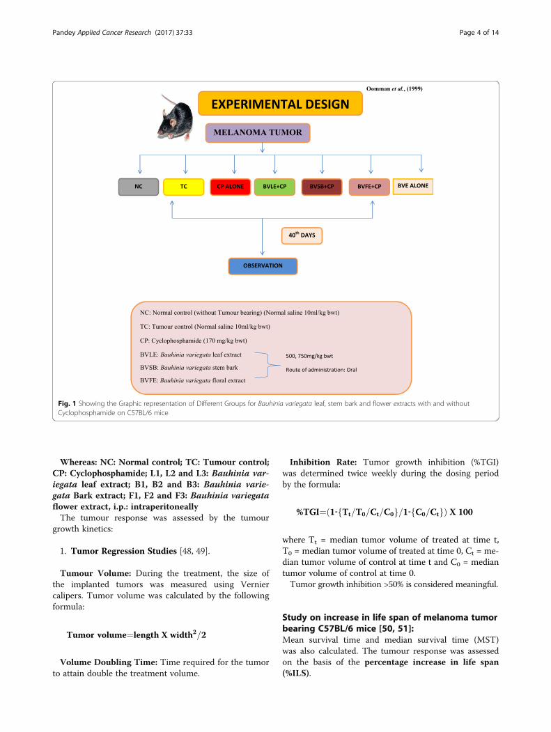
Fig. 1 Showing the Graphic representation of Different Groups for Bauhinia variegata leaf, stem bark and flower extracts with and withoutCyclophosphamide on C57BL/6 mice
Pandey Applied Cancer Research (2017) 37:33 Page 4 of 14
Whereas: NC: Normal control; TC: Tumour control;CP: Cyclophosphamide; L1, L2 and L3: Bauhinia var-iegata leaf extract; B1, B2 and B3: Bauhinia varie-gata Bark extract; F1, F2 and F3: Bauhinia variegataflower extract, i.p.: intraperitoneallyThe tumour response was assessed by the tumour
growth kinetics:
1. Tumor Regression Studies [48, 49].
Tumour Volume: During the treatment, the size ofthe implanted tumors was measured using Verniercalipers. Tumor volume was calculated by the followingformula:
Tumor volume¼length X width2=2
Volume Doubling Time: Time required for the tumorto attain double the treatment volume.
Inhibition Rate: Tumor growth inhibition (%TGI)was determined twice weekly during the dosing periodby the formula:
%TGI¼ 1‐ Tt=T0=Ct=C0f g=1‐ C0=Ctf gð Þ X 100
where Tt = median tumor volume of treated at time t,T0 = median tumor volume of treated at time 0, Ct = me-dian tumor volume of control at time t and C0 = mediantumor volume of control at time 0.Tumor growth inhibition >50% is considered meaningful.
Study on increase in life span of melanoma tumorbearing C57BL/6 mice [50, 51]:Mean survival time and median survival time (MST)was also calculated. The tumour response was assessedon the basis of the percentage increase in life span(%ILS).

Pandey Applied Cancer Research (2017) 37:33 Page 5 of 14
%ILS ¼ MST of Treated GroupMST of Control Group
� �−1� 100
whereas: MST = Mean survival time, ILS = Increase LifeSpanAn enhancement of life by 25% or more over that of con-
trol was considered as effective antitumor response [51].Antioxidant parameter was studied in all the groups at
the time of termination of the experiment (i.e., after41 days).
Determination of glutathione (GSH) levelGlutathione was evaluated by sacrificing all the experi-mental mice were on day 41st by cervical dislocationand liver and Kidney was removed.
Preparation of homogenatesAfter collection of blood samples [52], the mice weresacrificed. Then the liver and Kidney was excised, rinsedin ice cold normal saline followed by ice cold 10% KCl so-lution, blotted, dried and weighed. A 10% (w/v) homogen-ate was prepared in ice cold KCl solution and centrifugedat 1500 rpm for 15 min at 4 °C. The supernatant thus ob-tained was used for the estimation of glutathione (GSH)[53, 54] level were checked using respective kits (Sigma-Aldrich Co. LLC) according to manufacturer’s instruction.Reduced glutathione was used as a standard to calculate μmole GSH/100 g tissue.
Histopathological studiesAfter the completion of drug treatment (40 days), on theday 41st mice were sacrificed by cervical dislocation. Thetumor of three animals from each group was dissectedout, fixed in 10% buffered formalin for 12 h and processedfor histopathological examination. 4 μm-thick paraffinsections were cut and stained with hematoxylin and eosinand mounted in DPX (used as a synthetic resin mountingmedia). Sections were qualitatively assessed under thelight microscope for their architecture [55].
Statistical analysisResults of Statistical analysis are presented as Mean ± S.D.Statistical evaluation of data was performed by usingone-way analysis of variance (ANOVA) followed byDunnett’s multiple comparison test. P ≤ 0.05 was consid-ered statistically significant.
ResultsAcute oral toxicity testingThe present study conducted as per the OECD guidelines423 revealed that an acute toxicity test in which no deathoccurred in more than one of the six animals given a doseof 2000 mg /kg, the LD50 value can be considered greaterthan 2000 mg/ kg and less than 5000 mg/kg. In LD50
studies it were found that the different parts of B. varie-gata extract (leaf, stem bark and floral bud) did not pro-duce any signs of toxicity or mortality up to the dose level5000 mg/kg b.wt. Hence, the drug was considered to besafe up to the dose level of 2000 mg per kg bwt.
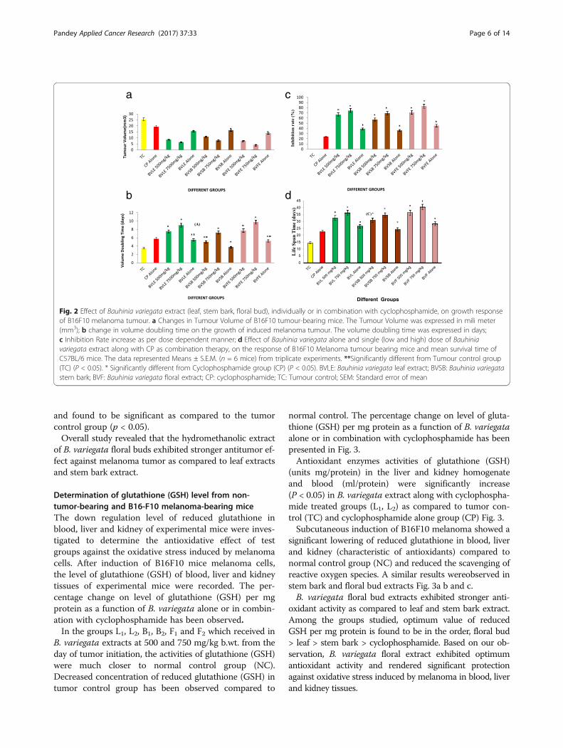
Effect of B. variegata in subcutaneous melanomaB16F10-bearing miceThe study of tumor volume of mice shown in Fig. 2a re-vealed that in the groups (L3, B3, and F3) which receivedhydromethanolic extract of different parts of B. varie-gata (leaf, stem bark, floral bud) at 500 and 750 mg/kg,b. wt. a reduction in the tumor volume was observed.On the day 40th of treatment, the reduction in thetumor volume was found to be significant (P < 0.05) forboth the doses compared to tumor control group (TC),which received normal saline, 10 mL/kg, b.wt and Cyclo-phosphamide alone group (CP).When hydromethanolic leaf extract of B. variegata at
500 and 750 mg/kg was used as an adjuvant to chemo-therapy groups (L1 and L2) receiving cyclophosphamide at170 mg/kg, there was a statistically significant (P < 0.05)reduction in tumor volume on 40th day of tumor induc-tion as compared to tumor control and cyclophosphamidealone group (A1and A2) as well as test drug group (L3).Simultaneously, B. variegata bark extract (B1, B2) andflower extract (F1 and F2) at the dose of 500 mg/kg and750 mg/kg with cyclophosphamide (CP) were also re-duced tumor volume as compared to tumor control andcyclophosphamide group (NC and CP) as well as test drugalone group (L3 B3 and F3) respectively.The tumor doubling time (in days) Fig. 2b of Cyclophos-
phamide group (CP) was increased, whereas B. variegataleaf extract (500 mg/kg and 750 mg/kg) along with CPgroups (L1 and L2), tumor doubling time were reduced.Simultaneously, B. variegata bark (B1 and B2) and flowerextract (F1 and F2) (500 mg/kg and 750 mg/kg) along withCP groups were also reduced tumor doubling timerespectively.The inhibition rate Fig. 2c of B. variegata leaf extract
alone (L3) and cyclophosphamide group (CP) weredecreased but when B. variegata leaf, stem bark, flowerextract (L1, L2, B1, B2, F1 and F2) along with CP were given,the inhibition rate were increased in tumor bearing mice ascompared to tumor control (TC) and cyclophosphamidegroup (CP) as well as test drug alone group (L3, B3 and C3)respectively.The life span Fig. 2d was also increased in B. variegata
leaf extract (500 mg/kg and 750 mg/kg) along with CPgroup (L1 and L2), at the same time the B. variegata bark(B1 and B2) and flower extract (F1 and F2) along withcyclophosphamide group, life span were also increase intumor bearing mice. The differences in the values of theresults of experimental groups were statistically analyzed
a
b
c
d
Fig. 2 Effect of Bauhinia variegata extract (leaf, stem bark, floral bud), individually or in combination with cyclophosphamide, on growth responseof B16F10 melanoma tumour. a Changes in Tumour Volume of B16F10 tumour-bearing mice. The Tumour Volume was expressed in mili meter(mm3); b change in volume doubling time on the growth of induced melanoma tumour. The volume doubling time was expressed in days;c Inhibition Rate increase as per dose dependent manner; d Effect of Bauhinia variegata alone and single (low and high) dose of Bauhiniavariegata extract along with CP as combination therapy, on the response of B16F10 Melanoma tumour bearing mice and mean survival time ofC57BL/6 mice. The data represented Means ± S.E.M. (n = 6 mice) from triplicate experiments. **Significantly different from Tumour control group(TC) (P < 0.05). * Significantly different from Cyclophosphamide group (CP) (P < 0.05). BVLE: Bauhinia variegata leaf extract; BVSB: Bauhinia variegatastem bark; BVF: Bauhinia variegata floral extract; CP: cyclophosphamide; TC: Tumour control; SEM: Standard error of mean
Pandey Applied Cancer Research (2017) 37:33 Page 6 of 14
and found to be significant as compared to the tumorcontrol group (p < 0.05).Overall study revealed that the hydromethanolic extract
of B. variegata floral buds exhibited stronger antitumor ef-fect against melanoma tumor as compared to leaf extractsand stem bark extract.
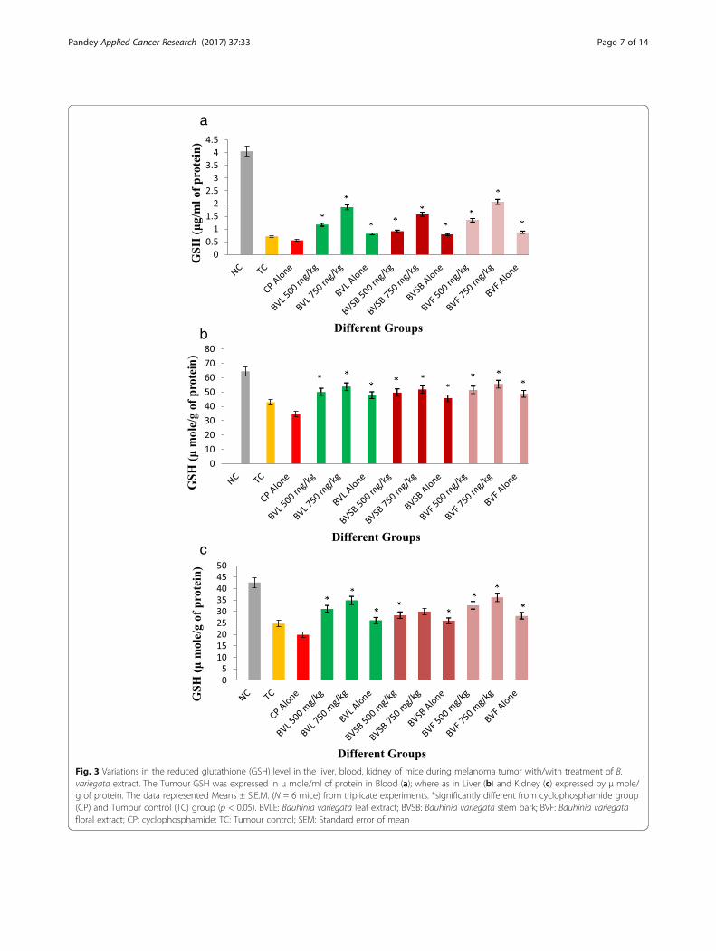
Determination of glutathione (GSH) level from non-tumor-bearing and B16-F10 melanoma-bearing miceThe down regulation level of reduced glutathione inblood, liver and kidney of experimental mice were inves-tigated to determine the antioxidative effect of testgroups against the oxidative stress induced by melanomacells. After induction of B16F10 mice melanoma cells,the level of glutathione (GSH) of blood, liver and kidneytissues of experimental mice were recorded. The per-centage change on level of glutathione (GSH) per mgprotein as a function of B. variegata alone or in combin-ation with cyclophosphamide has been observed.In the groups L1, L2, B1, B2, F1 and F2 which received in
B. variegata extracts at 500 and 750 mg/kg b.wt. from theday of tumor initiation, the activities of glutathione (GSH)were much closer to normal control group (NC).Decreased concentration of reduced glutathione (GSH) intumor control group has been observed compared to
normal control. The percentage change on level of gluta-thione (GSH) per mg protein as a function of B. variegataalone or in combination with cyclophosphamide has beenpresented in Fig. 3.Antioxidant enzymes activities of glutathione (GSH)
(units mg/protein) in the liver and kidney homogenateand blood (ml/protein) were significantly increase(P < 0.05) in B. variegata extract along with cyclophospha-mide treated groups (L1, L2) as compared to tumor con-trol (TC) and cyclophosphamide alone group (CP) Fig. 3.Subcutaneous induction of B16F10 melanoma showed a
significant lowering of reduced glutathione in blood, liverand kidney (characteristic of antioxidants) compared tonormal control group (NC) and reduced the scavenging ofreactive oxygen species. A similar results wereobserved instem bark and floral bud extracts Fig. 3a b and c.B. variegata floral bud extracts exhibited stronger anti-
oxidant activity as compared to leaf and stem bark extract.Among the groups studied, optimum value of reducedGSH per mg protein is found to be in the order, floral bud> leaf > stem bark > cyclophosphamide. Based on our ob-servation, B. variegata floral extract exhibited optimumantioxidant activity and rendered significant protectionagainst oxidative stress induced by melanoma in blood, liverand kidney tissues.
a
b
c
Fig. 3 Variations in the reduced glutathione (GSH) level in the liver, blood, kidney of mice during melanoma tumor with/with treatment of B.variegata extract. The Tumour GSH was expressed in μ mole/ml of protein in Blood (a); where as in Liver (b) and Kidney (c) expressed by μ mole/g of protein. The data represented Means ± S.E.M. (N = 6 mice) from triplicate experiments. *significantly different from cyclophosphamide group(CP) and Tumour control (TC) group (p < 0.05). BVLE: Bauhinia variegata leaf extract; BVSB: Bauhinia variegata stem bark; BVF: Bauhinia variegatafloral extract; CP: cyclophosphamide; TC: Tumour control; SEM: Standard error of mean
Pandey Applied Cancer Research (2017) 37:33 Page 7 of 14
Pandey Applied Cancer Research (2017) 37:33 Page 8 of 14
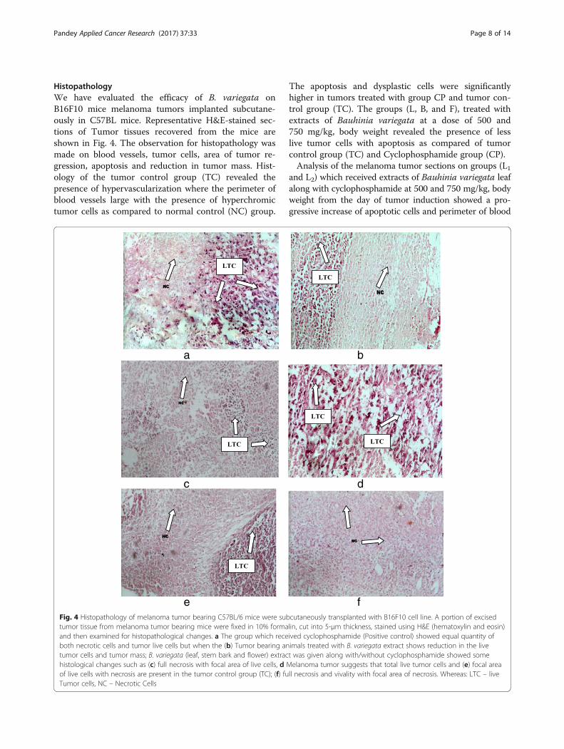
HistopathologyWe have evaluated the efficacy of B. variegata onB16F10 mice melanoma tumors implanted subcutane-ously in C57BL mice. Representative H&E-stained sec-tions of Tumor tissues recovered from the mice areshown in Fig. 4. The observation for histopathology wasmade on blood vessels, tumor cells, area of tumor re-gression, apoptosis and reduction in tumor mass. Hist-ology of the tumor control group (TC) revealed thepresence of hypervascularization where the perimeter ofblood vessels large with the presence of hyperchromictumor cells as compared to normal control (NC) group.
a
e
c
Fig. 4 Histopathology of melanoma tumor bearing C57BL/6 mice were subtumor tissue from melanoma tumor bearing mice were fixed in 10% formaand then examined for histopathological changes. a The group which receboth necrotic cells and tumor live cells but when the (b) Tumor bearing antumor cells and tumor mass; B. variegata (leaf, stem bark and flower) extrachistological changes such as (c) full necrosis with focal area of live cells, d Mof live cells with necrosis are present in the tumor control group (TC); (f) fuTumor cells, NC – Necrotic Cells
The apoptosis and dysplastic cells were significantlyhigher in tumors treated with group CP and tumor con-trol group (TC). The groups (L, B, and F), treated withextracts of Bauhinia variegata at a dose of 500 and750 mg/kg, body weight revealed the presence of lesslive tumor cells with apoptosis as compared of tumorcontrol group (TC) and Cyclophosphamide group (CP).Analysis of the melanoma tumor sections on groups (L1
and L2) which received extracts of Bauhinia variegata leafalong with cyclophosphamide at 500 and 750 mg/kg, bodyweight from the day of tumor induction showed a pro-gressive increase of apoptotic cells and perimeter of blood
b
f
d
cutaneously transplanted with B16F10 cell line. A portion of excisedlin, cut into 5-μm thickness, stained using H&E (hematoxylin and eosin)ived cyclophosphamide (Positive control) showed equal quantity ofimals treated with B. variegata extract shows reduction in the livet was given along with/without cyclophosphamide showed someelanoma tumor suggests that total live tumor cells and (e) focal areall necrosis and vivality with focal area of necrosis. Whereas: LTC – live
Pandey Applied Cancer Research (2017) 37:33 Page 9 of 14
vessels and vascular supply was less. A similar result wasobserved in stem bark and floral bud extracts also. Theincidence of dysplastic cells and apoptosis in the tumoralmost corresponded to the effect of tumor growth inhib-ition, suggesting that treatment resulted in tumor regres-sion by significant augmentation of apoptosis. However,flower extract of B. variegata were found more effective ascompared to leaf and stem bark extract.
DiscussionCancer is a growing health problem worldwide, with theintroduction of 6 million new cases every year. Manyapproaches are being tried through modulation of anti-tumor immune response, apoptosis, and antitumor pro-teins for cancer treatment [56]. Cancer cells lack thegrowth control of normal cells, exhibiting unlimited self-sufficient replication [57, 58]. For therapeutic effective-ness, drugs are being developed that act as biologicalmodifiers, regulating the cell cycle and promoting celldeath [59]. Plant derived compounds have been reportedto induce cell cycle arrest and cell death in many tumorcell lines [60–65].In recent years, the cancer rate has increased, espe-
cially in less developed countries. Identification and de-velopment of new chemotherapy drugs has been criticalfor cancer treatment. Plants have been a rich source ofnatural drugs [66, 67]. In addition, compounds derivedfrom plants are diverse in structure and bioactivity andexhibit low toxicity, therefore they play an importantrole in pharmaceutical research [68]. In cancer therapy,many plant-derived drugs such as vincristine, paclitaxeland taxol have been identified and developed. Severalworks on epidemiology and animal model studies dem-onstrated that natural compounds, which possess anti-oxidant or anti-inflammatory properties, could inhibitcarcinogenesis [69–72].According to the traditional recommendations and ex-
perimental studies, numerous medicinal plants have beenreported to have anticancer effect. Also antiproliferative,pro-apoptotic, anti-metastatic and anti-angiogenic effectsof several phytochemicals have been shown in in vitroexperiments or animal studies. However, only a smallnumber have been tested in cancerous patients andlimited evidence exists for their clinical effectiveness [73].In the current study, we have selected B. variegata ex-
tracts to treat the melanoma tumor. The mice weretreated with B. variegata extracts (stem bark, leaf andfloral bud) at a dose of 500 and 750 mg/kg, body weight.In the control group receiving normal saline, initially, aslow and steady growth in tumor volume was observedwith a drastic increase in tumor size after few days.Experimental tumor models are a critical pre-clinical
step for the development and evaluation of chemotherapyregimens for cancer. Interestingly, we report here that the
differential cytotoxic effect of these extracts was relatednot only to their chemical composition but may be alsodue to the nature of the tumor cells. In the present find-ing, the cancer treatment through chemotherapy with thecombination of B. variegata was found more effective ascompared to the extract alone. Earlier studies on herbswere reported that in cancer therapy many drugs whengiven alone give good results, however, when combinedwith other drugs exerting synergistic effect, the results arebetter. The objective of such approach is to minimize drugresistance and drug toxicity.The B. variegata leaf, stem bark and flower extracts
were studied on the inhibition of B16F10 melanomatumor bearing mice. Study with the melanoma tumourmodel showed the effect of cyclophosphamide alone, B.variegata leaf, stem bark and flower extract (500 and750 mg/kg body weight) along with Cyclophosphamideand B. variegata leaf, stem bark and flower extract alonegroups. These were significantly reduced tumor volumeand also increase survival time, inhibition rate and reduc-tion of tumor doubling time in all B. variegata leaf, stembark and flower extract as compare to tumour control(TC) and cyclophosphamide (CP) groups. Interestingly, Ireport here that the differential antitumor effect of theseextracts was related not only to their chemical compos-ition but may be also due to the nature of the tumor cells.Recent researches confirm the utility of herbs to bothcontrol cancer growth and to reduce side effects ofchemotherapy. In addition, some herbs can reverse multi-drug resistance [74].A majority of the carcinogenic agents are regarded as
powerful generators of free radicals leading to cancer.The reduction in tumor count may be due to effect inthe promotional phase of tumourgenesis which preventthe reduction of free radicals [75].Natural compounds have demonstrated strongest anti-
oxidant and anticancer activity with multifunctional activ-ity which also binds to and modulates activity of proteinkinase involved in signal transduction cascades, showcytotoxic and cytostatic activity towards cancer cells [76].The most quoted cancer prevention mechanism is viatheir activity, elicited either through direct free radical ab-sorption or through induction od antioxidant enzymessuch as superoxide dismutase (SOD), catalase and gluta-thione via a variety of molecular mechanisms [77, 78].Glutathione (GSH) plays an important role in a multi-
tude of cellular processes, including cell differentiation,proliferation, and apoptosis, and disturbances in GSHhomeostasis are involved in the etiology and progressionof many human diseases including cancer. While GSH de-ficiency, or a decrease in the GSH/glutathione disulphide(GSSG) ratio, leads to an increased susceptibility to oxida-tive stress implicated in the progression of cancer, elevatedGSH levels increase the antioxidant capacity and the
Pandey Applied Cancer Research (2017) 37:33 Page 10 of 14
resistance to oxidative stress as observed in many cancercells [79].On the basis of the present study, it can be suggested
that the B. variegata treated mice restores the changes inthe activity of the antioxidant enzymes and the level ofglutathione. Melanoma tumor group showed a sharp in-crease level of GSH in liver, kidney and Blood at B. varie-gata extract treated groups. It was observed that tumorcells produced more peroxides when they proliferateactively after inoculation of tumor. This rise in peroxidesindicated the occurrence of intensification of oxygen freeradical production [80]. Antioxidant molecules, such asGSH and vitamin E and C as well as antioxidant enzymessuch as SOD, Catalase and glutathione peroxidase, havebeen long believed to have protective and anticancer activ-ities by scavenging excess of free radicals. The liver is themain source of circulating plasma GSH (over 90% of thetotal GSH inflow) [81, 82]. As compared with non-tumor-bearing mice, GSH levels decrease in the brain, lung, liver,and kidney of B16-F10-bearing mice [83].In the current study, B. variegata therapy restored the
antioxidant levels and increases the GSH content intumor animals which may be due to the antioxidant andfree radicles scavenging ability of the plant. Many studiesreported that plant derived extracts containing antioxi-dant principles showed cytotoxicity towards tumor cellsand antitumor activity in experimental animals [84].Accumulating evidences [85–88] demonstrate that tumor
growth and lethality are dependent on angiogenesis. Anobservation of histological slides (Fig. 4) exhibits the de-crease in tumor growth in mice by the B. variegata extractwhich may be attributed to decreased host angiogenesis.B. variegata (leaf, stem bark and flower) extract alongwith/without cyclophosphamide groups showed somehistological changes such as full necrosis, necrosis withfocal area of live cells and vivality with focal area ofnecrosis, in C57BL/6 mice. A marked and dense micro-vasculature was observed in the tumor control group.Preclinical animal models have been used extensively
in the efficacy testing of potential chemopreventiveagents. Standardized statistical methodology has beenestablished to evaluate and compare the data from mostof these animal model experiments based on the variousendpoints [89]. A wide variety of naturally occurringsubstances have been shown to inhibit chemical carcino-genesis in animal models [90–92]. Nowadays, chemopre-vention has been an important approach to control theprocess of cancer induction. Therefore, there is a needfor exploring medicinal plants or other natural agentsthat can work as chemopreventive agents. The presentstudy demonstrates the chemopreventive potential of B.variegata (leaf, stem bark and flower) extract on B16F10melanoma tumour model in C57BL mice. The results ofthe present studies indicate that B. variegata (leaf, stem
bark and flower) extract is effective as anticarcinogenicagent. Our observation agrees with the previous resultsof antitumor activity of the ethanolic extract of B. varie-gata extract against Dalton’s ascetic lymphoma (DAL) inSwiss albino mice [93] and in N-notrosodiethylamine in-duced experimental liver tumour in rats and human can-cer cell lines [94]. Our findings are also in agreementwith the previous results of many herbal drugs such asalcoholic extract of Thuja occidentalis [95], aqueous-methanol (3:7) extract of Boerhaavia diffusa [96], metha-nolic extract of Withania somnifera roots [97], naturallyoccurring allyl and phenyl isothiocyanates [98], curcu-min [99], sulphorafane [100], B. variegata leaf extract[101], Piper longum against A549 cell line [102], Cinna-maldehyde and eugenol [103] etc. have been reported toinhibit metastasis.A numbers of drugs are used in cancer chemo and
radiotherapy, and most of them exhibit cell toxicity andcan induce genotoxic, carcinogenic, and teratogenic ef-fects in non-tumor cells [104]. These side effects limitthe use of conventional chemotherapeutic agents despitetheir high efficacy in treating cancerous cells. Therefore,the search for alternative drugs/molecules that are ef-fective and non-toxic in the treatment of cancers is animportant research area [105]. In fact, sincere efforts arebeing made to isolate bio-actives from medicinal plantsfor their potential in cancer treatment [106].There is emerging scientific evidence of herbal medi-
cines playing an important role in the supportive care ofcancer therapy [107]. Herbal extract have flavonoids, an-thraquinones, and saponins which might be responsiblefor exhibiting anticancer effect. There are some litera-tures which have reported biological interactions offlavonoids, polyphenols, or phenolic compounds withproteins, enzymes, and other biological processes in thecells that make them toxic to the tumour cell or serve asgrowth inhibitors for cancer cells [108].Moreover, flavonoids have an anti-proliferative role in
cancer through their effects on signal transduction incell proliferation and angiogenesis [109, 110]. Study hasbeen reported that presence of secondary metabolitessuch as terpenoids, phenolics, flavonoids, anthraqui-nones, saponins, tannins, and alkaloids in B. variegataleaf, stem bark and floral bud extracts [32, 111].Prevention and cure of diseases using phytochemicals
especially flavonoids are well known. Variety of flavonoidsfound in the nature possesses their own physical, chem-ical, and physiological properties. Structure function rela-tionship of flavonoids is epitome of major biologicalactivities. Medicinal efficacy of many flavonoids as anti-bacterial, hepatoprotective, anti-inflammatory, anticancer,and antiviral agents is well established. These substancesare more commonly used in the developing countries.Many flavonoids are shown to have antioxidative activity,
Pandey Applied Cancer Research (2017) 37:33 Page 11 of 14
free radical scavenging capacity, coronary heart diseaseprevention, hepatoprotective, anti-inflammatory, and anti-cancer activities [112].Studies have been reported that several naturally occur-
ring compounds exhibited antitumor promoting activity inB16F10 melanoma tumour model. Solanum lycopersicumfruit extracts has reported to inhibit the B16F10 melanomatumour in C57BL mice [113] and Lawsonia inermis leaf ex-tract has also been reported to possess anticarcinogenicproperty against B16F10 melanoma tumour model [114].Bauhinia variegata flower extract has been reported toshow Chemopreventive activity against DMBA-inducedskin Papillomagenesis in mice [115]. The Withania somni-feraand its bioactive fraction Withanolide D were studiedfor their anti-metastatic activity using B16F10 melanomacells in C57BL/6 mice. Keishi-ka-kei-to is a traditionalChinese herbal medicine which is reported to inhibit pul-monary metastasis in mice bearing B16F10 melanoma cellsthrough the stimulation of CD8+ Tcells [116].The mechanism of tumor growth reduction in vivo in-
duced by B. variegata seems to involve apoptosis induc-tion. Presently, I have shown that the hydrometholicextract of B. variegata have flavonoid compounds. Fi-nally, the isolation of the active principles of the hydro-methanolic extract of B. variegata is currently beingundertaken to investigate their cytotoxic, molecular andgenetic action mechanisms, which could provide mean-ingful perspectives for biomedical and future drug devel-opment research.
ConclusionTumors employ multiple mechanisms for their uncon-trolled proliferation, invasion, angiogenesis and metastasis.It is therefore logical to envision that a combination ofapproaches that target different mechanisms will be moreeffective at inhibiting tumor growth or destroying tumorsthan a single agent approach. As mentioned in the preced-ing section, few of the researches on tumor and angiogen-esis have been reported using different plants in an animalmodel other than melanoma tumor model (study modelof the present research work). The rapid increase inutilization of herbal remedies worldwide has been inspiredby several factors, including the concept that herbalproducts are safe and effective and so investigation onmedicinal plants is increasing day by day.In conclusion, the present piece of in vivo experiments
highlight the effectiveness of B. variegata combinationamong all the treatment studied and is found capable forreducing melanoma against reference drug cyclophospha-mide. Oral administration of B. variegata extract simul-taneously with tumor inoculation showed significantreduction in the tumor volume. The extract treatmentalso produced significant increase in the life span of tumorbearing C57BL/6 mice as compared to cyclophosphamide
alone and Tumor control groups. The activity of B. varie-gata was found in following order floral bud > leaf extract> stem bark extract. Further, the antitumor activity of B.variegata in mice may be attributed to the presence ofpolar phytoconstituents such as alkaloids, flavonoids, tan-nins, terpenoids, and glycosides present in the crude ex-tract of B. variegata. The endeavor of the present studywas to travel around the potential anti-tumor activity of B.variegata extract on melanoma tumor. The study was notonly supportive in determining the optimum dose extractemployed against melanoma tumors but also in the devel-opment of a new and a potential anti-cancer drug. Thehydromethanolic extract of B. variegata is an effective andpotent antitumor agent against human malenoma so itmay provide a poor man friendly and a drug of preferenceto the world. The use of new agents in clinical phasesneeds more investigations on its the molecular mechan-ism of action and potential usefulness of B. variegata asan agent for cancer therapy.
Abbreviationsb.wt.: Body weight; B16-F10: B16 melanoma F10 subline; BVFE: Bauhinia variegataFlower Extract; BVLE: Bauhinia variegata Leaf Extract; BVSE: Bauhinia variegata Stembark Extract; CYP: Cyclophosphamide; GSH: Glutathione; H&E: Haemotoxylin andEosin; I.P: Intraperitoneal; ILS: Increase in life span; IR: Inhibition rate; LTC: liveTumor cells; MST: Mean survival time; NC: Necrotic Cells; PBS: Phosphate BufferSaline; SC: Subcutaneous; VDT: Volume doubling time
AcknowledgementsThe authors are thankful to Dr. R.C. Agrawal (Guide) and Shri Madan MohanJoshi, chairman of the Jawaharlal Nehru Cancer Hospital and ResearchCentre, Bhopal for providing facilities to carry out the present work.
FundingThere was no funding source for this study. This quality study was approvedby the RDC (Research Degree Committee) of Barkatullah University, Bhopal,Madhya Pradesh India.
Availability of data and materialsThe data sets during and/or analyzed during the current study available fromthe corresponding author on reasonable request.
Authors’ contributionsSP carried out and supervised the molecular genetic studies, interpretationof data, participated in drafting the manuscript. Author read and approvedthe final manuscript.
Authors’ informationDr. Sonam Pandey obtained MSc (Microbiology) from Rani Durgavati University,Jabalpur, Madhya Pradesh, India and Ph.D (Bioscience) from BarkatullahUniversity, Bhopal, Madhya Pradesh, India. She is currently working as ResearchAssociate cum Project Coordinator (TRAE Cell) Gujarat State BiotechnologyMission, Gandhinagar, Gujarat, India. She has made contribution in the field ofToxicology, Carcinogenicity and evaluation of Herbal medicinal plants for thebenefits of cancer patient. Her area of expertise includes PreclinicalPharmacology, Toxicology, Carcinogenicity, Mutagenicity, Histopathology,Microbiology, Biotechnology, and Biochemistry.
Ethics approval and consent to participateThe study was approved by our institution internal Research EthicsCommittee.Project approval No: ProjectNo.43/Ref No. 670/225.IAE/2008
Consent for publicationI have consent form for this publication.
Pandey Applied Cancer Research (2017) 37:33 Page 12 of 14
Competing interestsThe author declares that she has no competing interests.
Publisher’s NoteSpringer Nature remains neutral with regard to jurisdictional claims inpublished maps and institutional affiliations.
Received: 23 February 2017 Accepted: 1 August 2017
References1. Jemal A, Siegel R, Ward E, Murray T, Xu J, Smigal C, Thun MJ. Cancer
statistics, 2006. CA cancer J. Clin. 2006(56):106–30.2. Balch CM, Soong SJ, Gershenwald JE, Thompson JF, Reintgen DS, Cascinelli N,
Urist M, McMasters KM, Ross MI, Kirkwood JM, et al. Prognostic factors analysisof 17, 600 melanoma patients: validation of the American joint committee oncancer melanoma staging system. J Clin Oncol. 2001;19:3622–34.
3. Macdonald JS. Toxicity of 5-fluorouracil. Oncology. 1999;13(7 Suppl 3):33–4.4. Rexroth G, Scotland V. Cardiac toxicity of 5-fluorouracil. Med Klin. 1994;
89(12):680–8.5. Rastogi N, Chag M, Ayyagari S. Myocardial ischemia after 5-fluorouracil
chemotherapy. Int J Cardiol. 1993;42(3):285–7.6. Aviles A, Arevila N, Diaz Maqueo JC, Gomez T, Garcia R, Nambo MJ. Late
cardiac toxicity of doxorubicin, epirubicin, and mitoxantrone therapy forHodgkin's disease in adults. Leuk Lymphoma. 1993;11(3–4):275–9.
7. Leo E, Arletti R, Forni F, Cameroni R. General and cardiac toxicity ofdoxorubicin-loaded gelatin nanoparticles. Farmaco. 1997;52(6–7):385–8.
8. Kilickap S, Akgul E, Aksoy S, Aytemir K, Barista I. Doxorubicin-induced seconddegree and complete atrioventricular block. Europace. 2005;7(3):227–30.
9. Manil L, Couvreur P, Mahieu P. Acute renal toxicity of doxorubicin (adriamycin)-loaded cyanoacrylate nanoparticles. Pharm Res. 1995;12(1):85–7.
10. Gibaud S, Andreux JP, Weingarten C, Renard M, Cou-vreur P. Increasedbone marrow toxicity of doxorubicin bound to nanoparticles. Eur J Cancer.1994;30A(6):820–6.
11. Adamson IY. Pulmonary toxicity of bleomycin. Environ Health Perspect.1976;16:119–25.
12. Parvinen LM, Kilkku P, Makinen E, Liukko P, Gronroos M. Factors affectingthe pulmonary toxicity of bleomycin. Acta Radiol Oncol. 1983;22(6):417–21.
13. Karam H, Hurbain-Kosmath I, Housset B. Direct toxic effect of bleomycin onalveolar type 2 cells. Toxicol Lett. 1995;76(2):155–63.
14. Cohen IS, Mosher MB, O'Keefe EJ, Klaus SN, De Conti RC. Cutaneous toxicityof bleomycin therapy. Arch Dermatol. 1973;107(4):553–5.
15. Fraiser LH, Kanekal S, Kehrer JP. Cyclophosphamide toxicity. Characterisingand avoiding the problem. Drugs. 1991;42(5):781–95.
16. Uma Devi P. Normal tissue protection in cancer therapy: progress andprospects. Acta Oncol. 1998;37:247.
17. Foster-Nora JA, Siden R. Amifostine for protection from antineoplastic drugtoxicity. Am J Health Syst Pharm. 1997;54:787.
18. Uma Devi P, Ganasoundari A. Radioprotective effect of leaf extract of Indianmedicinal plant Ocimum Sanctum. Indian J Exp Biol. 1995;33:205.
19. Goel HC, Prasad J, Sharma A, Singh B. Antitumor and radioprotective actionof Podophyllum Hexandrum, Indian. J Exp Biol. 1998;36:585.
20. Uma Devi P, Ganasoundari A, Rao BSS, Srinivasam KK. In vivoradioprotection by Podophuyllum flavonoids: survival of mice. Radiat Res.1999;515:74.
21. Osawa T, Kawakishi S, Namiki M. ln: Kuroda Y, Shankel, D M, Waters M D.Antimutagenesis and Anticarcinogenesis mechanism ll. Newyork: plenum.p. 139–53.
22. Di Carlo G, Mascolo N, lzzo AA, Capassao F. Flavonoids old and new aspectsof a class of natural therapeutic drugs. Life Sci. 1999;65:337–53.
23. Keith MW, Sally AL, Michael WS, Thomas JG, Garry MM. Taxus Spp. needlescontain amounts of tax oil comparable to the stem bark of taxus brevifolia:analysis and isolation. Nat Prod. 1990;53:1249–55.
24. Roja G, Heble MR. The quinoline alkaloid camptothecin from tissue culturesand mature trees of Nathapodytes foetida. Phytochemistry. 1994;36:65–6.
25. Fransworth NR, Akerele O, Bingel AS, Soejarto DD. And Guo. Z. Medicinalplants in therapy. Bull WHO. 1985;63:965–81.
26. Surh YJ. Molecular mechanisms of chemopreventive effects of selecteddietary and medical phenolic substances. Mutat Res. 1999;428:305–27.
27. Mashele S, Fuku SL. Evaluation of the antimutagenic and mutagenicproperties of Asparagus laricinus. Med Technol SA. 2011;25:33–6.
28. Youn MJ, Kim JK, Park SY, Kim Y, Park C, Kim ES. Potential anticancerproperties of the water extract of Inonotus obliquus by induction ofapoptosis in melanoma B16-F10 cells. J Ethnopharmacol. 2009;121:221–8.
29. Park HJ, Han ES, Park DK. The ethyl acetate extract of PGP (Phellinus linteusgrown on Panax ginseng) suppresses B16F10 melanoma cell proliferationthrough inducing cellular differentiation and apoptosis. J Ethnopharmacol.2010;132:115 21.
30. Hu W, Zhang C, Fang Y, Lou C. Anticancer properties of 10-hydroxycamptothecin in a murine melanoma pulmonary metastasis modelin vitro and in vivo. Toxicol in Vitro. 2011;25:513–20.
31. Pandey S. Ethno-pharmacological review of Bauhinia variegata: a potentialherbal drug. Res Rev: J Herb Sci. 2013;2(2):6–11.
32. Pandey S. Preliminary phytochemical screening and in vitro antibacterialactivity of Bauhinia variegata Linn. Against human pathogens. Asian Pac JTrop Dis. 2015;5(2):123–9.
33. Gupta R, Paarakh MP, Gavani U. Isolation of Phytoconstituents from theleaves of Bauhinia variegata Linn. J Pharm Res. 2009;2(8):1315–6.
34. Kumar D, Parcha V, Maithani A, Dhulia I. Effect and evaluation ofantihyperlipidemic activity of fractions of total methanol extract of Bauhiniavariegata (Linn.) leaves on triton WR-1339 (Tyloxapol) inducedhyperlipidemic rats. Int J Res Pharm Sci. 2011;2(4):493–7.
35. Vileges JH, DeMarchi E, Lancas EM. Phytochemical studies of somemedicinal plants. Anal. 1997;8:266–70.
36. Gamble JS. Flora of the presidency of madras, vol. 2. Calcutta: BotanicalSurvey of India; 1956.
37. Gupta AK, Chauhan JS. Constituents from the stem of Bauhinia variegata.Natl Acad Sci Lett. 1984;7:15–6.
38. Gupta AK, Vidyapati TJ, Chauhan JS. 5, 7- Dihydroxyflavanone-4-O-Z-L-rhamnopyranosyl-e-glucopyranoside from Bauhinia variegata. Indian JChem. 1979;18 B:85–6.
39. Sharma DD, Chawla MS, Negi SS. Chemical composition and nutritive valueof Bamboo saarundinacea and Bauhinia variegata tree leaves. J Res pharm.1968;5:253–8.
40. Singh RS. Pandey, HS, Ghanshyam.Two new long chain compounds fromBauhinia variegata Linn. Ind J Chem. 2006;45B:2151–3.
41. Hirano T, Oka K, Akiba M. Antiproliferative effect of synthetic and naturallyoccurring flavonoids on tumour cells of human carcinoma cells lines. ResComm Chem, Pathol Pharmacol. 1989;64:69–78.
42. Rajkapoor B, Jayakar B, Murgesh N. Sub chronic toxicity of plant extractBauhinia variegata on rats. J Ecotoxicol Env Monitor. 2004;14:71–4.
43. Rajkapoor B, Jayakar B, Murgeshand ND, Akthisekaran. Chemopreventionand cytotoxic effect of Bauhinia variegata against N-nitrosodiethylamineinduced liver tumors and human cancer cell lines. J Ethnopharmacol. 2006;104:407–9.
44. Parekh J, Chanda S. In vitro antimicrobial activity of Trapa natans L. fruit rindextracted in different solvents. Afr. J. Biotechnol. 2007;6(16):1905–9.
45. Organization for Economic Co-operation and Development. OECD guideline fortesting of chemicals. Acute oral toxicityacute toxic class method. OECDGuidelines for the Testing of Chemicals (No. 423). Health effects. 2010; 1(4): 1–14.
46. Ecobichon DJ. The basis of toxicology testing. New York: CRC Press; 1997. p.43–86.
47. Uma Devi P, Kamath R, Rao BSS. Radiosensitization of a mouse melanomaby withaferin a: In vivo studies. Ind J Exp Biol. 2000;38:432–7.
48. Uma Devi P, Guruprashad K. Influence of clamping –induced ischemia andreperfusion on the response of a mouse melanoma to radiation andhyperthermia. Int J Hyperth. 2001;17(4):357–67.
49. Rajkapoor B, Jayakar B, Murugesh N. Antitumor activity of Indigofera aspalathoideson ehrlich ascites carcinoma in mice. Ind J Pharmacol. 2004;36(1):38–40.
50. Uma Devi P, Soloman FE, Shardra AC. In vivo tumor inhibitory andradiosensitizing effects of an Indian medicinal plant, Plumbago rosea onexperimental mouse tumors. Ind J Exp Biol. 1994;32:523–8.
51. Geran RI, Greenberg NH, Mac Donald MM, Schumacher AM, Abbot BJ.Cancer Chemother Rep. 1972;3:1.
52. Beutler E, Duron O, Kellin BM. Improved method for the determination ofblood glutathione. J Lab Clin Med. 1963;61:882–8.
53. Ellman GL. Tissue sulfhydryl groups. Arch Biochem Biophys. 1959;82:70–7.54. Moron MS, Depiere JW, Mannervik B. Levels of GSH, GR and GST activities in
rat lung and liver. Biochemica Biophysica Acta. 1979;582:67–78.55. Krajian AA. Tissue cutting and staining. In: Frankel S, Reitman S, editors.
Gradwohl’s clinical laboratory methods and diagnosis. USA: The CV. MosbyCo. Saint Louis; 1963. p. 1639.
Pandey Applied Cancer Research (2017) 37:33 Page 13 of 14
56. Tascilar M, de Jong FA, Verweij J, Mathijssen RH. Complementary andalternative medicine during cancer treatment: beyond innocence.Oncologist. 2006;11:732–41.
57. Hartwell LH, Kastan MB. Cell cycle control and cancer. Science. 1994;266(5192):1821–8.
58. Vermeulen K, Van Bockstaele DR, Berneman ZN. The cell cycle: a review ofregulation, deregulation and therapeutic targets in cancer. Cell Prolif. 2003;36(3):131–49.
59. Zhang Y, Li Q, Ge Y, Chen Y, Chen J, Dong Y, et al. Silibinin triggersapoptosis and cell-cycle arrest of SGC7901 cells. Phytother Res. 2013;27(3):397–403.
60. Kroemer G, Galluzzi L, Vandenabeele P, Abrams J, Alnemri ES, Baehrecke EH,et al. Classification of cell death: recommendations of the nomenclaturecommittee on cell death 2009. Cell Death Differ. 2009;16(1):3–11.
61. Geethangili M, Rao YK, Fang SH, Tzeng YM. Cytotoxic constituents fromAndrographis Paniculata induce cell cycle arrest in jurkat cells. PhytotherRes. 2008;22(10):1336–41.
62. Nadova S, Miadokova E, Mucaji P, Grancai D, Cipak L. Growth inhibitoryeffect of ethyl acetate-soluble fraction of Cynara Cardunculus L. in leukemiacells involves cell cycle arrest, cytochrome c release and activation ofcaspases. Phytother Res. 2008;22(2):165–8.
63. Hosseini A, Ghorbani A. Cancer therapy with phytochemicals: evidence fromclinical studies. Avicenna J Phytomed. 2015;5(2):84–97.
64. Du B, Zhong X, Liao X, Xu W, Zhou X, Xu S. A new antitumorarabinopyranoside from Laurencia Majuscula induces G2/M cell cycle arrest.Phytother Res. 2010;24(10):1447–50.
65. Kim SJ, Min HY, Lee EJ, Kim YS, Bae K, Kang SS, et al. Growth inhibition andcell cycle arrest in the G0/G1 by schizandrin, a dibenzocyclooctadienelignan isolated from Schisandra Chinensis, on T47D human breast cancercells. Phytother Res. 2010;24(2):193–7.
66. Lee EJ, Kim WJ, Moon SK. Cordycepin suppresses TNF-alpha-inducedinvasion, migration and matrix metalloproteinase-9 expression in humanbladder cancer cells. Phytother Res. 2010;24(12):1755–1761. [PubMed].
67. Cragg GM, Newman DJ. Natural products: a continuing source of noveldrug leads. Biochim Biophys Acta. 2013;1830(6):3670–95.
68. Newman DJ, Cragg GM. Natural products as sources of new drugs over the30 years from 1981 to 2010. J Nat Prod. 2012;75(3):311–35.
69. Gali-Muhtasib H, Hmadi R, Kareh M, Tohme R, Darwiche N. Cell deathmechanisms of plant-derived anticancer drugs: beyond apoptosis.Apoptosis. 2015;20(12):1531–62.
70. Rajakrishnan V, Shiney SJ, Sudhakaran PR, Menon VP. Effect of curcuminon ethanol-induced stress on mononuclear cells. Phytother Res.2002;16(2):171–3.
71. Hanif R, Qiao L, Shiff SJ, Rigas B. Curcumin, a natural plant phenolic foodadditive, inhibits cell proliferation and induces cell cycle changes in colonadenocarcinoma cell lines by a prostaglandin-independent pathway. J LabClin Med. 1997;130(6):576–84.
72. Jiang MC, Yang-Yen HF, Yen JJ, Lin JK. Curcumin induces apoptosis inimmortalized NIH 3T3 and malignant cancer cell lines. Nutr Cancer. 1996;26(1):111–20.
73. Limtrakul P, Anuchapreeda S, Lipigorngoson S, Dunn FW. Inhibition ofcarcinogen induced c-ha-ras and c-fos proto-oncogenes expression bydietary curcumin. BMC Cancer. 2001;1(1):1.
74. Shu X, McCulloch M, Xiao H, Broffman M, Gao J. Chinese herbalmedicine and chemotherapy in the treatment of hepatocellularcarcinoma: a meta-analysis of randomized controlled trials. IntegrCancer Ther. 2005;4:219–29.
75. Wei H, Frenkel K. In vivo formation of oxidized DNA base in tumorpromoter-treated mouse skin. Cancer Res. 1991;51(16):4443–9.
76. Colic M, Pavelic KJ. Molecular mechanisms of anticancer activity of naturaldietetic products. Mol Med. 2000;78:333–6.
77. Reuland DJ, Khademi S, Castle CJ, et al. Upregulation of phase II enzymesthrough phytochemical activation of Nrf2 protects cardiomyocytes againstoxidant stress. Free Radic Biol Med. 2013;56:102–11.
78. Johnson I. Phytochemicals and cancer. Proc Nutr Soc. 2007;66:207–15.79. Nicola Traverso, Roberta Ricciarelli, Mariapaola Nitti, et al. “Role of
Glutathione in Cancer Progression and Chemoresistance”. Oxid Med CellLongev. 2013;2013:10. Article ID 972913. doi:10.1155/2013/972913
80. Navarro J, Obrador E, Pellicer JA, Asensi M, Vina J, Estrela JM. Bloodglutathione as an index of radiation-induced oxidative stress in mice andhumans. Free Radic Biol Med. 1997;22:1203–9.
81. Meister A. Glutathione deficiency produced by inhibition of its synthesis,and its reversal; applications in research and therapy. Pharmacol Ther.1991;51(2):155–94.
82. Obrador E, Carretero J, Ortega A, Medina I, Rodilla V, Pellicer JA, Estrela JM.γ-Glutamyl transpeptidase overexpression increases metastatic growth ofB16 melanoma cells in the mouse liver. Hepatol. 2002;35:74–81.
83. Griffith OW, Meister A. Glutathione: interorgan translocation, turnover, andmetabolism Proc. Natl Acad Sci USA. 1979;76:5606–10.
84. Ruby AJ, Kuttan G, Dinesh Babu K, Rajasekharan KN, Kuttan R. Anti-tumorand antioxidant activity of natural curcuminoids. Cancer Lett. 1995;94:79–83.
85. Folkman J. Angiogenesis research: from laboratory to clinic. Forum(Genova). 1999;9(Suppl 3):59–62.
86. Saaristo A, Karpanen T, Alitalo K. Mechanisms of angiogenesis and their use inthe inhibition of tumor growth and metastasis. Oncogene. 2000;19:6122–9.
87. Folkman J. Role of angiogenesis in tumor growth and metastasis. SeminOncol. 2002;29:15–8.
88. Chekenya M, Hjelstuen M, Enger PQ, Thorsen F, Jacob AL, Probst B, et al.The NG2 proteoglycan promotes angiogenesis dependent tumor growth inCNS by sequestering angiostatin. FASEB J. 2002;16:586–8.
89. Williams GW. Modulation of chemical carcinogenesis by xenobiotics. FundAppl Toxicol. 2004;4:325–44.
90. Wattenberg LW. Chemoprevention of cancer. Cancer Res. 1985;45:1–8.91. Unnikrishnan MC, Kuttan R. Tumor reducing and anticarcinogenic activity of
selected spices. Cancer Lett. 1990;51:85.92. Boone CW, Kelloff GJ, Malone WE. Identification of cancer chemotherapy
agents and their evaluation in animal models and human clinical trials: areview. Cancer Res. 1990;50:2–9.
93. Rajkapoor B, Jayakar B, Murugesh N. Antitumour activity of Bauhinia variegataon Dalton’s ascitic lymphoma. J Ethnopharmacol. 2003a;89(1):107–9.
94. Rajkapoor B, Jayakar B, Murgesh N, Sakthisekaran D. N-notrosodiethylamineinduced experimental liver tumour in rats and human cancer cell lines. JEthnopharmacol. 2006;104(3):407–9.
95. Sunila ES, Kuttan G. A preliminary study on antimetastatic activity of Thujaoccidentalis L. in mice model. Immunopharmacol Immunotoxicol. 2006;28:269–80.
96. Leyon PV, Lini CC, Kuttan G. Inhibitory effect of Boerhaavia diffusa onexperimental metastasis by B16F10 melanoma in C57BL/6 mice. Life Sci.2005;76:1339–49.
97. Leyon PV, Kuttan G. Effect of Withania somnifera on B16F-10 melanomainduced metastasis in mice. Phytother Res. 2004;18:118–22.
98. Manesh C, Kuttan G. Effect of naturally occurring allyl and phenylisothiocyanates in inhibition of experimental pulmonary metastasis inducedby B16F-10 melanoma cells. Fitoterapia. 2003;74:355–63.
99. Menon LG. Kuttan R, Kuttan G (1999). Anti-metastatic activity of curcuminand catechin. Cancer Lett. 1999;141:159–65.
100. Thejass P, Kuttan G. Anti-metastatic activity of sulphorafane. Life Sci.2006;78:3043–50.
101. Sharma UK, Sharma AK, Pandey AK. Protective effect of Bauhinia Variegataleaf extracts against oxidative damage, cell proliferation and bacterialgrowth. Proc Natl Acad Sci India sect B biol Sci. 2017;87(1):45–51.
102. Sharma AK, Kumar S, Chashoo G, Saxena AK, Pandey AK. Cell cycleinhibitory activity of Piper longum against A549 cell line and its protectiveeffect against metal-induced toxicity in rats. Ind J Biochem Biophys.2017;51(5):358–64.
103. Sharma U, Sharma AK, Pandey AK. Medicinal attributes of majorphenylpropanoids present in cinnamon. BMC Complement Altern Med.2016;16:156.
104. Philip PA. Experience with docetaxel in the treatment of gastric cancer.Semin Oncol. 2005;32:S24–38.
105. Tang W, Hemm I, Bertram B. Recent development of antitumor agents fromChinese herbal medicines. Part II. High molecular compounds (3) PlantaMed. 2003;69:193–201.103.
106. Kinghorn AD, Su BN, Jang DS, Chang LC, Lee D, Gu JQ. Natural inhibitors ofcarcinogenesis. Planta Med. 2004;70:691–705.
107. Manigauha A, Kharya MD, Ganesh N. In vivo antitumor potential of Ipomoeapes-caprae on melanoma cancer. Pharmacogn Mag. 2015;11(42):426–33.
108. Murray B, Carter R, Imrie C, Evans S, O’Suilleabhain C. Diclofenac reduces theincidence of acute pancreatitis after endoscopic retrogradecholangiopancreatography. Gastroenterology. 2003;124(7):1786–91.
109. Weber G, Shen F, Prajda N. Increased signal transduction activity anddown-regulation in human cancer cells. Anticancer Res. 1996;16(6A):271–3282.
Pandey Applied Cancer Research (2017) 37:33 Page 14 of 14
110. Fotsis T, Pepper MS, Aktas EE. Flavonoids, dietary derived inhibitors of cellproliferation and in vitro angiogenesis. Cancer Res. 1997;57(14):2916–21.
111. Mishra A, Sharma AK, Kumar S, Saxena AK, Pandey AK. Bauhinia variegataleaf extracts exhibit considerable antibacterial, antioxidant, and anticanceractivities. BioMed Res Intern. 2013;(2013):article Id 915436:1–10.
112. Kumar S, Pandey AK. Chemistry and biological activities of flavonoids: AnOverview. Sci World J. 2013;2013:article Id 162750:(1–16 ).
113. Agrawal RC, Jain R, Raja W, Ovais M. Anticarcinogenic effects of Solanumlycopersicum fruit extract on Swiss albino and C57BL mice. Asian Pac JCancer Prev. 2009;10:379–82.
114. Raja W, Agrawal RC, Ovais M. Chemopreventive action of Lawsonia inermisleaf extract on DMBA-induced skin papilloma and B16F10 melanomatumour. Pharmacologyonline. 2009;2:1243–9.
115. Pandey S, Agrawal RC. Chemopreventive potential of Bauhinia variegateflower extract against DMBA-induced skin Papillomagenesis in mice.Pharmacologyonline. 2010;1:39–46.
116. Suzuki F, Kobayashi M, Komatsu Y, Kato A, Pollard RB. Keishi-ka-kei-to, atraditional Chinese herbal medicine: inhibits pulmonary metastasis of B16melanoma. Anticancer Res. 1997;17(2A):873–8.
• We accept pre-submission inquiries
• Our selector tool helps you to find the most relevant journal
• We provide round the clock customer support
• Convenient online submission
• Thorough peer review
• Inclusion in PubMed and all major indexing services
• Maximum visibility for your research
Submit your manuscript atwww.biomedcentral.com/submit
Submit your next manuscript to BioMed Central and we will help you at every step: