in vivo effect of dna relaxation on the transcription of gene rpoh in escherichia coli
TRANSCRIPT
Ž .Biochimica et Biophysica Acta 1353 1997 79–83
In vivo effect of DNA relaxation on the transcription of gene rpoH inEscherichia coli
Fernando Lopez-Sanchez, Jesus Ramırez-Santos, M. Carmen Gomez-Eichelmann )´ ´ ´ ´ ´Departamento de Biologıa Molecular, Instituto de InÕestigaciones Biomedicas, UniÕersidad Nacional Autonoma de Mexico, P.O. Box´ ´ ´ ´
70-228, 04510 Mexico, D.F., Mexico
Received 25 February 1997; accepted 14 March 1997
Abstract
The in vivo effect of Novobiocin, a gyrase inhibitor, on the transcription of gene rpoH which codes for s 32 , the mainpositive regulator of the heat-shock response, was studied. Novobiocin induced a three-fold increase and a slight decrease inthe activity of the rpoH promoters P1 and P4, respectively. The Novobiocin-induced increase in the activity of promoter P1correlates with an increase in the amount of proteins s 32 and DnaK. These results suggest that the increase in expression ofthe heat-shock proteins induced by gyrase inhibitors is probably due to the increased activity of P1 on relaxed DNA. q 1997Elsevier Science B.V.
Keywords: E. coli; DNA supercoiling; rpoH-promoter
1. Introduction
Cells of almost any organism respond to abruptexposure to heat by a transient increase in the synthe-
Ž .sis of a set of proteins, the heat-shock proteins Hspw x1–3 . The main Hsp are constituents of the cellularmachinery of protein folding, translocation, repair
w xand degradation 1,2 . In Escherichia coli, the genesthat encode these proteins define the heat-shock stim-
w xulon 4 . Most of the approximately 40 genes of thisstimulon are under the control of the positive regula-
32 w xtor s encoded by rpoH 1,2,4 . However, some
) Corresponding author. E-mail: [email protected]
heat-shock genes have additional s 70 promoters andapproximately 10 genes, including rpoH and htrA,have a promoter for another s factor, s E or s 24,
w xthat is particularly active at high temperatures 5–8 .Induction of the s 32 heat-shock regulon in E. colioccurs primarily at the level of transcription; how-ever, the cellular concentration of s 32 involves tran-scriptional and translational regulation, and changes
32 w xin the stability of s 1–3 . The magnitude andkinetics of the change in s 32 concentration seem tobe sufficient to account for the changes in the synthe-
w xsis of heat-shock mRNAs 9 . The regulatory regionof gene rpoH, approximately 250 bp, contains four
w xpromoters: P1, P3, P4 and P5 10,11 , one putativeŽ . w xCRP cAMP receptor protein box 11 and two
w x 70DnaA boxes 12 . Promoters P1, P4 and P5 are s
0167-4781r97r$17.00 q 1997 Elsevier Science B.V. All rights reserved.Ž .PII S0167-4781 97 00054-7
( )F. Lopez-Sanchez et al.rBiochimica et Biophysica Acta 1353 1997 79–83´ ´80
E w xpromoters, while P3 is controlled by s 7,8 . Pro-moter P1 is located within the coding region and theterminator of the upstream gene ftsX of the operonftsYEX, suggesting a possible link between the termi-nation of transcription of this operon and P1 activityw x10,13 . At 308C, transcription of rpoH is mainlyfrom promoters P1 and P4; at 438C, P3 is stronglyinduced while transcription from P1 and P4 increasesslightly; at 508C, transcription from P1 and P4 shutsdown while that from P3 proceeds at a high levelw x10 . Promoter P5 is a rather weak catabolite-sensitivepromoter that is activated by ethanol or in the ab-sence of glucose. This activation requires both cAMP
w xand CRP 11 . The physiological role of this promoteris not yet known since adenyl cyclase mutants of E.
w xcoli show the heat-shock response 14 . Binding ofprotein DnaA to the DnaA boxes, both in vivo and in
w xvitro, inhibits transcription from P3 and P4 12 ,suggesting some coupling between expression ofrpoH and cell cycle.
The expression of several heat-shock genes ismodified by changes in the level of DNA supercoil-ing. Coumermycin A1, an inhibitor of the B subunitof gyrase, induces an increase in the amount of themain Hsp, GroE and DnaK, suggesting that the ex-pression of these proteins depends on the level of
w xDNA supercoiling 15,16 . This induction is not ob-w xserved in rpoH mutants 15,16 . These results sug-
gest that gyrase inhibitors induce an increase in thesynthesis of several Hsp by increasing the amount ofs 32. This implies that rpoH must have at least onepromoter that is more active in relaxed DNA. How-ever, there are controversial reports concerning theeffect of DNA supercoiling on the in vitro activity ofpromoters P1 and P4 of the rpoH gene. One researchgroup reported that the activity of P1 and P4 varied
w xby using linear or supercoiled DNA 10 ; anothergroup showed that P1 is not affected by DNA super-coiling and P4 decreases its activity in relaxed DNAw x17 .
In this study we analyzed the in vivo transcriptionof the rpoH gene and the amount of the s 32 factorŽ . Ž .32 000 kDa and protein DnaK 70 000 kDa in cellsgrowing in Luria–glucose broth or minimal mediumat 308C in the presence and in the absence of Novo-biocin, an inhibitor of the B subunit of gyrase. Underthese experimental conditions only P1 and P4 are
w xexpected to be active 10,11 .
2. Materials and methods
2.1. Bacterial strains, plasmid and growth conditions
The E. coli K12 strains used were: W3110 pro-Ž q.totroph, LE234 gyrB , LE316 Novobiocin-resistant
Ž . w xgyrB mutant 18 and R40 DrpoH30<kanÕal - 164
w x 3219,20 . Strain R40 lacks protein s but carries amutation that induces an enhanced constitutive syn-thesis of GroE and allows this strain to grow at 308Cw x19,20 . Plasmid pKV3 is a pBR322-derivative con-taining a HindIII-PÕuII fragment of 1.6 kbp with the
w xrpoH gene 21 . Cells were grown at 308C in LuriaŽ .broth containing 0.2% glucose Luria–glucose or in
M9 minimal medium containing 0.2% of glucose.Ž .When used, Novobiocin 80, 200 mgrml was added
to cultures 10 min before extracting the cells withphenol.
2.2. RNA isolation and Northern blot analysis
Total RNA was extracted from cells grown inLuria–glucose broth or M9 minimal medium withphenol at 658C, precipitated with 0.3 M sodiumacetate and ethanol and suspended in water. For
Ž .Northern blotting, RNA samples 30 mg were dena-tured with glyoxal and dimethyl sulfoxide and sepa-rated by electrophoresis through 1.5% agarose gelscontaining formaldehyde as described by Sambrook
w xet al. 22 . The gels were stained for 5–10 min withethidium bromide to visualize the rRNA precursors
Ž .and main rRNA 16S, 23S bands, then RNA wasŽtransferred to a nitrocellulose membrane Schleicher
.and Schuell, 0.45 mm by capillary elution. Prehy-bridization, hybridization and washes were carried
w xout as described by Sambrook et al. 22 . The hy-bridization was performed using the 900-bpHindIII-PÕuII restriction fragment of plasmid pVK3
w x wcontaining rpoH sequences 21 labeled with a-32 x ŽP dATP using random priming Amersham, Life
.Science . The blots were exposed to Kodak X-OmatKXK-1 films with an intensifying screen at y708C for1–3 days.
2.3. Sodium dodecyl sulphate-polyacrylamide gel( )electrophoresis SDS-PAGE and Western blot analy-
sis
One ml of a mid-logarithmic phase culture wasŽ .precipitated with 5% trichloroacetic acid TCA , sus-
( )F. Lopez-Sanchez et al.rBiochimica et Biophysica Acta 1353 1997 79–83´ ´ 81
w xpended in 40 ml of sample buffer 23 and 4 ml of 10Ž .mM phenylmethyl-sulfonyl fluoride PMSF . The
mixture was boiled for 3 min and the proteins sepa-rated by electrophoresis on 10% SDS-polyacrylamide
w xgels 23 . The amount of protein loaded on gels inexperiments using Anti-s 32 or Anti-DnaK waschecked by cutting and staining with Coomassie bluethe upper or lower part of the gel, respectively. Theremaining section of the gel was electroblotted. Pro-teins were electroblotted onto nitrocellulose mem-
Ž . w x 32branes Schleicher and Schuell, 0.45 mm 24 , s
and DnaK were detected using the ECL WesternŽ .Blotting System Amersham Life Science as well as
32 Žpolyclonal antibodies against s a generous giftfrom C.A. Gross, University of California, San Fran-
.cisco and a monoclonal antibody against DnaKŽ .StressGen, Biotechnologies Corp. Canada , respec-tively. The enhanced chemiluminescence wasrecorded on Kodak X-OmatK XK-1 films.
3. Results and discussion
3.1. Effect of NoÕobiocin on in ÕiÕo transcription ofgene rpoH
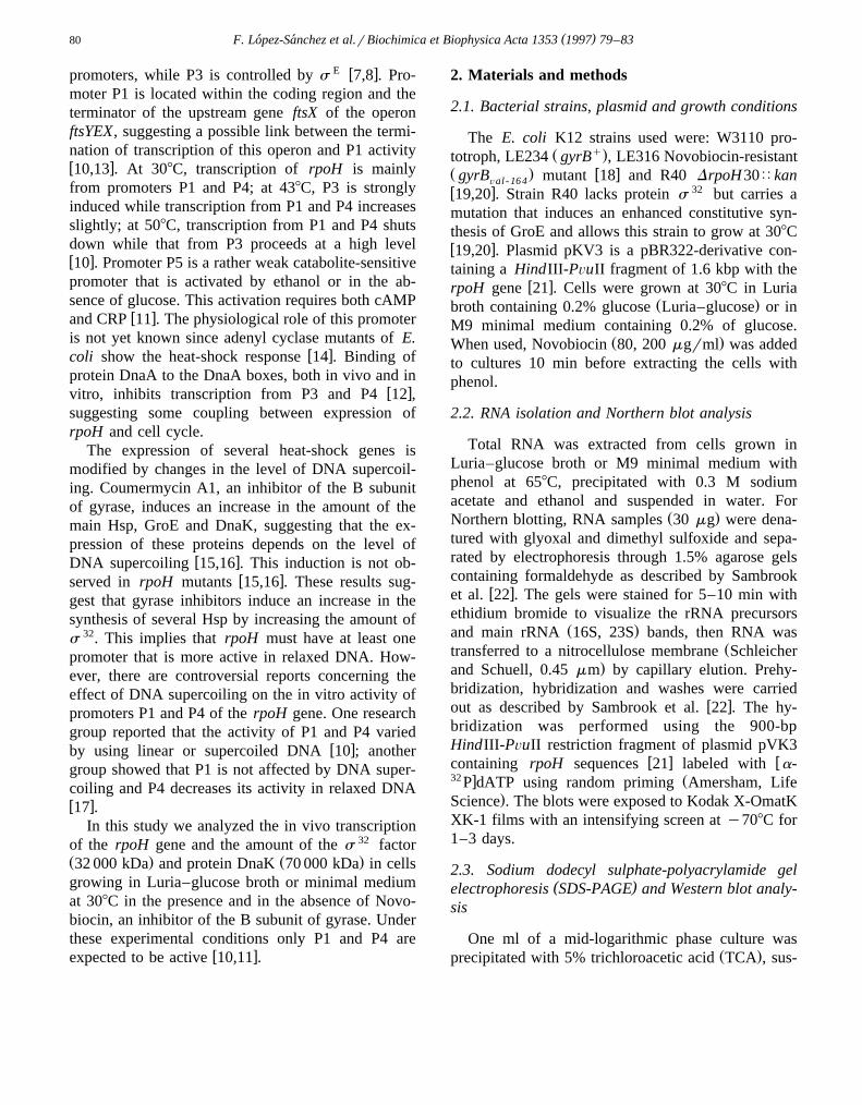
To study the in vivo transcription of gene rpoH,the amount of rpoH mRNA present in cells grown at308C in Luria–glucose broth or minimal medium wascompared with that of cells grown under the sameconditions but treated 10 min with Novobiocin. Ex-posure to Novobiocin for this period, induced a clear
Ž .relaxation of a reporter plasmid data not shown .Total RNA was separated by electrophoresis throughformaldehyde–agarose gels, transferred to nitrocellu-lose membranes and hybridized with a 32P-labeledrpoH-specific probe. Ethidium bromide staining ofthe gel and the re-hybridization of the membranewith 32P-labeled total DNA confirmed equal sampleloading of the gels. As shown in Fig. 1, two rpoHmRNA species of approximately 1400 and 1200 nu-cleotides were detected by Northern analysis. ThesemRNA species are similar to those previously de-
w xtected by Tilly et al. 25 . There was an approxi-mately three-fold increase in the amount of the 1400nucleotide mRNA and a small decrease in the amountof the 1200 nucleotide mRNA when cells were treatedwith Novobiocin, compared to control cells. Similar
Fig. 1. Effect of Novobiocin on the in vivo transcription of rpoH.Total RNA was phenol-extracted from W3110 cells grown at308C in Luria–glucose broth. For Northern blotting, RNA sam-
Ž .ples 30 mg were separated by electrophoresis through 1.5%agarose gels, transferred to nitrocellulose membranes and hy-bridized with a 32P-labeled 900-bp HindIII-PÕuII restrictionfragment of plasmid pVK3 containing rpoH sequences. Lane 1,control cells grown in Luria–glucose broth; lane 2, cells grown inLuria–glucose broth exposed to Novobiocin 80 mgrml for 10min. The relative migration of 16S rRNA, rpoH mRNA-1 andmRNA-4 are indicated on the left.
results were obtained with cells grown in Luria–glu-cose broth or minimal medium and with 80 and 200
Ž .mgrml Novobiocin data not shown . Under the ex-perimental conditions of 308C and Luria–glucosebroth or minimal medium, it is almost certain that
Žthese transcripts derived from promoters P1 1400. Ž . w xnucleotides and P4 1200 nucleotides 10,11 . To
verify that DNA gyrase and, therefore, DNA relax-ation are the mediators of the effect of Novobiocin onP1 activation, control experiments using the Novo-
Ž . w xbiocin-resistant strain LE316 gyrB 18 wereÕal -164
Ž .performed. In this strain, Novobiocin 80 mgrmlwas unable to induce a relaxation of a reporter plas-mid and to induce an increase in the amount of rpoH
Ž .mRNA-1 data not shown . These in vivo resultsshow that the amount of rpoH mRNA-1 is higher incells with relaxed DNA and that rpoH mRNA-4 isslightly more abundant in cells with supercoiled DNA.Results obtained here with P4 are similar to thosepreviously reported in experiments performed in vitro
w xby Ueshima et al. 17 ; however, results obtainedwith P1 disagree with those reported by this group.The main difference between the in vivo and in vitroexperiments is the presence, in the first case, of theentire operon ftsYEX upstream of gene rpoH and ofonly the ftsX-rpoH intercistronic region in the sec-ond. As mentioned before, the last codons corre-sponding to the FtsX protein and the ftsYEX termina-tor overlap the y35 and y10 regions, respectively,

( )F. Lopez-Sanchez et al.rBiochimica et Biophysica Acta 1353 1997 79–83´ ´82
w xof promoter P1 10,13 . Results obtained in vivo andpresented in this paper suggest that this overlap prob-ably affects the transcription of rpoH and that thetranscription unit that is sensitive to the level of DNAsupercoiling is the operon ftsYEX. The relaxation ofDNA induced by Novobiocin could decrease tran-scription of this operon allowing promoter P1 to betranscribed with higher efficiency. When this work
w xwas in progress, Kaneko et al. 26 reported thatDNA relaxation induced by expression of proteinLetD, an inhibitor of gyrase encoded by the F factor,causes an increase in the synthesis of s 32. Resultspresented here, propose that this increase is mainlydue to increased activity of P1 on relaxed DNA.
3.2. Effect of NoÕobiocin on the amount of proteinss 32 and DnaK
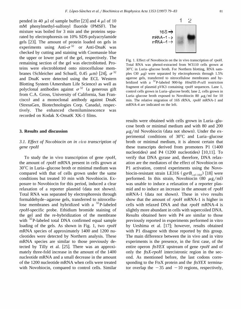
To verify if the increase in the amount of mRNAŽ .detected in cells treated with Novobiocin Fig. 1
corresponds to an increase in the amount of proteins 32, a Western blot analysis was performed. Ana-lyzed cells were grown on the same conditions usedfor the Northern blot analysis: Luria–glucose brothand 308C. Total protein samples were prepared, sepa-rated by SDS-PAGE and electroblotted on nitrocellu-lose membranes as described in Section 2.3. Thepolyclonal antibodies against s 32 detected, besidesome minor bands, a protein band of approximately32 000 kDa which is the only protein clearly induced
Fig. 2. Effect of Novobiocin on the amount of protein s 32. Totalprotein samples were prepared from W3110 cells grown at 308Cin Luria–glucose broth, separated by electrophoresis on 10%SDS-polyacrylamide gels, and electroblotted for Western analysisonto nitrocellulose membranes. Protein s 32 was detected usingpolyclonal antibodies against this protein and ECL Western
Ž .blotting reagents Amersham Life Science . Lane 1, control cellsŽ .308C ; lane 2, cells exposed to 438C for 6 min; lanes 3 and 4,cells grown at 308C and exposed for 10 min to Novobiocin 80and 200 mgrml, respectively.
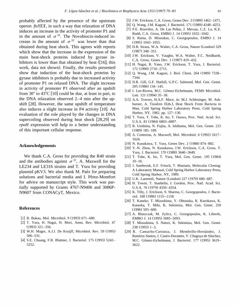
Fig. 3. Effect of Novobiocin on the amount of protein DnaK.Total protein samples were prepared from W3110 and R40 cellsgrown at 308C in Luria–glucose broth, separated by electrophore-sis on 10% SDS-polyacrylamide gels, and electroblotted forWestern analysis onto nitrocellulose membranes. Protein DnaKwas detected using monoclonal antibodies against this protein
Ž .and ECL Western blotting reagents Amersham Life Science .Lanes 1–4, W3110 cells; lanes 5–8, R40 cells. Lanes 1 and 5,
Ž .control cells 308C ; lanes 2 and 6, cells grown at 308C andexposed for 10 min to Novobiocin 80 mgrml; lanes 3 and 7,cells grown at 308C and exposed for 10 min to Novobiocin 200mgrml; lanes 4 and 8, cells exposed to 438C for 6 min.
Ž .after 6 min of cell exposure to 438C Fig. 2, lane 2 .This band is not observed in strain R40 which carries
w x Ž .an insertion in gene rpoH 19,20 data not shown .Novobiocin, 80 and 200 mgrml, induced an increase
32 Žin the amount of the s protein Fig. 2, lanes 3 and. 324 . The s increase induced by cell exposure to
Novobiocin was lower than that induced by 6-minŽ .exposure to 438C Fig. 2 . Therefore, the increase in
the amount of the mRNA from promoter P1 inducedby Novobiocin corresponds to an increase in theamount of s 32. These results suggest that the in-crease in heat-shock proteins GroEL and DnaK ob-
w xserved in cells exposed to Coumermycin 15,16 couldbe due to an increase in s 32. To test this, the amountof protein DnaK was determined by Western blotanalysis. This protein was selected since its gene,
32 w xdnaK , probably has only s promoters 20,27 .Gene groE has, in addition to the s 32 promoter, one
70 w xs promoter 20,27 . In strain W3110, Novobiocininduced an increase in the amount of protein DnaK;however, this increase is lower than that induced by
Ž .cell exposure to 438C Fig. 3, lanes 1–4 . ProteinDnaK was not detected in strain R40 which lacks
32 w x Ž .s 19,20 Fig. 3, lanes 5–8 .Results presented in this work show that Novo-
biocin induces a three-fold increase in the amount ofrpoH mRNA-1, a slight decrease in the amount ofrpoH mRNA-4 and an increase in the amount of
32 Ž .proteins s and DnaK Figs. 1–3 .These results suggest that during steady-state cell
growth at 308C, the in vivo transcription of rpoH is
( )F. Lopez-Sanchez et al.rBiochimica et Biophysica Acta 1353 1997 79–83´ ´ 83
probably affected by the presence of the upstreamoperon ftsYEX, in such a way that relaxation of DNAinduces an increase in the activity of promoter P1 andin the amount of s 32. The Novobiocin-induced in-crease in the amount of s 32 was lower than thatobtained during heat shock. This agrees with reportswhich show that the increase in the expression of themain heat-shock proteins induced by gyrase in-
Žw xhibitors is lower than that obtained by heat 16 ; this.work, data not shown . Finally, the results presented
show that induction of the heat-shock proteins bygyrase inhibitors is probably due to increased activityof promoter P1 on relaxed DNA. The slight increasein activity of promoter P1 observed after an upshift
w xfrom 308 to 438C 10 could be due, at least in part, tothe DNA relaxation observed 2–4 min after the up-
w xshift 28 . However, the same upshift of temperaturew xalso induces a slight increase in P4 activity 10 . An
evaluation of the role played by the changes in DNAw xsupercoiling observed during heat shock 28,29 on
rpoH expression will help to a better understandingof this important cellular response.
Acknowledgements
We thank C.A. Gross for providing the R40 strainand the antibodies against s 32, A. Maxwell for theLE234 and LE316 strains and T. Yura for providingplasmid pKV3. We also thank M. Paez for preparing´solutions and bacterial media and I. Perez-Montfort´for advice on manuscript style. This work was par-tially supported by Grants 4767-N9406 and 3086P-N9607 from CONACyT, Mexico.
References
w x Ž .1 B. Bukau, Mol. Microbiol. 9 1993 671–680.w x2 T. Yura, H. Nagai, H. Mori, Annu. Rev. Microbiol. 47
Ž .1993 321–350.w x Ž .3 W.H. Mager, A.J.J. De Kruijff, Microbiol. Rev. 59 1995
506–531.w x Ž .4 S.E. Chuang, F.R. Blattner, J. Bacteriol. 175 1993 5242–
5252.
w x Ž .5 J.W. Erickson, C.A. Gross, Genes Dev. 3 1989 1462–1471.w x Ž .6 Q. Wang, J.M. Kaguni, J. Bacteriol. 171 1989 4248–4253.w x7 P.E. Rouviere, A. De Las Penas, J. Mecsas, C.Z. Lu, K.E.` ˜
Ž .Rudd, C.A. Gross, EMBO J. 14 1995 1032–1042.w x8 S. Raina, D. Missiakas, C. Georgopoulos, EMBO J. 14
Ž .1995 1043–1055.w x Ž .9 D.B. Straus, W.A. Walter, C.A. Gross, Nature London 329
Ž .1987 348–351.w x10 J.W. Erickson, V. Vaughn, W.A. Walter, F.C. Neidhardt,
Ž .C.A. Gross, Genes Dev. 1 1987 419–432.w x11 H. Nagai, R. Yano, J.W. Erickson, T. Yura, J. Bacteriol.
Ž .172 1990 2710–2715.w x Ž .12 Q. Wang, J.M. Kaguni, J. Biol. Chem. 264 1989 7338–
7344.w x13 D.R. Gill, G.F. Hatfull, G.P.C. Salmond, Mol. Gen. Genet.
Ž .205 1986 134–145.w x14 I. Lee-Rivera, M.C. Gomez-Eichelmann, FEMS Microbiol.´
Ž .Lett. 121 1994 35–38.w x15 A.A. Travers, H.A.F. Mace, in: M.J. Schlesinger, M. Ash-
Ž .burner, A. Tissieres Eds. , Heat Shock From Bacteria to`Man, Cold Spring Harbor Laboratory Press, Cold SpringHarbor, NY, 1982, pp. 127–130.
w x16 T. Yura, T. Tobe, K. Ito, T. Osawa, Proc. Natl. Acad. Sci.Ž .U.S.A. 81 1984 6803–6807.
w x17 R. Ueshima, N. Fujita, A. Ishihama, Mol. Gen. Genet. 215Ž .1989 185–189.
w x Ž .18 A. Contreras, A. Maxwell, Mol. Microbiol. 6 1992 1617–1624.
w x Ž .19 N. Kusukawa, T. Yura, Genes Dev. 2 1988 874–882.w x20 Y.-N. Zhou, N. Kusukawa, J.W. Erickson, C.A. Gross, T.
Ž .Yura, J. Bacteriol. 170 1988 3640–3649.w x Ž .21 T. Tobe, K. Ito, T. Yura, Mol. Gen. Genet. 195 1984
10–16.w x22 J. Sambrook, E.F. Fritsch, T. Maniatis, Molecular Cloning:
A Laboratory Manual, Cold Spring Harbor Laboratory Press,Cold Spring Harbor, NY, 1989.
w x Ž . Ž .23 U.K. Laemmli, Nature London 227 1970 680–687.w x24 H. Towin, T. Staehelin, J. Gordon, Proc. Natl. Acad. Sci.
Ž .U.S.A. 76 1979 4350–4354.w x25 K. Tilly, J. Erickson, S. Sharma, C. Georgopoulos, J. Bacte-
Ž .riol. 168 1986 1155–1158.w x26 T. Kaneko, T. Mizushima, Y. Ohtusuka, K. Kurokawa, K.
Kataoka, T. Miki, K. Sekimizu, Mol. Gen. Genet. 250Ž .1996 593–600.
w x27 A. Blaszczak, M. Zylicz, C. Georgopoulos, K. Liberek,Ž .EMBO J. 14 1995 5085–5093.
w x28 T. Mizushima, S. Natori, K. Sekimizu, Mol. Gen. Genet.Ž .238 1993 1–5.
w x29 R. Camacho-Carranza, J. Membrillo-Hernandez, J.´Ramırez-Santos, J. Castro-Dorantes, V. Chagoya de Sanchez,´ ´
Ž .M.C. Gomez-Eichelmann, J. Bacteriol. 177 1995 3619–´3622.