in vivo il-10 gene delivery suppresses airway eosinophilia and
TRANSCRIPT
of April 14, 2018.This information is current as
Inflammationin a Mouse Model of Allergic Airway Antigen-Specific Systemic Immune ResponseMigration without Impairing the Down-Regulating APC Functions andAirway Eosinophilia and Hyperreactivity by In Vivo IL-10 Gene Delivery Suppresses
Jun-ichi Miyazaki and Kazuhiko YamamotoYoshinori Komagata, Katsuya Nagatani, Ryoichi Tanaka, Kazuyuki Nakagome, Makoto Dohi, Katsuhide Okunishi,
http://www.jimmunol.org/content/174/11/6955doi: 10.4049/jimmunol.174.11.6955
2005; 174:6955-6966; ;J Immunol
Referenceshttp://www.jimmunol.org/content/174/11/6955.full#ref-list-1
, 26 of which you can access for free at: cites 56 articlesThis article
average*
4 weeks from acceptance to publicationFast Publication! •
Every submission reviewed by practicing scientistsNo Triage! •
from submission to initial decisionRapid Reviews! 30 days* •
Submit online. ?The JIWhy
Subscriptionhttp://jimmunol.org/subscription
is online at: The Journal of ImmunologyInformation about subscribing to
Permissionshttp://www.aai.org/About/Publications/JI/copyright.htmlSubmit copyright permission requests at:
Email Alertshttp://jimmunol.org/alertsReceive free email-alerts when new articles cite this article. Sign up at:
Print ISSN: 0022-1767 Online ISSN: 1550-6606. Immunologists All rights reserved.Copyright © 2005 by The American Association of1451 Rockville Pike, Suite 650, Rockville, MD 20852The American Association of Immunologists, Inc.,
is published twice each month byThe Journal of Immunology
by guest on April 14, 2018
http://ww
w.jim
munol.org/
Dow
nloaded from
by guest on April 14, 2018
http://ww
w.jim
munol.org/
Dow
nloaded from
In Vivo IL-10 Gene Delivery Suppresses Airway Eosinophiliaand Hyperreactivity by Down-Regulating APC Functions andMigration without Impairing the Antigen-Specific SystemicImmune Response in a Mouse Model of AllergicAirway Inflammation1
Kazuyuki Nakagome,* Makoto Dohi,2* Katsuhide Okunishi,* Yoshinori Komagata,*Katsuya Nagatani,* Ryoichi Tanaka,* Jun-ichi Miyazaki,† and Kazuhiko Yamamoto*
IL-10 is an immunosuppressive cytokine. Although previous studies have reported that exogenous delivery of IL-10 reducedairway inflammation in experimental allergic airway inflammation, the mechanism of action has not been fully clarified. In thisreport, we elucidated a mechanism of action of IL-10 in vivo. BALB/c mice were immunized and aerosol challenged with OVA-Ag.We delivered the IL-10 gene to the mice before systemic sensitization or during aerosol Ag challenge by administering anIL-10-producing plasmid vector. Not only presensitization delivery of IL-10, as reported, but also delivery during inflammationstrongly suppressed the development of airway eosinophilia and hyperreactivity. Presensitization delivery suppressed the Ag-specific Th2-type immune response in both the lung and spleen. In contrast, delivery in the effector phase suppressed the Th2response only in the lung, whereas that in the spleen was not affected. IL-10 gene delivery did not induce the development of aregulatory phenotype of T cells or dendritic cells; rather, it suppressed the overall functions of CD11c� APCs of the lung such asAg-presenting capacity, cytokine production, and transportation of OVA-Ag to lymph nodes, thus attenuating Th2-mediatedallergic airway inflammation. Further, IL-10 revealed a distinct immunosuppressive effect in the presence of Ag and APCs. Theseresults suggest that suppression of APC function in the lung, the site of immune response, played a critical role in the IL-10-mediated suppression of Ag-induced airway inflammation and hyperreactivity. Therefore, if delivered selectively, IL-10 could sitespecifically suppress the Ag-specific immune response without affecting systemic immune responses. The Journal of Immunology,2005, 174: 6955–6966.
I nterleukin-10 is an important immunosuppressive cytokine.For example, IL-10 inhibits the production of proinflamma-tory cytokines such as IL-1�, IL-1�, IL-6, IL-8, IL-12,
TNF-�, and GM-CSF by monocytes and cytokines such as IL-1�,IL-6, IL-8, TNF-�, IFN-�, and GM-CSF by Th1 cells (1–6). IL-10down-regulates the expression of MHC class II molecules and co-stimulatory molecules on monocytes/macrophages and dendriticcells (DCs)3 (1, 7–10). IL-10 suppresses the production of cyto-kines by T cells through direct (11–13) and indirect (APC-depen-dent) mechanisms (4, 5, 7), although the direct effect on T cells hasbeen confirmed only in humans. These in vitro studies raise thepossibility that IL-10 could be beneficial for the treatment of var-
ious immune diseases in vivo. For example, both in human sub-jects and in animal models, treatment with IL-10 improved inflam-matory bowel disease (14–17) and psoriasis (18–20), which aremainly mediated by Th1 immune response. In individuals whohave received bee venom-specific immunotherapy or who recentlyhave received multiple bee stings, up-regulation of endogenousIL-10 may play an important role in preventing anaphylaxis (21,22). However, thus far, the inhibitory effects of IL-10 have beenanalyzed almost always in vitro, and the mechanisms of inhibitionin vivo have not been well examined. Therefore, the clinical ap-plication of IL-10 to treat immune disorders is difficult.
Recently, the role of IL-10 in the generation of regulatory T(Treg) cells has been highlighted. Groux et al. reported thatchronic activation of CD4� cells in the presence of IL-10 in vitroinduces the development of T-regulatory 1 (Tr1) cells, which pro-duce much IL-10 (23). Akbari et al. reported that stimulation ofCD4� cells with IL-10-producing DCs, which were generated af-ter exposure to inhaled Ag (24), induces IL-10-producing Tregcells (25). These reports suggest that IL-10-producing Treg cellsdevelop in the presence of IL-10. However, whether in vivo IL-10alone could induce the generation of Treg cells has not been fullyclarified.
Bronchial asthma is a chronic disorder characterized by eosin-ophilic airway inflammation, mucus hypersecretion, and an in-crease in airway hyperreactivity (AHR) (26). The process of air-way inflammation involves various types of cells, such aseosinophils, mast cells, T lymphocytes, and DCs (27, 28). Due to
*Department of Allergy and Rheumatology, Graduate School of Medicine, Universityof Tokyo, Tokyo, Japan; and †Division of Stem Cell Regulation Research, OsakaUniversity Graduate School of Medicine, Osaka, Japan
Received for publication July 8, 2004. Accepted for publication March 18, 2005.
The costs of publication of this article were defrayed in part by the payment of pagecharges. This article must therefore be hereby marked advertisement in accordancewith 18 U.S.C. Section 1734 solely to indicate this fact.1 This work was supported by Grant-in-aid 13670592 from the Ministry of Health,Welfare, and Labor of Japan.2 Address correspondence and reprint requests to Dr. Makoto Dohi, Department ofAllergy and Rheumatology, Graduate School of Medicine, University of Tokyo, 7-3-1Hongo, Bunkyo-ku, Tokyo 113-8655, Japan. E-mail address: [email protected] Abbreviations used in this paper: DC, dendritic cell; Treg, regulatory T; Tr1, Tregulatory 1; AHR, airway hyperreactivity; SA, physiological saline; Mch, metha-choline; Penh, enhanced pause; BALF, bronchoalveolar lavage fluid; MFI, mean flu-orescence intensity.
The Journal of Immunology
Copyright © 2005 by The American Association of Immunologists, Inc. 0022-1767/05/$02.00
by guest on April 14, 2018
http://ww
w.jim
munol.org/
Dow
nloaded from

its immunosuppressive effects, IL-10 is expected to down-regulatethe airway immune response. In humans, relative underproductionof IL-10 by alveolar macrophages and in the sputum of patientswith asthma has been reported (29, 30), which suggests an essen-tial role for IL-10 in regulating airway inflammation. However, therole of IL-10 in airway inflammation and AHR is not well under-stood in either humans or animal models. For example, the effectof deleting endogenous IL-10 on Ag-induced eosinophilic inflam-mation in mouse models of allergic airway inflammation has beencontroversial. Some reports have indicated that IL-10-deficientmice exhibit exaggerated eosinophilic airway inflammation pro-voked by a systemic sensitization to Ag and local inhalation of Ag(31, 32), whereas another report indicated that IL-10-deficientmice show diminished eosinophilic airway inflammation (33).In contrast, several reports have indicated that administrationof IL-10 to the lung suppresses eosinophilic airway inflammation.The instillation of recombinant murine IL-10 into the lung, admin-istered concurrently with Ag, inhibits eosinophilic airway inflam-mation (34, 35). CD4� Th cells engineered to produce IL-10prevent AHR and inflammation induced by the transfer of OVA-specific Th2 cells and OVA challenge, although their effect oneosinophilic inflammation was not confirmed in sensitized andchallenged mice (36). Further, administration of IL-10 cDNA intothe lung via an adenoviral vector, at least if done before sensiti-zation, inhibits eosinophilic inflammation and AHR (37). Al-though these studies show that exogenous delivery of IL-10 wouldhave some inhibitory effect on experimental allergic airway in-flammation, the mechanism of action in vivo, especially in thelung, has not been clarified.
The purpose of the present study was to elucidate the mecha-nism of IL-10 gene delivery in vivo. We found that IL-10 genedelivery, even after systemic Ag sensitization has been completed,suppressed Ag-induced eosinophilic airway inflammation andAHR. We also found that IL-10 gene delivery down-regulatedAPC functions in the lung and migration of Ag-captured CD11c�
cells to regional peribronchial lymph nodes, although it did notaffect the systemic immune response to Ag. These results indicatedthat the immunosuppressive effect of IL-10 is site specific, occur-ring in tissue where APCs encounter the Ag and thus an Ag-spe-cific immune response is driven.
Materials and MethodsMice
Male BALB/c and C57BL/6 mice were obtained from Charles River Japan.Male OVA TCR-transgenic DO11.10 mice and C57BL/6-IL10�/� micewere obtained from The Jackson Laboratory (Bar Harbor, ME). All animalexperiments were approved by the Animal Research Ethics Board of theDepartment of Allergy and Rheumatology, University of Tokyo (Tokyo,Japan).
Immunization of mice and evaluation of allergic airwayinflammation
Mice were immunized as reported previously (38, 39). Seven-week-oldanimals were sensitized with an i.p. injection of 2 �g of OVA (Sigma-Aldrich) plus 2 mg of aluminum hydroxide (alum) on days 0 and 11.Control mice received injections of physiological saline (SA) on days 0 and11. Some groups of mice were sacrificed on day 18 for analyses of immuneresponse in the spleen. In another experiment, for analyses of allergic air-way inflammation, mice were challenged with an aerosolized solution of3% OVA or PBS for 10 min from day 18 to day 20. On day 21, AHR tomethacholine (Mch) was measured with the enhanced pause (Penh) systemas described previously (38, 39). Results were expressed for each concen-tration of Mch as a percentage of the baseline Penh value after exposure toSA. Then, samples of serum and bronchoalveolar lavage fluid (BALF)were obtained. The lungs were cut out and fixed with 10% neutralizedbuffered formalin (Wako). Sections 3 �m thick were prepared and sub-jected to H&E staining.
Delivery of IL-10 into mice
In the present study, we delivered IL-10 in vivo using a hydrodynamics-based method through the i.v. injection of plasmid DNA with previouslyreported methods (40, 41). The plasmid pCAGGS-IL-10 (40) was ampli-fied in Escherichia coli and purified with a Qiagen Endo Free plasmid Gigakit (Qiagen). The empty plasmid pCAGGS was used as a control. PlasmidDNA in lactated Ringer’s solution (0.1 ml/g body weight) was injected i.v.into the tail. The injection was completed within 5 s. As reported originally,with this delivery system, the plasmid was trapped in the liver, and thecytokine was produced there and then moved into the bloodstream and tothe perfused organs (40, 41). For study of the kinetics of IL-10 gene de-livery, mice were sacrificed on days 1, 4, 7, 10, 13, 20, and 23 after theplasmid injection. Concentrations of IL-10 in serum and BALF were mea-sured using an ELISA kit (BD Pharmingen) according to the manufactur-er’s instructions. Some mice received plasmid DNA (100 �g; pCAGGS-IL-10 or control pCAGGS) i.v. on day �3 (before systemic immunization:pre) or on day 17 (during aerosol challenge; after systemic immunization:post).
Measurement of cytokines and Ig
IL-4, IL-5, IL-10, IL-12 p70, IL-13, IFN-�, TGF-�, and IgE concentrationswere measured with ELISA kits (IL-4, IL-5, IL-10, IL-12 p70, IFN-�, andIgE, BD Pharmingen; IL-13 and TGF-�, R & D Systems). OVA-specificIgE was measured using an ELISA kit for IgE except that the plate wascoated with OVA (1000 �g/ml) at 4°C overnight instead of anti-mouse IgEAb. The OVA-specific IgE standard was derived by pooling sera from fiveOVA-sensitized mice. Results were expressed as a percentage of the valueof the standard.
Preparation of spleen and lung cells
For the preparation of spleen cells, a 2-ml solution of 450 U of collagenase(Sigma-Aldrich) in complete DMEM (Life Technologies) supplementedwith 10% FCS, 10 mM HEPES (Life Technologies), 0.1 mM nonessentialamino acids (Life Technologies), 1 mM sodium pyruvate, 2 mM sodiumglutamate (Sigma-Aldrich), 100 U/ml penicillin (Sigma-Aldrich), 100�g/ml streptomycin (Sigma-Aldrich), and 5.0 � 10�5 M 2-ME (Sigma-Aldrich) were injected into the spleen, and the spleen was incubated at37°C for 20 min. Cell suspensions were collected by pressing the tissuesthrough a 70-�m pore size nylon filter into HBSS (Sigma-Aldrich) andcentrifuged. The cell pellets were resuspended in RBC lysis solution (Sig-ma-Aldrich) for 1 min and washed; then the cell suspensions were filteredagain to remove clumps and aggregates. Viable cell numbers were deter-mined by trypan blue dye exclusion. The preparation of lung cells wasconducted essentially using previously reported methods with a slight mod-ification (42, 43). Briefly, lungs were excised, minced, and digested for 90min at room temperature with 150 U of collagenase in complete DMEM.Cell suspensions were obtained by pressing the tissues through a 70-�mpore size nylon filter.
For the preparation of splenic CD4� T cells or DCs, spleen cells wereincubated with anti-CD4 mAb-coated or anti-CD11c mAb-coated mi-crobeads (Miltenyi Biotech). Bead-bound cells were then isolated usingmagnetic separation columns. The purities of the enriched CD4� andCD11c� cell populations were 95 and 85%, respectively (data not shown).More than 95% of the CD4� cells were CD3� T cells (data not shown). Forthe preparation of lung CD11c� cells, cells were purified using NycoPrep(AXIS-SHIELD) before incubation with anti-CD11c mAb-coated mi-crobeads, and bead-bound cells were then isolated. The purity of the en-riched CD11c� cells was 80% (data not shown). This purity was similar tothat of previous studies that isolated CD11c� cells from the lung (42, 43).
In vitro proliferation and cytokine assays
Spleen or lung cells were cultured (5 � 105 cells/well) with or withoutOVA in complete DMEM. In some experiments, positively selected CD4�
T cells (2.5 � 105 cells/well) were cultured with freshly isolated mitomy-cin C (Sigma-Aldrich)-treated splenocytes (2.5 � 105 cells/well) in thepresence or absence of OVA. After 72 or 96 h, the proliferation was as-sessed with a cell proliferation ELISA BrdU kit (Roche Applied Science).After 120 h, cytokine concentrations in the supernatants were measuredusing ELISA kits. To examine the direct effect of IL-10 on CD4� T cellfunctions in vitro, OVA-sensitized CD4� T cells were incubated withplate-bound anti CD3 Ab (10 �g/ml; BD Pharmingen) for 48 h or PMA (1ng/ml; Sigma-Aldrich) and ionomycin (1 �g/ml; Sigma-Aldrich) for 24 hin the presence or absence of IL-10. Then, the proliferation was assessed,and the IL-4 concentration in the supernatants was measured. To examinethe effect of coincubation with IL-10 on the Ag-specific immune response
6956 IL-10 SUPPRESSES AIRWAY EOSINOPHILIA AND AHR
by guest on April 14, 2018
http://ww
w.jim
munol.org/
Dow
nloaded from

of CD4� T cells, OVA-sensitized CD4� T cells were incubated with mi-tomycin C-treated splenocytes from naive mice and OVA (20 �g/ml) for72 h in the presence or absence of IL-10. In another experiment, to examinethe effect of preincubation with IL-10, OVA-sensitized spleen cells (5 �106 cells/ml) were preincubated with or without IL-10 (50 ng/ml) for 24 h.After IL-10 was removed from the culture medium, CD4� T cells werepositively selected from the spleen cell population and incubated with mi-tomycin C-treated splenocytes from naive mice and OVA for 72 h. Then,the proliferation was assessed and the IL-4 concentration was measured.For measurement of the production of cytokines by CD11c� cells, CD11c�
cells (1.25 � 105 cells/well) were incubated with LPS (1 �g/ml) or TNF-�(50 ng/ml) for 24 h, and then cytokine concentrations in the supernatantswere measured using ELISA kits.
RT-PCR
RNA was extracted from CD4� T cells by the acid-guanidium phenolchloroform method using Isogen (Nippon Gene). RT-PCR for Foxp3 or�-actin was performed using previously reported methods (38, 44). PCRfor Foxp3 consisted of 40 s of denaturation at 94°C, 1 min of annealing at60°C, and 1 min of extension at 72°C for 26 cycles. PCR for �-actinconsisted of 1 min of denaturation at 94°C, 1 min of annealing at 61°C, and1 min of extension at 72°C for 21 cycles. The sense primer for Foxp3 was5�-GGC GAA AGT GGC AGA GAG GTA T-3�, and the antisense primerwas 5�-AAG ACC CCA GTG GCA GCA GAA-3�. The sense primer for�-actin was 5�-TGG AAT CCT GTG GCA TCC ATG AAA C-3�, and theantisense primer was 5�-TAA AAC GCA GCT CAG TAA CAG TCCG-3�. PCR products were electrophoresed in a 3% agarose gel, and theresults were visualized by ethidium bromide staining. Densitometric anal-ysis of mRNA expression was performed using NIH Image software (Na-tional Insititutes of Health, Bethesda, MD). Normalized values for Foxp3mRNA expression in each sample were calculated as the relative quantityof Foxp3 divided by the relative quantity of �-actin.
Allogenic MLR and Ag-presenting capacity of CD11c� cells
For analyses of allogeneic MLR, mitomycin C-treated BALB/c (H-2d)CD11c� cells (1 � 104 or 2.5 � 104 cells/well) were cultured with posi-tively selected C57BL/6 (H-2b) CD4� T cells (2.5 � 105 cells/well) incomplete DMEM. In some experiments, C57BL/6 CD11c� cells (2.5 �104 cells/well) were cultured with BALB/c CD4� T cells (2.5 � 105 cells/well). For the measurement of OVA-Ag-presenting capacity, mitomycinC-treated CD11c� cells (2.5 � 104 cells/well) were cultured with DO11.10CD4� T cells (2.5 � 105 cells/well). In some experiments, CD11c� cells(1.25 � 105) from the spleen were cultured with OVA-sensitized CD4� Tcells (1.25 � 105 cells/well) and OVA (30 �g/ml). After 48–96 h, theproliferation was assessed with a cell proliferation ELISA BrdU kit. Formeasurements of the capacity of regional lymph node cells to activateOVA-specific TCR-transgenic CD4� T cells, mitomycin C-treated lymphnodes cells (2.5 � 105 cells/well) were cultured with DO11.10 CD4� Tcells (2.5 � 105 cells/well). After 72 h, the proliferation was assessed.
Flow cytometry
Spleen or lung cells (1 � 106) were incubated with PE anti-CD11c mAb(HL3; BD Pharmingen) and biotin anti-mouse MHC class II mAb (antiI-A/I-E mAb; 2G9; BD Pharmingen), anti-CD40 mAb (3/23; BD Pharm-ingen), anti-CD80 mAb (16-10A1; BD Pharmingen), or anti-CD86 mAb(GL1; BD Pharmingen) in the presence of anti-CD16/32 mAb (2.4G2; BDPharmingen) for 30 min and washed twice. Then, cells were incubated withFITC-streptavidin (BD Pharmingen) for 30 min and washed twice. Deadcells were excluded by propidium iodide staining. Briefly, cells were in-cubated with propidium iodide (2 �g/ml; Sigma-Aldrich) for 15 min atroom temperature. Then stained cells were analyzed by flow cytometry(EPICS XL System II).
Fluorescence study
Fluorescence microscopy was performed using previously reported meth-ods with some modification (45, 46). Mice were sensitized with OVA ondays 0 and 11. Some mice received pCAGGS-IL-10 or control pCAGGS(100 �g) i.v. on day 17. Mice were given FITC-labeled OVA (MolecularProbes), FITC-dextran (Sigma-Aldrich), or FITC alone intratracheally onday 20 (500 �g each). On day 21, the bronchial lymph nodes were excised,embedded in cryomolds containing Tissue-Tek OCT compound (SakuraFinetek), and frozen in liquid nitrogen. Sections were cut 5 �m thick bycryostat. Some sections were fixed in acetone and treated with PE anti-CD11c mAb. The sections were subsequently observed under a fluores-cence microscope (BX51).
Preparation of OVA-pulsed splenic DCs and transfer into naivemice
Splenic CD11c� cells were positively selected by magnetic cell sortingfrom naive mice and incubated with OVA (1000 �g/ml) in the presence orabsence of GM-CSF (10 ng/ml). IL-10 was added to some culture media(50 ng/ml). After 20 h, cells were collected and washed three times. Thecells were examined for Ag (OVA)-presenting capacity and cell surfacemarker expression. In some experiments, the cells were transferred intonaive mice i.v. (5 � 105). The mice were challenged with nebulized 3%OVA in PBS for 1 h twice daily on days 7, 8, and 15 and sacrificed on day16 for BALF analysis.
Statistics
Values are expressed as means � SEM. Statistical analyses were per-formed using a one- or two-way ANOVA followed by Fisher’s least sig-nificant difference test or Scheffe’s procedure. p �0.05 was consideredsignificant.
ResultsIL-10 expression in serum and in BALF after hydrodynamics-based gene delivery by i.v. injection of plasmid DNA
First, we examined the kinetics of IL-10 gene delivery after i.v.injection of the plasmid DNA. Samples were collected at the in-dicated times after the injection of pCAGGS-IL-10 or controlpCAGGS. Endogenous IL-10 concentrations in serum and inBALF from mice before gene delivery were 8.5 � 2.4 and 97.5 �28.1 pg/ml, respectively. The temporal pattern of IL-10 proteinexpression in serum (Fig. 1A) and in BALF (Fig. 1B) was con-firmed, as described previously (40). The level of IL-10 peaked 1day after the injection and gradually decreased thereafter. On day20, IL-10 delivery did not differ between mice that receivedpCAGGS-IL-10 and mice that received control pCAGGS.
In vivo IL-10 gene delivery suppresses Ag-induced immuneresponse of CD4� T cells and DC functions ex vivo
Next, we confirmed the effect of in vivo IL-10 gene delivery on theAg-induced immune response by conducting ex vivo analyses.
FIGURE 1. IL-10 concentrations in serum and in BALF after hydrody-namics-based gene delivery by i.v. injection of plasmid DNA. Mice re-ceived an i.v. injection of pCAGGS-IL-10 (IL-10) or control pCAGGSplasmid (CONT) on day 0. Concentrations of IL-10 in serum (A) and inBALF (B) were measured at the indicated times after the injection usingELISA. Values are presented as means � SEM for 6–10 mice per group.��, p � 0.01 and ���, p � 0.001 compared with the value of CONT.
6957The Journal of Immunology
by guest on April 14, 2018
http://ww
w.jim
munol.org/
Dow
nloaded from

Splenic CD4� T cells obtained from OVA-sensitized micestrongly proliferated (Fig. 2A) and produced Th1- and Th2-typecytokines (Fig. 2B), compared with those of SA-treated mice.Splenic CD4� T cells from OVA-sensitized and pCAGGS-IL-10-injected mice failed to proliferate (Fig. 2A) or produce Th1- andTh2-type cytokines (Fig. 2B) in response to OVA, in contrast tothose from the OVA-sensitized and control pCAGGS-injectedmice. Similar results of cell proliferation and cytokine productionwere obtained when we used whole spleen cells instead of CD4�
T cells (data not shown). Then, we examined the possibility thatthis suppression was mediated through production of Treg cells invivo. CD4� T cells from OVA-sensitized and pCAGGS-IL-10-injected mice did not produce IL-10 (Fig. 2B) or TGF-� (data notshown) in response to OVA, which suggested that this treatmentdid not induce the development of Tr1 (23)-like cells or Th3 (47)-like cells. In vivo IL-10 gene delivery also did not increase themRNA expression of Foxp3, a transcription factor that wasthought to be specifically expressed in naturally occurringCD4�CD25� T cells (Fig. 2C). In addition, IL-10 did not alter theratio of CD4�CD25� cells to CD4�CD25� cells either (data notshown). These results suggested that in vivo IL-10 gene deliverywould not induce the development of Treg cells. Next, we exam-ined the effect of IL-10 treatment on DC functions after Ag sen-sitization. IL-10 treatment decreased the MHC alloreactivity (Fig.2D), OVA-presenting capacity (Fig. 2E), and MHC class II ex-pression of splenic DCs (Fig. 2F). IL-10 treatment also suppressedCD40, CD80, and CD86 expression in splenic DCs, although therewas no significant difference in CD80 or CD86 expression (meanfluorescence intensity (MFI): CONT/OVA; CD40 71.1 � 5.6,CD80 56.0 � 4.4, CD86 51.0 � 3.6; vs IL-10/OVA; CD40 38.0 �3.3, CD80 33.0 � 4.0, CD86 29.3 � 4.1, respectively). Further,IL-10 treatment also suppressed the production of IL-10 in re-sponse to LPS (Fig. 2G) or TNF-� (Fig. 2H). These results sug-gested that IL-10 gene delivery induced an overall suppression ofDC activities, not a switch to a suppressive phenotype. They alsoindicated that our system of in vivo IL-10 gene delivery effectivelysuppressed the Ag-induced immune response.
IL-10 has a suppressive effect on CD4� T cells only in thepresence of Ag and APCs in vitro
As described above, IL-10 gene delivery suppressed DC functionand CD4� T cell priming in vivo. Then, we examined whetherIL-10 could directly suppress CD4� T cell functions in vitro.When OVA-sensitized splenic CD4� T cells were stimulated withplate-bound anti-CD3 Ab or PMA-ionomycin, IL-10 did not sup-press the proliferation or IL-4 production of CD4� T cells (Fig. 3,A and B). So IL-10 did not suppress CD4� T cells directly. Co-incubation of IL-10 with OVA and APCs significantly suppressedthe proliferation of CD4� T cells (Fig. 3, C and D). In contrast,pretreatment of OVA-sensitized spleen cells with IL-10, but with-out OVA, had no effect on the proliferation of CD4� T cells uponfurther OVA stimulation (Fig. 3, E and F). These results suggestedthat IL-10 has a suppressive effect on CD4� T cells only in thepresence of Ag and APCs.
In vivo IL-10 gene delivery, even after systemic Ag sensitizationhas been completed, suppresses Ag-induced eosinophilic airwayinflammation and AHR
We next elucidated the effect of IL-10 gene delivery on Th2-me-diated allergic inflammation using an experimental model of al-lergic airway inflammation. Mice were sensitized with OVA or SAand then challenged with nebulized OVA or PBS. Injection ofplasmid (pCAGGS-IL-10 or control pCAGGS) was performed be-fore the systemic Ag sensitization (pre, on day �3) or during the
aerosol challenge (after systemic Ag sensitization; post, on day17). In vivo IL-10 delivery in both cases almost completely di-minished the eosinophil number in BALF (Fig. 4A). Histologi-cally, a slight infiltration of inflammatory cells into the peribron-chial area was detected in both groups of pCAGGS-IL-10-injectedmice (Fig. 4B), but prominent infiltration of eosinophils into theperibronchial interstitial area and goblet cell formation of bron-chial epithelial cells, which were provoked in the OVA-sensitizedand OVA-challenged mice (Fig. 4B), were not observed. In themice that received control pCAGGS before the sensitization orduring the aerosol challenge, prominent infiltration of eosinophilsand goblet cell formation were observed, respectively (Fig. 4B).BALF IL-13 and TGF-� concentrations were also decreased inpCAGGS-IL-10-injected mice (Fig. 4C). Moreover, AHR to Mchdecreased in pCAGGS-IL-10-injected mice (Fig. 4D). These re-sults indicated that in vivo IL-10 gene delivery, not only beforesystemic sensitization but also during ongoing airway immune re-sponses, suppressed eosinophilic airway inflammation and AHR.
In vivo IL-10 gene delivery during aerosol challenge suppressesproduction of Th2 cytokines by lung cells, but not spleen cells
Next we examined the effect of IL-10 gene delivery on the immuneresponse in the lung and compared it with that in the spleen. Con-trol pCAGGS injection before sensitization or during aerosol chal-lenge did not significantly suppress Th2 cytokine production in thelung (Fig. 5A). IL-10 delivery before sensitization and that duringaerosol challenge suppressed the Th2 cytokine production by lungcells, although the suppression after IL-10 gene delivery duringaerosol challenge was moderate compared with that after deliverybefore sensitization (Fig. 5A). In contrast, in vivo IL-10 deliveryduring aerosol challenge did not suppress the cell proliferation orcytokine production of spleen cells, whereas IL-10 delivery beforesensitization suppressed them (Fig. 5, B and C). Further, in vivoIL-10 delivery during aerosol challenge did not suppress total orOVA-specific IgE production, whereas IL-10 delivery before sen-sitization suppressed both (Fig. 5D).
In vivo IL-10 gene delivery suppresses resident lung APCfunctions
To elucidate the difference in the mechanism of suppression be-tween lung and spleen, we examined lung APC functions. LungCD11c� cells obtained from OVA-sensitized and OVA-nebulizedmice enhanced the MHC alloreactivity (Fig. 6A), OVA-presentingcapacity (Fig. 6B), cytokine production (Fig. 6C), and expressionof MHC class II (Fig. 6, D and E), compared with those of SA-treated, PBS-nebulized mice. IL-10 delivery before sensitizationand that during aerosol challenge suppressed these resident lungAPC functions of CD11c� cells (Fig. 6, A–E). IL-10 treatment alsosuppressed the expression of other costimulatory molecules suchas CD40, CD80, and CD86 on lung CD11c� cells, although therewere no significant differences (MFI: CONT (pre), CD40 38.4 �3.4, CD80 52.9 � 4.4, CD86 38.0 � 2.6; IL-10 (pre), CD4027.7 � 3.6, CD80 43.2 � 3.7, CD86 28.1 � 3.1; CONT (post),CD40 37.6 � 4.0, CD80 58.1 � 6.4, CD86 37.2 � 3.2; IL-10(post), CD40 29.1 � 2.8, CD80 40.1 � 7.4, CD86 28.2 � 2.9).This is probably because OVA sensitization and challenge itselfdid not induce high levels of expression of these molecules. In thespleen, IL-10 delivery before sensitization suppressed the OVA-presenting capacity of DCs, whereas postsensitization delivery ofIL-10 did not suppress it (Fig. 6F).
6958 IL-10 SUPPRESSES AIRWAY EOSINOPHILIA AND AHR
by guest on April 14, 2018
http://ww
w.jim
munol.org/
Dow
nloaded from
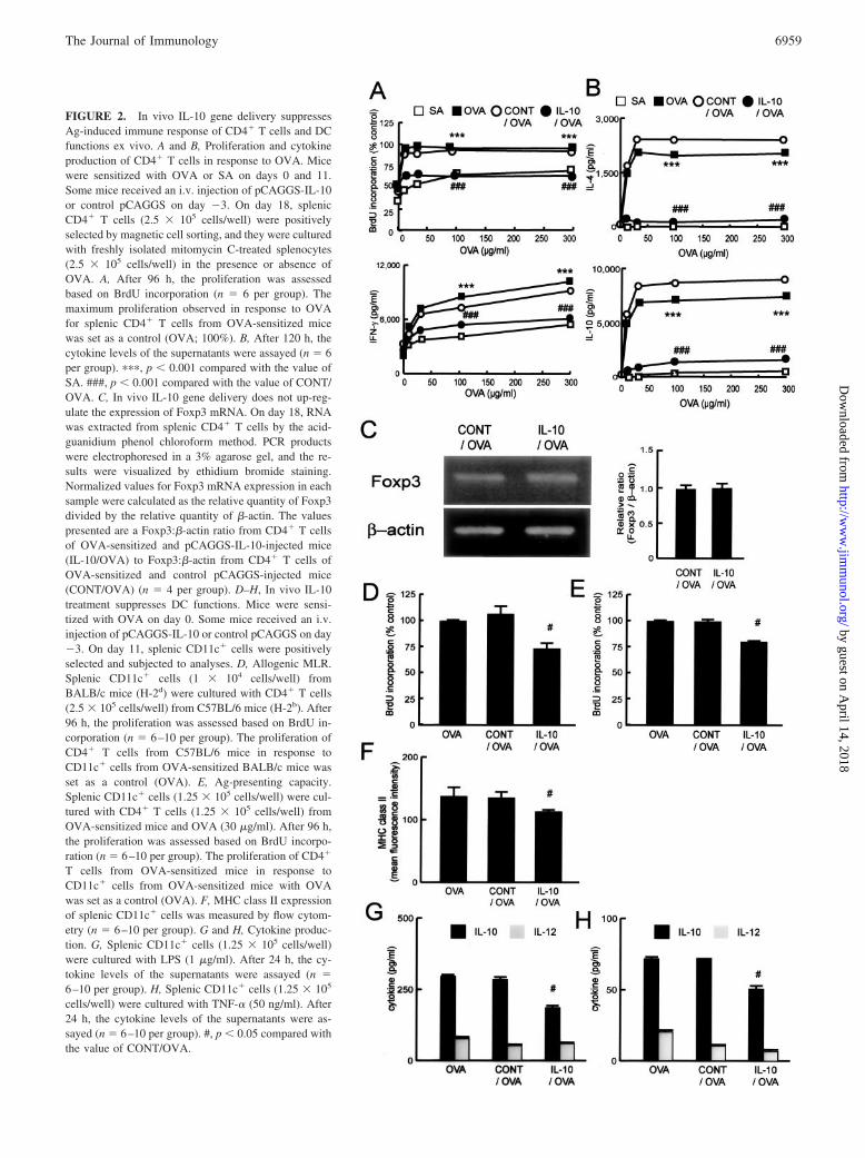
FIGURE 2. In vivo IL-10 gene delivery suppressesAg-induced immune response of CD4� T cells and DCfunctions ex vivo. A and B, Proliferation and cytokineproduction of CD4� T cells in response to OVA. Micewere sensitized with OVA or SA on days 0 and 11.Some mice received an i.v. injection of pCAGGS-IL-10or control pCAGGS on day �3. On day 18, splenicCD4� T cells (2.5 � 105 cells/well) were positivelyselected by magnetic cell sorting, and they were culturedwith freshly isolated mitomycin C-treated splenocytes(2.5 � 105 cells/well) in the presence or absence ofOVA. A, After 96 h, the proliferation was assessedbased on BrdU incorporation (n � 6 per group). Themaximum proliferation observed in response to OVAfor splenic CD4� T cells from OVA-sensitized micewas set as a control (OVA; 100%). B, After 120 h, thecytokine levels of the supernatants were assayed (n � 6per group). ���, p � 0.001 compared with the value ofSA. ###, p � 0.001 compared with the value of CONT/OVA. C, In vivo IL-10 gene delivery does not up-reg-ulate the expression of Foxp3 mRNA. On day 18, RNAwas extracted from splenic CD4� T cells by the acid-guanidium phenol chloroform method. PCR productswere electrophoresed in a 3% agarose gel, and the re-sults were visualized by ethidium bromide staining.Normalized values for Foxp3 mRNA expression in eachsample were calculated as the relative quantity of Foxp3divided by the relative quantity of �-actin. The valuespresented are a Foxp3:�-actin ratio from CD4� T cellsof OVA-sensitized and pCAGGS-IL-10-injected mice(IL-10/OVA) to Foxp3:�-actin from CD4� T cells ofOVA-sensitized and control pCAGGS-injected mice(CONT/OVA) (n � 4 per group). D–H, In vivo IL-10treatment suppresses DC functions. Mice were sensi-tized with OVA on day 0. Some mice received an i.v.injection of pCAGGS-IL-10 or control pCAGGS on day�3. On day 11, splenic CD11c� cells were positivelyselected and subjected to analyses. D, Allogenic MLR.Splenic CD11c� cells (1 � 104 cells/well) fromBALB/c mice (H-2d) were cultured with CD4� T cells(2.5 � 105 cells/well) from C57BL/6 mice (H-2b). After96 h, the proliferation was assessed based on BrdU in-corporation (n � 6–10 per group). The proliferation ofCD4� T cells from C57BL/6 mice in response toCD11c� cells from OVA-sensitized BALB/c mice wasset as a control (OVA). E, Ag-presenting capacity.Splenic CD11c� cells (1.25 � 105 cells/well) were cul-tured with CD4� T cells (1.25 � 105 cells/well) fromOVA-sensitized mice and OVA (30 �g/ml). After 96 h,the proliferation was assessed based on BrdU incorpo-ration (n � 6–10 per group). The proliferation of CD4�
T cells from OVA-sensitized mice in response toCD11c� cells from OVA-sensitized mice with OVAwas set as a control (OVA). F, MHC class II expressionof splenic CD11c� cells was measured by flow cytom-etry (n � 6–10 per group). G and H, Cytokine produc-tion. G, Splenic CD11c� cells (1.25 � 105 cells/well)were cultured with LPS (1 �g/ml). After 24 h, the cy-tokine levels of the supernatants were assayed (n �6–10 per group). H, Splenic CD11c� cells (1.25 � 105
cells/well) were cultured with TNF-� (50 ng/ml). After24 h, the cytokine levels of the supernatants were as-sayed (n � 6–10 per group). #, p � 0.05 compared withthe value of CONT/OVA.
6959The Journal of Immunology
by guest on April 14, 2018
http://ww
w.jim
munol.org/
Dow
nloaded from

In vivo IL-10 gene delivery suppresses migration of CD11c�
cells into draining lymph nodes
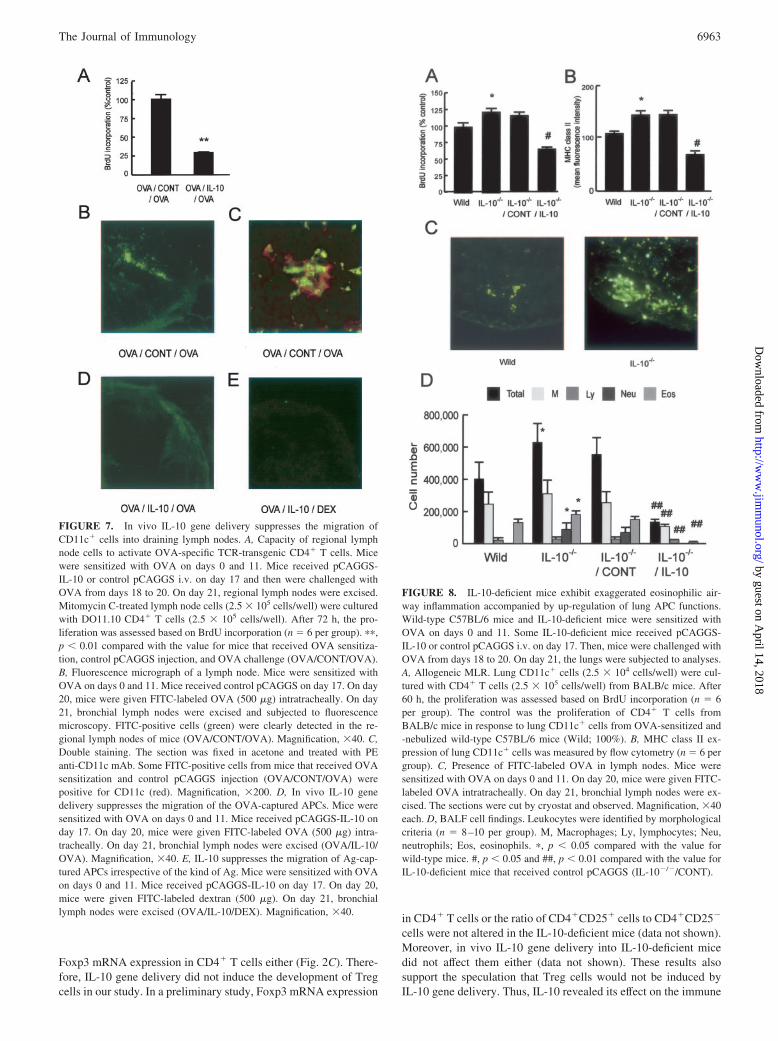
Next, we examined the effect of IL-10 gene delivery on the mi-gration of lung CD11c� cells into draining lymph nodes. First, weexamined the capacity of regional lymph node cells (APCs) toactivate OVA-specific TCR-transgenic CD4� T cells after inhala-tion of OVA. Lymph node cells from mice that received OVAsensitization, control pCAGGS injection, and OVA challenge in-duced a strong proliferation of DO11.10 CD4� T cells, whichsuggested that OVA-captured APCs actually migrated into lymphnodes (Fig. 7A). In vivo IL-10 gene delivery suppressed the pro-liferation of DO11.10 CD4� T cells (Fig. 7A), which suggestedthat IL-10 inhibited the migration of OVA-captured APCs intolymph nodes.
To confirm this, we examined the presence of OVA-capturedAPCs by fluorescence microscopy (45, 46). In mice that receivedthe OVA sensitization and control pCAGGS injection, after instil-lation of FITC-OVA, FITC signal was detected into paracorticalregions of the lymph nodes (Fig. 7B), whereas little signal wasdetected after instillation of FITC alone (data not shown). Next, weconfirmed the coexistence of OVA and CD11c� cells by doublestaining. Some areas in the lymph nodes were double-positive forFITC-OVA and CD11c, indicating the presence of the OVA-cap-tured CD11c� APCs (Fig. 7C). In vivo IL-10 gene delivery sup-pressed the migration of the OVA-captured APCs (Fig. 7D). Inanother experiment, FITC signal was also detected in the lymphnodes after instillation of FITC-dextran, instead of OVA, in micethat received OVA sensitization and control pCAGGS injection(data not shown). IL-10 treatment also suppressed this migration(Fig. 7E). Thus, IL-10 suppressed the migration of Ag-capturedAPCs irrespective of the kind of Ag.
IL-10-deficient mice exhibit up-regulated lung APC functionsand exaggerated eosinophilic airway inflammation
To further confirm the significant role of IL-10 in the lung immuneresponse, we elucidated the effect of deleting endogenous IL-10and the effect of IL-10 gene delivery during aerosol challenge intoIL-10-deficient mice. First, we examined lung APC functions andthe migration of CD11c� cells into regional lymph nodes in IL-10-deficient mice. Lung CD11c� cells from IL-10-deficient miceexhibited increased MHC alloreactivity (Fig. 8A) and MHC classII expression (Fig. 8B) compared with those from wild-type mice.In addition, the migration of CD11c� cells of IL-10-deficient miceinto regional lymph nodes dramatically increased (Fig. 8C). IL-10gene delivery into IL-10-deficient mice during aerosol challengesuppressed APC functions of lung CD11c� cells (Fig. 8, A and B)and the migration of CD11c� cells into regional lymph nodes (datanot shown). IL-10-deficient mice exhibited exaggerated neutro-philic as well as eosinophilic airway inflammation provoked by Agsystemic sensitization and local Ag inhalation (Fig. 8D), whichwas consistent with previous reports (31, 32). IL-10 gene deliveryinto IL-10-deficient mice during aerosol challenge suppressed theairway inflammation (Fig. 8D). These results also supported thefinding that IL-10 played a critical role in down-regulating residentlung APC functions and migration into regional lymph nodes.
(20 �g/ml) for 72 h. E, The proliferation was assessed based on BrdUincorporation (n � 6 per group). The proliferation of CD4� T cells inresponse to OVA in the absence of IL-10 was set as a control. F, IL-4concentration in the supernatants was measured by ELISA (n � 6 pergroup).
FIGURE 3. IL-10 has a suppressive effect on CD4� T cells only in thepresence of Ag and APCs. A and B, Direct effect of IL-10 on CD4� T cells.Mice were sensitized with OVA on days 0 and 11. On day 18, splenicCD4� T cells (1.25 � 105 cells/well) were incubated with PMA (1 ng/ml)and ionomycin (IONO, 1 �g/ml) for 24 h or plate-bound anti-CD3 Ab (10�g/ml) for 48 h in the presence or absence of IL-10. A, The proliferationwas assessed based on BrdU incorporation (n � 6 per group). The prolif-eration of CD4� T cells in response to PMA-ionomycin or plate-boundanti-CD3 Ab in the absence of IL-10 was set as a control (100%). B, IL-4concentration in the supernatants was measured by ELISA (n � 6 pergroup). C and D, IL-10 has a suppressive effect on CD4� T cells in thepresence of Ag and APCs. Mice were sensitized with OVA on days 0 and11. On day 18, splenic CD4� T cells (1.25 � 105 cells/well) were incu-bated with mitomycin C-treated splenocytes from naive mice (1.25 � 105
cells/well) and OVA (20 �g/ml) for 72 h in the presence or absence ofIL-10. C, The proliferation was assessed based on BrdU incorporation (n �6 per group). The proliferation of CD4� T cells in response to OVA in theabsence of IL-10 was set as a control. D, IL-4 concentration in the super-natants was measured by ELISA (n � 6 per group). #, p � 0.05 comparedwith the value in the absence of IL-10. E and F, IL-10 does not suppressCD4� T cell proliferation in the absence of Ag. Mice were sensitized withOVA on days 0 and 11. On day 18, spleen cells were preincubated with orwithout IL-10 for 24 h; then CD4� T cells (1.25 � 105 cells/well) werepositively selected from the spleen cells and incubated with mitomycinC-treated splenocytes from naive mice (1.25 � 105 cells/well) and OVA
6960 IL-10 SUPPRESSES AIRWAY EOSINOPHILIA AND AHR
by guest on April 14, 2018
http://ww
w.jim
munol.org/
Dow
nloaded from

FIGURE 4. In vivo IL-10 gene delivery before systemic sensitization(pre) and that during aerosol challenge (post) suppress eosinophilic (Eos)airway inflammation and AHR. Mice were sensitized with OVA or SA ondays 0 and 11. Some mice received an i.v. injection of pCAGGS-IL-10(IL-10) or control pCAGGS (CONT) on day �3 (pre) or on day 17 (post).Then, mice were challenged with 3% OVA or PBS for 10 min from day 18to day 20. On day 21, mice were analyzed. A, In vivo IL-10 deliverysuppresses eosinophil numbers in BALF. Bronchioalveolar lavage was per-formed (n � 11–12 per group). Leukocytes were identified by morphologicalcriteria. ���, p � 0.001 compared with the value of SA. ##, p � 0.01 and ###,p � 0.001 compared with the value of CONT. B, Histological findings. Lungswere excised and subjected to H&E staining. Magnification, �40 each. C,BALF cytokine concentrations. Supernatant of BALF was assayed for IL-13and TGF-� concentrations by ELISA (n � 11–12 per group). ��, p � 0.01compared with the value of SA. #, p � 0.05 and ##, p � 0.01 compared withthe value of CONT. D, In vivo IL-10 gene delivery suppresses AHR. AHR toMch was measured as described in Materials and Methods (n � 11–12 pergroup). Results were expressed for each Mch concentration as the percentageof the baseline Penh value after SA exposure. �, p � 0.05 compared with thevalue of SA. #, p � 0.05 compared with the value of CONT.
FIGURE 5. In vivo IL-10 gene delivery during aerosol challenge sup-presses Th2 cytokine production by lung cells, but not by spleen cells.Mice were treated as described in Fig. 4. On day 21, mice were analyzed.A, Cytokine production by lung cells. Lung cells were incubated (5 � 105
cells/well) with OVA (200 �g/ml). After 120 h, the cytokine levels of thesupernatants were assayed (n � 6 per group). �, p � 0.05 and ���, p �0.001 compared with the value of SA. #, p � 0.05, ##, p � 0.01, and ###,p � 0.001 compared with the value of CONT. B and C, Proliferation andcytokine production of spleen cells. Spleen cells were incubated (5 � 105
cells/well) with OVA. B, After 72 h, the proliferation was assessed basedon BrdU incorporation (n � 6 per group). The control was the maximumproliferation observed in response to OVA for spleen cells from OVA-sensitized mice (OVA; 100%). C, After 120 h, the cytokine levels of thesupernatants were assayed (n � 6 per group). ���, p � 0.001 comparedwith the value of SA. ###, p � 0.001 compared with the value of IL-10(pre). D, Serum IgE concentrations. On day 21, blood samples were ob-tained from the mice presented in Fig. 4, and total and OVA-specific IgEconcentrations were measured by ELISA (n � 11–12 per group). For mea-surement of OVA-specific IgE, sera from five OVA-sensitized mice wasset as a control. ���, p � 0.001 compared with the value of SA. ##, p �0.01 and ###, p � 0.001 compared with the value of CONT.
6961The Journal of Immunology
by guest on April 14, 2018
http://ww
w.jim
munol.org/
Dow
nloaded from

Treatment of OVA-pulsed DCs with IL-10 in vitro directlyprevents the development of eosinophilic airway inflammation
Suppression of splenic DC functions (Fig. 2, D–H) or lung APCfunctions (Fig. 6, A–E) by IL-10 gene delivery seems to be mild.Therefore, to confirm the significance of the suppression of APCfunctions by IL-10 to inhibit subsequent airway inflammation, wefinally performed a transfer experiment. Stimulation of DCs withOVA activated DCs, and GM-CSF treatment further up-regulatedthese functions such as OVA-Ag presenting capacity (Fig. 9A),MHC class II expression (Fig. 9B), and expression of cell surfacemarkers such as CD40, CD80, and CD86 (data not shown). Treat-ment with IL-10 in vitro suppressed these functions by almost 25%in both GM-CSF-treated and untreated DCs (Fig. 9, A and B, anddata not shown). The degree of suppression was equivalent to thesuppression of splenic DC functions (Fig. 2, D–H) or lung APCfunctions (Fig. 6, A–E) obtained by in vivo IL-10 gene delivery. Inan in vivo experiment, transfer of OVA-pulsed DCs into naivemice and subsequent OVA inhalation induced eosinophilic airwayinflammation, and GM-CSF-treatment of DCs increased the inten-sity of the eosinophilic airway inflammation (Fig. 9C). The trans-fer of DCs treated with IL-10 in vitro attenuated the eosinophilicairway inflammation (Fig. 9C). These results confirmed that thesuppression of APC functions by IL-10 could sufficiently inhibitthe development of subsequent eosinophilic inflammation in vivo.
DiscussionIn the current study, we demonstrated that in vivo IL-10 genedelivery suppresses Ag-induced eosinophilic airway inflammationand AHR even during airway Ag challenge, an effector phase ofthe immune response. In vivo IL-10 gene delivery during airwaychallenge suppressed local ongoing immune responses of the lungwithout impairing the Ag-specific systemic immune response, andthis suppression was mediated by down-regulating the functions ofresident lung APCs as well as the migration of CD11c� cells intoregional lymph nodes. The present study confirmed a significantrole for IL-10 as an Ag-specific, site-specific immune regulator.
First, we confirmed the effect of IL-10 on the Ag-induced im-mune response ex vivo (Fig. 2). IL-10 suppressed the Ag-inducedimmune response of CD4� T cells (Fig. 2, A and B) and DCfunctions (Fig. 2, D–H). Generally, IL-10 down-regulates the ex-pression of MHC class II and costimulatory molecules on mono-cytes/macrophages and DCs (1, 7–10, 48), thus suppressing theAg-induced immune response. Our data from spleen cells are con-sistent with these previous studies.
Chronic activation of CD4� T cells with Ag in the presence ofIL-10 induces the development of Tr1 cells, which produce IL-10,but not IL-2 and IL-4 (23). Some reports have indicated thatTGF-� converts CD4�CD25� T cells to Treg cells in vitro thoughthe induction of Foxp3 (44, 49). A recent report showed that CD4�
T cells expressing membrane-bound TGF-� and Foxp3 are in-volved in the tolerance induced by Ag inhalation (50). In the cur-rent study, IL-10 treatment did not up-regulate IL-10 production(Fig. 2B) or TGF-� production (data not shown) by CD4� T cells.In addition, in vivo IL-10 gene delivery did not enhance TGF-�production in the lung (Fig. 4C). Further, IL-10 did not increase
CD4� T cells (1.25 � 105 cells/well) from OVA-sensitized mice and OVA(30 �g/ml). After 96 h, the proliferation was assessed based on BrdUincorporation (n � 6–10 per group). The control was the proliferation ofCD4� T cells from OVA-sensitized mice in response to splenic CD11c�
cells from OVA-sensitized and -nebulized BALB/c mice and OVA (OVA).#, p � 0.05 compared with the value of CONT.
FIGURE 6. In vivo IL-10 gene delivery suppresses resident lung APCfunctions. Mice were treated as described in Fig. 4 and were evaluated onday 21. A, Allogenic MLR. Lung CD11c� cells (2.5 � 104 cells/well) werecultured with CD4� T cells (2.5 � 105 cells/well) from C57BL/6 mice.After 60 h, the proliferation was assessed based on BrdU incorporation(n � 6–10 per group). The control was the proliferation of CD4� T cellsfrom C57BL/6 mice responding to lung CD11c� cells from OVA-sensi-tized and -nebulized BALB/c mice (OVA; 100%). �, p � 0.05 comparedwith the value of SA. #, p � 0.05 compared with the value of CONT. B,OVA-presenting capacity of lung CD11c� cells. Lung CD11c� cells(2.5 � 104 cells/well) were cultured with CD4� T cells (2.5 � 105 cells/well) from DO11.10 mice. After 60 h, the proliferation was assessed basedon BrdU incorporation (n � 6–10 per group). The control was the prolif-eration of CD4� T cells from DO11.10 mice in response to lung CD11c�
cells from OVA-sensitized and -nebulized BALB/c mice (OVA). �, p �0.05 compared with the value of SA. #, p � 0.05 compared with the valueof CONT. C, Cytokine production. Lung CD11c� cells (1.25 � 105 cells/well) were cultured with LPS (1 �g/ml). After 24 h, the cytokine levels ofthe supernatants were assayed (n � 6–10 per group). ��, p � 0.01 com-pared with the value of SA. #, p � 0.05 compared with the value of CONT.D, MHC class II expression of lung CD11c� cells was measured by flowcytometry (n � 6–10 per group). �, p � 0.05 compared with the value ofSA. #, p � 0.05 compared with the value of CONT. E, Representativefigures of MHC class II expression. F, OVA-presenting capacity of splenicDCs. Splenic CD11c� cells (1.25 � 105 cells/well) were cultured with
6962 IL-10 SUPPRESSES AIRWAY EOSINOPHILIA AND AHR
by guest on April 14, 2018
http://ww
w.jim
munol.org/
Dow
nloaded from
Foxp3 mRNA expression in CD4� T cells either (Fig. 2C). There-fore, IL-10 gene delivery did not induce the development of Tregcells in our study. In a preliminary study, Foxp3 mRNA expression
in CD4� T cells or the ratio of CD4�CD25� cells to CD4�CD25�
cells were not altered in the IL-10-deficient mice (data not shown).Moreover, in vivo IL-10 gene delivery into IL-10-deficient micedid not affect them either (data not shown). These results alsosupport the speculation that Treg cells would not be induced byIL-10 gene delivery. Thus, IL-10 revealed its effect on the immune
FIGURE 7. In vivo IL-10 gene delivery suppresses the migration ofCD11c� cells into draining lymph nodes. A, Capacity of regional lymphnode cells to activate OVA-specific TCR-transgenic CD4� T cells. Micewere sensitized with OVA on days 0 and 11. Mice received pCAGGS-IL-10 or control pCAGGS i.v. on day 17 and then were challenged withOVA from days 18 to 20. On day 21, regional lymph nodes were excised.Mitomycin C-treated lymph node cells (2.5 � 105 cells/well) were culturedwith DO11.10 CD4� T cells (2.5 � 105 cells/well). After 72 h, the pro-liferation was assessed based on BrdU incorporation (n � 6 per group). ��,p � 0.01 compared with the value for mice that received OVA sensitiza-tion, control pCAGGS injection, and OVA challenge (OVA/CONT/OVA).B, Fluorescence micrograph of a lymph node. Mice were sensitized withOVA on days 0 and 11. Mice received control pCAGGS on day 17. On day20, mice were given FITC-labeled OVA (500 �g) intratracheally. On day21, bronchial lymph nodes were excised and subjected to fluorescencemicroscopy. FITC-positive cells (green) were clearly detected in the re-gional lymph nodes of mice (OVA/CONT/OVA). Magnification, �40. C,Double staining. The section was fixed in acetone and treated with PEanti-CD11c mAb. Some FITC-positive cells from mice that received OVAsensitization and control pCAGGS injection (OVA/CONT/OVA) werepositive for CD11c (red). Magnification, �200. D, In vivo IL-10 genedelivery suppresses the migration of the OVA-captured APCs. Mice weresensitized with OVA on days 0 and 11. Mice received pCAGGS-IL-10 onday 17. On day 20, mice were given FITC-labeled OVA (500 �g) intra-tracheally. On day 21, bronchial lymph nodes were excised (OVA/IL-10/OVA). Magnification, �40. E, IL-10 suppresses the migration of Ag-cap-tured APCs irrespective of the kind of Ag. Mice were sensitized with OVAon days 0 and 11. Mice received pCAGGS-IL-10 on day 17. On day 20,mice were given FITC-labeled dextran (500 �g). On day 21, bronchiallymph nodes were excised (OVA/IL-10/DEX). Magnification, �40.
FIGURE 8. IL-10-deficient mice exhibit exaggerated eosinophilic air-way inflammation accompanied by up-regulation of lung APC functions.Wild-type C57BL/6 mice and IL-10-deficient mice were sensitized withOVA on days 0 and 11. Some IL-10-deficient mice received pCAGGS-IL-10 or control pCAGGS i.v. on day 17. Then, mice were challenged withOVA from days 18 to 20. On day 21, the lungs were subjected to analyses.A, Allogeneic MLR. Lung CD11c� cells (2.5 � 104 cells/well) were cul-tured with CD4� T cells (2.5 � 105 cells/well) from BALB/c mice. After60 h, the proliferation was assessed based on BrdU incorporation (n � 6per group). The control was the proliferation of CD4� T cells fromBALB/c mice in response to lung CD11c� cells from OVA-sensitized and-nebulized wild-type C57BL/6 mice (Wild; 100%). B, MHC class II ex-pression of lung CD11c� cells was measured by flow cytometry (n � 6 pergroup). C, Presence of FITC-labeled OVA in lymph nodes. Mice weresensitized with OVA on days 0 and 11. On day 20, mice were given FITC-labeled OVA intratracheally. On day 21, bronchial lymph nodes were ex-cised. The sections were cut by cryostat and observed. Magnification, �40each. D, BALF cell findings. Leukocytes were identified by morphologicalcriteria (n � 8–10 per group). M, Macrophages; Ly, lymphocytes; Neu,neutrophils; Eos, eosinophils. �, p � 0.05 compared with the value forwild-type mice. #, p � 0.05 and ##, p � 0.01 compared with the value forIL-10-deficient mice that received control pCAGGS (IL-10�/�/CONT).
6963The Journal of Immunology
by guest on April 14, 2018
http://ww
w.jim
munol.org/
Dow
nloaded from

response by some mechanism other than inducing production ofTreg cells in our experimental system.
Repetitive exposure of the respiratory tract to Ag could inducesome kind of DCs that produce IL-10 to induce the development ofTreg cells (24, 25) and thus may work as tolerogenic DCs. In thepresent study, IL-10 gene delivery did not up-regulate, but rathersuppressed, the production of IL-10 by purified splenic DCs stim-ulated with LPS or TNF-� (Fig. 2, G and H). Thus, a single in-jection of the IL-10 expression plasmid did not suppress the OVA-induced immune response by generating IL-10-producingtolerogenic DCs.
In the current study, we delivered IL-10 via an expression plas-mid. Instillation of IL-10 protein into the lung also suppresseseosinophilic airway inflammation (34, 35). However, generally,the half-lives of recombinant cytokines are very short. In fact, thecirculating half-life of a systemically delivered rIL-10 protein isonly 20 min (51). Further, the degree of suppression of eosino-
philic inflammation by IL-10 protein was weak in previous reports(1/4–1/5 as compared with positive control) (34, 35). Instillationof IL-10 protein even increases AHR unexpectedly (32, 35). Incontrast, in this study, after a single injection of IL-10-expressingplasmid, both serum and BALF IL-10 concentrations remainedhigh for 5–7 days (Fig. 1). IL-10 gene delivery almost completelysuppressed eosinophilic inflammation (1/20–1/100 of control)(Fig. 4A) and it also suppressed AHR (Fig. 4D). Thus, IL-10 genedelivery was highly efficient and was superior to the instillation ofIL-10 protein for controlling airway inflammation and AHR. Inaddition, we demonstrated that, even after systemic sensitizationhad been completed, a high level of IL-10 strongly suppressedAg-induced eosinophilic inflammation (Fig. 4). In a previous re-port on the gene delivery system, IL-10 was effective only whenadministered before systemic sensitization (37). We, for the firsttime, clearly demonstrated the effect of IL-10 gene delivery aftersystemic sensitization had been completed, and the airway immuneresponse was ongoing, which could lead to the clinical applicationof gene delivery for patients with asthma.
In vivo IL-10 gene delivery during inhalation suppressed theproduction of Th2 cytokines by lung cells in response to OVA(Fig. 5A). However, it did not suppress the proliferation or cyto-kine production of spleen cells (Fig. 5, B and C). It did not sup-press OVA-specific IgE production either (Fig. 5D). In contrast, asexpected, in vivo IL-10 gene delivery before sensitization sup-pressed the production of cytokines in both the lung and spleen(Fig. 5, A–C). OVA-specific IgE production was also significantlysuppressed (Fig. 5D). In the case of presensitization delivery of theIL-10 plasmid, IL-10 suppressed the OVA-induced immune re-sponse from the initial stage of sensitization, so the overall re-sponse was suppressed both in the lung and in the spleen. There-fore, further immune responses induced by OVA inhalation wereaccordingly suppressed. In contrast, when IL-10 was deliveredduring OVA inhalation, systemic sensitization had already devel-oped in the spleen, and IgE synthesis had been accomplished.Therefore, presensitization delivery exhibited stronger suppressionthan postsensitization delivery (Figs. 4 and 5).
In addition, as we have demonstrated in studies in vitro, IL-10had a strong immunosuppressive effect where Ag and APCs co-existed (Fig. 3, C and D). Thus, when IL-10 was delivered duringOVA inhalation into the airway, it suppressed the immune re-sponse more strongly in the lung, although the delivery of IL-10was systemic. In other words, the immunosuppressive effect ofIL-10 is essentially the same between lung and spleen, however,the presence of Ag determined the intensity of suppression. Zuany-Amorim et al. (34) reported that the instillation of recombinantmurine IL-10 with Ag to the lung reduces Ag inhalation-inducedeosinophilic inflammation, whereas IL-10 alone does not show asuppressive effect. In another system, IL-10 delivered into the an-kle attenuated arthritis that was initiated by direct injection ofOVA-Ag into the ankle in presensitized mice without affecting theimmune response in the spleen (52). These studies support ourfindings. In other words, IL-10 acts as an immunosuppressive cy-tokine especially at the site of the immune response where both Agand APCs exist concurrently. If we could deliver the IL-10 geneselectively into the lung, the lung immune response would be spe-cifically suppressed, which should be further investigated in future.
To further clarify the mechanism of action of IL-10 in the lungimmune response, we next analyzed the effect of IL-10 on lungAPC function. Generally, lung APCs capture inhaled Ag in situ,mature, and migrate into draining lymph nodes to activate naive Tcells (45). Furthermore, recent reports have demonstrated thatsome resident DCs can mature and present Ag to T cells in the lung
FIGURE 9. OVA-pulsed, IL-10-treated DCs prevent the developmentof eosinophilic airway inflammation provoked by a combination of DCtransfer and subsequent OVA inhalation. A, IL-10-treated DCs impairOVA-presenting capacity. Splenic CD11c� cells were positively selectedby magnetic cell sorting from naive mice and incubated with OVA (1000�g/ml) in the presence or absence of GM-CSF (10 ng/ml). IL-10 was addedto some culture media (50 ng/ml). After 20 h, OVA-pulsed CD11c� cellswere collected (2.5 � 104) and cultured with CD4� T cells from DO11.10mice (2.5 � 105). After 48 h, the proliferation was assessed based on BrdUincorporation (n � 6 per group). The control was the proliferation of CD4�
T cells from DO11.10 mice in response to OVA-pulsed CD11c� cells(OVA; 100%). B, MHC class II expression of OVA-pulsed CD11c� cellswas measured by flow cytometry (n � 6 per group). C, OVA-pulsed andIL-10-treated DCs attenuate transfer-mediated eosinophilic airway inflam-mation. Syngeneic recipients received OVA-pulsed CD11c� cells (OVA),OVA-pulsed GM-CSF-treated CD11c� cells (GM), OVA-pulsed IL-10-treated CD11c� cells (IL-10), or OVA-pulsed GM-CSF- and IL-10-treatedCD11c� cells (GM/IL-10) on day 0 (5 � 105). The mice were challengedwith nebulized 3% OVA for 1 h twice daily on days 7, 8, and 15 andsacrificed on day 16 for BALF analysis (n � 6–8 per group). M, Macro-phages; Ly, lymphocytes; Neu, neutrophils; Eos, eosinophils. �, p � 0.05and ���, p � 0.001 compared with the value of OVA. #, p � 0.05 and ###,p � 0.001 compared with the value of GM.
6964 IL-10 SUPPRESSES AIRWAY EOSINOPHILIA AND AHR
by guest on April 14, 2018
http://ww
w.jim
munol.org/
Dow
nloaded from

before migration (53–55). In our study, IL-10 gene delivery sup-pressed resident lung APC functions such as Ag-presenting capac-ity and cytokine production (Fig. 6, A–C). The expression of MHCclass II molecule on APCs was significantly down-regulated (Fig.6, D and E). The expression of other costimulatory molecules suchas CD40, CD80, and CD86 was also down-regulated, although astatistically significant difference was not detected, probably due toa low baseline expression with OVA treatment. Moreover, in vivoIL-10 gene delivery also suppressed the migration of OVA-cap-tured CD11c� cells (DCs) into regional lymph nodes (Fig. 7).Taken together, at least in our experimental system, the mecha-nism of action of IL-10 was not to induce the regulatory function,but rather to suppress the overall function of APCs in the initialstage of Ag presentation and recognition. In contrast, the effect ofpostsensitization delivery of IL-10 on splenic DCs was slight andnot significant (Fig. 6F).
Next, we confirmed whether inhibition of APC function byIL-10 directly leads to inhibition of eosinophilic airway inflam-mation. In IL-10-deficient mice treated with OVA sensitizationand inhalation, lung APC function was up-regulated and strongereosinophilic airway inflammation was provoked compared withthat in wild-type mice (Fig. 8). Compensation by IL-10 gene de-livery again down-regulated the APC function and thus attenuatedeosinophilic airway inflammation. Further, in vitro treatment ofsplenic DCs with OVA activated the DCs, and the transfer of thesecells into naive mice followed by subsequent OVA inhalation pro-voked eosinophilic inflammation (Fig. 9). IL-10 treatment in vitrodown-regulated the Ag-presenting capacity and MHC class II ex-pression of DCs by 25–50% (Fig. 9, A and B). Then, the intensityof this transfer-induced eosinophilic airway inflammation wasstrongly attenuated by this IL-10 treatment (Fig. 9C). This sup-ports the reliability of data obtained from in vivo IL-10 gene de-livery into sensitized and challenged mice, in which the APC func-tion of lung cells was suppressed by 25–50% (Fig. 6, A–E), whichresulted in a strong suppression of airway inflammation and AHR(Fig. 4). Taken together, these results indicated that suppression ofAPC function by IL-10 sufficiently led to inhibition of eosinophilicairway inflammation and that this was one definite mechanism bywhich IL-10 inhibits eosinophilic inflammation.
As IL-10 is a multipotent immunosuppressive cytokine, it couldexhibit various effects on many resident cells and inflammatorycells, such as endothelial cells, monocytes/macrophages, lympho-cytes, and mast cells (1, 56). In our experimental system, mecha-nisms other than the suppression of APC function could be attrib-uted to the suppression of Ag-induced airway responses. Forexample, the possibility that IL-10 affected T cells directly cannotbe completely ruled out, because in humans, the direct effect ofIL-10 on T cell proliferation has been confirmed (11–13). In thecurrent study, however, IL-10 suppressed DC functions in vitro(50 ng/ml; Fig. 9, A and B), whereas this concentration of IL-10did not directly suppress CD4� T cell proliferation and cytokineproduction (Fig. 3, A and B). Thus, this possibility seems unlikely.IL-10 could also have modulated the expression of VCAM-1 onendothelial cells, thus reducing the recruitment of eosinophils intothe lung. These possibilities should also be considered. In addition,at present there is a limitation to the purification of CD11c� cellsfrom the lung. In the present study, the purity was 80–85%, whichwas similar to that in previous reports (42, 43). We confirmed thatthere was no difference in the purity of CD11c� cells among ex-perimental groups in the same experiment (data not shown). There-fore, we consider that our results on APC function are, not com-pletely but to a certain extent, reliable. However, the effect ofcontaminating cells should also be considered.
Strong Ag stimulation may produce qualitatively and quantita-tively different Ag responses from a modest stimulation. In a pre-liminary study, we examined various concentrations of OVA-Agand found that the protocol used in the current study induced amaximal response. Given that the purpose of this study was toexamine the therapeutic effect of IL-10, we applied this protocolthroughout the study to induce a specific intensity of immune re-sponse. However, differences in the intensity of Ag stimulationcould affect how IL-10 would suppress the Ag-induced immuneresponse.
In summary, IL-10 gene delivery achieved a high concentrationof protein in the lung and suppressed OVA-Ag-induced eosino-philic airway inflammation and AHR, even after sensitization hadbeen completed, by down-regulating lung APC functions and thesubsequent Th2 response. IL-10 exhibited its suppressive effect ina site-specific manner in the tissue where DCs encountered Ag andthus an Ag-specific immune response was driven. Therefore, ifdone selectively, IL-10 gene delivery could be a very effectivestrategy for regulating Ag-specific immune responses in a localenvironment, such as the lung, without affecting the systemicresponse.
AcknowledgmentsWe thank I. Makino for technical assistance.
DisclosuresThe authors have no financial conflict of interest.
References1. Moore, K. W., R. de Waal Malefyt, R. L. Coffman, and A. O’Garra. 2001. In-
terleukin-10 and the interleukin-10 receptor. Annu. Rev. Immunol. 19: 683–765.2. de Waal Malefyt, R., J. Abrams, B. Bennett, C. G. Figdor, and J. E. de Vries.
1991. Interleukin 10 (IL-10) inhibits cytokine synthesis by human monocytes: anautoregulatory role of IL-10 produced by monocytes. J. Exp. Med. 174:1209–1220.
3. Fiorentino, D. F., A. Zlotnik, T. R. Mosmann, M. Howard, and A. O’Garra. 1991.IL-10 inhibits cytokine production by activated macrophages. J. Immunol. 147:3815–3822.
4. Fiorentino, D. F., A. Zlotnik, P. Vieira, T. R. Mosmann, M. Howard,K. W. Moore, and A. O’Garra. 1991. IL-10 acts on the antigen-presenting cell toinhibit cytokine production by Th1 cells. J. Immunol. 146: 3444–3451.
5. Del Prete, G., M. De Carli, F. Almerigogna, M. G. Giudizi, R. Biagiotti, andS. Romagnani. 1993. Human IL-10 is produced by both type 1 helper (Th1) andtype 2 helper (Th2) T cell clones and inhibits their antigen-specific proliferationand cytokine production. J. Immunol. 150: 353–360.
6. D’Andrea, A., M. Aste-Amezaga, N. M. Valiante, X. Ma, M. Kubin, andG. Trinchieri. 1993. Interleukin 10 (IL-10) inhibits human lymphocyte interferon�-production by suppressing natural killer cell stimulatory factor/IL-12 synthesisin accessory cells. J. Exp. Med. 178: 1041–1048.
7. de Waal Malefyt, R., J. Haanen, H. Spits, M. G. Roncarolo, A. te Velde,C. Figdor, K. Johnson, R. Kastelein, H. Yssel, and J. E. de Vries. 1991. Inter-leukin 10 (IL-10) and viral IL-10 strongly reduce antigen-specific human T cellproliferation by diminishing the antigen-presenting capacity of monocytes viadownregulation of class II major histocompatibility complex expression. J. Exp.Med. 174: 915–924.
8. Willems, F., A. Marchant, J. P. Delville, C. Gerard, A. Delvaux, T. Velu,M. de Boer, and M. Goldman. 1994. Interleukin-10 inhibits B7 and intercellularadhesion molecule-1 expression on human monocytes. Eur. J. Immunol. 24:1007–1009.
9. Buelens, C., F. Willems, A. Delvaux, G. Pierard, J. P. Delville, T. Velu, andM. Goldman. 1995. Interleukin-10 differentially regulates B7-1 (CD80) and B7-2(CD86) expression on human peripheral blood dendritic cells. Eur. J. Immunol.25: 2668–2672.
10. Ding, L., P. S. Linsley, L. Y. Huang, R. N. Germain, and E. M. Shevach. 1993.IL-10 inhibits macrophage costimulatory activity by selectively inhibiting theup-regulation of B7 expression. J. Immunol. 151: 1224–1234.
11. Taga, K., H. Mostowski, and G. Tosato. 1993. Human interleukin-10 can directlyinhibit T-cell growth. Blood 81: 2964–2971.
12. de Waal Malefyt, R., H. Yssel, and J. E. de Vries. 1993. Direct effects of IL-10on subsets of human CD4� T cell clones and resting T cells: specific inhibitionof IL-2 production and proliferation. J. Immunol. 150: 4754–4765.
13. Schandene, L., C. Alonso-Vega, F. Willems, C. Gerard, A. Delvaux, T. Velu,R. Devos, M. de Boer, and M. Goldman. 1994. B7/CD28-dependent IL-5 pro-duction by human resting T cells is inhibited by IL-10. J. Immunol. 152:4368–4374.
6965The Journal of Immunology
by guest on April 14, 2018
http://ww
w.jim
munol.org/
Dow
nloaded from
14. Herfarth, H. H., S. P. Mohanty, H. C. Rath, S. Tonkonogy, and R. B. Sartor. 1996.Interleukin 10 suppresses experimental chronic, granulomatous inflammation in-duced by bacterial cell wall polymers. Gut 39: 836–845.
15. Barbara, G., Z. Xing, C. M. Hogaboam, J. Gauldie, and S. M. Collins. 2000.Interleukin 10 gene transfer prevents experimental colitis in rats. Gut 46:344–349.
16. Steidler, L., W. Hans, L. Schotte, S. Neirynck, F. Obermeier, W. Falk, W. Fiers,and E. Remaut. 2000. Treatment of murine colitis by Lactococcus lactis secretinginterleukin-10. Science 289: 1352–1355.
17. van Deventer, S. J., C. O. Elson, and R. N. Fedorak. 1997. Multiple doses ofintravenous interleukin 10 in steroid-refractory Crohn’s disease: Crohn’s DiseaseStudy Group. Gastroenterology 113: 383–389.
18. Asadullah, K., M. Friedrich, S. Hanneken, C. Rohrbach, H. Audring,A. Vergopoulos, M. Ebeling, W. D. Docke, H. D. Volk, and W. Sterry. 2001.Effects of systemic interleukin-10 therapy on psoriatic skin lesions: histologic,immunohistologic, and molecular biology findings. J. Invest. Dermatol. 116:721–727.
19. Asadullah, K., W. Sterry, K. Stephanek, D. Jasulaitis, M. Leupold, H. Audring,H. D. Volk, and W. D. Docke. 1998. IL-10 is a key cytokine in psoriasis. Proofof principle by IL-10 therapy: a new therapeutic approach. J. Clin. Invest. 101:783–794.
20. Asadullah, K., W. D. Docke, M. Ebeling, M. Friedrich, G. Belbe, H. Audring,H. D. Volk, and W. Sterry. 1999. Interleukin 10 treatment of psoriasis: clinicalresults of a phase 2 trial. Arch. Dermatol. 135: 187–192.
21. Akdis, C. A., T. Blesken, M. Akdis, B. Wuthrich, and K. Blaser. 1998. Role ofinterleukin 10 in specific immunotherapy. J. Clin. Invest. 102: 98–106.
22. Akdis, C. A., and K. Blaser. 2000. Mechanisms of allergen-specific immunother-apy. Allergy 55: 522–530.
23. Groux, H., A. O’Garra, M. Bigler, M. Rouleau, S. Antonenko, J. E. de Vries, andM. G. Roncarolo. 1997. A CD4� T-cell subset inhibits antigen-specific T-cellresponses and prevents colitis. Nature 389: 737–742.
24. Akbari, O., R. H. DeKruyff, and D. T. Umetsu. 2001. Pulmonary dendritic cellsproducing IL-10 mediate tolerance induced by respiratory exposure to antigen.Nat. Immunol. 2: 725–731.
25. Akbari, O., G. J. Freeman, E. H. Meyer, E. A. Greenfield, T. T. Chang,A. H. Sharpe, G. Berry, R. H. DeKruyff, and D. T. Umetsu. 2002. Antigen-specific regulatory T cells develop via the ICOS-ICOS-ligand pathway and in-hibit allergen-induced airway hyperreactivity. Nat. Med. 8: 1024–1032.
26. Bochner, B. S., B. J. Undem, and L. M. Lichtenstein. 1994. Immunological as-pects of allergic asthma. Annu. Rev. Immunol. 12: 295–335.
27. Humbert, M., G. Menz, S. Ying, C. J. Corrigan, D. S. Robinson, S. R. Durham,and A. B. Kay. 1999. The immunopathology of extrinsic (atopic) and intrinsic(non-atopic) asthma: more similarities than differences. Immunol. Today 20:528–533.
28. Moller, G. M., S. E. Overbeek, C. G. Van Helden-Meeuwsen, J. M. Van Haarst,E. P. Prens, P. G. Mulder, D. S. Postma, and H. C. Hoogsteden. 1996. Increasednumbers of dendritic cells in the bronchial mucosa of atopic asthmatic patients:downregulation by inhaled corticosteroids. Clin. Exp. Allergy 26: 517–524.
29. Takanashi, S., Y. Hasegawa, Y. Kanehira, K. Yamamoto, K. Fujimoto, K. Satoh,and K. Okamura. 1999. Interleukin-10 level in sputum is reduced in bronchialasthma, COPD and in smokers. Eur. Respir. J. 14: 309–314.
30. John, M., S. Lim, J. Seybold, P. Jose, A. Robichaud, B. O’Connor, P. J. Barnes,and K. F. Chung. 1998. Inhaled corticosteroids increase interleukin-10 but reducemacrophage inflammatory protein-1�, granulocyte-macrophage colony-stimulat-ing factor, and interferon-� release from alveolar macrophages in asthma.Am. J. Respir. Crit. Care Med. 157: 256–262.
31. Tournoy, K. G., J. C. Kips, and R. A. Pauwels. 2000. Endogenous interleukin-10suppresses allergen-induced airway inflammation and nonspecific airway respon-siveness. Clin. Exp. Allergy 30: 775–783.
32. Justice, J. P., Y. Shibata, S. Sur, J. Mustafa, M. Fan, and M. R. Van Scott. 2001.IL-10 gene knockout attenuates allergen-induced airway hyperresponsiveness inC57BL/6 mice. Am. J. Physiol. Lung Cell Mol. Physiol. 280:L363–L368.
33. Yang, X., S. Wang, Y. Fan, and X. Han. 2000. IL-10 deficiency prevents IL-5overproduction and eosinophilic inflammation in a murine model of asthma-likereaction. Eur. J. Immunol. 30: 382–391.
34. Zuany-Amorim, C., S. Haile, D. Leduc, C. Dumarey, M. Huerre, B. B. Vargaftig,and M. Pretolani. 1995. Interleukin-10 inhibits antigen-induced cellular recruit-ment into the airways of sensitized mice. J. Clin. Invest. 95: 2644–2651.
35. van Scott, M. R., J. P. Justice, J. F. Bradfield, E. Enright, A. Sigounas, and S. Sur.2000. IL-10 reduces Th2 cytokine production and eosinophilia but augmentsairway reactivity in allergic mice. Am. J. Physiol. Lung Cell Mol. Physiol. 278:L667–L674.
36. Oh, J. W., C. M. Seroogy, E. H. Meyer, O. Akbari, G. Berry, C. G. Fathman,R. H. Dekruyff, and D. T. Umetsu. 2002. CD4 T-helper cells engineered to pro-duce IL-10 prevent allergen-induced airway hyperreactivity and inflammation.J. Allergy Clin. Immunol. 110: 460–468.
37. Stampfli, M. R., M. Cwiartka, B. U. Gajewska, D. Alvarez, S. A. Ritz,M. D. Inman, Z. Xing, and M. Jordana. 1999. Interleukin-10 gene transfer to theairway regulates allergic mucosal sensitization in mice. Am. J. Respir. Cell Mol.Biol. 21: 586–596.
38. To, Y., M. Dohi, R. Tanaka, A. Sato, K. Nakagome, and K. Yamamoto. 2001.Early interleukin 4-dependent response can induce airway hyperreactivity beforedevelopment of airway inflammation in a mouse model of asthma. Lab. Invest.81: 1385–1396.
39. Okunishi, K., M. Dohi, K. Nakagome, R. Tanaka, and K. Yamamoto. 2004. Anovel role of cysteinyl leukotrienes to promote dendritic cell activation in theantigen-induced immune responses in the lung. J. Immunol. 173: 6393–6402.
40. Jiang, J., E. Yamato, and J. Miyazaki. 2001. Intravenous delivery of naked plas-mid DNA for in vivo cytokine expression. Biochem. Biophys. Res. Commun. 289:1088–1092.
41. Liu, D., and J. E. Knapp. 2001. Hydrodynamics-based gene delivery. Curr. Opin.Mol. Ther. 3: 192–197.
42. Dahl, M. E., K. Dabbagh, D. Liggitt, S. Kim, and D. B. Lewis. 2004. Viral-induced T helper type 1 responses enhance allergic disease by effects on lungdendritic cells. Nat. Immunol. 5: 337–343.
43. Dabbagh, K., M. E. Dahl, P. Stepick-Biek, and D. B. Lewis. 2002. Toll-likereceptor 4 is required for optimal development of Th2 immune responses: role ofdendritic cells. J. Immunol. 168: 4524–4530.
44. Fantini, M. C., C. Becker, G. Monteleone, F. Pallone, P. R. Galle, andM. F. Neurath. 2004. Cutting edge: TGF-� induces a regulatory phenotype inCD4�CD25� T cells through Foxp3 induction and down-regulation of Smad7.J. Immunol. 172: 5149–5153.
45. Vermaelen, K. Y., I. Carro-Muino, B. N. Lambrecht, and R. A. Pauwels. 2001.Specific migratory dendritic cells rapidly transport antigen from the airways tothe thoracic lymph nodes. J. Exp. Med. 193: 51–60.
46. Constant, S. L., J. L. Brogdon, D. A. Piggott, C. A. Herrick, I. Visintin,N. H. Ruddle, and K. Bottomly. 2002. Resident lung antigen-presenting cellshave the capacity to promote Th2 T cell differentiation in situ. J. Clin. Invest. 110:1441–1448.
47. Chen, Y., V. K. Kuchroo, J. Inobe, D. A. Hafler, and H. L. Weiner. 1994. Reg-ulatory T cell clones induced by oral tolerance: suppression of autoimmune en-cephalomyelitis. Science 265: 1237–1240.
48. Groux, H., F. Cottrez, M. Rouleau, S. Mauze, S. Antonenko, S. Hurst, T. McNeil,M. Bigler, M. G. Roncarolo, and R. L. Coffman. 1999. A transgenic model toanalyze the immunoregulatory role of IL-10 secreted by antigen-presenting cells.J. Immunol. 162: 1723–1729.
49. Chen, W., W. Jin, N. Hardegen, K. J. Lei, L. Li, N. Marinos, G. McGrady, andS. M. Wahl. 2003. Conversion of peripheral CD4�CD25� naive T cells toCD4�CD25� regulatory T cells by TGF-� induction of transcription factorFoxp3. J. Exp. Med. 198: 1875–1886.
50. Ostroukhova, M., C. Seguin-Devaux, T. B. Oriss, B. Dixon-McCarthy, L. Yang,B. T. Ameredes, T. E. Corcoran, and A. Ray. 2004. Tolerance induced by inhaledantigen involves CD4� T cells expressing membrane-bound TGF-� and FOXP3.J. Clin. Invest. 114: 28–38.
51. Chernoff, A. E., E. V. Granowitz, L. Shapiro, E. Vannier, G. Lonnemann,J. B. Angel, J. S. Kennedy, A. R. Rabson, S. M. Wolff, and C. A. Dinarello. 1995.A randomized, controlled trial of IL-10 in humans: inhibition of inflammatorycytokine production and immune responses. J. Immunol. 154: 5492–5499.
52. Setoguchi, K., Y. Misaki, Y. Araki, K. Fujio, K. Kawahata, T. Kitamura, andK. Yamamoto. 2000. Antigen-specific T cells transduced with IL-10 ameliorateexperimentally induced arthritis without impairing the systemic immune responseto the antigen. J. Immunol. 165: 5980–5986.
53. Vermaelen, K., and R. Pauwels. 2003. Accelerated airway dendritic cell matu-ration, trafficking, and elimination in a mouse model of asthma. Am. J. Respir.Cell Mol. Biol. 29: 405–409.
54. Huh, J. C., D. H. Strickland, F. L. Jahnsen, D. J. Turner, J. A. Thomas, S. Napoli,I. Tobagus, P. A. Stumbles, P. D. Sly, and P. G. Holt. 2003. Bidirectional inter-actions between antigen-bearing respiratory tract dendritic cells (DCs) and T cellsprecede the late phase reaction in experimental asthma: DC activation occurs inthe airway mucosa but not in the lung parenchyma. J. Exp. Med. 198: 19–30.
55. Byersdorfer, C. A., and D. D. Chaplin. 2001. Visualization of early APC/T cellinteractions in the mouse lung following intranasal challenge. J. Immunol. 167:6756–6764.
56. Asadullah, K., W. Sterry, and H. D. Volk. 2003. Interleukin-10 therapy: reviewof a new approach. Pharmacol. Rev. 55: 241–269.
6966 IL-10 SUPPRESSES AIRWAY EOSINOPHILIA AND AHR
by guest on April 14, 2018
http://ww
w.jim
munol.org/
Dow
nloaded from