in vivo near-infrared fluorescence imaging of integrin a in prostate
TRANSCRIPT

In Vivo Near-Infrared Fluorescence Imaging of Integrin a2b1
in Prostate Cancer with Cell-Penetrating-Peptide–ConjugatedDGEA Probe
Chiun-Wei Huang, Zibo Li, and Peter S. Conti
Molecular Imaging Center, Department of Radiology, University of Southern California, Los Angeles, California
The overexpression of integrin a2b1 has been demonstrated tocorrelate with prostate tumor aggressiveness and metastaticpotential. Recently, we reported that the DGEA peptide is apromising targeting ligand for near-infrared fluorescence andmicroPET imaging of integrin a2b1 expression in prostate can-cers. Here, we aimed to further improve the targeting efficacy ofthis peptide by incorporating a series of cell-penetrating pep-tides (CPPs) into the DGEA sequence. Methods: After the con-jugation with appropriate fluorescent dyes, the CPP-DGEApeptides were evaluated in human prostate cell lines (PC-3,CWR-22, and LNCaP) that contain different integrin a2b1
expression levels. In addition, to reduce excess kidney uptake,a carboxypeptidase-specific sequence Gly-Lys was incorpo-rated into the probe design, allowing for cleavage by the kidneybrush border enzymes of the CPP before uptake by proximaltubule cells. Results: Although the CPP motif greatly facilitatedthe translocation of CPP-DGEA without affecting binding spe-cificity in vitro, fluorescent dye–labeled CPP-DGEA demonstra-ted extremely high kidney uptake in vivo. Kidney uptake wasdramatically decreased after a carboxypeptidase-specific pep-tide linker (Gly-Lys) had been incorporated into the probedesign. The optimized probe demonstrated a prominent accu-mulation of activity in PC-3 tumor (integrin a2b1–positive).Receptor specificity was confirmed with blocking experimentsand evaluation in a CWR-22 control tumor model with low a2b1
expression. Conclusion: This study demonstrated that theintroduction of a CPP sequence can facilitate the internalizationof an integrin-targeted peptide probe in vitro. Moreover, a cleav-able peptide linker successfully reduced kidney uptake whilepreserving good tumor uptake in vivo.
Key Words: prostate cancer; integrin a2b1; cell penetratingpeptides; prognostic marker
J Nucl Med 2011; 52:1–8DOI: 10.2967/jnumed.111.091256
Malignant carcinoma of the prostate is the most fre-quently diagnosed noncutaneous malignancy and the sec-ond leading cause of cancer-related deaths among menin the United States (1). Although the prostate-specific-antigen screening test has greatly increased the number ofpatients identified with early-stage prostate cancer, who canbe cured by radical prostatectomy, about 40% of prostatecancers are still first detected at an advanced stage, and halfof these are found to be extracapsular at pathologic staging(2–4). Therefore, development of an accurate noninvasiveimaging technique to detect primary, recurrent, and residualprostate cancer is critical for the effective management ofthis group of patients.
In hormone-refractory prostate cancers, a2b1 integrin hasbeen implicated in multiple aspects of tumor progressionand metastasis (5–12). Because of the possible correlationbetween upregulation of integrin a2b1 and tumor progres-sion in human prostate cancer, noninvasive imaging ofthis receptor with radiolabeled peptides that specificallytarget integrin a2b1 could therefore be useful to decipherthe invasive potential of prostate cancers. Previously, wesuccessfully developed various integrin a2b1-targetedprobes for prostate cancer imaging based on DGEApeptides, and promising results were obtained in both opti-cal and PET imaging in vivo (13–15). However, these trac-ers cleared quickly through the renal pathway and thetumor washout rates were fast, as could be caused by thelack of a trapping mechanism. To enhance tumor uptakeand retention, an attractive solution is to facilitate the inter-nalization of DGEA peptides to integrin a2b1-positivecells. Cell-penetrating peptides (CPPs) have been used tofacilitate internalization and trapping of the labeled peptideprobe at the target site. A common property shared amongmost CPPs is the abundance of arginine residues withintheir sequences, which are cationic and critical for mem-brane-translocating abilities (16,17). The ability of argi-nine-containing peptides to penetrate cells has beenexploited by several groups. Simple arginine oligomers ofvarying lengths have been shown to effectively translocateacross cell membranes of a variety of cell types (16,18–22).In this study, a series of CPP-conjugated DGEA peptides
Received Apr. 4, 2011; revision accepted Aug. 17, 2011.For correspondence or reprints contact: Peter S. Conti, Department of
Radiology, University of Southern California, 1510 San Pablo St., Room 350,Los Angeles, CA 90033.E-mail: [email protected] online nnnn.COPYRIGHT ª 2011 by the Society of Nuclear Medicine, Inc.
CELL-PENETRATING INTEGRIN a2b1 PROBE • Huang et al. 1
jnm091256-sn n 11/3/11
Journal of Nuclear Medicine, published on November 7, 2011 as doi:10.2967/jnumed.111.091256
Copyright 2011 by Society of Nuclear Medicine.
by on April 3, 2019. For personal use only. jnm.snmjournals.org Downloaded from

was designed and evaluated for optical imaging of prostatecancer by targeting integrin a2b1.
MATERIALS AND METHODS
GeneralAll commercially available chemical reagents were used
without further purification. 5(6)-Carboxyfluorescein, 9-fluo-renylmethoxycarbonyl (Fmoc) amino acids, and Wang resinpreloaded with the Fmoc-Ala amino acid were purchased fromNovabiochem. Aspartic acid and glutamic acid were allprotected as the tert-butyl ester. Cy5.5 monofunctional N-hydroxysuccinimide ester (Cy5.5-NHS) was purchased fromAmersham Biosciences. The crude product was purified on ananalytic reversed-phase high-performance liquid chromatog-raphy system equipped with a dual ultraviolet absorbancedetector (model 2487; Waters) using a C18 reverse-phase(250 · 4.6 mm and 5 mm; Phenomenex) column. The flowwas 1 mL/min, with the mobile phase starting from 98% sol-vent A (0.1% trifluoroacetic acid in water) and 2% solvent B(0.1% trifluoroacetic acid in acetonitrile) (0–2 min) and end-ing with 40% solvent A and 60% solvent B at 30 min.
Synthesis of PeptidesAll peptides used in this study were chemically synthesized by
Fmoc solid-phase peptide synthesis on Wang resin. Fmoc-Lys(Fmoc) was used as a building block for branching polyargininepeptide structures. Four equivalents of Fmoc–amino acid deriv-atives, benzotriazole-1-yl-oxy-tris-pyrrolidino-phosphoniumhexa-fluoro phosphate (PyBOP), and hydroxybenzotriazole, and an8-fold molar excess of diisopropylethylamine (Sigma) per aminofunction, were used for each coupling. Acylation was performed for60 min, and reaction completion was confirmed by the trinitroben-zene sulfonic acid test. The Fmoc protective group on the a- ore-amino group was removed with 20% piperidine in dimethylfor-mamide (v/v) (Sigma). Dimethylformamide was used to wash theresin between each acylation and deprotection step. Deprotection ofthe peptide and cleavage from the resin were conducted by treat-ment with a 95:5 mixture of trifluoroacetic acid:H2O at room tem-perature for 3 h.
Fluorescent Dye Conjugation and PurificationFAM was coupled onto the exposed amino group at a 3-fold
excess in the presence of equimolar amounts of PyBOP and a6-fold molar excess of diisopropylethylamine for 2 h in the dark.The Cy5.5 conjugates were synthesized through conjugation ofCy5.5-NHS ester with the exposed amino group of the DGEApeptide probes. The Cy5.5-NHS, dissolved in dimethylformamide,was added to the fully protected DGEA peptide, which was still onthe resin, followed by the addition of diisopropylethylamine(10-fold). The reaction mixture was stirred overnight in the darkat room temperature. After the conjugation, unbound FAM orCy5.5 was removed by washing the resin with dimethylforma-mide, and the conjugated peptide DGEA was then cleaved fromthe resin by treating with cleavage solution (95% trifluoroaceticacid and 5% water) for 3 h. Crude peptides were precipitated andwashed twice with ice-cold diethylether and dissolved in 10%acetic acid in water before lyophilization. The conjugated peptideswere purified and characterized by analytic high-performanceliquid chromatography, and fractions containing dye conjugateswere collected, lyophilized, and stored in the dark at 220�C until
used. The purity of labeled peptides was more than 95% fromanalytic high-performance liquid chromatography analysis. The
identity of the products was ascertained using a LTQ Orbitrap
Hybrid Mass Spectrometer (Thermo).
Cell LinesThe human prostate cancer cell line PC-3 was obtained from
American Type Culture Collection and maintained at 37�C in a
humidified atmosphere containing 5% CO2 in F-12K medium and
10% fetal bovine serum (Life Technologies, Inc.). CWR-22 and
LNCaP cell lines were also from American Type Culture Collec-
tion and grown in RPMI-1640 with 10% fetal bovine serum in 5%
CO2 at 37�C. The LNCaP and CWR-22 human prostate cancer
cell lines express androgen receptors and prostate-specific anti-
gens and are stimulated by dihydroxytestosterone. The PC-3
human prostate cancer cell line was initiated from a bone meta-
stasis of a grade IV prostatic adenocarcinoma and displays low
testosterone-5-a-reductase activity.
Flow Cytometry StudyHuman prostate PC-3, CWR-22, and LNCaP cancer cells
were used to assess cell binding and internalization efficiencies
of a2b1-targeted peptide probes. To minimize nonspecific uptake
of peptides by pinocytosis, incubations were performed on ice
followed by flow cytometric analysis to allow rapid quantification
of fluorescence. To acquire the quantification of fluorescence by
flow cytometry (FACScan; Becton Dickinson), 10,000 cells were
counted and viable cells with similar size and granularity in the
forward- and side-scatter plots were analyzed. The fluorescence
profiles and the overall mean fluorescent intensities of the cells
within this region were obtained and analyzed using CellQuest
Software (Becton Dickinson).
In Vitro Fluorescence and ConfocalMicroscopy Studies
For fluorescence microscopy studies, PC-3, CWR-22, and
LNCaP prostate cells (1 · 105) were cultured on Falcon 4 chamber
vessel culture slides (BD Biosciences). After 24 h, the cells were
washed twice with phosphate-buffered saline and then incubated
at 25�C in the presence of an equimolar amount of fluorescent-
dye–labeled peptide for 30 min. After the incubation period, the
cells were washed 3 times with ice-cold phosphate-buffered saline.
For the blocking study, an excess of unconjugated DGEA peptide
was added to the binding medium before the addition of fluores-
cent-dye–labeled DGEA peptide conjugates. The fluorescence sig-
nals from the cells were recorded using an Axioskop 40
microscope (Carl Zeiss Micro-Imaging, Inc.) equipped with a flu-
orescein isothiocyanate filter set (exciter, 475/20 and 540/30 nm).
An AttoArc HBO 100-W microscopic illuminator (Carl Zeiss
Micro-Imaging, Inc.) was used as a light source for fluorescence
excitation. Images were taken using a thermoelectrically cooled
charged-coupled device (Micromax, model RTE/CCD-576;
Princeton Instruments, Inc.).PC-3 cells were further examined by confocal microscopy
using a Bio-Rad 1024 MRC instrument (LS&E Infrastructure
Unit). Green fluorescence was induced at a wavelength of 494
nm with a krypton and argon laser and detected at 550 nm. The
photomultiplier gain and laser power levels were set by adjusting
them such that the background fluorescence of cells incubatedwith phosphate-buffered saline was not visible.
2 THE JOURNAL OF NUCLEAR MEDICINE • Vol. 52 • No. 12 • December 2011
jnm091256-sn n 11/3/11
by on April 3, 2019. For personal use only. jnm.snmjournals.org Downloaded from

Tumor XenograftsAnimal procedures were performed according to a protocol
approved by the University of Southern California InstitutionalAnimal Care and Use Committee. In the procedure, 4- to 6-wk-old20- to 30-g noncastrated male athymic mice (BALB/c nu/nu) wereinjected subcutaneously with PC-3 and CWR-22 human prostatecancer cells (American Type Culture Collection) at a concentra-tion of 1 · 106 cells per 0.1 mL in the shoulder, and enough timewas allowed for tumors to grow to at least 3 mm in diameter(14–21 d after implantation). Tumor volume was calculated usingthe formula S2 · L/2, where S and L represent the small and largediameters, respectively, of the lesion. The mice were fed withTeklad Global 18% Protein Rodent Diet (Ralston Purina Co.) toreduce the autofluorescent signal for at least 11 d before theyunderwent any optical imaging, and they were maintained on acycle of 12 h of light and 12 h of darkness.
In Vitro Near-Infrared Optical Imaging of TumorsIn vivo fluorescence imaging was performed with an IVIS 200
small-animal imaging system (Xenogen). A Cy5.5 filter set wasused for acquiring the Cy5.5-conjugated DGEA peptide probefluorescence in vivo. Identical illumination settings (lamp voltage,filters, f-stop, field of views, and binning) were used for acquiringall images, and fluorescence emission was normalized to photons/s/cm2/steradian (sr). Images were acquired and analyzed usingLiving Image 4.0 software (Xenogen). For the control experiment,peptide probes were injected into 3 mice via the tail vein with1.5 nmol of Cy5.5-conjugated DGEA peptide probes, and theanimals underwent optical imaging at various time points afterinjection. For the blocking experiment, the mice (n 5 3) for eachprobe were also injected with a mixture of 10 mg/kg of unlabeledDGEA peptide and 1.5 nmol of Cy5.5-conjugated DGEA peptideprobes. All near-infrared fluorescence images were acquired usinga 1-s exposure time (an f-stop of 4). The mice of the experimentaland blocking groups were euthanized at 28 h after injection. Thetumor and major tissues and organs were dissected, and ex vivofluorescence images were obtained. To determine the localizationand distribution of the probe, the tumor was dissected, placed intissue holders, filled with Tissue-Tec optimal-cutting-temperaturecompound (Sakura Finetek USA, Inc.), and immediately frozen indry ice. Fresh-tumor-tissue slides (5 mm) were obtained with atissue slicer and imaged with fluorescence microscopy.
Data Processing and StatisticsAll data are given as the mean6 SD of 3 independent measure-
ments. Statistical analysis was performed with a Student t test.Statistical significance was assigned at the 95% confidence level,with a P value of less than 0.05.
RESULTS
In Vitro Characterization of CPPs-DGEA Peptide
FAM-DGEA, CPP-conjugated branched R4-FAM-DGEA peptide (½Fig: 1� Fig. 1), and nonsense peptide R4-FAM-AAAA were incubated with prostate cancer cell lines forflow cytometry analysis studies to evaluate the a2b1 integ-rin binding specificity and affinity. The flow cytometryresults demonstrated that the mean cell fluorescence inten-sities of PC-3 cells incubated with polyarginine peptide R4FAM-DGEA were almost 2 times higher than the FAM-
DGEA–incubated one, and the selectivity among differentcell lines was maintained. The nonsense peptide R4-FAM-AAAA resulted in only minimal fluorescent signal (Supple-mental Figs. 1A and 1B; supplemental materials areavailable online only at http://jnm.snmjournals.org).These observations clearly demonstrated that the CPPstructure can enhance the binding affinity of DGEA peptideprobes without compromising binding specificity amongthe tested prostate cancer cell lines.
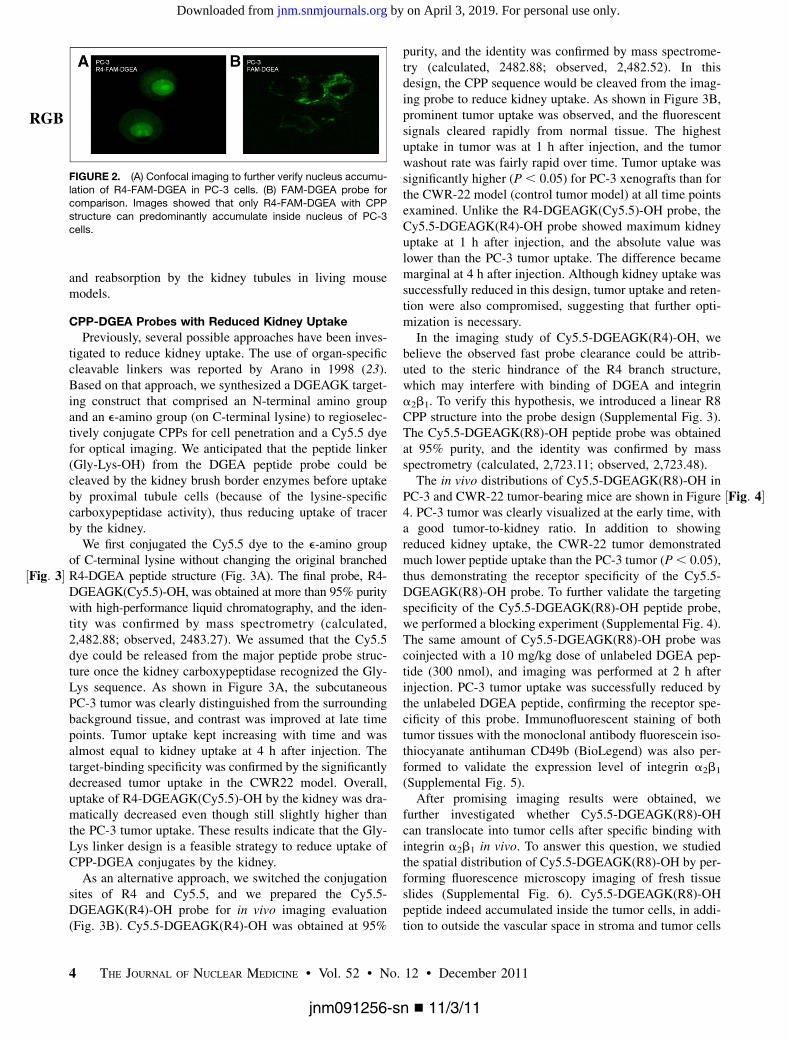
The binding specificities and subcellular localizationsof the R4-conjugated DGEA peptides were also examinedusing fluorescence microscopy, which showed that R4-FAM-DGEA bound distinctly to PC-3 cells whereas CWR-22and LNCaP showed substantially lower levels of cellularfluorescent signals (Supplemental Fig. 1C). In addition,binding of the fluorescent DGEA probes could be blockedwith the unlabeled DGEA peptide, doubly confirming thatR4-FAM-DGEA peptide binding is a2b1 integrin–specific.In these microscopy images, PC-3 cells incubated with R4-FAM-DGEA peptide unexpectedly seemed to show a strongsignal from nuclear sites. Therefore, the intracellularlocalization of R4-FAM-DGEA was further examined byconfocal microscopy. PC-3 cells that were incubated withR4-FAM-DGEA did exhibit a strong fluorescent signalinside the nucleus ( ½Fig: 2�Fig. 2). In comparison, the signals wereconfined mostly to the cytosol and cell membrane whenFAM-DGEA peptides were used.
In Vitro Characterization of CPP-DGEA Peptides byFluorescence Imaging
Receptor-specific uptake was evaluated in vivo usingathymic nude mice bearing either the integrin a2b1-positivePC-3 tumor or the control tumor CWR-22. Optical imagingwas performed with an IVIS 200 small-animal imaging sys-tem (Xenogen). The whole animal became fluorescentimmediately after injection, and the subcutaneous PC-3tumor could clearly be delineated from the surroundingbackground tissue from 30 min to 24 h after injection. Inaddition, the CWR22 tumor demonstrated lower uptake thanthe PC-3 tumor for up to 4 h after injection, but the differ-ence was not significant except at the first time point. In bothtumor models, most of the injected fluorescent probes weretrapped in the kidney (Supplemental Fig. 2). We believe thistrapping could be caused by the rapid filtration of the probes
FIGURE 1. Peptide structureof R4-dye-DGEA. Peptide was
labeled with FAM dye for in
vitro study and Cy5.5 for in
vivo imaging experiments.
RGB
CELL-PENETRATING INTEGRIN a2b1 PROBE • Huang et al. 3
jnm091256-sn n 11/3/11
by on April 3, 2019. For personal use only. jnm.snmjournals.org Downloaded from
and reabsorption by the kidney tubules in living mousemodels.
CPP-DGEA Probes with Reduced Kidney Uptake
Previously, several possible approaches have been inves-tigated to reduce kidney uptake. The use of organ-specificcleavable linkers was reported by Arano in 1998 (23).Based on that approach, we synthesized a DGEAGK target-ing construct that comprised an N-terminal amino groupand an e-amino group (on C-terminal lysine) to regioselec-tively conjugate CPPs for cell penetration and a Cy5.5 dyefor optical imaging. We anticipated that the peptide linker(Gly-Lys-OH) from the DGEA peptide probe could becleaved by the kidney brush border enzymes before uptakeby proximal tubule cells (because of the lysine-specificcarboxypeptidase activity), thus reducing uptake of tracerby the kidney.We first conjugated the Cy5.5 dye to the e-amino group
of C-terminal lysine without changing the original branchedR4-DGEA peptide structure (½Fig: 3� Fig. 3A). The final probe, R4-DGEAGK(Cy5.5)-OH, was obtained at more than 95% puritywith high-performance liquid chromatography, and the iden-tity was confirmed by mass spectrometry (calculated,2,482.88; observed, 2483.27). We assumed that the Cy5.5dye could be released from the major peptide probe struc-ture once the kidney carboxypeptidase recognized the Gly-Lys sequence. As shown in Figure 3A, the subcutaneousPC-3 tumor was clearly distinguished from the surroundingbackground tissue, and contrast was improved at late timepoints. Tumor uptake kept increasing with time and wasalmost equal to kidney uptake at 4 h after injection. Thetarget-binding specificity was confirmed by the significantlydecreased tumor uptake in the CWR22 model. Overall,uptake of R4-DGEAGK(Cy5.5)-OH by the kidney was dra-matically decreased even though still slightly higher thanthe PC-3 tumor uptake. These results indicate that the Gly-Lys linker design is a feasible strategy to reduce uptake ofCPP-DGEA conjugates by the kidney.As an alternative approach, we switched the conjugation
sites of R4 and Cy5.5, and we prepared the Cy5.5-DGEAGK(R4)-OH probe for in vivo imaging evaluation(Fig. 3B). Cy5.5-DGEAGK(R4)-OH was obtained at 95%
purity, and the identity was confirmed by mass spectrome-try (calculated, 2482.88; observed, 2,482.52). In thisdesign, the CPP sequence would be cleaved from the imag-ing probe to reduce kidney uptake. As shown in Figure 3B,prominent tumor uptake was observed, and the fluorescentsignals cleared rapidly from normal tissue. The highestuptake in tumor was at 1 h after injection, and the tumorwashout rate was fairly rapid over time. Tumor uptake wassignificantly higher (P , 0.05) for PC-3 xenografts than forthe CWR-22 model (control tumor model) at all time pointsexamined. Unlike the R4-DGEAGK(Cy5.5)-OH probe, theCy5.5-DGEAGK(R4)-OH probe showed maximum kidneyuptake at 1 h after injection, and the absolute value waslower than the PC-3 tumor uptake. The difference becamemarginal at 4 h after injection. Although kidney uptake wassuccessfully reduced in this design, tumor uptake and reten-tion were also compromised, suggesting that further opti-mization is necessary.
In the imaging study of Cy5.5-DGEAGK(R4)-OH, webelieve the observed fast probe clearance could be attrib-uted to the steric hindrance of the R4 branch structure,which may interfere with binding of DGEA and integrina2b1. To verify this hypothesis, we introduced a linear R8CPP structure into the probe design (Supplemental Fig. 3).The Cy5.5-DGEAGK(R8)-OH peptide probe was obtainedat 95% purity, and the identity was confirmed by massspectrometry (calculated, 2,723.11; observed, 2,723.48).
The in vivo distributions of Cy5.5-DGEAGK(R8)-OH inPC-3 and CWR-22 tumor-bearing mice are shown in ½Fig: 4�Figure4. PC-3 tumor was clearly visualized at the early time, witha good tumor-to-kidney ratio. In addition to showingreduced kidney uptake, the CWR-22 tumor demonstratedmuch lower peptide uptake than the PC-3 tumor (P, 0.05),thus demonstrating the receptor specificity of the Cy5.5-DGEAGK(R8)-OH probe. To further validate the targetingspecificity of the Cy5.5-DGEAGK(R8)-OH peptide probe,we performed a blocking experiment (Supplemental Fig. 4).The same amount of Cy5.5-DGEAGK(R8)-OH probe wascoinjected with a 10 mg/kg dose of unlabeled DGEA pep-tide (300 nmol), and imaging was performed at 2 h afterinjection. PC-3 tumor uptake was successfully reduced bythe unlabeled DGEA peptide, confirming the receptor spe-cificity of this probe. Immunofluorescent staining of bothtumor tissues with the monoclonal antibody fluorescein iso-thiocyanate antihuman CD49b (BioLegend) was also per-formed to validate the expression level of integrin a2b1
(Supplemental Fig. 5).After promising imaging results were obtained, we
further investigated whether Cy5.5-DGEAGK(R8)-OHcan translocate into tumor cells after specific binding withintegrin a2b1 in vivo. To answer this question, we studiedthe spatial distribution of Cy5.5-DGEAGK(R8)-OH by per-forming fluorescence microscopy imaging of fresh tissueslides (Supplemental Fig. 6). Cy5.5-DGEAGK(R8)-OHpeptide indeed accumulated inside the tumor cells, in addi-tion to outside the vascular space in stroma and tumor cells
FIGURE 2. (A) Confocal imaging to further verify nucleus accumu-lation of R4-FAM-DGEA in PC-3 cells. (B) FAM-DGEA probe for
comparison. Images showed that only R4-FAM-DGEA with CPP
structure can predominantly accumulate inside nucleus of PC-3cells.
RGB
4 THE JOURNAL OF NUCLEAR MEDICINE • Vol. 52 • No. 12 • December 2011
jnm091256-sn n 11/3/11
by on April 3, 2019. For personal use only. jnm.snmjournals.org Downloaded from

(4,6-diamino-2-phenylindole was used for nuclear stainingand is shown in blue in the figure).
DISCUSSION
Progression of prostate cancer primarily involves theformation of secondary metastatic lesions to bone. The roleof integrin a2b1 in tumor invasion and metastasis has beenimplicated by its increased expression in more aggressiveprostate cancer. Previously, we reported the synthesis andbiologic evaluation of a series of near-infrared fluorescentoptical probes and PET tracers for integrin a2b1-targetedimaging based on DGEA peptides. The DGEA-based pep-
tide was concluded to be a promising ligand for integrina2b1 imaging in vivo. However, the fast washout rate fromthe tumor may somewhat limit application. We hypothe-sized that tumor retention might be greatly increased byintroducing a selective trapping mechanism.
CPP-mediated bio-cargo delivery into living cells hasattracted great attention in the last few decades (24–26).This novel technology has demonstrated great potentialboth for basic research in cellular biology and for therapeu-tic application. By hybridization of these CPPs geneticallyor chemically, efficient intracellular delivery of various oli-gopeptides and proteins has been achieved in vitro (27–31).Inspired by these results, we proposed that the CPP-conjugated
FIGURE 3. Schematic structure of cleavable CPP-DGEA probe design that consisted of brush border peptidase–sensitive linker (Gly-Lys-
OH), DGEA-targeting sequence, and Cy5.5 and CPPs. In vivo fluorescence imaging and quantification plots are shown for athymic nude
mice bearing subcutaneous PC-3 or CWR-22 xenografts (n 5 3 for each model) after intravenous injection of 1.5 nmol of R4-DGEAGK(Cy5.5)-OH (A) or Cy5.5-DGEAGK(R4)-OH (B). Specificity of peptide uptake between 2 tumor models is not compromised. PC-3 tumor
displayed higher peptide uptake and better tumor-to-normal contrast than that of CWR-22 tumor for both peptide probes. Kidney accu-
mulation dropped dramatically after introduction of enzyme-specific linker.
RGB
CELL-PENETRATING INTEGRIN a2b1 PROBE • Huang et al. 5
jnm091256-sn n 11/3/11
by on April 3, 2019. For personal use only. jnm.snmjournals.org Downloaded from

DGEA peptides may be able to cross cell membranes and betrapped inside the cell. This potential would greatly expandoptions for the design of our integrin a2b1-targeted imagingagent.In our initial approach, we conjugated the CPP and Cy5.5
dye to the distant N-terminal and e-amino group of Lys andobtained the branched R4-K(Cy5.5)-DGEA peptide as aprototype probe. The branched polyarginine R4 structurewas tested first because the branched structure may leadto a higher translocation ability, whereas linear counterpartswith the same amount of arginine do not show significantinternalization. Flow cytometry experiments demonstratedthat the introduction of the additional R4 unit effectivelyenhanced cell-labeling efficiency without affecting bindingspecificity. Moreover, confocal imaging of the PC-3 cellalso demonstrated the significantly increased cell retentionof R4-FAM-DGEA, compared with FAM-DGEA. In fact, itwas previously shown that integrin-targeted probes, such ascyanine dye–labeled cyclic RGD peptides, could be internal-ized to only a limited extent (32) and that the internalizationis most likely attributable to the increased lipophilicity of thetagging of fluorescent dye (33). Clearly, the increaseduptake for R4-FAM-DGEA should be caused mainlyby incorporation of the CPP motif. The cell-penetratingproperty of this CPP-conjugated peptide probe not onlyimproves integrin a2b1-targeted binding but also providesa potential opportunity for therapy application. However,whether the nuclear penetration was related to a2b1 integrin
targeting is not fully understood. Further biologic investi-gations are required to elucidate the mechanism.
Despite the improved in vitro behavior of R4-K(dye)-DGEA, it demonstrated a prominent and persistent kidneyuptake in vivo. Previous studies have demonstrated that thehigh kidney uptake of some imaging agents is related to theoverall positive charge of the probes (34–36). Because wedid observe much lower kidney uptake of our previouslyreported Cy5.5-DGEA probe, the prolonged kidney uptakeof CPP-modified DGEA might be attributed to the positivecharge on the CPP motif. Although it is a common phenom-enon for peptide probes to have relatively high kidneyuptake, potential clinical therapeutic applications might belimited for agents such as R4-DGEA, since the kidney usu-ally is the dose-limiting organ. Therefore, a CPP-conjugatedDGEA probe with improved in vivo pharmacokinetic behav-ior is needed to create an effective integrin a2b1-targetedcarrier for both diagnosis and therapy applications.
Previously, Arano reported that the Gly-Lys linker couldbe specifically cleaved by the brush border carboxypepti-dase in the kidney (23). Here, we adopted this peptide linkerstrategy (Gly-Lys) in our integrin a2b1-targeted probedesign. R4-DGEAGK(Cy5.5)-OH, Cy5.5-DGEAGK(R4)-OH, and Cy5.5-DGEAGK(R8)-OH were prepared and eval-uated in PC-3 and CWR22 tumor models. Kidney uptakewas dramatically decreased in these CPP-DGEA peptidesmodified with the G-K linker. Tumor-targeting specificitywas not affected in the tested prostate cancer models. These
FIGURE 4. (A) In vivo fluorescence imag-ing of athymic nude mice bearing subcuta-
neous PC-3 or CWR-22 xenografts (n 5 3
for each model) after intravenous injection of
1.5 nmol of Cy5.5-DGEAGK(R8)-OH. (B)Positive and control tumor models indicate
that specificity of peptide probe was not
compromised. PC-3 tumor showed higherprobe uptake than that of CWR-22 tumor,
and kidney uptake decreased dramatically
because of cleavable linker. Compared with
branch R4, linear R8 conjugate demonstra-ted improved tumor retention.
RGB
6 THE JOURNAL OF NUCLEAR MEDICINE • Vol. 52 • No. 12 • December 2011
jnm091256-sn n 11/3/11
by on April 3, 2019. For personal use only. jnm.snmjournals.org Downloaded from

imaging results demonstrated that the use of a cleavablepeptide linker between the fluorescent dye–labeled DGEAtracer and polyarginine motif (CPP) is a feasible approachto lower kidney uptake while preserving tumor contrast.Among all the CPP-conjugated peptides, Cy5.5-DGEAGGK(R8)-OH is the most promising imaging agent because it hasthe highest tumor uptake and lowest kidney uptake. Cur-rently, we are developing a radiolabeled tracer based on thisconstruct. In summary, we have successfully developedCPP-DGEA–based optical agents for near-infrared fluores-cence imaging of integrin a2b1 expression in prostate can-cer. We also demonstrated that kidney uptake of these newlydesigned probes could be effectively reduced after introduc-tion of an organ-specific linker. The incorporation of addi-tional strategies, such as the administration of L- or D-lysine,may further lower kidney uptake. Moreover, in view of thefavorable trapping phenomena we have observed in thisresearch, these integrin a2b1-specific peptide probes notonly may be used as diagnostic imaging agents but alsomay serve as an efficient drug delivery carrier for prostatecancer therapy.
CONCLUSION
Our results clearly demonstrated the great potential ofusing a CPP motif to enhance the tumor targeting andinternalization capabilities of DGEA peptides. Moreover,it was shown that a cleavable peptide linker could success-fully reduce kidney uptake while preserving good tumoruptake in vivo. The success of this research could lead to aselective and highly effective diagnostic imaging agent toevaluate the stage of prostate cancer, help us more appro-priately select patients considered for potential antiintegrina2b1-based treatment, and allow the evaluation of diseasecourse and therapeutic efficacy at the earliest stages oftreatment. In view of their favorable trapping phenomena,the integrin a2b1-specific peptides developed in this studymay also serve as an efficient drug delivery carrier forprostate cancer therapy.
DISCLOSURE STATEMENT
The costs of publication of this article were defrayed inpart by the payment of page charges. Therefore, and solelyto indicate this fact, this article is hereby marked “adver-tisement” in accordance with 18 USC section 1734.
ACKNOWLEDGMENTS
This study was supported in part by the MolecularImaging Center at USC, the James H. Zumberge FacultyResearch and Innovation Fund, the National Cancer Insti-tute (P30CA014089), ACS/IRG pilot project funds IRG-58-007-48, and research grant DE-SC0002353 from theDepartment of Energy. No other potential conflict of interestrelevant to this article was reported.
REFERENCES
1. The Surveillance, Epidemiology, and End Results Program: cancer of the pros-
tate statistics 2010. National Cancer Institute Web site. Available at: http://seer.
cancer.gov/statfacts/html/prost.html. Accessed October 19, 2011.
2. Pashayan N, Pharoah P, Neal DE, et al. Stage shift in PSA detected prostate
cancers: effect modification by Gleason score. J Med Screen. 2009;16:98–101.
3. Oesterling JE. Prostate specific antigen: a critical assessment of the most useful
tumor marker for adenocarcinoma of the prostate. J Urol. 1991;145:907–923.
4. Potosky AL, Miller BA, Albertsen PC, Kramer BS. The role of increasing de-
tection in the rising incidence of prostate cancer. JAMA. 1995;273:548–552.
5. Lochter A, Navre M, Werb Z, Bissell MJ. alpha1 and alpha2 integrins mediate
invasive activity of mouse mammary carcinoma cells through regulation of stro-
melysin-1 expression. Mol Biol Cell. 1999;10:271–282.
6. Kirkland SC, Ying H. Alpha2beta1 integrin regulates lineage commitment in
multipotent human colorectal cancer cells. J Biol Chem. 2008;283:27612–27619.
7. Patrawala L, Calhoun-Davis T, Schneider-Broussard R, Tang DG. Hierarchical
organization of prostate cancer cells in xenograft tumors: the CD441a2b11
cell population is enriched in tumor-initiating cells. Cancer Res. 2007;67:
6796–6805.
8. Kostenuik PJ, Sanchez-Sweatman O, Orr FW, Singh G. Bone cell matrix pro-
motes the adhesion of human prostatic carcinoma cells via the alpha 2 beta 1
integrin. Clin Exp Metastasis. 1996;14:19–26.
9. Slack-Davis JK, Parsons JT. Emerging views of integrin signaling: implications
for prostate cancer. J Cell Biochem. 2004;91:41–46.
10. Chung LWK, Baseman A, Assikis V, Zhau HE. Molecular insights into prostate
cancer progression: the missing link of tumor microenvironment. J Urol.
2005;173:10–20.
11. Hall CL, Dai J, van Golen KL, Keller ET, Long MW, Type I collagen receptor
(a2b1) signaling promotes the growth of human prostate cancer cells within the
bone. Cancer Res. 2006;66:8648–8654.
12. Kiefer JA, Farach-Carson MC. Type I collagen-mediated proliferation of PC3
prostate carcinoma cell line: implications for enhanced growth in the bone mi-
croenvironment. Matrix Biol. 2001;20:429–437.
13. Huang CW, Li Z, Cai H, Shahinian T, Conti PS. Biological stability evaluation of
the a2b1 receptor imaging agents: diamsar and DOTA conjugated DGEA pep-
tide. Bioconjug Chem. 2011;22:256–263.
14. Huang C, Li Z, Cai H, Chen K, Shahinian T, Conti PS. Design, synthesis and
validation of integrin a2b1-targeted probe for microPET imaging of prostate
cancer. Eur J Nucl Med Mol Imaging. 2011;38:1313–1321.
15. Huang CW, Li Z, Cai H, Shahinian T, Conti PS. Novel a2b1 integrin-targeted
peptide probes for prostate cancer imaging. Mol Imaging. 2011;10:284–294.
16. Rothbard JB, Kreider E, VanDeusen CL, Wright L, Wylie BL, Wender PA.
Arginine-rich molecular transporters for drug delivery: role of backbone spacing
in cellular uptake. J Med Chem. 2002;45:3612–3618.
17. Sakai N, Matile S. Anion-mediated transfer of polyarginine across liquid and
bilayer membranes. J Am Chem Soc. 2003;125:14348–14356.
18. Futaki S, Suzuki T, Ohashi W, et al. Arginine-rich peptides: an abundant source
of membrane-permeable peptides having potential as carriers for intracellular
protein delivery. J Biol Chem. 2001;276:5836–5840.
19. Futaki S. Arginine-rich peptides: potential for intracellular delivery of macro-
molecules and the mystery of the translocation mechanisms. Int J Pharm.
2002;245:1–7.
20. Futaki S. Membrane-permeable arginine-rich peptides and the translocation
mechanisms. Adv Drug Deliv Rev. 2005;57:547–558.
21. Takayama K, Suehisa Y, Fujita T, et al. Oligoarginine-based prodrugs with self-
cleavable spacers for caco-2 cell permeation. Chem Pharm Bull (Tokyo). 2008;
56:1515–1520.
22. Futaki S, Goto S, Suzuki T, Nakase I, Sugiura Y. Structural variety of membrane
permeable peptides. Curr Protein Pept Sci. 2003;4:87–96.
23. Arano Y. Strategies to reduce renal radioactivity levels of antibody fragments.
Q J Nucl Med. 1998;42:262–270.
24. Vives E. Present and future of cell-penetrating peptide mediated delivery sys-
tems: “Is the Trojan horse too wild to go only to Troy? J Control Release.
2005;109:77–85.
25. Foerg C, Merkle HP. On the biomedical promise of cell penetrating peptides:
limits versus prospects. J Pharm Sci. 2008;97:144–162.
26. Mae M, Langel U. Cell-penetrating peptides as vectors for peptide, protein and
oligonucleotide delivery. Curr Opin Pharmacol. 2006;6:509–514.
27. Vives E, Brodin P, Lebleu B. A truncated HIV-1 Tat protein basic domain rapidly
translocates through the plasma membrane and accumulates in the cell nucleus.
J Biol Chem. 1997;272:16010–16017.
28. Fawell S, Seery J, Daikh Y, et al. Tat-mediated delivery of heterologous proteins
into cells. Proc Natl Acad Sci USA. 1994;91:664–668.
CELL-PENETRATING INTEGRIN a2b1 PROBE • Huang et al. 7
jnm091256-sn n 11/3/11
by on April 3, 2019. For personal use only. jnm.snmjournals.org Downloaded from

29. Derossi D, Joliot AH, Chassaing G, Prochiantz A. The third helix of the Anten-
napedia homeodomain translocates through biological membranes. J Biol Chem.
1994;269:10444–10450.
30. Mitchell DJ, Kim DT, Steinman L, Fathman CG, Rothbard JB. Polyarginine
enters cells more efficiently than other polycationic homopolymers. J Pept
Res. 2000;56:318–325.
31. Nagahara H, Vocero-Akbani AM, Snyder EL, et al. Transduction of full-length
TAT fusion proteins into mammalian cells: TAT-p27Kip1 induces cell migration.
Nat Med. 1998;4:1449–1452.
32. Sancey L, Garanger E, Foillard S, et al. Clustering and internalization of
integrin avb3 with a tetrameric RGD-synthetic peptide. Mol Ther. 2009;17:
837–843.
33. Baker M, Ntam C, Reese CT, et al. Internalization of near-infrared fluorescent
dyes within isolated leukocyte populations. Int J Environ Res Public Health.
2006;3:31–37.
34. Akizawa H, Uehara T, Arano Y. Renal uptake and metabolism of radiopharma-
ceuticals derived from peptides and proteins. Adv Drug Deliv Rev. 2008;60:
1319–1328.
35. Behr TM, Sharkey RM, Juweid ME, et al. Reduction of the renal uptake of
radiolabeled monoclonal antibody fragments by cationic amino acids and their
derivatives. Cancer Res. 1995;55:3825–3834.
36. Boswell CA, Tesar DB, Mukhyala K, Theil F, Fielder PJ, Khawli LA. Effects of
charge on antibody tissue distribution and pharmacokinetics. Bioconjug Chem.
2010;21:2153–2163.
8 THE JOURNAL OF NUCLEAR MEDICINE • Vol. 52 • No. 12 • December 2011
jnm091256-sn n 11/3/11
by on April 3, 2019. For personal use only. jnm.snmjournals.org Downloaded from

Doi: 10.2967/jnumed.111.091256Published online: November 7, 2011.J Nucl Med. Chiun-Wei Huang, Zibo Li and Peter S. Conti
Conjugated DGEA Probe−Cell-Penetrating-Peptidewith in Prostate Cancer 1β2α Near-Infrared Fluorescence Imaging of Integrin In Vivo
http://jnm.snmjournals.org/content/early/2011/11/07/jnumed.111.091256This article and updated information are available at:
http://jnm.snmjournals.org/site/subscriptions/online.xhtml
Information about subscriptions to JNM can be found at:
http://jnm.snmjournals.org/site/misc/permission.xhtmlInformation about reproducing figures, tables, or other portions of this article can be found online at:
the manuscript and the final, published version.typesetting, proofreading, and author review. This process may lead to differences between the accepted version of
ahead of print area, they will be prepared for print and online publication, which includes copyediting,JNMthe copyedited, nor have they appeared in a print or online issue of the journal. Once the accepted manuscripts appear in
. They have not beenJNM ahead of print articles have been peer reviewed and accepted for publication in JNM
(Print ISSN: 0161-5505, Online ISSN: 2159-662X)1850 Samuel Morse Drive, Reston, VA 20190.SNMMI | Society of Nuclear Medicine and Molecular Imaging
is published monthly.The Journal of Nuclear Medicine
© Copyright 2011 SNMMI; all rights reserved.
by on April 3, 2019. For personal use only. jnm.snmjournals.org Downloaded from