in vivo phosphorylation of adaptors regulates their
TRANSCRIPT

In Vivo Phosphorylation of Adaptors Regulates Their Interaction with Clathrin Andrew Wilde and Frances M. Brodsky
The G.W. Hooper Foundation, Department of Immunology and Microbiology, and Departments of Biopharmaceutical Sciences and Pharmaceutical Chemistry, University of California, San Francisco 94143-0552
Abstract. The coat proteins of clathrin-coated vesicles (CCV) spontaneously self-assemble in vitro, but, in vivo, their self-assembly must be regulated. To deter- mine whether phosphorylation might influence coat formation in the cell, the in vivo phosphorylation state of CCV coat proteins was analyzed. Individual compo- nents of the CCV coat were isolated by immunoprecip- itation from Madin-Darby bovine kidney cells, labeled with [32P]orthophosphate under normal culture condi- tions. The predominant phosphoproteins identified were subunits of the AP1 and AP2 adaptors. These in- cluded three of the four 100-kD adaptor subunits, et and 132 of AP2 and 131 of AP1, but not the ~/subunit of AP1. In addition, the ix1 and p.2 subunits of AP1 and AP2 were phosphorylated under these conditions. Lower levels of in vivo phosphorylation were detected for the clathrin heavy and light chains. Analysis of phosphory-
lation sites of the 100-kD adaptor subunits indicated they were phosphorylated on serines in their hinge re- gions, domains that have been implicated in clathrin binding. In vitro clathrin-binding assays revealed that, upon phosphorylation, adaptors no longer bind to clathrin. In vivo analysis further revealed that adaptors with phosphorylated 100-kD subunits predominated in the cytosol, in comparison with adaptors associated with cellular membranes, and that phosphorylated t32 subunits of AP2 were exclusively cytosolic. Kinase ac- tivity, which converts adaptors to a phosphorylated state in which they no longer bind clathrin, was found associated with the CCV coat. These results suggest that adaptor phosphorylation influences adaptor-clath- rin interactions in vivo and could have a role in control- ling coat disassembly and reassembly.
C LATHRIN-MEDIATED vesicular transport occurs at
two sites within the cell. At the plasma membrane, clathrin-coated vesicles (CCV) 1 facilitate recep-
tor-mediated endocytosis (Pearse and Robinson, 1990) to internalize soluble extracellular molecules such as nutri- ents, antigens, and growth factors and to down-regulate their receptors. CCV are also formed at the TGN where they sort newly synthesized lysosomal enzymes to the en- docytic pathway and play a role in secretory granule bio- genesis (Pearse and Robinson, 1990). The CCV coat is formed by polymerization of triskelion-shaped clathrin molecules into a lattice, catalyzed by adaptor molecules. The AP2 and AP1 adaptors nucleate clathrin assembly at the plasma membrane and the TGN, respectively, and play a role in selecting integral membrane proteins for
Address all correspondence to Frances M. Brodsky, G.W. Hooper Foun- dation, Department of Immunology and Microbiology, University of Cali- fornia at San Francisco, 513 Parnassus Avenue, Room HSW-1527, Box 0552, San Francisco, CA.
1. Abbreviat ions used in this paper: CCV, clathrin-coated vesicle; CIAP, calf intestinal alkaline phosphatase; MDBK, Madin-Darby bovine kidney; PVDF, polyvinylidene difluoride.
concentration in CCVs. Purified clathrin and adaptor mol- ecules, when sufficiently concentrated, will coassemble un- der physiological conditions in vitro (Keen, 1990).
The cellular assembly of coat proteins needs to be tightly regulated to ensure thai the coat forms only at the membrane to initiate vesiculation, and that coat disassem- bly occurs only after the CCV has formed. Thus, there must be regulatory factors in the cell to ensure that clath- rin and adaptors interact at the appropriate cellular loca- tion but avoid coassembly in the cytosol. Results from several studies have suggested CCV coat protein phosphor- ylation may play a role in controlling assembly state in vivo (Georgieva-Hanson et al., 1988; Pypaert et al., 1991). Endocytosis is inhibited during mitosis by cytosolic factors that block CCV invagination in an in vitro assay, an effect that could be partially reproduced by addition of purified cdc2 kinase to the assay (Pypaert et al., 1991). The mitotic inhibition of CCV invagination in vitro was reversed by di- luting cytosol, and this reversal was sensitive to okadaic acid, a protein phosphatase inhibitor. Furthermore, phos- phorylation of coat proteins by CCV-associated kinases destabilizes the clathrin coat of isolated brain CCV (Geor- gieva-Hanson et al., 1988). Together, these data indicate phosphorylation may influence CCV assembly or disas-
© The Rockefeller University Press, 0021-9525/96/11/635/11 $2.00 The Journal of Cell Biology, Volume 135, Number 3, November 1996 635~45 635
Dow
nloaded from http://rupress.org/jcb/article-pdf/135/3/635/1266062/635.pdf by guest on 28 N
ovember 2021

sembly, but the target molecules for the implicated kinases and phosphatases have not yet been identified.
Conversely, in vitro studies have established that some CCV components can be phosphorylated by CCV-associ- ated kinases, but the functional significance of the phos- phorylation and whether it occurs in vivo has yet to be es- tablished (Morris et al., 1990). Each heterotetrameric adaptor has two 100-kD subunits (designated a and [32 [[3] in AP2, and [31 [13'] and ~/in AP1) and one medium and one small subunit (designated Izl [AP47] and or1 [AP19] in AP1, and Ix2 [AP50] and ~r2 [AP17] in AP2). When the ki- nase activity of purified CCV is stimulated, it is possible to phosphorylate all four 100-kD adaptor subunits. The me- dium subunits of AP1 and AP2 have both been reported to become phosphorylated in vitro (Merrese et al., 1990; Pauloin and Thurieau, 1993). Indeed, Ix1 was reported to have an intrinsic casein kinase II-like activity (Merrese et al., 1990). or1 and ~2 have not been found to be phosphory- lated in any conditions. In vitro phosphorylation of clath- rin, which comprises a 180-kD heavy chain and two ho- mologous light chains, LCa and LCb, has revealed a casein kinase II activity associated with CCV that phosphorylates LCb but not LCa. The target site in LCb was mapped to serines 11 and 13, in the amino-terminal region, which are absent in LCa (Hill et al., 1988). In vitro, the clathrin heavy chain can be phosphorylated by pp60 vsrc (Mooi- broek et al., 1992).
A limited number of studies on the in vivo phosphoryla- tion of CCV coat proteins have been carried out in special- ized cell types such as neurons and reticulocytes (Bar-Zvi et al., 1988; Corvera and Capocasale, 1990; Keen and Black, 1986). One study found that in cultured neurons, the 100-kD CCV coat proteins are phosphorylated, as well as the Ix2 but not Ix1 (Keen and Black, 1986). It was not es- tablished which of the 100-kD proteins became phosphor- ylated, as antibodies distinguishing the different 100-kD proteins were not available at the time these studies were carried out. In reticulocytes, it was found that the distribu- tion of phosphorylated 100 and 50-kD CCV proteins ap- parently correlated with their assembly state, further sup- porting the idea that phosphorylation of adaptors may influence CCV formation (Bar-Zvi et al r, 1988). Again, due to lack of appropriate reagents, the phosphorylated adaptor subunits were not identified. A third study re- ported that, in chick embryo fibroblast cells transformed with Rous sarcoma virus, phosphorylation of the clathrin heavy chain occurred on tyrosine and serine residues (Martin-Perez et al., 1989). Tyrosine phosphorylation of the clathrin light chains was also shown to occur upon the stimulation of cells with EGF (Mooibroek et al., 1992). Each of these studies of clathrin phosphorylation impli- cated pp60 vsrc as the tyrosine kinase responsible.
The data summarized above demonstrate the potential for many CCV coat proteins to be phosphorylated both in vivo and in vitro, and hint at a possible role for phosphory- lation in cellular regulation of CCV coat assembly or dis- sassembly. It was therefore of interest to establish more accurately which CCV coat protein subunits are modified by phosphorylation in vivo, and to correlate their modifi- cation with coat protein interactions. Here we identify the CCV coat protein subunits that are phosphorylated in vivo and show that phosphorylation of adaptors is a negative
regulator of their interaction with clathrin. This phosphor- ylation effect was reproduced in vitro by stimulation of CCV-associated kinase activity. !a addition, the [32 sub- units of AP2 adaptors in the cytosol were all phosphory- lated, while the 132 subunits of membrane-associated AP2 were predominantly dephosphorylated, further supporting a role for phosphorylation in regulating the assembly state of CCV coat proteins in the cell.
Materials and Methods
Materials Clathrin and adaptors were purified using a method based on that of Man- fredi and Bazari (1987) with the modifications described by Chang et al. (1993). All chemicals were purchased from Sigma Chemical Co. (St. Louis, MO) unless otherwise stated in the text. The antibodies X22, LCB.1, TD.1, X16, and AP.6 were generated in this laboratory (see Table I). Antibodies 100/1, 100/2, and 100/3, and antibodies GD/1 and RY/1 were obtained from E. Ungewickell and L. Traub (Washington University, St. Louis, MO), respectively. Antibodies AC1-Mll and AP50-2 were ob- tained from M. Robinson (University of Cambridge, UK).
Cell Culture, Phosphate Labeling, and Immunoprecipitation Madin-Darby bovine kidney (MDBK) cells, obtained from the American Type Culture Collection (Rockville, MD), were cultured in DME (Chang et al., 1993) to 80-90% confluency. Cells were washed in phosphate-free DME (GIBCO BRL, Gaithersburg, MD), and then labeled for 3 h in a medium containing 250 mCi/ml [32p]orthophosphate (Dupont-New En- gland Nuclear, Wilmington, DE). Cells were then solubilized in 0.5 ml of IP buffer (0.5M Tris/HC1, pH 7.2, 1% Triton X-100, 20 mM EDTA, 10 mM NaF, 30 mM Na4PPi, 2 mM benzamidine, 1 mM Na3VO 4, lmg/ml pepsta- tin A, aprotinin, and teupeptin, and 0.1 mM PMSF) and centrifuged (13,000 g, 10 rain, 4°C); the supernatant was removed and used for immu- noprecipitation by antibodies prebound to protein A-sepharose (Pharma- cia, Uppsala, Sweden). Immunoprecipitates were analyzed by SDS-PAGE (Laemmli, 1970) and by Western blotting and subsequent autoradiogra- phy using x-ray film (X-OMAT; Eastman-Kodak Co., Rochester, NY).
Phosphoamino Acid Analysis MDBK cells were labeled as described previously, and the immunoprecip- itated proteins were analyzed by SDS-PAGE and Western blotting of the protein onto polyvinylidene difluoride (PVDF) membrane (Immobilon-P; Millipore Corp., Bedford, MA). Phosphorylated proteins were excised from the membrane and heated at ll0°C for 1.5 h in 6 N HC1 (Pierce Chemical Co., Rockford, IL). The resultant solution of hydrolyzed protein was analyzed by one-dimensional thin-layer electrophoresis on 0.1 mm plates (EM Separations, Gibbstown, NJ) using 10% acetic acid and 1% pyridine running buffer to separate the different phosphorylated amino acids. The migration of standards of the phosphoamino acids serine, thre- onine, and tyrosine was visualized with ninhydrin, and the migration of 32P-containing phosphoamino acids was determined by autoradiography.
Fractionation of Labeled Cells MDBK cells were labeled with [32p]orthophosphate as above. Cells were harvested in 100 mM MES, pH 6.8, 1 mM EGTA, 0.5 mM MgC12, and 0.2 mM DTT and lysed by repeated freeze thaw in liquid nitrogen. Cell ly- sate was centrifuged (1,000 g, 10 min), to remove intact cells and nuclei, and the supernatant was removed and centrifuged (100,000 g, 30 min) to separate membranes (pellet) from the cytosol (supernatant). To the cyto- solic fraction, Tris/HC1, pH 7.2, and Triton X-100 were added to final con- centrations of 0.5 M and 1%, respectively, before being used for immuno- precipitation as described above. The membrane fraction was solubilized in IP buffer and centrifuged (13,000 g, 10 min), and the supernatant was used for immunoprecipitation as described before. Immunoprecipitations were analyzed by SDS-PAGE, Western blotting, and autoradiography. The amount of protein found in the different fractions was determined by immunoblotting using the ECL detection system (Amersham Corp., Ar-
The Journal of Cell Biology, Volume 135, 1996 636
Dow
nloaded from http://rupress.org/jcb/article-pdf/135/3/635/1266062/635.pdf by guest on 28 N
ovember 2021

lington Heights. IL) and by quantitation using the Molecular Dynamics Personal Densitometer (Sunnyvale, CA). The relative levels of 32p-labeled proteins in each of the fractions were determined using a phosphorimager (Molecular Dynamics). To determine the ratio of phosphorylated proteins in the membrane as compared with those in the cytosol, the phosphorim- ager values were normalized according to the protein levels determined from the immunoblot experiments.
Two-dimensional Gel Electrophoresis Proteins were resolved in the first dimension by isoelectric focusing on a 3.3% acrylamide gel containing 9 M urea and 2% ampholytes (Pharma- cia). The ampholyte mixture was a 3:1 mix of the pH 5.0-7.0 ampholytes and the pH 3.5-10 ampholytes, respectively. Proteins were then separated on the basis of size on a standard 8% acrylamide SDS-PAGE gel (O'Far- rell, 1975) and transferred onto nitrocellulose before detection by immu- noblotting.
Protease Digestion of Immunoprecipitated Adaptors MDBK ceils were labeled with [32p]orthophosphate, as above, and AP1 and AP2 were immunoprecipitated with the 100/3 and AP.6 antibodies. Immunoprecipitated coat proteins were resuspended in 50 pJ of 50 mM Tris/HCI, pH 8.0, and digested at 37°C for either l, 3, or 5 min by the addi- tion of either 0.025 mg of elastase or 0.1 mg of trypsin. The resultant digest was analyzed by SDS-PAGE, Western blotting, and autoradiography.
Clathrin Cage-binding Assay Purified clathrin was concentrated to >1 mg/ml, and then dialyzed over- night against 100 mM MES, pH 6.5, 1 mM EGTA, 0.5 mM MgCI 2, 3 mM CaCl2, and 0.2 mM DTT at 4°C to allow clathrin cages to assemble. The cages were harvested by centrifugation (100,000 g, 15 min), and the pel- leted cages were resuspended in the dialysis buffer. Clathrin cages were added to cytosol made from [32p]orthophosphate-labeled MDBK cells har- vested in 100 mM MES, pH 6.8, 1 mM EGTA, 0.5 mM MgCI 2, and 02 mM DTT, and incubated for 3 h at 4°C. Cages were then pelleted and solubi- lized; adaptors were then immunoprecipitated from both the pellet and the supernatant; and immunoprecipitates were analyzed by SDS-PAGE, Western blotting, and autoradiography. In experiments where cytosol from nonradiolabeled cells was tested for cage binding, the presence of adaptors in pellets or supernatants was analyzed directly by SDS-PAGE and immunoblotting.
Dephosphorylation of Cytosolic Adaptors Cytosol was prepared from MDBK cells labeled with [32P]orthophosphate as above in the absence of phosphatase inhibitors. Adaptors were dephos- phorylated by the addition of calf intestinal alkaline phosphatase (CIAP) (Boehringer Mannheim Biochemicals, Indianapolis, IN), 5 U/ml, and in- cubated at 37°C for 30 min. The cytosol was then used in the cage-binding assay described above. Mock treatment was by incubation at 37°C for 30 rain without adding CLAP.
Stimulation of Clathrin-coated Vesicle-associated Kinase Activity CCVs were purified from bovine brain using the method described by Bauxbaum and Woodman (1995). Proteins were stripped from purified CCVs using 0.5 M Tris, pH 7.4. Kinase activity was stimulated as de- scribed by Morris et al. (1990), by the addition of KC1, MgC12, polylysine, and ATP to final concentrations of 0.15 M, 5 mM 100 l~g/ml and 100 ~M, respectively, and by diluting the stripped protein solution such that the fi- nal Tris concentration was 100 mM. This solution was then incubated at room temperature for 30 min. The reaction was stopped by the addition of EDTA to a final concentration of 20 mM.
Results
Adaptor Subunits Are Major CCV Phosphoproteins The purpose of this study was to identify components of the CCV coat that are phosphorylated in vivo and to in- vestigate the role of these modifications in regulating coat
assembly. In recent years, our laboratory and others have developed and characterized antibodies specific for each of the subunits of clathrin and the AP1 and AP2 adaptors (Table I). These antibodies were used to immunoprecipi- tate components of the CCV coat from solubilized MDBK cells, biosynthetically labeled with [32p]orthophosphate. Analysis of the immunoprecipitated proteins after SDS- PAGE and autoradiography revealed that, under these steady state culture conditions, components of both adap- tors and clathrin are constitutively phosphorylated, and that several adaptor subunits appear to be the dominant targets for phosphorylation among the CCV components analyzed (Fig. 1).
To verify which subunits of CCV proteins were phos- phorylated, immunoprecipitates were further analyzed by immunoblotting with specific antibodies, as well as by au- toradiography, and the resulting films were overlaid (Fig. 2). Immunoprecipitated 100-kD adaptor subunits were re- solved on SDS-PAGE gels containing 6 M urea before this analysis (Ahle and Ungewickell, 1986). Once phosphory- lated polypeptides were identified from both adaptors and clathrin, gel bands were excised and hydrolyzed, and the phosphoamino acid composition was determined by thin- layer electrophoresis (Fig. 3).
In AP1, the 131 subunit was phosphorylated (Fig. 2 A), and its phosphorylation was shown to be exclusive to serine residues (Fig. 3 A). In AP2, both the et and 132 sub- units were found to be phosphorylated (Fig. 2 B) on serine residues (Fig. 3 A). Quantitation of the amount of [32p]phosphate incorporated in the et and 132 subunits showed that the 132 subunit contained twice as much phos- phate as a (data not shown). For both AP1 and AP2, the medium subunits were also phosphorylated (Fig. 2, C and D). The I~1 subunit of AP1 was phosphorylated equally on serine and threonine residues. In contrast, the t~2 subunit of AP2 was phosphorylated predominantly on serine resi- dues with only a minor amount of threonine phosphoryla- tion (Fig. 3 B).
Figure 1. In v ivo -phosphory la t ed C C V coat proteins. Au to rad io - graph of the immunoprecipi ta ted coat prote in componen t s of C C V from M D B K cells labeled in vivo with [32p]orthophosphate. A P 2 immunoprec ip i t a t ed with an t ibody AP.6 ( lane 1). Clathr in immu- noprec ip i ta ted with an t ibody X22 (lane 2). Immunoprec ip i t a t ion using a con t ro l an t ibody D A 6 that recognizes the ct chain of class II his tocompatibi l i ty molecules (not expressed in M D B K cells) ( lane 3). AP1 immunoprec ip i t a t ed with an t ibody 100/3 (lane 4). Immunoprec ip i t a t ion was carr ied out in the p resence of 0.5 M Tris, p H 7.0, to p reven t coat pro te in assembly. I m m u n o p r e - cipitates were analyzed on a t 0 % ac ry lamide -SDS gel. The mi- grat ion of molecular weight s tandards in ki lodal tons (kD) is indi- ca ted on the le f thand side of this figure and all subsequen t figures.
Wilde and Brodsky Adaptor Phosphorylation Regulates Clathrin Binding 637
Dow
nloaded from http://rupress.org/jcb/article-pdf/135/3/635/1266062/635.pdf by guest on 28 N
ovember 2021

Table L Antibodies Used in This Study
Antibody Antigen Reference
100/1 131 subunit of AP1 and (Ahle et al., 1988) 132 subunit of AP2
100/2 ct subunit of AP2 (Ahle et al., 1988) 100/3 ",/subunit of AP1 (Ahle et al., 1988) AC1-M11 tx subunit of AP2 (Robinson, 1987) AP.6 ~t subunit of AP2 (Chin et al., 1989) G/DI 131 subunit of AP1 and (Traub et al., 1995)
132 subunit of AP2 RY/1 Ixl of API gift from L. Traub AP50-2 p,2 of AP2 (Page and Robinson, 1995) X16 Clathrin light chain a(LCa) (Brodsky, 1985) X22 Clathrin heavy chain (Brodsky, 1985) LCB. 1 Clathrin light chain b (LCb) (Brodsky et al., 1987) TD.1 Clathrin heavy chain (Niithke et al., 1992)
Under the same steady state culture conditions, clathrin heavy chain was weakly phosphorylated (Fig. 2 E), pre- dominantly on tyrosine residues but with some serine phosphorylation (Fig. 3 C). Clathrin light chains, LCa and LCb, were both phosphorylated (Fig. 2/7) on serine resi- dues (Fig. 3 C). LCb was the predominant target for phos- phorylation in clathrin, correlating with in vitro findings that LCb has a target site for CCV-associated casein kinase II activity that is absent in the LCa sequence (Hill et al., 1988).
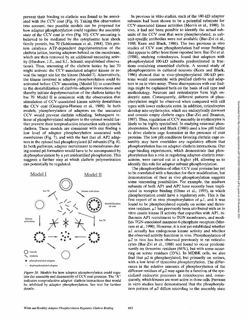
Identification of the Phosphorylated Domains of the lO0-kD Adaptor Subunits
The 100-kD subunits of adaptors have proline/glycine-rich hinge regions within which are proteolysis sites that allow the adaptors to be fragmented into their constituent do- mains. Limited cleavage releases the globular "ear" do- mains, from the "head" of the adaptors, which comprises the NH2-terminal 60-70-kD portion of each 100-kD sub- unit plus the medium and small subunits. A number of studies have assigned specific functions to the proteolyti- cally defined domains of adaptors (Goodman and Keen, 1995; Keen and Beck, 1989; Shih et al., 1995; Traub et al., 1995). The head domains of both AP1 and AP2 have local- ization specificity for the intracellular membrane where each type of adaptor is found (Page and Robinson, 1995; Peeler et al., 1993), The hinge regions of both 131 and 132, as well as sequences in the et chain of AP2, can interact with clathrin (Goodman and Keen, 1995; Shih et al., 1995). The domains in which adaptors are phosphorylated were identified to establish a potential functional consequence for these modifications.
AP1 or AP2 adaptors were separately immunoprecipi- tated from 32p-labeled MDBK cells and cleaved with either trypsin or elastase, and the resulting fragments were ana- lyzed by SDS-PAGE and immunoblotting (Fig. 4). The tryp- sin cleavage site lies in the hinge domain of the 100-kD adaptor subunits, closest to the head domain, and proteo- lysis with trypsin produces a low molecular weight frag- ment, consisting of the hinge and ear domains, and a high molecular weight fragment, consisting of the head frag- ment alone (SchrOder and Ungewickell, 1991). The elas- tase cleavage site lies at the opposite end of the hinge do-
Figure 2. Identification of the.phosphorylated subunits within each of the CCV coat protein complexes. MDBK cells were la- beled in vivo with [32p]orthophosphate, and components of the CCV coat were immunoprecipitated. The individual subunits of each precipitated complex were separated by SDS-PAGE and analyzed both by Western blotting and autoradiography. (A) AP1 immunoprecipitated wit h antibody 100/3 and analyzed on an 8% acrylamide--SDS gel, com aining 6 M urea, followed by auto- radiography (lane 1), immurtbblotting with antibody 100/3 to de- tect the y subunit (lane 2),:arid immunoblotting with the antibody 100/1 to detect the 131 (/3') subunit (lane 3). (B) AP2 immunopre- cipitated with antibody AP:6 and analyzed on an 8% acrylamide- SDS gel, containing 6 M urea, followed by autoradiography (lane 1), immunobtotting ~,ith antibody AC1-Mll to detect the ¢t subunit (lane 2), and immunoblotting with the antibody 100/1 to detect the 132 (/3) subu~t (lane 3). (C) AP1 immunoprecipitated with antibody 100/3 and analyzed on a 10% acrylamide-SDS gel, followed by autoradiography (lane 1) and immunoblotting with antibody RY/1 against txl (lane 2). (D) AP2 immunoprecipitated using antibody AP.6 and analyzed on a 10% acrylamide-SDS gel, followed by autoradiography (lane 1) and immunoblotting with antibody AP50-2 against W2 (lane 2). (E) Clathrin immunopre- cipitated using the antibody X22 and analyzed on an 8% acryl- amide-SDS gel, followed by autoradiography (lane 1) and by im- munoblotting with the clathrin heavy chain-specific antibody TD.1 (lane 2). (F) Clathrin immunoprecipitated with the anti- body X22 and analyzed on a 12% acrylamide-SDS gel, followed by autoradiography (lane 1), immunoblotting with the antibody LCB.1 that recogn~es LCb (lane 2), and immunoblotting with the antibody X16 that recognizes LCa (lane 3).
main closest to the ear domain. Cleavage with elastase produces a low molecular weight fragment, consisting of the ear domain alone, and a high molecular weight frag- ment, consisting of the head and hinge domains (Shih et al., 1995). Analysis of phosphorylated AP1 digested with trypsin revealed a 32p-labeled fragment corresponding to the ear plus hinge domains (Fig. 4 A), while elastase diges-
The Journal of Cell Biology, Volume 135, 1996 638
Dow
nloaded from http://rupress.org/jcb/article-pdf/135/3/635/1266062/635.pdf by guest on 28 N
ovember 2021
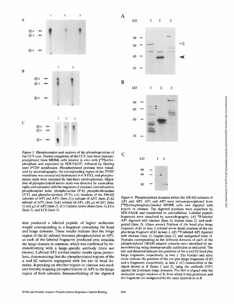
Figure 3. Phosphoamino acid analysis of the phosphoproteins of the CCV coat. Protein complexes of the CCV coat were immuno- precipitated from MDBK cells labeled in vivo with [32p]ortho- phosphate and separated by SDS-PAGE, followed by blotting onto PVDF membranes. Phosphorylated proteins were visual- ized by autoradiography, the corresponding region of the PVDF membrane was excised and hydrolyzed in 6 N HCI, and phospho- amino acids were analyzed by thin-layer electrophoresis. Migra- tion of phosphorylated amino acids was detected by autoradiog- raphy and compared with the migration of standard, nonradioactive phosphoamino acids, phospho-serine (P-S), phospho-threonine (P-T), and phospho-tyrosine (P-Y). (A) Analysis of the 100-kD subunits of AP1 and AP2: (lane 1) a subunit of AP2; (lane 2) 132 subunit of AP2; (lane 3) 131 subunit of AP1. (B) Ixl of AP1 (lane 1) and 1~2 of AP2 (lane 2). (C) Clathrin heavy chain (lane 1), LCa (lane 2), and LCb (lane 3).
tion produced a labeled peptide of higher molecular weight corresponding to a fragment containing the head and hinge domains. These results indicate that the hinge region of the 131 subunit becomes phosphorylated in AP1, as each of the labeled fragments produced only contains the hinge domain in common, which was confirmed by im- munoblott ing with a hinge-specific antibody (data not shown). Labeled AP2 yielded similar results upon proteo- lysis, demonstrating that the phosphorylated regions of the et and 132 subunits segregated with the ear or head do- mains, depending on whether trypsin or elastase was used, and thereby mapping phosphorylat ion of AP2 to the hinge region of both subunits. Immunoblot t ing of the digested
Figure 4. Phosphorylated domains within the 100-kD subunits of AP1 and AP2. AP1 and AP2 were immunoprecipitated from [32p]orthophosphate-labeled MDBK cells and digested with trypsin or elastase. The digested products were separated by SDS-PAGE and transferred to nitrocellulose. Labeled peptide fragments were visualized by autoradiography. (A) 32p-labeled AP1 digested with elastase (lane 1), trypsin (lane 2), and undi- gested (lane 3). (Open arrow) Position of the head plus hinge fragment of 131 in lane 1; (closed arrow head) position of the ear plus hinge fragment of 131 in lane 2. (B) 32p-labeled AP2 digested with elastase (lane 1), trypsin (lane 2), and undigested (lane 3). Peptides corresponding to the different domains of each of the phosphorylated 100-kD adaptor subunits were identified by im- munoblotting using domain-specific antibodies as indicated. The star and diamond indicate the positions of the a and 132 head plus hinge fragments, respectively, in lane 1. The bracket and open circle indicate the position of the ear plus hinge fragments of 132 and a fragments, respectively, in lane 2. (C) Immunoblot of the digest shown in B (lanes 1 and 2), using the antibody GD/1 against the 13-subunit hinge domains. The blot is aligned with the molecular weight markers of B, from which it was produced, and the fragments are designated by the same symbols as in B.
Wilde and Brodsky Adaptor Phosphorylation Regulates Clathrin Binding 639
Dow
nloaded from http://rupress.org/jcb/article-pdf/135/3/635/1266062/635.pdf by guest on 28 N
ovember 2021

Figure 5. Adaptors phosphorylated in vivo are impaired in their ability to bind to clathrin cages. (-4) Cytosol was prepared from [32p]orthophosphate-labeled MDBK cells and incubated in the presence or absence of preformed clathrin cages produced by po- lymerization of purified bovine brain clathrin. Cages were har- vested by centrifugation, and adaptors were first immunoprecipi- tated (using antibody 100/3 for AP1 and antibody AP.6 for AP2) from both the pellet (P) and the soluble fraction (S); then the im- munoprecipitates were analyzed by SDS-PAGE, immunoblotting (lower panel), and autoradiography (upper panel). AP1 was de- tected using the antibody 100/3, and AP2 was detected using the antibody 100/2. (B) Cytosol was prepared from MDBK ceils and incubated in the presence or absence of preformed clathrin cages with or without purified adaptors. The solution was centrifuged, and both the pelletable (P) and soluble (S) material was analyzed directly by SDS-PAGE and immunoblotting. AP1 was detected by immunoblotting with the 100/3 antibody. AP2 was detected by immunoblotting with the 100/2 antibody.
AP2 products with an antibody recognizing the hinge do- main of the 132 subunits (Traub et al., 1995) showed the hinge region segregated along with the 3Zp-labeled pep- tides into the low molecular weight trypsin fragment and the high molecular weight elastase fragment of the 132 sub- unit (Fig. 4 C). The same digestion patterns were seen for phosphorylated adaptors immunoprecipitated from la- beled normal rat kidney cells, establishing that hinge phos- phorylation was not cell specific (data not shown). Fur- thermore, in vivo-phosphorylated AP1 and AP2 from MDBK and normal rat kidney cells, fragmented using CNBr, produced phosphopeptide maps consistent with the phosphorylation site being in the hinge region (data not shown). The hinge regions of the ~, 131, and 132 subunits of adaptors extend approximately between amino acid resi- dues 600 and 750 in each of these 100-kD subunits (Kirch-
hausen et al., 1989). As serine residues within these do- mains do not lie within classical kinase consensus sequences, their phosphorylation must be mediated by a novel kinase or a known kinase using a nonclassical consensus site.
Phosphorylated Adaptors Have Impaired Binding to Clathrin Cages
Phosphorylation of the 131 and 132 subunits within their clathrin-binding hinge region suggested that one role of adaptor phosphorylation might be to regulate the interac- tion between adaptors and clathrin within cells. To investi- gate this possibility, an in vitro clathrin-binding assay was used. Purified bovine brain clathrin was polymerized into polyhedral cages, which were added to cytosol from 32p_ labeled MDBK cells. This cage-binding assay can detect adaptor-clathrin interactions by cosedimentation of adap- tors with preformed clathrin cages (Ahle and Ungewickell, 1989). The presence of bound (peUetable) or unbound (nonpelletable) adaptors was assayed by immunoprecipi- ration followed by immunoblotting and autoradiography (Fig. 5 A). Phosphorylated adaptors, derived from MDBK cytosol, were only associated with the supernatant of these reactions and absent from pellets of clathrin cages. Blot- ting for adaptor subunits indicated that no adaptors from the cytosol bound to the preformed cages in this experi- ment, suggesting that cytosolic adaptors were impaired in their ability to associate with clathrin cages. This was con- firmed using cytosol from unlabeled MDBK cells (Fig. 5 B). No adaptors of cytosolic origin bound to clathrin cages, al- though when purified adaptors were added to cytosol in the same reaction, adaptor binding to clathrin cages was detected. This demonstrated that cytosol does not contain inhibitory factors that prevent binding of adaptors to clathrin cages and further suggested that cytosolic, but not purified adaptors, are impaired for clathrin binding, possi- bly as a result of modification.
Purified and cytosolic adaptors would be expected to differ in their states of phosphorylation. Adaptors purified from bovine brain CCV have been previously shown to be dephosphorylated by a potent phosphatase activity that copurifies with adaptors and efficiently dephosphorylates adaptors within the time course of purification (Morris et al., 1990). Furthermore, the purified adaptors tested for clathrin binding in the above experiment were isolated in the absence of phosphatase inhibitors, while the adaptors present in the cytosol were clearly phosphorylated to some extent (Fig. 5 A). If phosphorylation prevents cytosolic adaptors from binding to clathrin, then dephosphorylation should induce clathrin binding. To test this theory, condi- tions for dephosphorylation of cytosolic adaptors were es- tablished, using CIAP. Dephosphorylation of cytosolic adaptors, detected by loss of 32p-label without comparable loss of protein, was achieved after incubation with 5 U/ml for 30 min at 37°C (Fig. 6 A). Unlabeled cytosol was then prepared from MDBK cells, incubated with CIAP under the same conditions, and the cytosolic adaptors were tested for binding to clathrin cages (Fig. 6 B). The CIAP- treated cytosolic adaptors were rendered competent to bind cages, while mock-treated cytosolic adaptors did not. These experiments indicate that phosphorylated adaptors are impaired in binding to clathrin and suggest that phos-
The Journal of Cell Biology, Volume 135. 1996 640
Dow
nloaded from http://rupress.org/jcb/article-pdf/135/3/635/1266062/635.pdf by guest on 28 N
ovember 2021

Figure 6. CIAP will dephosphorylate cytosolic adaptor com- plexes, allowing them to bind to clathrin cages. (A) Cytosol pre- pared from [32p]orthophosphate-labeled MDBK ceils was treated with ClAP to establish whether the ClAP was effective at de- phosphorylation. AP1 or AP2 was first immunoprecipitated, us- ing the antibodies 100/3 and AP.6, respectively, from cytosol, and then analyzed by SDS-PAGE, immunoblotting, and autoradiog- raphy. AP1 was detected using the antibody 100/3, and AP2 was detected using the antibody 100/2. (B) Cytosol was prepared from MDBK cells, treated or mock treated with calf intestinal phosphatase, and then incubated in the presence or absence of preformed clathrin cages. Material was pelleted by centrifugation and analyzed directly by SDS-PAGE and immunoblotting for AP1 using the antibody 100/3 or for AP2 using the antibody 100/2.
Figure 7. Cellular localization of phosphorylated adaptors. MDBK cells were labeled with [32p]orthophosphate, and then membrane and cytosol fractions were isolated. AP1 and AP2 were immunoprecipitated from the fractions using the antibodies 100/3 and AP.6, respectively, and analyzed by SDS acrylamide gel containing 6 M urea, followed by Western blotting onto nitro- cellulose and autoradiography. (Lanes 1 and 3) Membrane frac- tions. (Lanes 2 and 4) Cytosolic fractions.
impaired ability of cytosolic adaptors to bind clathrin (Fig. 5), suggested that most cytosolic adaptors may be phosphory- lated in the 131 or 132 hinge region that influences clathrin binding. Therefore, the degree of phosphorylation of the [32 subunit of AP2 was investigated in membrane and cy- tosolic fractions of M D B K cells, using two-dimensional electrophoresis (Fig. 8). Isoelectric focusing, followed by S D S - P A G E and autoradiography, revealed that the [32 subunit of AP2, immunoprecipitated from 32P-labeled MDBK cytosol, was present in two phosphorylated forms and
phorylation of adaptors could influence the assembly state of CCV coat proteins within the cell.
Cellular Localization of Phosphorylated Adaptors
Further evidence for a cellular role of adaptor phosphory- lation in regulating adaptor-clathrin interactions was sought in vivo. First, the intracellular distribution of phosphory- lated adaptors was investigated to determine whether phosphorylated adaptors predominated in the cytosol. M D B K cells were labeled with [32p]orthophosphate and, after freeze-thaw lysis, separated by differential centrifu- gation into membrane and cytosol fractions. AP1 or AP2 adaptors were immunoprecipitated from each fraction and analyzed by S D S - P A G E and autoradiography (Fig. 7). In addition, the degree of phosphorylation of each of the adaptor subunits in each fraction was measured using a Molecular Dynamics phosphorimager. This value was then normalized for protein concentration as determined by es- timating the relative amount of protein in each fraction by quantitative immunoblotting. Phosphorylated adaptor sub- units of both AP1 (131) and AP2 (et and 132) predominated in the cytosol fraction relative to their presence in the membrane fraction, in ratios of >3:1. This distribution cor- related with a tendency of dephosphorylated adaptors to be coassembled with clathrin on cellular membranes.
The predominance of phosphorylated [31 and 132 adap- tor subunits in the cytosol (Fig. 7), in conjunction with the
Figure 8. Two-dimensional gel electrophoresis of the 132 subunit of AP2 from membrane and cytosolic fractions. 32p-labeled (A) or unlabeled (B) MDBK cells were lysed by freeze thawing, and membrane and cytosol fractions were prepared. In each of the fractions, AP2 was analyzed by isoelectric focusing in the first di- mension and SDS-PAGE in the second dimension. (A) AP2 was immunoprecipitated from the cytosolic fraction of [32p]ortho- phosphate-labeled MDBK cells with the antibody AP.6 and ana- lyzed by two-dimensional gel electrophoresis, followed by auto- radiography and immunoblotting with the 100/1 antibody to detect the 132 subunit of AP2. (B) AP2 was immunoprecipitated with AP.6, and the 132 subunit was detected by immunoblotting with the antibody 100/1. (Lane 1) 132 subunit immunoprecipitated from MDBK membranes. (Lane 2) 132 subunit immunoprecipi- tated separately from membrane and cytosolic fractions of MDBK cells. These immunoprecipitates were then mixed and analyzed on the same two-dimensional gel. (Lane 3) 132 subunit immuno- precipitated from MDBK cytosol. (Arrows) Equivalent spots in the two experiments; i.e., the major form of 132 in the cytosol, which is phosphorylated, but to a lesser extent than the minor form visible in A, which is more acidic and not detected by blot- ting in B.
Wilde and Brodsky Adaptor Phosphorylation Regulates Clathrin Binding 641
Dow
nloaded from http://rupress.org/jcb/article-pdf/135/3/635/1266062/635.pdf by guest on 28 N
ovember 2021

therefore must have at least two phosphorylation sites (Fig. 8 A). Immunoblotting indicated that no dephosphor- ylated 132 subunit was present in the cytosol, and that the only dectectable 132 subunit protein in the cytosol corre- sponded to the two phosphorylated forms detected by au- toradiography. Blotting also indicated that the hyperphos- phorylated form of [32 subunit constituted a minor subpopulation. In contrast, when the 132 subunit of AP2 isolated from the membrane fraction was analyzed, it mi- grated to a less acidic position than either phosphorylated form. When the two samples were mixed (AP2 from mem- brane and AP2 from cytosol) and analyzed together on the same gel, it was confirmed that a distinctly charged form of 132 subunit predominated in each of the cellular subfrac- tions. The more acidic form was exclusive to the cytosol, comigrating with the major phosphorylated form (not the hyperphosphorylated form). A less acidic form, which is consistent with migration of dephosphorylated 132, was ex- clusive to membrane-associated AP2 (Fig. 8 B). These re- suits indicate that all the 132 subunits in the cytosol are phosphorylated, and the membrane-associated 132 is de- phosphorylated.
Location of the Adaptor Kinase
Previous studies have shown that many different kinase activities are associated with the CCV coat (Bar-Zvi and Branton, 1986; Campbell et al., 1984; Merrese et al., 1990; Pauloin et al., 1982). Coat proteins were extracted from purified CCV, and their kinase activity was stimulated to determine whether associated kinase activities could phos- phorylate the extracted adaptors and affect their ability to bind clathrin. Kinase activity was low unless polylysine was added to the reaction, an observation consistent with previous studies (Morris et al., 1990). Addition of polyly-
sine resulted in phosphorylation of 100-kD adaptor sub- units present in the extracted coat proteins (data not shown). Adaptors phosphorylated in this manner did not bind to preformed clathrin cages, in contrast with adaptors that were extracted from CCV without stimulation of kinase activity (Fig. 9). This indicates that, within the clathrin coat, there is a kinase capable of modifying adaptors in a manner analogous to that achieved by in vivo phosphory- lation.
Discussion
The results reported in this study identify the individual CCV coat proteins that become phosphorylated in vivo (Table II). Phosphorylation of adaptors prevents their binding to clathrin. Thus, one physiological function of adaptor subunit phosphorylation is apparently to regulate the assembly state of CCV coat proteins. The a, 131, 132, p~l, and Ix2 subunits of the adaptors were all identified as phosphoproteins. However, the most likely targets for the phosphorylation that regulates adaptor-clathrin binding in vivo are the 100-kD adaptor subunits. All three, et, 131, and 132 subunits, have been implicated in clathrin binding (Goodman and Keen, 1995; Shih et al., 1995; Traub et al., 1995), and all three are phosphorylated in their hinge re- gions (Fig. 4). The hinge region of the 131 and 132 subunits have been previously identified as clathrin-binding do- mains (Shih et al., 1995), and they are more homologous to each other than to ct, whose counterpart ~/subunit in AP1 is not phosphorylated. Therefore, we suggest that the site at which phosphorylation regulates interaction between the adaptors and clathrin is most likely the hinge region of both the 131 and 132 subunits.
The hinge regions of the 131 and 132 subunits (approxi- mately residues 600-750) are rich in proline and glycine. Hinge phosphorylation might influence the orientation of the adaptor head and ear domains, which arc separated by the hinge region and thereby conceal the clathrin-binding site. The differential phosphorylation of the et and ~/sub- units could have a role in one of several distinct cellular functions attributed to the et and ~ subunits. The ct sub- unit, but not ~, has been shown to interact with dynamin (Wang et al., 1995), as well as clathrin. Differential phos- phorylation of a and ~ is also consistent with the known differential regulation of membrane interactions of AP1 and AP2 adaptors (Robinson and Kreis, 1992; Wong and Brodsky, 1992).
Kinase activity that can phosphorylate adaptors and
Figure 9. In vitro, a CCV-associated kinase phosphorylates adap- tors and impairs their binding to preformed clathrin cages. Coat proteins were extracted from CCV with 0.5 M Tris, pH 7.0, and diluted 10-fold. CCV-associated kinase activity was then stimu- lated (+) or not ( - ) , by the addition of poly-L-lysine and ATP. Phosphorylated (+) or unphosphorylated ( - ) CCV coat proteins were incubated in the presence or absence of preformed clatbrin cages. The clathrin cages were harvested by centrifugation and analyzed directly by SDS-PAGE and immunoblotting to detect the presence of bound adaptors with the anti-AP1 antibody 100/3 or the anti-AP2 antibody 100/2.
Table 11. CCV Phosphoprotein Properties
Coat protein Phosphoamino acid Domain
ct subunit, AP2 S hinge 131 subunit, API S hinge 132 subunit, AP2 S hinge p,1 of AP 1 S T nd tx2 of AP2 S (T) nd LCa S nd LCb S nd Clathrin heavy chain Y S nd
S, serine; T, threonine; Y, tyrosine; nd. not done. Parentheses indicate a low level of phosphorylation.
The Journal of Cell Biology, Volume 135. 1996 642
Dow
nloaded from http://rupress.org/jcb/article-pdf/135/3/635/1266062/635.pdf by guest on 28 N
ovember 2021
prevent their binding to clathrin was found to be associ- ated with the CCV coat (Fig. 9). Taking this observation into account, two possible models can be proposed for how adaptor phosphorylation could regulate the assembly state of the CCV coat in vivo (Fig. 10). CCV uncoating is believed to be mediated by the cytoplasmic heat shock family protein, hsc 70 (Schlossman et al., 1984). This pro- tein catalyzes ATP-dependent depolymerization of the clathrin lattice, leaving adaptors behind on the membrane, which are then removed by an additional uncoating activ- ity (Hinshaw, J.E., and S.L. Schmid, unpublished observa- tions). Thus, uncoating of the clathrin lattice by hsc 70 might activate the CCV-associated adaptor kinase or re- veal the target site for the kinase (Model I). Alternatively, the kinase involved in adaptor phosphorylation could be activated before CCV uncoating (Model II) to contribute to the destabilization of clathrin-adaptor interactions and thereby initiate depolymerization of the clathrin lattice by hsc 70. Model II is consistent with the observation that stimulation of CCV-associated kinase activity destabilizes the CCV coat (Georgieva-Hanson et al., 1988). In both models, phosphorylation of adaptors on the uncoated CCV would prevent clathrin rebinding. Subsequent re- lease of phosphorylated adaptors to the cytosol would fur- ther prevent their nonproductive interaction with cytosolic clathrin. These models are consistent with our finding a low level of adaptor phosphorylation associated with membranes (Fig. 7), and with the fact that all AP2 adap- tors in the cytosol had phosphorylated 132 subunits (Fig. 8). In both pathways, adaptor recruitment to membranes dur- ing coated pit formation would have to be accompanied by dephosphorylation by a yet unidentified phosphatase. This suggests a further step at which clathrin polymerization can potentially be regulated.
Figure 10. Models for how adaptor phosphorylation could regu- late the assembly and disassembly of CCV coat proteins. The "X" indicates nonproductive adaptor-clathrin interactions that would be inhibited by adaptor phosphorylation. See text for further details.
In previous in vitro studies, each of the 100-kD adaptor subunits had been shown to be a potential substrate for CCV-associated kinase activities (Morris et al., 1990). In vivo, it had not been possible to identify the actual sub- units of the CCV coat that were phosphorylated, as sub- unit-specific antibodies were not available (Bar-Zvi et al., 1988; Keen and Black, 1986). The two previous in vivo studies of CCV coat phosphorylation had some findings that appear to differ from those reported here. Bar-Zvi et al. (1988), studying reticulocytes, found that adaptors with phosphorylated 100-kD subunits predominated in frac- tions containing assembled clathrin. A second study of phosphoproteins in cultured neurons (Keen and Black, 1986) showed that in vivo-phosphorylated 100-kD pro- teins would coassemble with purified clathrin and adap- tors in an in vitro assay. These discrepancies with our find- ings might be explained both on the basis of cell type and methodology. Neurons and reticulocytes have high en- docytic rates. Consequently, different patterns of phos- phorylation might be observed when compared with cell types with lower endocytic rates. In addition, reticulocytes develop into erythrocytes, which are endocytically dormant and contain empty clathrin cages (Bar-Zvi and Branton, 1987). Thus, regulation of CCV assembly in erythrocytes is likely to be highly specialized. In studying neuronal phos- phoproteins, Keen and Black (1986) used a low pH buffer to drive clathrin cage formation in the presence of coat proteins. The low pH conditions favoring clathrin cage as- sembly may have overidden any regulatory effects that phosphorylation has on adaptor--clathrin interactions. Our cage-binding experiments, which demonstrate that phos- phorylation has a role in regulating adaptor-clathrin inter- actions, were carried out at a higher pH, allowing us to identify this role for adaptor subunit phosphorylation.
The phosphorylation of other CCV coat proteins has yet to be correlated with a function for their modification, but demonstration of their in vivo phosphorylation suggests some interesting possibilities. For example, the medium subunits of both AP1 and AP2 have recently been impli- cated in receptor binding (Ohno et al., 1995), in which phosphorylation could have a regulatory role. This is the first report of in vivo phosphorylation of ~1, and it was found to be phosphorylated equally on serine and threo- nine residues. Izl has previously been attributed with an in vitro casein kinase II activity that copurifies with AP1, in- fluences AP1 recruitment to TGN membranes, and modi- fies TGN-associated mannose-6-phosphate receptors (Mer- rese et al., 1990). However, it is not yet established whether p.1 actually has endogenous kinase activity and whether the observed activity functions in vivo. Phosphorylation of ~z2 in vivo has been observed previously in rat reticulo- cytes (Bar-Zvi et al., 1988) and found to occur predomi- nantly on threonine residues (66%), but with some occur- ring on serine residues (33%). In MDBK cells, we also find that Ix2 is phosphorylated, but primarily on serines, with a low level of threonine phosphorylation. The differ- ences in the relative amounts of phosphorylation of the different residues of Ix2 may again be a function of the spe- cialized endocytic processes in reticulocytes and, conse- quently, which kinases are most active in those cells. Previous in vitro studies have demonstrated that the phosphoryla- tion pattern of p.2 differs according to the assembly state
Wilde and Brodsky Adaptor Phosphorylation Regulates Clathrin Binding 643
Dow
nloaded from http://rupress.org/jcb/article-pdf/135/3/635/1266062/635.pdf by guest on 28 N
ovember 2021
of AP2. When in the CCV, )x2 was observed to be phos- phorylated on threonines, but in the disassembled coat, Ix2 was phosphorylated predominantly on serines with only a minor amount occurring on threonine residues (Campbell et al., 1984). This suggests that different kinases may be able to act on 1~2 depending on its cellular location.
It is also worth considering the role that in vivo phos- phorylation might have in influencing clathrin function. A previous study reported that clathrin heavy chain was phosphorylated in Rous sarcoma virus-transformed cells but not in untransformed cells, suggesting it was a sub- strate for pp60 ..... (Martin-Perez et al., 1989). In MDBK ceils, we observed tyrosine phosphorylation of clathrin heavy chain, implicating cellular tyrosine kinases. Due to the low level of clathrin heavy chain phosphorylation, it was not possible to determine where the phosphorylated form predominated. However, as only a minor population of the clathrin heavy chain was phosphorylated, phosphor- ylation may affect only a specialized function of a subset of cellular clathrin. Several minor pools of clathrin have been described: one being found on endosomes (Stoorvogel et al., 1996), another found at sites of cell adhesion (Nicol and Nermut, 1987), and a third contributing to the forma- tion of pentagons rather than hexagons in the polymerized clathrin lattice (Crowther et al., 1976; Heuser, 1989). Inter- estingly, pp60 csrc and other SRC family protein tyrosine kinases are localized to endosomes and to adhesion plaques, as well as to the plasma membrane, where they would be available to modify such clathrin subpopulations. In addition, each of the clathrin light chains, LCa and LCb, are phosphorylated in vivo, on serine residues. LCb was more heavily phosphorylated than LCa, as observed in reticulocytes (Bar-Zvi et al., 1988), and consistent with the unique casein kinase II consensus site, in the NH2 termi- nus of LCb (Hill et al., 1988). The level of clathrin light chain phosphorylation varied between experiments, so it was not possible to establish where phosphorylated forms were localized within the cell. The clathrin light chains are thought to modulate clathrin assembly, and their phosphor- ylation might regulate aspects of this modulating role or again influence the function of specialized subsets of clathrin.
The data presented here indicate that adaptor phosphor- ylation and dephosphorylation could influence the assem- bly/disassembly cycle of the CCV coat within the cell. The potential for phosphorylation to control additional steps in CCV formation is also demonstrated. Identification of the kinases and phosphatases involved in the cycling of CCV coat proteins on and off the membrane will provide fur- ther insight into how this process is regulated.
We thank D. Riethof for assistance with protein purification. We are also grateful for the helpful discussion from members of the Brodsky labora- tory, in particular S. Morris.
This work was supported by grants GM38093 and GM26691 from the National Institutes of Health to F.M. Brodsky. A. Wilde was supported by a postdoctoral fellowship (9558) from the American Heart Association (California affiliate).
Received for publication 4 June 1996 and in revised form 15 August 1996.
References
Able, S., and E. Ungewickell. 1986. Purification and properties of a new clath- rin assembly protein. EMBO (Eur. Mol. Biol. Organ.) J. 5:3143-3149.
Ahle, S., and E. Ungewickell. 1989. Identification of a clathrin binding subunit
in the HA2 adaptor protein complex. J. Biol. Chem. 264:20089-20093. Ahle, S., A. Mann, U. Eichelsbacher, and E. Ungewickell. 1988. Structural rela-
tionships between clathrin assembly proteins from the Golgi and the plasma membrane. EMBO (Eur. Mol. Biol. Organ.) J. 7:919-929.
Bar-Zvi, D., and D. Branton. 1986. Clathrin-coated vesicles contain two protein kinase activities. Phosphorylation of clathrin I~-light chain by casein kinase II. J. Biol. Chem. 261:9614-9621.
Bar-Zvi, D., and D. Branton. 1987. Assembled clathrin in erythrocytes. J. Biol. Chem. 262:17719-17723.
Bar-Zvi, D., S.T. Mosley, and D. Branton. 1988. In vivo phosphorylation of clathrin-coated vesicle proteins from rat reticulocytes. J. Biol. Chem. 263: 4408--4415.
Brodsky, F.M. 1985. Clathrin structure characterized with monoclonal antibod- ies. I. Analysis of multiple antigenic sites. J. Cell Biol. 101:2047-2054.
Brodsky, F.M., C.J. Galloway, G.S. Blank, A.P. Jackson, H.-F. Scow, K. Drick- amer, and P. Parham. 1987. Localization of clathrin light-chain sequences mediating heavy-chain binding and coated vesicle diversity. Nature (Lond.). 326:203-205.
Campbell, C., J. Squicciarini, M. Shia, P.F. Pilch, and R.E. Fine. 1984. Identifi- cation of a protein kinase as an intrinsic component of rat liver coated vesi- cles. Biochemistry. 23:4420-4426.
Chang, M.P., W.G. Mallet, K.E. Mostov, and F.M. Brodsky. 1993. Adaptor self- aggregation, adaptor-receptor recognition and binding of ~t-adaptin subunits to the plasma membrane contribute to recruitment of adaptor (AP2) compo- nents of clathrin-coated pits. EMBO (Eur. Mol. Biol. Organ.) J. 12:2169- 2180.
Chin, D.J., R.M. Straubinger, S. Acton, I. N~thke, and F.M. Brodsky. 1989. 100- kDa polypeptides in peripheral clathrin-coated vesicles are required for re- ceptor-mediated endocytosis. Proc. Natl. Acad. Sci. USA. 86:9289-9293.
Corvera, S., and R.J. Capocasale. 1990. Enhanced phosphorylation of a coated vesicle polypeptide in response to insulin stimulation of rat adipocytes. Z Biol. Chem. 265:15963-15969.
Crowther, R.A., J.T. Finch, and B.M.F. Pearse. 1976. On the structure of coated vesicles. J. Mol. Biol. 103:785-798.
Georgieva-Hanson, V., W.J. Schook, and S. Puszkin. 1988. Brain coated vesicle destabilization and phosphorylation of coat proteins. J. Neuroehem. 50: 307-315.
Goodman, O.B., and J.H. Keen. 1995. The a chain of the AP-2 adaptor is a clathrin binding subunit. J. Biol. Chem. 270:23768-23773.
Heuser, J. 1989. Effects of cytoplasmic acidification on clathrin lattice morphol- ogy. J. Cell Biol. 108:401-411.
Hill, B.L., K. Drickamer, F.M. Brodsky, and P. Parham. 1988. Identification of the phosphorylation sites of clathrin light chain LC b. £ BioL Chem. 263: 5499-5501.
Keen, J.H. 1990. Clathrin and associated assembly and disassembly proteins. Annu. Rev. Biochem. 59:415--438.
Keen, J.H., and K.A. Beck. 1989. Identification of the clathrin-binding domain of assembly protein AP-2. Biochem. Biophys. Res. Commun. 158:17-23.
Keen, J.H., and M.M. Black. 1986. The phosphorylation of coated membrane proteins in intact neurons. J. Cell Biol. 102:1325-1333.
Kirchhausen, T., K.L. Nathanson, W. Matsui, A. Vaisberg, E.P. Chow, C. Burne, J.H. Keen, and A.E. Davis. 1989. Structural and functional division into two domains of the large (100- to l l5-kDa) chains of the clathrin-associ- ated protein complex AP-2. Proc. Natl. Acad. Sci. USA. 86:2612-2616.
Laemmli, U.K. 1970. Cleavage of structural proteins during the assembly of the head of bacteriophage T4. Nature (Lond.). 227:680-685.
Manfredi, J.J. and W.L. Bazari. 1987. Purification and characterization of two distinct complexes of assembly polypeptides from calf brain coated vesicles that differ in their polypeptide composition and kinase activities. J. Biol. Chem. 262:12182-12188
Martin-Perez, J., D. Bar-Zvi, D. Branton, and R.L. Erikson. 1989. Transforma- tion by Rous sarcoma virus induces clathrin heavy chain phosphorylation. J. Cell BioL 109:577-584.
Merrese, S., T. Ludwig, R. Frank, and B. Hoflack. 1990. Phosphorylation of the cytoplasmic domain of the bovine cation-independent mannose 6-phosphate receptor. J. Biol. Chem. 265:18833-18842.
Mooibroek, M.J., H.-C. Cheng, and J.H. Wang. 1992. Differential in vitro phos- phorylation of clathrin light chains by epidermal growth factor receptor as- sociated protein tyrosine kinase and a pp60 c-src related tyrosine kiuase. Arch. Biochem. Biophys. 292:448~55.
Morris, S.A., A. Mann, and E. Ungewickell. 1990. Analysis of 100-180-kDa phosphoproteins in elathrin-coated vesicles from bovine brain. J. Biol. Chem. 265:3354-3357.
N~ithke, I.S., J. Heuser, A. Lupas, J. Stock, C.W. Turck, and F.M. Brodsky. 1992. Folding and trimerization of clathrin subunits at the triskelion hub. Cell. 68:899-910.
Nicol, A., and M.V. Nermut. 1987. A new type of substratum adhesion struc- ture in NRK cells revealed by correlated interference reflection and electron microscopy. Eur. J. Cell Biol. 43:348--357.
O'Farrell, P.H. 1975. High resolution two-dimensional electrophoresis of pro- teins. J. Biol. Chem. 250:4007-4021.
Ohno, H., J. Stewart, M.-C. Fournier, H. Bosshart, I. Rhee, S. Miyatake, T. Saito, A. Galluser, T. Kirchhausen, and J.S. Bonifacino. 1995. Interaction of tyrosine-based sorting signals with clathrin-associated proteins. Science (Wash. DC). 269:1872-1875.
The Journal of Cell Biology, Volume 135, 1996 644
Dow
nloaded from http://rupress.org/jcb/article-pdf/135/3/635/1266062/635.pdf by guest on 28 N
ovember 2021

Page, L.J., and M.S. Robinson. 1995. Targeting signals and subunit interactions in coated vesicle adaptor complexes. J. Cell Biol. 131:619-630.
Pauloin, A., and C. Thurieau. 1993. The 50 kDa protein subunit of assembly polypeptide (AP) AP-2 adaptor from clathrin-coated vesicles is phosphory- lated on threonine-156 by AP-1 and a soluble AP50 kinase which co-purifies with the assembly polypeptides. Biochem. J. 296:409-415.
Pauloin, A., I. Bernier, and P. Jolles. 1982. Presence of cyclic nueleotide-Ca 2+ independent protein kinase in bovine brain coated vesicles. Nature (Lond.). 298:574-576.
Pearse, B.M.F., and M.S. Robinson. 1990. Clathrin, adaptors and sorting. Annu. Rev. Cell Biol. 6:151-171.
Peeler, J.S., W.C. Donzell, and R.G.W. Anderson. 1993. The appendage do- main of the AP-2 subunit is not required for assembly or invagination of clathrin-coated pits. J. Cell BioL 120:47-54.
Pypaert, M., D. Mundy, E. Souter, J.-C. Labb6, and G. Warren. 1991. Mitotic cytosol inhibits invagination of coated pits in broken mitotic cells. J. Cell Biol. 114:1159-1166.
Robinson, M.S. 1987. 100-kD coated vesicle proteins: molecular heterogeneity and intracellular distribution studied with monoclonal antibodies. J. Cell Biol. 104:887-895.
Robinson, M.S., and T.E. Kreis. 1992. Recruitment of coat proteins onto Golgi membranes in intact and permeabilized cells: effects of brefeldin A and
G-protein activators. Cell, 69:129-138. Schlossman, D.M., S.L. Schmid, W.A. Braell, and J.E. Rothman. 1984. An en-
zyme that removes clathrin coats: purification of an uncoating ATPase. J. Cell Biol. 99:723-733.
Schr6der, S., and E. Ungewickell. 1991. Subunit interaction and function of clathrin-coated vesicle adaptors from the Golgi and the plasma membrane. Z Biol. Chem. 266:7910-7918.
Shih, W., A. Gallusser, and T. Kirchhausen. 1995. A clathrin-binding site in the hinge of the 132 chain of mammalian AP-2 complexes. J. Biol, Chem. 270: 31083-31090.
Stoorvogel, W., V. Oorschot, and H.J. Geuze. 1996. A novel class of clathrin- coated vesicles budding from endosomes. J. Cell Biol. 132:21-33.
Traub, L.M., S. Kornfeld, and E. Ungewickell. 1995. Different domains of the AP-1 adaptor complex are required for Golgi membrane binding and clath- rin recruitment. Z BioL Chem. 270:4933-4942.
Wang, L.H., T.C. Stidhof, and R.G.W. Anderson. 1995. The appendage domain of c~-adaptin is a high affinity binding site for dynamin. J. Biol. Chem. 270: 10079-10083.
Wong, D.H., and F.M. Brodsky. 1992. 100-kD proteins of Golgi and trans-Golgi network-associated coated vesicles have related but distinct membrane bind- ing properties. J. Cell Biol. 117:1171-1179.
Wilde and Brodsky Adaptor Phosphorylation Regulates Clathrin Binding 645
Dow
nloaded from http://rupress.org/jcb/article-pdf/135/3/635/1266062/635.pdf by guest on 28 N
ovember 2021