inactivation of airborne microorganisms using dielectric
TRANSCRIPT
1
Inactivation of Airborne Microorganisms using Dielectric Barrier Discharge Technology
Jaione Romero-Mangado1, Felipe Soberon2, Sami Sainio3, Dennis Nordlund4, Ian T. Saunders1,
Gurusharan Singh5, Graham Deane2, Kevin Maughan2, Stephen Daniels5, David Loftus1, M.
Meyyappan1, Jessica Koehne1, Ram P. Gandhiraman1*
1NASA Ames Research Center, Moffett Field, CA-94035, USA
2 Novaerus Inc. 111 N. Canal St. Suite 165, Chicago, IL 60606, USA
3 Department of Electrical Engineering and Automation, School of Electrical Engineering, Aalto
University, 02150, Espoo, Finland
4Stanford Synchrotron Radiation Lightsource, Stanford Linear Accelerator Center, Menlo Park,
CA 94025, USA
5National Center for Plasma Science and Technology, Dublin City University, Dublin-9, Ireland
Abstract
The primary goal of this work is to study the efficacy of dielectric barrier discharge (DBD)
technology for deactivating airborne micro-organisms. E. coli nebulised in air was exposed to the
DBD and the bacterial sample collected from air was analyzed in detail. Reculturing of the
collected bacteria showed deactivation of E. coli. In order to understand the mechanisms of
bacterial deactivation, the bacterial samples collected from air in the vicinity of the DBD were
analysed for surface chemical change and morphological change upon DBD treatment. Scanning
electron microscopy imaging of the E. coli shows that the bacteria undergoes physical distortion
to varying degrees from formation of pores to severe structural distortion. Fourier transform
infrared spectroscopy analysis showed that the bacteria underwent severe oxidation resulting in

2
formation of hydroxyl groups and carbonyl groups and a significant reduction of amine
functionalities and phosphate groups. High resolution N1s core level photoemission
spectroscopic measurements confirmed the presence of oxide peak and a decrease in the peak
intensity of the protonated amine compared to untreated E.coli suggest oxidation of the outer
layer of the cell wall.
*Corresponding author email: [email protected] Ph: +1 650 604 4702
Key words: air decontamination, E. coli, sterilization, dielectric barrier discharge, plasma

3
Introduction
Nosocomial infections have become a worldwide public health concern, resulting in prolonged
hospitalisation of patients, increased human suffering and spiraling healthcare costs. Recent
estimates place the annual cost of nosocomial infections at over $45 billion every year to the
U.S. healthcare system alone.1 Decontamination of air is an important area of research with
significant impact on several environments including hospitals, long duration space flights and
other enclosed areas that are prone for microbial contamination.2 Ross et. al. analyzed air
samples from the homes of 44 asthmatic individuals and concluded that increased concentrations
of gram negative bacteria present in the air increased the severity of asthma.3 The most
commonly used technologies for air cleaning include high efficiency particulate air filters, UV
irradiation, chemicals (spray, gel), steam (autoclave), and gas spray (ozone, hydrogen
peroxide).4-11 These methods, while popular, often provide sub-optimal outcomes such as
creating bacterial resistance, developing mutagenic outcomes if bacteria are under-exposed and
are responsible for numerous health hazards when operated incorrectly (for example, radiation
exposure in the case of UV). Prolonged exposure to radiation could cause skin irritation in
operators, patients and staff.12,13
Air filters depend on the mechanical filtration of airborne particles and their drawback is that the
contaminated air has to pass through the filter, trapping clusters of bacteria but consequently
causing a localized bio-hazard for the maintenance staff. Standard HEPA filtration may also
allow individual bacteria to pass through its porous surface once the original clusters have
desiccated, and is unable to arrest the transit of smaller pathogenic organisms such as viruses.
Another problem with traditional air filtration is that there is a significant drop in air pressure
across the filter medium, increasing the cost of continuous operation over their life cycle and

4
requiring filter replacement on a frequent basis. UV irradiation for air disinfection comes in
several forms, including full intensity irradiation of room with no occupants in it and shielded
UV irradiation which is done with no restriction to human occupation.14 One of the biggest
drawbacks of UV irradiation is that it requires direct ‘line of sight’ exposure for it to be effective,
and ‘shadowing’ may occur when used to treat airborne bacteria, a process whereby the exposed
upper layers of a bacterial cluster are deactivated while providing a protective shield for the
bacteria below them, particularly those that form biofilms.15 While the above technologies are
often used on their own, they may be combined; for example, a combination of high efficiency
particulate air filters with UV irradiation was shown to be effective in reducing 60% of the bio-
aerosols in a contaminated room.16 Pal et. al., reported titanium dioxide nanoparticle mediated
photocatalytic inactivation of airborne E. coli in a continuous flow reactor,17,18 and fluorescent
light inactivation of gram negative and gram positive bacteria.19 Kowalski et. al. reported killing
E.coli in 10-480 seconds using high concentrations of ozone (300-631 ppm).20
In recent years, cold atmospheric pressure plasmas have gained significant recognition in the
field of healthcare, for example in the treatment of living cells, sterilization, wound healing and
blood coagulation21. The plasmas are nontoxic, provide rapid and continuous anti-bacterial
treatment, leave no residues to clean up and are easy to scale up, compared to other approaches.
The dielectric barrier discharge (DBD) based air decontamination method requires the air and
biomolecules to pass in close proximity to the discharge source and several synergistic
decontamination mechanisms are believed to increase the overall decontamination efficacy22-25.
Here, we present a DBD based air decontamination technology using a novel electrode for
generating the discharge. The study aims to explore the efficacy of DBD technology for
deactivating airborne micro-organisms and assess the effects of the atmospheric pressure plasma

5
specifically on E. coli micro-organisms. In order to understand the mechanisms of bacterial
deactivation it is necessary to examine the bacterial surface chemical and topographical change
upon plasma treatment. Topographical features have been observed here using scanning electron
microscopy (SEM). Understanding the relation of surface chemical change to cell death is crucial
and therefore, a detailed analysis of the chemical changes to the proteins, lypopolysaccharides
and the outer cell wall of the E. coli upon exposure to DBD has been carried out using fourier
transform infrared spectroscopy (FTIR) and x ray photoelectron spectroscopy (XPS).
E. coli bacteria was chosen as a representative organism for this study because of its prevalence
in many human occupied spaces, its rod-like structure makes it easier to see morphological
surface changes and its propensity to cause infectious diseases; including wound infections,
neonatal meningitis, inflammation of abdominal wall, bacteria and pneumonia.
Methods
Materials:
Escherichia coli (Migula) Castellani and Chalmers (ATCC 25922) was rehydrated in 1 ml of
Tryptic soy Broth (BD 211825). The aliquot was aseptically transferred into a tube containing 5
ml of Tryptic Soy Broth and incubated overnight with shaking at 37℃. The bacterial suspension
was then centrifuged at 1000 rpm for 10 minutes, the supernatant was discarded and the cells
were washed down four times with distilled water. The cell culture was transferred to a tube and
vortexed to re-suspend the bacterial cell pellet in distilled water.
Experimental Setup:

6
A commercially available plasma based air decontamination system (NV200, Novaerus Inc.) was
used. The DBD electrode consists of two coaxial cylindrical mesh coils (304 stainless steel, 0.2
mm diameter wire) separated by a dielectric, borosilicate glass tube (Fig. 1). The glass tube has
the following dimensions: 80 mm length, 22.5 mm internal diameter, and 28 mm outer diameter.
A high alternating voltage, 4 kV, is applied to the coils via a step-up transformer. A fan inside
the NV200 unit is used to draw the air containing bioaerosol into the unit. The DBD tool was
placed inside a Bio-Safety Cabinet (Nuaire, Class II, Type A2, Model NU-425-400) and a
compressor nebulizer (OMROM Compressor nebulizer model NE-C29-E) was attached to the
input of the system in order to aerosolize the bacterial particles for testing. All vents located on
the system were sealed and a platform was placed over the top of the system to ensure the
aerosolized particles had a direct route through the system. The bacterial suspension containing
E.coli and distilled water of quantity 1 ml was transferred to the OMROM compressor nebulizer.
The aerosolized particles were fed through the input and any viable particles were collected at
the output in Tryptic Soy Agar plates (BD 236950). The agar plates were left in front of the
output of the DBD NV200 tool and were replaced every minute for 5 minutes. The agar plates
were incubated overnight at 37℃ and after the incubation, colony forming units were observed
on the plates as shown in figure 2a. In another set of experiment, the agar plates were placed
continuously for 1 to 5 minutes and the colony forming unit counts upon reculture is shown in
figure 2b. Two different sized petri dishes 100 mm diameter and 60 mm diameter were used. For
the experiment in Figure 2a, 100 mm diameter petri dishes were used with both the fan and
electrodes turned ON and OFF simultaneously. For the experiment in Figure 2b, 60 mm diameter
petri dishes were used with plasma ON and OFF, while having the fan ON for both the cases.
There is no scientific reason for the use of different sized petri dishes. The concentration of E.

7
coli in each plate was calculated using the following formula: bacteria/ml= number of colonies x
dilution of sample.
Characterization:
For SEM imaging, bacterial cells on silicon wafer were fixed in a solution of 2.5%
Glutaraldehyde (Sigma-Aldrich) in Phosphate Buffered Saline (ATCC) for 2 hours and as a
second fixative procedure, in a 2% Osmium Tetroxide (Ted Pella, Inc.) in PBS for 1.5 hours. The
samples were then dehydrated in gradually increasing concentrations of ethanol from 60% to
100% in deionized water and chemically dried using Hexamethyldisilazane (Sigma-Aldrich) for
5 minutes. In order to preserve the samples from reacting with water from humidity, they were
placed in a vacuum desiccator overnight. SEM imaging was performed using S4800 scanning
electron microscope (Hitachi, Pleasanton, CA).
FTIR measurements were carried out using a Perkin Elmer Spectrum GX system. As the
transmission mode measurement was carried out, single side polished silicon wafers were used
as substrates. An untreated silicon substrate was used as a background substrate for the
measurement. Each measurement was an average of 200 scans.
The N1s core level x ray photo emission spectroscopy (XPS) measurements were performed on
beamline 10-1 (bending magnet endstation, spherical grating monochromator) at the Stanford
Synchrotron Radiation Lightsource (SSRL).26 A gold grid in the beam path upstream of the
chamber was used for normalization of the incoming flux. The samples were mounted on an
aluminum stick with carbon tape and all the measurements were done under UHV conditions (<
1x10⁻⁸ torr) in a generic XPS chamber. Peak fitting was performed using Casa XPS using Shirley
background and Gaussian-Lorentzian distribution. For XPS measurements, the bacterial samples
8
collected on silicon wafer were freeze dried using Labconco’s FreeZone 4.5 Liter Freeze Dry
Systems.
Results
Figure 1 shows the (a) Schematic of the NV200 DBD system, (b) diagram of the assembled coil,
(c) cross section illustrating the air flow around the dielectric barrier discharge, (d) the coil parts,
and (e) the experimental setup for airborne inactivation of E. coli and sample collection. The bio-
aerosol containing nebulized E. coli was directed at the top of the DBD tool which has an
internal fan that sucks the contaminated air towards the DBD placed inside the tool. The
decontaminated air passes out of the bottom outlet. The deactivated bacteria was collected near
the bottom outlet in a silicon wafer and agar plates for characterization and re-culturing
respectively. The purpose of a partial shield between the nebulizer and the bottom outlet in the
DBD tool is to prevent direct settlement of bacteria on to the silicon wafer and the culture plate.
The DBD design shown in Figure 1 (right) contains metal electrodes separated by a dielectric.
Figure 2 shows the reduction numbers of nebulized e.coli exposed to DBD. E.coli exposed to
DBD was collected, re-cultured and counted. Comparison of the e.coli counts with samples
collected in DBD OFF state shows a clear reduction of air borne e.coli exposed to DBD. Figure 2
shows two plots (Left) both fan and electrode were either ON and OFF together and (Right) fan
was ON throughout the experiment and the electrode was turned ON and OFF independently. In
both the cases, the DBD ON results in deactivation of the airborne E. coli. The difference in the
E. coli counts between the left and right is due to the difference in the surface area made
available for collection of the air borne microbes i.e. the difference in petri dish size between the
experiments.

9
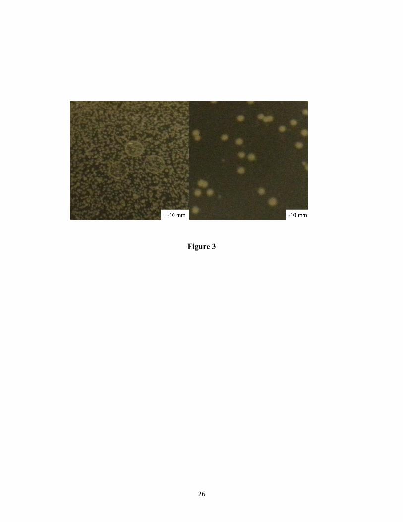
Figure 3 shows a photograph of captured E. coli in DBD OFF and DBD ON states displaying the
deactivation of air borne E. coli. Figure 4 shows SEM images of the samples at various stages.
The nebulized E. coli was exposed to DBD and collected from air on a silicon wafer. Upon
fixing of the microbes using the procedure described in the experimental section earlier, SEM
imaging was carried out on several sets of samples to study the effect of DBD on the airborne
bacteria. Untreated E. coli is smooth with intact surface and the DBD-treated bacteria are rough
with holes. The DBD alters the topographical features of E. coli to a significant extent ranging
from formation of big holes to severe structural deformation resulting in cell death. Physical
distortion to the bacterial cell wall happens to varying degrees. It is hypothesized that the degree
of distortion depends on the proximity of the bacteria to the electric field and the plasma
discharge source. Formation of holes and structural deformation of sterilized e-coli similar to
our observation has been reported by others using techniques such as biocide emusions27 and
photocatalysis.28
The FTIR data for the untreated and DBD-treated E.coli are shown in figure 5. The peak
attributions are made based on the vast FTIR spectroscopy literature on intact and damaged
bacterial cells.29-34The broad asymmetric absorption peak between 3000 and 3500 cm-1
comprises of multiple bands. Though the peaks were not distinctly resolved, the asymmetric
peak shape indicates multiple components. The characteristic peaks at 3305 and 3066 cm−1 are
assigned to amide I and amide II, respectively. A shoulder peak observed around 1730 cm-1
corresponds to C=O stretching vibrations of ester functional groups probably from the lipids and
fatty acids. The vibration bands centered around 1653 cm-1 and 1550 cm-1 correspond to amide
vibrations associated with proteins. The amide I band centered around 1653 cm-1 can be
resolved into three separate peaks at 1636 cm-1, 1655 cm-1 and 1687 cm-1. The 1655 cm-1 peak is

10
attributed to C=O stretching vibration of amides in the α helical content of the protein, 1636 cm-1
correspond to β sheets and 1687 cm-1 is related to antiparallel β sheets and aggregated strands.35-
37 The amide II band centered around 1550 cm-1 band is attributed to N-H deformation of amides
associated with proteins including contributions from the C-N stretching vibrations of the peptide
group. The absorption peaks between 2800 and 3000 cm-1 correspond to the stretching vibrations
of alkyl groups (CH2 and CH3) present in the bacterial cell wall fatty acids. The peaks at 2960
cm-1 and 2923 cm-1 correspond to asymmetric stretching vibration of CH3 and CH2 and 2870 cm-
1 and 2851 cm-1 correspond to symmetric stretching vibration of CH3 and CH2 respectively. The
peaks at 1460 and 1394 cm-1 correspond to CH3 and CH2 asymmetric and symmetric
deformation of proteins. The presence of a band at 1402 cm-1 has been attributed to the C-O
stretching vibration of carboxylic groups.38 The peak at 1240 cm-1 is due to the P=O asymmetric
stretching mode of the phospholipid phosphodiester backbone of nucleic acids DNA and RNA.39
The region between 1000 and 1100 cm-1 with peaks centered around 1040 and 1100 cm-1 has
multiple components corresponding to the C-O-C and C-O-P stretching of diverse
polysaccharides groups and P=O symmetric stretching overlapping with the vibrations of the
sugar rings of lipopolysaccharides. The peak at 1033 cm-1 is attributed to C-OH of
phosphorylated proteins and associated alcohols. We are not certain about the new intense peak
centered around 1850 cm-1 in the DBD treated E. coli. The carbonyl C=O stretching vibrations of
esters and carboxylic groups from lipids and fatty acids, in general, appear around 1740 cm-1. As
we did not find any literature for attributing the 1850 and 1975 cm-1 vibrational bands, these
peaks are left unassigned. Appearance of a new band around 930 cm-1 in the DBD treated E. coli
is due to the formation of OH species caused by oxidation. An adjacent peak around 956 cm-1 is
attributed to asymmetric stretching vibration of O-P-O functionalities.
11
Figure 6 shows the N1s core level x ray photoemission spectrum (high resolution scan) of
untreated and DBD treated E. coli. The N1s peak of untreated E. coli can be resolved in to two
components and that of DBD treated E. coli can be resolved in to three components. The major
peak at 399.8 eV is due to non-protonated nitrogen as in amines and amides. The low intensity
peak at 401.4 eV is due to protonated amines which are likely from the basic amino acid
sequence.40-43
Discussion
DBD based inactivation of the air borne E. coli as observed in figures 2 and 3 can be caused by
multiple factors. The possible contributions arising from topographical and chemical changes are
discussed in detail below. The gram negative bacteria contain cytoplasmic membrane,
peptidoglycan layer and outer membrane. The ability of the cell to withstand high internal
osmotic pressure and the maintenance of the cell shape is due to peptidoglycan layer that consists
of amino sugars and amino acids.44,45 The outer membrane contains structural proteins, receptor
molecules and phospholipids and acts as a selective permeability barrier containing hydrophilic
diffusion channels.46-49 It has been reported that the change in permeability of the membrane
during the inactivation process results in leakage of Potassium ion (K+), a vital component
involved in protein synthesis and essential for cell viability.50,51 Formation of pores, as observed
in the SEM images, is more likely to have caused potassium ion leak resulting in cell death. The
structural deformation suggests extensive chemical degradation of the microbe through the
action of the charged species generated by DBD.
The role of hydroxyl radicals and reactive oxygen species in E.coli inactivation has been studied
by several research groups.52,53 The extents of this molecular damage were assessed using
12
infrared spectroscopy by observing the major chemical components such as C=O and N-H
vibrations of amides associated with proteins, C=O vibrations of esters from lipids, CH
vibrations of the bacterial cell walls, P=O vibrations of the phosphodiester of nucleic acids and
the C-O-C vibrations of polysaccharides.29 The formation of free and hydrogen bonded hydroxyl
groups in the decontaminated E.coli has been observed here.
Exposure of the nebulized bacteria in air to DBD resulted in a significant change in chemical
structure. Significant reduction in the intensity of broad stretching vibrations of amine between
3000 to 3500cm-1 and the formation of hydrogen bonded and free hydroxyl groups between 3500
to 4000 cm-1 is very evident. A broad shoulder peak centered around 3850 cm-1 and an intense
peak centered around 3665 cm-1 correspond to O-H functionalities. The C=O stretching vibration
and N-H deformation of amides associated with proteins in the range of 1655 and 1550 cm-1
show a very clear variation. An enormous increase in the intensity of the peak centered around
1100 cm-1 could be attributed to oxidation of P=O, C-O-C and C-O-P groups of
lipopolysaccharides and other structural proteins and receptor molecules present in the outer
layer. In the DBD treated E. coli, appearance of a new band caused by oxidation has been
observed at around 930 cm-1 due to the OH species and an adjacent peak around 956 cm-1
attributed to asymmetric stretching vibration of O-P-O functionalities. All these observations
suggest damage to the outer leaflet of the E. coli due to the exposure to DBD.
In both the untreated and DBD treated E. coli, the N1s core level photoemission spectra confirms
the presence of non-protonated nitrogen as in amines and amides and protonated amines which
are likely from the basic amino acid sequence. For the DBD-treated E. coli, an additional peak at
402.8 eV corresponding to oxidized species is prominent. Also, the peak intensity corresponding
to the protonated amine in DBD-treated E. coli is decreased compared to that in untreated E. coli.

13
As the probing depth for XPS measurement is about 10 nm, the confirmation of oxide peak in the
N1s high resolution scan and a decrease in the peak intensity of the protonated amine suggest
oxidation of the outer layer of the cell wall.
The inactivation mechanism of the reactive oxygen species is based on its effect on cell
membrane and intra cellular substances. As hydroxyl groups are permeable to cell membrane,
the hydroxyl species penetrate the E. coli cell membrane, react with cytoplasmic substances and
cause chromosomal DNA damage resulting in the deactivation54. This effect is clearly observed
in figure 5 with the disappearance of one of the amide related peaks and an intense carbonyl peak
formation.
Conclusion
The efficacy of DBD technology for decontamination of air borne microorganisms was tested
and proved positive. The DBD technology is rapid and the inactivation occurs immediately upon
an organism transporting through or making contact with the electric field and/or any radicals
generated by DBD. The short time needed for inactivation of bacterial cells increases the
usefulness of the plasma in everyday settings such as hospital and workplace environments. The
effect of DBD on the morphology and surface chemistry of E. coli was studied in order to
understand the underlying mechanism in the decontamination process. The electromagnetic field
around the DBD coil causes severe distortion of the morphology of E. coli to varying degrees
from formation of pores to shrinking and elongation of the cell structure possibly resulting in
leakage of potassium ion. The oxidation and destruction components of protein, DNA and
lipopolysaccharides present in the outer leaflet of the cell wall are evident from the FTIR
spectroscopy and XPS.

14
Acknowledgement
Dr. R.P. Gandhiraman is with Universities Space Research Association subcontracted to NASA
Ames Research Center under NASA cooperative agreement. Use of the Stanford Synchrotron
Radiation Lightsource, SLAC National Accelerator Laboratory, is supported by the U.S.
Department of Energy, Office of Science, Office of Basic Energy Sciences under Contract No.
DE-AC02-76SF00515. The SSRL Structural Molecular Biology Program is supported by the
DOE Office of Biological and Environmental Research, and by the National Institutes of Health,
National Institute of General Medical Sciences (including P41GM103393). The contents of this
publication are solely the responsibility of the authors and do not necessarily represent the
official views of NIGMS or NIH.
Conflict of interest statement
None declared
Funding source
Part of this research is funded by Novaerus Inc

15
References
1. R D Scott II, Centers for Disease Control and Prevention, The Direct Cost of Healthcare-
Associated Infections in U.S. Hospitals and the Benefits of Prevention, 2009
2. W.R. Jarvis. Selected aspects of the socioeconomic impact of nosocomial infections:
morbidity, mortality, cost, and prevention. Infect Control Hosp Epidemiol 1996; 17(8): 552-7.
3. M.A. Ross, L. Curtis, P.A. Scheff et al. Association of asthma symptoms and severity with
indoor bioaerosols. Allergy 2000; 55(8):705-11.
4. Philip E. Hockberger. A History of Ultraviolet Photobiology for Humans, Animals and
Microorganisms. J. Photochem. Photobiol. 2002; 76(6): 561–579
5. A. Davies, , T. Pottage, A. Bennett, J. Walker. Gaseous and air decontamination technologies
for Clostridium difficile in the healthcare environment. . J. Hosp. Infect. 2011; 77(3): 199-203
6. A. E. Humphrey. Air Sterilization. Adv. Appl. Microbiol. 1960; 2: 301–311
7. S. Aiba and A. Yamamoto. Distribution of bacterial cells within fibrous air sterilization filters.
J Biochem Microbiol. 1959; 1 (2): 129–141

16
8. S. Roy, J. Gendron, M.-C. Delhoménie, L. Bibeau, M. Heitz, R. Brzezinski. Pseudomonas
putida as the dominant toluene-degrading bacterial species during air decontamination by
biofiltration. Appl. Microbiol. Biotechnol. 2003; 61 (4): 366-373
10. Y. Paz. Application of TiO2 photocatalysis for air treatment: Patents’ overview. Appl. Catal.,
B 2010; 99 (3–4): 448–460
11. J. Peral, X. Domènech, D. F. Ollis. Heterogeneous Photocatalysis for Purification,
Decontamination and Deodorization of Air. J. Chem. Technol. Biotechnol. 1997; 70(2):117–
140.
12. E. Bermudez, J. B. Mangum, Wong B A, et al. Pulmonary responses of mice, rats, and
hamsters to subchronic inhalation of ultrafine titanium dioxide particles. Toxicol Sci, 2004; 77:
347―357
13. S. H. Sharma. Nanoneuroscience: Emerging concepts onnanoneuro toxicity and
nanoneuroprotection. Nanomed. 2007; 2: 753 ―758
14. E. Levetin, R. Shaughnessy, C.A. Rogers, R. Scheir. Effectiveness of germicidal UV
radiation for reducing fungal contamination within air-handling units. Appl Environ Microbiol
2001; 67(8): 3712-5

17
15. M. Vleugels, G. Shama, X.T.Deng, E.Greenacre, T. Brocklehurst, M.G. Kong.
Atmospheric plasma inactivation of biofilm-forming bacteria for food safety control. IEEE
Trans. Plasma Sci., 2005; 33: 824-828
16. E. Kujundzic, D.A. Zander, M. Hernandez, L.T. Angenent, D.E. Henderson, S.L. Miller.
Effects of ceiling-mounted HEPA-UV air filters on airborne bacteria concentrations in an indoor
therapy pool building. J Air Waste Manag Assoc 2005; 55(2): 210-218
17. A. Pal, S.O. Pehkonen, L.E. Yu, M.B. Ray. Photocatalytic Inactivation of Airborne Bacteria
in a Continuous-Flow Reactor. Ind Eng Chem Res 2008; 47 (20): 7580-7585
18. A. Pal , X. Min, L.E Yu, S.O. Pehkonen, M.B. Ray. Photocatalytic Inactivation of
Bioaerosols by TiO2 Coated Membrane. Int. J. Chem. Reactor Eng.2005; 3: 1
19. A. Pal, S.O. Pehkonen, L.E. Yu, M.B. Ray. Photocatalytic inactivation of Gram-positive and
Gram-negative bacteria using fluorescent light. J Photoch Photobio A 2007; 186: 335-341
20. W.J. Kowalski, W.P. Bahnfleth, T.S. Whittam. Bactericidal effects of high airborne ozone
concentrations on Escherichia coli and Staphylococcus aureus. Ozone Sci. Eng. 1998; 20: 205-
221

18
21. M G Kong, G Kroesen, G Morfill, T Nosenko, T Shimizu, J van Dijk, J L Zimmermann.
Plasma medicine: an introductory review. New J. Phys. 2009; 11: 115012
22. M.J. Gallagher, N. Vaze, S. Gangoli, V.N. Vasilets, A.F. Gutsol, T.N. Milovanova,
S. Anandan, D.M. Murasko, A.A. Fridman. Rapid Inactivation of Airborne Bacteria Using
Atmospheric Pressure Dielectric Barrier Grating Discharge. IEEE Transactions on Plasma
Science. 2007 ; 35(5): 1501 - 1510
23. N.D. Vaze, K.P. Arjunan, Michael J. Gallagher, V.N. Vasilets, A. Gutsol, A. Fridman, S.
Anandan. Air and Water Sterilization using Non-Thermal Plasma. , 2007. ICOPS 2007. IEEE
34th International Conference on Plasma Science, DOI: 10.1109/PPPS.2007.4346053 : 2007 ,
Page(s): 747
24. N. Vaze, S. Park, G. Fridman, A. Fridman, Direct exposure to a single filament of DBD
plasma leads to the inactivation of airborne bacteria. 2010 Abstracts IEEE International
Conference on Plasma Science. DOI: 10.1109/PLASMA.2010.5533905
Publication Year: 2010 , Page(s): 1
25.Y. Liang, Y.Wu, K. Sun, Q. Chen, F. Shen, J. Zhang, M. Yao, T. Zhu, J. Fang. Rapid
Inactivation of Biological Species in the Air using Atmospheric Pressure Nonthermal Plasma.
Environ. Sci. Technol., 2012; 46 (6): 3360–3368
26. K.G. Tirsell, V.P. Karpenko. A General Purpose Sub-kev X-ray Facility at the Stanford
Synchrotron Radiation Laboratory. Nucl Instrum Methods Phys Res Sect A 1990; 291: 511–517.

19
27. R. Vyhnalkova, A. Eisenberg, T.G.M. van de Ven. Deactivation Efficiency of Stabilized
Bactericidal Emulsions. Langmuir 2011; 27(18): 11296–11305
28. J. Ren, W.Z. Wang, L. Zhang, J. Chang, S. Hu. Photocatalytic inactivation of bacteria by
photocatalyst Bi2WO6 under visible light. Catal. Commun.2009; 10: 1940−1943
29. H.M. Al-Qadiri, M.A. Al-Holy, M. Lin, N.I. Alami, A.G. Cavinato, B.A. Rasco. Rapid
Detection and Identification of Pseudomonas aeruginosa and Escherichia coli as Pure and
Mixed Cultures in Bottled Drinking Water Using Fourier Transform Infrared Spectroscopy and
Multivariate Analysis. J. Agric. Food Chem 2006; 54 (16): 5749-5754
30. J. Schmitt, H.C. Flemming. FTIR-spectroscopy in microbial and material analysis. Int.
Biodeterior. Biodegrad.1998; 41 (1): 1-11.
31. M. Kansiz, P. Heraud, B. Wood, F. Burden, J. Beardall, D. McNaughton. Fourier transform
infrared microspectroscopy and chemometrics as a tool for the discrimination of cyanobacterial
strains. Phytochemistry 1999; 52 (3): 407-417.
32. L.P. Choo-Smith, K. Maquelin, T.V. Vreeswijk et al. Investigating microbial (micro) colony
heterogeneity by vibrational spectroscopy. Appl. Environ. Microbiol 2001; 67 (4): 1461-1469.
20
33. M. Lin, M. Al-Holy, H. Al-Qadiri et al. Discrimination of intact and injured Listeria
monocytogenes by Fourier transform infrared spectroscopy and principal component analysis. J.
Agric. Food. Chem. 2004; 52: 5769-5772
34. J.J. Ojeda, M.E. Romero-Gonzalez, R.T. Bachmann, R.G.J. Edyvean, S.A. Banwart.
Characterization of the Cell Surface and Cell Wall Chemistry of Drinking Water Bacteria by
Combining XPS, FTIR Spectroscopy, Modeling, and Potentiometric Titrations. Langmuir 2008;
24: 4032-4040
35. G. Vedantham, H.G. Sparks, S.U. Sane, S. Tzannis, T.M. Przybycien. A holistic approach for
protein secondary structure estimation from infrared spectra in H(2)O solutions. Anal. Biochem.
2000; 285: 33-49
36. J. Kiwi, V. Nadtochenko. Evidence for the Mechanism of Photocatalytic Degradation of the
Bacterial Wall Membrane at the TiO2 Interface by ATR-FTIR and Laser Kinetic Spectroscopy.
Langmuir 2005; 21 (10): 4631-4641
37. A.R. Badireddy, B.R. Korpol, S. Chellam et al. Spectroscopic Characterization of
Extracellular Polymeric Substances from Escherichia coli and Serratia marcescens: Suppression
Using Sub-Inhibitory Concentrations of Bismuth Thiols. Biomacromolecules 2008; 9 (11):
3079–3089

21
38. V. Guine, L. Spadini, G. Sarret et al. Zinc sorption to three gram-negative bacteria:
Combined titration, modeling and EXAFS study. Environ. Sci. Technol. 2006; 40: 1806-1813.
39. W. Jiang, A. Saxena, B. Song, B.B. Ward, T.J. Beveridge, S.C.B Myneni. Elucidation of
functional groups on gram-positive and gram-negative bacterial surfaces using infrared
spectroscopy. Langmuir 2004; 20: 11433-11442.
40. A. R. Badireddy, B. R. Korpol, S. Chellam, P. L. Gassman, M. H. Engelhard, A. S. Lea, and
K. M. Rosso. Spectroscopic Characterization of Extracellular Polymeric Substances from
Escherichia coli and Serratia marcescens: Suppression Using Sub-Inhibitory Concentrations of
Bismuth Thiols. Biomacromolecules 2008, 9, 3079–3089
41.K. M. R. Kallury, M. Thompson, C. P. Tripp and M. L. Hair. Interaction of Silicon Surfaces
Silanized with Octadecylchlorosilanes with Octadecanoic Acid and Octadecanamine Studied by
Ellipsometry, X-ray Photoelectron Spectroscopy, and Reflectance Fourier Transform Infrared
Spectroscopy. Langmuir 1992,8, 947-954
42.K. Bierbaum, M. Kinzler, Ch. Woll, M. Grunze, G. Hahner, S. Heid and F. Effenberger. A
Near Edge X-ray Absorption Fine Structure Spectroscopy and X-ray Photoelectron Spectroscopy
Study of the Film Properties of Self-Assembled Monolayers of Organosilanes on Oxidized Si(
100) A Near Edge X-ray Absorption Fine Structure Spectroscopy and X-ray Photoelectron
Spectroscopy Study of the Film Properties of Self-Assembled Monolayers of Organosilanes on
Oxidized Si( 100). Langmuir 1995,11, 512-518

22
43.K. Syres, A. Thomas, F. Bondino, M. Malvestuto, and M. Grätzel. Dopamine Adsorption on
Anatase TiO2(101): A Photoemission and NEXAFS Spectroscopy Study. Langmuir 2010, 26,
14548–14555
44. K.H. Schleifer, O. Kandler. Peptidoglycan types of bacterial cell walls and their taxonomic
implications. Bacteriology Reviews 1972; 36 (4): 407- 477
45. R. Benz, K. Bauer. Permeation of hydrophilic molecules through the outer membrane of
gram-negative bacteria. Eur. J. Biochem.1988; 16: 1-19
46. A. Brooks, J. Yau, S. Pham. Stringent Response Changes Cell Membrane Permeability in
Escherichia coli but does not Develop Cross Tolerance to Kanamycin, Tetracycline and
Ampicillin. Journal of Experimental Microbiology and Immunology 2011; 15: 30 – 35
47. R.E. Hancock. The bacterial outer membrane as a drug barrier. Trends Microbiol 1997; 5 (1):
37-42
48. T. Nakae, J. Ishii, M. Tokunaga. Subunit structure of functional porin oligomers that form
permeability channels in the outer membrane of Escherichia coli. J Biol Chem 1979; 254 (5):
1457-1461

23
49. H. Nikaido. Prevention of drug access to bacterial targets - permeability barriers and active
efflux. Science 1994; 264: 382-388
50. W. Wang, Y. Yu, T. An. Visible-Light-Driven Photocatalytic Inactivation of E.coli K-12 by
Bismuth Vanadate Nanotubes: Bactericidal Performance and Mechanism. Environ. Sci. Technol.
2012; 46 (8): 4599-4606
51. P. Wua, J.A. Imlay, J.K. Shang. Mechanisms of Escherichia coli inactivation on palladium-
modified nitrogen-doped titanium oxide. Biomaterials 2010; 31(29): 7526-7533
52. H.Yang, X. Li, Q. Zhao, G. Chen, C.L. Raston. Role of Hydroxyl Radicals and Mechanism
of Escherichia coli. Inactivation on Ag/AgBr/TiO2 Nanotube Array Electrode under Visible
Light Irradiation. Environ. Sci. Technol. 2012; 46 (7): 4042−4050
53. P.G. Wu, J.A. Imlay, J.K. Shang. Mechanism of Escherichia coli inactivation on palladium-
modified nitrogen-doped titanium dioxide. Biomaterials 2010; 31 (29): 7526−7533.
54. K. Ishizaki, K. Sawadaishi, K. Miura, N. Shinriki. Effect of ozone on plasmid DNA of
Escherichia coli in situ. Water Research 1987; 21(7): 823-828

24
Figures
Figure 1

25
Figure 2
26
Figure 3

27
Figure 4

28
Figure 5
29
Figure 6

30
Figure captions
Figure 1. (a) Schematic of the NV200 DBD system, (b) diagram of the assembled coil, (c) cross
section illustrating the air flow around the dielectric barrier discharge, (d) the coil parts, and (e )
the experimental setup for inactivation E. coli and sample collection
Figure 2. E. coli counts captured from air in an agar plate and re-cultured. (a) both DBD and fan
ON, control experiment with both DBD and fan OFF in 100 mm diameter agar plate, (b) DBD
and fan ON, Control experiment with DBD OFF and Fan ON in 60 mm diameter agar plate.
Figure 3. Photograph of the E. coli captured from air and re-cultured in a 100 mm diameter petri
dish containing tryptic soy agar (left) control experiment with DBD OFF (right) DBD ON
Figure 4. Scanning electron microscopic image of (a,b) untreated E.coli and (c-h) E. coli passed
through the DBD and captured from air
Figure 5. FTIR spectroscopy analysis of untreated and DBD-treated E. coli. Peak attribution: 1,
2 - OH functionalities due to oxidation of hydrocarbons, 3, 6- Vibrations of Amine
functionalities (amine and amide), 4: alkyl CH2 and CH3 vibrations, 5: unknown, 7,8: CH
vibration, 9: Phosphate groups present in cell membrane, 10: P=O, C-O-C, C-O-P, 11: OH
functionality.
Figure 6. N 1s Core level x ray photoemission spectrum (XPS) of (left) untreated E. coli and
(right) inactivated E. coli