incomplete recovery of myocyte contractile function...
TRANSCRIPT

Incomplete Recovery of Myocyte Contractile Function Despite Improvement of
Myocardial Architecture With Left Ventricular Assist Device Support
Ambardekar et al: Myocyte recovery after LVAD
Amrut V. Ambardekar MD; John S. Walker PhD; Lori A. Walker PhD; Joseph C.
Cleveland Jr. MD; Brian D. Lowes MD, PhD; Peter M. Buttrick MD
Divisions of Cardiology and Cardiothoracic Surgery
University of Colorado Denver, Aurora, CO
Correspondence to: Amrut V. Ambardekar, MD Division of Cardiology, University of Colorado Denver 12631 E. 17th Avenue, Room 7102, Campus Box B-130 Aurora, CO 80045 Email: [email protected] Telephone: 303-883-4278 Fax: 303-724-2094
Journal Subject Codes: [37] CV surgery: transplantation, ventricular assistance, cardiomyopathy; [105] Myocardial biology: Contractile function; [107] Myocardial biology: Biochemistry and metabolism; [110] Congestive Heart Failure
ra, CO
rd
Avenue Room 7102 Campus Box B 130
e to:rdekar, MD
diology, University of Colorado Denver Avenue Room 7102 Campus Box B-130
by guest on June 14, 2018http://circheartfailure.ahajournals.org/
Dow
nloaded from
Abstract
Background Unloading a failing heart with a left ventricular assist device (LVAD) can
improve ejection fraction (EF) and left ventricular (LV) size; however, recovery with
LVAD explantation is rare. We hypothesized that evaluation of myocyte contractility
and biochemistry at the sarcomere level before and after LVAD may explain organ level
changes.
Methods and Results Paired LV tissue samples were frozen from 8 patients with
nonischemic cardiomyopathy at LVAD implantation (Before LVAD) and prior to
transplant (After LVAD). These were compared to 8 nonfailing hearts. Isolated skinned
myocytes were purified, attached to a force transducer, and dimensions, maximal calcium
saturated force (Fmax), calcium sensitivity, and myofilament cooperativity were
assessed. Relative isoform abundance and phosphorylation levels of sarcomeric
contractile proteins were measured. With LVAD support, the unloaded EF improved
(10.0±1.0 to 25.6±11.0%, p=0.007), LV size decreased (LVIDd 7.6±1.2 to 4.9±1.4cm,
p<0.001), and myocyte dimensions decreased (cross-sectional area 1247±346 to
638±254 m2, p=0.001). Fmax improved after LVAD (3.6±0.9 to 7.3±1.8mN/mm2,
p<0.001), but was still lower than nonfailing (7.3±1.8 vs. 17.6±1.8mN/mm2, p<0.001).
An increase in troponin I (TnI) phosphorylation after LVAD was noted, but protein
kinase C phosphorylation of TnI decreased. Biochemical changes of other sarcomeric
proteins were not observed after LVAD.
Conclusions There is significant improvement in LV and myocyte size with LVAD,
but there is only partial recovery of EF and myocyte contractility. LVAD support was
only associated with biochemical changes in TnI. This suggests that alternate
mechanisms might contribute to contractile changes after LVAD and that additional
interventions may be needed to alter biochemical remodeling of the sarcomere to further
enhance myofilament and organ level recovery.
Key Words: mechanical circulatory support, remodeling heart failure, mechanical
unloading, heart failure
imensions, mamaaaaaaxxxxxxx
coooooooopepepepepepeperararararararatititititititivivivivivivivitytytytytytyty wwww w ww
ve isoform abundance and phosphorylation levels of sarcom
i m
6
m 6
=0 001) Fmax improved after LVAD (3 6±0 9 to 7 3±1 8mN
ve isoform abundance and phosphorylation levels of sarcom
ins were measured. With LVAD support, the unloaded EF im
6±11.0%, p=0.007), LV size decreased (LVIDd 7.6±1.2 to 4
myocyte dimensions decreased (cross-sectional area 1247±346
=0 001) Fmax improved after LVAD (3 6±0 9 to 7 3±1 8mN
by guest on June 14, 2018http://circheartfailure.ahajournals.org/
Dow
nloaded from
Abbreviations
LVAD=left ventricular assist device, EF=ejection fraction, LV=left ventricle,
LVIDd=left ventricular internal dimension end-diastole, TnI=troponin I, HF=heart
failure, MyBPC=myosin binding protein C, TnT=troponin T, Tm=tropomyosin, MLC-
2=regulatory myosin light chain, MLC-1=essential myosin light chain, PKA=protein
kinase A, PKC=protein kinase C, SD=standard deviation
by guest on June 14, 2018http://circheartfailure.ahajournals.org/
Dow
nloaded from

The left ventricular assist device (LVAD) is a well established therapy for patients
with end-stage heart failure (HF). The mechanical unloading of the left ventricle with the
LVAD and the subsequent restoration of cardiac output results in improvements in HF
symptoms, functional status, quality of life, and end-organ perfusion. In addition to these
systemic effects, some patients undergoing LVAD support demonstrate improved
function of the native left ventricle (LV), termed reverse remodeling. Such organ level
improvements include decreased LV chamber size, decreased LV mass, and improved
LV ejection fraction (EF)1 and have been accompanied by systemic effects such as
normalization of catecholamine levels,2 natriuretic peptides levels,3 and circulating
cytokines like TNF- .1,4
Some of these clinical markers of reverse remodeling at the organ level have also
been demonstrated at the cellular and molecular level. Previous studies have noted
reduction in myocyte hypertrophy5,6 as well as improvement in overall cardiac histology7
after LVAD support. Furthermore, in vitro studies have demonstrated improvements in
myocardial contractile properties, restoration of adrenergic receptor density and
responsiveness, and improved calcium handling with LVAD support.8-11
As both clinical and cellular evidence suggested the plausibility of reversing end-
stage disease, the concept arose that LVADs may be used as a bridge to recovery in HF
patients. Indeed, this was met with great enthusiasm after a single center’s report of the
successful explantation of LVADs in 11 of 15 patients with nonischemic cardiomyopathy
treated with a combination of unloading with a LVAD followed by conventional HF
medical therapy and the selective 2 adrenergic receptor agonist clenbuterol.12 However,
such high rates of recovery were not observed in the multi-institutional LVAD Working
evels, and cirrrrcucuccccc
v
t e
o a
these clinical markers of reverse remodeling at the organ lev
ted at the cellular and molecular level. Previous studies have
ocyte hypertrophy5,6 as well as improvement in overall cardia
by guest on June 14, 2018http://circheartfailure.ahajournals.org/
Dow
nloaded from
Group study where only 6 of 67 patients (9%) underwent LVAD explantation for
recovery.1
At this time, the mechanisms of reverse remodeling remain poorly understood,
and it appears that restoring myocardial contractility may be more complex than what had
been initially considered. Therefore, we postulated that evaluation of myocyte
contractility and biochemistry at the most fundamental contractile level of the heart, the
sarcomere, before and after LVAD placement might reveal an explanation for this
phenomenon. Our first goal was to assess sarcomeric contractile properties by measuring
direct isometric forces on skinned isolated cell preparations. Second, since the cardiac
sarcomeric proteins myosin binding protein C (MyBPC), troponin T (TnT), troponin I
(TnI), tropomyosin (Tm), and regulatory myosin light chain (MLC-2) are known to effect
contractility,13 we assessed whether post-translational modifications and/or other
alterations in these proteins were affected with LVAD support.
Methods
Patient Selection and Tissue Acquisition
Paired LV tissue samples were collected and flash frozen from 8 patients with
end-stage nonischemic cardiomyopathy at the time of LVAD implantation (Before
LVAD) and prior to cardiac transplantation (After LVAD). Nonfailing control samples
were obtained from 8 donor hearts that were harvested for transplant but then unused for
non-cardiac reasons. The tissue obtained at the time of LVAD implantation consisted of
a core sample from the LV apex – at the in-flow cannula of the LVAD. The tissue
collected at the time of the cardiac transplant consisted of an analogously sized fragment
Second, sinceeeeeee t t h
poninin n n n n n n T T T T TTT (T(T(T(T(T(T(TnTnTnTnTnTnTnT),),),),),),), t
s
w o
e
sin (Tm), and regulatory myosin light chain (MLC-2) are kn
we assessed whether post-translational modifications and/or o
ese proteins were affected with LVAD support.
by guest on June 14, 2018http://circheartfailure.ahajournals.org/
Dow
nloaded from
excised from the explanted heart. All tissue samples were immediately flash frozen in
liquid nitrogen in the operating room (without the use of any cardioplegia solutions) and
transported to a -80 ºC freezer where they were stored until use. This storage method has
been shown to preserve sarcomeric contractile protein phosphorylation state and also
allow for the accurate assessment of the myocyte contractile parameters that are
described below.14-16 The ischemia time from removal of LV tissue from the patient to
freezing in liquid nitrogen for the Before LVAD samples was less than 10 seconds and
for the After LVAD and Nonfailing samples was less than 10 minutes. The Colorado
Multicenter Institutional Review Board approved the protocol for the collection, storage,
and analysis of human tissue.
Medical records were retrospectively reviewed by a trained physician to obtain
demographic and clinical data. Echocardiographic and hemodynamic data were obtained
from the medical record at the time closest to before LVAD implantation (to ensure
conditions reflecting the time the Before LVAD tissue was obtained) followed by the
time closest to cardiac transplantation (to ensure conditions reflecting the time the After
LVAD tissue was obtained). The data obtained from patients being supported with a
LVAD reflect the device settings that were clinically indicated at the time for the
patients, and represent the combined effects of native LV function as well as LVAD
related unloading.
Myocyte Contractility Measurements
Details of the myocyte isolation and experimentation protocols have been
previously described.14,17-19 Briefly, myocytes were purified from the frozen LV samples
ol for the collececccccctti
r n
d w
records were retrospectively reviewed by a trained physician
d clinical data. Echocardiographic and hemodynamic data w
l record at the time closest to before LVAD implantation (to
by guest on June 14, 2018http://circheartfailure.ahajournals.org/
Dow
nloaded from

by mechanical homogenization and subsequently permeabilized with 0.3% Triton X-100.
The resulting suspension was placed on the stage of an inverted microscope, and in order
to ensure adequate tissue quality, myocyte fragments with an organized myofibril pattern
with clear sarcomeric striations were chosen for the contractility experiments.15 These
isolated skinned myocyte fragments were then attached to a force transducer and motor
and mechanical experiments were conducted. A representative photomicrograph is
shown in Figure 1. The length and width of the skinned myocyte fragment were
measured when it was attached to the force transducer in relaxing solution and a side
view mirror was used to measure the thickness. Cross-sectional area was calculated
using an elliptical approximation. The isolated myocytes were stored on ice and used
within 20 hours of isolation. All experiments were performed at 15 °C and since
sarcomere length can effect force measurements, the sarcomere length was set to 2.1 um
for all experiments.18
Myocytes were exposed in random order to six different activation solutions
containing varying concentrations of calcium (pCa 9.0 to 4.5) and the developed force for
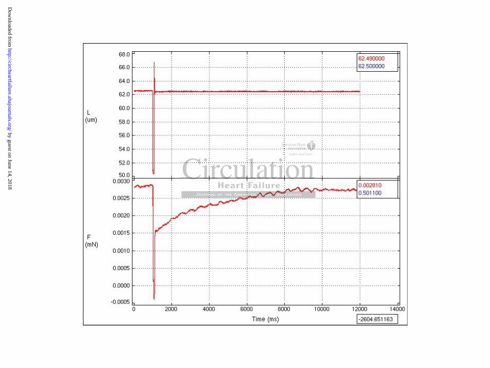
each of the calcium concentrations was recorded as shown in Figure 2a. The developed
force at the maximal calcium concentration was measured before and after obtaining the
measurements of force at the other 6 calcium concentrations. If there was >15% decline
in force between the first and last force measurement at the maximal calcium
concentration, we considered this as evidence of cell fragment degradation and this data
was excluded from the analysis. Using these measurements, a force-pCa curve was
plotted (Figure 2b) and the Hill equation was fit to derive the following contractile
parameters: Fmax (the maximal calcium saturated developed force normalized to the
nal area was cccccccaalaaaaa
re stotototototot rerererererered d d d d d ononononononon i i i i i iicececcccc
o s
h can effect force measurements, the sarcomere length was se
n
of isolation. All experiments were performed at 15 °C and s
h can effect force measurements, the sarcomere length was se
nts.18
by guest on June 14, 2018http://circheartfailure.ahajournals.org/
Dow
nloaded from

cross-sectional area of the myocyte), pCa50 (the calcium concentration at which the force
is half maximal, a measure of calcium sensitivity), and Hill coefficient (the slope of the
calcium-force relation, an index of myofilament cooperative activation). In order to
minimize measurement variations for any single isolated myocyte, the individual patient
contractile data reflect the average of measurement recordings for 3 to 5 myocytes per
patient.
Sarcomeric Protein Phosphorylation Assessment
The remaining myocyte preparations not used for mechanical study were acetone
precipitated to clamp their phosphorylation state and homogenized in 8M urea, 2.5M
thiourea, 4% CHAPS, 10 mM EDTA, and a mixture of protease and phosphatase
inhibitors as previously described.20 Protein concentration of these samples was
measured using a BCA Protein Assay Kit (Thermo Scientific).
Phosphorylation levels of the cardiac sarcomeric proteins were determined by
separating the proteins with 12% 1D-SDS-PAGE and fixing and staining with a
phosphoprotein stain (ProQ Diamond Phosphoprotein Gel Stain, Invitrogen). After
imaging, the same gels were rinsed and stained with a total protein stain (BioSafe
Coomassie Blue, BioRad). All gels were imaged using a Typhoon 9410 Gel Imager (GE
Lifesciences) and protein optical densities were measured using ImageJ (version 1.42
NIH). In order to adjust for subtle differences in protein loading, the phosphorylation
levels were calculated by dividing the optical density of the each sarcomeric protein on
the ProQ diamond gel by the optical density of the same protein on the Coomassie gel.
chanical study y y y yyy wwww
enizeeeeeeed d d d d d ininininininin 8 8 8 8 88 8M M M M M MM ururuuuuu e
H a
v w
a
HAPS, 10 mM EDTA, and a mixture of protease and phospha
viously described.20 Protein concentration of these samples w
a BCA Protein Assay Kit (Thermo Scientific).
by guest on June 14, 2018http://circheartfailure.ahajournals.org/
Dow
nloaded from

In the case of MyBPC, the total protein stain optical density merges with the abundant
protein myosin, so the phosphorylation level of MyBPC on the phosphoprotein stain was
normalized to essential myosin light chain (MLC-1) as prior studies have reported.20
Additional Assessment of Sarcomeric Proteins
Site-specific phosphorylation of troponin I was assessed by Western blots.
Proteins were separated with 12% 1D-SDS-PAGE as above and transferred to PVDF
membranes. After the membranes were blocked in 5% BSA and rinsed with TBST, they
were incubated overnight at 4 °C with a phosphospecific primary antibody to TnI
phosphorylated at the putative protein kinase A (PKA) site, Serine 22,23 (Cell Signaling
1:1000) or to troponin I phosphorylated at the putative protein kinase C (PKC) site Serine
43 using an epitope specific phosphoserine antibody (Abcam 1:1000) as previously
described.20 The blots were then washed and incubated with secondary antibody (anti-
mouse from Sigma, 1:10000) for one hour at room temperature, washed, and visualized
using enhanced chemiluminescence. Membranes were stripped and subsequently
incubated in primary total cardiac TnI antibody (Fitzgerald, Inc 1:2500). Site specific
phosphorylation was calculated by dividing the optical density of the phosphospecific
antibody blot by the density of the total protein blot.
Changes in myosin heavy chain isoforms were assessed as previously described20
by separating proteins using modified 6% 1D-SDS-PAGE and subsequent staining with
BioSafe Coomassie Blue total protein stain. MLC-1 and MLC-2 phosphorylation and
isoforms changes were assessed using 2D-SDS-PAGE21 and percent phosphorylated
MLC-2 and percent atrial isoform of MLC-1 were calculated.
mary antibodyyyyyyy t t tttttoo
Serinenenenenenen 2 2 2 2 2 222,2,2,2,2,2,2,23232323232323 (C(C(C(C(C(C(Ce
C
o v
e b
ponin I phosphorylated at the putative protein kinase C (PKC
ope specific phosphoserine antibody (Abff cam 1:1000) as prev
e blots were then washed and incubated with secondary antib
by guest on June 14, 2018http://circheartfailure.ahajournals.org/
Dow
nloaded from

Changes in TnT isoform expression were assessed using by modified Western
blots as previously described.22 The primary cardiac TnT isoform antibody used was Ab-
1 (Clone 13-11, Thermo scientific 1:1000).
Statistical Analysis
Results are expressed as mean ± standard deviation (SD). The paired t-test was
used to compare differences between before and after LVAD implantation. The unpaired
t-test was used to compare differences between before LVAD implantation failing and
nonfailing and between after LVAD implantation failing and nonfailing groups.
Statistical significance was defined as a two-tailed P-value of less than 0.05. The R
version 2.9.1 (Vienna, Austria) statistical program was used for all analyses.
Results
Patient Characteristics
The baseline characteristics of patients with end-stage nonischemic
cardiomyopathy requiring LVAD as a bridge to transplant are provided in Table 1. All
patients were inotrope dependent with NYHA class IV HF at the time of LVAD
implantation, and 7 of 8 patients required mechanical support with an intra-aortic balloon
pump. The mean duration for the diagnosis of HF at the time of LVAD implantation was
59±22 months. Both first generation pulsatile displacement LVADs (HeartMate XVE,
Thoratec, Pleasanton, CA) and second generation continuous axial flow LVADs
(HeartMate II, Thoratec, Pleasanton, CA) were utilized. The mean duration of LVAD
support was 143±41 days. LVAD support resulted in significant reductions in LV
nonfailing grrououooooo
f lesssssssss t tttttthahahahahahah n n n nnnn 0.0.0.0.0.00 05050505050505.
iienna, Austria) statistical program was used for all analyses.
by guest on June 14, 2018http://circheartfailure.ahajournals.org/
Dow
nloaded from

echocardiographic dimensions, improvement in EF, and normalization of several
hemodynamic measures as summarized in Table 2.
The 8 nonfailing patients (3 male and 5 female) were organ donors whose hearts
were not utilized for transplant for non-cardiac reasons. The mean age of this group was
48 ± 8 years, the mean EF 70.6 ± 5.4%, and none of these patients had any prior cardiac
history. Seven (87.5%) of these patients were on adrenergic vasopressor agents during
the period of brain death and as organs were being harvested.
Myocyte Size and Contractility Parameters
Significant differences were noted for all myocyte fragment dimensions as
summarized in Table 3. Myocytes obtained from patients before LVAD implant were
significantly larger than myocytes from patients after LVAD implantation. However, the
myocytes from patients after LVAD implantation were still larger than the myocytes
obtained from nonfailing patients. Fmax doubled with LVAD support (3.6 ± 0.9 vs. 7.3 ±
1.8 mN/mm2, p<0.001), but was still less than half of nonfailing subjects (7.3 ± 1.8 vs.
17.6 ± 1.8 mN/mm2, p<0.001) as described in Table 4. The pCa50 was unchanged in all
groups and the Hill coefficient was reduced in both LVAD groups relative to controls.
Sarcomeric Proteins and Phosphorylation Levels
Total TnI phosphorylation increased with LVAD support (12.8 ± 4.1 vs. 21.6 ±
9.4 OD units, p=0.030) (Table 5 and Figure 3). While there were no significant
differences noted in the Serine 22,23 site specific phosphorylation of TnI, Serine 43 site
specific phosphorylation of TnI decreased after LVAD (7.5 ± 2.2 vs. 5.2 ± 1.9 OD units,
agmeeeeeeentntntntntntnt d d d d d d imimimimimimimenenenenenenensssssisis o
T p
g H
p m
Table 3. Myocytes obtained from patients before LVAD imp
ger than myocytes from patients after LVAD implantation. H
patients after LVAD implantation were still larger than the m
by guest on June 14, 2018http://circheartfailure.ahajournals.org/
Dow
nloaded from

p=0.044) (Figure 4). Total phosphorylation levels of MyBPC were higher in both failing
groups compared with nonfailing donors (Table 5). Phosphorylation levels of TnT, Tm,
and MLC-2 were not significantly different before and after LVAD (Table 5, and Figure
3).
There were no changes noted on 2D-SDS-PAGE in the percent phosphorylation
of MLC-2 before and after LVAD (43±3 vs. 41±2%, p=0.341) and in the percent atrial
isoform of MLC-1 before and after LVAD (23±5 vs. 21±7%, p=0.711). Furthermore, no
differences were noted in myosin heavy chain isoforms before and after LVAD with only
the -isoform being expressed in both groups of patients (Figure 5). Finally there was no
evidence of altered TnT isoform expression in the before LVAD, after LVAD, and
nonfailing groups. Total TnT content was unchanged between these groups.
Discussion
The main finding of this study is that in isolated skinned myocytes, there is a
marked reduction in maximum developed force in patients with end-stage nonischemic
cardiomyopathy compared to nonfailing donors. This marked reduction in force is only
partially improved with LVAD support. Furthermore, there is strong evidence that
structural changes accompany these changes in force at the organ level (reduction in LV
size) as well as at the cellular level (reduction of all myocyte dimensions) and these
correlate with functional changes at both the organ level (improvement in EF) and
cellular level (improvement in maximum force). Despite these significant changes in
structural and mechanical parameters, unloading of the LV with LVAD support did not
gure 5). Finallllllly y yyyyy
AD,D,,,,,, a a a a a a aftftftftftftftererererererr L L L L L LLVAVAVAVAVAVAVAD
pps. Total TnT content was unchanged between these groups.
by guest on June 14, 2018http://circheartfailure.ahajournals.org/
Dow
nloaded from

change the biochemical properties of the sarcomere other than a change in TnI
phosphorylation.
Our findings confirm that contractile dysfunction in human heart failure at least in
part resides at the level of the cardiac myofilament. This is independent of cell loss or
changes in the cardiac interstitium and certainly argues that therapies to treat heart failure
need to acknowledge sarcomeric function. To place these changes in context, a prior
report using a similar experimental protocol in human samples reported ~30% reductions
in myocyte Fmax in diabetics with preserved EF compared to control patients (14.6±1.7
vs. 20.6±3.7 mN/mm2, p<0.05).14 The Fmax of the failing groups before and after LVAD
from the current study are more dramatic (~60%) than seen in this prior report, consistent
with the severe reductions in contractile function at the organ level as assessed by EF.
There are a number of cellular and molecular changes in the heart that occur with
LVAD support and these encompass the most basic aspects of genetic regulation and
involve a complex, interconnected cascade of changes.23 While there have been two
prior studies that have assessed for changes in myocyte contractility with LVAD, this is
the first study to directly measure isometric force of skinned myocytes from paired
samples before and after LVAD support. The two prior studies (one of which used paired
pre/post LVAD samples) involved patients with both ischemic and
nonischemic/idiopathic cardiomyopathies. Using in vitro motility and unloaded cell
shortening assays, the authors reported improvements in contractility in myocytes
obtained from patients with HF vs. patients with HF supported by LVAD.8,9 Our
assessment of myocyte contractility also differs from the two prior reports in that we
limited our analysis to only those patients with nonischemic cardiomyopathy (a global
roups before ananananannndd
n thihiiiiiis s s s s s prprprprprprprioioioioiooior r r r r r r rererererererepopppppp
reductions in contractile function at the organ level as asses e
e a
a a
reductions in contractile function at the organ level as assesse
e a number of cellular and molecular changes in the heart tha
and these encompass the most basic aspects of genetic regula
by guest on June 14, 2018http://circheartfailure.ahajournals.org/
Dow
nloaded from

pathologic process that involves the entire LV) as opposed to ischemic cardiomyopathy
(a regional pathologic process based on areas of infarct/ischemia from coronary artery
disease). The prior reports included data from patients with ischemic cardiomyopathy,
and the before LVAD tissue (from the LV apex) would be inherently different than the
after LVAD tissue depending on the regions of LV infarction making comparisons
difficult. The results of our study confirm that among patients with nonischemic
cardiomyopathy, improvement in contractility at the sarcomeric level is possible with
LVAD support; however normalization of forces to levels seen in nonfailing subjects
does not occur using standard LVAD protocols. This is concordant with organ level
assessment of contractility where EF improved from 10.0 to 25.6% with LVAD support;
which is still half that seen in a nonfailing control hearts. This may in part explain
clinically why the majority of LVADs cannot be successfully explanted.
The cellular mechanisms by which contractility is depressed in these HF samples
and why there is only partial reversal with mechanical unloading is not unequivocally
answered in this study, although there is suggestive evidence that points towards both
structural and biochemical mechanisms. The fact that the Hill coefficient, a marker of
protein cooperativity, was reduced in both HF groups, coupled with the changes in
MyBPC phosphorylation suggest that functional interactions between actin and myosin
might have been compromised in both HF groups.24 This hypothesis is supported by the
cell morphology data which suggest that sarcomere cross-bridge (and perhaps lattice)
spacing is tightly coupled with contractile performance both in the two HF groups and
also in the donor heart specimens, a finding which reflects previous studies
ordant with ororrrrrrggggggg
25.66% % % % % %% wiwiwiwiwiwiwiththththththth L L LLLLLVVVVVAVV
l x
h
ular mechanisms by which contractility is depressed in the e
f that seen in a nonfailing control hearts. This may in part ex
he majority of LVADs cannot be successfully explanted.
ular mechanisms by which contractility is depressed in these
by guest on June 14, 2018http://circheartfailure.ahajournals.org/
Dow
nloaded from

demonstrating that contractile parameters in isolated muscle were influenced in a step-
wise fashion by osmotic determinants that influenced lattice architecture.25,26
The mechanism for the partial recovery of contractile function following LVAD
support might be in part reflective of contractile protein phosphorylation changes. Total
TnI phosphorylation was lower before LVAD support compared to after LVAD and
nonfailing samples which may be due to down-regulation of -adrenergic receptors in
inotrope dependent patients with end-stage HF. The increase in TnI phosphorylation
after LVAD might be reflective of normalization of -adrenergic receptor function.
Despite this, there was an increase in TnI phosphorylation at serine 43—a putative PKC
site—in the pre LVAD samples which decreased following LVAD support. This finding
is consistent with a substantial literature that suggests that heart failure, both in humans
and in experimental animals, is associated with an increase in PKC activation,27,28 and
that PKC dependent phosphorylation of TnI is associated with depressed myofilament
force development.29-31 The finding also provides a plausible explanation as to why
phosphatase treatment of myofilament preparations from HF patients improves
contractile performance despite overall low levels of overall contractile protein
phosphorylation.9 The fact that there was no change seen in TnI phosphorylation at the
putative PKA site, serine 22,23 despite postulated changes in adrenergic receptor activity
before and after LVAD, may reflect the somewhat promiscuous nature of this site which
can be targeted both by PKA and PKC,29 the former likely increasing after LVAD and the
later declining.
Not seen in this study were changes in phosphorylation of other contractile
proteins or isoform shifts before and after LVAD support. In particular, and in contrast
serine 43 a pupuppppp
LVADADADADDDD s s s s sssupupupupupupuppopopopopopoportrtrtrtrtrtrt. .
h a substantial literature that suggests that heart failure, both
ntal animals, is associated with an increase in PKC activation
d
h a substantial literature that suggests that heart failure, both
ntal animals, is associated with an increase in PKC activation
dent phosphorylation of TnI is associated with depressed my
by guest on June 14, 2018http://circheartfailure.ahajournals.org/
Dow
nloaded from

to studies in experimental animals, we did not see a shift at the protein level in the /
myosin heavy chain isoform ratio or in troponin T isoform distribution. This is
particularly important to note as mechanical unloading of the normal LV in experimental
animals does result in atrophy associated with a recapitulation of the fetal gene program32
whereas in this study, unloading was associated with a normalization of ventricular size
without isoform changes.
Limitations
We acknowledge a number of limitations to this study. The overall number of
patients in the analysis is small and all were from a single center’s LVAD program.
There were no differences in the results between patients supported with pulsatile-flow
HeartMate XVE pumps versus continuous-flow HeartMate II pumps, although our small
numbers preclude accurate subgroup analysis to definitively test for differences in the
type of mechanical unloading. In addition, the echocardiographic measures of LV size
and EF during LVAD support were obtained from retrospective review and were thus
obtained on the LVAD settings that were clinically indicated for the patient. While these
assessments do partially reflect the influence of the LVAD, some component of native
LV function also contributes as on pump measures of LV size and EF are often used in
the initial screening assessments to determine whether patients should have further
testing to assess for recovery.33,34 Myocyte dimensions were measured from the isolated
skinned myocyte fragments that were used for mechanical force measurements rather
than a specific histological stain of intact tissue. Despite this different methodology, the
cross-sectional areas obtained in this study were very similar to those reported recently
y. The overallllllll n n n n
nter’r’ssssss LVLVLVLVLVLVLVADADADADADADAD p p p ppppro
i s
g
d c
ifferences in the results between patients supported with puls
pumps versus continuous-flow HeartMate II pumps, althoug
de accurate subgroup analysis to definitively test for differenc
by guest on June 14, 2018http://circheartfailure.ahajournals.org/
Dow
nloaded from

using such traditional histological techniques6 and the overall trends are well in line with
other published reports.5
The nonfailing tissue was obtained from organ donors, and the majority of these
patients were receiving adrenergic agents prior to and during harvest, so adrenergic
mediated pathways were likely activated in these patients. Furthermore, concomitant
medical therapy during LVAD support, such as -blockade, varied and may have
influenced some of the sarcomeric mechanical properties as has been previously
demonstrated.35 In addition, while we believe the comparison between LVAD (before
and after) and nonfailing donor hearts are illuminating, it is worth noting that the tissue
procurement strategies were by necessity somewhat different in that the nonfailing and
after LVAD hearts were excised en bloc whereas the before LVAD specimens were taken
from small tissue cores obtained from beating hearts. Finally, the before and after LVAD
samples were obtained in the operating room after the patient was induced with general
anesthesia and placed on cardiopulmonary bypass. These procedures may have affected
the sarcomeric protein phosphorylation background. Despite these limitations, the
current study remains the most comprehensive examination of the effects of LVAD
support on the sarcomere and thus merits attention.
Conclusion
There is significant improvement in LV and myocyte size with LVAD support,
but there is only partial recovery of EF and myocyte contractility. The architectural
changes of LVAD support were associated with a limited spectrum of biochemical
changes which suggest that both lattice spacing and PKC activation might contribute to
worth noting thhhhhhataaaaaa
in thththththththatatatatatatat t t t t t thehehehehehehe n nnnnnnonononoonoo f
r n
e d
b w
rts were excised en bloc whereas the before LVAD specimen
e cores obtained from beating hearts. Finally, the before and
btained in the operating room after the patient was induced w
by guest on June 14, 2018http://circheartfailure.ahajournals.org/
Dow
nloaded from

contractile dysfunction. These data further suggest that additional interventions beyond
mechanical unloading may be needed to alter biochemical remodeling of the sarcomere to
further enhance myofilament and organ level recovery.
by guest on June 14, 2018http://circheartfailure.ahajournals.org/
Dow
nloaded from

Sources of Funding
Dr. Ambardekar is supported by a Research Fellowship Award from the Heart Failure
Society of America (St. Paul, MN). Additional work on this review was supported by
grants from the National Institutes of Health (HL077195 and HL101435).
Disclosures
None.
by guest on June 14, 2018http://circheartfailure.ahajournals.org/
Dow
nloaded from

References
1. Maybaum S, Mancini D, Xydas S, Starling RC, Aaronson K, Pagani FD, Miller
LW, Margulies K, McRee S, Frazier OH, Torre-Amione G. Cardiac improvement
during mechanical circulatory support: A prospective multicenter study of the
LVAD working group. Circulation. 2007;115:2497-2505.
2. Burkhoff D, Klotz S, Mancini DM. LVAD-induced reverse remodeling: Basic
and clinical implications for myocardial recovery. J of Card Failure.
2006;12:227-239.
3. Bruggink AH, de Jonge N, van Oosterhout MF, Van Wichen DF, de Koning E,
Lahpor JR, Kemperman H, Gmelig-Meyling FH, de Weger RA. Brain natriuretic
peptide is produced both by cardiomyocytes and cells infiltrating the heart in
patients with severe heart failure supported by a left ventricular assist device. J
Heart Lung Transplant. 2006;25:174-180.
4. Torre-Amione G, Stetson SJ, Youker KA, Durand JB, Radovancevic B, Delgado
RM, Frazier OH, Entman ML, Noon GP. Decreased expression of tumor necrosis
factor- in failing human myocardium after mechanical circulatory support: A
potential mechanism for cardiac recovery. Circulation. 1999;100:1189-1193.
5. Zafeiridis A, Jeevanandam V, Houser SR, Margulies KB. Regression of cellular
hypertrophy after left ventricular assist device support. Circulation.
1998;98:656-662.
6. Drakos SG, Kfoury AG, Hammond EH, Reid BB, Revelo MP, Rasmusson BY,
Whitehead KJ, Salama ME, Selzman CH, Stehlik J, Clayson SE, Bristow MR,
Renlund DG, Li DY. Impact of mechanical unloading on microvasculature and
Wichen DF, dddde e eeeee K
Wegegegegeeeer r r r rr RARARARARARARA. . . . . . B BBBBBBrararrrrr i
s
w t
u
s produced both by cardiomyocytes and cells infiltrating the
with severe heart failure supported by a left ventricular assist
ung Transplant. 2006;25:174-180.
by guest on June 14, 2018http://circheartfailure.ahajournals.org/
Dow
nloaded from

associated central remodeling features of the failing human heart. J Am Coll
Cardiol. 2010;56:382-391.
7. Rose AG, Park SJ. Pathology in patients with ventricular assist devices: A study
of 21 autopsies, 24 ventricular apical core biopsies and 24 explanted hearts.
Cardiovascular Pathology. 2005;14:19-23.
8. Dipla K, Mattiello JA, Jeevanandam V, Houser SR, Margulies KB. Myocyte
recovery after mechanical circulatory support in humans with end-stage heart
failure. Circulation. 1998;97:2316-2322.
9. Noguchi T, Hunlich M, Camp PC, Begin KJ, El-Zaru M, Patten R, Leavitt BJ,
Ittleman FP, Alpert NR, LeWinter MM, VanBuren P. Thin filament-based
modulation of contractile performance in human heart failure. Circulation.
2004;110:982-987.
10. Klotz S, Barbone A, Reiken S, Holmes JW, Naka Y, Oz MC, Marks AR,
Burkhoff D. Left ventricular assist device support normalizes left and right
ventricular beta-adrenergic pathway properties. J Am Coll Cardiol. 2005;45:668-
76.
11. Chaudhary KW, Rossman EI, Piacentino V, Kenessey A, Weber C, Gaughan JP,
Ojamaa K, Klein I, Bers DM, Houser SR, Margulies KB. Altered myocardial
Ca2+ cycling after left ventricular assist device support in the failing human heart.
J Am Coll Cardiol. 2004;44:837-45.
12. Birks EJ, Tansley PD, Hardy J, George RS, Bowles CT, Burke M, Banner NR,
Khaghani A, Yacoub MH. Left ventricular assist device and drug therapy for the
reversal of heart failure. N Engl J Med. 2006;355:1873-84.
M, Patten R, , LeLLLLLL
Thhhhhhhininininininin f f f f f f fililililllamamamamamamamenenenenenenent-t-
l
0
B A
on of contractile performance in human heart failure. Circul
0:982-987.
Barbone A, Reiken S, Holmes JW, Naka Y, Oz MC, Marks A
by guest on June 14, 2018http://circheartfailure.ahajournals.org/
Dow
nloaded from

13. Jin W, Brown AT, Murphy AM. Cardiac myofilaments: From proteome to
pathophysiology. Proteomics Clin Appl. 2008;2:800-810.
14. Jweied EE, McKinney RD, Walker LA, Brodsky I, Geha AS, Massad MG,
Buttrick PM, de Tombe PP. Depressed cardiac myofilament function in human
diabetes mellitus. Am J Physiol Heart Circ Physiol. 2005;289:H2478-H2483.
15. Jweied E, de Tombe P, Buttrick PM. The use of human cardiac tissue in
biophysical research: The risks of translation. J Mol Cell Cardiol. 2007;42:722-
726.
16. Walker LA, Medway AM, Walker JS, Cleveland JC, Buttrick PM. Tissue
procurement strategies affect the protein biochemistry of human heart samples. J
Muscle Res Cell Motil. 2011;31:309-314.
17. Fan D, Wannenburg T, de Tombe PP. Decreased myocyte tension development
and calcium responsiveness in rat right ventricular pressure overload. Circulation.
1997;95:1077-1083.
18. Dobesh DP, Konhilas JP, de Tombe PP. Cooperative activation in cardiac
muscle: Impact of sarcomere length. Am J Physiol Heart Circ Physiol.
2002;282:H1055-H1062.
19. Walker JS, Li X, Buttick PM. Analysing force-pCa curves. J Muscle Res Cell
Motil. 2010;31:59-69.
20. Walker LA, Walker JS, Ambler SK, Buttrick PM. Stage-specific changes in
myofilament protein phosphorylation following myocardial infarction in mice. J
Mol Cell Cardiol. 2010;48:1180-1186.
Buttrick PM. TTTTTTT
y of f huhuhuhuhuhuhumamamamamamaman n nnnnn heheheheheheheara
R
W e
.
Res Cell Motil. 2011;31:309-314.
Wannenburg T, de Tombe PP. Decreased myocyte tension de
um responsiveness in rat right ventricular pressure overload.tt
by guest on June 14, 2018http://circheartfailure.ahajournals.org/
Dow
nloaded from

21. Scruggs SB, Hinken AC, Thawornkaiwong A, Robbins J, Walker LA, de Tombe
PP, Geenen DL, Buttrick PM, Solaro RJ. Ablation of ventricular myosin
regulatory light chain phosphorylation in mice causes cardiac dysfunction in Situ
and affects neighboring myofilament protein phosphorylation. J Biol Chem.
2009;284:5097-5106.
22. Anderson PAW, Malouf NN, Oakeley AE, Pagani ED, Allen PD. Troponin T
isoform expression in humans: A comparison among normal and failing adult
heart, fetal heart, and adult and fetal skeletal muscle. Circ Res. 1991;69:1226-
1233.
23. Ambardekar AV, Buttrick PM. Reverse remodeling with left ventricular assist
devices: A review of clinical, cellular, and molecular effects. Circ Heart Failure.
2011;4:224-233.
24. Sadayappan S, Osinska H, Klevitsky R, Lorenz JN, Sargent M, Molkentin JD,
Seidman CE, Seidman JG, Robbins J. Cardiac myosin binding protein C
phosphorylation is cardioprotective. Proc Natl Acad Sci USA. 2006;103:16918-
16923.
25. Konhilas JP, Irving TC, de Tombe PP. Myofilament calcium sensitivity in
skinned rat cardiac trabeculae: Role of interfilament spacing. Circ Res.
2002;90:59-65.
26. Farman GP, Walker JS, de Tombe PP, Irving TC. Impact of osmotic compression
on sarcomere structure and myofilament calcium sensitivity of isolated rat
myocardium. Am J Physiol Heart Circ Physiol. 2006;291:H1847-H1855.
with h leleleleleleeftftftftftftf veveveveveveventntntntntntntririririririricuc
A e
p k
A review of clinical, cellular, and molecular effects. Circ He
24-233.
pan S, Osinska H, Klevitsky R, Lorenz JN, Sargent M, Molk
by guest on June 14, 2018http://circheartfailure.ahajournals.org/
Dow
nloaded from
27. Bowling N, Walsh RA, Song G, Estridge T, Sandusky GE, Fouts RL, Mintze K,
Pickard T, Roden R, Bristow MR, Sabbah HN, Mizrahi JL, Gromo G, King GL,
Vlahos CJ. Increased protein kinase C activity and expression of Ca2+-sensitive
isoforms in the failing human heart. Circulation. 1999;99:384-391.
28. Goldspink PH, Montgomery DE, Walker LA, Urboniene D, McKinney RD,
Geenen DL, Solaro RJ, Buttrick PM. Protein kinase C overexpression alters
myofilament properties and composition during the progression of heart failure.
Circ Res. 2004;95:424-432.
29. Roman BB, Goldspink PH, Spaite E, Urboniene D, McKinney R, Geenen DL,
Solaro RJ, Buttrick PM. Inhibition of PKC phosphorylation of cTnI improves
cardiac performance in vivo. Am J Physiol Heart Circ Physiol. 2004;286:H2089-
H2095.
30. Belin RJ, Sumandea MP, Allen EJ, Schoenfelt K, Wang H, Solaro RJ, de Tombe
PP. Augmented protein kinase C- -induced myofilament protein phosphorylation
contributes to myofilament dysfunction in experimental congestive heart failure.
Circ Res. 2007;101:195-204.
31. Liu Q, Chen X, MacDonnell SM, Kranias EG, Lorenz JN, Leitges M, Houser SR,
Molkentin JD. Protein kinase C , but not PKC or PKC , regulate contractility
and heart failure susceptibility: Implications for ruboxistaurin as a novel
therapeutic approach. Circ Res. 2009;105:194-200.
32. Sharma S, Ying J, Razeghi P, Stepkowski S, Taegtmeyer H. Atrophic remodeling
of the transplanted rat heart. Cardiology. 2006;105:128-136.
McKinney R, GGGGGGGeeeeeeee
ylattioioioioioioion n n n n n n ofofofofofoff c c c c c ccTnTnTnTnTnTnTnIIII I I i
p rformance in vivo. Am J P siol Heart Circ Ph iol. 20 4
, Sumandea MP, Allen EJ, Schoenfelt K, Wan H, Solaro J
performance in vivo. Am J Physiol Heart Circ Physiol. 2004
, Sumandea MP, Allen EJ, Schoenfelt K, Wang H, Solaro RJ
by guest on June 14, 2018http://circheartfailure.ahajournals.org/
Dow
nloaded from

33. Dandel M, Weng Y, Siniawski H, Potapov E, Drews T, Lehmkuhl HB, Knosalla
C, Hetzer R. Prediction of cardiac stability after weaning from left ventricular
assist devices in patients with idiopathic dilated cardiomyopathy. Circulation.
2008:118[suppl 1]:S94-S105.
34. Formica P, Murthy S, Edwards P, Goldstein D, Maybaum S. A structured 3-step
approach to evaluate cardiac recovery with continuous flow circulatory support. J
Heart Lung Transplant. 2010;29:1440-1442.
35. Hamdani N, Paulus WJ, van Heerebeek L, Borbely A, Boontje NM, Zuidwijk MJ,
Bronzwaer JGF, Simonides WS, Niessen HWM, Stienen GJM, van der Velden J.
Distinct myocardial effects of beta-blocker therapy in heart failure with normal
and reduced left ventricular ejection fraction. Eur Heart J. 2009;30:1863-1872.
nen GJM, vannnn d d ddd d d
n heaaaaaaartrtrtrtrtrt f f f f fffaiaiaiaiaiaiilululululululurerererererere w w w wwwwi
c 1ced left ventricular ejection fraction. Eur Heart J. 2009;30:1
by guest on June 14, 2018http://circheartfailure.ahajournals.org/
Dow
nloaded from

Table 1. Baseline Patient Characteristics
Patient Characteristics (n=8) Mean age (years) 40.5 ± 11.5 Male gender 7 (88%) Caucasian 4 (50%) African American 4 (50%) Mean duration of HF prior to LVAD (months) 56 ± 22 NYHA Class IV 8 (100%) Mean duration of LVAD support (days) 143 ± 41 HeartMate XVE LVAD* 5 (62%) HeartMate II LVAD* 3 (39%)
Medical Therapy or Interventions Before LVAD Intravenous inotropic agent 8 (100%) Intravenous vasodilator 5 (62%) Beta-blocker 0 (0%) ACE inhibitor/ARB 2 (25%) Aldosterone antagonist 7 (88%) Diuretic 8 (100%) Implantable cardioverter-defibrillator 4 (50%) Cardiac resynchronization therapy 2 (25%) Intraaortic balloon pump 7 (88%)
Medical Therapy during LVAD Support Intravenous inotropic agent 0 (0%) -blocker 6 (75%) ACE inhibitor/ARB 7 (88%) Aldosterone antagonist 7 (88%) Diuretic 6 (75%)
HF=Heart Failure, LVAD=left ventricular assist device, NYHA=New York Heart Association, ACE=angiotensin-converting enzyme, ARB=angiotensin II receptor blocker *Thoratec, Pleasanton, CA
(6(6(6(6(6(6(62%2%2%2%2%2%2%) ) ) ) ) ))0 (00%)%)%)%)%)%)%)
(25%5%%%%%%)))))))( )
rcl
( )tagonist 7 (88%)
8 (100%) rdioverter-defibrillator 4 (50%)
chronization therapy 2 (25%)loon pump 7 (88%)
by guest on June 14, 2018http://circheartfailure.ahajournals.org/
Dow
nloaded from

Table 2. Echocardiographic and Hemodynamic Parameters Before and After LVAD Support
Before LVAD
After LVAD
P-value
Ejection fraction (%) 10.0 ± 1 25.6 ± 11 0.007 Left ventricular end-systolic internal dimension (cm)
6.8 ± 1.2 4.1 ± 1.3 <0.001
Left ventricular end-diastolic internal dimension (cm)
7.6 ± 1.2 4.9 ± 1.4 <0.001
Heart rate (beats/min) 96 ± 27 79 ± 14 0.13 Mean arterial pressure (mmHg) 72 ± 6 90 ± 12 0.016 Right atrial pressure (mmHg) 16.8 ± 6.9 1.3 ± 0.6 <0.001 Mean pulmonary artery pressure (mmHg)
40 ± 8 19 ± 6 0.006
Pulmonary capillary-wedge pressure (mmHg)
27 ± 7 5 ± 3 <0.001
Cardiac output (liters/min) 2.9 ± 0.8 4.7 ± 0.8 0.024 Cardiac index (liters/min/m2) 1.5 ± 0.4 2.5 ± 0.7 0.16
55 ± ± 3 33333
4 7777777 ±±±±±±± 0000000 8888888((liters/min/m2) 1.5 ± 0.4 2.5 ± 0.7
by guest on June 14, 2018http://circheartfailure.ahajournals.org/
Dow
nloaded from

Table 3. Isolated Skinned Myocyte Fragment Morphometric Data
Before LVAD After LVAD Nonfailing Length ( m) 105 ± 21 86 ± 14* 75 ± 10† Width ( m) 41 ± 6 33 ± 7* 23 ± 3† Thickness ( m) 37 ± 6 24 ± 5* 17 ± 4† Cross-sectional Area ( m2)
1247 ± 346 638 ± 254* 330 ± 111†
Volume ( m3) 138660 ± 63460 56923 ± 29858* 25435 ± 10803†
*P-value <0.05 for comparison between Before LVAD and After LVAD †P-value <0.05 for comparisons between Nonfailing vs. Before LVAD and for Nonfailing vs. After LVAD.
by guest on June 14, 2018http://circheartfailure.ahajournals.org/
Dow
nloaded from

Table 4. Mean contractile parameters among control patients and patients with nonischemic cardiomyopathy before and after LVAD implantation.
Before LVAD
After LVAD
P-value* Nonfailing
Fmax (mN/mm2) 3.6 ± 0.9 7.3 ± 1.8 <0.001 17.6 ± 1.8† pCa50 5.89 ± 0.08 6.03 ± 0.28 0.257 5.85 ± 0.14 Hill coefficient 1.64 ± 0.28 1.55 ± 0.45 0.579 2.30 ± 0.81‡
The Fmax is the maximum calcium-saturated developed force normalized to the cross-sectional area of the myocytes, pCa50 is the calcium concentration at which the force is half maximal and represents a measure of myofilament calcium sensitivity, Hill coefficient is the slope of the calcium-force relation and is an index of myofilament cooperative activation. *P-value for comparison between Before LVAD and After LVAD †P<0.001 for comparisons of Fmax between Before LVAD vs. Nonfailing and After LVAD vs. Nonfailing ‡P<0.05 for comparisons of Hill coefficient between Before LVAD vs. Nonfailing and After LVAD vs. Nonfailing
VAD ss. NoNoNoNoNoNoNonfnfnfnfnfnfnfaiaiaiaiaiaiailililililililingngngngngngng a aaaaaannnnnnn
m risons of Hill coefficient between Before LVAD vs. Nonfmparisons of Hill coefficient between Before LVAD vs. NonfNonfailing
by guest on June 14, 2018http://circheartfailure.ahajournals.org/
Dow
nloaded from

Table 5. Phosphorylation levels of sarcomeric proteins in patients with nonischemic cardiomyopathy before and after LVAD implantation.
Before LVAD
After LVAD
P-value* Nonfailing
Myosin Binding Protein C
6.1 ± 1.2 6.7 ± 1.0 0.328 4.6 ± 0.6†
Troponin T 19.2 ± 3.0 18.0 ± 3.8 0.562 20.9 ± 7.0 Troponin I 12.8 ± 4.1 21.6 ± 9.4 0.030 13.2 ± 1.9 Tropomyosin 7.9 ± 2.6 8.5 ± 2.9 0.431 8.0 ± 1.9 Regulatory Myosin Light Chain
0.5 ± 0.2 0.7 ± 0.3 0.109 0.6 ± 0.2
Serine 22,23 phosphorylated Troponin I
3.7 ± 2.3 3.4 ± 1.5 0.747 2.8 ± 0.6
Serine 43 phosphorylated Troponin I
7.5 ± 2.2 5.2 ± 1.9 0.044 12.0 ± 1.0‡
Phosphorylation levels expressed in arbitrary optical density units. *P-value for comparison between Before LVAD and After LVAD †P<0.01 for comparisons of Myosin Binding Protein C between Before LVAD vs. Nonfailing and After LVAD vs. Nonfailing ‡P<0.001 for comparisons of Serine 43 phosphorylated Troponin I between Before LVAD vs. Nonfailing and After LVAD vs. Nonfailing
1212.0.0 ± 1 1.0.0.0.0.0.0.0‡‡
mm AA
i f S i 43 h h l t d T i I b t B
levels expressed in arbitrary optical density units.
mparison between Before LVAD and After LVAD mparisons of Myosin Binding Protein C between Before LVAAfter LVAD vs. Nonfailing
i f S i 43 h h l t d T i I b t B
by guest on June 14, 2018http://circheartfailure.ahajournals.org/
Dow
nloaded from

Figure Legends
Figure 1. Photomicrograph example of an isolated skinned myocyte fragment attached to
the force transducer and motor in relaxing solution for mechanical contractile
experiments. The visible sarcomere striations were set at 2.1 m for all myocyte
experiments.
Figure 2.
(a) Representative tracing of force measurements from the experimental protocol. After
the myocyte had achieved a steady force in the calcium containing activation solution, it
was rapidly step shortened to break cross-bridges (top tracing) and the maximum force
for the particular activation solution was then recorded (bottom tracing).
(b) Representative plot of the force-calcium relation. Force is shown plotted against the
inverse log of the calcium concentration (pCa). The Fmax (the maximal calcium
saturated developed force normalized to the cross-sectional area of the myocyte), pCa50
(the calcium concentration at which the force is half maximal, a measure of calcium
sensitivity), and Hill coefficient (the slope of the calcium-force relation, an index of
myofilament cooperative activation) for each myocyte were derived from these force-pCa
curves.
Figure 3. 12% 1D-SDS-PAGE images stained with a total phosphoprotein stain (ProQ
Diamond Phosphoprotein Gel Stain) in panel A and a total protein stain (BioSafe
xperimental pprororororororot
ainingngngngngngng acacacacacacactititititititivavavavavavavatititititititionooooo
m
r
shortened to break cross-bridges (top tracing) and the maxim
r activation solution was then recorded (bottom tracing).
by guest on June 14, 2018http://circheartfailure.ahajournals.org/
Dow
nloaded from

Coomassie Blue) in panel B demonstrate the increase in total TnI phosphorylation with
LVAD support and higher MyBPC phosphorylation in both failing samples compared
with nonfailing donors. Sarcomeric protein phosphorylation levels were calculated by
dividing the optical density of the each sarcomeric protein on the ProQ diamond gel by
the optical density of the same protein on the Coomassie gel. MyBPC was normalized to
MLC-1 as noted in the text.
NF=nonfailing, MyBPC=myosin binding protein C, TnT=troponin T, Tm=tropomyosin,
TnI=troponin I, MLC-1=myosin light chain 1, MLC-2=myosin light chain 2
Figure 4. Western blots of phosphorylated troponin I (TnI) demonstrate while there were
no significant differences in Serine 22,23 site specific phosphorylation of TnI, Serine 43
site specific phosphorylation of TnI decreased after LVAD. Samples were separated on
12.5% SDS-PAGE and probed with antibodies against phosphoserine 22/23,
phosphoserine 43, and cardiac TnI (for total protein). NF=nonfailing.
Figure 5. 6% 1D-SDS-PAGE separation of and myosin before and after LVAD
demonstrates that only the -isoform is expressed in both groups. The last lane is a
mixture of 50% nonfailing human with 50% nonfailing mouse and illustrates adequate
separation of and myosin isoforms.
in light chain n 22 22222
l
f n
s e
rn blots of phosphorylated troponin I (TnI) demonstrate whil
fferences in Serine 22,23 site specific phosphorylation of Tn
sphorylation of TnI decreased after LVAD. Samples were se
by guest on June 14, 2018http://circheartfailure.ahajournals.org/
Dow
nloaded from

by guest on June 14, 2018http://circheartfailure.ahajournals.org/
Dow
nloaded from
by guest on June 14, 2018http://circheartfailure.ahajournals.org/
Dow
nloaded from

by guest on June 14, 2018http://circheartfailure.ahajournals.org/
Dow
nloaded from

by guest on June 14, 2018http://circheartfailure.ahajournals.org/
Dow
nloaded from

by guest on June 14, 2018http://circheartfailure.ahajournals.org/
Dow
nloaded from

by guest on June 14, 2018http://circheartfailure.ahajournals.org/
Dow
nloaded from

and Peter M. ButtrickAmrut V. Ambardekar, John S. Walker, Lori A. Walker, Joseph C. Cleveland, Jr., Brian D. Lowes
Architecture With Left Ventricular Assist Device SupportIncomplete Recovery of Myocyte Contractile Function Despite Improvement of Myocardial
Print ISSN: 1941-3289. Online ISSN: 1941-3297 Copyright © 2011 American Heart Association, Inc. All rights reserved.
is published by the American Heart Association, 7272 Greenville Avenue, Dallas, TX 75231Circulation: Heart Failure published online May 3, 2011;Circ Heart Fail.
http://circheartfailure.ahajournals.org/content/early/2011/05/03/CIRCHEARTFAILURE.111.961326World Wide Web at:
The online version of this article, along with updated information and services, is located on the
http://circheartfailure.ahajournals.org//subscriptions/
is online at: Circulation: Heart Failure Information about subscribing to Subscriptions:
http://www.lww.com/reprints Information about reprints can be found online at: Reprints:
document. Permissions and Rights Question and Answer process is available in the
click Request Permissions in the middle column of the Web page under Services. Further information about thisEditorial Office. Once the online version of the published article for which permission is being requested is located,
can be obtained via RightsLink, a service of the Copyright Clearance Center, not theCirculation: Heart Failure Requests for permissions to reproduce figures, tables, or portions of articles originally published inPermissions:
by guest on June 14, 2018http://circheartfailure.ahajournals.org/
Dow
nloaded from