incorporation of l-leucine--14c immunoglobulins biopsies...
TRANSCRIPT

Incorporation of L-Leucine--14C into
Immunoglobulins by Jejunal Biopsies of Patients with
Celiac Sprue and Other Gastrointestinal Diseases
P. M. LoEB, W. STROBER,Z. M. FALaEXuK, and L. LASTRE
From the Digestive and Hereditary Diseases Branch, National Institute ofArthritis and Metabolic Diseases, and the Immunophysiology Section,Metabolism Branch, National Cancer Institute, National Institutes of Health,Bethesda, Maryland 20014
A BSTRA CT Incorporation of L-leucine-4C into pro-teins and immunoglobulins in vitro was determined injejunal biopsy specimens from normal volunteers, pa-tients with celiac sprue before and after introduction ofgluten into the diet, patients with Whipple's disease inremission, and patients with immune deficiency states.
Values for incorporation of L-leucine-4C into totaland soluble protein by biopsies from five celiac spruepatients on a gluten-free diet were within, or slightlyabove, the 95% confidence limits for control data. Onepatient with celiac sprue and with normal intestinalhistology had a normal value for incorporation intoIgA; the other four patients with flat mucosas hadelevated values. In Whipple's disease in remission,values for incorporation into total protein and IgAwere within the control limits, whereas incorporationinto soluble protein was increased. Patients with hypo-gammaglobulinemia or IgA deficiency had normal orelevated values for incorporation into total and solubleproteins; in these cases, however, no incorporation intoIgA was detected.
Biopsies from the four celiac sprue patients studiedrevealed that with introduction of gluten into the diet(a) incorporation into total protein, soluble protein, or
both, increased; (b) incorporation into IgA increasedin all patients, and in two instances the increase was
greater than the increase in incorporation into totalprotein; and (c) incorporation into IgM increased inall patients. The changes during gluten administrationusually occurred before changes in gastrointestinal ab-
Presented in part before the National Meeting of theAmerican Federation for Clinical Research, Atlantic City,N. J., 3 May 1970.
Received for publication 20 July 1970 and in revised form22 October 1970.
sorptive function or in concentration of IgA in serumcould be detected.
These results indicate that gluten challenge stimulatesincreased local intestinal synthesis of immunoglobulinsin patients with celiac sprue. The reaction occurswithin days and it is possible that it plays a primaryrole in the pathogenesis of the disease.
INTRODUCTIONIt has become increasingly clear that the gastrointestinaltract can respond to exogenous antigens by the localproduction of antibodies which are secreted into thegastrointestinal tract lumen. Thus, oral immunizationwith live polio virus results in the appearance of anti-bodies in the gastrointestinal secretions, whereas paren-teral immunization with killed virus produces an in-crease in serum antibodies, but does not produce de-tectable gastrointestinal antibodies (1, 2). Since IgAis the major immunoglobulin found in gastrointestinalsecretions (3), and since IgA-containing cells are thepredominant lymphoid cells in the gastrointestinal mu-cosa (4, 5), it appears that local immune responsesinvolving the gastrointestinal tract are mediated pri-marily through the IgA immunoglobulin system.
It is possible that the gastrointestinal immune systemparticipates in the production of gastrointestinal dis-ease. This could occur through the local elaboration ofantibodies which react with gastrointestinal tissue orwith dietary or bacterial protein bound to such tissue,with consequent cell destruction. A mechanism such asthis might play a role in the pathophysiology of celiacsprue. Thus, it could be postulated that in this disease,gluten, or some other antigen, becomes bound to theintestinal epithelium, that antibodies specific for this
The Journal of Clinical Investigation Volume 50 1971 559

antigen are then synthesized within the intestinal wall,and these antibodies react with the bound antigen toproduce tissue damage. It would be important in pur-suing such an hypothesis to know whether a system forlocal production of antibodies exists within the intestinalmucosa, and if it does, whether it responds when thepatient is exposed to dietary gluten. In the present studywe explored these questions by determining the in-corporation in vitro of L-leucine--"C into immunoglobu-lins by specimens of jejunal mucosa from patients withceliac sprue and other diseases as a measure of gastro-intestinal immunoglobulin synthesis. We found that insome patients with celiac sprue in remission while ongluten-free diets, local intestinal IgA synthesis exceededthat of control subjects and that in all celiac sprue pa-tients intestinal production of IgA and IgM increasedsoon after reintroduction of gluten into the patients'diet.
METHODS
PatientsThe 17 individuals studied included 6 normal volunteers,
2 patients with Whipple's disease in remission, 2 patientswith selective IgA deficiency, 2 patients with hypogamma-globulinemia, and 5 patients with celiac sprue. The 6 volun-teers included 5 men and 1 woman ranging in age from20 to 26 yr. The Whipple's disease patients included twoCaucasian males, one 45 yr old and one 47 yr old; bothpatients had been in remission for 4 yr and were free ofgastrointestinal symptoms. One of the patients with selectiveIgA deficiency was a 10-yr-old Negro girl with ataxia telan-giectasia; this patient had a history of recurrent respiratoryinfection, but no laboratory or clinical evidence of gastro-intestinal disease. The second patient with selective IgAdeficiency was a 45-yr-old Caucasion woman with calciummalabsorption unassociated with other gastrointestinal ab-normalities and with a history of recurrent respiratory in-fection. The two patients with hypogammaglobulinemia in-cluded a 38-yr-old Caucasian woman with late onset diseaseand a 24-yr-old Caucasian man with hypogammaglobulinemiasince childhood but without a family history of hypogam-maglobulinemia; both of these patients had recurrent respi-ratory infections and bronchiectasis but no history of gas-trointestinal disease.
Five patients with celiac sprue were studied includingfour white women ranging in ages from 45 to 64 yr, and aCaucasian boy, age 12. The diagnosis of celiac sprue wasbased on clinical and biochemical evidence of fat malab-sorption and histological demonstration of villous flatteningand mononuclear infiltration of the proximal jejunal mu-cosa. After removal of gluten from the diet, the steatorrheadisappeared, clinical symptoms abated, and histological ex-amination of the jejunal mucosa demonstrated marked im-provement in villous architecture. Furthermore, in all fivepatients reintroduction of gluten into the diet led to areturn of the clinical, biochemical, and histological abnor-malities. At the time of this study, the patients had beenon gluten-free diets for 1-5 yr, although L. H. had notadhered rigidly to her diet.
Clinical proceduresJejunal biopsies were obtained with a four-hole multi-
purpose biopsy instrument, the capsule of which was posi-tioned near the ligament of Treitz under fluoroscopicexamination (6). Each patient was biopsied one to fourtimes, and an average of three specimens were obtainedwith each biopsy procedure. At least one specimen fromeach patient was used for microscopic examination. Speci-mens used for studies of protein synthesis were placedimmediately on iced aluminum foil, gently blotted fourtimes, weighed, and transferred to the incubation mediumwithin 10 min after obtaining the biopsy.
Stool fats were determined by the method of Van derKamer, ten Bokkel Huinink, and Weijers (7). The D-xylosetolerance test was performed by the administration of 25 gof the pentose and subsequent measurement of xylose ina 5 hr urine specimen. Serum immunoglobulins were mea-sured quantitatively by the radial immunodiffusion technique(8).
Assay of incorporation of L-leucine-14C intototal protein and immunoglobulinsIncubation conditions; total and soluble proteins. L-Leu-
cine-Y4C incorporation into protein by jejunal mucosa invitro was measured by a modification (9) of the method ofManchester and Young (10). Each tissue specimen was in-cubated in 1.5 ml of Krebs-Ringer bicarbonate buffer (11)(modified to include one-half the recommended calciumconcentration) to which was added: 0.05 ml of a solutioncontaining 50 ,umoles each of 20 amino acids including L-leucine, 0.25 ml of 0.85% NaCl solution, and 0.2 ml of0.85%o NaCl solution containing 10 ,uCi of uniformly la-beled L-leucine-14C (34.6 ,moles of L-leucine (New Eng-land Nuclear, Boston, Mass.). Incubations were carried outin an atmosphere of 95% 02 and 5% C02 for periods oftime ranging from 15 to 90 min. Incubations were termi-nated by transfer of the biopsy specimen, together with theincubation fluid, to an iced tissue homogenizer containing1.0 ml of a solution containing 0.5% sodium deoxycholate(added to solubilize microsomal proteins [12]) and 10 mML-leucine. The specimens were homogenized immediatelywith 20 strokes and brought to a volume of 12.5 ml with0.15 M NaCl-phosphate buffer pH 7 containing 10 mML-leucine. Aliquots were taken in duplicate for determinationof protein content (13), and for determination of L-leucine-"'C incorporation into total protein. The homoge-nates were centrifuged at 100,000 g for 60 min, and dupli-cate aliquots were taken from the supernate for determina-tion of L-leucine-l'C incorporation into immunoglobulins.
To measure L-leucine-1C incorporation into total andsoluble proteins, protein was precipitated from the initialtissue homogenate (total protein) and the supernatant solu-tions of these homogenates (soluble protein), which hadbeen centrifuged at 100,000 g. Proteins were precipitated byaddition of 20% TCA1 containing 10 mML-leucine to anequal volume of homogenate or of the 100,000 g super-natant solution. The precipitates were separated by centrifu-gation at 2600 g, and then resuspended in 10% TCA con-taining 5 mM L-leucine, heated at 90°C in a water bathfor 15 min, washed three times with the TCA-leucine solu-
'Abbreviations used in this paper: DPO, 2,5-diphenyl-oxazole; NCS, N-chlorsuccinimide; PAS, para-aminosali-cylic acid; RGG, rabbit gammaglobulin; TCA, trichloro-acetic acid.
560 P. M. Loeb, W. Strober, Z. M. Falkhuk, and L. Laster

tion and once each with ethanol: ether (1: 1) and ether.The dry precipitates were dissolved in 1 ml NCS solvent,and the NCS solution was dissolved in 13 ml of 0.435%oDPOand toluene in glass counting vials for determinationof radioactivity in a liquid scintillation spectrometer. Valuesfor counts per minute were converted to disintegrations perminute by the channels ratio method (14).
Immunological reagents. Specific antisera were preparedin order to measure incorporation of L-leucine-j"C into im-munoglobulins. Anti-IgA was produced by immunizationof a sheep with myeloma IgA. The anti-IgA antiserum wasabsorbed with human IgG and serum deficient in IgA, andits specificity was determined by Ouchterlony double dif-fusion and radioimmunoelectrophoresis against IgG, IgM,albumin, and IgA. Anti-IgM was produced by immuniza-tion of a sheep with IgM obtained from a patient withWaldenstr6m's macroglobulinemia. The anti-IgM antiserumwas absorbed with human IgG, and its specificity was de-termined by Ouchterlony double diffusion against IgG, IgA,albumin, and IgM. In addition, the specificity of the anti-IgA and anti-IgM antisera was confirmed by immunopre-cipitation under the experimental conditions described belowagainst 'I-labeled IgA, IgM, IgG, IgE, and human serumalbumin. Anti-IgA precipitated 99.8%o of the IgA-1'I andless than 0.5% of the other labeled proteins (which hadapproximately the same specific activity as the IgA-6'I).Anti-IgM precipitated 100% of the IgM-'lI and less than2%o of the other labeled proteins.
Measurement of L-leucine--4C incorporation into IgA and1gM. Incorporation of L-leucine-j"C into IgA and IgMwas determined by counting immune precipitates formed bythese immunoglobulins and their specific antibodies to IgAand IgM. The precipitations were performed in 3-ml cen-trifuge tubes, in duplicate, on 1 ml aliquots of soluble pro-tein fraction (100,000 g supernatant solution). To insurecomplete precipitation of the "C-labeled IgA and IgM,cold carrier immunoglobulins were added in proportionsdetermined experimentally by precipitation curve analysisto results in antibody excess. For every determination made,complete precipitation of the immunoglobulin was confirmedby the complete precipitation of radioiodinated IgA andIgM in parallel tubes. The tubes containing the soluble pro-tein, the specific antibody, and its carrier antigen were in-cubated for 1 hr at 370C and 48 hr at 40C. Precipitates thatresulted were separated by centrifugation at 2600 g at 40C,and washed three times with 0.15 M NaCl-phosphate bufferpH 7.0 containing 10 mML-leucine. A 20% TCA solutioncontaining 10 mML-leucine was added to the precipitates,and the procedure described above for determination oftotal and soluble protein synthesis was performed.
When various antigens unrelated to gastrointestinal pro-teins (such as bovine serum albumin or rabbit gamma-globulin [RGG]) were added to antibodies (anti-bovineserum albumin or anti-rabbit gammaglobulin [anti-RGG]),the precipitates contained radioactivity bound nonspecifi-cally (nonspecific counts). The amount of "C-labeled sub-stances bound by these antigen-antibody complexes wererelated only to the total amount of 'C-labeled proteinsoriginally present in the solutions. These nonspecific countsconstituted a blank for which the assay procedure had tobe corrected. Therefore, the values for total radioactivityprecipitated with anti-IgA and anti-IgM were correctedfor nonspecific counts by subtraction of the values forcounts brought down nonspecifically by an unrelated anti-body-antigen system, in this case RGG-anti-RGG, deter-mined in duplicate in parallel experiments. The nonspeci-ficity of the RGG-anti-RGG binding was confirmed byimmunoprecipitation under experimental conditions in thepresence of 'SI-labeled IgA, IgM, IgG, IgE, or humanserum albumin. RGG-anti-RGG bound less than 2%o of eachof the labeled proteins except IgG-'"I; 4.5% of the addedIgG-'lI was bound.
To prove that the values for nonspecific counts boundby the specific anti-immunoglobulin precipitates (IgA-anti-IgA or IgM-anti-IgM precipitates) were the same as thevalues for the counts bound by the RGG-anti-RGG pre-cipitates, the following experiment was performed. IgA-anti-IgA precipitates were formed in two separate tubescontaining labeled biopsy homogenates by adding anti-IgAand carrier IgA. The precipitates bound labeled IgA aswell as nonspecific counts. The precipitates were then re-moved and additional anti-IgA and carrier IgA were addedto the supernatant solution in one tube and anti-RGG andcarrier RGG were added to the second tube. The newprecipitates bound an equal number of counts, indicatingthat they had an equivalent capacity to bind nonspecificcounts when labeled IgA was not present.
To prove that RGG-anti-RGG precipitates do not bindlabeled IgA, the values for IgA counts were determinedby two different procedures (Table I) and compared. Inprocedure I, which was used routinely in the present studies,two tubes were prepared, each containing labeled homoge-nate. Anti-IgA and carrier IgA were added to tube 1 andanti-RGG and carrier RGGwere added to tube 2. Thevalue for counts brought down by the RGG-anti-RGG pre-cipitate was subtracted from the value for counts broughtdown by the IgA-anti-IgA precipitate to obtain a value of1013 cpm for the amount of labeled IgA in the homogenate.
TABLE IEvaluation of Method for Determination of Incorporation of L-Leucine-14C into Immunoglobulins:
Studies of Nonspecific Counts
Tube
1 2
Precipitate Precipitate IgAProcedure Addition (a) Addition (b) (a) - (b)
cpm cPm cpmI IgA, anti-IgA 1673 RGG, anti-RGG 660 1013
II A RGG, anti-RGG 625 RGG, anti-RGG 695B IgA, anti-IgA 1290 RGG, anti-RGG 248 1042
L-Leucine-'4C Incorporation into Immunoglobulins in Celiac Sprue 561

6
43
30 60 90TIME (Minutes)
B
30 60 90
FIGURE 1 Incorporation of L-leucine-'4C into total proteinas a function of time. (A) Linear plot. (B) Log-log plot.
In procedure II, which was carried out in two steps, A andB, anti-RGG and carrier RGGwere first (step A) addedto two tubes (1 and 2) each containing labeled homogenate,and the resulting precipitates were removed by centrifuga-tion. In step B, anti-IgA and carrier IgA were added to thesupernatant solution remaining in tube 1, and anti-RGGand carrier RGGwere added to the supernatant solutionremaining in tube 2. As in procedure I, the value forcounts bound by the RGG-anti-RGG precipitate was sub-tracted from the value for counts bound by the IgA-anti-IgA precipitate to obtain the value for specific counts in thehomogenate (1042 cpm). This value was equal, within ex-perimental error, to that obtained without prior exposureof the labeled homogenate to RGG-anti-RGG precipitation(procedure I). This indicated that RGG-anti-RGG pre-cipitates do not bind labeled IgA.
Relation of L-leucine-"C incorporation to protein andimmunoglobulin synthesis. Incorporation of L-leucine-1'Cinto a protein can be related quantitatively to the synthesisof the protein only if the specific activity of the amino acidprecursor pool and the degradation rate of the protein areknown (15). We attempted to diminish variation in theamino acid precursor pool in the present studies by addinga mixture of amino acids containing unlabeled leucine tothe incubation mixtures. We did not, however, make directmeasurements of intracellular precursor pool specific ac-tivity or protein degradation rates; therefore, conclusionswe draw about rates of synthesis of proteins from theincorporation data are dependent on the reasonable assump-tion that these activities and rates were constant.
When an inhibitor of protein synthesis, puromycin (Nu-tritional Biochemicals Corp., Cleveland, Ohio), was addedto the incubation medium in a final concentration of 1mmole/ml, L-leucine-14C, incorporation into total protein wasreduced by 89%o, incorporation into soluble protein by 88%,incorporation into IgA by 93%o, and incorporation into IgMby 100%. These observations provide evidence that L-leu-cine-"C incorporation reflects protein synthesis.
Kinetics of L-leucine-"C incorporation into proteins.When the data are plotted on a linear graph, the relationbetween incorporation of L-leucine-14C into protein (disinte-grations per minute per milligram tissue protein) andincubation time is represented by a curved line (Fig. 1 A).When plotted on a log-log graph, the data fall on a straightline (Fig. 1 B). The fact that linearity is obtained byplotting the data in logarithmic form implies that for every
fractional change in time there is a constant fractionalchange in incorporation over the time period studied. Thiswould be expected if the amino acid incorporation processwere the product of multiple linear steps.
Statistical analysis of data. Data for incorporation ofL-leucine-14C were plotted logarithmically and fitted withregression lines by means of the least squares technique(Fig. 2) in order to compare results obtained in differentstudies. The regression lines were parallel for patients andnormal individuals for any given protein studied. Thus, anytime point on the regression lines could be used for compari-son of studies and the 60 min value was chosen. P values fordifferences between results obtained in any two studies werecalculated f rom the average variations of experimentalvalues from the regression lines.
RESULTS
Studies of normal volunteers and patients withceliac sprue in remission, Whipple's disease inremission, and immune deficiencyHistological and laboratory findings. Histological
examination of the jejunal biopsies taken from thenormal volunteers and the patients with IgA deficiencyand Whipple's disease revealed normal villous archi-tecture and plasma cell populations. There was a per-sistence of PAS-positive macrophages in the mucosaof the patients with Whipple's disease although no bacil-lary bodies were present. In biopsies obtained from thepatients with hypogammaglobulinemia, there were noplasma cells present, but the mucosal architecture was
1008060
40
A-C> 20
C>10a- 8en 6
cj, 4LA
0m
_-
30 60 90TIME (Minutes)
FIGURE 2 Incorporation of L-leucine--4C into IgA by je-junal mucosa from patient M. C., as a function of time.Regression lines such as shown here were plotted for eachstudy of each patient. Since the lines were parallel foreach protein, it was feasible to select an arbitrary time,60 min, for comparison of data.
562 P. M. Loeb, W. Strober, Z. M. Falchuk, and L. Laster
e. I-L

otherwise normal. In the specimens of four of the fivepatients with celiac sprue in remission there was some
persistent flattening of the villi although the liningepithelial cells were columnar and had basally orientednuclei. The biopsy specimen of the fifth patient (G. K.)with celiac sprue had normal villi.
Results of measurements of fat and carbohydrateabsorption (stool fat, serum carotene, D-xylose absorp-tion) were normal in the normal volunteers, in the pa-
tients with Whipple's disease in remission, and in thepatients with immune deficiency states. Only one of thepatients (L. H.) with celiac sprue had laboratory evi-dence of malabsorption. This patient did not adhererigidly to the gluten-free diet. Results of measurementsof the concentration of IgA in serum (Table II) were
normal in all patients studied except for one patient(A. C.) with celiac sprue in remission, and the patientswith immune deficiency states. A. C. had an elevatedconcentration of IgA in serum, and the patients withhypogammaglobulinemia, the patient with ataxia telan-giectasia, and the patient with isolated calcium malab-sorption had no detectable IgA in their serum.
Incorporation of L-leucine-`4C into total and solubleprotein. The mean control value for intestinal incor-poration of L-leucine-14C into total protein (Fig. 3 A)established in eight studies of six normal volunteerswas 156,700 dpm/mg protein per hr with 95% confi-dence limits (mean ±2 SD) of 103,400-237,300. The pa-
tients with celiac sprue were studied when they were
in remission on a gluten-free diet. The value for G. K.,the patient with normal intestinal histology, and for
two of the four patients with villous flattening were
within the 95% confidence limit of the control data,whereas the values for the remaining two patients were
elevated. The values for the patients with Whipple'sdisease in remission and patients with hypogammaglobu-linemia were within the 95% confidence limits of the
TABLE I ISerum IgA Concentration in the Patients with
Celiac Sprue
Serum IgA
Gluten-Gluten-free containing
Subject diet diet
mg/mlNormal volunteers (50) 2.6 1.1 (SD)
L. B. 2.25 2.70M. C. 2.70 2.90G. K. 1.10 1.05L. H. 2.85 3.00A. C.* 8.80
* Not challenged with gluten-containing diet.
3
I(n
IdJ
I.-
a
C>
ztoI-
0.CL
0-
40
A. INCORPORATIONINTO TOTAL PROTEIN
LB0
*LH
2
4
3
2
B. INCORPORATIONINTO SOLUBLE
0
LtLH
PROTEIN
FIGURE 3 Incorporation of L-leucine-"'C into (A) total and(B) soluble protein by jejunal mucosa from normal volun-teers and patients.
control data. The values for the two patients with iso-lated IgA deficiency were somewhat elevated.
The values for incorporation of L-leucine-14C intosoluble protein (Fig. 3 B) revealed generally similarresults. Once again, the values for two of the fivepatients with celiac sprue were elevated, as were thevalues for each patient with isolated IgA deficiency.In this case, however, the values for the patients withWhipple's disease and for one of the patients withhypogammaglobulinemia were greater than the upper
95% confidence limits of the control data.Incorporation. of L-leucine-14C into IgA and IgM.
The mean control value for intestinal incorporation ofL-leucine,-.4C into IgA (Fig. 4 A) was 4853 dpM/mg
L-Leucine-'4C Incorporation into Immunoglobulins in Celiac Sprue
NORMAL CELIAC WHIPPLES NPOCANA- ISOLATEDVOLUNTEERS SPRUE DISEASE IN CLOSLIN- IgA
(LUTEN RMISSION EMIA DEFICIENCi
FREEDIET)
563

40
30
20
w
I-z
a
0
ma
a.-C~
V2
W
.-
2
I..
10
0
B. INCORPORATIONINTO IgM
IL:o I10 -
5
1Ff0
0
*ACcACI
'F fK~# lGj0
NORMAL CELIAC VWIPPLE'S HYPOCAMMA. ISOLATEDOLVNTEER SPIVE DISEASE IN GLOBULIN- 1gA
(GLUTEN REMISSION ENIA DEFICIENCY
FREE
DIET)
FIGURE 4 Incorporation of L-leucinex-C into (A) IgA and(B) IgM by normal volunteers and patients.
protein per hr with 95% confidence limits (mean ±2SD) of 3400-6900. Thus, in normal individuals the valuefor incorporation of labeled amino acid into IgA was
approximately 3% of the value for incorporation intototal protein. Four of the five patients with celiac spruehad abnormally high values for intestinal incorporationof L-leucine--4C into IgA while they were on gluten-free diets. The value for the fifth patient, G. K., withnormal jeju-nal histology, was within the 95% confidencelimits of the control data. The value for one of thepatients with Whipple's disease was within the 95%confidence limits of the control data; the value for theother patient was slightly greater than the upper limit.Wecould detect no significant (P < 0.05 for the differ-ence between the observed value and zero) incorpora-tion of labeled leucine into IgA for the patients with
hypogammaglobulinemia or for the patients with isolatedIgA deficiency.
For all but one of the normal volunteers studied,the intestinal incorporation of L-leucine--"C into IgM(Fig. 4 B) could not be distinguished from zero (P <0.05). Incorporation into IgM was detected for thefive patients with celiac sprue on a gluten-free diet withvalues approximately 5-20% of the corresponding valuesfor incorporation into IgA. This percentage range isconsistent with the determined ratio of IgA-containingcells to IgM-containing cells detected in intestinal mu-cosa by use of specific fluorescent antiserum (4). Noincorporation of labeled leucine into IgM was detectedfor the patients with hypogammaglobulinemia, whereasthe value for one of the patients with isolated IgA de-ficiency (ataxia telangiectasia) was relatively high, 7800dpm/mg protein per hr. The intestinal tissue from theother patient with isolated IgA deficiency failed to in-corporate L-leucine--'4C into IgM.
The inability to detect incorporation of L-leucine-14Cinto IgA by biopsy specimens from the patients withIgA deficiency and hypogammaglobulinemia is evidencethat their ability to synthesize IgA in the gastrointes-tinal tract is impaired and is consistent with previousfindings of decreased, or absent, IgA immunofluores-cence of plasma cells in the intestinal mucosa of pa-tients deficient in serum and secretory IgA (3).
Day-to-day variation. To examine variation of L-leucine-j4C incorporation by jejunal specimens on twodifferent days, seven patients, two with celiac sprue,two normal controls, two patients with Whipple's dis-ease in remission, and one with hypogammaglobulinemia,were studied. No significant (P <0.05) variation wasobserved for any of the protein classes measured ex-
cept in the case of one patient with Whipple's diseasein remission who showed significant changes in incorpo-ration into total and soluble protein but not into IgA.Because of the lack of variation, it seemed reasonableto compare studies of patients with celiac sprue beforeand after addition of gluten to the diet, and to attributesignificant differences in observed results to effects ofgluten rather than to biological variation.
Studies in patients with celiac sprue challengedwith dietary glutenFour of the five patients with celiac sprue who had
been studied while in remission were fed gluten-contain-ing diets for short periods of time (6-16 days). Duringthe challenge, repeat determinations were made of stoolfat excretion, serum carotene concentration, D-xylose ab-sorption, and serum IgA concentration. Repeat jejunalbiopsies were obtained during and at the termination ofthe gluten challenge, and the incorporation in vitro of
564 P. M. Loeb, W. Strober, Z. M. Falchuk, and L. Laster
l

LHGLUTENADDED
5 HOURURINARY
D-XYLOSEEXCRETION
9
SERUMCAROTENE
Fq/Io0 ml
JEJUNALBIOPSY
STOOLFATq /doy
1O
5
300
200
100
GLUTENADDEO
:ormc/
GLUTENADDED
GK LB
Nl ,o.2/by1He >~~~~-7 GLUTENADDED
I
, ,*'.''5 ~ 3l'
4 5- ---
DAYS -15-9-3 3915 -6-3 3 6 -15-9-3 3 9 -9-6-3 3 6 9DAS 0 0 o
FIGURE 5 Indices of intestinal absorption of fat and carbohydratebefore and during gluten challenge in patients with celiac sprue.
L-leucine--4C into total protein, soluble protein, IgA,and IgM was determined.
Histological and laboratory findings. Histologicalexamination of the jejunal biopsy specimens obtainedfrom the four patients during gluten challenge demon-strated no change in villus-crypt ratios or mucosal thick-ness. However, the intestinal epithelial cells became morecuboidal and the nuclei were displaced from their normalbasal location.
With one exception, there was no apparent change inthe determined parameters of intestinal absorption of fatand carbohydrate during the period of gluten administra-tion at the time jejunal biopsies were taken (Fig. 5).L. H., who had slight steatorrhea at the termination ofthe gluten administration, excreted 22.6 g of fat per dayin her stool at the termination of the gluten challenge.The concentration of IgA in serum did not change dur-ing the period of gluten administration (Table II).
Incorporation of L-leucine-4C into total protein andsoluble protein. After patient G. K., with normal jejunalhistology, was on a gluten-containing diet for 6 days,his intestinal incorporation of L-leucine-Y4C into totalprotein was 60% higher than it was when he was on agluten-free diet (Fig. 6A). Although the higher valuewas still within the 95% confidence limit of the controldata, the increase was statistically significant (P < 0.05).Of the three patients with flattened intestinal mucosastudied, only one (M. C.) had a statistically significant
(P < 0.05) increase (300%) in incorporation of la-beled leucine into total protein. This increase was evi-dent on the 12th day after institution of the gluten-containing diet but not on the 9th day. The values forthe other two patients, L. B. and L. H., increased, but notsignificantly (30 and 4%, respectively).
The effects of a gluten-containing diet on intestinalincorporation of L-leucine-14C into soluble protein (Fig.6 B) were generally similar to the effects on incorpora-tion into total protein. A 30% increase in incorporationwas detected for G. K. but this was not significant. Sig-nificant (P <0.05) increases were observed for M. C.(300%) and for L. H. (50% after 16 days). The in-crease (30%) observed for L. B. was not significant.
In summary, it appears that for three of the four pa-tients studied there was an increase in incorporation ofL-leucine--'4C into total protein, soluble protein, or both,after a brief exposure to a gluten-containing diet. Thesefindings complement earlier studies (16) which showedincreased incorporation of L-leucine-"C into total pro-tein by jejunal biopsy specimens of, patients withceliac sprue. The increased L-leucine-1'C incorporationinto protein shown in those and the present studies can berelated to the abnormal cell turnover observed byCreamer (17) who investigated mucosal biopsies ofceliac sprue patients by use of radioautography and bycounting mitoses.
L-Leucine-'4C Incorporation into Immunoglobulins in Celiac Sp-rue
PATIENT IMC
-0 j ---q-_ w _ _
i j I
565
It--

4A- TotolP~ le
4 .7
b 3x ~~LbD
0 MC
B- So lublePro/e,,
> 4-
3-C- c~~~FMC LBI
I~~~~~~~~~~~~~~~~~~~~~~~~~~~~~~~~~~~
ILE TV |~~~~~~~~~~~~~~~~~~~~~~~~~~~~~~~~
LI, 1K.- - K
Is 1' i - -' - I I..L
0 8 16 0 8 16 0 8 16 0 8 16DAYS ON GLUTEN
FIGURE 6 Incorporation of L-leucine-14C into (A) totalprotein and (B) soluble protein before (day 0) and duringgluten challenge by patients with celiac sprue.
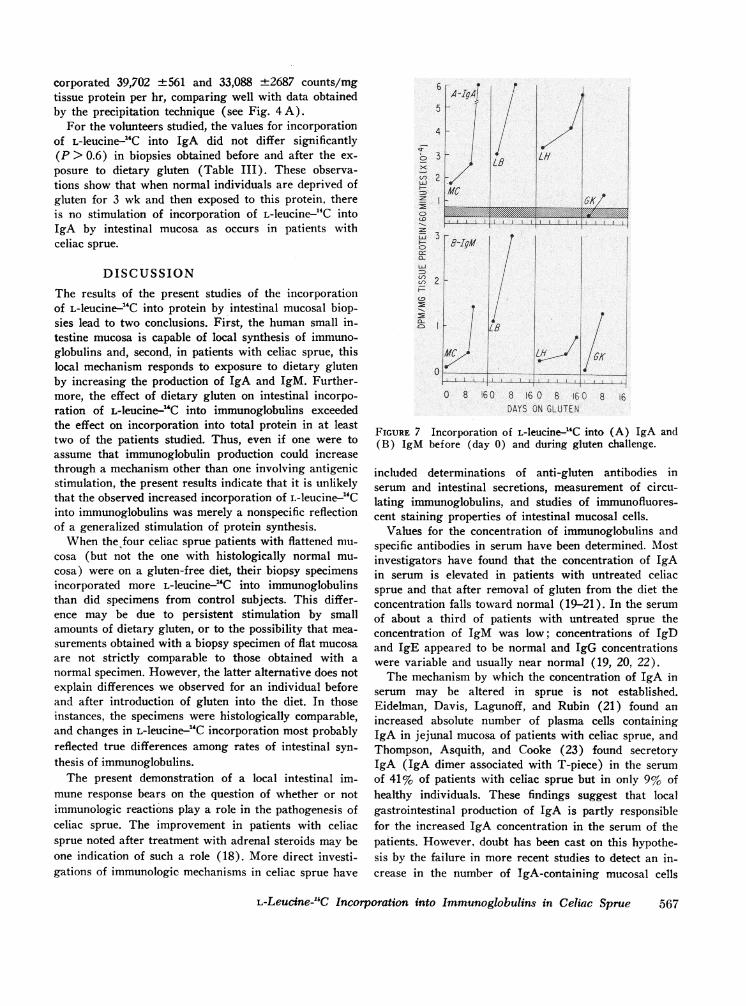
Incorporation of L-leucine-'4C into IgA. Intestinal in-corporation of L-leucine---4C into IgA increased signifi-cantly for all four patients after they were challengedwith gluten-containing diets (Fig. 7 A). Patient G. K.,whose value for incorporation into IgA was within the95% confidence limits of the control data before chal-lenge, had a 3-fold increase in incorporation (from 3912to 12,970 dpm/mg protein per hr) after challenge. Theother patients, whose values for incorporation into IgAwere well above the 95% confidence limits of the con-trol data while they were on a gluten-free diet, had a2- to 3-fold increase (P < 0.05) in the value for L-leu-cine-'4C incorporation into IgA after challenge with agluten-containing diet. For three of the four patients theincrease in incorporation of L-leucine-2"C into IgAoccurred before gastrointestinal absorptive changes weredetected.
Because the incorporation of L-leucine--"C into totalprotein increased in some of the patients with celiacsprue after gluten challenge, we determined whether thefractional change in incorporation into IgA was greaterthan the fractional change in incorporation into totalprotein. In the studies of G. K., incorporation into IgAincreased 3.3-fold, whereas incorporation into total pro-tein increased 1.6-fold after gluten challenge. Thus, thefractional change in incorporation into IgA was twice thefractional change in incorporation into total protein. Inthe studies of the other three patients, the fractional
increase in incorporation of L-leucine-14C into IgA ex-ceeded that of incorporation into total protein by factorsof 1.6, 1.5, and 1.1. In all, the fractional change in in-corporation into IgA was significantly greater than thechange in incorporation into total protein for two of thefour patients (P < 0.1).
Incorporation of L-leucine--"C into IgM. There wasinsufficient data to determine day-to-day variation in thestudies of incorporation of L-leucine-14C into IgM. Ifone assumes that, as in the case of IgA, such variationwas negligible, each of the patients studied showed a sig-nificant (P <0.05) increase in incorporation of L-leu-cine-14C into IgM (Fig. 7B).
Studies in normal volunteers maintained on agluten-free diet and then challenged withdietary glutenWe considered the possibility that the effect we ob-
served by gluten on intestinal immunoglobulin synthesisin patients with celiac sprue was a nonspecific phenome-non attributable to abrupt reexposure to gluten after aperiod of gluten deprivation. As a control investigation,we studied three normal volunteers who were fed gluten-free diets for 3 wk, subjected to intestinal biopsies, placedon normal gluten-containing diets, and then subjected tointestinal biopsies again after 7 days. During thesestudies the fecal excretion of fat by the subjects did notchange significantly.
In the study of the first volunteer the biopsy specimenswere assayed for IgA synthesis as described underMethods. In the studies of the remaining two volunteersa modified and improved assay procedure was used.2In this procedure the biopsy specimens were incubatedwith L-leucine--'4C as before, but the newly synthesized,labeled IgA was isolated by use of a complex comprisingspecific anti-IgA antibodies covalently coupled to cellu-lose. After the incubation with L-leucine--14C, the biopsyspecimen was homogenized and exposed to the anti-IgA-cellulose complex so that the 14C-labeled IgA was boundspecifically to the complex. The complex, together withthe bound IgA-14C, was isolated by filtration and counteddirectly on a filter paper disc. The value for nonspecificbinding in this procedure was obtained from the numberof counts bound after incubation of labeled homogenatewith anti-IgA-cellulose previously exposed to an ex-cess of unlabeled IgA. The range of values obtained byuse of this method for incorporation of L-leucine--4Cinto IgA by intestinal biopsy specimens from normalvolunteers, as well as from celiac sprue patients, agreesclosely with the range of values obtained by the proceduredescribed above under Methods. Thus, biopsy specimensfrom two celiac sprue patients on gluten-free diets in-
2 Falchuk, Z. M., W. Strober, and L. Laster. Unpublishedobservations.
566 P. M. Loeb, W. Strober, Z. M. Falchuk, and L. Laster
IUA
-.1II -14-
corporated 39,702 4-561 and 33,088 ±2687 counts/mgtissue protein per hr, comparing well with data obtainedby the precipitation technique (see Fig. 4A).
For the volunteers studied, the values for incorporationof L-leucine--14C into IgA did not differ significantly(P > 0.6) in biopsies obtained before and after the ex-posure to dietary gluten (Table III). These observa-tions show that when normal individuals are deprived ofgluten for 3 wk and then exposed to this protein, thereis no stimulation of incorporation of L-leucine--14C intoIgA by intestinal mucosa as occurs in patients withceliac sprue.
DISCUSSION
The results of the present studies of the incorporationof L-leucine--4C into protein by intestinal mucosal biop-sies lead to two conclusions. First, the human small in-testine mucosa is capable of local synthesis of immuno-globulins and, second, in patients with celiac sprue, thislocal mechanism responds to exposure to dietary glutenby increasing the production of IgA and IgM. Further-more, the effect of dietary gluten on intestinal incorpo-ration of L-leucine-14C into immunoglobulins exceededthe effect on incorporation into total protein in at leasttwo of the patients studied. Thus, even if one were toassume that immunoglobulin production could increasethrough a mechanism other than one involving antigenicstimulation, the present results indicate that it is unlikelythat the observed increased incorporation of L-leucine-14Cinto immunoglobulins was merely a nonspecific reflectionof a generalized stimulation of protein synthesis.
When the four celiac sprue patients with flattened mu-cosa (but not the one with histologically normal mu-cosa) were on a gluten-free diet, their biopsy specimensincorporated more L-leucine--4C into immunoglobulinsthan did specimens from control subjects. This differ-ence may be due to persistent stimulation by smallamounts of dietary gluten, or to the possibility that mea-surements obtained with a biopsy specimen of flat mucosaare not strictly comparable to those obtained with anormal specimen. However, the latter alternative does notexplain differences we observed for an individual beforeand after introduction of gluten into the diet. In thoseinstances, the specimens were histologically comparable,and changes in L-leucine--14C incorporation most probablyreflected true differences among rates of intestinal syn-thesis of immunoglobulins.
The present demonstration of a local intestinal im-mune response bears on the question of whether or notimmunologic reactions play a role in the pathogenesis ofceliac sprue. The improvement in patients with celiacsprue noted after treatment with adrenal steroids may beone indication of such a role (18). More direct investi-gations of immunologic mechanisms in celiac sprue have
U,
LAJ
z
CD
LO
6 r- A-v
5 -_
4 -M
3r
2-
MC
Fi 3r0 2
i I2
-- 1
p
ILB
/ILH
i6K/
0
O 8 16 0 8 16 0 8 16 C 8 6DAYS ON GLUTEN
FIGURE 7 Incorporation of L-leucine-4C into (A) IgA and(B) IgM before (day 0) and during gluten challenge.
included determinations of anti-gluten antibodies inserum and intestinal secretions, measurement of circu-lating immunoglobulins, and studies of immunofluores-cent staining properties of intestinal mucosal cells.
Values for the concentration of immunoglobulins andspecific antibodies in serum have been determined. Mostinvestigators have found that the concentration of IgAin serum is elevated in patients with untreated celiacsprue and that after removal of gluten from the diet theconcentration falls toward normal (19-21). In the serumof about a third of patients with untreated sprue theconcentration of IgM was low; concentrations of IgDand IgE appeared to be normal and IgG concentrationswere variable and usually near normal (19, 20, 22).
The mechanism by which the concentration of IgA inserum may be altered in sprue is not established.Eidelman, Davis, Lagunoff, and Rubin (21) found anincreased absolute number of plasma cells containingIgA in jejunal mucosa of patients with celiac sprue, andThompson, Asquith, and Cooke (23) found secretoryIgA (IgA dimer associated with T-piece) in the serumof 41% of patients with celiac sprue but in only 9% ofhealthy individuals. These findings suggest that localgastrointestinal production of IgA is partly responsiblefor the increased IgA concentration in the serum of thepatients. However, doubt has been cast on this hypothe-sis by the failure in more recent studies to detect an in-crease in the number of IgA-containing mucosal cells
L-Leucine-14C Incorporation into Immunoglobulins in Celiac Sprue
I IL
11
--I III
567

TABLE IIIIncorporation of L-Leucine-14C into IgA of Normal
Volunteers Pre- and Postgluten Challenge
Prechallenge Postchallenge
dpm/mg protein per hrD. L.* 5328 6925H. B.$ 5936 ±1212 7912 ±4836A. Y.: 3103 ±339 3064 ±t356
* Assayed by immune precipitation as described underMethods.t Assayed by anti-IgA-cellulose.
(23) and by the observation that the presence of secre-tory IgA in serum did not correlate with the concentra-tion of IgA in serum or with the activity of the disease.
The other abnormality in serum immunoglobulins, thereduced concentration of IgM in celiac sprue patients,is also not well understood. Despite the low concentra-tion of IgM in serum, and despite the detection of de-creased total body synthesis of IgM in this disease, as de-termined by turnover studies (24), the number of in-testinal mucosal plasma cells containing IgM was foundto be increased (20). The concentration of IgM in se-rum does not appear related to the severity of the dis-ease (19, 22) or to the presence of splenic atrophy (24,25).
Specific circulating antibodies to gluten and glutenfractions were found by several investigators in celiacsprue (26-29). However, their relation to the gastro-intestinal lesion in celiac sprue was questioned becausecirculating antibodies to other dietary proteins, such asmilk proteins, were also demonstrated. Furthermore,anti-gluten antibodies were found in other diseases inwhich mucosal integrity is disrupted, such as ulcerativecolitis. These findings suggest that circulating anti-bodies to gluten are attributable, at least in part, to ab-normal permeability of the intestinal mucosa in celiacsprue to various dietary proteins, and to subsequentstimulation of internal lymphoid tissue by such proteins.
Local gastrointestinal production of anti-gluten anti-bodies has been regarded as unlikely because of the fail-ure to detect gluten-binding antibodies in jejunal biopsyspecimens by use of immunofluorescence techniques (5).More recently, however, Katz, Kantor, and Herskovic(30) found antibodies to a trypsin-pepsin digest of glutenin the jejunal aspirates of a high percentage of patientswith untreated celiac sprue. The immunoglobulin classof these antibodies was not determined so that certaininferences regarding their origin and significance can-not be made. If the anti-gluten antibodies were in theIgA class, it would indicate that they originated in localintestinal- immune systems because very little IgA istransported from the systemic circulation into the ex-ternal secretions (31). However, if they were in the
IgG class, it would be possible that they were trans-ported from the serum, since about 50% of IgG immuno-globulins in external secretions are derived from theserum (31).
The present studies indicate that increased immuno-globulin synthesis does indeed occur locally in the gastro-intestinal tract after gluten challenge, and moreover, thatthe increase in synthesis takes place soon after the in-troduction of gluten into the diet, and generally beforefunctional gastrointestinal disturbances can be observed.These findings lend greater weight to the suppositionthat immune mechanisms are involved as one of the pri-mary factors in the pathophysiology of celiac sprue.Thus, the proximity of an immune mechanism to the siteof initial exposure to gluten makes it reasonable to pos-tulate that an immunological reaction plays a role in theinitiation of the intestinal lesion.
The additional immunoglobulins produced after glutenchallenge may be specific for gluten, for other exogenousproteins, or for proteins of the epithelium which gainaccess to antibody-producing cells through the disruptiveaction of gluten. Since Rubin, Fauci, Sleisenger, andJeffries (5) presented evidence that gluten binds to theepithelial cells of patients with celiac sprue but not thoseof normal individuals, it may well be that the excess im-munoglobulins are composed largely of anti-gluten anti-bodies. Such antibodies would be free to react with glu-ten on epithelial cell membranes and could thereby leadto immune destruction of the epithelial cells.
ACKNOWLEDGMENTSWewish to thank Dr. M. Hamilton for statistical analysisof the data. We also wish to thank Miss C. Roman fortechnical assistance.
REFERENCES1. Keller, R., J. E. Dwyer, W. Oh, and D. D'Amodio.
1969. Intestinal IgA neutralizing antibodies in newborninfants following poliovirus immunization. Pediatrics.43: 330.
2. Ogra, P. L., D. T. Karzon, F. Righthand, and M.MacGillivray. 1968. Immunoglobulin response in serumand secretions after immunization with live and inacti-vated poliovaccine and natural infection. N. Engl. J.Med. 279: 893.
3. Tomasi, T. B., Jr., and J. Bienenstock. 1968. Secretoryimmunoglobulins. Advan. Immunol. 9: 1.
4. Crabbe, P. A., and J. F. Heremans. 1966. The distribu-tion of immunoglobulin-containing cells along the humangastrointestinal tract. Gastroenterology. 51: 305.
5. Rubin, W., A. S. Fauci, M. H. Sleisenger, and G. H.Jeffries. 1965. Immunofluorescent studies in adult celiacdisease. J. Clin. Invest. 44: 475.
6. Brandborg, L. L., G. E. Rubin, and W. E. Quinton.1959. A multipurpose instrument for suction biopsy ofthe esophagus, stomach, small bowel, and colon. Gastro-enterology. 37: 1.
568 -P, M. Loeb, W. Strober, Z. M. Falchuk, and L. Laster

7. Van de Kamer, J. H., H. ten Bokkel Huinink, andH. A. Weyers. 1949. Rapid method for the determina-tion of fat in feces. J. Biol. Chem. 177: 347.
8. Fahey, J. L., and E. M. McKelvey. 1965. Quantitativedetermination of serum immunoglobulins in antibody-agar plates. J. Immunol. 94: 84.
9. Warshaw, A. L., L. Laster, and N. R. Shulman. 1967.Protein synthesis by human platelets. J. Biol. Chem.242: 2094.
10. Manchester, K. L., and F. G. Young. 1958. The effectof insulin on incorporation of amino acids into proteinof normal rat diaphragm in vitro. Biochem. J. 70: 353.
11. Umbreit, W. W., R. H. Burris, and J. F. Stauffer. 1957.Manometric Techniques; a Manuel Describing MethodsApplicable to the Study of Tissue Metabolism. BurgessPublishing Company, Minneapolis. 3rd edition. 149.
12. Swenson, R. M., and M. Kern. 1968. The synthesis andsecretion of -y-globulin by lymph node cells. III. Theslow acquisition of the carbohydrate moiety of 'y-globu-lin and its relationship to secretion. Proc. Nat. Acad.Sci. U. S. A. 59: 546.
13. Lowry, 0. H., N. J. Rosebrough, A. L. Farr, and R. J.Randall. 1951. Protein measurement with the folin phenolreagent. J. Biol. Chem. 193: 265.
14. Bruno, G. A., and J. E. Christian. 1961. Correction forquenching associated with liquid scintillation counting.Anal. Chem. 33: 650.
15. Zilversmit, D. B., C. Entenman, and M. C. Fishler. 1943.On the calculation of 'turnover time' and 'turnover rate'from experiments involving the use of labeling agents.J. Gen. Physiol. 26: 325.
16. Warshaw, A. L., and L. Laster. 1968. Protein synthesisby mucosa of the human small intestine in vitro: effectof disease and an inhibitory effect of gliadin in gluten-sensitive enteropathy. J. Clin. Invest. 47: 100a. (Abstr.)
17. Creamer, B. 1962. Dynamics of the mucosa of the smallintestine in idiopathic steatorrhea. Gut. 3: 295.
18. Lepore, M. J. 1958. Long-term or maintenance adrenalsteroid therapy in non-tropical sprue. Amer. J. Med.25: 381.
19. Asquith, P., R. A. Thompson, and W. T. Cooke. 1969.Serum-immunoglobulins in adult coeliac disease. Lancet.2:129.
20. Hobbs, J. R., G. W. Hepner, A. P. Douglas, P. A.Crabbe, and S. G. 0. Johansson. 1969. Immunologicalmystery of coeliac disease. Lancet. 2: 649.
21. Eidelman, S., S. D. Davis, D. Lagunoff, and C. E. Rubin.1966. The relationship between intestinal plasma cellsand serum immunoglobulin A (IgA) in man. J. Clin.Invest. 45: 1003.
22. Hobbs, J. R., and G. W. Hepner. 1968. Deficiency of'yM-globulin in coeliac disease. Lancet. 1: 217.
23. Thompson, R. A., P. Asquith, and W. T. Cooke, 1969.Secretary IgA in the serum. Lancet. 2: 517.
24. Brown, D. L., A. G. Cooper, and G. W. Hepner. 1969.IgM Metabolism in coeliac disease. Lancet. 1: 858.
25. McCarthy, C. F., I. D. Fraser, K. T. Evans, and A. E.Read. 1966. Lymphoreticular dysfunction in idiopathicsteatorrhea. Gut. 7:140.
26. Heiner, D. C., M. E. Lahey, J. F. Wilson, J. W. Ger-rard, H. Shwachman, and K-T. Khaw. 1962. Precipitinsto antigens of wheat and cow's milk in celiac disease.J. Pediat. 61: 813.
27. Taylor, K. B., S. C. Truelove, and R. Wright. 1964.Serologic reactions to gluten and cow's milk proteins ingastrointestinal disease. Gastroenterology. 46: 99.
28. Alarcon-Segovia, D., T. Herskovic, K. G. Wakim, P.A. Green, and H. H. Scudamore. 1964. Presence of circu-lating antibodies to gluten and milk fractions in patientswith nontropical sprue. Amer. J. Med. 36: 485.
29. Berger, E., and E. Freudenberg. 1961. Bemerkungenuiber die antigen Eigenschaften von Abbaustufen desGliadins. Ann. Paediat. 196: 238.
30. Katz, J., F. S. Kantor, and T. Herskovic. 1968. Intestinalantibodies to wheat fractions in celiac disease. Ann.Intern. Med. 69: 1149.
31. Strober, W., R. M. Blaese, and T. A. Waldmann. 1970.The origin of salivary IgA. J. Lab. Clin. Med. 75: 856.
L-Leucine-14C Incorporation into Immunoglobulins in Celiac Sprue 569