increased nmda-induced excitability during ethanol withdrawal: a behavioural and histological study
TRANSCRIPT
BRAIN RESEARCH
E L S E V I E R Brain Research 674 (1995) 91-96
Research report
Increased NMDA-induced excitability during ethanol withdrawal: a behavioural and histological study
Mark Davidson *, Brian Shanley, Peter Wilce Alcohol Research Unit, Department of Biochemistry, University of Queensland, Queensland 4072, Australia
Accepted 6 December 1994
Abstract
Intrahippocampal injections of N-methyl-D-aspartic acid (NMDA) leads to neurodegeneration in a dose-dependent manner. Chronic administration of ethanol to animals leads to CNS tolerance and dependence. Hyperexcitability following ethanol withdrawal is thought to be related to increased sensitivity of the NMDA receptors. The purpose of this study was to investigate this predisposition to hyperexcitability by intrahippocampal injection of low doses of NMDA. Using control and ethanol-withdrawn male Wistar rats, behavioural indices were determined immediately after injection and morphological damage was assessed after a period of recovery. There was significantly increased hyperactivity in the ethanol-treated rats immediately after injection. Morphological damage resulting from 5 nmol of NMDA was significantly greater in the CA3 region of the hippocampus in these animals. These data support the hypothesis that ethanol dependence and subsequent withdrawal is associated with increased sensitivity to NMDA which may underlie ethanol withdrawal-associated brain damage.
Keywords: N-Methyl-D-aspartate; Ethanol; Rat hippocampus; Alcohol withdrawal; Excitotoxicity
I. Introduct ion
The neurobiological changes underlying the abnor- mal behaviour observed in ethanol dependence and withdrawal appear to involve several t ransmit ter recep- tor systems in the brain. It is now well established that acute ethanol exposure inhibits the excitatory action of glutamate at the N-methyl-o-aspar ta te ( N M D A ) and kainate types of glutamate receptor, and enhances 7-aminobutyrate (GABA) action at the inhibitory GABAA/benzod iazep ine receptor complex. A working model of the long-term actions of ethanol is based on adaptive changes that upregulate N M D A receptor function and reduce G A B A A receptor function [29]. These changes offset the acute action of ethanol in the central nervous system but result in neuronal hyperex- citability and the characteristic withdrawal syndrome when the inhibitory effect of ethanol is removed. The action of ethanol on the N M D A receptor is of particu-
* Corresponding author. Fax: (61) (07) 365-4699.
0006-8993/95/$09.50 © 1995 Elsevier Science B.V. All rights reserved SSDI 0006-8993(94)01440-X
lar interest, since the influx of calcium through the receptor ion-channel during withdrawal-induced hyper- activity may not only initiate withdrawal-associated changes in gene expression but also result in excito- toxic cell loss.
There is considerable experimental support for this model. Acute ethanol exposure inhibits a number of NMDA-recep tor mediated events in cultured cells in- cluding NMDA-responsive currents [20,34], NMDA- mediated Ca 2+ influx, generation of cGMP [13] and increases in [Ca2+]i [7]. In tissue preparations, ethanol inhibits NMDA-st imulated [3H]noradrenaline release in rat brain cortex [8], [3H]norepinephrine release in rat cortical [9] and hippocampal slices [35] and endoge- nous dopamine release from striatal slices [36]. In vivo, chronic ethanol t rea tment of both rats and mice results in increased binding of the non-competit ive N M D A receptor antagonist, MK801, to hippocampal mem- branes [11,12,30,31,33] and a 65% increase in immuno- reactivity of the major subunit of the receptor, NM- DAR1 [32]. When N M D A receptor function is blocked pharmacologically, ethanol withdrawal-associated be- haviour is inhibited [5,11,19,26].

92 M. Dacidson et al. / Brain Research 674 (1995) 91-96
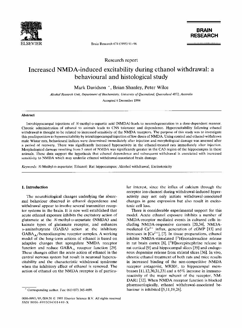
Fig. 1. Effect of increasing doses of NMDA on morphological damage in the rat hippocampus. NMDA (1,5,10 and 20 nmol) was injected into the left hippocampus of halothane anaesthetised control rats. After a 7-day recovery period, animals were perfused with paraformaldehyde. Brains were removed, cryoprotected, embedded into OCT and 50/zm sections cut from the area surrounding the injection site. A = 1 nmol NMDA; B = 5 nmol NMDA; C = 10 nmol NMDA; D = 20 nmol NMDA. Arrow heads show needle tracts whereas arrows indicate areas of damage. Bar = 200 tzm.
T h e h y p e r a c t i v i t y o f t h e r e c e p t o r d u r i n g e t h a n o l 20
w i t h d r a w a l h a s b e e n d i r e c t l y r e l a t e d to a n i n c r e a s e d w a:
s u s c e p t i b i l i t y to N M D A - i n d u c e d b e h a v i o u r a n d exc i to - 0 ¢3
toxic d a m a g e . S a n n a e t al. ( 1993) [30] i n j e c t e d ra t s , tn
u n d e r g o i n g l a t e w i t h d r a w a l ( 1 2 - 2 4 h) , w i t h N M D A
(i .c.v.) a n d f o u n d s i g n i f i c a n t p o t e n t i a t i o n t o g e t h e r w i t h ,,-10
r e d u c e d s e i z u r e t h r e s h o l d , a n d r e d u c e d l a t e n c y o f c o n - o m
v u l s i o n s . W e a l so f o u n d i n c r e a s e d n e u r o t o x i c i t y a f t e r >
i n j e c t i o n o f N M D A i n t o t h e h i p p o c a m p u s o f l a t e w i t h - = tL I
d r a w n r a t s [6]. T h e a i m o f t h i s s t u d y w a s to f u r t h e r m
c h a r a c t e r i s e t h i s in v ivo s y s t e m by a s s e s s i n g b e -
h a v i o u r a l c h a n g e s a n d h i s t o l o g i c a l l y e x a m i n i n g exc i to -
tox ic d a m a g e , r e s u l t i n g f r o m i n t r a h i p p o c a m p a l in jec -
t i o n s o f m i n i m a l d o s e s o f N M D A in c o n t r o l s a n d
a n i m a l s u n d e r g o i n g e t h a n o l - w i t h d r a w a l .
2. M a t e r i a l s a n d m e t h o d s
2.1. Chronic e thanol t rea tment
A d u l t m a l e W i s t a r r a t s ( 2 1 0 - 2 2 0 g; n = 38) w e r e
o b t a i n e d f r o m t h e C e n t r a l A n i m a l B r e e d i n g H o u s e ,
T
I I I I
5 10 20 30
NMDA (nmoles)
• C O N T R O L - - I - - E T H A N O L
Fig. 2. Behavioural analysis of NMDA-injected control and ethanol- withdrawn rats. After NMDA injection animals were carefully ob- served for 1 h for any behavioural changes. All animals recieved an accumulated behavioural score as descibed in section 2. Statistical analysis of behavioural changes was carried out by two-way analysis of variance. The results were significantly different for treatment (F1,68 = 41.6, P < 0.001) and dose (F4,68 = 13.2, P < 0.001). A post hoc Bonferroni t-test showed significance at the 5 nmol dose (P < 0.05). n = 4-13.

M. Davidson et al. / Brain Research 674 (1995) 91-96 93
University of Queensland. Chronic ethanol t reatment was achieved using the inhalation method described previously [6]. Rats were housed in a perspex chamber and given free access to water and a commercial ro- dent diet (18% protein) while being exposed to an increasing concentration of ethanol vapour (range 10- 25 m g / m i n / l air) in an air flow of 10 l /min . A blood alcohol level of 2 m g / m l or greater was maintained for at least 5 days, before ethanol was withdrawn. Control animals were pair-fed throughout the experiment.
2.2. Leisoning procedure and histological assessment
As ethanol is well known to inhibit N M D A receptor function, injection at early times when animals have a significant blood alcohol level, was not considered. The behavioural and epileptiform manifestations of with- drawal are maximal at 8 -12 h after removal of ethanol. We have already shown that injection during early withdrawal leads to significant mortality [6]. Further, Sanna et al. [30] showed that MK-801 binding was maximal at 9 -24 h after withdrawal. Based on our earlier work and of that of Sanna et al. we chose 24 h as the time point for our experiments. Rats were anaesthetised with 1.5% halothane in 0 2 and placed in a Kopf stereotaxic frame. Injections were made into the left hippocampus through a burr hole at the coor-
dinates: - 3 . 8 mm bregma, 2.6 m m lateral and 3.6 mm ventral, [27] using a 5 /x l Hamil ton syringe. N M D A in phosphate-buffered saline (PBS) neutralised to pH 7.0 with 0.1 M NaOH, was injected in a volume of 1 /zl over a 1 min period. The needle was left in place for a further 1 min before being slowly withdrawn and the scalp apposed with sutures. Animal behavioural pat- terns were monitored for lh after surgery. Seven days after injection, animals were perfused through the left cardiac ventricle with 100 ml of PBS, followed by 100 ml of 4% (w/v) paraformaldehyde in PBS. Isolated brains were left in the same solution overnight before being cryoprotected in 30% (w/v) sucrose in PBS, and embedded into OCT compound (Tissue-Tek, USA). Coronal sections (50 /zm) around the injection site were cut on an IEC cryostat and stored in 0.1% (w/v) sodium azide in PBS at 4°C. Sections were mounted on poly-L-lysine coated slides and stained with thionin. Stained sections were cleared in xylene, coverslipped under DPX prior to examination and photographed with a Nikon microphot-FXA.
2.3. Behavioural analysis
Control animals and animals during late withdrawal were injected with N M D A (1-30 nmol) as described above, and observed carefully for behavioural changes
CONTROL - 5 nmoles CONTROL - 5 nmoles
ETHANOL - 5 nmoles ETHANOL - 5 nmoles WITHDRAWAL WITHDRAWAL
Fig. 3. Representative photomicrographs of control and ethanol-treated animals injected with 5 nmol of NMDA.

94 M. Da vidson et al. / Brain Research 674 (1995) 91-96
over the following hour. Behavioural scoring was modi- fied from the scales used by Matsumoto et al. [24] and Danysz et al. [5]. All animals received a cumulative behavioural score based on the following criteria: 0 = no change; 1 = hunched body, isolated wet dog shakes (WDS), piloerection, rotation on the spot, jaw chatter- ing, tremors, hypoactivity, extensive grooming, tail stiff- ening, isolated jumps, slow rotation around cage; 2 = general hyperactivity, rapid rotation around cage, se- ries of WDS, vocalisation, minor escape reaction; 3 = barrel rolls, wild running, rearing and falling, moderate escape reaction; 4 = tonic-clonic seizures, strong es- cape reaction; 5 = death. Animals received a score if anyone of these behaviours was detected. Statistical analysis of behavioral changes was carried out by two- way analysis of variance for dose and ethanol effects and by one-way analysis of variance, followed by a post hoc Bonferroni t-test for multiple comparisons. This was performed using SigmaStat software (Jandel Scien- tific, CA, USA).
2.4. Morphological analysis
Damage to the hippocampi was assessed morpho- metrically using a 1 × objective on a Nikon microscope and JAVA image analysis software (Jandel Scientific, CA, USA). A video image of the hippocamus was displayed and the length (in mm) of the C A 1/CA2 , CA3 and dentate gryrus regions was measured. The damaged region was measured and expressed as a % of the total distance for the particular region. The degree of damage was assessed in 10-20 Nissl-stained sections around the injection site. Sections with the greatest percentage damage, usually closely associated with the injection site, were utilized for comparisons between groups and then subjected to Student 's t-test.
3. Results
The results of initial experiments to examine dose- dependent induction of behaviours and neurotoxicity by N M D A in the hippocampus of control animals are shown in Figs. 1 and 2. The amount of damage result- ing from 1 nmole N M D A (Fig. 1A) was not different from that produced by an injection of vehicle. Injection of 5, 10 and 20 nmoles of N M D A resulted in signifi- cant damage (Fig. 1B,C,D). The amount of damage in the hippocampus of the control animals correlated with the intensity of NMDA-induced behaviours which in- creased dose-dependently. Considering these two ob- servations, it was decided to compare the behaviours induced in control and withdrawn animals by a range of doses of NMDA, and to assess excitotoxic damage in the two groups using a dose of 5 nmol. Behavioural analysis (Fig. 2) shows a significant enhancement of
IJ.I o <
._1 <
X
125
100
75 -
5 0
25
8 C A I / C A 2 CA3
5 n m e l e s N M D A
CONTROL / ETHANOL
Fig. 4. Maximal damage after injection of 5 nmol of NMDA into the hippocampus of control and ethanol-withdrawn rats. Analysis of distance of damage in the CA1 +CA2, CA3 and dentate gyrus regions was undertaken. The CA1 + CA2 region of the hippocampus was damaged in most animals but to a lesser extent than that in the CA3 region. The data in the CA1 + CA2 region showed a trend for increased sensitivity although this was not significant (P > 0.13). The CA3 region was however significant ( n - 5 , * P < 0.05, Student's t-test). The 5 nmol dose showed no consistent damage of the dentate gyrus in either the control or ethanol-treated animals.
behaviours in the animals undergoing ethanol with- drawal (FI,68 = 41.6, P < 0.001) and a dose-related ef- fect (F4,68= 13.2, P<0 .001) . Interestingly, after 10 nmol of NMDA, 3 out of 10 animals in the ethanol- treated group had a marked seizure, whereas control animals exhibited a seizure response only at a dose of 20 nmol. At the latter dose there was a significant mortality in the ethanol withdrawn group. Fig. 3 shows representative photomicrographs of hippocampi from control and ethanol-treated animals injected with 5 nmoles of NMDA. Quantitative data from all animals is displayed in Fig. 4. At a dose of 5 nmol, the CA3 region of the hippocampus had increased damage com- pared to the CA1 + CA2 region in both control and ethanol-withdrawn animals. Since the injection needle was directed at this area, the apparent succeptability of the CA3 may in fact be artifactual. Minimal damage was seen in the dentate gyrus (Fig. 3). Damage in the CA3 region was significant increased in the animals that had been withdrawn from ethanol ( P < 0.05, Stu- dent 's t-test; Figs. 3 and 4). There was also a trend towards increased sensitivity of the CA1 + CA2 region in withdrawn animals but the difference was not statis- tically significant.

M. Davidson et al. / Brain Research 674 (1995) 91-96 95
4. Discussion
Receptor binding studies using NMDA antagonists have demonstrated a high density of NMDA receptors in the hippocampus [17,25]. Biochemical, behavioural and binding studies have shown these receptors to have an important role in ethanol withdrawal hyperexcitabil- ity [11,12,30,31]. Ethanol withdrawal-associated seizures are inhibited by administration of NMDA antagonists [10,11,19,26,33]. Our studies show a clear dose-related increase in behavioural modification following in- trahippocampal NMDA administration which is poten- tiated after ethanol withdrawal. Danysz et al. [5] using behavioural analysis based on escape reactions and seizure activity, failed to show a proconvulsant effect, following injection of 2 nmol NMDA (i.c.v.) into rats 14 h into the withdrawal period. Sanna et al. [30] however, showed a dose-dependent increase in sensitivity to NMDA-induced seizures, 24 h into the withdrawal period. In our study we used a cumulative hyperactivity score to accommodate the many behavioural changes induced by NMDA and to reflect the overall hyperex- citability in the animals. We were able to demonstrate a significant enhancement of NMDA-induced be- havioural changes during withdrawal, supporting the concept of increased NMDA receptor function result- ing from chronic ethanol treatment.
Ethanol given as a single acute dose is neuroprotec- tive due to its inhibitory actions at the NMDA receptor and potentiation of the GABA a receptor. This effect has been reported both in cell culture systems [4,21] and in vivo using susceptibility to seizures induced after NMDA injection [5] as an assay. Chronic treat- ment of cultured cells with ethanol, however, results in an increased susceptibility to NMDA measured by several parameters [1,3,15,16]. This predisposition to excitotoxic damage after chronic ethanol t reatment is thought to result from an up-regulation of NMDA receptor function. We have previously shown increased toxicity in vivo after NMDA injection into the hip- pocampus [6] of rats after chronic ethanol treatment. Studies with MK801 binding [11,12,30,31,33] and gluta- mate binding [31] have demonstrated increased recep- tors in the hippocampus after chronic ethanol treat- ment, while Trevisan et al. [32] recently showed a 65% increase in NMDAR1 immunoreactivity. Whether this upregulation of receptor density corresponds to an increase in receptor function remains to be answered, but the results presented here would certainly support this concept.
Subgroups of human alcoholics have a susceptibility to brain damage and cognitive deficits. In a recent report Hunt [14] stressed that brain damage may not be due to chronic ethanol treatment per se, but rather to the ethanol withdrawal syndrome. Neuronal death in mamillary bodies and the thalamus of alcoholics has
been related to thiamin deficiency but damage to hip- pocampal and cerebral cortical neurons is thought to reflect withdrawal episodes. Repeated withdrawal has a cumulative effect and creates more severe future withdrawal reactions, possibly by a kindling phe- nomenon [2]. Interestingly, the CA3 region has been shown to be particularly sensitive to NMDA-induced excitotoxicity after kindling [22]. This region also showed a high level of NMDA-induced damage after withdrawal which may indicate increased excititoxic activity in this area.
What remains unanswered is the mechanism under- lying the increase in sensitivity after ethanol treatment. Obviously the density of NMDA receptors may be critical but other factors may also be important. Changes in sensitivity may reflect changes in subunit composition of the NMDA receptor complex. Cer- tainly, variation in NMDA receptor subunit composi- tion is correlated with differential sensitivity to ethanol [18,23] and electrogenic kindling results in changes in NMDA receptor subunit gene expression, especially in the hippocampus [28]. Thus alteration in subunit com- position may prove to be an important consequence of ethanol withdrawal and may underlie the kindling phe- nomenon.
In summary, our results show correlation between neuronal damage induced by exogenously applied NMDA in the hippocampus of control and ethanol- treated rats and characteristic behaviours. Further, we have demonstrated increased sensitivity of hippocam- pal regions, particularly the CA3 region, during ethanol withdrawal. This may reflect an upregulation of NMDA receptor function. The enhanced NMDA channel func- tion and subsequent susceptibility to excitotoxic dam- age during withdrawal represents a mechanism for ethanol-induced neuronal cell death leading to re- duced neuronal density, and brain size together with cognitive and motor deficits in the human alcoholic.
Acknowledgements
This work was supported by the National Health and Medical Research Council of Australia in the form of a project grant.
References
[1] Ahern, K.B., Lustig, H.S. and Greenberg, D.A., Enhancement of NMDA toxicity and calcium responses by chronic exposure of cultured cortical neurons to ethanol, Neurosci. Lett., 165 (1994) 211-214.
[2] Ballenger, J.C. and Post, R.M., Kindling as a model for alcohol withdrawal syndromes, Br. J. Psychiatry, 133 (1978) 1-14.
[3] Chandler, L.J., Newsom, H., Sumners, C. and Crews, F., Chronic ethanol exposure potentiates NMDA excitotoxicity in cerebral cortical neurons, J. Neurochem., 60 (1993) 1578-1581.

96 M. Davidson et al. /Brain Research 674 (1995) 91-96
[4] Chandler, L.J., Sumners, C. and Crews, F.T., Ethanol inhibits NMDA receptor-mediated excitotoxicity in rat primary neuronal cultures, Alcohol. Clin. Exp. Res., 17 (1993) 54-60.
[5] Danysz, W., Dyr, W., Janowska, E., Glazewski, S. and Kos- towski, W., The involvement of NMDA receptors in acute and chronic effects of ethanol, Alcohol. Clin. Exp. Res., 16 (1992) 499-504.
[6] Davidson, M., Wilce, P. and Shanley, B.C., Increased sensitivity of the hippocampus in ethanol-dependent rats to toxic effect of N-methyl-D-aspartic acid in vivo, Brain Res., 606 (1993) 5-9.
[7] Dildy, J.E. and Leslie, S.W., Ethanol inhibits NMDA-induced increases in intracellular Ca 2÷ in dissociated brain cells, Brain Res. 499 (1989) 383-387.
[8] Fink, K. and Gothert, M., Inhibition of N-methyl-D-aspartic-in- duced noradrenaline release by alcohols is related to their hydrophobicity, Eur. J. Pharrnacol., 191 (1990) 225-229.
[9] Gonzales, R.A. and Woodwood, J.J., Ethanol inhibits N-methyl- D-aspartate stimulated [3H]norepinephrine release from rat cor- tical slices, J. Pharmacol. Exp. Ther., 253 (1990) 1138-1144.
[10] Grant, K.A., Snell, L.D., Rogawski, M.A., Thurkauf, A. and Tabakoff, B., Comparison of the effects of the uncompetitive N-methyl-D-aspartate antagonist ( _+ )-5-aminocarbonyl-10,11-di- hydro-5H-dibenzo[a,d]cyclohepten-5,10-imine (ADCL) with its structural analogs dizocilpine (MK-801) and carbamazepine on ethanol withdrawal seizures, J. Pharmacol. Exp. Ther., 260 (1992) 1017-1022.
[11] Grant, K.A., Valverius, P., Hudspith, M. and Tabakoff, B., Ethanol withdrawal seizures and the NMDA receptor complex, Eur. J. Pharmacol. 176 (1990) 289-296.
[12] Gulya, K., Grant, K.A., Valverius, P., Hoffman, P.L. and Tabakoff, B., Brain regional specificity and time course of changes in the NMDA receptor-ionophore complex during ethanol withdrawal, Brain Res., 547 (1991) 129-134.
[13] Hoffman, P.L., Rabe, C.S., Moses, F. and Tabakoff, B., N- Methyl-D-aspartate receptors and ethanol: inhibition of calcium flux and cyclic GMP production, J. Neurochem., 52 (1989) 1937-1940.
[14] Hunt, W.A., Are binge drinkers more at risk of developing brain damage? Alcohol, 10 (1993) 559-561.
[15] Iorio, K.R., Reinlib, L., Tabakoff, B. and Hoffman P.L., Chronic exposure of cerebellar granule cells to ethanol results in in- creased N-methyl-D-aspartate receptor function, Mol. Pharma- col. 41 (1992) 1142-1148.
[16] lorio, K.R., Tabakoff, B. and Hoffman, P.L., Glutamate-in- duced neurotoxicity is increased in cerebellar granule cells ex- posed chronically to ethanol, Eur. J. Pharmacol. 248 (1993) 209-212.
[17] Jarvis, M.F., Murphy, D.E. and Williams, M., Quantitative auto- radiography of NMDA receptors in rat brain using [3H]CPP: comparison with [3H]TCP binding sites, Eur. J. Pharmacol. 141 (1987) 149-152.
[18] Koltchine, V., Anantharam, V., Wilson, A., Bayley, H. and Treistman, S.N., Homomeric assemblies of NMDAR1 splice variants are sensitive to ethanol, Neurosci. Lett., 152 (1993) 13-16.
[19] Liljequist, S., The competitive NMDA receptor antagonist, CGP 39551, inhibits ethanol withdrawal seizures, Eur. J. Pharmacol., 192 (1991) 197-198.
[20] Lovinger, D.M., White, G. and Weight, F.F. Ethanol inhibits
NMDA-activated ion current in hippocampal neurons, Science, 243 (1989) 1721-1724.
[21] Lustig, H.S., Chan, J. and Greenberg, D.A., Ethanol inhibits excitotoxicity in cerebral cortical cultures, Neurosci. Lett., 135 (1992) 259-261.
[22] Martin, D., McNamara, J.O. and Nadler, J.V., Kindling en- hances sensitivity of CA3 hippocampal pyramidal cells to NMDA, J. Neurosci., 12 (1992) 1928-1935.
[23] Masood, K., Wu, C., Brauneis, U. and Weight, F.F., Differential ethanol sensitivity of recombinant N-methyl-D-aspartate recep- tor subunits, Mol. Pharmacol., 45 (1994) 324-329.
[24] Matsumoto, I., Davidson, M. and Wilce, P.A., Polyamine-en- hanced NMDA receptor activity: effect of ethanol, Eur. J. Pharmacol., 247 (1993) 289-294.
[25] Monaghan, D.T. and Cotman, C.W., Distribution of N-methyl- D-aspartate-sensitive L-[3H]glutamate binding sites in rat brain, J. Neurosci., 5 (1985) 2909-2919.
[26] Morrisett, R.A., Rezvani, A.H., Overstreet, D., Janowsky, D.S., Wilson, W.A. and Swartzwelder, H.S., MK-801 potently inhibits alcohol withdrawal seizures in rats, Eur. J. Pharmacol,, 176 (1990) 103-105.
[27] Paxinos, G. and Watson, C., The Rat Brain in Stereotaxic Coordi- nates, 2nd edn., Academic Press, Sydney, 1986.
[28] Pratt, G.D., Kokala, M., Bengzon, J., Kokala, Z, Fritschy, J. -M., Mohler, H. and Lindvall, O., Differential regulation of N- methyl-D-aspartate receptor subunit messenger RNAs in kin- dling-induced epileptogenesis, Neurosc&nce, 57 (1993) 307-318.
[29] Samson H.H. and Harris, R.A., Neurobiology of alcohol abuse, Trends PHarmacol. Sci., 13 (1992) 206-211.
[30] Sanna, E., Serra, M., Cossu, A., Colombo, G., Follesa, P., Cuccheddu, T., Concas, A. and Biggio, G., Chronic ethanol intoxication induces differential effects on GABA and NMDA receptor function in the rat brain, Alcohol. Clin. Exp. Res., 17 (1993) 115-123.
[31] Snell, L.D., Tabakoff, B. and Hoffman, P.L., Radioligand bind- ing to the N-methyl-D-aspartate receptor/ionophore complex: alterations by ethanol in vitro and by chronic in vivo ethanol ingestion, Brain Res., 602 (1993) 91-98.
[32] Trevisan, L., Fitzgerald, L.W., Brose, N., Gasic, G.P., Heine- mann, S.F., Duman, R.S. and Nestler, E.J., Chronic ingestion of ethanol up-regulates NMDAR1 receptor subunit immuno- reactivity in rat hippocampos, J. Neurochem., 62 (1994) 1635- 1638.
[33] Valverius, P. Crabbe, J.C., Hoffman, P.L. and Tabakoff, B., NMDA receptors in mice bred to be prone or resistant to ethanol withdrawal seizures, Eur. J. Pharmacol. 184 (1990) 185- 189.
[34] White, G., Lovinger, D.M. and Weight, F.F., Ethanol inhibits NMDA-activated current but does not alter GABA-activated current in an isolated adult mammalian neuron, Brain Res., 507 (1990) 332-336.
[35] Woodward, J.J., A comparison of the effects of ethanol and the competitive glycine antagonist 7-chlorokynurenic acid on N- methyl-o-aspartic acid-induced neurotransmitter release from rat hippocampal slices, J. Neurochem., 62 (1994) 987-991.
[36] Woodward, J.J. and Gonzales, R.A., Ethanol inhibition of N- methyl-D-aspartate stimulated endogenous dopamine release from rat striatal slices: reversal by glycine, Z Neurochem., 54 (1990) 712-715.