increased serotonin transporter expression reduces fear
TRANSCRIPT

Increased Serotonin Transporter Expression Reduces Fear and
Recruitment of Parvalbumin Interneurons of the Amygdala
Marco Bocchio1, Giulia Fucsina1, Lydia Oikonomidis1,2,3,4, Stephen B McHugh3, David M Bannerman3,Trevor Sharp2 and Marco Capogna*,1
1MRC Brain Network Dynamics Unit, Department of Pharmacology, University of Oxford, Oxford, UK; 2Department of Pharmacology, University ofOxford, Oxford, UK; 3Department of Experimental Psychology, University of Oxford, Oxford, UK
Genetic association studies suggest that variations in the 5-hydroxytryptamine (5-HT; serotonin) transporter (5-HTT) gene are associatedwith susceptibility to psychiatric disorders such as anxiety or posttraumatic stress disorder. Individuals carrying high 5-HTT-expressing genevariants display low amygdala reactivity to fearful stimuli. Mice overexpressing the 5-HTT (5-HTTOE), an animal model of this humanvariation, show impaired fear, together with reduced fear-evoked theta oscillations in the basolateral amygdala (BLA). However, it is unclearhow variation in 5-HTT gene expression impacts on the microcircuitry of the BLA to change behavior. We addressed this issue byinvestigating the activity of parvalbumin (PV)-expressing interneurons (PVINs), the biggest IN population in the basal amygdala (BA). Wefound that increased 5-HTT expression impairs the recruitment of PVINs (measured by their c-Fos immunoreactivity) during fear.Ex vivo patch-clamp recordings demonstrated that the depolarizing effect of 5-HT on PVINs was mediated by 5-HT2A receptor.In 5-HTTOE mice, 5-HT-evoked depolarization of PVINs and synaptic inhibition of principal cells, which provide the major output of theBA, were impaired. This deficit was because of reduced 5-HT2A function and not because of increased 5-HT uptake. Collectively, thesefindings provide novel cellular mechanisms that are likely to contribute to differences in emotional behaviors linked with genetic variationsof the 5-HTT.Neuropsychopharmacology (2015) 40, 3015–3026; doi:10.1038/npp.2015.157; published online 8 July 2015
���������������������������������������������
INTRODUCTION
Linking genes and neural circuits to understand individualdifferences in emotional behavior and, ultimately, suscepti-bility to psychiatric disorders are a growing challengein biological psychiatry. The 5-hydroxytryptamine transpor-ter (5-HTT) is the key controller of the clearance of5-hydroxytryptamine (5-HT; serotonin) from the synapticcleft (Blakely et al, 1994), and represents the primary targetfor the pharmacological treatment of mood and anxietydisorders (Ballenger, 1999). The large natural variation in 5-HTT expression in the human population, up to sevenfoldbetween individuals (Lundberg et al, 2007), may in part begenetically driven (Hu et al, 2006; Lesch et al, 1996; Murphyet al, 2013). Several common variants of the human 5-HTTgene putatively determine 5-HTT expression levels, and havebeen linked to alterations in emotionality. Of particularinterest is a 44 bp insertion/deletion mutation in the 5-HTTgene promoter region (known as the 5-HTT gene linked to
the polymorphic region or 5-HTTLPR), which gives rise tolong (l) and short (s) alleles. Specifically, many (but not all)studies associate a low 5-HTT-expressing polymorphism(s/s) with greater risk for anxiety-related traits (Lesch et al,1996) and posttraumatic stress disorder (Lee et al, 2005),whereas a high 5-HTT-expressing variation (l/l) associateswith reduced anxiety and depression susceptibility (Leschet al, 1996), especially when combined with environmentalfactors (Caspi et al, 2003; Uher and McGuffin, 2010).Importantly, variations of the 5-HTT gene have been
associated with altered neural pathways linked to fear: forinstance, l/l carriers demonstrate reduced activation of theamygdala in response to fearful faces (Hariri et al, 2002),together with altered functional connectivity betweenamygdala and medial prefrontal cortex (mPFC) (Canliet al, 2005; Pezawas et al, 2005), although not all studiesare consistent with these findings (Murphy et al, 2013).While human 5-HTT gene association studies are compli-
cated by multiple environmental and demographic factors,transgenic mouse models allow for a more controlledanalysis of the impact of 5-HTT gene variation on behaviorand neural function. The 5-HTT knockout (5-HTTKO) micedisplay increased trait anxiety (Holmes et al, 2003) andimpaired fear extinction recall (Wellman et al, 2007),whereas the 5-HTT-overexpressing (5-HTTOE) mice arecharacterized by a low anxiety phenotype (Jennings et al,2006) and impaired fear memory (Barkus et al, 2014; Line
*Correspondence: Professor M Capogna, MRC Brain NetworkDynamics Unit, Department of Pharmacology, University of Oxford,Mansfield Road, Oxford OX1 3TH, UK, Tel: +44 1865 271897, Fax:+44 1865 271647, E-mail: [email protected] address: Department of Physiology, Development andNeuroscience, University of Cambridge, UK.Received 30 January 2015; revised 20 May 2015; accepted 26 May2015; accepted article preview online 8 June 2015
Neuropsychopharmacology (2015) 40, 3015–3026© 2015 American College of Neuropsychopharmacology. All rights reserved 0893-133X/15
www.neuropsychopharmacology.org

et al, 2014). Anatomical and electrophysiological evidencehas confirmed that differential 5-HTT expression can alterstructure and function of the amygdala, a brain regionencoding fear. More specifically, 5-HTTKO mice displayaltered synchrony between the basolateral amygdala (BLA)and the mPFC (Narayanan et al, 2011), as well as abnormaldendritic spine density in principal neurons (PNs) of theBLA (Wellman et al, 2007). Recently, recordings from theBLA of 5-HTTOE mice revealed attenuated fear-evoked thetaoscillations (Barkus et al, 2014). Collectively, these studiessuggest that alterations in 5-HTT gene expression impact onthe microcircuitry of the BLA; however, the mechanisms areunknown.Timed inhibition from GABAergic interneurons (INs), in
particular parvalbumin-expressing INs (PVINs), to PNs isthought to be involved in the control of theta oscillations(Courtin et al, 2014; Klausberger et al, 2003). 5-HT releasedfrom axons originating from cell bodies in the midbrain isbelieved to modulate BLA activity primarily through therecruitment of local INs (Rainnie, 1999). Importantly, midbrain5-HT neurons can fire phase locking to theta oscillations(Kocsis and Vertes, 1992) and modulate limbic theta rhythms(Jackson et al, 2008). Taken together, these data suggest that theevidence for reduced theta power in response to aversive cuesin the BLA of 5-HTTOE mice could be a sign of dysfunctionalinhibitory inputs from GABAergic INs to PNs.To test this hypothesis, we investigated the activity of
PVINs, a GABAergic population accounting for more than40% of all the INs in the rodent basal amygdala (BA)(McDonald and Mascagni, 2001). In particular, we examinedwhether BA PVINs of 5-HTTOE mice would displaydifferential recruitment during fear memory retrieval com-bined with altered sensitivity to 5-HT.
MATERIALS AND METHODS
Subjects
We used female 5-HTTOE mice and littermate wild-type(WT) controls (C57BL/6 ×CBA background, 4–6 monthsage for electrophysiology, 4–11 months age for behavior).A detailed description of how 5-HTTOE mice weregenerated can be found in Jennings et al., 2006. Mice werehoused 2–6 per cage on a 12 h light/dark cycle and at atemperature of 19–22 °C with free access to food and water.Testing took place during the light cycle. Mice were handledin the 2 days before fear conditioning began. The experi-ments were conducted in accordance with the UnitedKingdom Animals Scientific Procedures Act (1986) underproject licenses PPL 30/3061 and 30/3068 and were approvedby local ethical review at the University of Oxford.
Fear Conditioning
Mice received five training trials (paired group: 30 s tonefollowed by 0.3 mA, 0.5 s shock; unpaired group: tone andshock never paired together; tone-alone group: 30 s toneonly) in one context followed 24 h later by one tone-alonepresentation in a novel context. Mice were then individuallyhoused in a darkened room for 2 h, after which they wereperfused transcardially with 0.1 M phosphate–buffered saline(PBS, pH 7.3) followed by a fixative solution containing
4% paraformaldehyde (PFA) and 15% (v/v) saturated picricacid in 0.1 M phosphate buffer (PB; pH 7.3). Brains wereextracted, postfixed overnight at 4 °C in the same fixativesolution, and processed as described in the Immunohisto-chemistry section. For a detailed description of the fearconditioning paradigm and apparatus see SupplementaryMaterials and Methods.
Fear Conditioning Data Analysis
Freezing behavior was measured using a script in NIH Image(Barkus et al, 2014), which analyzed consecutive videoframes (1 Hz sampling) for pixel changes and assigned afreezing score if the % pixel change was below a set thresholdcalibrated for an absence of movement except for breathing.This automated system has over 80% concordance withexperimenter ratings of freezing behavior and gives anunbiased measure of immobility, as described elsewhere(Richmond et al., 1998). We calculated a freezing differencescore by subtracting freezing during the preconditionedstimulus (CS) period from freezing during CS presentation.Positive freezing difference scores therefore indicate in-creased freezing compared with the pre-CS period, whereasnegative difference scores indicate decreased freezing com-pared with the pre-CS period.
Acute Slice Preparation
Naïve WT and 5-HTTOE mice were decapitated under deepisoflurane anesthesia (4% in O2). The brains were rapidlyremoved and immersed in ice-cold oxygenated cuttingsolution with the following composition (in mM): 0.5 CaCl2,10 glucose, 2.5 KCl, 7 MgCl2, 85 NaCl, 25 NaHCO3, 1.25NaH2PO4, and 65 sucrose (all from Sigma-Aldrich) saturatedwith 95% O2 and 5% CO2 at pH 7.3. Coronal slices of 325 μmthickness including the BLA (−1.4 to − 2 mm from bregma)were sliced at 4 °C using a microtome (Microm HM 650V;Thermo Fisher), and then incubated at 36 °C for 30 min.During the incubation period, the cutting solution wassubstituted at a rate of ~ 5 ml/min with oxygenated artificialcerebrospinal fluid (ACSF) composed of (in mM): 2.5 CaCl2,10 glucose, 3.5 KCl, 1.5 MgSO4, 130 NaCl, 24 NaHCO3, and1.25 NaH2PO4 saturated with 95% O2 and 5% CO2 at pH 7.3.
Electrophysiology
Slices were transferred to a submerged recording chamberand continuously perfused with oxygenated ACSF at a rate of~ 5 ml/min at 34± 1 °C. Neurons were visualized with anupright Axioskop microscope (Zeiss) using phase-contrastmicroscopy under a LUMPlanFI 60 × immersion objective(Olympus). Micropipettes (5–6MΩ) were pulled fromborosilicate glass capillaries (GC120F, 1.2 mm o.d.; ClarkeElectromedical Instruments) with a DMZ puller (Zeitz-instrumente GmbH). Somatic whole-cell patch-clamprecordings were performed from visually identifiedneurons in the BA, characterized by higher number ofPVINs (McDonald and Mascagni, 2001) and denseserotonergic innervation (Asan et al, 2013). Electrodes werefilled with an intracellular solution composed of (in mM):126 K-gluconate, 4 KCl, 4 ATP-Mg, 0.3 GTP-Na2, 10Na2-phosphocreatine, 10 HEPES, and 0.5% (w/v) biocytin
5-HTT overexpression impairs amygdala PV interneuronsM Bocchio et al
3016
Neuropsychopharmacology
(all from Sigma-Aldrich). Electrophysiological signals wereamplified using an EPC9/2 amplifier (HEKA Electronik) andacquired using Patchmaster software (HEKA Electronik).Recordings were accepted only when the initial sealresistance was 42 GΩ and the series resistance did notchange 420% throughout the experiment. PNs weredistinguished from INs by their larger somata (⩾20 μmdiameter), lower (⩽200MΩ) Rin, adapting firing patterns inresponse to prolonged current injections, broad spikes(~1 ms half-width), and higher membrane time constant(τ), in line with previous reports (Sosulina et al, 2006). Forinhibitory postsynaptic currents (IPSCs) and sinusoidalcurrent injections recording see Supplementary Materialsand Methods. The experimenters were blind to genotypes. Atthe end of the recording, slices were fixed overnight at 4 °C in4% PFA and 15% saturated picric acid in 0.1 M PB. After24 h, slices were embedded in gelatin and resectioned into60 μm sections with a VT-1000 vibrating microtome (Leica).
Electrophysiological Data Analysis
Analysis of synaptic currents and intrinsic membraneproperties were performed using IGOR Pro 6 (Wavemetrics).IPSCs were detected using TaroTools toolbox for Igor Pro(https://sites.google.com/site/tarotoolsregister/). Electrophy-siological parameters of single neurons were analyzed asdescribed previously (Manko et al, 2012). For a detaileddescription see Supplementary Materials and Methods.
Immunohistochemistry
Brains from fear-conditioned animals were cut into 60-μm-thick coronal sections. Sections were stained with the followingprimary antibodies: guinea-pig anti-PV (1 : 2000; SynapticSystems) and rabbit anti-c-Fos antibodies (1 : 500; Abcam).Recorded INs were stained with Cy3- (Life Technologies)
or Alexa 488-conjugated (Invitrogen) streptavidin (1 : 1000–3000). Somatic immunoreactivity was assessed using guinea-pig anti-PV (1 : 2000; Synaptic Systems) or goat anti-PV(1 : 500; Abcam) primary antibodies. Immunoreactivity ofthe recorded neurons was visualized using an epifluorescencemicroscope (AxioImager M2; Zeiss) or a laser-scanning con-focal microscope (LSM 710; Zeiss). For a detailed descriptionof immunohistochemical procedures see SupplementaryMaterials and Methods.
Quantification of c-Fos- and PV-Positive Neurons
Sections containing the BA from fear-conditioned mice werescanned using an epifluorescence microscope (AxioImagerM2; Zeiss) under a 40 × objective. Counting of c-Fos- andPV-positive neurons was performed in StereoInvestigator(MBF Bioscience). The experimenter was blind to genotypesand behavioral testing conditions. For a detailed descriptionof the quantification method see Supplementary Materialsand Methods.
Statistical Analysis
Data are presented as means± SEM values. Distributionspassing the Shapiro–Wilk test for normality were comparedusing Student’s t-tests and ANOVAs with Bonferroni
multiple comparisons correction. Non-normal distributionswere compared using Mann–Whitney test and Kruskall–Wallis test with Dunn’s multiple comparisons correction.Cumulative distributions of inter-IPSCs intervals werecompared using Kolmogorov-Smirnov test. Statistical analy-sis was performed with SPSS (IBM) and Po0.05 wasconsidered statistically significant.
Drugs
Serotonin hydrochloride, MDL 72 222, RS 102221, MDL100 907, SR95531, and CGP 54626 hydrochloride werepurchased from Tocris Bioscience UK. Picrotoxin, kynurenicacid, and α-methylserotonin maleate salt were purchasedfrom Sigma-Aldrich UK.
RESULTS
5-HTT Overexpression Leads to Impaired PVINActivation by Fear Memory
In the BLA, PVINs are excited by the CS during fearconditioning (Wolff et al, 2014). Moreover, as PVINs arestrongly involved in the generation of local theta oscillations,we postulated that abnormal CS-evoked theta activity in5-HTTOE mice (Barkus et al, 2014) might reflect reducedrecruitment of PVINs.To test this hypothesis, we subjected 5-HTTOE and
littermate WT mice to an auditory fear conditioningparadigm (Figure 1a). During the fear memory test session,the onset of the tone CS evoked a large increase in freezing inmice in the paired condition, with a slight increase infreezing in mice in the unpaired condition, and either noincrease (WT) or a slight decrease in freezing (5-HTTOE)in mice in the tone-alone condition (Figure 1a and Supple-mentary Figure S1). Statistical analysis confirmed theseobservations. Mice in the paired condition showed asignificantly larger increase in freezing with cue onsetcompared with mice in the tone-alone and unpairedconditions (phase (pretone vs tone) × training conditioninteraction: F (1,38)= 20.4; Po0.001). These differenceswere present only during tone presentation (paired vsunpaired: Po0.05; paired vs tone-alone: Po0.001), with nodifferences in freezing during the pretone periods (allP40.5). Although the three-way interaction between geno-type, training condition, and phase did not reach significance(F (2,50)= 2.0, P40.05), we performed separate ANOVAs(with factors of genotype and phase) for each trainingcondition. In the paired condition, there was a genotype ×phase interaction (F (1,24)= 7.2, Po0.05) reflecting thelarger increase in freezing from the pre-CS to CS period inWT compared with 5-HTTOE mice. Performing a t-test onthe freezing ‘difference score’ (CS freezing− pre-CS freezing)confirmed that the change in freezing evoked by the CSwas greater in WTs compared with that in 5-HTTOEs(t(24)= 2.7, Po0.05; Figure 1a). For the ANOVAs per-formed on the unpaired and tone-alone condition, there wasno effect of genotype or interaction between phase andgenotype, with WTs and 5-HTTOEs showing similarresponses in these training conditions (Figure 1a). Thus, inthe paired condition, cue-evoked freezing was lower in
5-HTT overexpression impairs amygdala PV interneuronsM Bocchio et al
3017
Neuropsychopharmacology

5-HTTOE mice, consistent with previous reports (Barkuset al, 2014; Line et al, 2014).Two hours after the fear memory test session, mice were
perfused and sections containing the BA were immuno-stained for both c-Fos, a marker of neuronal activation,and PV to probe the activation of PVINs during fearmemory retrieval. Somatic c-Fos immunoreactivity wasdetected in the BA of both WT and 5-HTTOE mice
(Figure 1b). We observed a significant effect of conditionin the amount of c-Fos-expressing (c-Fos+) neurons(F (2, 24)= 5.4, Po0.05). Mice in the paired condition dis-played a greater number of c-Fos+ cells compared with micein the unpaired and tone-alone conditions (both Po0.05;Figure 1d). These results suggest that fear memory retrievalreliably recruited neurons in the BLA in both WT and5-HTTOE mice.
Figure 1 Increased 5-hydroxytryptamine transporter (5-HTT) expression leads to reduced fear recruitment of parvalbumin-expressing interneurons(PVINs) in the basal amygdala (BA). (a) Reduced freezing responses in 5-HTT-overexpressing (5-HTTOE) mice in the conditioned stimulus–unconditionedstimulus (CS–US) paired condition during fear testing session (paired: wild type (WT), n= 14, 5-HTTOE, n= 12; unpaired: WT, n= 7, 5-HTTOE, n= 7; tonealone: WT, n= 8, 5-HTTOE, n= 8). (b) Representative c-Fos expression in the BA following the fear testing session. Arrows indicate c-Fos+ neurons. Scalebar: 100 μm. (c) Immunoreactivity of a representative neuron for PV and c-Fos. Scale bar: 10 μm. (d) Overall numbers of c-Fos+ neurons in the basolateralamygdala (BLA) do not differ between WT and 5-HTTOE mice following the fear testing session (n= 5 per genotype and condition). (e) Overall numbers ofPV+ neurons in the BA do not differ between WT and 5-HTTOE mice following the fear testing session (n= 5 per genotype). (f) Reduced numbers of c-Fos+/PV+ in the BA of 5-HTTOE in the paired condition. No significant difference between WT and 5-HTTOE in the unpaired and tone-alone condition(n= 5 per genotype and condition). *Po0.05. The color reproduction of this figure is available on the Neuropsychopharmacology journal online.
Figure 2 5-Hydroxytryptamine (5-HT)-evoked depolarization of parvalbumin-expressing interneurons (PVINs) of the basolateral amygdala (BLA) isimpaired in 5-hydroxytryptamine transporter-overexpressing (5-HTTOE) mice. (a) Recording configuration and schematic connectivity between PVINs andprincipal neurons (PNs). (b) Representative PVIN filled with neurobiotin during recording. Scale bar: 50 μm. (c) Representative immunoreactivity for PV of abiocytin-filled neuron (same neuron as in b) (d) Five superimposed voltage responses to depolarizing current injections of representative PVINs from a WT(160 pA, 300 ms, left) and 5-HTTOE mouse (120 pA, 300 ms, right) during control, bath application of 5-HT (50 μM), and washout. In control, the currentintensities used were set just below threshold to evoke an action potential. (e) Time course of the effect of 5-HT on the membrane potential of WT and5-HTTOE PVINs. (f) 5-HTTOE PVINs (n= 5 from five animals) are significantly less depolarized by 5-HT compared with WT PVINs (n= 7 from six animals).(g) Time course of the effect of 5-HT on the input resistance of WT and 5-HTTOE PVINs. (h) 5-HT causes a significantly weaker increase in input resistance in5-HTTOE compared with WT PVINs. *Po0.05. The color reproduction of this figure is available on the Neuropsychopharmacology journal online.
5-HTT overexpression impairs amygdala PV interneuronsM Bocchio et al
3018
Neuropsychopharmacology

Although WT and 5-HTTOE mice displayed similarnumbers of c-Fos+ cells (main effect of genotype F (1, 24)=0.9, P40.1; Figure 1d), c-Fos+/PV+ colabeled cells (Figure 1c)were far more numerous in WT mice compared with5-HTTOE mice following fear memory recall (Figure 1f; main
effect of genotype: F (1, 24)= 8.8, Po0.01). Post hoc compa-risons revealed that WT mice had a greater proportion of c-Fos+/PV+ cells compared with 5-HTTOE mice (Po0.05) in thepaired condition. We did not observe a significant differencein the unpaired (P40.1) and tone-alone (P40.9) conditions,
5-HTT overexpression impairs amygdala PV interneuronsM Bocchio et al
3019
Neuropsychopharmacology
although in the former condition the number of activatedPVINs was lower in 5-HTTOE mice. This difference was notdriven by a significantly reduced number of c-Fos+ (Figure 1d)or PV+ cells (Figure 1e) in 5-HTTOE mice, arguing that5-HTTOE mice displayed a cell-type, behavior-specific deficitin the activation of PVINs of the BA evoked by fear.
5-HTT Overexpression Leads to Impaired 5-HTExcitation of PVIN
Next, we asked whether reduced fear activation of PVINs inthe BA of 5-HTTOE mice was accompanied by alterations atthe physiological level. As 5-HTT manipulations result inaltered synaptic 5-HT levels, and fear conditioning causesrelease of 5-HT in the BLA (Zanoveli et al, 2009), weinvestigated the 5-HT sensitivity of PVINs in 5-HTTOE andWT mice. Acute coronal brain slices were prepared and INswere recorded in the BA in whole-cell mode (Figure 2a).Neurons were filled with biocytin (Figure 2b) to allowpost hoc immunohistochemical identification of PVINs(Figure 2c). Importantly, we found no significant differencein membrane properties of PVINs between WT and5-HTTOE mice (Supplementary Table S1).All PVINs recorded from the BA of WT mice depolarized in
response to bath application of 5-HT (50 μM, n= 7 from sixanimals; Figures 2d–f) and displayed an increase in the inputresistance (Figure 2g and h). Depolarization and change ininput resistance were blocked by the 5-HT2A receptor-selectiveantagonist MDL 100 907 (150 nM, Po0.05, n= 5 from fouranimals; Supplementary Figure S2), consistent with previousstudies reporting the localization of 5-HT2A receptors on thisIN type (Jiang et al, 2009; McDonald and Mascagni, 2007).These effects were caused by a direct effect of 5-HT on PVINsbecause they were also detected in the presence of synapticblockers (3mM kynurenic acid, 10 μM SR95531, and 5 μMCGP 54626; Supplementary Figure S2). In contrast, BA PVINsof 5-HTTOE mice (n= 5 from five animals) displayedsignificantly reduced depolarization and a reduced change ininput resistance in response to 5-HT compared with PVINs ofWT mice (Po0.05 for both parameters; Figure 2d–h). Theseresults suggest a link between the impaired recruitment ofPVINs of the BA in 5-HTTOE mice during fear and a specificdeficit in sensitivity to 5-HT in the same neuron type.
5-HTT Overexpression Causes Reduced 5-HT-EvokedInhibitory Drive onto PNs
As PVINs form the major IN population in the BLA and arethe source of a significant inhibitory input to PNs (Woodruff
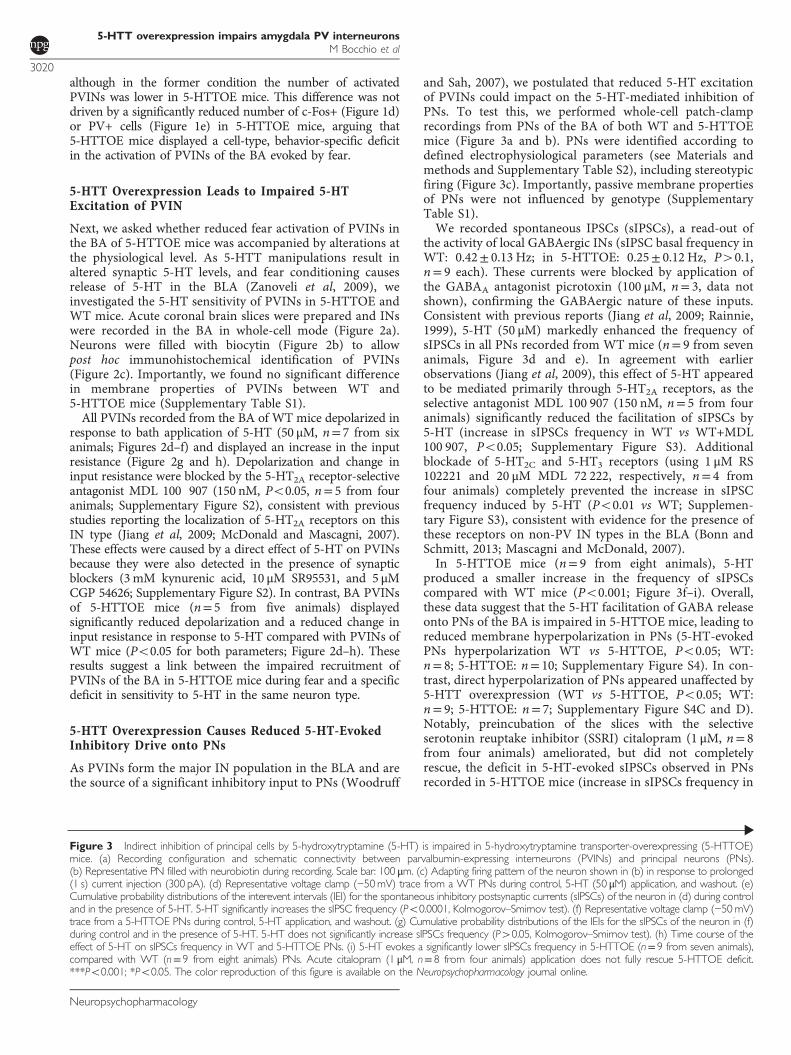
and Sah, 2007), we postulated that reduced 5-HT excitationof PVINs could impact on the 5-HT-mediated inhibition ofPNs. To test this, we performed whole-cell patch-clamprecordings from PNs of the BA of both WT and 5-HTTOEmice (Figure 3a and b). PNs were identified according todefined electrophysiological parameters (see Materials andmethods and Supplementary Table S2), including stereotypicfiring (Figure 3c). Importantly, passive membrane propertiesof PNs were not influenced by genotype (SupplementaryTable S1).We recorded spontaneous IPSCs (sIPSCs), a read-out of
the activity of local GABAergic INs (sIPSC basal frequency inWT: 0.42± 0.13 Hz; in 5-HTTOE: 0.25± 0.12 Hz, P40.1,n= 9 each). These currents were blocked by application ofthe GABAA antagonist picrotoxin (100 μM, n= 3, data notshown), confirming the GABAergic nature of these inputs.Consistent with previous reports (Jiang et al, 2009; Rainnie,1999), 5-HT (50 μM) markedly enhanced the frequency ofsIPSCs in all PNs recorded from WT mice (n= 9 from sevenanimals, Figure 3d and e). In agreement with earlierobservations (Jiang et al, 2009), this effect of 5-HT appearedto be mediated primarily through 5-HT2A receptors, as theselective antagonist MDL 100 907 (150 nM, n= 5 from fouranimals) significantly reduced the facilitation of sIPSCs by5-HT (increase in sIPSCs frequency in WT vs WT+MDL100 907, Po0.05; Supplementary Figure S3). Additionalblockade of 5-HT2C and 5-HT3 receptors (using 1 μM RS102221 and 20 μM MDL 72 222, respectively, n= 4 fromfour animals) completely prevented the increase in sIPSCfrequency induced by 5-HT (Po0.01 vs WT; Supplemen-tary Figure S3), consistent with evidence for the presence ofthese receptors on non-PV IN types in the BLA (Bonn andSchmitt, 2013; Mascagni and McDonald, 2007).In 5-HTTOE mice (n= 9 from eight animals), 5-HT
produced a smaller increase in the frequency of sIPSCscompared with WT mice (Po0.001; Figure 3f–i). Overall,these data suggest that the 5-HT facilitation of GABA releaseonto PNs of the BA is impaired in 5-HTTOE mice, leading toreduced membrane hyperpolarization in PNs (5-HT-evokedPNs hyperpolarization WT vs 5-HTTOE, Po0.05; WT:n= 8; 5-HTTOE: n= 10; Supplementary Figure S4). In con-trast, direct hyperpolarization of PNs appeared unaffected by5-HTT overexpression (WT vs 5-HTTOE, Po0.05; WT:n= 9; 5-HTTOE: n= 7; Supplementary Figure S4C and D).Notably, preincubation of the slices with the selectiveserotonin reuptake inhibitor (SSRI) citalopram (1 μM, n= 8from four animals) ameliorated, but did not completelyrescue, the deficit in 5-HT-evoked sIPSCs observed in PNsrecorded in 5-HTTOE mice (increase in sIPSCs frequency in
Figure 3 Indirect inhibition of principal cells by 5-hydroxytryptamine (5-HT) is impaired in 5-hydroxytryptamine transporter-overexpressing (5-HTTOE)mice. (a) Recording configuration and schematic connectivity between parvalbumin-expressing interneurons (PVINs) and principal neurons (PNs).(b) Representative PN filled with neurobiotin during recording. Scale bar: 100 μm. (c) Adapting firing pattern of the neuron shown in (b) in response to prolonged(1 s) current injection (300 pA). (d) Representative voltage clamp (−50mV) trace from a WT PNs during control, 5-HT (50 μM) application, and washout. (e)Cumulative probability distributions of the interevent intervals (IEI) for the spontaneous inhibitory postsynaptic currents (sIPSCs) of the neuron in (d) during controland in the presence of 5-HT. 5-HT significantly increases the sIPSC frequency (Po0.0001, Kolmogorov–Smirnov test). (f) Representative voltage clamp (−50mV)trace from a 5-HTTOE PNs during control, 5-HT application, and washout. (g) Cumulative probability distributions of the IEIs for the sIPSCs of the neuron in (f)during control and in the presence of 5-HT. 5-HT does not significantly increase sIPSCs frequency (P40.05, Kolmogorov–Smirnov test). (h) Time course of theeffect of 5-HT on sIPSCs frequency in WT and 5-HTTOE PNs. (i) 5-HT evokes a significantly lower sIPSCs frequency in 5-HTTOE (n= 9 from seven animals),compared with WT (n= 9 from eight animals) PNs. Acute citalopram (1 μM, n= 8 from four animals) application does not fully rescue 5-HTTOE deficit.***Po0.001; *Po0.05. The color reproduction of this figure is available on the Neuropsychopharmacology journal online.
5-HTT overexpression impairs amygdala PV interneuronsM Bocchio et al
3020
Neuropsychopharmacology
5-HTTOE vs 5-HTTOE+citalopram, P40.05; Figure 3hand i). These data suggest that 5-HTTOE mice display adeficit in 5-HT-induced inhibition of PNs of the BA by
GABAergic INs. This deficit is likely to be mediated, at leastin part, by impaired 5-HT-mediated depolarization ofPVINs. This loss of sensitivity to 5-HT in the 5-HTTOE
5-HTT overexpression impairs amygdala PV interneuronsM Bocchio et al
3021
Neuropsychopharmacology

mice is not entirely explained by an ongoing increase in thereuptake of 5-HT, as it could not be completely rescued bypharmacological blockade of the 5-HTT.
Impaired 5-HT2A Function Underlies Deficit in5-HT-Evoked Inhibition of PNs
We reasoned that the reduced 5-HT-evoked excitation ofPVINs observed in the 5-HTTOE mice could be caused by aloss of function at the level of 5-HT2A receptors. Crucially,the 5-HT2A receptor is the main mediator of the excitatoryeffect of 5-HT on BLA INs, and this receptor is locatedprimarily on PVINs (Jiang et al, 2009; McDonald andMascagni, 2007). Thus, we asked whether reduced 5-HT2A
receptor function could account for impaired 5-HT-inducedexcitation of PVINs in 5-HTTOE mice.The 5-HT2A agonist α-methyl-5-HT was applied while
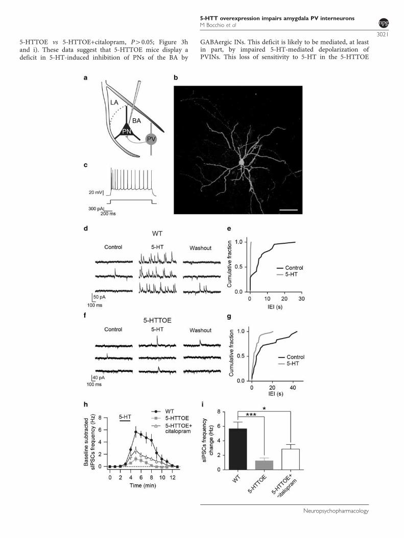
recording sIPSCs on PNs (sIPSC basal frequency in WT:0.04± 0.01 Hz; in 5-HTTOE: 0.07± 0.01 Hz, P40.1, n= 13each). In WT mice, bath application of α-methyl-5-HT(30 μM, n= 13 from four animals) increased the frequency ofsIPSCs (Figure 4a and b). This effect was almost entirelyabolished by MDL 100 907 (150 nM, n= 5; increase insIPSCs frequency WT vs WT+MDL 100 907, Po0.01;Figure 4c, d, g, and h), indicating that α-methyl-5-HT actsselectively on 5-HT2A receptors to facilitate GABA release,presumably by depolarization of PVINs (Jiang et al, 2009). Incomparison with WT mice, in 5-HTTOE mice application ofα-methyl-5-HT produced a much smaller increase in thesIPSC frequency in PNs (Po0.01, n= 13 from three animals;Figure 4e–h). Taken together, these data demonstrate that, inthe BA, increased 5-HTT expression is associated withimpaired 5-HT2A receptor-mediated activation of INsinnervating PNs. As PVINs are the main IN type expressing5-HT2A receptors in the BA, this deficit likely arises fromdysfunctional PVINs.
5-HTT Overexpression Causes Deficient 5-HTModulation of PNs Firing
Finally, to understand the functional consequences ofreduced 5-HT-driven inhibition of PNs, we examined5-HT modulation of PN firing in the two genotypes. Weinjected theta frequency sinusoidal currents that evokedfiring in ~ 50% of the oscillation peaks. Application of 50 μM5-HT reliably suppressed the firing of PNs from WT mice(n= 6 from three animals; Figure 5a and c). In contrast, thereduction in firing rate evoked by 5-HT was significantlyweaker in PNs from 5-HTTOE mice (Po0.01, n= 5 fromtwo animals, Figure 5b–d). Hence, 5-HTT overexpressioncauses a differential 5-HT modulation of PNs firing, likelyaffecting BA excitatory output (Figure 5e and f).
DISCUSSION
The present study demonstrates two novel findings on howvariation in 5-HTT expression impacts upon the micro-circuitry of the BA. First, the c-Fos data show that increased5-HTT expression results in reduced recruitment of BAPVINs during fear. Second, the electrophysiological resultsdemonstrate a selective impairment in 5-HT-evoked excita-tion of PVINs, leading to reduced inhibition of PNs. Thus,
increased 5-HTT expression leads to a loss of 5-HT controlover PVINs and therefore over PN firing, likely affecting BAoutput to the central amygdala and the processing of fear(Figure 5e and f). To our knowledge, these findings providethe first evidence of changes in the function of neurochemi-cally defined neuron types because of genetic alterations in5-HTT expression.PVINs of the BLA increase their firing rates in response to
CS presentation during fear conditioning (Wolff et al, 2014).Our data are consistent with this finding because presenta-tion of a tone predicting a footshock significantly increasedc-Fos expression in BA PVINs in WT mice. PVINs target theperisomatic region of PNs (Bienvenu et al, 2012), inhibitingand synchronizing their firing (Popescu and Paré, 2011;Ryan et al, 2012; Woodruff and Sah, 2007) and disinhibitingtheir dendrites (Wolff et al, 2014). Hence, they tightly controlthe BLA output to the central amygdala, orchestrating fearresponses. Crucially, inhibiting PVINs during the CSweakens fear learning (Wolff et al, 2014). Thus, reducedfreezing responses to the CS observed in 5-HTTOE mice (seealso Barkus et al, 2014; Line et al, 2014) could be directlylinked with reduced excitation of PVINs during the CS.Although the present study does not demonstrate a causalrelationship between reduced PVIN activation and reducedfear in 5-HTTOE mice, a defective encoding of the CS byPVINs could (1) fail to disinhibit PN dendrites, by lackof direct inhibition of somatostatin-expressing neurons,which are also inhibited by PVINs (Wolff et al, 2014);(2) contribute to recently reported weaker theta oscillations(Barkus et al, 2014); and (3) lead to reduced fear (currentstudy; Barkus et al., 2014; Line et al., 2014). Furthermore, wecannot fully resolve whether PVIN activation arises from fearmemory recall, sensitization by footshock, or fear general-ization to the tone or the testing context. This is becausetone-evoked freezing and PVIN activation in WT mice wereweak but not absent in the unpaired condition. Futureexperiments could test whether manipulation of PVINs inthe BA is sufficient to rescue the fear deficits in 5-HTTOE mice.The present study also provides a putative mechanism
underpinning the reduced excitation of PVINs of the BA in5-HTTOE mice, namely reduced sensitivity to 5-HT. Thiswas evident in electrophysiological experiments showing thatboth 5-HT-evoked depolarization of PVINs associated withthe closure of a membrane conductance as well as 5-HT-evoked IPSCs in PNs, were reduced in 5-HTTOE comparedwith WT mice. These deficits are likely caused by reducedfunctionality of 5-HT2A receptors, as sensitivity to the direct-acting 5-HT2A receptor agonist, α-methyl-5-HT, was alsoimpaired. Reduced 5-HT2A function could arise from eitherdownregulation or impaired receptor–effector coupling(Béïque et al, 2004). Surprisingly, 5-HTT blockade failed torescue fully the loss of sensitivity to 5-HT in the 5-HTTOEmice. Therefore, the 5-HT deficit in the BA associated withincreased 5-HTT expression appears to be dependent moreon 5-HT2A receptor function compared with that on reduced5-HT extracellular levels mediated by greater reuptake in5-HTTOE mice.The impaired sensitivity of PVINs in 5-HTTOE mice to
5-HT could be responsible for reduced c-Fos/PV coexpres-sion evoked by fear memory retrieval. This view is appealingbecause fear activates 5-HT neurons in the midbrain
5-HTT overexpression impairs amygdala PV interneuronsM Bocchio et al
3022
Neuropsychopharmacology
Figure 4 Serotonin 2A (5-HT2A)-mediated inhibition of principal neurons (PNs) is impaired in 5-hydroxytryptamine transporter-overexpressing(5-HTTOE) mice. (a) Representative voltage clamp (−50 mV) trace from a wild-type (WT) PNs during control, α-methyl-5-HT (30 μM) application, andwashout. (b) Cumulative probability distributions of the interevent intervals (IEIs) for the inhibitory postsynaptic currents (sIPSCs) of the neuron in (d) duringcontrol and in the presence of α-methyl-5-HT, a 5-HT2 agonist. α-Methyl-5-HT significantly increases the sIPSCs frequency (Po0.0001, Kolmogorov–Smirnovtest). (c) Representative voltage-clamp (−50 mV) trace from a WT PNs in the presence of the 5-HT2A antagonist MDL 100 907 (150 nM) during control,α-methyl-5-HT application, and washout. (d) Cumulative probability distributions of the IEIs for the sIPSCs of the neuron in (d) during control and in presenceof α-methyl-5-HT. This drug does not significantly increase the sIPSCs frequency (P40.05, Kolmogorov–Smirnov test). (e) Representative voltage-clamp(−50 mV) trace from 5-HTTOE PNs during control, α-methyl-5-HT application, and washout. (f) Cumulative probability distributions of the IEIs for the sIPSCsof the neuron in (f) during control and in the presence of α-methyl-5-HT. This drug does not significantly increase sIPSC frequency (P40.05, Kolmogorov–Smirnov test). (g) Time course of the effect of α-methyl-5-HT on sIPSCs frequency in WT (with and without 5-HT2A antagonist MDL 100 907) and5-HTTOE PNs. (h) α-Methyl-5-HT evokes a significantly higher sIPSCs frequency in WT (n= 13 from four animals) compared with 5-HTTOE (n= 13 from3 animals) PNs. MDL 100 907 blocks the effect of α-methyl-5-HT (n= 5 from two animals). **Po0.01. The color reproduction of this figure is available onthe Neuropsychopharmacology journal online.
5-HTT overexpression impairs amygdala PV interneuronsM Bocchio et al
3023
Neuropsychopharmacology

(Spannuth et al, 2011) and triggers the release of 5-HT in theBLA (Zanoveli et al, 2009). Additionally, midbrain 5-HTneurons can fire phase locked to limbic theta oscillations(Kocsis and Vertes, 1992). Thus, their impaired excitation ofPVINs in 5-HTTOE mice could contribute to the reductionof theta rhythms in the BLA of these mice (Barkus et al,2014). This is consistent with the notion that the power oftheta rhythm is much more influenced by the interplaybetween two synaptic current generators, namely dendriticexcitation vs PV-perisomatic inhibition of PNs, rather thanby PNs spiking per se (Buzsáki, 2002). However, othercircuits could also contribute to the weaker activation ofPVINs during fear memory retrieval. For instance, 5-HTToverexpression might alter the function of other areas suchas the mPFC and the hippocampus, which are also targetedby 5-HT fibers and project to the BLA (Hensler, 2006).Unlike PVINs, we did not detect genotypic differences inthe overall number of c-Fos+ neurons in the CS–US pairedcondition, despite PNs receiving impaired inhibition.Although the majority of these activated neurons likelyrepresent PNs, it is important to keep in mind that non-PVIN populations might be included. Furthermore, impairedPVIN output could result in a different temporal distributionof action potentials fired by PNs rather than a long-lastingreduction in their firing rates, with c-Fos protein express-ion lacking the temporal resolution to reveal such subtlechanges.Our data provide compelling evidence linking increased
expression of the 5-HTT with abnormal PVIN function. The5-HT2A receptor, which depolarizes PVINs, appears to have
a key role in this relationship. Although we cannot fullyexclude the possibility that other IN types also contribute tothe deficient 5-HT-driven inhibitory inputs to PNs, PVINsare the major GABAergic cell type expressing 5-HT2A
receptors in the BLA, in addition to a small number ofsomatostatin-expressing INs (McDonald and Mascagni,2007). Adaptive changes in 5-HT2A receptor functionbecause of pharmacological or genetic manipulation of the5-HTT are consistent with previous studies (Jennings et al,2008; Li et al, 2003; Marek et al, 2005; Sarkar et al, 2013).Importantly, the direct inhibition by 5-HT of PNs wasunaffected in 5-HTTOE mice. This inhibitory effect on BAPNs by 5-HT mirrors the hyperpolarization of corticalpyramidal cells mediated by 5-HT1A receptors (Araneda andAndrade, 1991). Altered 5-HT levels during developmentbecause of higher 5-HT reuptake may be responsible for theloss of 5-HT2A function in PVINs. In contrast, 5-HT1A
receptors have been shown to develop later in corticalpyramidal cells (Béïque et al, 2004), thus their laterexpression on BA PNs might prevent receptor exposureduring a sensitive developmental period (Gross et al, 2002).Further experiments examining PVIN function duringdevelopment are necessary to corroborate this hypothesis.In summary, our results suggest a novel mechanism
through which increased expression of the 5-HTT gene canalter the microcircuitry of the amygdala, namely by changingthe function of PVINs. Probing the effects of geneticvariation in the 5-HTT or SSRI treatment on neural circuitsin limbic areas may open new horizons for understandingthe biological basis of emotional behavior.
Figure 5 Increased 5-hydroxytryptamine transporter (5-HTT) expression alters the output of basal amygdala (BA) principal neurons (PNs). (a) Sinusoidalcurrent (160 pA intensity) injected at theta frequency (6 Hz) in a representative wild-type (WT) PN current clamped at − 60 mV. Application of5-hydroxytryptamine (5-HT) (50 μM) causes hyperpolarization (2–3 mV) and suppression of firing. (b) Sinusoidal current injection (190 pA) at thetafrequency (6 Hz) in a representative 5-HTT-overexpressing (5-HTTOE) PN recorded in the current clamp at − 60 mV. Application of 5-HT produces aweaker hyperpolarization and fails to suppress firing. (c) Time course of the effect of 5-HT on the firing rate of WT and 5-HTTOE PNs (sinusoidal currentinjections shown in (a) and (b) delivered every 10 s). (d) 5-HT evokes a significantly stronger depression of firing rate in WT (n= 6 from three animals)compared with 5-HTTOE (n= 5 from two animals) PNs. (e and f) Model of amygdala circuit alterations in 5-HTTOE mice. (e) Dorsal raphe firing, leading tothe release of 5-HT in the BA, excites parvalbumin-expressing interneurons (PVINs) through 5-HT2A receptors, leading to indirect perisomatic inhibitionof PNs. (f) Increased 5-HTT expression, characterized by lower 5-HT tone because of more efficient 5-HT reuptake, causes reduced 5-HT2A receptorfunction on PVINs, leading to a failure of dorsal raphe activity to recruit PVINs. This could affect the encoding of sensory stimuli (conditioned stimulus (CS)and unconditioned stimulus (US)) by the PNs, dampening fear responses. **Po0.01. The color reproduction of this figure is available on theNeuropsychopharmacology journal online.
5-HTT overexpression impairs amygdala PV interneuronsM Bocchio et al
3024
Neuropsychopharmacology

FUNDING AND DISCLOSURE
This work was supported by the Medical Research Council,UK (to MC) (award MC_UU_12020/2) and a WellcomeTrust Senior Fellowship award (to DMB) (Grant No.087736). Giulia Fucsina was a student of the Master Degreein Neuroscience at the University of Trieste, Italy, and wassupported by the Erasmus Placement Exchange. LydiaOikonomidis was an MSc Neuroscience student at theUniversity of Oxford. The authors declare no conflict ofinterest.
ACKNOWLEDGMENTS
We thank K Whitworth, L Norman, J Janson, B Micklem,and L Black for excellent technical support.
REFERENCES
Araneda R, Andrade R (1991). 5-Hydroxytryptamine2 and5-hydroxytryptamine 1A receptors mediate opposing responseson membrane excitability in rat association cortex. Neuroscience40: 399–412.
Asan E, Steinke M, Lesch K (2013). Serotonergic innervation of theamygdala: targets, receptors, and implications for stress andanxiety. Histochem Cell Biol 139: 785–813.
Ballenger JC (1999). Current treatments of the anxiety disordersin adults. Biol Psychiatry 46: 1579–1594.
Barkus C, Line SJ, Huber A, Capitao L, Lima J, Jennings K et al(2014). Variation in serotonin transporter expression modulatesfear-evoked hemodynamic responses and theta-frequency neuro-nal oscillations in the amygdala. Biol Psychiatry 75: 901–908.
Béïque J-C, Campbell B, Perring P, Hamblin MW, Walker P,Mladenovic L et al (2004). Serotonergic regulation of membranepotential in developing rat prefrontal cortex: coordinatedexpression of 5-hydroxytryptamine (5-HT)1A, 5-HT2A, and5-HT7 receptors. J Neurosci 24: 4807–4817.
Bienvenu TCM, Busti D, Magill PJ, Ferraguti F, Capogna M (2012).Cell-type-specific recruitment of amygdala interneurons tohippocampal theta rhythm and noxious stimuli in vivo. Neuron74: 1059–1074.
Blakely RD, De Felice LJ, Hartzell HC (1994). Molecular physiologyof norepinephrine and serotonin transporters. J Exp Biol 196:263–281.
Bonn M, Schmitt A (2013). Serotonergic innervation and serotoninreceptor expression of NPY-producing neurons in the rat lateraland basolateral amygdaloid nuclei. Brain Struct 218: 421–435.
Buzsáki G (2002). Theta oscillations in the hippocampus. Neuron33: 325–340.
Canli T, Omura K, Haas BW, Fallgatter A, Constable RT, Lesch KP(2005). Beyond affect: a role for genetic variation of the serotonintransporter in neural activation during a cognitive attention task.Proc Natl Acad Sci USA 102: 12224–12229.
Caspi A, Sugden K, Moffitt TE, Taylor A, Craig IW, Harrington Het al (2003). Influence of life stress on depression: moderation bya polymorphism in the 5-HTT gene. Science 301: 386–389.
Courtin J, Chaudun F, Rozeske RR, Karalis N, Gonzalez-Campo C,Wurtz H et al (2014). Prefrontal parvalbumin interneurons shapeneuronal activity to drive fear expression. Nature 505: 92–96.
Gross C, Zhuang X, Stark K, Ramboz S, Oosting R, Kirby L et al(2002). Serotonin1A receptor acts during development toestablish normal anxiety-like behaviour in the adult. Nature 416:396–400.
Hariri AR, Mattay VS, Tessitore A, Kolachana B, Fera F, GoldmanD et al (2002). Serotonin transporter genetic variation and theresponse of the human amygdala. Science 297: 400–403.
Hensler JG (2006). Serotonergic modulation of the limbic system.Neurosci Biobehav Rev 30: 203–214.
Holmes A, Lit Q, Murphy DL, Gold E, Crawley JN (2003).Abnormal anxiety-related behavior in serotonin transporter nullmutant mice: the influence of genetic background. Genes BrainBehav 2: 365–380.
Hu X-Z, Lipsky RH, Zhu G, Akhtar LA, Taubman J, Greenberg BDet al (2006). Serotonin transporter promoter gain-of-functiongenotypes are linked to obsessive-compulsive disorder. Am J HumGenet 78: 815–826.
Jackson J, Dickson CT, Bland BH (2008). Median raphe stimulationdisrupts hippocampal theta via rapid inhibition and state-dependent phase reset of theta-related neural circuitry.J Neurophysiol 99: 3009–3026.
Jennings KA, Loder MK, Sheward WJ, Pei Q, Deacon RMJ,Benson MA et al (2006). Increased expression of the 5-HTtransporter confers a low-anxiety phenotype linked to decreased5-HT transmission. J Neurosci 26: 8955–8964.
Jennings KA, Sheward WJ, Harmar AJ, Sharp T (2008). Evidencethat genetic variation in 5-HT transporter expression is linked tochanges in 5-HT2A receptor function. Neuropharmacology 54:776–783.
Jiang X, Xing G, Yang C, Verma A, Zhang L, Li H (2009).Stress impairs 5-HT2A receptor-mediated serotonergic facilita-tion of GABA release in juvenile rat basolateral amygdala.Neuropsychopharmacology 34: 410–423.
Klausberger T, Magill PJ, Márton LF, Roberts JDB, Cobden PM,Buzsáki G et al (2003). Brain-state- and cell-type-specificfiring of hippocampal interneurons in vivo. Nature 421:844–848.
Kocsis B, Vertes RP (1992). Dorsal raphe neurons: synchronousdischarge with the theta rhythm of the hippocampus in the freelybehaving rat. J Neurophysiol 68: 1463–1467.
Lee H-J, Lee M-S, Kang R-H, Kim H, Kim S-D, Kee B-S et al (2005).Influence of the serotonin transporter promoter gene polymor-phism on susceptibility to posttraumatic stress disorder. DepressAnxiety 21: 135–139.
Lesch KP, Bengel D, Heils A, Sabol SZ, Greenberg BD, Petri S et al(1996). Association of anxiety-related traits with a polymorphismin the serotonin transporter gene regulatory region. Science 274:1527–1531.
Li Q, Wichems CH, Ma L, Van de Kar LD, Garcia F, Murphy DL(2003). Brain region-specific alterations of 5-HT2A and 5-HT2Creceptors in serotonin transporter knockout mice. J Neurochem84: 1256–1265.
Line SJ, Barkus C, Rawlings N, Jennings K, McHugh S, Sharp T et al(2014). Reduced sensitivity to both positive and negativereinforcement in mice over-expressing the 5-hydroxytryptaminetransporter. Eur J Neurosci 40: 3735–3745.
Lundberg J, Borg J, Halldin C, Farde L (2007). A PET study onregional coexpression of 5-HT1A receptors and 5-HTT in thehuman brain. Psychopharmacology (Berl) 195: 425–433.
Manko M, Bienvenu TCM, Dalezios Y, Capogna M (2012). Neuro-gliaform cells of amygdala: a source of slow phasic inhibition inthe basolateral complex. J Physiol 590: 5611–5627.
Marek GJ, Martin-Ruiz R, Abo A, Artigas F (2005). The selective5-HT2A receptor antagonist M100907 enhances antidepressant-like behavioral effects of the SSRI fluoxetine. Neuropsycho-pharmacology 30: 2205–2215.
Mascagni F, McDonald AJ (2007). A novel subpopulation of 5-HTtype 3A receptor subunit immunoreactive interneurons in the ratbasolateral amygdala. Neuroscience 144: 1015–1024.
McDonald AJ, Mascagni F (2001). Colocalization of calcium-binding proteins and GABA in neurons of the rat basolateralamygdala. Neuroscience 105: 681–693.
McDonald AJ, Mascagni F (2007). Neuronal localization of 5-HTtype 2A receptor immunoreactivity in the rat basolateralamygdala. Neuroscience 146: 306–320.
5-HTT overexpression impairs amygdala PV interneuronsM Bocchio et al
3025
Neuropsychopharmacology

Murphy SE, Norbury R, Godlewska BR, Cowen PJ, Mannie ZM,Harmer CJ et al (2013). The effect of the serotonin transporterpolymorphism (5-HTTLPR) on amygdala function: a meta-analysis. Mol Psychiatry 18: 512–520.
Narayanan V, Heiming RS, Jansen F, Lesting J, Sachser N,Pape H-C et al (2011). Social defeat: impact on fear extinctionand amygdala-prefrontal cortical theta synchrony in 5-HTTdeficient mice. PLoS One 6: e22600.
Pezawas L, Meyer-Lindenberg A, Drabant EM, Verchinski B a,Munoz KE, Kolachana BS et al (2005). 5-HTTLPR polymor-phism impacts human cingulate-amygdala interactions: a geneticsusceptibility mechanism for depression. Nat Neurosci 8:828–834.
Popescu AT, Paré D (2011). Synaptic interactions underlyingsynchronized inhibition in the basal amygdala: evidence forexistence of two types of projection cells. J Neurophysiol 105:687–696.
Rainnie DG (1999). Serotonergic modulation of neurotransmissionin the rat basolateral amygdala. J Neurophysiol 82: 69–85.
Richmond MA, Murphy CA, Pouzet B, Schmid P, Rawlins JNP,Feldon J (1998). A computer controlled analysis of freezingbehaviour. J Neurosci Methods 86: 91–99.
Ryan SJ, Ehrlich DE, Jasnow AM, Daftary S, Madsen TE, Rainnie DG(2012). Spike-timing precision and neuronal synchrony areenhanced by an interaction between synaptic inhibition andmembrane oscillations in the amygdala. PLoS One 7: e35320.
Sarkar A, Chachra P, Vaidya VA (2013). Postnatal fluoxetine-evoked anxiety is prevented by concomitant 5-HT2A/C receptor
blockade and mimicked by postnatal 5-HT2A/C receptorstimulation. Biol Psychiatry 76: 858–868.
Sosulina L, Meis S, Seifert G, Steinhäuser C, Pape H-C (2006).Classification of projection neurons and interneurons in the ratlateral amygdala based upon cluster analysis. Mol Cell Neurosci33: 57–67.
Spannuth BM, Hale MW, Evans AK, Lukkes JL, Campeau S,Lowry CA (2011). Investigation of a central nucleus of theamygdala/dorsal raphe nucleus serotonergic circuit implicated infear-potentiated startle. Neuroscience 179: 104–119.
Uher R, McGuffin P (2010). The moderation by the serotonintransporter gene of environmental adversity in the etiology ofdepression: 2009 update. Mol Psychiatry 15: 18–22.
Wellman CL, Izquierdo A, Garrett JE, Martin KP, Carroll J,Millstein R et al (2007). Impaired stress-coping and fearextinction and abnormal corticolimbic morphology in serotonintransporter knock-out mice. J Neurosci 27: 684–691.
Wolff SBE, Gründemann J, Tovote P, Krabbe S, Jacobson G a,Müller C et al (2014). Amygdala interneuron subtypes controlfear learning through disinhibition. Nature 509: 453–458.
Woodruff AR, Sah P (2007). Inhibition and synchronization ofbasal amygdala principal neuron spiking by parvalbumin-positiveinterneurons. J Neurophysiol 98: 2956–2961.
Zanoveli JM, Carvalho MC, Cunha JM, Brandão ML (2009).Extracellular serotonin level in the basolateral nucleus of theamygdala and dorsal periaqueductal gray under unconditionedand conditioned fear states: an in vivo microdialysis study. BrainRes 1294: 106–115.
Supplementary Information accompanies the paper on the Neuropsychopharmacology website (http://www.nature.com/npp)
5-HTT overexpression impairs amygdala PV interneuronsM Bocchio et al
3026
Neuropsychopharmacology