induced pluripotent stem cell-based therapy for age
TRANSCRIPT
Induced Pluripotent Stem Cell-Based Therapy for Age-Related Macular Degeneration Peter Bracha, Nicholas A. Moore & Thomas A. Ciulla
1. Background
2. Age-related macular degeneration pathogenesis
3. Induced pluripotent stem cells
4. Retinal pigment epithelium transplantation4.1. Retinal pigment epithelium physiology 4.2. History of adjuvant retinal pigment epithelium transplantation in choroidal neovascular membrane resection 4.3. Preclinical stem cell-based retinal pigment epithelium studies 4.4. Human studies of human embryonic stem cell-derived retinal pigment epithelium transplantation 4.5. Human studies of induced pluripotent stem cell-derived retinal pigment epithelium transplantation
5. Retinal progenitor cell transplantation
6. Challenges to effective transplantation6.1. Surgical complications 6.2. Transplant immunology 6.3. Systemic immunosuppression 6.4. Aging Bruch’s Membrane
7. Ongoing studies
8. Conclusion
9. Expert Opinion
___________________________________________________________________
This is the author's manuscript of the article published in final edited form as:
Bracha, P., Moore, N. A., & Ciulla, T. A. (2017). Induced pluripotent stem cell-based therapy for age-related macular degeneration. Expert Opinion on Biological Therapy, 17(9), 1113–1126. https://doi.org/10.1080/14712598.2017.1346079

[Type here]
Abstract Introduction: In age-related macular degeneration (AMD), induced pluripotent stem cell (iPSC)-based therapies have advanced from the bench to human trials over the last decade. This novel therapy has the potential to replace or regenerate damaged tissue, such as retinal pigment epithelium (RPE) to treat geographic atrophy or to limit choroidal neovascularization. Areas covered: Induced PSCs can be derived from the introduction of transcription factors to adult cells under specific cell culture conditions, which can then be differentiated into various cell types, including RPE cells. Induced PSC-derived RPE cells exhibit ion transport, membrane potential, polarized vascular endothelial growth factor (VEGF) secretion and gene expression that is similar to native RPE cells and have principally been investigated as subretinal transplants to replace or replenish diseased RPE. It remains to be seen if stem cell-based RPE cells can replicate the myriad of functions, including immunomodulatory effects, of native RPE cells in the human eye. Expert opinion: Induced PSC-based RPE transplantation is a promising investigational therapy in the treatment of AMD, however significant progress is required prior to broad application and major pathophysiological hurdles may eventually limit its success in AMD.

[Type here]
1. Background Age-related macular degeneration (AMD) is one of the leading causes of blindness globally. By the
year 2020, an estimated 196 million people will have AMD and 11 million will have significant vision loss [1]. AMD can broadly be categorized into atrophic and neovascular (nAMD) forms. Atrophic AMD is characterized by the development of drusen and retinal pigment epithelial (RPE) changes early in the disease course, and with loss of RPE (geographic atrophy, GA) and associated severe vision loss in advanced disease. Currently no effective treatment exists for GA. Neovascular AMD is characterized by choroidal neovascularization (CNV) causing central vision loss from macular exudation; subsequently, despite treatment, fibrosis and/or RPE atrophy develop in nearly half of patients by 2 years, resulting in severe permanent central vision loss [2].
Anti-vascular endothelial growth factor (anti-VEGF) therapy is currently the standard of care for nAMD. The original ANCHOR (Antibody for the Treatment of Predominantly Classic CNV in AMD Study) and MARINA (Minimally Classic/Occult Trial of the Anti-VEGF Antibody Ranibizumab in the Treatment of nAMD) trials of monthly ranibizumab respectively yielded 11.3 and 7.2 Early Treatment of Diabetic Retinopathy Study (ETDRS) letters of improvement at one year [3, 4]. However, the subsequent open label extension HORIZON trial ( An Open-Label Extension Trial of Ranibizumab for Choroidal Neovascularization Secondary to Age-Related Macular Degeneration) showed insidious loss of vision, yielding a mean visual acuity (VA) improvement of only 2 ETDRS letters after four years [5]. Similarly, in a follow-up study of the CATT (Comparison of AMD Treatment Trials) trial, patients experienced a mean loss of 3 ETDRS letters five years after initiating anti-VEGF treatment [6]. “Real world” studies of anti-VEGF therapy in nAMD have reported even less favorable visual outcomes [7-19]. Several of these “real world” studies, based on chart reviews, electronic medical records (EMR) or claims analysis, demonstrate that nAMD patients generally lose vision by the 2- to 5-year time point despite anti-VEGF therapy [9-13, 16, 19, 20]. While the cause of these discrepancies between “real world” studies and RCTs is unknown, patient characteristics and undertreatment are implicated [7-9, 13, 14]. Several of these “real world” nAMD studies observed a 20-30% loss to follow-up in the first year [8, 10, 14, 16], highlighting the treatment burden. Additionally, some “real world” nAMD studies suggest that poor visual outcomes account for patient drop out [10, 12, 14] and that despite treatment, patients with better baseline VA experience greater loss of vision than patients with worse baseline VA [12, 14, 16, 18]. This experience over the past decade demonstrates the limitations of anti-VEGF agents and the unmet need for more effective therapy.
Stem cells, currently under investigation for the treatment of AMD, are characterized by the ability to differentiate into multiple cell lineages and an unlimited self-renewal capacity [21]. Pluripotent stem cells (PSCs), by definition, are able to differentiate into all endodermal, mesodermal and ectodermal lineages. Induced pluripotent stem cells (iPSCs) are a subtype of pluripotent stem cells and are intriguing in that they can be derived from a previously differentiated cell source and have the potential for reduced ethical controversy over human embryonic stem cell (hESC)-based therapies, and may negate immunological issues associated with hESC-based therapies. The eye is an ideal candidate for stem cell research because the clear ocular media allows for direct visualization of transplanted cells and the eye is a relatively immune privileged site; furthermore, the size of the eye requires smaller quantities of therapeutic tissue in comparison to other organs. In AMD, stem cells have the potential for replacing or regenerating lost tissue, such as RPE replacement to treat GA and to limit CNV. They have the potential to produce growth factors and cytokines such as brain derived neurotrophic factor (BDNF) that have a supportive paracrine effect on local structures within the macula. Exciting research into stem cell-based therapy for AMD has been performed over the last several decades and will be reviewed here. Emphasis

[Type here]
will be placed on research into iPSCs, but context will be provided through a discussion of other stem-cell based therapies.
2. AMD pathogenesis
A basic understanding AMD pathology provides context to better evaluate the therapeutic role of stem cell-based therapies. AMD pathogenesis involves a complex mixture of structural and functional changes as a result of aging [22], chronic oxidative stress [23] and pathologic inflammation [23]. This etiologic milieu eventually results in the clinical appearance of drusen and RPE changes, and eventually vision threatening GA and CNV.
Aging results in numerous changes to the RPE, Bruch’s membrane (BM) and choriocapillaris (CC), which could adversely affect the health of any transplanted stem cells. With age, the RPE accumulates residual bodies containing lipofuscin, resulting in a reduced phagocytic capacity and an impairment of lysosomal function [24, 25]. A generalized age-related loss of RPE cells increases the metabolic demand on each cell [26]. Bruch’s membrane is a 5-layered structure between the RPE and CC. With age, the thickness [27], the amount of non-collagenous proteinaceous material [28] and the amount of lipids increases [29]. The BM’s central elastic layer becomes thinned and calcified [30], increasing the risk of BM breaks that facilitate CNV ingrowth through a breach of the anti-angiogenic barrier [31]. Additionally, BM heparan sulfate quantities decrease with age, resulting in less Complement Factor H (CFH) binding; as discussed below, this factor moderates the complement cascade and decreased local levels may potentiate the inflammatory component of AMD pathogenesis [32]. These structural and biochemical changes affect the transport of molecules and growth factors across BM and likely directly affect the interaction between BM and both the RPE and CC, including any stem cell derived RPE transplant. The choriocapillaris is the innermost layer of the choroid and supplies the oxygen and nutrients and eliminates the waste of the BM, RPE and photoreceptors. The CC density and vessel diameter decreases with age along with the total choroidal thickness [28, 33]. Choriocapillaris endothelial density decreases [34], supporting the notion that vascular changes may contribute to AMD. Overall, these age-related changes of the RPE-BM-CC complex contribute to AMD pathogenesis and are one of several factors necessary for pathologic manifestations.
Oxidative stress is likely a major contributor to the development of AMD based on epidemiologic and clinical studies [23]. Tobacco use is a well-known risk factor for AMD. On a mechanistic level, chronic accumulation of reactive oxygen species (ROS) can be detrimental to an organism through oxidization of DNA, proteins and lipids [35]. ROS can be generated through exposure to UV light, ionizing radiation and normal cellular enzymatic reactions [36]. The balance towards more oxidative stress is present in patients with AMD. Levels of plasma glutathione, an antioxidant, are low in patients with AMD [37]. ROS formation is high due to the high oxygen tension observed in the retina [36] and also due to the constant exposure to light [36]. Additionally, nutritional supplementation with the antioxidants Vitamin C and Vitamin E reduces the risk of progression of moderate AMD [38]. Cumulative oxidative stress over decades likely contributes to the impaired function of RPE-BM-CC complex. Additionally, of particular relevance to stem cell-based therapies, any subretinally transplanted cells will be exposed to the same oxidative stress, potentially affecting cellular behavior and survival.
An impaired inflammatory response is a key component in AMD pathogenesis, potentially further complicating stem cell based transplantation. Patients with AMD are known to have elevated systemic inflammatory biomarkers such as C-reactive protein (CRP), interleukin-6 (IL-6), and homocysteine [39, 40], and surgically excised CNV has been shown to contain inflammatory cells [41]. The inflammation
[Type here]
appears to be mediated through the complement cascade of the immune system. Normally, the complement cascade detects and destroys blood born bacteria. It can be activated via an antibody-dependent pathway (the “classical pathway”), an antibody-independent pathway (the “alternate pathway”), or on binding to specific carbohydrates on the surface of microorganisms (the “lectin” pathway). Complement was first implicated in the pathogenesis of AMD in 2005, when a common variant in complement factor H (CFH) gene was noted to be strongly associated with AMD, with individuals homozygous for the risk allele possessing a 7.4 fold increased risk [42, 43]; several other genetic variants in complement genes have since been associated with AMD [44-46]. CFH is a natural inhibitor of factor C3 activation, which is central to the three pathways. Complement is involved in drusen formation , and genetic variations in CFH can lead to less effective modulation of complement pathway activity, leading to greater precipitation of complement protein observed in drusen [47]. The final common pathway of complement activation, factor C5, is cleaved to form key terminal fragments (C5a and C5b-9) regardless of which pathway (classical, alternate or lectin) induced their generation. The C5a fragment, observed in drusen of AMD patients, is an important inflammatory activator, recruiting inflammatory cells and inducing vascular endothelial growth factor (VEGF) expression from RPE cells [48]. C5b is involved in the formation of the membrane attack complex (MAC: C5b-9), which causes cell death through disruption of the cell membrane. In humans, deposition of MAC in Bruch’s membrane and choriocapillaris increases significantly with aging and with AMD [49]. When choroidal endothelial cells are exposed to MAC, cell lysis occurs and the surviving cells express VEGF and matrix metalloproteinases (MMP); thus, MAC deposition in the choriocapillaris may play a role in atrophic and neovascular AMD, respectively [50]. Consequently, both C5a and MAC may promote CNV, and this is supported by experimental models of CNV, in which inhibition of C5a and MAC formation prevents the development of CNV [51, 52].
AMD pathogenesis is a chronic, complex process that affects the RPE-BM-CC complex and stem cell-based therapies will need to overcome key structural and functional changes. Additionally, if placed subretinally, any transplant will be exposed to a toxic environment, potentially affecting the survival and physiology of the transplanted cells. 3. Induced pluripotent stem cells
Human embryonic stem cells (hESCs) were first cultured in 1998 and have the potential to differentiate into all cell types [53]. They are a promising source for stem-cell based therapy but raise potential ethical considerations due to their harvesting during embryogenesis when there is potential for full development. In 2006, Takahashi and Yamanaka discovered a method of inducing a pluripotent state from terminally differentiated mouse fibroblasts, potentially addressing this issue. The introduction of four transcription factors, Oct3/4, Sox2, c-Myc and Klf4 to adult murine fibroblasts under embryonic cell culture conditions, resulted in an induced pluripotent stem cell (iPSC) state as confirmed by the ability to differentiate into all three germ layers upon transplantation into adult mice and mouse embryos [54]. One year later, two separate groups were able to create iPSCs from adult human cells, another major scientific milestone [55, 56].
Numerous advances in the efficiency and safety of iPSC generation has occurred over the last decade. Various groups have developed methods for improving the efficiency of pluripotency induction through the use of factors such as valproic acid [57], SV40 large T antigen [58], and microRNA [59]. The development of iPSCs has also become safer. The original use of Myc transcription factor in the formation of iPSCs created concern that constitutively active Myc, an

[Type here]
oncogene, could result in tumor formation. This problem was solved in 2008 by Nakagawa et al. when they demonstrated a method of iPSCs production in mouse and human fibroblasts without the use of Myc [60]. Similarly, the safety of using retroviral vectors for creating iPSCs, and concern about retroviral integration into the DNA with unpredictable sequelae such as creating a tumorigenic state, was overcome through numerous methods including the use of non-integrating episomal vectors [61], adenoviruses [62], the Sendai virus [63], piggyback transposition [64], minicircles [65], mRNA transfection [66, 67], microRNAs [68], transfection of a single multiprotein expression vectors [69], chemicals [70] and reprogramming proteins [71], among other techniques. The landscape of methods to effectively and efficiently induce a pluripotent state is expanding rapidly and a thorough discussion is beyond the scope of this review.
These advances over the last decade helped pave the way for the translation of iPSC-based animal and human studies. In addition to cell therapy, iPSCs have other applications, including disease modeling and drug discovery.
4. Retinal pigment epithelium transplantation RPE replacement is of interest in AMD therapeutics research given its numerous key roles in retinal support and function, as well as its pathophysiologic roles in AMD. This section reviews RPE physiology, the history of RPE transplantation, preclinical studies, and the most recently published human studies. 4.1. Retinal pigment epithelium physiology
The RPE, a monolayer of hexagonal cells, plays numerous roles in retinal homeostasis. It is a polar layer with the apical portion facing the outer segments of the photoreceptors and the basal surface facing BM. The polarity of RPE cells aids in the transport of ions, water and metabolic products from the subretinal space to the CC [72]. It also helps in the delivery of glucose and fatty acids to the outer retina [72]. Voltage-dependent ion conductance at the apical membrane creates the appropriate ion composition in the subretinal space necessary for photoreceptor excitability [72]. Tight junctions help maintain polarity in addition to forming the outer blood-retinal barrier [73]. RPE cells apically project microvilli that facilitate retinal adhesion and processing of shed outer photoreceptor segments. These shed photoreceptor outer segments are processed by the RPE through phagocytosis and subsequently recycled to the photoreceptors or eliminated to the CC [74]. Multiple steps in the retinoid cycle are performed by the RPE cells and provide photoreceptors with 11-cis-retinal [75]. Melanin is abundantly found in RPE cells and functions by preventing light reflection and as a free radical scavenger [72].
The RPE physiologically secretes numerous growth factors. Pigment epithelium-derived factor (PEDF) has anti-angiogenic and neurotrophic properties and is principally secreted apically by the RPE [72]. VEGF, at low levels, is secreted on the basal surface of the RPE and prevents endothelial cell apoptosis of the CC [72]. Tissue inhibitors of metalloproteinases (TIMP-1 and TIMP-3) are secreted to stabilize the endothelium, Bruch’s membrane and the CC extracellular matrix [72]. Fibroblast growth factor and ciliary neurotrophic factors are secreted in response to RPE damage and have neuroprotective roles. Contributions to immune privilege and inflammation modulation is performed through the secretion of interleukins and cytokines and through the presentation of cell membrane proteins [72, 76]. Consequently, the RPE plays multiple roles in the physiology of the CC and retina through the secretion of growth factors, while also facilitating ocular immune privilege through the secretion of interleukins and cytokines [72, 77].

[Type here]
These paracrine functions of the RPE are critical for the health of the neurosensory retina. This discussion is pertinent because most stem cell based therapies in AMD strive to replace or supplement diseased RPE cells. While critical functions dependent on proper orientation and structure may be challenging to replicate with stem-cell based therapy, the paracrine functions could potentially be more easily supplanted.
4.2. History of adjuvant retinal pigment epithelium transplantation in choroidal neovascular membrane resection
This section provides a brief discussion on the history of RPE transplants and the relevance to stem-cell based transplantation. Subfoveal neovascular membrane resection was one of numerous investigational therapies for nAMD in the 1990s [78, 79]. A major limitation of this procedure was the associated extraction of RPE and the subsequent development of GA with a poor visual prognosis [80-82]. To limit this atrophy, investigators attempted various methods of adjuvant RPE transplantation following CNV resection, including homologous fetal RPE transplantation [83], homologous adult RPE transplantation [84, 85], macular translocation [86], autologous RPE transplantation [84, 87, 88], choroidal-RPE transplantation [89, 90], and Bruch’s membrane-RPE transplantation [91]. With the advent of anti-VEGF therapy, the investigative fervor of RPE transplantation subsided, but numerous lessons can be gleaned from those studies.
Intraoperatively, manipulation of RPE grafts makes transplantation technically difficult. Maaijwee et al. published a series of 84 RPE and choroidal translocation procedures, in which 56 were associated with an inadequate positioning or incomplete flattening of the graft [92]. The graft was often partly folded or wrinkled, and 12 cases required extraction and reinsertion [92]. Joussen et al. noted that of 45 eyes receiving an autologous translocation of peripheral choroid and RPE, two patients intraoperatively and one patient postoperatively were observed to have an upside-down insertion of the graft [89]. These technical difficulties will similarly be encountered in stem-cell based therapies utilizing subretinal RPE sheets that require precise orientation, but not in stem cell-based therapies utilizing intravitreal or subretinal cellular suspensions.
Immediately post-operatively, a high rate of subretinal hemorrhage occurred in several published series. MacLaren et al. noted that 4 of 12 patients who underwent an autologous RPE and choroidal transplantation experienced a subretinal hemorrhage within 1 week of surgery [93]. Similarly, Joussen et al. observed that of 45 eyes treated with autologous choroidal and RPE transplantation, 18 experienced a massive subretinal hemorrhage postoperatively [89]. Stem-cell based therapies that create iatrogenic subretinal spaces (i.e. retinal detachment “blebs” via vitrectomy) and inject transplants through retinotomies may pose similar risks. However, a likely contributing factor to subretinal hemorrhage was the CNV resection performed prior to transplantation, which all ongoing stem cell-based studies currently avoid.
A major limitation in early RPE transplantation studies was the rejection of transplants. In a study of non-immunosuppressed allogenic fetal subretinal RPE transplants, the majority of eyes with atrophic AMD rejected the transplant and all patients with nAMD rejected the transplant [85]. Despite the relative immune privilege of the subretinal space, the immunogenicity of fetal RPE cells resulted in an unacceptable rate of rejection. The higher rejection rate for nAMD patients was thought by authors to be due new blood vessels disrupting the blood-retinal barrier. Tezel et al., seeking to overcome this immunological hurdle, investigated 12 patient receiving allogenic, adult RPE transplants and added triple immunosuppression with corticosteroids, cyclosporine and azathioprine postoperatively [94]. None of the 12 transplants experienced rejection [94], suggesting
[Type here]
the need for immunosuppression in these allogenic transplants. In contrast, autologous transplants with low immunogenicity, were found to have low rates of rejection [88]. Stem cell-based therapy will need to account for the immune response to transplanted tissue, particularly if allogenic tissue is utilized.
Several case series have noted a high rate of proliferative vitreoretinopathy (PVR) following RPE transplant. For example, Joussen et al. observed that 17 of 45 eyes that underwent an autologous choroidal and RPE transplantation experienced PVR [89]. Several other case series involving autologous choroidal and RPE transplantation and macular translocation surgeries have noted high rates of PVR [90, 95-97]. PVR is likely fostered by the extensive manipulation of the retina, RPE and choroid during harvesting and may also be related to extensive manipulation of the graft during insertion through the macular retinotomy. In comparison, allograft RPE transplant studies suggest lower rates of PVR [98], likely due to smaller retinotomies and without the need to harvest tissue from the surgical eye. We suspect that stem-cell based therapies will have similar PVR rates as the allograft studies because of less manipulation of RPE and choroid and smaller retinotomies.
4.3. Preclinical stem-cell based retinal pigment epithelium studies Studies investigating adjuvant RPE transplants during CNV resection performed during the
1990s and early 2000s demonstrated the feasibility of subretinal transplantation, as well as the associated challenges. This section will review preclinical studies of stem cell-based RPE therapy, which demonstrated proof of mechanism prior to human studies.
The manufacturing of stem cell-based RPE transplants requires RPE cells to be created from progenitor cells, a complex process that has undergone significant evolution over the last two decades. One of the earliest and simplest methods of RPE differentiation was to allow pluripotent stem cells to undergo “spontaneous” differentiation [99]. While producing a highly pure RPE yield, the spontaneous method was lengthy and inefficient, and supplementation with various factors has improved efficiency [100, 101]. Early on, RPE differentiation utilized co-culture with mouse cells [102], which raised reasonable ethical and clinical concerns if this resulting RPE were to be implanted into human eyes. This hurdle was overcome in 2012 by using human fibroblast feeder cells [103] as opposed to non-human feeder cells, and later by methods obviating the need for feeder cells [104]. Differentiation of desired cells is a time-consuming process, and from 2013-2016, numerous investigators have published successively more efficient methods of RPE differentiation [105-111].
Improved efficiency and effectiveness in the production of stem cell-derived RPE cells led to numerous animal studies. Stem cell-based RPE grafts have been successfully transplanted subretinally in rats [101, 112-115], mice [116], rabbits [117] and pigs [118]. The successful surgical implantation [112-114, 116-118], survival [112-114, 116-118], and in several publications the integration [117] and functional rescue of retinal function in AMD animal models [112, 113, 116, 117] paved the way for human trials.
Stem cell-derived RPE cells are mostly considered “RPE cells” based on morphology, immunostaining, electron microscopic analysis and a few functional studies. Kokkinaki et al., demonstrated that iPSC-derived RPE cells exhibit ion transport, membrane potential, polarized VEGF secretion and gene expression that is similar to native RPE [119]. Others have noted that iPSC-derived RPE cells have similar mRNA expression, protein expression and rod outer segment phagocytosis when compared to fetal human RPE cells [106]. In contrast, Sugino et al. noted that
[Type here]
ESC-derived RPE behaved differently than fetal RPE when placed on aged human Bruch’s membrane [120]. Additionally, Liao et al. demonstrated that the molecular signature of hESC-derived RPE cells are different from fetal RPE cells and an even greater difference existed between human iPSC-derived RPE cells and fetal RPE cells [121]. It remains to be seen if stem cell-based RPE cells can replicate the myriad of functions, including immunomodulatory effects, of native RPE cells in the human eye.
4.4. Human studies of human embryonic stem cell-derived retinal pigment epithelium transplantation
The first human studies of stem cell-based RPE transplants in AMD were published in 2012 [122]. Schwartz et al. performed two prospective clinical trials assessing the safety and tolerability of subretinal transplantation of hESC-derived RPE cells in patients with Stargardt macular dystrophy and dry AMD [123]. Nine subjects with atrophic AMD and nine with Stargardt macular dystrophy were included and all had VA measuring from 20/200 to hand motion [123]. Patients underwent pars plana vitrectomy, followed by injection of 150 microliters of RPE cellular suspension into the subretinal space at preselected sites of transition between diseased and healthy retina [123]. Patients received either 50,000 cells, 100,000 cells or 150,000 cells and were immunosuppressed with tacrolimus and mycophenolate mofetil from 1 week before surgery until 12 weeks following surgery [123].
Following surgery, 72% of patients had increased subretinal pigmentation at the location of transplantation, suggesting presence of the injected cells [123]. OCT imaging of this hyperpigmented region revealed findings consistent with a layer of cells lining Bruch’s membrane [123]. At 6 months, the VA in AMD patients improved by 15 letters in four eyes, 11-14 letters in two eyes and remained stable in three eyes [123]. No significant changes were seen in visual field, static perimetry, electroretinography, or reading speed [123]. No signs of acute rejection were noted, such as prominent lymphocyte infiltration, acute or chronic moderate grade non-infectious uveitis, hyperacute rejection, cystoid macular edema, encapsulation of the transplanted cells, or whitening of the transplanted area [123]. No eyes developed abnormal growth suggestive of a teratoma, a tumor comprised of 2 or more germ layers which could originate from stem cells; and no eyes developed PVR or a retinal detachment, although four patients developed a cataract and one developed endophthalmitis [123]. At four years, no teratoma formation or abnormal differentiation of cells occurred [124].
These results suggest the tolerability of subretinal stem cell-derived RPE therapy. Adverse events were acceptable and, importantly, there was no evidence of tumor formation or rejection. A follow-up study is currently underway that will include patients with better baseline VA, which should theoretically yield better outcomes given lower amounts of preexisting RPE-BM-CC complex damage.
In 2015, Song et al. published preliminary results of subretinal hESC-derived RPE transplantation in two patients with advanced atrophic AMD and two patients with Stargardt disease [125]. In two patients, surgical complications included difficulty creating a retinotomy site and a retinal bleb encroaching the fovea, both of which required a secondary retinotomy site [125]. In one patient, a small subretinal hemorrhage was noted postoperatively that resolved by 13 weeks [125]. Immunosuppression was stopped in one patient because of kidney damage and myelosuppression [125]. No patients developed a teratoma or immune rejection [125]. This study demonstrated the technical difficulties of performing subretinal surgery as well as the systemic risks

[Type here]
of immunosuppression. Similar to Schwartz et al., importantly, no rejection or teratoma formation was noted.
4.5. Human studies of pluripotent stem cell-based retinal pigment epithelium transplantation The use of iPSC-derived RPE transplants in human trials has lagged behind the use of ESCs. The first human trial using iPSCs-derived RPE subretinal transplants was initiated by RIKEN, a research institute in Kobe, Japan in September 2014 [126]. A 70-year-old Japanese woman became the first person in the world to receive an iPSC-derived therapy [127]. Of note, she did not receive immunosuppression [127], in comparison to the patients in ESC-derived RPE transplantation studies [123, 125]. Additionally, the patient demonstrated no adverse effects at one year, as the transplanted sheets remained intact and her vision decline had stabilized [128, 129]. This study proved short-lived when it was suspended the subsequent year [126]. The reasons for the suspension were multifactorial and included regulatory changes in Japan, but of particular concern was the development of mutations in the second patient’s iPSCs that were not detectable in the patient’s original fibroblasts [126]. The genomic instability of iPSCs has been documented by other investigators [130] and the potential tumorigenicity of these cells needs to be considered in human trials. Of note, and emphasized by the investigators of the RIKEN study, mutations are not necessarily tumorigenic [127]. In 2016, RIKEN planned to resume the study, with a significant modification. Instead of autologous cells, they are investigating HLA-matched allogenic iPSC-derived RPE cells [126]. These cells will not only be verified for genomic stability but the process of cell production will be expedited [126]. The use of autologous cells are estimated to require three months to develop and cost approximately $1,000,000 from harvesting to intraocular implantation [127]. Thus, the use of HLA-matched allogenic stem cell derived transplants are more likely to succeed financially and have the potential to be safer. A downside of using allogenic cells is the increased risk of rejection and the likely need for immunosuppression. Another study using iPSC-derived RPE cells is currently underway at Moorfields Eye Hospital in England (Table 1) and other groups in the United States are working on garnering FDA approval for a US-based study [127].
5. Retinal progenitor cell transplantation An alternative to subretinal RPE transplantation is the subretinal transplantation of retinal progenitor cells. In 2002, Zhao et al. published a method of differentiating ESCs into retinal neurons [131, 132]. As in stem cell-based RPE research, improved methods of photoreceptor differentiation occurred over the ensuing decade [133, 134]. Additionally, various groups described methods of differentiating iPSCs into photoreceptors [100, 135, 136], allowing for potentially fewer ethical and immunologic concerns. Numerous preclinical studies have demonstrated the ability of neural precursor cells to integrate into the retina of animal models. Precursor cells have been derived from a variety of animal sources including adult rat brain [137, 138], rat retina [139], mouse embryo [140-143], mouse brain [144], mouse spinal cord [145], mouse retina [146], adult mouse induced iPSCs [147], human brain [148, 149] and human embryos [150]. These various cell sources have been injected into the vitreous cavities and subretinal spaces of numerous rat and mouse retinal degeneration models for AMD [137-154]. Overall, the transplanted progenitor cells have demonstrated the ability to survive, integrate and develop various degrees of retinal differentiation [137-139, 142, 144-146, 150-152, 154, 155]. Several
[Type here]
small animal studies have even demonstrated improved retinal function following transplantation [149, 154-158]. In large animals, investigators have published the subretinal hESC-based photoreceptor transplantation in healthy squirrel monkeys, and found the transplants were able to survive 3 months and demonstrated in vivo imaging evidence of axonal projections extending into the host retina, optic nerve and retrolaminar optic nerve [159]. Subretinal transplantation of photoreceptors is traditionally thought to result in a degree of migration and integration of the transplant within the host retina. Recently, however, research into preclinical models of photoreceptor transplantation have elucidated a novel mechanism of host-transplant cellular interaction. Various authors have demonstrated that transplanted photoreceptors transfer proteins and/or RNA with the host tissue, without fusion of nuclei and with significantly less integration of transplanted photoreceptors than previously thought [160-162]. This novel finding has brought into question the prior understanding and conclusions of photoreceptor transplantation in animal models and the mechanism of how these transplants improve vision, in particular if there is a significant degree of integration of transplanted cells. Additionally, this discovery reveals a possible therapeutic mechanism, whereby transplanted cells transfer needed proteins and genes rather than direct tissue replacement. In 2006, MacLaren et al., published a landmark study identifying the ideal ontogenetic stage of donor cells for successful rod photoreceptor transplantation [163]. They harvested retinal progenitor cells from normal mice and subretinally transplanted them into adult mice, neonatal mice and in three disease models [163]. They varied the age of maturation of transplanted cells and noted that progenitor cells that were aged outside the ideal ontogenetic stage did not integrate into the retina [163]. These findings highlight a catch-22 situation, where if the transplant is not terminally differentiated, then the eye has increased risk of teratoma formation; in contrast, if the transplant is terminally differentiated, then it is unlikely to properly integrate into the host retina. Regarding safety, the intraocular transplantation of non-terminally differentiated pluripotent stem cell-based photoreceptor progenitors increases the risk of teratoma formation in several animal models [146, 155]. This observation is not surprising because of the unlimited replicative potential of pluripotent stem cells and their ability to differentiate into all cell types. This is in contrast to the transplantation of terminally-differentiated RPE cells, which through differentiation, lose the ability for further differentiation or replication. Surprisingly, even neurally-selected mouse ESCs can induce tumor formation after subretinal transplantation [140]. In this study, tumors developed 2 months following surgery even though 2 weeks after engraftment there was no hint of pathology [140]. The authors speculated that despite attempting for a pure selection of neural precursor cells, a few of the less differentiated cells were likely injected into animal eyes as well, resulting in the teratoma formation [140]. If non-terminally-differentiated stem-cell based therapies are to be used in humans, the possibility of less-differentiated cells contaminating the treatment sample must be considered. The major limitation of these animal studies is the difficulty in simulating complex human pathologies such as AMD. Animal models of human ocular pathology usually have one pathologic process resulting in visual impairment. In contrast, AMD results in damage the CC, BM and RPE as well as local immunological changes, which is difficult to replicate in an animal model. Additionally, numerous studies investigated transplantation into young animal eyes [137, 139, 141, 148, 149], which have a different microenvironment than aged eyes. Thus, the translatability of retinal progenitor cell
[Type here]
transplantation in animal models to human studies has significant limitations and there have been no human studies to date.
6 Challenges to effective transplantation 6.1 Surgical complications
Subretinal surgery is technically difficult and exposes patients to potential risks. Song et al., while performing subretinal transplants of ESC-derived RPE cells, had difficulty in creating a retinotomy site in two of four patients, requiring the creation of a secondary retinotomy site at a different location [125]. Each retinotomy site theoretically increases the risk of complications, including scotoma, hemorrhage, retinal detachment and PVR. In one patient, the retinotomy bleb encroached the fovea, requiring another retinotomy site [125]. In contrast, Schwartz et al., did not publish similar difficulties in 18 patients [123]. The methods of each investigator were similar, with both performing a pars plana vitrectomy, inducing of posterior vitreous detachment and injecting 150 microliters of suspended cells into the subretinal space using a 38-G cannula. However, it is likely that these authors had small differences in technique or surgical instruments to account for these differences. . Regardless, surgical challenges will undoubtedly be encountered with subretinal transplantations and these risks need to be weighed against the potential benefits.
Theoretically, fewer surgical complications should be encountered with bone marrow stem cell (BMSC)-derived therapy, an alternative stem cell source. BMSC-based treatments have largely been investigated as intravitreal therapies because the transplant is injected for a supportive paracrine effect, as opposed to a cell replacement role. However, postoperatively, patients may be at increased risk of proliferation of cells within the vitreous, as was noted in several animal models [164, 165] as well as in humans [166], and particularly with mesenchymal stem cells.
Several case reports of adverse events following BMSC-based therapy have been published. Following intravitreal injection of CD34-positive stem cells, a 71-year-old female patient developed a visually significant epiretinal membrane within four months [167]. Another patient, a 60-year-old man with Stargardt disease, developed a retinal detachment 2 months following subretinal injection of autologous bone marrow-derived mesenchymal stem cells [168]. Most recently, Kuriyan et al., published a tragic case series of three patients who experienced retinal detachment and profound loss of vision following adipose tissue-derived mesenchymal stem cell intravitreal transplants at one center in Florida [166]. Consequently, despite the more invasive nature of the subretinal approach with iPSC therapy, it may pose lower overall risk than the intravitreal approach used with BMSC-derived stem cells, which has led to significant proliferation and fibrosis within the vitreous cavity, followed by retinal detachment in a subset of patients.
6.2 Transplant immunology
The eye is relatively immunologically privileged, making it an intriguing option in the investigation of stem cell-based therapies [169-172]. The mechanism for this immune privilege is complex and incompletely understood, but the RPE monolayer likely plays a critical role and is known to express and secrete transforming growth factor-beta, somatostatin, thrombospondin, PEDF and CD86, all of which either directly or indirectly modulate the immune response [173]. On a cellular level, RPE cells can induce T cell apoptosis and modulate macrophage activation by PEDF production [174, 175].
[Type here]
Ocular pathology can alter the immunological balance, as increased immunogenicity was observed in RPE transplant studies during the 1990s. Patients with nAMD had a significantly higher rate of RPE transplant rejection than those with atrophic AMD, possibly due to a CNV-induced break in the blood-retinal barrier [85]. In addition to a break in the blood-retinal barrier, patients with atrophic AMD had a high rate of rejection, albeit lower than in nAMD, suggesting a separate mechanism for ocular immunological impairment [85]. Immune suppression with corticosteroids, cyclosporine and azathioprine eliminated the immune rejection of adult RPE allografts [94], and this method will likely be necessary in humans who receive allograft transplants.
Induced PSC-derived cells have demonstrated reduced immunogenicity [176, 177], one of their advantages over ESC-based therapy. Furthermore, the use of autologous iPSCs is hypothesized to reduce the risk of immune rejection due to self-identification of implanted cells. The disadvantage of autologous induction of pluripotency followed by the differentiation of cells into acceptable transplants is its time-consuming and expensive nature. An alternative role for iPSCs is allogenic transplantation using HLA-matched iPSCs, similar to HLA-matched organ transplantation. Preclinical studies have supported this idea by demonstrating that iPSC-derived RPE cells lack a T cell response [178] and a non-immunosuppressed animal model utilizing MHC-matched subretinal iPSC-derived RPE transplants showed no evidence of rejection [179]. In contrast, an animal model using MHC-mismatched transplants resulted in an immune attack around the graft and nearby retinal tissue [179]. In preparation for human studies, generation of iPSCs from donors who are HLA-matched with ~20% of the Japanese population has been developed [180].
6.3 Systemic immunosuppression Immunosuppression improves the survival of transplanted tissues but carries a significant
risk. Schwartz et al. utilized low-dose tacrolimus (target blood levels of 3-7 ng/mL) and mycophenolate mofetil (MMF, ranging 0.5 g – 2 g orally/day) one week prior to transplantation [123]. At six weeks, tacrolimus was discontinued and MMF was continued for an additional six weeks [123]. Several patients needed to discontinue or modify therapy due to adverse events [123]. One patient developed severe shortness of breath, moderate fatigue, moderate diarrhea and severe stomach aches, and consequently MMF was discontinued [123]. A second patient developed a UTI, resulting in the discontinuation of both medications [123]. A third patient developed endophthalmitis and immunosuppression was stopped [123].
Song et al. similarly noted significant adverse events from systemic immunosuppression [125]. Their immunosuppressive protocol was comparable to Schwartz et al., with MMF and tacrolimus started one week prior to surgery and continued for 6 weeks postoperatively [125]. Subsequently, tacrolimus was discontinued and MMF was continued an additional 7 weeks [125]. In one patient, immunosuppression was stopped at 4 weeks postoperatively due to acute kidney injury, elevated potassium levels, bone marrow suppression and diarrhea [125]. Another patient developed pneumonia, which was treated successfully with oral antibiotics, and immunosuppression was not stopped [125]. A third patient developed herpetic vesicles on the right arm, which was possibly related to MMF immunosuppression, but immunosuppression was not stopped [125].
These studies demonstrate that immunosuppressive medications are not without risks. The use of autologous or allogenic, HLA-matched iPSC-derived cell therapy may reduce the immune response to transplanted cells and minimize the need for potent and long-term immunosuppression. The ideal immunosuppressive medications, duration and dose remains to be determined.
[Type here]
6.4 Aging Bruch’s Membrane AMD pathogenesis results in significant changes to the retina, RPE, BM and choroid, which
may limit successful subretinal RPE transplantation. Given the close anatomic and functional interactions between transplanted RPE and BM, the changes to BM are of prime importance. In vitro studies have demonstrated that aged BM alters the gene expression profile of human RPE [181], reduces the likelihood of attachment to BM [182] and increases RPE apoptosis [183, 184].
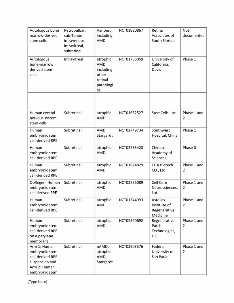
Various attempts have been attempted to overcome these limitations. Extensive research has been conducted to develop an effective film to support stem cell-based RPE cell growth and transplantation [185-193]. The goals of this film are to create an artificial attachment for stem-cell based RPE cells that would negate the need for attachment to diseased BM, as well as providing structural and physiological support. The major limitation of the film is its potential adverse effect on subretinal physiology and, for films that biodegrade, the ability of RPE cells to attach to remaining BM following dissolution of the film. The use of films has transitioned to clinical research, as can be seen in Table 1, with two groups using a film on which RPE cells are transplanted.
Other methods to improve RPE cellular adherence to aged BM include altering integrin expression on RPE cells [194, 195], modifying the extracellular matrix of RPE cells prior to transplantation [196], and modifying native BM of treated eyes prior to transplantation [197]. While demonstrating preclinical effectiveness, these methods have not yet translated to human studies.
7 Ongoing studies Table 1 includes a list of ongoing or recently completed studies investigating various types of stem cell-derived therapies in atrophic AMD and nAMD. A wide variety of cell sources are included in these studies. Table 1. List of ongoing or recently completed stem-cell based therapies in AMD.
Type of Stem Cell Therapy
Transplantation Method
Ocular Pathology
Clinicaltrials.gov Number
Sponsor Study phase
Autologous induced pluripotent stem cell-derived RPE cells
Subretinal atrophic AMD
NCT02464956 Moorfields Eye Hospital NHS Foundation Trust
Phase 1
Allogenic HLA-matched induced pluripotent stem cell-derived RPE cells
Subretinal nAMD -- RIKEN Phase 1
Autologous bone-marrow derived stem cells
Intravitreal nAMD, atrophic AMD, Stargardt
NCT01518127 University of Sao Paulo
Phase 1 and 2
Autologous bone-marrow derived stem cells
Intravitreal atrophic AMD
NCT02016508 Al-Azhar University
Phase 1 and 2
[Type here]
Autologous bone marrow-derived stem cells
Retrobulbar, sub-Tenon, intravenous, intravitreal, subretinal
Various, including AMD
NCT01920867 Retina Associates of South Florida
Not documented
Autologous bone-marrow derived stem cells
Intravitreal atrophic AMD including other retinal pathologies
NCT01736059 University of California, Davis
Phase 1
Human central nervous system stem cells
Subretinal atrophic AMD
NCT01632527 StemCells, Inc. Phase 1 and 2
Human embryonic stem cell-derived RPE
Subretinal AMD, Stargardt
NCT02749734 Southwest Hospital, China
Phase 1
Human embryonic stem cell-derived RPE
Subretinal atrophic AMD
NCT02755428 Chinese Academy of Sciences
Phase 0
Human embryonic stem cell-derived RPE
Subretinal atrophic AMD
NCT01674829 CHA Biotech CO., Ltd
Phase 1 and 2
OpRegen: Human embryonic stem-cell derived RPE
Subretinal atrophic AMD
NCT02286089 Cell Cure Neurosciences, Ltd.
Phase 1 and 2
Human embryonic stem cell-derived RPE
Subretinal atrophic AMD
NCT01344993 Astellas Institute of Regenerative Medicine
Phase 1 and 2
Human embryonic stem cell-derived RPE on a parylene membrane
Subretinal atrophic AMD
NCT02590692 Regenerative Patch Technologies, LLC
Phase 1 and 2
Arm 1: Human embryonic stem cell-derived RPE suspension and Arm 2: Human embryonic stem
Subretinal nAMD, atrophic AMD, Stargardt
NCT02903576 Federal University of Sao Paulo
Phase 1 and 2
[Type here]
cell-derived RPE seeded on a substrate
8 Conclusion
The limitations of anti-VEGF therapy in the treatment of nAMD and the lack of effective treatment for nonexudative AMD demonstrate the need for new therapies. Stem cell-based therapies are an intriguing therapeutic option and have been extensively investigated over the last several decades. Induced PSC therapies are of particular interest because of their reduced immunogenicity and their potential for reduced ethical controversy over hESC-based therapies. A decade of preclinical studies improving the safety and efficiency of iPSC-derived RPE transplants paved the way for human trials. The investigational treatment of a 70-year-old Japanese woman was the first published utilization of iPSCs in humans, and is a major investigational milestone. The major limitation of applying stem cell-based therapies to patients with AMD is the chronic and complex disease process that needs to be overcome. Despite these limitations, RIKEN and Moorefields Eye Hospital are planning on further studies utilizing iPSCs in the treatment nAMD and non-neovascular AMD, and their results will be eagerly awaited.
9 Expert Opinion The use of iPSCs was first performed on a Japanese female with end-stage AMD, and this marked a
major milestone as the first application of iPSC-based therapy in humans. The preceding decade of advancement in the generation and differentiation of iPSCs, which started in 2006 with the discovery of Yamanaka factors, as well as previous research into RPE transplantation, has allowed for the rapid translation of bench research to clinical trials.
Induced PSC-derived transplants are a promising investigational therapy in AMD research due to the potential for reduced ethical controversy over hESC-based therapies, as well as their reduced immunogenicity. A dampened immune response could improve the survival and function of transplants as well as potentially minimize the need for chronic powerful perioperative systemic immunosuppressive agents. Research into hESCs has demonstrated the potential harms associated with immunosuppression following subretinal transplantation, as reviewed above.
Disadvantages of iPSC-based therapies include their time-consuming and expensive production, particularly for autologous transplants, as well as their relative genomic instability. The use of autologous cells are estimated to require three months to develop and cost approximately $1,000,000 from harvesting to intraocular implantation [127]. The use of allogenic HLA-matched cells allows for a more efficient and economical application, as well as allowing for a more thorough investigation of potential mutations, and consequently likely represents the future of iPSC-based therapies for human AMD research. RIKEN stopped their trial due to genomic instability noted in a batch of iPSC-based RPE tissue. Despite the unclear clinical significance of this finding, they halted further research and revamped their methods prior to continuing their trial.
Induced PSCs have principally been investigated as subretinal transplants to replace or replenish diseased RPE cells. The advantages of this approach is that RPE cells are damaged in AMD and their replacement could theoretically improve photoreceptor function and survival. However, the limitations are numerous. Native RPE cells have a number of functions and it remains to be seen if iPSC-derived RPE

[Type here]
cells are able to replicate these roles. Arguably, transplanted cells may not need to replicate all functions to be therapeutically successful, but it remains to be seen if they are able to replicate the essential roles necessary for a reduction in disease progression. Additionally, the replacement of RPE cells may not overcome the AMD-related damage of the CC and BM, and AMD pathology might progress despite a successful transplantation. Furthermore, the long-term survival and proper functioning of transplanted cells in an environment of increased oxidative stress, increased immunogenicity and surrounding tissue damage remains to be seen.
Induced PSCs may have a more promising role in utilizing paracrine effects than direct tissue replacement. It will be interesting to learn if iPSC-derived tissue can be genetically modified to produce locally-acting growth factors. We suspect that stem-cell based therapies for AMD, including iPSCs-based therapies, will likely move in the direction of utilizing paracrine effects. This is due to the complexity of AMD pathogenesis and the questionable benefit of replacing a defective layer such as the RPE when the surrounding tissue is damaged and exposed to increased oxidative stress and pathologic immunogenicity. Pathologies that are less chronic and cause less extensive tissue damage may be more amenable to direct tissue replacement. Alternatively, advancements in our understanding of photoreceptor transplant interaction with host tissue, in particular the transfer of proteins and/or RNA from transplanted photoreceptors to host tissue as was discovered recently, may hold promise. Additionally, choroidal cell generation from iPSCs has recently been published [198], and a combination of tissues may eventually be more effective.
Despite our view that the paracrine effects of transplanted tissue holds the most promise for AMD treatment at this moment, rapid advancements in the field of stem cell biology and research just beyond the horizon could easily change this perspective. Overall, the research into iPSC-based therapy is in its nascency. Significant progress is required prior to broad application and major pathophysiological hurdles may eventually limit its success in AMD.
Article Highlights
• In both neovascular and advanced non-neovascular age-related macular degeneration (AMD), stem cell therapy has the potential to improve on current therapeutic limitations.
• Induced pluripotent stem cells (iPSCs) are promising in that they are derived from adult human cells, ameliorating the need for embryonic stem cells (ESCs) and have the potential for reduced immunogenicity.
• Retinal pigment epithelial (RPE) tissue, derived from iPSCs, has undergone the most extensive research in AMD therapy and holds the most promise for application in humans.
• Previous studies investigating adjuvant RPE transplantation combined with choroidal neovascular membrane resection have provided excellent lessons for the application of iPSC-based therapies.
• The use of iPSCs was first performed on a Japanese female with end-stage AMD, and this marked a major milestone as the first application of an iPSC-based therapy in humans.
• Significantly more research is needed to determine the efficacy and safety of transplantation of stem cells in AMD.
[Type here]
References 1. Wong, W.L., X. Su, X. Li, et al., Global prevalence of age-related macular degeneration and
disease burden projection for 2020 and 2040: a systematic review and meta-analysis. Lancet Glob Health, 2014. 2(2): p. e106-16.
2. Martin, D.F., M.G. Maguire, S.L. Fine, et al., Ranibizumab and bevacizumab for treatment of neovascular age-related macular degeneration: two-year results. Ophthalmology, 2012. 119(7): p. 1388-98.
3. Brown, D.M., P.K. Kaiser, M. Michels, et al., Ranibizumab versus verteporfin for neovascular age-related macular degeneration. N Engl J Med, 2006. 355(14): p. 1432-44.
4. Rosenfeld, P.J., D.M. Brown, J.S. Heier, et al., Ranibizumab for neovascular age-related macular degeneration. N Engl J Med, 2006. 355(14): p. 1419-31.
5. Singer, M.A., C.C. Awh, S. Sadda, et al., HORIZON: an open-label extension trial of ranibizumab for choroidal neovascularization secondary to age-related macular degeneration. Ophthalmology, 2012. 119(6): p. 1175-83.
6. Maguire, M.G., D.F. Martin, G.S. Ying, et al., Five-Year Outcomes with Anti-Vascular Endothelial Growth Factor Treatment of Neovascular Age-Related Macular Degeneration: The Comparison of Age-Related Macular Degeneration Treatments Trials. Ophthalmology, 2016. 123(8): p. 1751-61.
7. Cohen, S.Y., G. Mimoun, H. Oubraham, et al., Changes in visual acuity in patients with wet age-related macular degeneration treated with intravitreal ranibizumab in daily clinical practice: the LUMIERE study. Retina, 2013. 33(3): p. 474-81.
8. Hjelmqvist, L., C. Lindberg, P. Kanulf, et al., One-year outcomes using ranibizumab for neovascular age-related macular degeneration: results of a prospective and retrospective observational multicentre study. J Ophthalmol, 2011. 2011: p. 405724.
9. Holz, F.G., R. Tadayoni, S. Beatty, et al., Multi-country real-life experience of anti-vascular endothelial growth factor therapy for wet age-related macular degeneration. Br J Ophthalmol, 2015. 99(2): p. 220-6.
10. Kruger Falk, M., H. Kemp, and T.L. Sorensen, Four-year treatment results of neovascular age-related macular degeneration with ranibizumab and causes for discontinuation of treatment. Am J Ophthalmol, 2013. 155(1): p. 89-95.e3.
11. Rakic, J.M., A. Leys, H. Brie, et al., Real-world variability in ranibizumab treatment and associated clinical, quality of life, and safety outcomes over 24 months in patients with neovascular age-related macular degeneration: the HELIOS study. Clin Ophthalmol, 2013. 7: p. 1849-58.
12. Rasmussen, A., S.B. Bloch, J. Fuchs, et al., A 4-year longitudinal study of 555 patients treated with ranibizumab for neovascular age-related macular degeneration. Ophthalmology, 2013. 120(12): p. 2630-6.
13. Souied, E.H., H. Oubraham, G. Mimoun, et al., Changes in visual acuity in patients with wet age-related macular degeneration treated with intravitreal ranibizumab in daily clinical practice: The TWIN Study. Retina, 2015. 35(9): p. 1743-9.
14. Talks, J.S., A.J. Lotery, F. Ghanchi, et al., First-Year Visual Acuity Outcomes in the United Kingdom of Providing Aflibercept According to the VIEW Study Protocol for Age-Related Macular Degeneration. Ophthalmology, 2015.
15. van Asten, F., K.U. Evers-Birkenkamp, J.J. van Lith-Verhoeven, et al., A prospective, observational, open-label, multicentre study to investigate the daily treatment practice of ranibizumab in patients with neovascular age-related macular degeneration. Acta Ophthalmol, 2015. 93(2): p. 126-33.
16. The neovascular age-related macular degeneration database: multicenter study of 92 976 ranibizumab injections: report 1: visual acuity. Ophthalmology, 2014. 121(5): p. 1092-101.

[Type here]
17. Zarranz-Ventura, J., G. Liew, R.L. Johnston, et al., The neovascular age-related macular degeneration database: report 2: incidence, management, and visual outcomes of second treated eyes. Ophthalmology, 2014. 121(10): p. 1966-75.
18. Ross, A.H., P.H. Donachie, A. Sallam, et al., Which visual acuity measurements define high-quality care for patients with neovascular age-related macular degeneration treated with ranibizumab? Eye (Lond), 2013. 27(1): p. 56-64.
19. Arevalo, J.F., A.F. Lasave, L. Wu, et al., Intravitreal bevacizumab for choroidal neovasculariztion in age-related macular degeneration: 5-Year Results of The Pan-American Collaborative Retina Study Group. Retina, 2016. 36(5): p. 859-67.
20. Zarranz-Ventura, J., G. Liew, R.L. Johnston, et al., The neovascular age-related macular degeneration database: report 2: incidence, management, and visual outcomes of second treated eyes. Ophthalmology, 2014. 121(10): p. 1966-75.
21. Mimeault, M., R. Hauke, and S.K. Batra, Stem cells: a revolution in therapeutics-recent advances in stem cell biology and their therapeutic applications in regenerative medicine and cancer therapies. Clin Pharmacol Ther, 2007. 82(3): p. 252-64.
22. Zarbin, M.A., Current concepts in the pathogenesis of age-related macular degeneration. Arch Ophthalmol, 2004. 122(4): p. 598-614.
23. Ambati, J., B.K. Ambati, S.H. Yoo, et al., Age-related macular degeneration: etiology, pathogenesis, and therapeutic strategies. Surv Ophthalmol, 2003. 48(3): p. 257-93.
24. Kinnunen, K., G. Petrovski, M.C. Moe, et al., Molecular mechanisms of retinal pigment epithelium damage and development of age-related macular degeneration. Acta Ophthalmol, 2012. 90(4): p. 299-309.
25. Eldred, G.E. and M.L. Katz, Fluorophores of the human retinal pigment epithelium: separation and spectral characterization. Exp Eye Res, 1988. 47(1): p. 71-86.
26. Dorey, C.K., G. Wu, D. Ebenstein, et al., Cell loss in the aging retina. Relationship to lipofuscin accumulation and macular degeneration. Invest Ophthalmol Vis Sci, 1989. 30(8): p. 1691-9.
27. Karwatowski, W.S., T.E. Jeffries, V.C. Duance, et al., Preparation of Bruch's membrane and analysis of the age-related changes in the structural collagens. Br J Ophthalmol, 1995. 79(10): p. 944-52.
28. Ramrattan, R.S., T.L. van der Schaft, C.M. Mooy, et al., Morphometric analysis of Bruch's membrane, the choriocapillaris, and the choroid in aging. Invest Ophthalmol Vis Sci, 1994. 35(6): p. 2857-64.
29. Pauleikhoff, D., C.A. Harper, J. Marshall, et al., Aging changes in Bruch's membrane. A histochemical and morphologic study. Ophthalmology, 1990. 97(2): p. 171-8.
30. Loffler, K.U. and W.R. Lee, Basal linear deposit in the human macula. Graefes Arch Clin Exp Ophthalmol, 1986. 224(6): p. 493-501.
31. Chong, N.H., J. Keonin, P.J. Luthert, et al., Decreased thickness and integrity of the macular elastic layer of Bruch's membrane correspond to the distribution of lesions associated with age-related macular degeneration. Am J Pathol, 2005. 166(1): p. 241-51.
32. Keenan, T.D., C.E. Pickford, R.J. Holley, et al., Age-dependent changes in heparan sulfate in human Bruch's membrane: implications for age-related macular degeneration. Invest Ophthalmol Vis Sci, 2014. 55(8): p. 5370-9.
33. Wakatsuki, Y., A. Shinojima, A. Kawamura, et al., Correlation of Aging and Segmental Choroidal Thickness Measurement using Swept Source Optical Coherence Tomography in Healthy Eyes. PLoS One, 2015. 10(12): p. e0144156.
34. Chirco, K.R., E.H. Sohn, E.M. Stone, et al., Structural and molecular changes in the aging choroid: implications for age-related macular degeneration. Eye (Lond), 2017. 31(1): p. 10-25.
[Type here]
35. Evans, M.D., M. Dizdaroglu, and M.S. Cooke, Oxidative DNA damage and disease: induction, repair and significance. Mutat Res, 2004. 567(1): p. 1-61.
36. Blasiak, J., G. Petrovski, Z. Vereb, et al., Oxidative stress, hypoxia, and autophagy in the neovascular processes of age-related macular degeneration. Biomed Res Int, 2014. 2014: p. 768026.
37. Samiec, P.S., C. Drews-Botsch, E.W. Flagg, et al., Glutathione in human plasma: decline in association with aging, age-related macular degeneration, and diabetes. Free Radic Biol Med, 1998. 24(5): p. 699-704.
38. A randomized, placebo-controlled, clinical trial of high-dose supplementation with vitamins C and E, beta carotene, and zinc for age-related macular degeneration and vision loss: AREDS report no. 8. Arch Ophthalmol, 2001. 119(10): p. 1417-36.
39. Seddon, J.M., S. George, B. Rosner, et al., Progression of age-related macular degeneration: prospective assessment of C-reactive protein, interleukin 6, and other cardiovascular biomarkers. Arch Ophthalmol, 2005. 123(6): p. 774-82.
40. Bok, D., Evidence for an inflammatory process in age-related macular degeneration gains new support. Proc Natl Acad Sci U S A, 2005. 102(20): p. 7053-4.
41. Grossniklaus, H.E., P.H. Miskala, W.R. Green, et al., Histopathologic and ultrastructural features of surgically excised subfoveal choroidal neovascular lesions: submacular surgery trials report no. 7. Arch Ophthalmol, 2005. 123(7): p. 914-21.
42. Klein, R.J., C. Zeiss, E.Y. Chew, et al., Complement factor H polymorphism in age-related macular degeneration. Science, 2005. 308(5720): p. 385-9.
43. Haines, J.L., M.A. Hauser, S. Schmidt, et al., Complement factor H variant increases the risk of age-related macular degeneration. Science, 2005. 308(5720): p. 419-21.
44. Ambati, J., J.P. Atkinson, and B.D. Gelfand, Immunology of age-related macular degeneration. Nat Rev Immunol, 2013. 13(6): p. 438-51.
45. Khandhadia, S., V. Cipriani, J.R. Yates, et al., Age-related macular degeneration and the complement system. Immunobiology, 2012. 217(2): p. 127-46.
46. van Lookeren Campagne, M., J. LeCouter, B.L. Yaspan, et al., Mechanisms of age-related macular degeneration and therapeutic opportunities. J Pathol, 2014. 232(2): p. 151-64.
47. Francis, P.J., D.W. Schultz, S. Hamon, et al., Haplotypes in the complement factor H (CFH) gene: associations with drusen and advanced age-related macular degeneration. PLoS One, 2007. 2(11): p. e1197.
48. Nozaki, M., B.J. Raisler, E. Sakurai, et al., Drusen complement components C3a and C5a promote choroidal neovascularization. Proc Natl Acad Sci U S A, 2006. 103(7): p. 2328-33.
49. Mullins, R.F., D.P. Schoo, E.H. Sohn, et al., The membrane attack complex in aging human choriocapillaris: relationship to macular degeneration and choroidal thinning. Am J Pathol, 2014. 184(11): p. 3142-53.
50. Benzaquen, L.R., A. Nicholson-Weller, and J.A. Halperin, Terminal complement proteins C5b-9 release basic fibroblast growth factor and platelet-derived growth factor from endothelial cells. J Exp Med, 1994. 179(3): p. 985-92.
51. Bora, P.S., J.H. Sohn, J.M. Cruz, et al., Role of complement and complement membrane attack complex in laser-induced choroidal neovascularization. J Immunol, 2005. 174(1): p. 491-7.
52. Tsutsumi, C., K.H. Sonoda, K. Egashira, et al., The critical role of ocular-infiltrating macrophages in the development of choroidal neovascularization. J Leukoc Biol, 2003. 74(1): p. 25-32.
53. Thomson, J.A., J. Itskovitz-Eldor, S.S. Shapiro, et al., Embryonic stem cell lines derived from human blastocysts. Science, 1998. 282(5391): p. 1145-7.
54. Takahashi, K. and S. Yamanaka, Induction of pluripotent stem cells from mouse embryonic and adult fibroblast cultures by defined factors. Cell, 2006. 126(4): p. 663-76.
[Type here]
55. Takahashi, K., K. Tanabe, M. Ohnuki, et al., Induction of pluripotent stem cells from adult human fibroblasts by defined factors. Cell, 2007. 131(5): p. 861-72.
56. Yu, J., M.A. Vodyanik, K. Smuga-Otto, et al., Induced pluripotent stem cell lines derived from human somatic cells. Science, 2007. 318(5858): p. 1917-20.
57. Huangfu, D., K. Osafune, R. Maehr, et al., Induction of pluripotent stem cells from primary human fibroblasts with only Oct4 and Sox2. Nat Biotechnol, 2008. 26(11): p. 1269-75.
58. Mali, P., Z. Ye, H.H. Hommond, et al., Improved efficiency and pace of generating induced pluripotent stem cells from human adult and fetal fibroblasts. Stem Cells, 2008. 26(8): p. 1998-2005.
59. Judson, R.L., J.E. Babiarz, M. Venere, et al., Embryonic stem cell-specific microRNAs promote induced pluripotency. Nat Biotechnol, 2009. 27(5): p. 459-61.
60. Nakagawa, M., M. Koyanagi, K. Tanabe, et al., Generation of induced pluripotent stem cells without Myc from mouse and human fibroblasts. Nat Biotechnol, 2008. 26(1): p. 101-6.
61. Yu, J., K. Hu, K. Smuga-Otto, et al., Human induced pluripotent stem cells free of vector and transgene sequences. Science, 2009. 324(5928): p. 797-801.
62. Stadtfeld, M., M. Nagaya, J. Utikal, et al., Induced pluripotent stem cells generated without viral integration. Science, 2008. 322(5903): p. 945-9.
63. Fusaki, N., H. Ban, A. Nishiyama, et al., Efficient induction of transgene-free human pluripotent stem cells using a vector based on Sendai virus, an RNA virus that does not integrate into the host genome. Proc Jpn Acad Ser B Phys Biol Sci, 2009. 85(8): p. 348-62.
64. Woltjen, K., I.P. Michael, P. Mohseni, et al., piggyBac transposition reprograms fibroblasts to induced pluripotent stem cells. Nature, 2009. 458(7239): p. 766-70.
65. Jia, F., K.D. Wilson, N. Sun, et al., A nonviral minicircle vector for deriving human iPS cells. Nat Methods, 2010. 7(3): p. 197-9.
66. Warren, L., P.D. Manos, T. Ahfeldt, et al., Highly efficient reprogramming to pluripotency and directed differentiation of human cells with synthetic modified mRNA. Cell Stem Cell, 2010. 7(5): p. 618-30.
67. Malik, N. and M.S. Rao, A review of the methods for human iPSC derivation. Methods Mol Biol, 2013. 997: p. 23-33.
68. Miyoshi, N., H. Ishii, H. Nagano, et al., Reprogramming of mouse and human cells to pluripotency using mature microRNAs. Cell Stem Cell, 2011. 8(6): p. 633-8.
69. Kaji, K., K. Norrby, A. Paca, et al., Virus-free induction of pluripotency and subsequent excision of reprogramming factors. Nature, 2009. 458(7239): p. 771-5.
70. Lyssiotis, C.A., R.K. Foreman, J. Staerk, et al., Reprogramming of murine fibroblasts to induced pluripotent stem cells with chemical complementation of Klf4. Proc Natl Acad Sci U S A, 2009. 106(22): p. 8912-7.
71. Kim, D., C.H. Kim, J.I. Moon, et al., Generation of human induced pluripotent stem cells by direct delivery of reprogramming proteins. Cell Stem Cell, 2009. 4(6): p. 472-6.
72. Strauss, O., The retinal pigment epithelium in visual function. Physiol Rev, 2005. 85(3): p. 845-81. 73. Runkle, E.A. and D.A. Antonetti, The blood-retinal barrier: structure and functional significance.
Methods Mol Biol, 2011. 686: p. 133-48. 74. Young, R.W. and D. Bok, Participation of the retinal pigment epithelium in the rod outer segment
renewal process. J Cell Biol, 1969. 42(2): p. 392-403. 75. Baehr, W., S.M. Wu, A.C. Bird, et al., The retinoid cycle and retina disease. Vision Res, 2003.
43(28): p. 2957-8. 76. Holtkamp, G.M., A. Kijlstra, R. Peek, et al., Retinal pigment epithelium-immune system
interactions: cytokine production and cytokine-induced changes. Prog Retin Eye Res, 2001. 20(1): p. 29-48.
[Type here]
77. Ishida, K., N. Panjwani, Z. Cao, et al., Participation of pigment epithelium in ocular immune privilege. 3. Epithelia cultured from iris, ciliary body, and retina suppress T-cell activation by partially non-overlapping mechanisms. Ocul Immunol Inflamm, 2003. 11(2): p. 91-105.
78. Berger, A.S. and H.J. Kaplan, Clinical experience with the surgical removal of subfoveal neovascular membranes. Short-term postoperative results. Ophthalmology, 1992. 99(6): p. 969-75; discussion 975-6.
79. Lambert, H.M., A. Capone, Jr., T.M. Aaberg, et al., Surgical excision of subfoveal neovascular membranes in age-related macular degeneration. Am J Ophthalmol, 1992. 113(3): p. 257-62.
80. Grossniklaus, H.E., A.K. Hutchinson, A. Capone, Jr., et al., Clinicopathologic features of surgically excised choroidal neovascular membranes. Ophthalmology, 1994. 101(6): p. 1099-111.
81. Spraul, C.W., G.E. Lang, H.E. Grossniklaus, et al., Histologic and morphometric analysis of the choroid, Bruch's membrane, and retinal pigment epithelium in postmortem eyes with age-related macular degeneration and histologic examination of surgically excised choroidal neovascular membranes. Surv Ophthalmol, 1999. 44 Suppl 1: p. S10-32.
82. Castellarin, A.A., M. Nasir, I.K. Sugino, et al., Progressive presumed choriocapillaris atrophy after surgery for age-related macular degeneration. Retina, 1998. 18(2): p. 143-9.
83. Algvere, P.V., L. Berglin, P. Gouras, et al., Transplantation of fetal retinal pigment epithelium in age-related macular degeneration with subfoveal neovascularization. Graefes Arch Clin Exp Ophthalmol, 1994. 232(12): p. 707-16.
84. Peyman, G.A., K.J. Blinder, C.L. Paris, et al., A technique for retinal pigment epithelium transplantation for age-related macular degeneration secondary to extensive subfoveal scarring. Ophthalmic Surg, 1991. 22(2): p. 102-8.
85. Algvere, P.V., L. Berglin, P. Gouras, et al., Transplantation of RPE in age-related macular degeneration: observations in disciform lesions and dry RPE atrophy. Graefes Arch Clin Exp Ophthalmol, 1997. 235(3): p. 149-58.
86. Pieramici, D.J., E. De Juan, Jr., G.Y. Fujii, et al., Limited inferior macular translocation for the treatment of subfoveal choroidal neovascularization secondary to age-related macular degeneration. Am J Ophthalmol, 2000. 130(4): p. 419-28.
87. Binder, S., U. Stolba, I. Krebs, et al., Transplantation of autologous retinal pigment epithelium in eyes with foveal neovascularization resulting from age-related macular degeneration: a pilot study. Am J Ophthalmol, 2002. 133(2): p. 215-25.
88. Binder, S., I. Krebs, R.D. Hilgers, et al., Outcome of transplantation of autologous retinal pigment epithelium in age-related macular degeneration: a prospective trial. Invest Ophthalmol Vis Sci, 2004. 45(11): p. 4151-60.
89. Joussen, A.M., F.M. Heussen, S. Joeres, et al., Autologous translocation of the choroid and retinal pigment epithelium in age-related macular degeneration. Am J Ophthalmol, 2006. 142(1): p. 17-30.
90. Joussen, A.M., S. Joeres, N. Fawzy, et al., Autologous translocation of the choroid and retinal pigment epithelium in patients with geographic atrophy. Ophthalmology, 2007. 114(3): p. 551-60.
91. Lu, Y., L. Han, C. Wang, et al., A comparison of autologous transplantation of retinal pigment epithelium (RPE) monolayer sheet graft with RPE-Bruch's membrane complex graft in neovascular age-related macular degeneration. Acta Ophthalmol, 2016.
92. Maaijwee, K., H. Heimann, T. Missotten, et al., Retinal pigment epithelium and choroid translocation in patients with exudative age-related macular degeneration: long-term results. Graefes Arch Clin Exp Ophthalmol, 2007. 245(11): p. 1681-9.
[Type here]
93. MacLaren, R.E., G.S. Uppal, K.S. Balaggan, et al., Autologous transplantation of the retinal pigment epithelium and choroid in the treatment of neovascular age-related macular degeneration. Ophthalmology, 2007. 114(3): p. 561-70.
94. Tezel, T.H., L.V. Del Priore, A.S. Berger, et al., Adult retinal pigment epithelial transplantation in exudative age-related macular degeneration. Am J Ophthalmol, 2007. 143(4): p. 584-95.
95. Stanga, P.E., A. Kychenthal, F.W. Fitzke, et al., Retinal pigment epithelium translocation after choroidal neovascular membrane removal in age-related macular degeneration. Ophthalmology, 2002. 109(8): p. 1492-8.
96. Akduman, L., M.P. Karavellas, J.C. MacDonald, et al., Macular translocation with retinotomy and retinal rotation for exudative age-related macular degeneration. Retina, 1999. 19(5): p. 418-23.
97. van Meurs, J.C., E. ter Averst, L.J. Hofland, et al., Autologous peripheral retinal pigment epithelium translocation in patients with subfoveal neovascular membranes. Br J Ophthalmol, 2004. 88(1): p. 110-3.
98. Algvere, P.V., P. Gouras, and E. Dafgard Kopp, Long-term outcome of RPE allografts in non-immunosuppressed patients with AMD. Eur J Ophthalmol, 1999. 9(3): p. 217-30.
99. Klimanskaya, I., J. Hipp, K.A. Rezai, et al., Derivation and comparative assessment of retinal pigment epithelium from human embryonic stem cells using transcriptomics. Cloning Stem Cells, 2004. 6(3): p. 217-45.
100. Osakada, F., Z.B. Jin, Y. Hirami, et al., In vitro differentiation of retinal cells from human pluripotent stem cells by small-molecule induction. J Cell Sci, 2009. 122(Pt 17): p. 3169-79.
101. Idelson, M., R. Alper, A. Obolensky, et al., Directed differentiation of human embryonic stem cells into functional retinal pigment epithelium cells. Cell Stem Cell, 2009. 5(4): p. 396-408.
102. Kawasaki, H., H. Suemori, K. Mizuseki, et al., Generation of dopaminergic neurons and pigmented epithelia from primate ES cells by stromal cell-derived inducing activity. Proc Natl Acad Sci U S A, 2002. 99(3): p. 1580-5.
103. Zhang, Y.S., Z.Y. Lu, Y. Yu, et al., Derivation, culture and retinal pigment epithelial differentiation of human embryonic stem cells using human fibroblast feeder cells. J Assist Reprod Genet, 2012. 29(8): p. 735-44.
104. Cho, M.S., S.J. Kim, S.Y. Ku, et al., Generation of retinal pigment epithelial cells from human embryonic stem cell-derived spherical neural masses. Stem Cell Res, 2012. 9(2): p. 101-9.
105. Maruotti, J., K. Wahlin, D. Gorrell, et al., A simple and scalable process for the differentiation of retinal pigment epithelium from human pluripotent stem cells. Stem Cells Transl Med, 2013. 2(5): p. 341-54.
106. Buchholz, D.E., S.T. Hikita, T.J. Rowland, et al., Derivation of functional retinal pigmented epithelium from induced pluripotent stem cells. Stem Cells, 2009. 27(10): p. 2427-34.
107. Lane, A., L.R. Philip, L. Ruban, et al., Engineering efficient retinal pigment epithelium differentiation from human pluripotent stem cells. Stem Cells Transl Med, 2014. 3(11): p. 1295-304.
108. Leach, L.L., D.E. Buchholz, V.P. Nadar, et al., Canonical/beta-catenin Wnt pathway activation improves retinal pigmented epithelium derivation from human embryonic stem cells. Invest Ophthalmol Vis Sci, 2015. 56(2): p. 1002-13.
109. Maruotti, J., S.R. Sripathi, K. Bharti, et al., Small-molecule-directed, efficient generation of retinal pigment epithelium from human pluripotent stem cells. Proc Natl Acad Sci U S A, 2015. 112(35): p. 10950-5.
110. Gong, J., M.A. Fields, E.F. Moreira, et al., Differentiation of Human Protein-Induced Pluripotent Stem Cells toward a Retinal Pigment Epithelial Cell Fate. PLoS One, 2015. 10(11): p. e0143272.
111. Choudhary, P., H. Booth, A. Gutteridge, et al., Directing Differentiation of Pluripotent Stem Cells Toward Retinal Pigment Epithelium Lineage. Stem Cells Transl Med, 2016.
[Type here]
112. Haruta, M., Y. Sasai, H. Kawasaki, et al., In vitro and in vivo characterization of pigment epithelial cells differentiated from primate embryonic stem cells. Invest Ophthalmol Vis Sci, 2004. 45(3): p. 1020-5.
113. Carr, A.J., A.A. Vugler, S.T. Hikita, et al., Protective effects of human iPS-derived retinal pigment epithelium cell transplantation in the retinal dystrophic rat. PLoS One, 2009. 4(12): p. e8152.
114. Vugler, A., A.J. Carr, J. Lawrence, et al., Elucidating the phenomenon of HESC-derived RPE: anatomy of cell genesis, expansion and retinal transplantation. Exp Neurol, 2008. 214(2): p. 347-61.
115. Lund, R.D., S. Wang, I. Klimanskaya, et al., Human embryonic stem cell-derived cells rescue visual function in dystrophic RCS rats. Cloning Stem Cells, 2006. 8(3): p. 189-99.
116. Sun, J., M. Mandai, H. Kamao, et al., Protective Effects of Human iPS-Derived Retinal Pigmented Epithelial Cells in Comparison with Human Mesenchymal Stromal Cells and Human Neural Stem Cells on the Degenerating Retina in rd1 mice. Stem Cells, 2015. 33(5): p. 1543-53.
117. Plaza Reyes, A., S. Petrus-Reurer, L. Antonsson, et al., Xeno-Free and Defined Human Embryonic Stem Cell-Derived Retinal Pigment Epithelial Cells Functionally Integrate in a Large-Eyed Preclinical Model. Stem Cell Reports, 2016. 6(1): p. 9-17.
118. Koss, M.J., P. Falabella, F.R. Stefanini, et al., Subretinal implantation of a monolayer of human embryonic stem cell-derived retinal pigment epithelium: a feasibility and safety study in Yucatan minipigs. Graefes Arch Clin Exp Ophthalmol, 2016. 254(8): p. 1553-65.
119. Kokkinaki, M., N. Sahibzada, and N. Golestaneh, Human induced pluripotent stem-derived retinal pigment epithelium (RPE) cells exhibit ion transport, membrane potential, polarized vascular endothelial growth factor secretion, and gene expression pattern similar to native RPE. Stem Cells, 2011. 29(5): p. 825-35.
120. Sugino, I.K., Q. Sun, J. Wang, et al., Comparison of FRPE and human embryonic stem cell-derived RPE behavior on aged human Bruch's membrane. Invest Ophthalmol Vis Sci, 2011. 52(8): p. 4979-97.
121. Liao, J.L., J. Yu, K. Huang, et al., Molecular signature of primary retinal pigment epithelium and stem-cell-derived RPE cells. Hum Mol Genet, 2010. 19(21): p. 4229-38.
122. Schwartz, S.D., J.P. Hubschman, G. Heilwell, et al., Embryonic stem cell trials for macular degeneration: a preliminary report. Lancet, 2012. 379(9817): p. 713-20.
123. Schwartz, S.D., C.D. Regillo, B.L. Lam, et al., Human embryonic stem cell-derived retinal pigment epithelium in patients with age-related macular degeneration and Stargardt's macular dystrophy: follow-up of two open-label phase 1/2 studies. Lancet, 2015. 385(9967): p. 509-16.
124. Schwartz, S.D., G. Tan, H. Hosseini, et al., Subretinal Transplantation of Embryonic Stem Cell-Derived Retinal Pigment Epithelium for the Treatment of Macular Degeneration: An Assessment at 4 Years. Invest Ophthalmol Vis Sci, 2016. 57(5): p. ORSFc1-9.
125. Song, W.K., K.M. Park, H.J. Kim, et al., Treatment of macular degeneration using embryonic stem cell-derived retinal pigment epithelium: preliminary results in Asian patients. Stem Cell Reports, 2015. 4(5): p. 860-72.
126. Garber, K., RIKEN suspends first clinical trial involving induced pluripotent stem cells. Nat Biotechnol, 2015. 33(9): p. 890-1.
127. Chakradhar, S., An eye to the future: Researchers debate best path for stem cell-derived therapies. Nat Med, 2016. 22(2): p. 116-9.
128. Foss, A.J., L.J. Scott, C.A. Rogers, et al., Changes in intraocular pressure in study and fellow eyes in the IVAN trial. Br J Ophthalmol, 2016.
129. Mandai, M., A. Watanabe, Y. Kurimoto, et al., Autologous Induced Stem-Cell-Derived Retinal Cells for Macular Degeneration. N Engl J Med, 2017. 376(11): p. 1038-1046.

[Type here]
130. Yoshihara, M., Y. Hayashizaki, and Y. Murakawa, Genomic Instability of iPSCs: Challenges Towards Their Clinical Applications. Stem Cell Rev, 2016.
131. Zhao, X., J. Liu, and I. Ahmad, Differentiation of embryonic stem cells into retinal neurons. Biochem Biophys Res Commun, 2002. 297(2): p. 177-84.
132. Zhao, X., J. Liu, and I. Ahmad, Differentiation of embryonic stem cells to retinal cells in vitro. Methods Mol Biol, 2006. 330: p. 401-16.
133. Yanai, A., C. Laver, A.W. Joe, et al., Efficient Production of Photoreceptor Precursor Cells from Human Embryonic Stem Cells. Methods Mol Biol, 2016. 1307: p. 357-69.
134. Lamba, D.A., M.O. Karl, C.B. Ware, et al., Efficient generation of retinal progenitor cells from human embryonic stem cells. Proc Natl Acad Sci U S A, 2006. 103(34): p. 12769-74.
135. Hirami, Y., F. Osakada, K. Takahashi, et al., Generation of retinal cells from mouse and human induced pluripotent stem cells. Neurosci Lett, 2009. 458(3): p. 126-31.
136. Meyer, J.S., R.L. Shearer, E.E. Capowski, et al., Modeling early retinal development with human embryonic and induced pluripotent stem cells. Proc Natl Acad Sci U S A, 2009. 106(39): p. 16698-703.
137. Takahashi, M., T.D. Palmer, J. Takahashi, et al., Widespread integration and survival of adult-derived neural progenitor cells in the developing optic retina. Mol Cell Neurosci, 1998. 12(6): p. 340-8.
138. Guo, Y., P. Saloupis, S.J. Shaw, et al., Engraftment of adult neural progenitor cells transplanted to rat retina injured by transient ischemia. Invest Ophthalmol Vis Sci, 2003. 44(7): p. 3194-201.
139. Qiu, G., M.J. Seiler, C. Mui, et al., Photoreceptor differentiation and integration of retinal progenitor cells transplanted into transgenic rats. Exp Eye Res, 2005. 80(4): p. 515-25.
140. Arnhold, S., H. Klein, I. Semkova, et al., Neurally selected embryonic stem cells induce tumor formation after long-term survival following engraftment into the subretinal space. Invest Ophthalmol Vis Sci, 2004. 45(12): p. 4251-5.
141. Schraermeyer, U., G. Thumann, T. Luther, et al., Subretinally transplanted embryonic stem cells rescue photoreceptor cells from degeneration in the RCS rats. Cell Transplant, 2001. 10(8): p. 673-80.
142. Meyer, J.S., M.L. Katz, J.A. Maruniak, et al., Neural differentiation of mouse embryonic stem cells in vitro and after transplantation into eyes of mutant mice with rapid retinal degeneration. Brain Res, 2004. 1014(1-2): p. 131-44.
143. West, E.L., A. Gonzalez-Cordero, C. Hippert, et al., Defining the integration capacity of embryonic stem cell-derived photoreceptor precursors. Stem Cells, 2012. 30(7): p. 1424-35.
144. Mizumoto, H., K. Mizumoto, M.A. Shatos, et al., Retinal transplantation of neural progenitor cells derived from the brain of GFP transgenic mice. Vision Res, 2003. 43(16): p. 1699-708.
145. Pressmar, S., M. Ader, G. Richard, et al., The fate of heterotopically grafted neural precursor cells in the normal and dystrophic adult mouse retina. Invest Ophthalmol Vis Sci, 2001. 42(13): p. 3311-9.
146. Li, T., M. Lewallen, S. Chen, et al., Multipotent stem cells isolated from the adult mouse retina are capable of producing functional photoreceptor cells. Cell Res, 2013. 23(6): p. 788-802.
147. Tucker, B.A., I.H. Park, S.D. Qi, et al., Transplantation of adult mouse iPS cell-derived photoreceptor precursors restores retinal structure and function in degenerative mice. PLoS One, 2011. 6(4): p. e18992.
148. Wang, S., S. Girman, B. Lu, et al., Long-term vision rescue by human neural progenitors in a rat model of photoreceptor degeneration. Invest Ophthalmol Vis Sci, 2008. 49(7): p. 3201-6.
149. McGill, T.J., B. Cottam, B. Lu, et al., Transplantation of human central nervous system stem cells - neuroprotection in retinal degeneration. Eur J Neurosci, 2012. 35(3): p. 468-77.
[Type here]
150. Banin, E., A. Obolensky, M. Idelson, et al., Retinal incorporation and differentiation of neural precursors derived from human embryonic stem cells. Stem Cells, 2006. 24(2): p. 246-57.
151. Young, M.J., J. Ray, S.J. Whiteley, et al., Neuronal differentiation and morphological integration of hippocampal progenitor cells transplanted to the retina of immature and mature dystrophic rats. Mol Cell Neurosci, 2000. 16(3): p. 197-205.
152. Nishida, A., M. Takahashi, H. Tanihara, et al., Incorporation and differentiation of hippocampus-derived neural stem cells transplanted in injured adult rat retina. Invest Ophthalmol Vis Sci, 2000. 41(13): p. 4268-74.
153. Wojciechowski, A.B., U. Englund, C. Lundberg, et al., Subretinal transplantation of brain-derived precursor cells to young RCS rats promotes photoreceptor cell survival. Exp Eye Res, 2002. 75(1): p. 23-37.
154. Mandai, M., M. Fujii, T. Hashiguchi, et al., iPSC-Derived Retina Transplants Improve Vision in rd1 End-Stage Retinal-Degeneration Mice. Stem Cell Reports, 2017. 8(1): p. 69-83.
155. Chaudhry, G.R., C. Fecek, M.M. Lai, et al., Fate of embryonic stem cell derivatives implanted into the vitreous of a slow retinal degenerative mouse model. Stem Cells Dev, 2009. 18(2): p. 247-58.
156. Lamba, D.A., A. McUsic, R.K. Hirata, et al., Generation, purification and transplantation of photoreceptors derived from human induced pluripotent stem cells. PLoS One, 2010. 5(1): p. e8763.
157. Lamba, D.A., J. Gust, and T.A. Reh, Transplantation of human embryonic stem cell-derived photoreceptors restores some visual function in Crx-deficient mice. Cell Stem Cell, 2009. 4(1): p. 73-9.
158. Wang, S., B. Lu, S. Girman, et al., Non-invasive stem cell therapy in a rat model for retinal degeneration and vascular pathology. PLoS One, 2010. 5(2): p. e9200.
159. Chao, J.R., D.A. Lamba, T.R. Klesert, et al., Transplantation of Human Embryonic Stem Cell-Derived Retinal Cells into the Subretinal Space of a Non-Human Primate. Transl Vis Sci Technol, 2017. 6(3): p. 4.
160. Santos-Ferreira, T., S. Llonch, O. Borsch, et al., Retinal transplantation of photoreceptors results in donor-host cytoplasmic exchange. Nat Commun, 2016. 7: p. 13028.
161. Singh, M.S., J. Balmer, A.R. Barnard, et al., Transplanted photoreceptor precursors transfer proteins to host photoreceptors by a mechanism of cytoplasmic fusion. Nat Commun, 2016. 7: p. 13537.
162. Pearson, R.A., A. Gonzalez-Cordero, E.L. West, et al., Donor and host photoreceptors engage in material transfer following transplantation of post-mitotic photoreceptor precursors. Nat Commun, 2016. 7: p. 13029.
163. MacLaren, R.E., R.A. Pearson, A. MacNeil, et al., Retinal repair by transplantation of photoreceptor precursors. Nature, 2006. 444(7116): p. 203-7.
164. Moisseiev, E., Z. Smit-McBride, S. Oltjen, et al., Intravitreal Administration of Human Bone Marrow CD34+ Stem Cells in a Murine Model of Retinal Degeneration. Invest Ophthalmol Vis Sci, 2016. 57(10): p. 4125-35.
165. Park, S.S., E. Moisseiev, G. Bauer, et al., Advances in bone marrow stem cell therapy for retinal dysfunction. Prog Retin Eye Res, 2016.
166. Kuriyan, A.E., T.A. Albini, J.H. Townsend, et al., Vision Loss after Intravitreal Injection of Autologous "Stem Cells" for AMD. N Engl J Med, 2017. 376(11): p. 1047-1053.
167. Kim, J.Y., Y.S. You, S.H. Kim, et al., Epiretinal membrane formation after intravitreal autologous stem cell implantation in a retinitis pigmentosa pagient. Retin Cases Brief Rep, 2016.
168. Leung, E.H., H.W. Flynn, Jr., T.A. Albini, et al., Retinal Detachment After Subretinal Stem Cell Transplantation. Ophthalmic Surg Lasers Imaging Retina, 2016. 47(6): p. 600-1.

[Type here]
169. Grisanti, S., M. Ishioka, M. Kosiewicz, et al., Immunity and immune privilege elicited by cultured retinal pigment epithelial cell transplants. Invest Ophthalmol Vis Sci, 1997. 38(8): p. 1619-26.
170. Zhang, X. and D. Bok, Transplantation of retinal pigment epithelial cells and immune response in the subretinal space. Invest Ophthalmol Vis Sci, 1998. 39(6): p. 1021-7.
171. Jiang, L.Q. and J.W. Streilein, Immune responses elicited by transplantation and tissue-restricted antigens expressed on retinal tissues implanted subconjunctivally. Transplantation, 1991. 52(3): p. 513-9.
172. Drukker, M., Immunogenicity of human embryonic stem cells: can we achieve tolerance? Springer Semin Immunopathol, 2004. 26(1-2): p. 201-13.
173. Zamiri, P., S. Sugita, and J.W. Streilein, Immunosuppressive properties of the pigmented epithelial cells and the subretinal space. Chem Immunol Allergy, 2007. 92: p. 86-93.
174. Jorgensen, A., A.K. Wiencke, M. la Cour, et al., Human retinal pigment epithelial cell-induced apoptosis in activated T cells. Invest Ophthalmol Vis Sci, 1998. 39(9): p. 1590-9.
175. Zamiri, P., S. Masli, J.W. Streilein, et al., Pigment epithelial growth factor suppresses inflammation by modulating macrophage activation. Invest Ophthalmol Vis Sci, 2006. 47(9): p. 3912-8.
176. Araki, R., M. Uda, Y. Hoki, et al., Negligible immunogenicity of terminally differentiated cells derived from induced pluripotent or embryonic stem cells. Nature, 2013. 494(7435): p. 100-4.
177. Guha, P., J.W. Morgan, G. Mostoslavsky, et al., Lack of immune response to differentiated cells derived from syngeneic induced pluripotent stem cells. Cell Stem Cell, 2013. 12(4): p. 407-12.
178. Sugita, S., Y. Iwasaki, K. Makabe, et al., Lack of T Cell Response to iPSC-Derived Retinal Pigment Epithelial Cells from HLA Homozygous Donors. Stem Cell Reports, 2016. 7(4): p. 619-634.
179. Sugita, S., Y. Iwasaki, K. Makabe, et al., Successful Transplantation of Retinal Pigment Epithelial Cells from MHC Homozygote iPSCs in MHC-Matched Models. Stem Cell Reports, 2016. 7(4): p. 635-648.
180. Okita, K., Y. Matsumura, Y. Sato, et al., A more efficient method to generate integration-free human iPS cells. Nat Methods, 2011. 8(5): p. 409-12.
181. Cai, H. and L.V. Del Priore, Bruch membrane aging alters the gene expression profile of human retinal pigment epithelium. Curr Eye Res, 2006. 31(2): p. 181-9.
182. Del Priore, L.V. and T.H. Tezel, Reattachment rate of human retinal pigment epithelium to layers of human Bruch's membrane. Arch Ophthalmol, 1998. 116(3): p. 335-41.
183. Tezel, T.H. and L.V. Del Priore, Reattachment to a substrate prevents apoptosis of human retinal pigment epithelium. Graefes Arch Clin Exp Ophthalmol, 1997. 235(1): p. 41-7.
184. Gullapalli, V.K., I.K. Sugino, Y. Van Patten, et al., Impaired RPE survival on aged submacular human Bruch's membrane. Exp Eye Res, 2005. 80(2): p. 235-48.
185. Lu, L., C.A. Garcia, and A.G. Mikos, Retinal pigment epithelium cell culture on thin biodegradable poly(DL-lactic-co-glycolic acid) films. J Biomater Sci Polym Ed, 1998. 9(11): p. 1187-205.
186. Cai, S., M.E. Smith, S.M. Redenti, et al., Mouse retinal progenitor cell dynamics on electrospun poly (-caprolactone). J Biomater Sci Polym Ed, 2012. 23(11): p. 1451-65.
187. Tomita, M., E. Lavik, H. Klassen, et al., Biodegradable polymer composite grafts promote the survival and differentiation of retinal progenitor cells. Stem Cells, 2005. 23(10): p. 1579-88.
188. Lu, B., Y.C. Tai, and M.S. Humayun, Microdevice-based cell therapy for age-related macular degeneration. Dev Ophthalmol, 2014. 53: p. 155-66.
189. Warnke, P.H., M. Alamein, S. Skabo, et al., Primordium of an artificial Bruch's membrane made of nanofibers for engineering of retinal pigment epithelium cell monolayers. Acta Biomater, 2013. 9(12): p. 9414-22.
[Type here]
190. Lawley, E., P. Baranov, and M. Young, Hybrid vitronectin-mimicking polycaprolactone scaffolds for human retinal progenitor cell differentiation and transplantation. J Biomater Appl, 2015. 29(6): p. 894-902.
191. Yao, J., C.W. Ko, P.Y. Baranov, et al., Enhanced differentiation and delivery of mouse retinal progenitor cells using a micropatterned biodegradable thin-film polycaprolactone scaffold. Tissue Eng Part A, 2015. 21(7-8): p. 1247-60.
192. Calejo, M.T., T. Ilmarinen, H. Jongprasitkul, et al., Honeycomb porous films as permeable scaffold materials for human embryonic stem cell-derived retinal pigment epithelium. J Biomed Mater Res A, 2016. 104(7): p. 1646-56.
193. Peng, C.H., J.H. Chuang, M.L. Wang, et al., Laminin modification subretinal bio-scaffold remodels retinal pigment epithelium-driven microenvironment in vitro and in vivo. Oncotarget, 2016. 7(40): p. 64631-64648.
194. Gullapalli, V.K., I.K. Sugino, and M.A. Zarbin, Culture-induced increase in alpha integrin subunit expression in retinal pigment epithelium is important for improved resurfacing of aged human Bruch's membrane. Exp Eye Res, 2008. 86(2): p. 189-200.
195. Fang, I.M., C.H. Yang, C.M. Yang, et al., Overexpression of integrin alpha6 and beta4 enhances adhesion and proliferation of human retinal pigment epithelial cells on layers of porcine Bruch's membrane. Exp Eye Res, 2009. 88(1): p. 12-21.
196. Sugino, I.K., A. Rapista, Q. Sun, et al., A method to enhance cell survival on Bruch's membrane in eyes affected by age and age-related macular degeneration. Invest Ophthalmol Vis Sci, 2011. 52(13): p. 9598-609.
197. Tezel, T.H., L.V. Del Priore, and H.J. Kaplan, Reengineering of aged Bruch's membrane to enhance retinal pigment epithelium repopulation. Invest Ophthalmol Vis Sci, 2004. 45(9): p. 3337-48.
198. Songstad, A.E., L.A. Wiley, K. Duong, et al., Generating iPSC-Derived Choroidal Endothelial Cells to Study Age-Related Macular Degeneration. Invest Ophthalmol Vis Sci, 2015. 56(13): p. 8258-67.