inducible microrna-155 feedback promotes type i … · inducible microrna-155 feedback promotes...
TRANSCRIPT
of September 25, 2018.This information is current as
Cytokine Signaling 1Immunity by Targeting Suppressor ofType I IFN Signaling in Antiviral Innate Inducible microRNA-155 Feedback Promotes
Dong Li, Feng Ma, Zhugang Wang and Xuetao CaoPin Wang, Jin Hou, Li Lin, Chunmei Wang, Xingguang Liu,
http://www.jimmunol.org/content/185/10/6226doi: 10.4049/jimmunol.1000491October 2010;
2010; 185:6226-6233; Prepublished online 11J Immunol
MaterialSupplementary
1.DC1http://www.jimmunol.org/content/suppl/2010/10/12/jimmunol.100049
Referenceshttp://www.jimmunol.org/content/185/10/6226.full#ref-list-1
, 23 of which you can access for free at: cites 48 articlesThis article
average*
4 weeks from acceptance to publicationFast Publication! •
Every submission reviewed by practicing scientistsNo Triage! •
from submission to initial decisionRapid Reviews! 30 days* •
Submit online. ?The JIWhy
Subscriptionhttp://jimmunol.org/subscription
is online at: The Journal of ImmunologyInformation about subscribing to
Permissionshttp://www.aai.org/About/Publications/JI/copyright.htmlSubmit copyright permission requests at:
Email Alertshttp://jimmunol.org/alertsReceive free email-alerts when new articles cite this article. Sign up at:
Print ISSN: 0022-1767 Online ISSN: 1550-6606. Immunologists, Inc. All rights reserved.Copyright © 2010 by The American Association of1451 Rockville Pike, Suite 650, Rockville, MD 20852The American Association of Immunologists, Inc.,
is published twice each month byThe Journal of Immunology
by guest on September 25, 2018
http://ww
w.jim
munol.org/
Dow
nloaded from
by guest on September 25, 2018
http://ww
w.jim
munol.org/
Dow
nloaded from

The Journal of Immunology
Inducible microRNA-155 Feedback Promotes Type I IFNSignaling in Antiviral Innate Immunity by TargetingSuppressor of Cytokine Signaling 1
Pin Wang,*,1 Jin Hou,*,1 Li Lin,† Chunmei Wang,* Xingguang Liu,* Dong Li,†
Feng Ma,† Zhugang Wang,‡ and Xuetao Cao*,†
Effective recognition of viral infection and subsequent triggering of antiviral innate immune responses are essential for the host
antiviral defense, which is tightly regulated bymultiple regulators, includingmicroRNAs. Our previous study showed that a panel of
microRNAs, including miR-155, was markedly upregulated in macrophages upon vesicular stomatitis virus infection; however, the
biological function of miR-155 during viral infection remains unknown. In this paper, we show that RNA virus infection induces
miR-155 expression in macrophages via TLR/MyD88-independent but retinoic acid-inducible gene I/JNK/NF-kB–dependent
pathway. And the inducible miR-155 feedback promotes type I IFN signaling, thus suppressing viral replication. Furthermore,
suppressor of cytokine signaling 1 (SOCS1), a canonical negative regulator of type I IFN signaling, is targeted by miR-155 in
macrophages, and SOCS1 knockdown mediates the enhancing effect of miR-155 on type I IFN-mediated antiviral response.
Therefore, we demonstrate that inducible miR-155 feedback positively regulates host antiviral innate immune response by pro-
moting type I IFN signaling via targeting SOCS1. The Journal of Immunology, 2010, 185: 6226–6233.
Upon viral infection, host innate immunity is the first lineof antiviral defense, designed to recognize viral compo-nents and produce proinflammatory cytokines and type I
IFN (1–3). Type I IFN plays critical roles in antiviral immuneresponse mainly through induction of cellular resistance to viralinfection and apoptosis of virus-infected cells. Hence, type I IFNis extensively applied to the therapy of viral infection. However,the outcome of IFN therapy varies with different individuals de-pending on different disease stages and the factors influencing hostresponses to IFN. Although IFN signaling pathways have beeninvestigated extensively so far, the underlying mechanisms forthe regulation of this pathway are still not fully characterized.JAK/STAT cascade, the well-established cardinal pathway trans-mitting IFN signals, was found to be tightly regulated by severalmechanisms. Among them, some molecules, such as suppressor
of cytokine signaling (SOCS) family members, can negatively reg-ulate type I IFN signaling (4, 5). However, there are few reportsabout the regulation of type I IFN signaling and subsequent anti-viral innate immunity by microRNAs (miRNAs) to date.miRNAs are an abundant class of highly conserved small non-
coding RNAs. They function mainly through suppressing target
genes expression by binding to the 39-untranslational region (UTR)
of target mRNAs to induce degradation or suppress translation. It
has been demonstrated that miRNAs participate in various bio-
logical processes, including innate and adaptive immune responses
(6–8). Using genetic approaches, miR-155 has been demonstrated
to have an indispensable role in humeral and cellular immunity (9–
15). Regarding innate immunity and inflammatory response, miR-
155 expression is induced by TLR signals, such as TLR2, TLR3,
TLR4, and TLR9, or stimulations from cytokines, such as IL-1,
TNF-a, and IFN-b, indicating the participation of miR-155 in these
immune responses (13–15). Although the fact that miR-155 ex-
pression is induced by stimulation of poly(I:C) or IFN-b implies the
involvement of miR-155 in antiviral responses, there is no report by
now about the regulation and underlying mechanism of type I IFN
signaling and subsequent type I IFN-mediated antiviral innate im-
munity by the inducible miR-155.On the basis of our previous observation that a panel of miRNAs
including miR-146a and miR-155 were upregulated in murine peri-
toneal macrophages upon vesicular stomatitis virus (VSV) challenge
(16), in this study, we found that the induction of miR-155 by VSV
infection was through a TLR/MyD88-independent but retinoic acid-
inducible gene I (RIG-I)/JNK/NF-kB–dependent mechanism. Up-
regulated miR-155 suppressed SOCS1 expression in macrophages
and subsequently enhanced type I IFN effector gene expression
and type I IFN-mediated antiviral response, thus suppressing viral
replication. We for the first time, to our knowledge, demonstrate
that the inducible miR-155, upregulated upon RNA virus in-
fection, acts as a feedback positive regulator of type I IFN sig-
naling in antiviral immunity by targeting SOCS1.
*National Key Laboratory of Medical Immunology and Institute of Immunology,Second Military Medical University; ‡Department of Medical Genetics, ShanghaiJiao Tong University School of Medicine, Shanghai; and †Institute of Immunology,Zhejiang University School of Medicine, Hangzhou, China
1P.W. and J.H. contributed equally to this work.
Received for publication February 12, 2010. Accepted for publication September 10,2010.
This work was supported by National 115 Key Project for Hepatitis B Virus ResearchGrant 2008ZX10002-008, National Natural Science Foundation of China Grant30721091, and National Key Basic Research Program of China Grant 2007CB512403.
Address correspondence and reprint requests to Dr. Xuetao Cao, National Key Lab-oratory of Medical Immunology and Institute of Immunology, Second Military Med-ical University, 800 Xiangyin Road, Shanghai 200433, China. E-mail address: [email protected]
The online version of this article contains supplemental material.
Abbreviations used in this paper: BMDC, bone marrow-derived dendritic cell; IP-10,inflammatory protein 10; ISG, IFN-stimulated gene; miRNA, microRNA; MOI, mul-tiplicity of infection; N.D., not detected; PDTC, pyrrolidinecarbodithoic acid; RIG-I,retinoic acid-inducible gene I; SeV, Sendai virus; siRNA, small interfering RNA;SOCS, suppressor of cytokine signaling; TCID50, 50% tissue culture infectious dose;UTR, untranslational region; VSV, vesicular stomatitis virus.
Copyright� 2010 by TheAmericanAssociation of Immunologists, Inc. 0022-1767/10/$16.00
www.jimmunol.org/cgi/doi/10.4049/jimmunol.1000491
by guest on September 25, 2018
http://ww
w.jim
munol.org/
Dow
nloaded from

Materials and MethodsMice and reagents
C57BL/6 mice (6–8 wk) were purchased from Joint Ventures Sipper BKExperimental Animal Company (Shanghai, China). TLR3 knockout micewere described previously (16). TLR4, TLR9, and MyD88 knockout micewere provided by Prof. S. Akira (Osaka University, Osaka, Japan). RIG-Iknockout mice were generated as described previously (17). All animalexperiments were performed in accordance with the National Institutes ofHealth Guide for the Care and Use of Laboratory Animals, with the ap-proval of the Scientific Investigation Board of Second Military MedicalUniversity (Shanghai, China). VSV was a gift from Prof. W. Pan (SecondMilitary Medical University, Shanghai, China), and Sendai virus (SeV)was from Prof. B. Sun (Chinese Academy of Sciences, Shanghai, China).Pyrrolidinecarbodithoic acid, an inhibitor of NF-kB, SB203580, an in-hibitor of p38, PD98059, an MEK/ERK inhibitor, and SP600125, a JNKinhibitor, were from Calbiochem (San Diego, CA). Murine rIFN-b wasdescribed previously (16). Abs specific to SOCS1 and HRP-coupled sec-ondary Abs were from Santa Cruz Biotechnology (Santa Cruz, CA). Absspecific to STAT1, JAK1, phosphorylated STAT1, and phosphorylated JAK1were from Cell Signaling Technology (Danvers, MA). Abs specific to STAT2and phosphorylated STAT2 were from Abcam (Cambridge, U.K.). Ab spe-cific to b-actin was from Sigma-Aldrich (St. Louis, MO).
Cell culture and transfection
Murine macrophage cell line RAW264.7 was obtained from the AmericanType Culture Collection (Manassas, VA) and cultured as described pre-viously (18). Thioglycolate-elicited murine peritoneal macrophages wereprepared and cultured as described previously (19, 20). A total of 0.5 ml2 3 105 cells were seeded into each well of 24-well plates, or a total of3 ml 1 3 106 cells were seeded into each well of 6-well plates and in-cubated overnight and then transfected as described previously (16). Bonemarrow-derived dendritic cells (BMDCs) and splenocytes were generatedas described previously (21). SOCS1 stably overexpressed RAW264.7 cellclones were selected in 600 mg/ml G418 for 3–4 wk and then confirmedby Western blot analysis for the expression of SOCS1.
Prediction of miR-155 targeting sites in VSV RNA in silico
RNA22 miRNA target (http://cbcsrv.watson.ibm.com/rna22_targets.html)detection (22) was used to predict miR-155 target site in VSV RNA se-quences with settings as follows. Maximum number of allowed unpairedbases: 0 in seed/nucleus of 6 nt; minimum number of paired-up bases inheteroduplex: 12; and maximum folding energy for heteroduplex (Kcal/mol): 225.
miR-155 mimic and inhibitor
miR-155 mimic (dsRNA oligonucleotides) and miR-155 inhibitor (single-stranded chemically modified oligonucleotides) from GenePharma (Shang-hai, China) were used for the overexpression and inhibition of miR-155in murine macrophages, respectively. Macrophages described above weretransfected as previously described (16) at a final concentration of 10 nM.Negative control mimic or inhibitor (GenePharma) was transfected as theirmatched controls.
RNA interference
The SOCS1-specific small interfering RNA (siRNA) (siSOCS1) was 59-CUA CCU GAG UUC CUU CCC CTT-39 (sense) and 59-GGG GAA GGAACU CAG GUAGTT-39 (antisense). The scrambled control RNA sequenceswere described previously (16). siRNA duplexes were transfected into mu-rine peritoneal macrophages at a final concentration of 10 nM as describedpreviously (18).
RNA quantification
Total RNA, containing miRNA, was extracted, reverse-transcribed, andreal-time PCR amplificated as described previously (16). For miRNAanalysis, reverse transcription primers for miR-155 were 59-GTC GTATCC AGT GCA GGG TCC GAG GTA TTC GCA CTG GAT ACG ACCCCC TA-39. Quantitative PCR primers were 59-CTC GTG GTTAAT GCTAAT TGT GA-39 (forward) and 59-GTG CAG GGT CCG AGG T-39(reverse). The relative expression level of miRNA was obtained as de-scribed previously (16). For murine b-actin, IFN-b, and IFN-4a mRNAanalysis, the primers were described previously (19, 20); for murineSOCS1, the primers were 59-CTG CGG CTT CTA TTG GGG AC-39(forward) and 59-AAA AGG CAG TCG AAG GTC TCG-39 (reverse); formurine inflammatory protein 10 (IP-10), the primers were 59-CCA AGTGCT GCC GTC ATT TTC-39 (forward) and 59-GGC TCG CAG GGATGATTT CAA-39 (reverse); for murine IFN-stimulated gene 15 (ISG15),the primers were 59-GGT GTC CGT GAC TAA CTC CAT-39 (forward)and 59-TGG AAA GGG TAA GAC CGT CCT-39 (reverse); for VSVIndiana serotype, the primers were described previously (16). Data werenormalized to the level of b-actin expression in each sample.
ELISA
A total of 2 3 105 cells in 0.5 ml culture medium were seeded into eachwell of 24-well plates and incubated overnight and then transfected asdescribed above. After 48 h, the cells were infected with VSV or SeV forthe indicated time periods. The concentration of IFN-b in culture super-natants was measured as described previously (16).
Western blot analysis
The cells were washed twice with cold PBS and lysed with cell lysis buffer(Cell Signaling Technology) supplemented with protease inhibitor mixture(Calbiochem). Protein concentrations of the cell lysis extracts were mea-sured with BCA assay (Pierce, Rockford, IL) and equalized with the ex-traction reagent. Equal amount of the extracts were loaded and subjected toSDS-PAGE, transferred onto nitrocellulose membranes, and then blottedas described previously (19, 20).
VSV yield qualification
Macrophages were transfected as described above and infected by VSV asindicated. A total of 0.1 ml cultural supernatants were serially diluted on themonolayer of BHK21 cells, which were obtained from the American TypeCulture Collection, and 13 104 cells were seeded into 96-well plates a daybefore measurement. Fifty percent tissue culture infectious dose (TCID50)was measured 3 d later as described previously (16).
FIGURE 1. RNA virus infection induces signifi-
cant upregulation of miR-155 in murine macrophages
and dendritic cells. A and B, Murine peritoneal mac-
rophages were infected with VSV at MOI 1 (A) or
SeV at MOI 10 (B) for indicated time. Expressions of
miR-155 was measured by quantitative RT-PCR and
normalized to that of U6 in each sample. C and D,
Murine peritoneal macrophages were infected with
VSV (C) or SeV (D) at indicated MOI for 36 h.
Expression of miR-155 was measured as in A. E,
Murine splenocytes (left panel) and BMDCs (right
panel) were infected with VSVat MOI 1 for indicated
time, and miR-155 expression was measured as de-
scribed in A. Data are shown as mean 6 SD (n = 3)
of one representative experiment. Similar results were
obtained in three independent experiments. MOI, mul-
tiplicity of infection.
The Journal of Immunology 6227
by guest on September 25, 2018
http://ww
w.jim
munol.org/
Dow
nloaded from

Statistical analysis
Statistical significance was determined by Student t test, with p , 0.05considered to be statistically significant.
ResultsRNA virus infection upregulates miR-155 expression inmacrophages and dendritic cells
We previously reported that a panel of miRNAs was markedlyupregulated upon VSVinfection in macrophages, and miR-155 wasone of the mostly upregulated (16). However, the role of induciblemiR-155 expression in antiviral immune response is still notknown. The kinetic induction of mature miR-155 following VSVchallenge indicated that miR-155 was a VSV infection responsivegene in macrophages, and its induction reached the peak at ∼24 hafter VSV challenge (Fig. 1A). SeV, another RNA virus, also in-duced miR-155 expression in macrophages with similar kinetics(Fig. 1B). Furthermore, these RNA viruses both induced miR-155expression in a dose-dependent manner (Fig. 1C, 1D). Also, miR-
155 expression was upregulated by VSV infection in murinesplenocytes and BMDCs (Fig. 1E). So, expression of miR-155 canbe upregulated in APCs in response to RNA virus infection.
RNA virus infection upregulates miR-155 expression via RIG-I/JNK/NF-kB but not TLR/MyD88 pathway
We next investigated the underlying mechanism by which miR-155was induced. As it was reported that signals from TLR2, TLR3,TLR4, and TLR9 induced miR-155 expression through MyD88-or TRIF-dependent signaling pathways (13), we exclude the pos-sibility that activation of TLR signals contribute to VSV-inducedmiR-155 expression. Peritoneal macrophages from TLR3-, TLR4-,TLR9-, or MyD88-deficient mice showed identical induced miR-155 expression following VSV infection as compared with wild-type macrophages (Fig. 2A). However, VSV-induced miR-155 ex-pression was markedly impaired in RIG-I–deficient macrophagesand splenocytes (Fig. 2B), suggesting that inducible miR-155expression was dependent on RIG-I signaling. As control, LPS-induced miR-155 expression was impaired by TLR4 deficiencybut not significantly influenced by RIG-I deficiency (SupplementalFig. 1).Because induction of miR-155 was reported to be JNK de-
pendent (13), we further investigated the effect of JNK inhibitoron VSV-induced miR-155 expression in macrophages. As shownin Fig. 2C, inhibition of JNK efficiently impaired VSV-inducedmiR-155 expression. In addition, inhibition of NF-kB also partiallyimpaired VSV-induced miR-155 upregulation, and combined in-hibition of JNK and NF-kB suppressed VSV-induced miR-155expression more significantly. However, inhibition of p38 or ERKhad little effect on the induction of miR-155 (Fig. 2C). Taken to-gether, these results suggest that VSV infection upregulates miR-155 expression in macrophages mainly through RIG-I/JNK/NF-kB–dependent pathway.
FIGURE 2. VSV infection induces miR-155 expression in macrophages
via RIG-I/JNK/NF-kB but not TLR/MyD88 pathway. A, Peritoneal mac-
rophages from wild-type–, TLR3-, TLR4-, TLR9-, or MyD88-deficient
mice were infected with VSV at MOI 10 for indicated time. Expression of
miR-155 was measured as in A. B, Murine peritoneal macrophages or
splenocytes from wild-type or RIG-I–deficient mice were infected with
VSVat MOI 10 for indicated time, and miR-155 expression was measured
as in A. C, Murine peritoneal macrophages were pretreated with DMSO,
SB203580 (10 mM), PD98059 (10 mM), PDTC (100 mM), or SP600125
(10 mM) as indicated for 30 min and then infected with VSVat MOI 10 for
indicated time. miR-155 expression was measured as in A. Data are shown
as mean 6 SD (n = 3) of one representative experiment. Similar results
were obtained in three independent experiments. ppp , 0.01; pp , 0.05.
PDTC, pyrrolidinecarbodithoic acid.
FIGURE 3. miR-155 attenuates VSV replication in macrophages. A and
B, Murine peritoneal macrophages transfected with control inhibitor or miR-
155 inhibitor (A) or with control mimic or miR-155 mimic (B) were infected
by VSVat MOI 10 for 1 h and washed, then added with fresh medium. After
72 h, VSV TCID50 in cultural supernatants were measured. C and D, Murine
peritoneal macrophages were treated as in A, and intracellular VSV RNA
replicates were qualified using quantitative RT-PCR and normalized to that
of b-actin in each sample. Data are shown as mean 6 SD (n = 3) of one
representative experiment. Similar results were obtained in three independent
experiments. ppp , 0.01; pp , 0.05. N.D., not detected.
6228 miR-155 PROMOTES IFN SIGNALING
by guest on September 25, 2018
http://ww
w.jim
munol.org/
Dow
nloaded from

miR-155 feedback attenuates the viral replication inmacrophages
To investigate the biological significance of upregulated miR-155during viral infection, we further examined the effect of miR-155on VSV replication in macrophages. By measuring VSV TCID50 inthe cultural supernatants of the infected macrophages, we foundthat inhibition of induced miR-155 facilitated VSV replication,whereas overexpression of miR-155 suppressed VSV replication(Fig. 3A, 3B). Consistent with the data of VSV TCID50 assay incultural supernatants, intracellular VSV RNA replicates were alsoincreased by miR-155 inhibition and decreased by miR-155 over-expression (Fig. 3C, 3D). Thus, we conclude that induced miR-155expression in turn, as a positive feedback, attenuates VSV replica-tion in VSV-infected macrophages.
miR-155 attenuates viral propagation mainly throughenhancing type I IFN signaling in macrophages
To gain an insight into the mechanism of how miR-155 inductionattenuated viral propagation, we analyzed in silico with RNA22miRNA target detection (22) using both VSV sense and antisenseRNA sequences but found no potential target sites in VSV RNA(data not shown). Hence, miR-155 is less likely to target VSVRNA directly, as miR-24 and miR-93 have been shown to do so
(23). We next examined type I IFN production in VSV-infectedmacrophages and found no significant difference in either mRNAor protein level of IFN-b when miR-155 was inhibited or over-expressed (Fig. 4A, 4B), although there was a slight increase inIFN-b production when miR-155 was overexpressed. Similar re-sults were also obtained when IFN-4a mRNA level was examined(Fig. 4C). Finally, we focused on the effect of miR-155 on type IIFN downstream signaling. After type I IFN binds to their re-ceptors, IFN-ab receptors, JAK/STAT pathway is activated, and
the STATs are phosphorylated and translocated into nucleus,resulting in transcription of IFN-stimulated genes. The kineticphosphorylation of STAT1 was detected after VSV challenge inmacrophages transfected with miR-155 mimic or miR-155 in-hibitor. As shown in Fig. 4D, VSV-induced STAT1 phosphoryla-tion was promoted by miR-155 overexpression while inhibited bymiR-155 inhibition. We also examined expression of IFN-inducedimmune regulatory genes, such as ISG15 and IP-10. In macro-phages treated with rIFN-b, we found that induced expressions ofISG15 and IP-10 were increased by miR-155 overexpression anddecreased by miR-155 inhibition (Fig. 4E). These results suggestthat inducible miR-155 in response to RNA virus infection pro-motes type I IFN signaling but does not significantly affect type IIFN production.
FIGURE 4. miR-155 promotes type I IFN signaling rather than enhances type I IFN production. A and B, A total of 0.5 ml 2 3 105 murine peritoneal
macrophages were transfected with control mimic or miR-155 mimic (A) or control inhibitor or miR-155 inhibitor (B) as indicated. After 48 h, cells were
infected by VSV at MOI 10 for indicated time. IFN-b mRNA expression (left panel) was measured by quantitative RT-PCR and normalized to that of
b-actin in each sample. IFN-b in supernatants (right panel) was measured by ELISA. C, Murine peritoneal macrophages were transfected and infected as in
A and B, and IFN-4a mRNA expression was measured as in A and B. D, Murine peritoneal macrophages were transfected as in A and B and then infected by
VSV at MOI 10. Phosphorylated STAT1 was detected by immunoblot at the indicated time and total STAT1 as an input control. E, Macrophages were
transfected as in A and B. After 48 h, recombinant murine IFN-b (100 U/ml) was added for indicated time, and ISG15 and IP-10 mRNA expression were
measured as in A and B. Data are shown as mean 6 SD (n = 3) of one representative experiment. Similar results were obtained in three independent
experiments. ppp , 0.01; :p . 0.05.
The Journal of Immunology 6229
by guest on September 25, 2018
http://ww
w.jim
munol.org/
Dow
nloaded from

miR-155 targets SOCS1 in virus-infected macrophages
We next investigated what was the major target of miR-155 thatcould modulate type I IFN signaling. We found that miR-155 wasone of the broadly conserved miRNAs that putatively targets con-served sites of murine SOCS1’s 39-UTR by computational pre-diction via TargetScan (www.targetscan.org) (Supplemental Fig. 2).SOCS1 has been identified to negatively regulate various immuneresponses and signaling pathways, including type I IFN signaling(24). SOCS1 mRNA level accumulates sharply in no more than 24 hafter VSV challenge (Fig. 5A), but its protein level hardly changesor even slightly decreases over a time course of 48 h (Fig. 5B),suggesting that the expression of SOCS1 in macrophages is reg-ulated by a posttranscriptional mechanism, presumably miRNAinhibition. Hence, we hypothesized that miR-155 might suppressSOCS1 translation in macrophages after VSV challenge.Then we examined whether the protein level of SOCS1 in mac-
rophages was targeted and regulated by miR-155. As expected,SOCS1 expression was increased by miR-155 inhibition and de-creased by miR-155 overexpression (Fig. 5C). To present directevidence that inducible miR-155 during VSV infection suppressedSOCS1 protein expression, we examined the kinetic level ofSOCS1 protein and mRNA after VSV challenge when induciblemiR-155 was inhibited. As shown in Fig. 5D and 5E, miR-155inhibitor rescued SOCS1 mRNA translation upon VSV challenge,and SOCS1 mRNA kinetic level was not affected significantly. AsSOCS1 was reported to be recruited to the JAKs and in turnblock JAKs’ tyrosine kinase activity and downstream STAT phos-phorylation (25, 26), so we further proved that miR-155 regulated
type I IFN signaling via SOCS1 by examining kinetic phosphory-lation of JAK1 and STAT1/2 in response to rmIFN-b. As shown inFig. 5F, inhibition of inducible miR-155 suppressed STAT1/2 andJAK1 phosphorylation, whereas overexpression of miR-155 en-hanced STAT1/2 and JAK1 phosphorylation, which is consistentwith the regulatory mechanism of SOCS1 (27, 28). These resultswere consistent with a recent report showing that miR-155 directlytargeted the potential conserved target site in SOCS1 mRNA 39-UTR through reporter gene assay (29). Taken together, these datashow that endogenous SOCS1 is targeted and directly regulated bymiR-155 in macrophages.
Antiviral function of miR-155 is mainly through targetingSOCS1
To demonstrate the role of targeting SOCS1 in the antiviralfunction of miR-155, we carried out experiments of SOCS1 RNAinterference knockdown and overexpression. And we confirmedthat SOCS1 siRNA effectively inhibited its expression in murineperitoneal macrophages (Fig. 6A), whereas SOCS1-stably overex-pressed cell clone of RAW264.7 macrophages, which expressedSOCS1 mRNA without its 39-UTR sequence, had significantly el-evated SOCS1 protein expression (Fig. 6B).In murine peritoneal macrophages, SOCS1 knockdown attenu-
ated VSV replication, which resembled the effect of miR-155overexpression (Fig. 6C). And VSV replication increased bymiR-155 inhibition was rescued by SOCS1 knockdown (Fig. 6D).Also, rIFN-b–induced ISG15 expression is reduced by miR-155inhibition and further rescued by SOCS1 knockdown (Fig. 6E).
FIGURE 5. miR-155 targets murine SOCS1
in VSV-infected macrophages. A and B, Murine
peritoneal macrophages were infected with VSV
at MOI 10 for indicated time. SOCS1 mRNA
expression was measured as in Fig. 4A (A), and
SOCS1 protein level and b-actin were detected
by immunoblot (B). C, Murine peritoneal mac-
rophages were transfected with control mimic or
miR-155 mimic (top panel), or control inhibitor
or miR-155 inhibitor (bottom panel) as indicated.
After 48 h, SOCS1 was detected by immunoblot
as in B. D and E, Murine peritoneal macrophages
were transfected with control inhibitor or miR-
155 inhibitor as indicated; 12 h after transfection,
cells were infected with VSV at MOI 10 for in-
dicated time. SOCS1 mRNA expression was
measured as in Fig. 4A (D), and SOCS1 protein
level and b-actin were detected by immunoblot
(E). F, Macrophages were transfected as in Fig.
4A and 4B. After 48 h, recombinant murine IFN-
b (100 U/ml) was added, and SOCS1 and tyro-
sine phosphorylation of STAT1, STAT2, and
JAK1 were detected by immunoblot at indicated
time and total STAT1, STAT2, and JAK1 as input
controls. Data are shown as mean 6 SD (n = 3)
of one representative experiment. Similar results
were obtained in at least three independent ex-
periments.
6230 miR-155 PROMOTES IFN SIGNALING
by guest on September 25, 2018
http://ww
w.jim
munol.org/
Dow
nloaded from
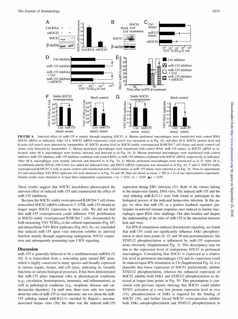
These results suggest that SOCS1 knockdown phenocopied theantiviral effect of induced miR-155 and counteracted the effect ofmiR-155 inhibition.Because the SOCS1-stably overexpressed RAW264.7 cell clones
transcribed SOCS1 mRNAwithout its 39-UTR, miR-155 should nolonger target SOCS1 expression in these cells. We did not findthat miR-155 overexpression could influence VSV proliferationin SOCS1-stably overexpressed RAW264.7 cells, documented byboth measuring VSV TCID50 in the cultural supernatants (Fig. 6F)and intracellular VSV RNA replicates (Fig. 6G). So, we concludedthat induced miR-155 upon viral infection exhibits its antiviralfunction mainly through suppressing endogenous SOCS1 expres-sion and subsequently promoting type I IFN signaling.
DiscussionmiR-155 is generally believed to be a multifunctional miRNA (9,10). It is transcribed from a noncoding gene named BIC gene,which is highly conserved in many species and broadly expressedin various organs, tissues, and cell types, indicating its versatilefunctions in various biological processes. It has been demonstratedthat miR-155 plays important roles in physiological conditions(e.g., circulation, hematopoiesis, immunity, and inflammation), aswell as pathological conditions (e.g., neoplastic diseases and car-diovascular disorders). Up until now, there were only two reportsabout the roles of miR-155 in viral infection: one was about the miR-155 ortholog named miR-K12-11 encoded by Kaposi’s sarcoma-associated herpes virus (30); the other was the induced miR-155
expression during EBV infection (31). Both of the viruses belongto the herpesvirus family, DNA virus. The induced miR-155 and theviral ortholog miR-K12-11 were both found to participate in thebiological process of the indicated herpesvirus infection. In this pa-per, we show that miR-155, as a positive feedback regulator, par-ticipates in antiviral immune responses once induced in murine mac-rophages upon RNA virus challenge. Our data broaden and deepenthe understanding of the roles of miR-155 in the interaction betweenhost and virus.For IFN-b stimulation-induced downstream signaling, we found
that miR-155 could not significantly influence JAK1 phosphory-lation at short time points (0, 15, and 30 min poststimulation), butSTAT1/2 phosphorylation is influenced by miR-155 expressionmore obviously (Supplemental Fig. 3). This discrepancy may bedue to the expression level of endogenous SOCS1 in peritonealmacrophages. Considering that SOCS1 is expressed at a relativelow level in peritoneal macrophages (32) and its expression couldbe induced upon IFN stimulation in 2 h (Supplemental Fig. 4), it isplausible that lower expression of SOCS1 preferentially inhibitsSTAT1/2 phosphorylation, whereas the enhanced expression ofSOCS1 inhibits both JAK1 and STAT1/2 phosphorylation as de-tected at longer time points in Fig. 5F. This presumption is con-sistent with previous reports showing that SOCS1 could inhibitSTAT1 activation at a very low protein expression level in vivo(33), phosphorylation of JAKs is required for the binding ofSOCS1 (34), and further forced SOCS1 overexpression inhibitsboth JAKs autophosphorylation and STAT1/2 phosphorylation in
FIGURE 6. Antiviral effect of miR-155 is mainly through targeting SOCS1. A, Murine peritoneal macrophages were transfected with control RNA
SOCS1 siRNA as indicated. After 24 h, SOCS1 mRNA expression (right panel) was measured as in Fig. 4A, and after 48 h, SOCS1 protein level and
b-actin (left panel) were detected by immunoblot. B, SOCS1 protein level in SOCS1-stably overexpressed RAW264.7 cell clones and mock control cell
clones were detected by immunoblot. C, Murine peritoneal macrophages were transfected with control RNA, miR-155 mimic, or SOCS1 siRNA as in-
dicated. After 48 h, macrophages were treated, infected, and detected as in Fig. 3A. D, Murine peritoneal macrophages were transfected with control
inhibitor, miR-155 inhibitor, miR-155 inhibitor combined with control RNA, or miR-155 inhibitor combined with SOCS1 siRNA, respectively, as indicated.
After 48 h, macrophages were treated, infected, and detected as in Fig. 3A. E, Murine peritoneal macrophages were transfected as in D. After 48 h,
recombinant murine IFN-b (100 U/ml) was added for indicated time, and ISG15 mRNA expression was measured as in Fig. 4A. F and G, SOCS1-stably
overexpressed RAW267.4 cells or mock control cells transfected with control mimic or miR-155 mimic were infected as in Fig. 3A. Virus in supernatants
(F) and intracellular VSV RNA replicates (G) were detected as in Fig. 3A and 3B. Data are shown as mean 6 SD (n = 3) of one representative experiment.
Similar results were obtained in at least three independent experiments. ppp , 0.01; pp , 0.05; :p . 0.05.
The Journal of Immunology 6231
by guest on September 25, 2018
http://ww
w.jim
munol.org/
Dow
nloaded from

vitro (27, 28). However, the detailed regulatory mechanism medi-ating this discrepancy remains to be investigated in the future.Up until now, several genes have been identified as targets of
miR-155, through which the function of miR-155 is revealed.However, the target profile of miR-155 does not seem to end so far.The major targets of one specific miRNA always changes in dif-ferent physical or pathological conditions, or different cell types,to implement different functions accordingly. miR-155 has beensuggested to participate in different biological processes by tar-geting different genes expression, such as PU.1 (11, 35), AID (36,37), SHIP1 (38), and SMAD5 (39). In this paper, we show thatmiR-155 directly targets SOCS1 expression in VSV-infectedmacrophages to strengthen IFN effects, which is consistent witha recent report revealing that miR-155 expression is required forregulatory T cell activity also via targeting SOCS1 expression(29). It seems that the essential roles of miR-155 elucidated bytargeting SOCS1 expression are not confined to regulatory T cellsor macrophages. Because SOCS1 is a general regulator of cellsignaling downstream lots of cytokines, such as IL-2, IL-4, IL-6, IFN-g, growth hormone, and erythropoietin, thus modulatingvarious biological functions of immune cells (4), the finding thatmiR-155 targeting SOCS1 may explain, to some extent, some ofthe pathological phenotypes in miR-155 knockout mice or miR-155 transgenic mice [e.g., impaired hematopoietic homeostasis(40), lymphoblastic leukemia (41), increased lung airway remodel-ing (10), and unbalanced cytokine production (12)]. Also, miR-155targeting SOCS1 may contribute to the pathogenesis of many humanhemopoietic tumors with overexpressed miR-155 (42–44). In addi-tion, overexpressed SOCS1 lacking miR-155 suppression in in-competent immunocytes is probably the explanation for immunetolerance or immunodeficiency in patients with poor response to IFNantiviral or antitumor therapy. Predictably, upregulation of miR-155expression in activated immunocytes may facilitate antitumor re-sponses, whereas, in contrast, enhanced reactivity of immunocytes toproliferative cytokines mediated by miR-155 overexpression mayotherwise contribute to the increased risk of suffering hemopoietictumors. These predictions need to be validated further through ex-tensive experiments in the future.Induced cytokine production is required for the elimination of
pathogen infection; nevertheless, it is clarified that their over-production or overreaction may result in local or systematic pa-thology. For instance, solid evidence reveals that host immuneresponses contribute to the pathogenesis of human seasonal in-fluenza A virus disease (45). Hence, the suppression of SOCS1 byinducible miR-155 strengthens antiviral immunity and elicits moreeffective immune responses as a positive aspect. In contrast, miR-155 overexpression may also probably initiate excessive antiviralresponses and contribute to viral infection-induced tissue damage,which is supported by studies demonstrating that SOCS1 nega-tively regulates innate immune responses triggered by influenza Avirus (46) and SOCS1 protects mice against lethal poxvirus in-fection (47) and Chlamydia pneumoniae infection (48). InducedmiR-155 expression upon viral infection may be a double-edgedsword, because it may participate in both physiological antiviralimmune response and pathological viral infection-induced tissuedamage. And maybe this is the reason why miR-155 is moder-ately, neither vigorously nor slightly, induced in macrophages andother APCs upon RNA virus challenge, and deregulation of miR-155 probably contributes to the pathogenesis of viral infection-induced pathological responses.Our previous work documented that miR-146a, upregulated
upon VSV challenge, inhibited RIG-I–dependent type I IFN pro-duction as a negative feedback regulator in antiviral immunity(16), whereas in this study, the inducible miR-155 promoted type I
IFN signaling as a positive feedback regulator. Taking these workstogether, we further confirmed that antiviral immune response wasunder accurate and sophisticated regulation by multiple regulators,both negatively and positively, like miR-146a and miR-155. It isprobable that we are far from revealing the last miRNA thatmodulates antiviral immunity. And each miRNA may act as a coun-terbalance to the other, so the overall effect of all these miRNAson IFN-mediated antiviral response may be not apparent. Thismay be the reason why Dicer-deficient macrophages, which lead-ing to deficiency of all miRNAs, showed no obviously alteredIFN-mediated antiviral response (23). In addition, besides thesesignificantly induced miRNAs after VSV challenge, we could notexclude the possibility that the less markedly altered miRNAs alsoinfluence host antiviral response via the cumulative impact of allor most of them, if single one has little effect.Host favorable defense against viral infection requires not only
appropriate induction of antiviral cytokines but also appropriatecellular responses to them. We show in this study that the induciblemiR-155 promotes host cell response to type I IFN in antiviralimmunity by targeting SOCS1, further indicating that different hostmiR-155 expression levels may contribute to different responses toIFN therapy of viral infections among individuals. Hence, miR-155may be a potential therapeutic target in viral infections or diseasessubjected to type I IFN therapy, which needs to be identified in thefuture.
AcknowledgmentsWe thank Prof. Shizuo Akira for providing TLR4, TLR9, or MyD88 knock-
out mice, Prof. Wei Pan for providing VSV, Prof. Bing Sun for providing
SeV, Mei Jin and Yan Li for their excellent technical assistance, and
Prof. Jiahuai Han, Genhong Chen, Dr. Xiaoping Su, Dr. Taoyong Chen,
and Dr. Qian Zhang for their helpful discussions.
DisclosuresThe authors have no financial conflicts of interest.
References1. Akira, S., S. Uematsu, and O. Takeuchi. 2006. Pathogen recognition and innate
immunity. Cell 124: 783–801.2. Beutler, B., C. Eidenschenk, K. Crozat, J. L. Imler, O. Takeuchi, J. A. Hoffmann,
and S. Akira. 2007. Genetic analysis of resistance to viral infection. Nat. Rev.Immunol. 7: 753–766.
3. Takeuchi, O., and S. Akira. 2008. MDA5/RIG-I and virus recognition. Curr.Opin. Immunol. 20: 17–22.
4. Alexander, W. S., and D. J. Hilton. 2004. The role of suppressors of cytokinesignaling (SOCS) proteins in regulation of the immune response. Annu. Rev.Immunol. 22: 503–529.
5. Croker, B. A., H. Kiu, and S. E. Nicholson. 2008. SOCS regulation of the JAK/STAT signalling pathway. Semin. Cell Dev. Biol. 19: 414–422.
6. O’Connell, R. M., D. S. Rao, A. A. Chaudhuri, and D. Baltimore. 2010. Phys-iological and pathological roles for microRNAs in the immune system. Nat. Rev.Immunol. 10: 111–122.
7. Baltimore, D., M. P. Boldin, R. M. O’Connell, D. S. Rao, and K. D. Taganov.2008. MicroRNAs: new regulators of immune cell development and function.Nat. Immunol. 9: 839–845.
8. Xiao, C., and K. Rajewsky. 2009. MicroRNA control in the immune system:basic principles. Cell 136: 26–36.
9. Faraoni, I., F. R. Antonetti, J. Cardone, and E. Bonmassar. 2009. miR-155 gene:a typical multifunctional microRNA. Biochim. Biophys. Acta 1792: 497–505.
10. Rodriguez, A., E. Vigorito, S. Clare, M. V. Warren, P. Couttet, D. R. Soond,S. van Dongen, R. J. Grocock, P. P. Das, E. A. Miska, et al. 2007. Requirement ofbic/microRNA-155 for normal immune function. Science 316: 608–611.
11. Vigorito, E., K. L. Perks, C. Abreu-Goodger, S. Bunting, Z. Xiang, S. Kohlhaas,P. P. Das, E. A. Miska, A. Rodriguez, A. Bradley, et al. 2007. microRNA-155regulates the generation of immunoglobulin class-switched plasma cells. Im-munity 27: 847–859.
12. Thai, T. H., D. P. Calado, S. Casola, K. M. Ansel, C. Xiao, Y. Xue, A. Murphy,D. Frendewey, D. Valenzuela, J. L. Kutok, et al. 2007. Regulation of the germinalcenter response by microRNA-155. Science 316: 604–608.
13. O’Connell, R. M., K. D. Taganov, M. P. Boldin, G. Cheng, and D. Baltimore.2007. MicroRNA-155 is induced during the macrophage inflammatory response.Proc. Natl. Acad. Sci. USA 104: 1604–1609.
6232 miR-155 PROMOTES IFN SIGNALING
by guest on September 25, 2018
http://ww
w.jim
munol.org/
Dow
nloaded from

14. Tili, E., J. J. Michaille, A. Cimino, S. Costinean, C. D. Dumitru, B. Adair,M. Fabbri, H. Alder, C. G. Liu, G. A. Calin, and C. M. Croce. 2007. Modulationof miR-155 and miR-125b levels following lipopolysaccharide/TNF-a stimula-tion and their possible roles in regulating the response to endotoxin shock. J.Immunol. 179: 5082–5089.
15. Ceppi, M., P. M. Pereira, I. Dunand-Sauthier, E. Barras, W. Reith, M. A. Santos,and P. Pierre. 2009. MicroRNA-155 modulates the interleukin-1 signalingpathway in activated human monocyte-derived dendritic cells. Proc. Natl. Acad.Sci. USA 106: 2735–2740.
16. Hou, J., P. Wang, L. Lin, X. Liu, F. Ma, H. An, Z. Wang, and X. Cao. 2009.MicroRNA-146a feedback inhibits RIG-I–dependent Type I IFN production inmacrophages by targeting TRAF6, IRAK1, and IRAK2. J. Immunol. 183: 2150–2158.
17. Wang, Y., H. X. Zhang, Y. P. Sun, Z. X. Liu, X. S. Liu, L. Wang, S. Y. Lu,H. Kong, Q. L. Liu, X. H. Li, et al. 2007. Rig-I–/– mice develop colitis associatedwith downregulation of Gai2. Cell Res. 17: 858–868.
18. Wang, C., T. Chen, J. Zhang, M. Yang, N. Li, X. Xu, and X. Cao. 2009. The E3ubiquitin ligase Nrdp1 “preferentially” promotes TLR-mediated production oftype I interferon. Nat. Immunol. 10: 744–752.
19. Liu, X., M. Yao, N. Li, C. Wang, Y. Zheng, and X. Cao. 2008. CaMKII promotesTLR-triggered proinflammatory cytokine and type I interferon production bydirectly binding and activating TAK1 and IRF3 in macrophages. Blood 112:4961–4970.
20. An, H., J. Hou, J. Zhou, W. Zhao, H. Xu, Y. Zheng, Y. Yu, S. Liu, and X. Cao.2008. Phosphatase SHP-1 promotes TLR- and RIG-I–activated production oftype I interferon by inhibiting the kinase IRAK1. Nat. Immunol. 9: 542–550.
21. Xia, S., Z. Guo, X. Xu, H. Yi, Q. Wang, and X. Cao. 2008. Hepatic microen-vironment programs hematopoietic progenitor differentiation into regulatorydendritic cells, maintaining liver tolerance. Blood 112: 3175–3185.
22. Miranda, K. C., T. Huynh, Y. Tay, Y. S. Ang, W. L. Tam, A. M. Thomson,B. Lim, and I. Rigoutsos. 2006. A pattern-based method for the identification ofMicroRNA binding sites and their corresponding heteroduplexes. Cell 126:1203–1217.
23. Otsuka, M., Q. Jing, P. Georgel, L. New, J. Chen, J. Mols, Y. J. Kang, Z. Jiang,X. Du, R. Cook, et al. 2007. Hypersusceptibility to vesicular stomatitis virusinfection in Dicer1-deficient mice is due to impaired miR24 and miR93 ex-pression. Immunity 27: 123–134.
24. Dimitriou, I. D., L. Clemenza, A. J. Scotter, G. Chen, F. M. Guerra, andR. Rottapel. 2008. Putting out the fire: coordinated suppression of the innate andadaptive immune systems by SOCS1 and SOCS3 proteins. Immunol. Rev. 224:265–283.
25. Yoshimura, A., T. Naka, and M. Kubo. 2007. SOCS proteins, cytokine signallingand immune regulation. Nat. Rev. Immunol. 7: 454–465.
26. Zitzmann, K., S. Brand, E. N. De Toni, S. Baehs, B. Goke, J. Meinecke,G. Spottl, H. H. Meyer, and C. J. Auernhammer. 2007. SOCS1 silencingenhances antitumor activity of type I IFNs by regulating apoptosis in neuroen-docrine tumor cells. Cancer Res. 67: 5025–5032.
27. Naka, T., M. Narazaki, M. Hirata, T. Matsumoto, S. Minamoto, A. Aono,N. Nishimoto, T. Kajita, T. Taga, K. Yoshizaki, et al. 1997. Structure andfunction of a new STAT-induced STAT inhibitor. Nature 387: 924–929.
28. Endo, T. A., M. Masuhara, M. Yokouchi, R. Suzuki, H. Sakamoto, K. Mitsui,A. Matsumoto, S. Tanimura, M. Ohtsubo, H. Misawa, et al. 1997. A new proteincontaining an SH2 domain that inhibits JAK kinases. Nature 387: 921–924.
29. Lu, L. F., T. H. Thai, D. P. Calado, A. Chaudhry, M. Kubo, K. Tanaka,G. B. Loeb, H. Lee, A. Yoshimura, K. Rajewsky, and A. Y. Rudensky. 2009.Foxp3-dependent microRNA155 confers competitive fitness to regulatory T cellsby targeting SOCS1 protein. Immunity 30: 80–91.
30. Gottwein, E., N. Mukherjee, C. Sachse, C. Frenzel, W. H. Majoros, J. T. Chi,R. Braich, M. Manoharan, J. Soutschek, U. Ohler, and B. R. Cullen. 2007. Aviral microRNA functions as an orthologue of cellular miR-155. Nature 450:1096–1099.
31. Lu, F., A. Weidmer, C. G. Liu, S. Volinia, C. M. Croce, and P. M. Lieberman.2008. Epstein-Barr virus-induced miR-155 attenuates NF-kB signaling andstabilizes latent virus persistence. J. Virol. 82: 10436–10443.
32. Baetz, A., M. Frey, K. Heeg, and A. H. Dalpke. 2004. Suppressor of cytokinesignaling (SOCS) proteins indirectly regulate Toll-like receptor signaling ininnate immune cells. J. Biol. Chem. 279: 54708–54715.
33. Song, M. M., and K. Shuai. 1998. The suppressor of cytokine signaling (SOCS)1 and SOCS3 but not SOCS2 proteins inhibit interferon-mediated antiviral andantiproliferative activities. J. Biol. Chem. 273: 35056–35062.
34. Yasukawa, H., H. Misawa, H. Sakamoto, M. Masuhara, A. Sasaki, T. Wakioka,S. Ohtsuka, T. Imaizumi, T. Matsuda, J. N. Ihle, and A. Yoshimura. 1999. TheJAK-binding protein JAB inhibits Janus tyrosine kinase activity through bindingin the activation loop. EMBO J. 18: 1309–1320.
35. Martinez-Nunez, R. T., F. Louafi, P. S. Friedmann, and T. Sanchez-Elsner. 2009.MicroRNA-155 modulates the pathogen binding ability of dendritic cells (DCs)by down-regulation of DC-specific intercellular adhesion molecule-3 grabbingnon-integrin (DC-SIGN). J. Biol. Chem. 284: 16334–16342.
36. Dorsett, Y., K. M. McBride, M. Jankovic, A. Gazumyan, T. H. Thai,D. F. Robbiani, M. Di Virgilio, B. R. San-Martin, G. Heidkamp, T. A. Schwickert,et al. 2008. MicroRNA-155 suppresses activation-induced cytidine deaminase-mediated Myc-Igh translocation. Immunity 28: 630–638.
37. Teng, G., P. Hakimpour, P. Landgraf, A. Rice, T. Tuschl, R. Casellas, andF. N. Papavasiliou. 2008. MicroRNA-155 is a negative regulator of activation-induced cytidine deaminase. Immunity 28: 621–629.
38. O’Connell, R. M., A. A. Chaudhuri, D. S. Rao, and D. Baltimore. 2009. Inositolphosphatase SHIP1 is a primary target of miR-155. Proc. Natl. Acad. Sci. USA106: 7113–7118.
39. Rai, D., S. W. Kim, M. R. McKeller, P. L. Dahia, and R. C. Aguiar. 2010.Targeting of SMAD5 links microRNA-155 to the TGF-b pathway and lym-phomagenesis. Proc. Natl. Acad. Sci. USA 107: 3111–3116.
40. O’Connell, R. M., D. S. Rao, A. A. Chaudhuri, M. P. Boldin, K. D. Taganov,J. Nicoll, R. L. Paquette, and D. Baltimore. 2008. Sustained expression ofmicroRNA-155 in hematopoietic stem cells causes a myeloproliferative disorder.J. Exp. Med. 205: 585–594.
41. Costinean, S., N. Zanesi, Y. Pekarsky, E. Tili, S. Volinia, N. Heerema, andC. M. Croce. 2006. Pre-B cell proliferation and lymphoblastic leukemia/high-grade lymphoma in Em-miR155 transgenic mice. Proc. Natl. Acad. Sci. USA103: 7024–7029.
42. Garzon, R., M. Garofalo, M. P. Martelli, R. Briesewitz, L. Wang, C. Fernandez-Cymering, S. Volinia, C. G. Liu, S. Schnittger, T. Haferlach, et al. 2008. Dis-tinctive microRNA signature of acute myeloid leukemia bearing cytoplasmicmutated nucleophosmin. Proc. Natl. Acad. Sci. USA 105: 3945–3950.
43. Metzler, M., M. Wilda, K. Busch, S. Viehmann, and A. Borkhardt. 2004. Highexpression of precursor microRNA-155/BIC RNA in children with Burkittlymphoma. Genes Chromosomes Cancer 39: 167–169.
44. Eis, P. S., W. Tam, L. Sun, A. Chadburn, Z. Li, M. F. Gomez, E. Lund, andJ. E. Dahlberg. 2005. Accumulation of miR-155 and BIC RNA in human B celllymphomas. Proc. Natl. Acad. Sci. USA 102: 3627–3632.
45. Lee, N., C. K. Wong, P. K. Chan, S. W. Lun, G. Lui, B. Wong, D. S. Hui,C. W. Lam, C. S. Cockram, K. W. Choi, et al. 2007. Hypercytokinemia andhyperactivation of phospho-p38 mitogen-activated protein kinase in severe hu-man influenza A virus infection. Clin. Infect. Dis. 45: 723–731.
46. Pothlichet, J., M. Chignard, and M. Si-Tahar. 2008. Cutting edge: innate immuneresponse triggered by influenza A virus is negatively regulated by SOCS1 andSOCS3 through a RIG-I/IFNAR1–dependent pathway. J. Immunol. 180: 2034–2038.
47. Ahmed, C. M., R. Dabelic, L. W. Waiboci, L. D. Jager, L. L. Heron, andH. M. Johnson. 2009. SOCS-1 mimetics protect mice against lethal poxvirusinfection: identification of a novel endogenous antiviral system. J. Virol. 83:1402–1415.
48. Yang, T., P. Stark, K. Janik, H. Wigzell, and M. E. Rottenberg. 2008. SOCS-1protects against Chlamydia pneumoniae-induced lethal inflammation but ham-pers effective bacterial clearance. J. Immunol. 180: 4040–4049.
The Journal of Immunology 6233
by guest on September 25, 2018
http://ww
w.jim
munol.org/
Dow
nloaded from