infarction in the territory of the anterior choroidal artery · infarction in the territory of the...
TRANSCRIPT

Brain (1986), 109, 1071-1085
INFARCTION IN THE TERRITORY OF THEANTERIOR CHOROIDAL ARTERY
A CLINICAL AND COMPUTERIZED TOMOGRAPHICSTUDY OF 16 CASES
by J. P. DECROIX, 1 PH. GRAVELEAU, 2 M. MASSON1 and J. CAMBIER1
(From the 1Clinique Neurologique, Hopital Beaujon, Clichy, and 2the Service de Neurologic, HopitalFoch, Suresnes, France)
SUMMARY
Sixteen cases of the anterior choroidal artery syndrome are reported. In its completed form, this raresyndrome combines the triad of hemiplegia, hemianaesthesia, and homonymous hemianopia. CTexamination confirms the diagnosis by revealing an area of reduced density situated in the posteriorlimb of the internal capsule, sparing the thalamus medially and encroaching upon the tip of the globuspallidus laterally, and corresponding to the distribution of the anterior choroidal artery. Incompleteforms of the syndrome are more frequent. Left-sided spatial neglect may accompany right-sidedlesions, as may slight disorders of speech in left-sided lesions. Clinical-anatomical correlations arediscussed.
INTRODUCTION
The syndrome of the anterior choroidal artery was described in 1925 by Foix et al.,although 2 cases had been reported by Kolisko in 1891. Subsequent cases, withanatomical confirmation, were published by Poppi (1928), Ley (1932), Abbie (1933),and Buge et al. (1979), making a total of 7 cases. In its complete form, the syndromecomprises hemiplegia, hemianaesthesia, and homonymous hemianopia contra-lateral to the lesion. The absence of a cognitive deficit classically permits thedifferential diagnosis from cortical and subcortical lesions. In 1983 and 1984 wereported 5 cases and described the clinical, neuropsychological and computerizedtomographic (CT) signs (Masson et al., 1983; Viader et al., 1984). There have beensubsequent descriptions by Ward et al. (1984) and Derouesne et al. (1985). Thepresent study of 11 new cases makes it possible to distinguish the principal signsand their variations related to infarction in the territory of the anterior choroidalartery.
Correspondence to: Dr J. P. Decroix, Clinique Neurologique, Hopital Beaujon, 100 Bd du General Leclerc,92110Clichy, France.
© Oxford University Press 1986
Downloaded from https://academic.oup.com/brain/article-abstract/109/6/1071/300343/INFARCTION-IN-THE-TERRITORY-OF-THE-ANTERIORby gueston 24 September 2017

1072 5. P. DECROIX AND OTHERS
CASE REPORTS
The clinical observations are summarized in Table 1 and the CT scans in fig. 4. Cases 1,2,4, 6 and 15have already been published (Cambier et al., 1983; Masson et al., 1983; Viader et al., 1984).
Case 1 (N 2260 D)A right-handed man, aged 62 years, was admitted to hospital on September 13, 1981, with a left
hemiplegia of sudden onset. He had previously had untreated arterial hypertension and diabetes.Examination on admission revealed a massive left hemiplegia, anaesthesia for all sensory modalitiesdown the left half of the body, and a left homonymous hemianopia.
TABLE 1. CLINICAL SYMPTOMS
lase1234
56
78
910111213141516
Sex/Age(yrs)M/62M/53F/64F/68
M/7IF/68
M/45F/67
F/83F/60F/75M/46M/72F/42M/84F/71
Side oflesion
RRLR
RL
LL
LRRLLLLL
Hemiplegia
SevereSevere
ModerateSevere
BrachiofacialModerate
ModerateModerate
ModerateSevere
BrachiofacialSevere
00
SevereModerate
. Sensorydeficit
SevereSevere
ModerateSevere
ModerateModerate
ModerateModerate
Moderate000
SevereSevere
ModerateModerate
Homonymoushemianopia
+++
Visualextinction
00
00
00000000
Languagedeficit
0000
0'Thalamic'
aphasiaDysarthria'Thalamic'
aphasiaDysarthriaDysarthria
0Dysarthria
00
DysarthriaDysarthria
Neglectsyndrome
SevereSevere
0Moderate
00
00
00000000
Neuropsychological examination was undertaken between the second and ninth days after thestroke. Orientation in time and space was satisfactory, but he showed severe visual neglect to the left;the head and eyes were permanently turned to the right. He could look to the left on command, turningfirst his head and then his eyes. When asked to cross out marks, only those on the extreme right-handedge of the page were deleted. When asked to describe a picture, only the right-hand quarter wasidentified. The placing of principal towns on an outline map of France was correctly performed, despitesome imprecision; this test did not reveal left visual neglect, as there was no displacement of town sitesto the right. Voluntary drawing (circle, square, cube, bicycle, flower) was defective and imprecise; thepictures were nevertheless complete except for the flower, which lacked petals on the left. Whencopying a picture, the drawing was identical, but neglect of the left part was more evident. There was apartial agnosia for pictures which seemed to depend on the left visual neglect: out of 21 pictures, 11were correctly identified immediately, 5 after one or more mistakes, and 5 were not identified orwrongly identified. In Poppelreuter's test, performance was better when the picture was presented in itsnormal form than when enlarged; in the latter case the left part was then ignored. There was no agnosiafor objects or colours, and no prosopagnosia. Language was normal. Reading of letters gave rise to aconstant error, as W was identified as N. Syllables were correctly identified if they comprised only twoletters, although the first was sometimes neglected. From 3 letters upwards, paralexia was frequentowing to neglect of one or more letters at the beginning of the syllable. Reading of words was correct
Downloaded from https://academic.oup.com/brain/article-abstract/109/6/1071/300343/INFARCTION-IN-THE-TERRITORY-OF-THE-ANTERIORby gueston 24 September 2017

ANTERIOR CHOROIDAL ARTERY INFARCTION 1073
with the exception of rare but significant mistakes: mappemonde was read monde and esquimau as mau.In general, mistakes were more frequent when the right-hand part of the word, in isolation from theleft, was meaningful: plateau was read as eau and cartable as table. When reading a text, the left-handpart was neglected and there were mistakes in changing line; the result was incoherent. Dichoticlistening revealed a total extinction on the left. There was motor impersistence, but no anosognosia.
CT scan without contrast (17/9/81) showed an area of reduced density in the posterior limb of theright internal capsule. ECG revealed left ventricular hypertrophy. The neurological signs andsymptoms remained virtually unchanged. At discharge after one month the hemiplegia was as severeas on admission, hypoaesthesia for all modalities persisted down the left half of the body, and thehemianopia was unchanged, but the neglect syndrome had improved.
Case 2 (N 2191 D)A right-handed man, aged 53 years, was admitted to hospital following a left hemiplegia of sudden
onset on August 7, 1981. He had had a primary tuberculous infection at the age of 20. Examination onadmission showed a severe left hemiplegia, anaesthesia for all sensory modalities down the left half ofthe body, and a left homonymous hemianopia. Blood pressure was normal.
Neuropsychological examination was carried out between the days 26 and 28 after the stroke.Orientation in space and time was satisfactory. There was severe visual neglect on the left. The headand eyes were spontaneously turned to the right, but the patient could look to the left on command,
FIG. 1. Case 2. Spontaneous drawings.
Downloaded from https://academic.oup.com/brain/article-abstract/109/6/1071/300343/INFARCTION-IN-THE-TERRITORY-OF-THE-ANTERIORby gueston 24 September 2017

1074 J. P. DECROIX AND OTHERS
turning first his head and then his eyes. When asked to cross out marks on paper, only those on theextreme right of the page were deleted. When asked to describe complex images, only the right fifth ofthe picture was identified. Placement of towns on an outline map of France was correct, as werefreehand drawings of a cube, a flower and a bicycle (fig. 1). However, when copying the same picturesfrom a model, the left-hand part was completely neglected (fig. 2). Copying Rey's figure produced
FIG. 2. Case 2. Copied drawings.
disorganization of the left part of the figure. There was partial agnosia for image recognition, with 10mistakes in 32 pictures. In Poppelreuter's test, performance was better when the design was presentedin its normal form than when enlarged, when the left part was neglected. There was no agnosia forobjects, colours or prosopagnosia. Speech was normal. When writing spontaneously or from dictation,the patient did not use the left-hand side of the paper. When copying from a text, only the right-handpart of the sentences were copied. There were reading deficits, such that Y was identified as I; all otherletters were correctly identified. Reading of syllables revealed neglect of the left-hand part, particularlyif the syllable comprised more than three letters: oul was read as ul, drain as ain, splan as plan and stroncas tronc. Reading of logatomes revealed the same deficit. Mistakes appeared in the reading of words,particularly when the right-hand part, isolated from the left, retained a meaning (chevrefeuille was readasfeuille). When reading a text, the left-hand part was completely ignored. The patient was aware ofthe resulting incoherence but was unable to correct it. There was motor impersistence: the patient wasunable to keep his eyes closed on command, and every verbal communication automatically producedopening of the eyes, despite repeated requests to close them. Immediately after admission, there wasanosognosia.
Cervical Doppler, ECG, Holter and echocardiogram were normal. CT scan without contrast(21/8/81) showed an area of diminished density in the posterior limb of the right internal capsule(fig. 3), which was unchanged by injection of contrast. Right carotid angiography did not revealatheroma in the cervical or cranial vessels and the right anterior choroidal artery was visible andapparently normal. The neurological deficits did not change. A month later, the patient suffered aninfarct in the territory of the left middle cerebral artery, suggesting a diagnosis of multiple emboli ofcardiac origin, despite the negative cardiac findings.
Downloaded from https://academic.oup.com/brain/article-abstract/109/6/1071/300343/INFARCTION-IN-THE-TERRITORY-OF-THE-ANTERIORby gueston 24 September 2017

ANTERIOR CHOROIDAL ARTERY INFARCTION 1075
Case 3 (N 5713 D)
A woman, aged 64 years, was admitted on February 14, 1983, with a right hemiplegia. There wasno significant previous history. The right hemiplegia had developed suddenly on the previous day.Examination on admission showed an incomplete right hemiplegia, anaesthesia for pin prick overthe whole right half of the body, a defect of deep sensation on the right and a right homonymoushemianopia. There was no speech deficit. Blood pressure was normal. ECG and Doppler examinationof the cervical vessels were normal. Fasting blood sugar was slightly elevated at 8.7 mmol/1. CT scan(15/2/1983) showed diminished density in the posterior limb of the left internal capsule, unchanged byinjection of contrast. On discharge a month later the patient was able to walk without help, the motordeficit was not severe, the hemianopia had disappeared but the sensory deficit persisted.
FIG. 3. Case 2. CT scan, showing area of reduced density in posteriorlimb of right internal capsule.
Case 4 (N 2889 D)
A woman, aged 68 years, was admitted on December 25, 1981. Her past history included calcifiedaortic narrowing. A left hemiplegia of sudden onset had developed on December 22, 1981. Onadmission, examination revealed a total left hemiplegia, anaesthesia of all sensory modalities down theleft half of the body, and left visual extinction. Blood pressure was normal.
Neuropsychological examination was performed between days 11 and 16 after the stroke. Orienta-tion in time and space was normal. At the beginning of her stay in hospital, the patient manifested leftvisual neglect, with the head and eyes most frequently turned to the right. When asked to enumeratethe people in the room, she 'forgot' those on her left. There was left visual extinction, but this featurewas notably improved by the time of neuropsychological examination. Crossing out marks across apage was normal. However, when asked to bisect a horizontal line, there was a distinct shift to the right.When asked to describe pictures, details on the extreme left were omitted. Placing of the principaltowns on an outline map of France was correctly carried out. Spontaneous drawing (circle, square,bicycle, house, flower) was defective and imprecise, but slightly better when copying. Copying of Rey'sfigure was disorganized, with omission of detail on the extreme left. There was no agnosia for pictures,objects, or colours and no prosopagnosia. Speech was normal. Spontaneous and dictated writingrevealed a failure to use the left side of the paper. Reading of letters, syllables and words was normal.Reading a text revealed difficulties in returning to the line. Mental arithmetic was unaffected. Inarithmetical calculations, the operational sign placed at the left was sometimes neglected. There wasan anosognosia when she was first in hospital. She suffered from motor impersistence.
Blood pressure was normal. ECG showed arrhythmia due to atrial fibrillation, but she returned to
Downloaded from https://academic.oup.com/brain/article-abstract/109/6/1071/300343/INFARCTION-IN-THE-TERRITORY-OF-THE-ANTERIORby gueston 24 September 2017

1076 J. P. DECROIX AND OTHERS
Case 9 (4 days)
Case 1 (4 days) Case 2 (14 days) Case 3 (2 days) Case 4 (6 days)
Case 5 (6 days) Case 6 (42 days) Case 7 (7 days) Case 8 (39 days)
Case 10 (2 days) Case 11 (11 days) Case 12 (3 days)
Case 13 (6 days) Case 14 (1 day) Case 15 (19 days) Case 16 (66 days)
Fio. 4. Diagrams illustrating CT appearances in all 16 patients: time between onset of symptoms and CT scan isindicated in brackets.
sinus rhythm the following day. No biochemical abnormalities were detected. Doppler examinationshowed moderate stenosis at the origin of the right internal carotid artery, without reversal of flow inthe ophthalmic artery. CT scan (28/12/82) showed diminished density in the posterior limb of the rightinternal capsule unchanged by injection of contrast. On discharge one month later, there was noalteration in symptoms.
Case 5 (N 1073 E)A man, aged 71 years, was admitted on March 12, 1980, with a left hemiplegja. There was a history of
hypertension and of a rapidly regressing left hemiplegia in 1975. On the evening of March 11,1980, he
Downloaded from https://academic.oup.com/brain/article-abstract/109/6/1071/300343/INFARCTION-IN-THE-TERRITORY-OF-THE-ANTERIORby gueston 24 September 2017

ANTERIOR CHOROIDAL ARTERY INFARCTION 1077
experienced paraesthesiae in the left leg, and on waking the next morning was hcmiplegic. Examinationon admission showed a left hemiplegia with brachiofacial predominance, diminished pin pricksensation over the whole left half of the body, but no deficit of position sense. There were no visual fielddisturbances and higher cerebral function was unaffected. ECG was normal. Doppler examinationshowed stenosis of the left internal carotid artery. Blood sugar was raised to 7.92 mmol/1. CT scan(17/3/1980) showed reduced density in the posterior limb of the right internal capsule. The symptomswere unchanged on discharge fifteen days later.
Case <J(N 2320 E)
A woman, aged 68 years, was admitted on October 26,1976 because of a right hemiplegia which haddeveloped suddenly the previous day, together with a speech deficit. Past history revealed arterialhypertension which had been treated since 1967. Examination on admission showed moderate righthemiparesis and hypoaesthesia for all sensory modalities over the right half of the body. Visual fieldswere normal. The patient did not speak spontaneously, but would briefly answer questions. Theinformational content of her spontaneous speech was good, but language production was slow withsome words missing and rare semantic paraphasia. When asked to name 25 pictures, some semanticparaphasia and perseveration were noted. Words in alphabetical series or categories revealed some lossof verbal fluency and the existence of perseveration. Repetition, comprehension of spoken and writtenlanguage, spelling, and mental arithmetic were all normal.
ECG was normal. CT scan (7/12/1976) showed an area of reduced density in the posterior limb of theleft internal capsule, which was thinly ringed after injection of contrast. Five years later, she still hada moderate right hemiparesis with hypoaesthesia of the right half of the body; semantic paraphasiaremained evident in her speech.
Case 7 (N 2334 E)
A man, aged 45 years, was admitted on June 30, 1984, with right hemiplegia. There was a history ofrenal calculus. The right hemiplegia developed suddenly on June 26, 1984 together with a languagedeficit which regressed in 24 h. On admission, examination revealed moderate right hemiplegia, anddiminished pin prick sensation over the right half of the body without any loss of position sense. Thevisual fields were normal and there was a mild dysarthria. Blood pressure was moderatey raised. ECGshowed left ventricular enlargement. Doppler examination revealed moderate stenosis at the origin ofthe left internal carotid artery. CT scan (3/7/1984) showed diminished density in the posterior limb ofthe left internal capsule. On discharge ten days later, the symptoms were unchanged.
Case 8 (N 875 E)
A woman, aged 67 years, was admitted on February 20, 1984, with a right hemiplegia. She hadpreviously complained of frequent palpitations which were treated with amiodarone. A righthemiplegia of sudden onset had developed on January 28, 1984. Examination on admission showeda moderate right hemiplegia, diminished pin prick sensation down the right half of the body withastereognosis and impaired joint position sense. Visual fields were normal. There was slight dysarthria,with a reduction in fluency and in initiating speech. ECG was normal. Doppler examinationdemonstrated moderate stenosis at the origin of both internal carotid arteries. CT scan (8/3/1984)showed uptake of contrast in the posterior limb of the left internal capsule. Three months later, therewas considerable improvement and she was able to walk without support.
Case 9 (N 356 E)
A woman, aged 83 years, was admitted on September 4, 1983, with a right hemiplegia. There was aprevious history of diabetes treated by sulphamides since 1958, and moderate renal insufficiency. OnAugust 5, 1983, she had experienced headaches. On August 7 she experienced dysarthria and washemiplegic on waking the next morning. On admission, she had a complete right hemiplegia withdiminished pin prick sensation over the right half of the body without disturbance of joint position
Downloaded from https://academic.oup.com/brain/article-abstract/109/6/1071/300343/INFARCTION-IN-THE-TERRITORY-OF-THE-ANTERIORby gueston 24 September 2017

1078 J. P. DECROIX AND OTHERS
sense. There was no hemianopia, but there was dysarthria. ECG was normal. Blood sugar was raised to10.3 mmol/1. Doppler examination revealed moderate stenosis at the origin of the left internal carotidartery. CT scan (11/8/83) showed a nonenhancing area of reduced density in the posterior limb of theleft internal capsule. Four months later the hemiplegia was still severe but the sensory disturbances haddisappeared.
Case 10 (N 2028 E)
A woman, aged 60 years, was admitted on March 18, 1984, with a left hemiplegia with which shehad woken that morning. There was no significant previous history. Examination on admissionrevealed a total left hemiplegia, with no disturbance of sensation or visual fields, and no neglectsyndrome. Blood pressure was normal. ECG showed some ventricular ectopic beats. Dopplerexamination revealed stenosis at the origin of the right internal carotid artery without reversal of flowin the ophthalmic artery. Blood sugar was 6.6 mmol/1. CT scan (20/3/1984) showed an area of reduceddensity in the posterior limb of the right internal capsule. Two months later the lower limb hadrecovered sufficiently to allow walking, but the upper limb showed little change.
Case 11 (N 5704 D)
A woman, aged 75 years, was admitted on March 1, 1983, with a left hemiplegia which had comeon in two episodes on February 24, 1983. She had previously had a gastrectomy for peptic ulcer.Examination on admission revealed a left hemiplegia with brachiofacial predominance, withoutdisturbances of sensation or of the visual fields and without a neglect syndrome. ECG was normal.Doppler examination showed moderate stenosis of the left internal carotid artery. CT scan (7/3/1983)revealed an area of reduced density in the posterior limb of the right internal capsule. Two monthslater, there was good recovery in the lower limb, but none in the upper.
Case 12 (84/9695)
A man, aged 46 years, was admitted on June 5, 1984, with a right hemiplegia with which he hadwoken that morning. He had previously suffered from severe hypertension. Examination on admissionshowed a complete right hemiplegia with no sensory or visual field disturbances; he was dysarthric.ECG showed enlargement of the left ventricle. Doppler examination was normal. CT scan on June 8,1984, revealed an area of reduced density in the posterior limb of the left internal capsule. One monthlater there was considerable recovery in the lower limb, permitting walking with a severely spastic gait,but little recovery in the upper limb.
Case 13 (84/1347)
A man, aged 72 years, was admitted on January 21, 1984, with left hemianaesthesia. Arterialhypertension had recently been diagnosed. On the day of admission, the patient had suddenly feltweakness in the right lower limb and a disturbance of balance. Examination on admission showed totalnondissociated hemianaesthesia in the right half of the body, without any motor deficit or asymmetryof tendon reflexes and without visual field defects. ECG, biochemistry and a Doppler examinationwere all normal. CT scan (27/1/1984) showed an area of reduced density in the posterior limb of theleft internal capsule. On discharge fifteen days later he still had hypaesthesia of the whole left half ofthe body.
Case 14 (84/7010)
A woman, aged 42 years, was admitted on April 22, 1984, with right hemianaesthesia; there was nosignificant previous history. She noticed swelling of her right leg on the morning of admission, and hadhad palpitations the previous evening. Examination on admission revealed undissociated hemi-anaesthesia of the whole right half of the body, without motor disturbance or visual field defect. ECG,biochemistry, and Doppler testing were normal. CT scan (22/4/1984) showed reduced density in theposterior limb of the left internal capsule. On discharge fifteen days later she had completely recovered.
Downloaded from https://academic.oup.com/brain/article-abstract/109/6/1071/300343/INFARCTION-IN-THE-TERRITORY-OF-THE-ANTERIORby gueston 24 September 2017

ANTERIOR CHOROIDAL ARTERY INFARCTION 1079
Case 15 (N 324 E)
A right-handed man, aged 84 years, with non-insulin-dependent diabetes, was admitted on June 18,1982, with a right hcmiparcsis. Eight days previously he had suddenly had the impression of 'seeingthree people instead of one'; this lasted only a few seconds. An hour later, he felt formication first in hisright upper, then his right lower limb. On waking the next morning, he had a right hemiparesis,difficulty in speaking, and diplopia. The deficit became complete in the course of the day. Examinationon admission revealed a total right hemiplegia and hypaesthesia for all sensory modalities on the sameside. Visual fields were normal. There was dysarthria without dysphasia. The patient complained ofvertical diplopia on looking in all directions, which was attributed to a defect in upward movement ofthe left eye. CT scan (29/6/1982) showed reduced density in the posterior limb of the left internalcapsule.
Case 76(84/21142)
A hypertensive woman, aged 71 years, with non-insulin-dependent diabetes, was admitted onDecember 10, 1984, because of the sudden onset 48 h earlier of a right hemiparesis with an initial lossof speech. Examination revealed a moderate right hemiparesis with brachiofacial predominance.Initially, there was ataxia of the right arm with difficulty in grasping an object with the eyes shut. Jointposition sense, cutaneous sensation and the visual fields were normal. There was dysarthria, difficultyin organizing conversation and in recalling series of words (9 names of animals in 1 min), and aconstructional apraxia. CT scan on December 11, 1984, showed a small area of reduced density in theanterior limb of the right internal capsule. A second scan (12/2/1985) showed persistence of thisappearance, together with a new area of reduced density in the posterior limb of the left internalcapsule. ECG was normal. Doppler examination coupled with echography showed diffuse athero-sclerosis of the carotid and a calcified plaque at the left carotid bifurcation. There was gradual partialregression of the sensorimotor deficits and of the dysarthria.
D I S C U S S I O N
The anterior choroidal artery is a branch of the internal carotid, and usually arisesabove the origin of the posterior communicating artery. Despite its long course itsdiameter is very narrow. Running at first between the brainstem and the uncus of thetemporal lobe, it enters the inferior (temporal) horn of the lateral ventricle and endsin its choroid plexus, its branches anastomosing with those of the posterolateralchoroidal artery. According to Abbie (1933), Carpenter et al. (1954), Lazorthes(1961), and Rhoton et al. (1979), it supplies the whole of the internal segment andpart of the external segment of the globus pallidus; the posterior two-thirds of theposterior limb of the internal capsule, together with its retrolenticular segment,including the origins of the optic and auditory radiations; the optic tract; the lateralhalf of the lateral geniculate body; the most rostral part of the cerebral peduncle; theuncus; part of the amygdaloid nucleus; the anterior part of the hippocampus andfascia dentata; the tail of the caudate nucleus; and the choroid plexus of the inferiorhorn of the lateral ventricle. It does not participate in the supply of the thalamusproper (Percheron, 1977). There are anastomoses between the anterior choroidalartery and the posterior communicating, posterior and middle cerebral arteries, butmost importantly with the posterolateral choroidal artery in the choroid plexus andlateral geniculate body.
Downloaded from https://academic.oup.com/brain/article-abstract/109/6/1071/300343/INFARCTION-IN-THE-TERRITORY-OF-THE-ANTERIORby gueston 24 September 2017

1080 J. P. DECROIX AND OTHERS
In all cases described, CT scanning showed areas of reduced density, signifyingrecent ischaemia, in the posterior limb of the internal capsule, sparing the thalamusmedially but encroaching on the globus pallidus laterally (fig. 3). The site of thisdiminished density corresponds exactly with the area of supply of the anteriorchoroidal artery as described above, and as reconstructed by Damasio (1983) ondiagrams of regular CT cuts. Its distribution differs from the capsuloputamino-caudate territory involved in infarcts of the deep branches of the middle cerebralartery.
TABLE 2. CASES DESCRIBED IN THE LITERATURE
Kolisko (I) Kolisko (2) Foixetal.(1891) (1891) (1925)
HemiplegiaHemianaesthesiaHomonymous
hemianopiaOther signs
Observations
Poppi(1928)
0
Ptosiscontralateral
to thehemiplegia
Associatedlesions in the
territorysupplied by the
paramedianartery of themamillary
peduncle (arteryof Foix)
Ley Abbie(1932) (1933)
X XSuperior
homonymousquadrantanopiacontralateral to
hemiplegiaBilaterallyaffected
Buge et al.(1979)
000
Pseudobulbarsyndrome;paralysis ofvertical gaze
Bilaterallyaffected
Involvement of the whole territory supplied by the anterior choroidal arteryproduces massive hemiplegia, hemianaesthesia for all sensory modalities, and ahomonymous hemianopia; this is the clinical picture originally described by Foix etal. in 1925, and in 3 of the 7 cases described in the subsequent literature (Table 2).Three of our patients (Cases 1 -3) correspond to this picture. The hemiplegia isusually severe and equal throughout the affected half of the body. The anteriorchoroidal artery supplies the upper motor neuron pathway at two sites: the cerebralpeduncle and the posterior limb of the internal capsule, where it is now known thatthe pyramidal tract occupies the posterior half (Brion and Guiot, 1964; Englanger etal., 1975; Hanaway and Young, 1977; Ross, 1980), which is supplied by the anteriorchoroidal artery. Anatomical examination shows that it is the capsular lesion whichis usually responsible. The sensory deficit is of variable intensity, not dissociated,and involves the whole body half. It is related to lesions of the superior thalamicradiations situated in the thalamolenticular portion of the posterior limb of theinternal capsule. A homonymous hemianopia can be the consequence of separatelesions at any of the three points at which the anterior choroidal artery supplies thevisual pathway: the optic tract, the lateral geniculate body, and the optic radiation.
Downloaded from https://academic.oup.com/brain/article-abstract/109/6/1071/300343/INFARCTION-IN-THE-TERRITORY-OF-THE-ANTERIORby gueston 24 September 2017
ANTERIOR CHOROIDAL ARTERY INFARCTION 1081
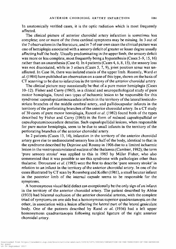
In anatomically verified cases, it is the optic radiation which is most frequentlyaffected.
The clinical picture of anterior choroidal artery infarction is sometimes lesscomplete; one or more of the three cardinal symptoms may be missing. In 3 out ofthe 7 observations in the literature, and in 7 of our own cases the clinical picture wasone of hemiplegia associated with a sensory deficit of greater or lesser degree usuallyaffecting half the body. Usually predominating in the upper limb, the sensory deficitwas more or less complete, most frequently being a hypaesthesia (Cases 5-9, 15,16)rather than an anaesthesia (Case 4). In 4 patients (Cases 4,6, 8,15), the sensory losswas not dissociated, while in 3 others (Cases 5, 7, 9), joint position sense was un-affected. In Case 16, there was isolated ataxia of the upper limb. Recently, Ward etal. (1984) have published an observation on a case of this type, shown on the basis ofCT scanning to be due to infarction in the territory of the anterior choroidal artery.
The clinical picture may occasionally be that of a pure motor hemiplegia (Cases10-12). Fisher and Curry (1965), in a clinical and neuropathological study of puremotor hemiplegia, found two types of ischaemic lesion to be responsible for thissyndrome: capsuloputaminocaudate infarcts in the territory of the lateral lenticulo-striate branches of the middle cerebral artery, and pallidocapsular infarcts in theterritory of the perforating branches of the anterior choroidal artery. In a CT studyof 30 cases of pure motor hemiplegia, Rascol et al. (1982) found both of the typesdescribed by Fisher and Curry (1965) in the form of reduced capsulopallidal orcapsuloputaminocaudate densities. Such capsulopallidal lesions, when responsiblefor pure motor hemiplegia, seem to be due to small infarcts in the territory of theperforating branches of the anterior choroidal artery.
In 2 patients (Cases 13, 14), infarction in the territory of the anterior choroidalartery gave rise to undissociated sensory loss in half of the body, identical to that inthe syndrome described by Dejerine and Roussy in 1906 due to a limited ischaemiclesion in the ventroposterolateral nucleus of the thalamus (Cambier, 1982); the term'pure sensory stroke' was applied to this in 1965 by Miller Fisher, who alsocommented that it was possible to see this syndrome with pathologies other thanthalamic. Derouesne et al. (1985) were the first to describe 'pure sensory stroke' inrelation to an infarct in the territory of the anterior choroidal artery. In one of thecases illustrated by CT scan by Rosenberg and Koller (1981), a small lacunar infarctin the posterior limb of the internal capsule seems to be responsible for thesymptoms.
A homonymous visual field defect can exceptionally be the only sign of an infarctin the territory of the anterior choroidal artery. The patient described by Abbie(1933) had bilateral occlusion of the anterior choroidal arteries, with the completetriad of symptoms on one side but a homonymous superior quadrantanopia on theother, in association with a lesion affecting the lateral part of the lateral geniculatebody. One of the patients described by Rand et al. (1956) had a left upperhomonymous quadrantanopia following surgical ligature of the right anteriorchoroidal artery.
Downloaded from https://academic.oup.com/brain/article-abstract/109/6/1071/300343/INFARCTION-IN-THE-TERRITORY-OF-THE-ANTERIORby gueston 24 September 2017

1082 J. P. DECROIX AND OTHERS
Bilateral anterior choroidal artery infarcts are very rare, and their clinicalpresentation is very variable. Apart from the case of Abbie (1933), alluded to above,Buge et al. (1979) have reported a case of bilateral anterior choroidal infarctionwhich presented as a major pseudobulbar syndrome without any motor deficit in thelimbs. Oculomotor palsies have been described in cases of anterior choroidal arteryinfarction. Thus Poppi (1928) observed ptosis contralateral to the hemiplegiaassociated with infarction of the posterior limb of the internal capsule, cerebralpeduncle and third nerve nucleus. Buge et al. (1979) noted paralysis of upwardmovement in a case of bilateral infarction in the territory of the anterior choroidalartery. In our Case 15 (previously published by Viader et al., 1984), there wasdiplopia in association with defective upward eye movement on the side of thelesion.
Surgical ligature of the anterior choroidal artery was formerly employed for thetreatment of Parkinson's disease (Cooper, 1954; Rand et al., 1956) and could beentirely asymptomatic. This variability in clinical effects is a consequence of the richanastomotic connections of the anterior choroidal artery with other vessels in itsneighbourhood.
The aetiology of infarcts is frequently uncertain. In 2 of our patients (Cases 2and 4), emboli of cardiac origin were probable. In 4 patients (Cases 7-10), carotidstenosis, confirmed by Doppler examination, was found on the side of the lesion,suggesting an embolus arising from the stenosed portion of artery. In 2 patients(Cases 12 and 16), lacunar infarcts due to arteriolar lipohyalinosis may be suspected.In the cases examined anatomically by Ley (1932) and by Buge et al. (1979), therewas atheromatous stenosis of the internal carotid arteries, but the anterior choroidalartery was unaffected.
Language disorders following anterior choroidal artery infarction are inconstantand minor. They are often limited to a mild dysarthria (Cases 7-10, 12, 15, 16); lessfrequently (Cases 6,8) they resemble the symptoms of thalamic aphasia (Cambier etal., 1982a), with loss of verbal fluency, difficulty in organizing speech, rare semanticparaphasias, but sparing of repetition and comprehension. The interruption ofthalamocortical connections between thalamic nuclei, particularly the pulvinar andpostrolandic cortical language areas may account for its occurrence (Penfield andRoberts, 1959; Cambier et al., 1982a).
In 3 cases with right-sided lesions, we have observed various degrees of lefthemispatial neglect, similar to that seen in circumscribed parietal lesions (Hecaen etal., 1956; Heilman et al., 1971; Dehen and Cambier, 1980) or thalamic pathology(Watson and Heilman, 1979; Watson et al., 1981; Cambier et al., 19826; Cambierand Graveleau, 1985). The very characteristic behaviour is seen essentially invisuospatial activities in which the attentional component predominates, such ascrossing out symbols, the description and copying of pictures, copying of complexRey's figure (Rey, 1959; Pillon, 1981) and the description of Poppelreuter's over-lapping drawings test (Walsh, 1978). It is absent or less marked, on the other hand,when the patient is engaged in intentional activities, such as placing towns on an
Downloaded from https://academic.oup.com/brain/article-abstract/109/6/1071/300343/INFARCTION-IN-THE-TERRITORY-OF-THE-ANTERIORby gueston 24 September 2017

ANTERIOR CHOROIDAL ARTERY INFARCTION 1083
outline map or spontaneous drawing. Lesions of the posterior limb of the internalcapsule such as are brought about by infarction in the territory of the anteriorchoroidal artery interrupt the connections between the thalamus and the primarysensory and associative postrolandic cortex (Carpenter, 1976), particularly thosebetween the pulvinar and the inferior parietal lobule (Mauguiere et al., 1978) whichare important in attentional functions (Mesulam, 1981; Heilman et al., 1985). Theappearance of a neglect syndrome as a consequence of the interruption of theseconnections underlines the importance of thalamocortical mechanisms in theregulation of attentional behaviour directed towards the contralateral half of space.The lack of interruption of connections between the thalamus and the frontal cortexis probably responsible for the absence, or the very slight extent, of spatial neglect insome intentional activities.
R E F E R E N C E S
ABBIE A A (1933) The clinical significance of the anterior choroidal artery. Brain, 56, 233-246.BRION S, GUIOT G (1964) Topographie des faisceaux de projection du cortex dans la capsule interne
et dans le pedoncule cerebral: etude des degenerescences secondaires dans la sclerose lateraleamyotrophique et la maladie de Pick. Revue Neurologique, 110, 123-144.
BUGE A, ESCOUROLLE R, HAUW J J, RANCUREL G, GRAY F, TEMPIER P (1979) Syndrome pseudo-
bulbaire aigu par infarctus bilateral limite du territoire des arteres choroidiennes anterieures.Revue Neurologique, 135, 313-318.
CAMBIER J (1982) Le syndrome de Dejerine-Roussy. Revue Neurologique, 138, 979-988.CAMBIER J, ELGHOZI D, GRAVELEAU PH (1982a) Neuropsychologie des Lisions du Thalamus. Rapport de
Neurologie Prisente au Congris de Psychiatric et de Neurologic de Langue Francaise. LXXX"Session, Barcelone 7 au 12 Juin 1982. Paris: Masson.
CAMBIER J, MASSON M, GRAVELEAU PH, ELGHOZI D (19826) Semiologie de negligence lors des lesionsischemiques dans le territoire de l'artere cerebrale posterieure droite. Role de la lesion thalamique.Revue Neurologique, 138, 631-648.
CAMBIER J, GRAVELEAU PH (1985) Thalamic syndromes. In: Handbook of Clinical Neurology, Volume45. Edited by P. J. Vinken, G. W. Bruyn and H. L. Klawans. Amsterdam: Elscvier, pp. 87-98.
CAMBIER J, GRAVELEAU PH, DECROEX J P, ELGHOZI D, MASSON M (1983) Lc syndrome de l'artere
choroidienne anterieure: etude neuropsychologique de 4 cas. Revue Neurologique, 139, 553-559.CARPENTER M B (1976) Human Neuroanatomy. 7th edition. Baltimore: Williams and Wilkins,
pp. 463-464.CARPENTER M B, NOBACK C R, MOSS M L (1954) The anterior choroidal artery: its origins, course,
distribution and variations. Archives of Neurology and Psychiatry, Chicago, 71, 714-722.COOPER I S (1954) Surgical occlusion of the anterior choroidal artery in parkinsonism. Surgery,
Gynecology and Obstetrics, 99, 207-219.DAMASIO H (1983) A computed tomographic guide to the identification of cerebral vascular territories.
Archives of Neurology, Chicago, 40, 138-142.DEHEN H, CAMBIER J (1980) Negligence somesthesique, visuelle, auditive et impersistance motrice par
lesion ischemique limitee hemisphfcrique droite. Novvelle Presse Midicale, 9, 249.DEIERINE J, ROUSSY G (1906) Le syndrome thalamique. Revue Neurologique, 14, 521-532.DEROUESNE C, YELNIK A, CASTAIGNE P (1985) Deficit sensitif isole par infarctus dans le territoire de
l'artere choroidienne anterieure. Revue Neurologique, 141, 311-314.ENGLANGER R N, NETSKY M G, ADELMAN L S (1975) Location of human pyramidal tract in the
internal capsule: anatomic evidence. Neurology, Minneapolis, 25, 823-826.
Downloaded from https://academic.oup.com/brain/article-abstract/109/6/1071/300343/INFARCTION-IN-THE-TERRITORY-OF-THE-ANTERIORby gueston 24 September 2017

1084 J. P. DECROIX AND OTHERS
FISHER C M (1965) Pure sensory stroke involving face, arm and leg. Neurology, Minneapolis, 15,76-80.FISHER C M, CURRY H B (1965) Pure motor hemiplegia of vascular origin. Archives of Neurology,
Chicago, 13,30-44.Foix CH, CHAVANY J A, HILLEMAND P, SCHIFF-WERTHEIMER S (1925) Obliteration de l'artere
choroldienne anterieure. Ramollissement de son territoire cerebral. Hemiplegie, hemianesthesie,hemianopsie. Bulletin de la Societi d'Ophtalmologie de Parb, 37, 221-223.
HANAWAY J, YOUNG R R (1977) Localization of the pyramidal tract in the internal capsule of man.Journal of the Neurological Sciences, 34, 63-70.
HECAEN H, PENFIELD W, BERTRAND C, MALMO R (1956) The syndrome of apractognosia due to
lesions of the minor cerebral hemisphere. Archives of Neurology and Psychiatry, Chicago, 75,400-434.
HEILMAN K. M, PANDYA D N, KAROL E A, GESCHWIND N (1971) Auditory inattention. Archives of
Neurology, Chicago, 24, 323-325.HFJLMAN K M, VALENSTFJN E, WATSON R T (1985) The neglect syndrome. In: Handbook of Clinical
Neurology, Volume 45. Edited by P. J. Vinken, G. W. Bruyn and H. L. KJawans. Amsterdam:Elsevier, pp. 153-183.
KOLISKO A (1891) Uber die Beziehung der Arteria choroidea anterior zum hinteren Schenkel der innerenKapseldes Gehirnes. Vienna: A. Holder.
LAZORTHES G (1961) Voscularisation et Circulation de Ceribrales. Paris: Masson.LEY J (1932) Contribution a l'etude du ramollissement cerebral, envisagee au oint de vue de la
pathogenie de l'ictus apoplectique. Journal de Neurologic et de Psychiatric, 32, 785 and 895.MASSON M, DECROIX J P, HENIN D, DAIROU R, GRAVELEAU PH, CAMBIER J (1983) Syndrome de l'artere
choroldienne anterieure: etude clinique et tomodensitometrique de 4 cas. Revue Newologique,139, 547-552.
MAUGUIERE F, BALEYDIER C, GARDE A (1978) Organisation anatomique et fonctionnelle des airescorticales 'associatives' 7,21 et 22 chez le singe: a propos de la mise en evidence par la technique dumarquage par la peroxydase de projections corticales du pulvinar. Revue Neurologique, 134,93-102.
MESULAM M-M (1981) A cortical network for directed attention and unilateral neglect. Annals ofNeurology, 10, 309-325.
PENFIELD W, ROBERTS L (1959) Speech and Brain-Mechanisms. Princeton: Princeton University Pressand London: Oxford University Press.
PERCHERON G (1977) Les arteres du thalamus humain: les arteres choroldiennes. Revue Neurologique,133, 547-558.
PILLON B (1981) Troubles visuo-constructifs et methodes de compensation: resultats de 85 patientsatteints de lesions cerebrates. Neuropsychologia, 19, 375-383.
POPPI U (1928) La sindrome anatomo-clinica conseguente a lesione dell'arteria choroidea anteriore.Rivista di Neurologia, 1, 466-475.
RAND R W, BROWN W J, STERN W E (1956) Surgical occlusion of the anterior choroidal arteries inparkinsonism: clinical and neuropathologic findings. Neurology, Minneapolis, 6, 390-401.
RASCOLA, CLANETM, MENELFEC, GUDLAUDB, BONAFEA(1982) Pure motor hemiplegia: CT study of30 cases. Stroke, 13, 11-17.
REY A (1959) Le Test de Copie de Figure Complexe. Paris: Centre de Psychologie Appliquee.RHOTON A L, FUJII K, FRADD B (1979) Microsurgical anatomy of the anterior choroidal artery.
Surgical Neurology, 12, 171 -187.ROSENBERG N L, KOLLER R (1981) Computerized tomography and pure sensory stroke. Neurology,
New York, 31,217-220.Ross E D (1980) Localization of the pyramidal tract in the internal capsule by whole brain dissection.
Neurology, New York, 30, 59-64.VIADER F, MASSON M, MARION M H, CAMBIER J (1984) Infarctus cerebral dans le territoire de l'artere
choroldienne anterieure avec trouble oculomoteur. Revue Neurologique, 140, 668-670.
Downloaded from https://academic.oup.com/brain/article-abstract/109/6/1071/300343/INFARCTION-IN-THE-TERRITORY-OF-THE-ANTERIORby gueston 24 September 2017

ANTERIOR CHOROIDAL ARTERY INFARCTION 1085
WALSH K W (1978) Neuropsychology. A Clinical Approach. Edinburgh: Churchill Livingstone,pp. 250-251.
WARD T N, BERNAT J L, GOLDSTEIN A S (1984) Occlusion of the anterior choroidal artery. Journal ofNeurology, Neurosurgery and Psychiatry, 47, 1048-1049.
WATSON R T, HEILMAN K M (1979) Thalamic neglect. Neurology, New York, 29, 690-694.WATSON R T, VALENSTETN E, HHLMAN K M (1981) Thalamic neglect: possible role of the medial
thalamus and nucleus reticularis in behavior. Archives of Neurology, Chicago, 38, 501-506.
(Received September 24, 1985. Revised November 29, 1985. Accepted December 20, 1985)
Downloaded from https://academic.oup.com/brain/article-abstract/109/6/1071/300343/INFARCTION-IN-THE-TERRITORY-OF-THE-ANTERIORby gueston 24 September 2017