infection control and sterilization unit · sterilization failure polymicrobial ventriculitis...
TRANSCRIPT

INFECTION CONTROL and STERILIZATION UNIT
A. Arzu Sayıner, MDClinical Microbiology DepartmentMedical FacultyDokuz Eylul UniversityIzmir, TURKEY

Hospital acquired infections 3 – 10 / 100 hospitalized patients USA > 2 million patients / year Cross-infections 11 – 35% At least 20% preventable Percentage of infections related to
the sterilization units ?? Zero risk should be the standard
MMWR, 1992 Harbarth S, J Hosp Infect, 2003

Outline
Sterilization failures Complex surgery equipment Endoscopes Reprocessing single use devices Prions

Goal of infection control
Reduce infection risk Patients Employees Others
Sterilization Unit Infection control team+

Sterilization unit Cleaning Decontamination
InspectionAssembly
PackagingSterilization
StorageDelivery
Properly prepared, sterile medical devices
Principles
Methods
Agents
Limitations
Regulations
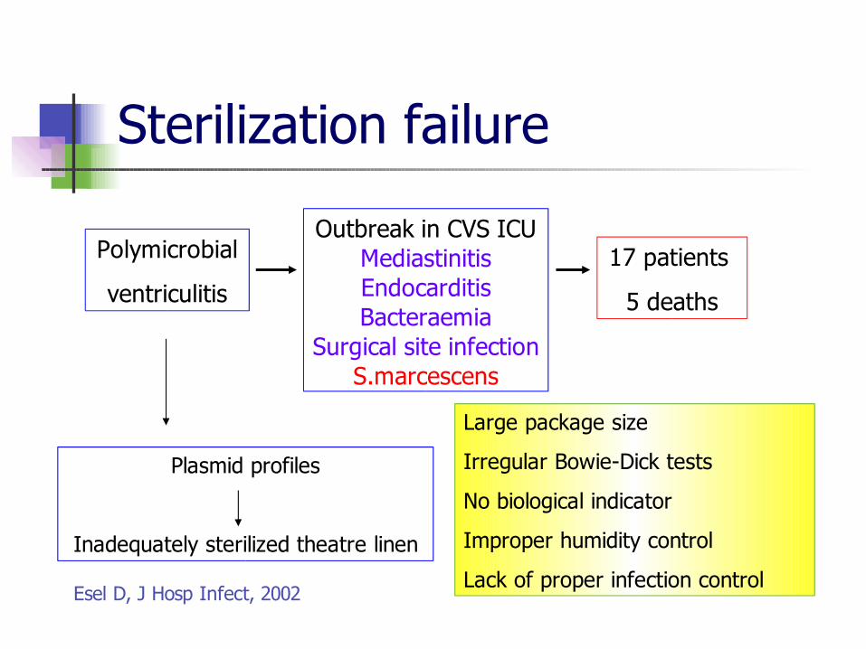
Sterilization failure
Polymicrobial
ventriculitis
Outbreak in CVS ICUMediastinitisEndocarditisBacteraemia
Surgical site infectionS.marcescens
17 patients
5 deaths
Plasmid profiles
Inadequately sterilized theatre linen
Large package size
Irregular Bowie-Dick tests
No biological indicator
Improper humidity control
Lack of proper infection controlEsel D, J Hosp Infect, 2002

Complex structured devices
Disinfection / Sterilization Difficult Labor-intensive Requires attention to details
Narrow and long lumens Twisted structure with crevices Heat-sensitive material, lubricants Small pieces, difficult to detach

Complex structured devicesPhacoemulsification instruments 32 sets
16% 22% moderately severely contaminated contaminated
• lens capsule• man-made fibres • squamous cells • bacteria and fungal elements • red blood cells• proteinaceous material.
Leslie T, Eye
2003

Endoscopes
PATIENT Normal flora E. coli Klebsiella spp. Colonizing organism Serratia spp. Infection Salmonella spp. M.tuberculosis Hepatitis B or C virus HIV
ENVIRONMENT Pseudomonas spp. Atypical mycobacteria
Failure of cleaning and disinfection
Infection transmission
Ann Intern Med, 1993Am J Infect Control, 2000

Endoscopes - Problems of disinfection
Complex structure of endoscopes and accessories
Compliance with established reprocessing guidelines
Endoscopy, 2000
Infect Control Hosp Epidemiol, 2003

Disinfection of endoscopes
26 hospitals in USA 78% failure to sterilize biopsy forceps 71 GIS endoscopes bacterial cultures of internal channels after disinfection
24% grew ≥105 colonies
Kaczmarek RG, Am J Med, 1992

Endoscopes - Failures of disinfection
Inadequate manual CLEANING Cleaning of all channels (flushing, brushing)
Inadequate disinfection Lack of full immersion in the disinfectant solution Short duration of immersion Unappropriate disinfectant
Inadequate rinsing and drying Lack of sterilization of accessories Use of automated endoscope reprocessors
Contaminated reprocessor (water bottles and tubes) Improper connection / usage

Endoscope-related transmission Colonoscopy – HCV
Biopsy suction channel was not cleanedAccessories were not autoclaved5 min. immersion in 2% gluteraldehyde
Bronchoscopy – M. tuberculosisPoor manual cleaningPartial immersion in the disinfectantFailure to sterilize biopsy forceps
Gastroscopy – HBV, H.pylori, Trichosporon spp.
Langenberg W, J Infect Dis, 1990 Bronowicki JP, NEJM, 1997
Michele TM, JAMA, 1997 Agerton T, JAMA, 1997 Wenzel R, JAMA, 1997
Larson JL, Infect Control Hosp Epidemiol, 2003

Patient is not infected but the culture of the sample taken by the endoscope is positive
Pathogens P. aeruginosa, S. marcescens, M. tuberculosis and atypical mycobacteria
Possible results Transmission Colonization or infection Unnecessary investigations Unnecessary treatment
Silva CV, Infect Control Hosp Epidemiol, 2003Bennett SN, Am J Respir Crit Care Med, 1994
Endoscope related pseudo-infections

Microbial reservoirs
Biofilm formation Layer of bacteria (tightly attached to each
other and the underlying surface) and extracellular material
Difficult to clean Protection from disinfection and sterilization
Importance of mechanical cleaning

A biopsy forceps after cleaning, demonstrating residual organic soil
Am J Infect Control, 2000
Biofilm: Microbial life on surfaces commtechlab.msu.edu

Reuse of single use devices (SUD)
Cost saving
Waste minimization
Infection
Endotoxic reactions
Toxic residues
Loss of device integrity
Increased employee risks
Controversial but common practice Collignon PJ, MJA, 1996 and 2003
Muscarella LF, Gastroenterol Nurs, 2001

Studies related to reuseIn favorCatheters Bloom,1997; Kozarek, 1996 Browne, 1997; Druce 2003
Sphincterotomes, papillotomes Cohen,1997; Wilcox 1998
Coagulation probes Roach,1999
Spinal needles Penna, 2000
Against
Endoscope stopcocks Wilson,2000
Biopsy forceps, papillotomes, stone baskets Heeg,2001; Hambrick, 2001
Laparoscopic devices Roth,2002
Reuse-related infections
Frequency is unknown May be undetected
Long incubation period, Asymptomatic nature of blood-borne viral
infections Difficulty to trace infections back to
reused device

Reuse of catheters
Balloon catheterscontaminated with viruses (echo- and adenovirus)
Culture and PCR
Detectable virus after cleaning + sterilization (glutaraldehyde) Luijt DS, Eur Heart J, 2001

Reuse - Questions Which device is suitable for reuse? (instructions of
the manufacturer, decision of a central body, etc) Is it cost effective? What are the risks? (quality assurance and research) What are the standards and regulations? Is the patient informed? Is there a validated cleaning / sterilization process
and a guideline? Is there a standardised assessment process? Is there a tracking system for the outcome?
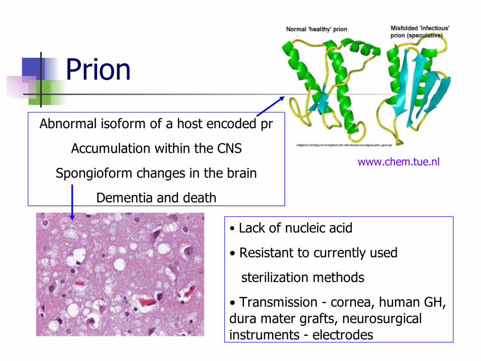
www.chem.tue.nl
• Lack of nucleic acid
• Resistant to currently used
sterilization methods
• Transmission - cornea, human GH, dura mater grafts, neurosurgical instruments - electrodes
Prion
Abnormal isoform of a host encoded pr
Accumulation within the CNS
Spongioform changes in the brain
Dementia and death

vCJD protein
Present in lymphoid tissue starting from early (asymptomatic) stages of disease Tonsil Spleen Lymph nodes Appendix
Incubation period : years

vCJD pr - Risk of transmission
Possibility of cross-infection with surgical instruments contaminated with lymphoid, neural, ocular tissues
UNKNOWNS
• Number of infected people (prevalence)
• Quantity of prions that can cause cross-infection
• Infectivity of the tissues involved in procedures
• Amount of reduction of infective tissue with the decontamination / sterilization procedures

Using disposables (may be only in UK) Which procedures?Are the disposables as effective as reusable
equipment? Special decontamination – sterilization
methodsHow to decide when to use them?
Separated equipment for diagnosed patientsEndoscopes
Lowering the risk of transmission

Method of sterilization for prion
Combinations of Immersion in NaOH or sodium hypochlorite (at
different temperatures and durations) Autoclaving at 121°C (30 min – 1 hour) or 132°C www.who.int/emc-documents, 2003
Collaboration between the surgery team, infection control group, sterilization unit
Type of operation, tissue involved, risk level of the patient
Summary
Sterilization units’ success has a leading role in the prevention of health-care associated infections.
Problems Sterilization of the complex surgery
equipment and endoscopes Reprocessing single use devices Inactivating prions

Success of the sterilization unit
Skilled personnel Asepsis, cleaning, disinfection, sterilization
Adequate space and equipment Standardized written protocols Quality control - assurance Continous education Communication and collaboration
between departments
