infectious diseases affecting the nervous systemmgkmicro.com/biol257/lecture16.pdf · who ÒwinsÓ...
TRANSCRIPT
Infectious Diseases Affecting
the Nervous System

Figure 19.1
CNS:
central
nervous
system
PNS:
peripheral
nervous
system
Defenses:
Bone
Microglial cells
Macrophages
Meniges (cushion of CSF)
Blood-brain barrier
Defenses:
The Nervous System and its Defenses
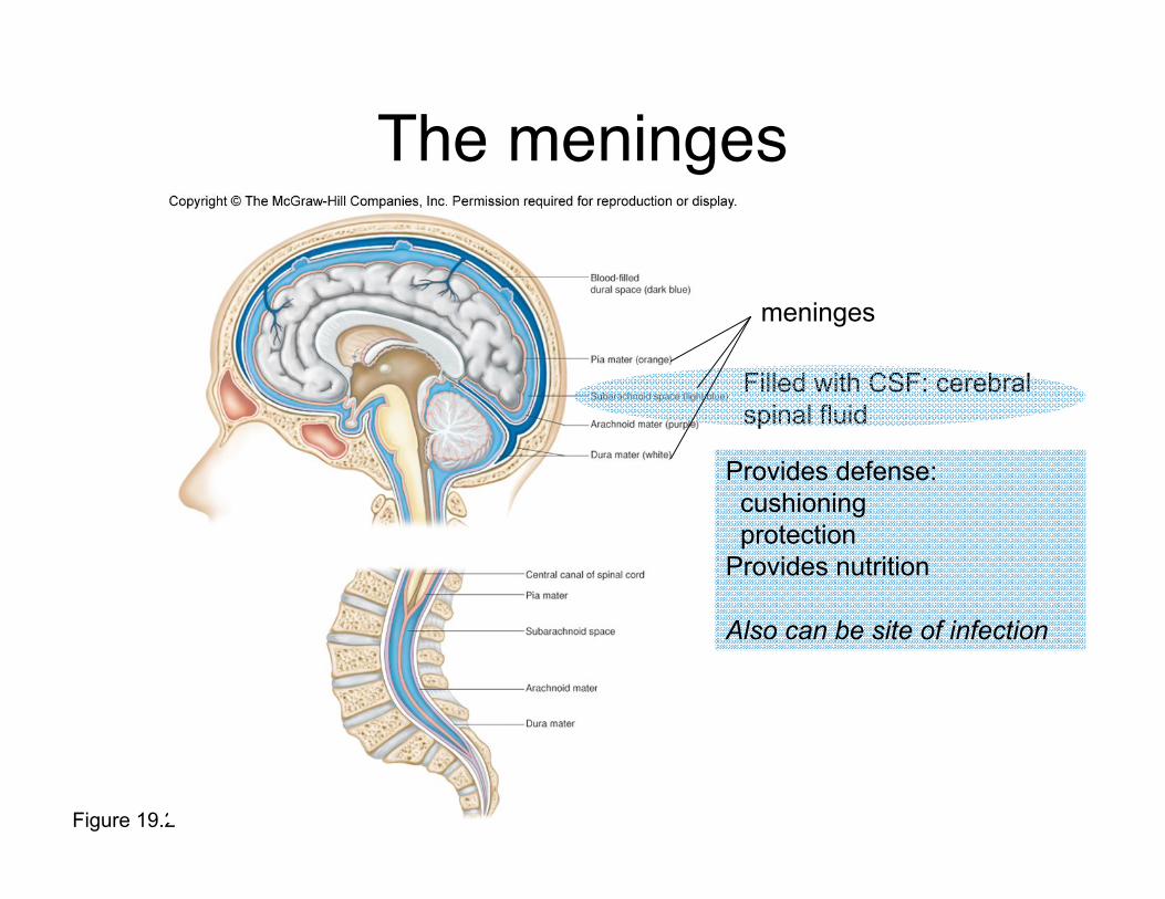
Figure 19.2
The meninges
Filled with CSF: cerebral
spinal fluid
meninges
Provides defense:
cushioning
protection
Provides nutrition
Also can be site of infection
Normal Biota of the Nervous System
none

Who “wins” from nervoussystem infections?
In almost all cases, neither
the host nor the pathogen.

Meningitis
• Inflammation of the meninges
• Many microorganisms can cause meningitis
• More serious forms caused by bacteria
• If it is suspected, lumbar puncture isperformed to obtain CSF
• Typical symptoms: headache, painful orstiff neck, fever, and usually an increasednumber of white blood cells in the CSF
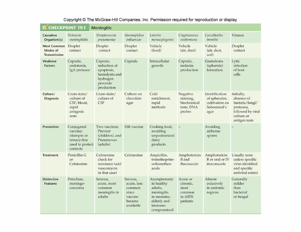

Neisseria meningitides
• Gram-negative diplococcic lined up side by side
• Commonly known as meningococcus
• Often associated with epidemic meningitis
• Causes most serious form of acute meningitis
Causes of meningitis
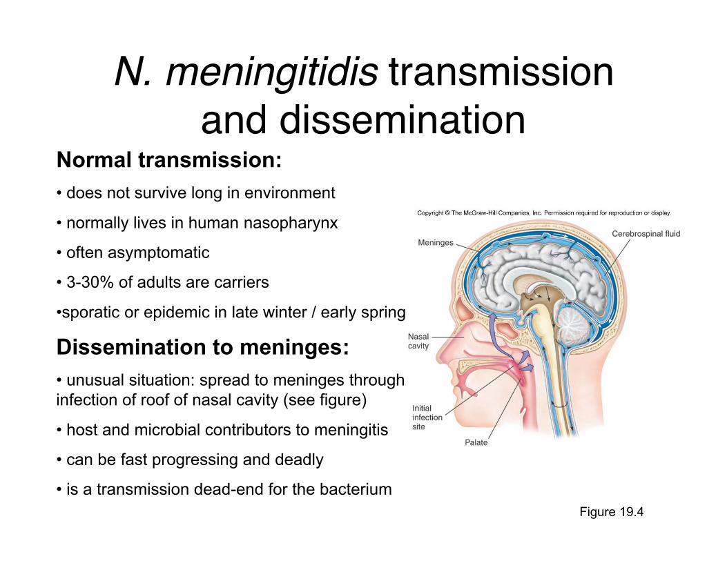
Figure 19.4
N. meningitidis transmissionand dissemination
Normal transmission:
• does not survive long in environment
• normally lives in human nasopharynx
• often asymptomatic
• 3-30% of adults are carriers
•sporatic or epidemic in late winter / early spring
Dissemination to meninges:
• unusual situation: spread to meninges through
infection of roof of nasal cavity (see figure)
• host and microbial contributors to meningitis
• can be fast progressing and deadly
• is a transmission dead-end for the bacterium

Figure 19.5
Meningitis caused by N.meningitidis must treated FASTDiagnosis:First priority: rule out or identify N. meningitidis
gram negative
rapid tests for capsular polysaccarides
oxidase testing (N. meningitidis is oxidase positive)
need to differentiate from N. gonorrhoeae
causes petechiae (diagnostic symptom)
Treatment:Penicillin G (high dose, IV)
Petechiae: small red or purple spots on skin
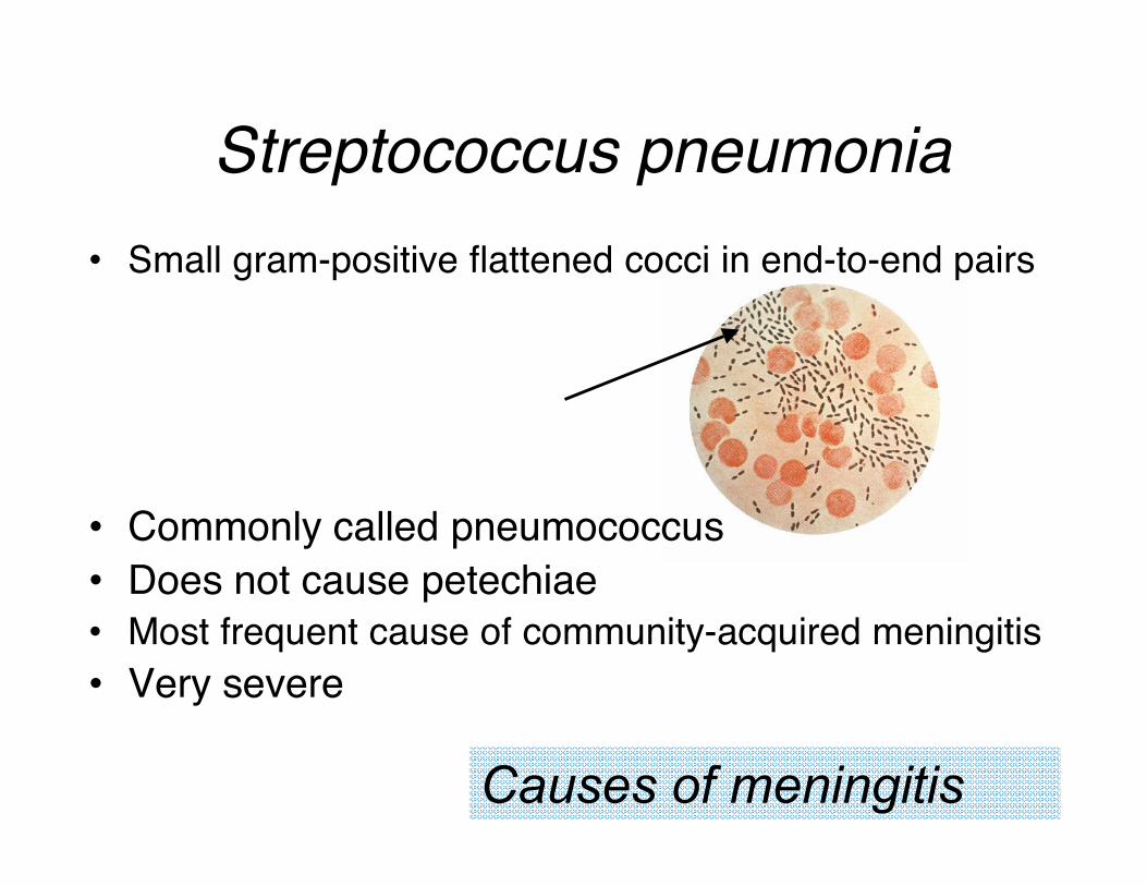
• Small gram-positive flattened cocci in end-to-end pairs
• Commonly called pneumococcus
• Does not cause petechiae
• Most frequent cause of community-acquired meningitis
• Very severe
Streptococcus pneumonia
Causes of meningitis
S. pneumonia diagnosis and treatment
Diagnosis:
•Also major causes of bacterial pneumonias
•Often accompanied by pneumococcal pneumonia
•Usually enters meninges through bloodstream (via lungs)
Treatment:
•Drug resistance common: susceptibility must be tested
•Two vaccines available

Haemophilus influenza• Tiny gram-negative pleomorphic rods
• Fastidious: sensitive to drying, temperatureextremes, disinfectants
• Causes severe meningitis
• Symptoms: similar to N. meningitidis-caused meningitis
Causes of meningitis

H. influenza diagnosis and treatment
Diagnosis:
• Like meningococcus, part of normal nasopharyngial flora
• Meningitis most common in children 3-5 years
• Rarely epidemic (instead is sporatic)
Treatment:
• Vaccine recommended for all children over 2 months
• Even with treatment 33% suffer residual damage

Listeria monocytogenes• Gram-positive, ranges in morphology from
coccobacilli to long filaments in palisades formation
• Not fastidious: resistant to cold, heat, salt, pHextremes, and bile
• Reservoir unknown: environmental, foodborne
Causes of meningitis
L. monocytogenes diagnosis and
treatmentSymptoms:
• In normal adults- mild infection with nonspecific symptoms
of fever, diarrhea, and sore throat
• In elderly or immunocompromised patients, fetuses, orneonates can affect the brain and meninges
•Diagnosis:
• Difficult to isolate: use of cold enrichment
• Recently: rapid non-culture based techniques
Treatment:
• Prevention via pasteurization, cooking of foods
• Antibiotic treatment

Cryptococcus neoformans• Fungus with spherical to ovoid shape and a large capsule
• More chronic form of meningitis, gradual symptom onset
• Headache- most common symptom; also nausea, stiff neck
• Environmental - common in human habitats
Causes of meningitis
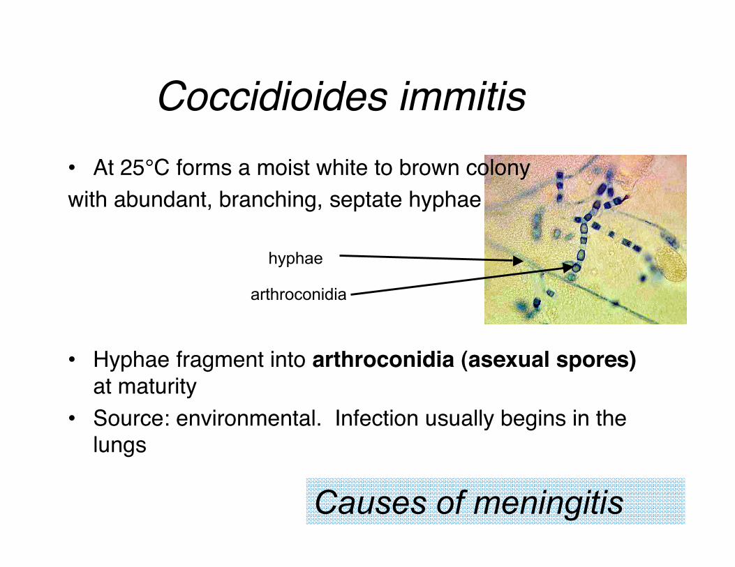
Coccidioides immitis
• At 25°C forms a moist white to brown colony
with abundant, branching, septate hyphae
• Hyphae fragment into arthroconidia (asexual spores)at maturity
• Source: environmental. Infection usually begins in thelungs
Causes of meningitis
hyphae
arthroconidia
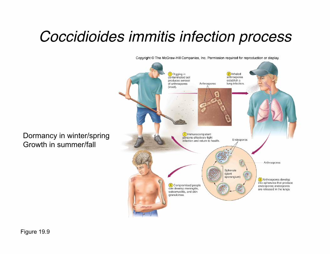
Figure 19.9
Coccidioides immitis infection process
Dormancy in winter/spring
Growth in summer/fall
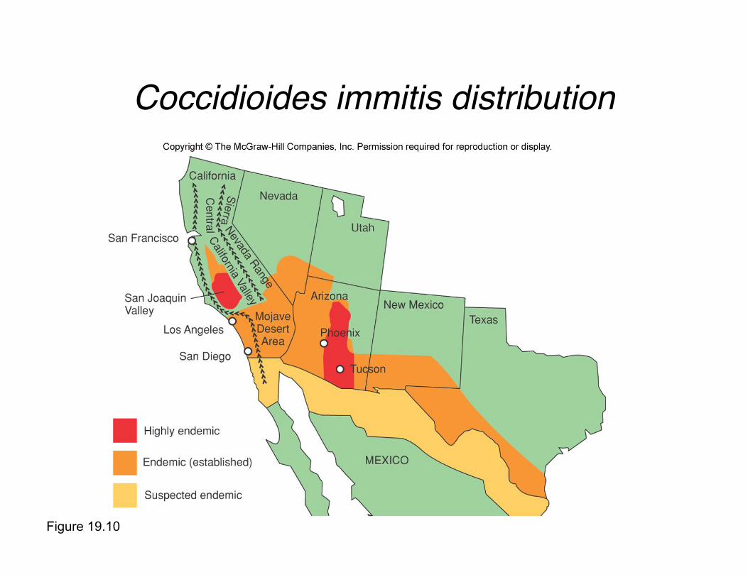
Coccidioides immitis distribution
Figure 19.10
Viruses
• Aseptic meningitis
• Majority of cases occur in children
• 90% caused by enteroviruses
• Generally milder than bacterial or fungal
meningitis
Causes of meningitis

Neonatal Meningitis
• Almost always a result of infection
transmitted by the mother, either in utero
or during passage through the birth canal

Meningoencephalitis
• Encephalitis: inflammation of the brain
• Two microorganisms cause
meningoencephalitis (both amoebas)
– Naegleria fowleri
– Acanthamoeba

Naegleria fowleri
• Causes primary amoebic meningoencephalitis (PAM)
• Small, flask-shaped amoeba
• Forms a rounded, thick-walled, uninucleate cyst
• Resistant to temperature extremes and mildchlorination
Causes of meningoencepalitis

Naegleria fowleri in the environment
• Very common - maychildren are carriers, anddisease is extremely rare
•Infection begins whenamoebas are forced intohuman nasal passages as aresult of swimming, diving, orother aquatic activities
• Amoeba burrows in to thenasal mucosa, multiplies, andmigrates into the brain andsurrounding structures
• Fatal within a week,treatment usually futile
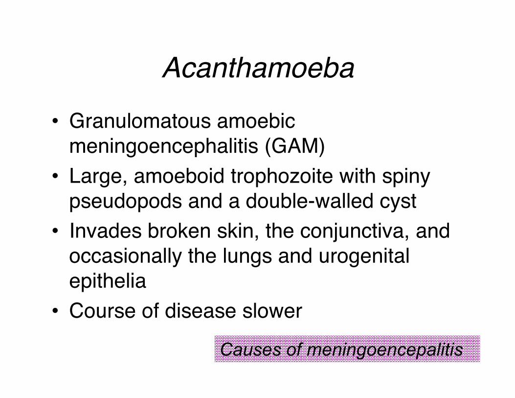
Acanthamoeba
• Granulomatous amoebic
meningoencephalitis (GAM)
• Large, amoeboid trophozoite with spiny
pseudopods and a double-walled cyst
• Invades broken skin, the conjunctiva, and
occasionally the lungs and urogenital
epithelia
• Course of disease slower
Causes of meningoencepalitis
Encephalitis
• Encephalitis can present as acute or subacute
• Always a serious condition
• Acute: almost always caused by viral infection
Acute encephalitis signs and symptoms vary but may include
behavior changes, confusion, decreased consciousness,
seizures
• Subacute: can be caused by protozoans,viruses, prions
Asymptomatic at first, thereafter symptoms and signs vary, often
less striking

• Borne by insects feeding on the blood of hosts
• Most common outcome: acute fever, often
accompanied by rash
Western equine encephalitis (WEE)
Eastern equine encephalitis (EEE) (high case fatality)
California encephalitis (may include two viruses)
St. Louis encephalitis (SLE) (most common US viral cause)
West Nile encephalitis
Causes of acute encephalitis
Arborviruses

Herpes Simplex
Virus
• Can cause encephalitis in newborns
born to HSV-positive mothers
• Prognosis is poor
Causes of acute encephalitis

JC Virus
• Infection is common, pathology rare
• In patients with immune dysfunction,
cause progressive multifocal
leukoencephalopathy (PML)- uncommon
but generally fatal
Causes of acute encephalitis

Toxoplasma gondii
• Flagellated parasite
• Most common cause of subacute encephalitis
• Most cases go unnoticed
• In the fetus and immunodeficient people, severeand often fatal
• Asymptomatic or marked by mild symptomssuch as sore throat, lymph node enlargement,and low-grade fever
Causes of subacute encephalitis
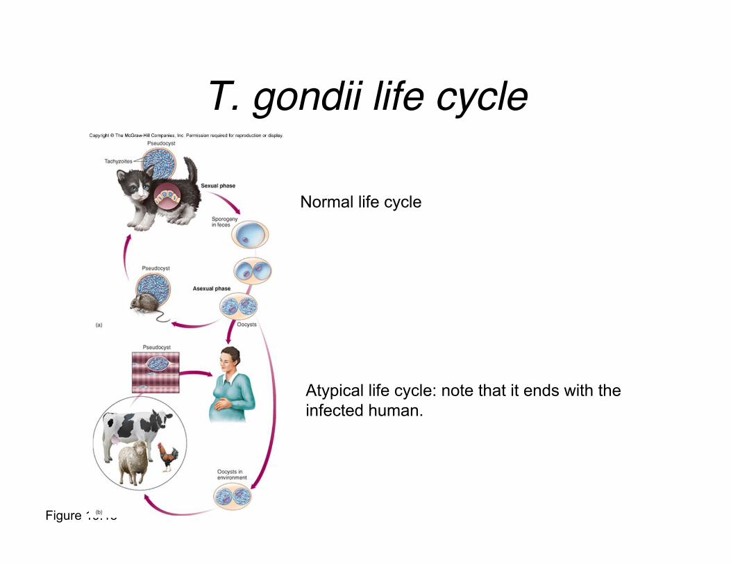
Figure 19.13
T. gondii life cycle
Normal life cycle
Atypical life cycle: note that it ends with the
infected human.
Measles Virus: SubacuteSclerosing Panencephalitis
(SSPE)
• Occurs years after an initial measles
episode
• Seems to be caused by direct viral
invasion of neural tissue
• Reason for persistence of the virus insome people is unclear
Causes of subacute encephalitis

Prions• Transmissible spongiform encephalopathies (TSEs):
neurodegenerative diseases with long incubation periods
but rapid progression once they begin
• Proteins with “contagious” altered structure
• Highly resistant to chemicals radiation and heat
• Human TSEs
– Creutzfeldt-Jakob disease (CJD)
– Gerstmann-Strussler-Scheinker disease
– Fatal familial insomnia
Causes of subacute encephalitis

Figure 19.14
Effects of prions on brain tissue

Other Disease of the NervousSystem
• Rabies (caused by rabies virus)
• Poliomyelitis (caused by poliovirus)
• Tenanus (caused by bacterium Clostridium tetani)
Toxin causes spasms
• Botulism (caused bacterium Clostridium botulinum)
Toxin causes flaccid paralysis
• African Sleeping sickness (caused by protozoan
Trypanosoma brucei)
Rabies• Exception to “no one wins” rule: normally a nervous
sytem disease of mammals
• Slow, progressive zoonotic disease characterized byfatal encephalitis
• Average incubation time: 1-2 months or more
• Prodromal phase begins with fever, nausea, vomiting,headache, fatigue, and other nonspecific symptoms
• Furious rabies– Periods of agitation, disorientation, seizures, and twitching
– Spasms in the neck and pharyngeal muscles lead to hydrophobia
• Dumb rabies– Patient is not hyperactive but is paralyzed, disoriented and stuporous
• Both forms progress to the coma phase, resulting indeath, unless vaccination precedes symptoms.

Figure 19.15
Rabies virus is related to VSV
It’s an enveloped negative sense RNA virus.

Poliomyelitis• Acute enteroviral infection of the spinal cord
• Can cause neuromuscular paralysis
• Often affects small children
• Most infections are contained as short-term, mild viremia
• Some develop mild nonspecific symptoms of fever, headache,nausea, sore throat, and myalgia
• Then spreads along specific pathways in the spinal cord and brain
• Neurotropic: the virus infiltrates the motor neurons of the anteriorhorn of the spinal cord
• Nonparalytic: invasion but not destruction of nervous tissue
• Paralytic: various degrees of flaccid paralysis
• Rare cases: bulbar poliomyelitis

Figure 19.18
Poliovirus transmission
Fecal-oral transmission
Escape into the nervous system
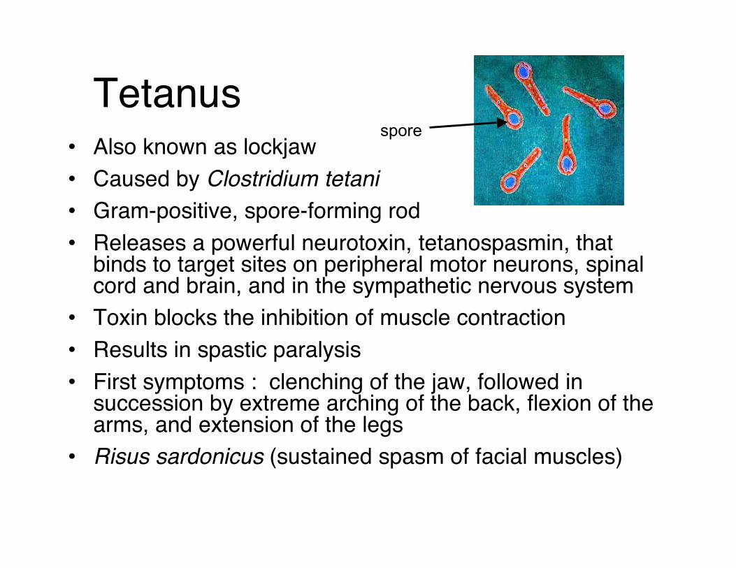
Tetanus• Also known as lockjaw
• Caused by Clostridium tetani
• Gram-positive, spore-forming rod
• Releases a powerful neurotoxin, tetanospasmin, thatbinds to target sites on peripheral motor neurons, spinalcord and brain, and in the sympathetic nervous system
• Toxin blocks the inhibition of muscle contraction
• Results in spastic paralysis
• First symptoms : clenching of the jaw, followed insuccession by extreme arching of the back, flexion of thearms, and extension of the legs
• Risus sardonicus (sustained spasm of facial muscles)
spore

Figure 19.21
• Soil-borne
• Entry of bacterium into (anaerobic) wound is required
Neonatal tetanus associated with ash or mud on ubilical stump
• Effective vaccine available
• Treatment with antibiotics and passive TIG (tetanus immune globulin)
BotulismCause: spore forming bacterium Clostridium
botulinum
Three major forms– Food-borne botulism (not an infection)
• Ingestion of preformed botulinum exotoxin from bacteriagrowing in an anaerobic environment (e.g. canned foods)
• Results in an intoxication affecting neuromuscularjunctions
– Infant botulism (true infection)• Entrance of botulinum toxin into the bloodstream after
spore enters the gut and establishes infection
– Wound botulism (true infection)• Entrance of botulinum toxin into the bloodstream after
anaerobic infection of a wound from environmental source
• Symptoms: double vision, difficulty in
swallowing, dizziness; later symptoms
include descending muscular paralysis
and respiratory compromise

Figure 19.23
Mechanism of toxin!s effect

African Sleeping
Sickness• Caused by Trypanosoma brucei protozoan
• Also called trypanosomiasis
• Escape of immune system by antigenic shift
• Affects the lymphatic system and areas surroundingblood vessels
• Usually a long asymptomatic period precedes onset ofsymptoms
• Symptoms include intermittent fever, enlarged spleen,swollen lymph nodes, and joint pain
• Central nervous system is affected with personality andbehavioral changes that progress to lassitude and sleepdisturbances

Figure 19.24
T. brucei infection cycle
CNS damage occurs over years
of infection. It is not required for
the protozoan to complete its life
cycle.