infectious keratitis - feiz.mui.ac.ir ulcer.pdf · sub conj ab: imminent scleral spread/...
TRANSCRIPT

Infectious keratitisA.KhalilianM.D

Natural eye defense
Eyelids, blinking
Tear film ( Ig, complement, enzymes)
Corneal epithelium
Normal ocular flora
MALT in conjectiva
Intact corneal sensation

Predisposing factors Contact lens (most common. 10 fold risk)
Trauma
Contaminated ocular med
Impaired defense mechanisms( Sj ,ocp,…)
Tap water, eyewash solutions
Chronic anesthetics abuse
Malnutrition,DM, collagen vascular dis

Contact lens induced ulcer
EW > RGP = daily wear Overnight lens use; major RF ( 15)
Apkakia, corneal graft, chronic keratopathy,fail to practice lens care regiment,worn CL
Pseudomonas sp ; most common
Corneal abrasion, epith thining, epith mitosis, anerobic metabolism, hypoxia

pathogenesis
Adhesions
S areus: use adhesins to bind collagen/fibronectin of bowman layer
P aeruginosa : directly binds (pili) to receptors exposed of injured epith
CED,abrasion S.areus- S.pnemoniaeS.aeruginosa

Pathogenesis( cont…)Invasion
LPS are the major mediators of inflm
Local production of cytokins/chemokins
Migration of neutrophils from limbus
Enzyme released by PMN
Activation of MMPs
Microbial proteas: destroy stroma

Invasion through intact epithelium
N gonorrhoeae
N meningitidis
Corynebacterium diphtheriae
Haemophilus aegyptius
Listeria monocyrogenes

Clinical presentation
Bacterial corneal ulcer shows:
I. Sharp epith demarcation
II. underlying dense supporative stromal inflm with indinstinct edges
III. surrounded by stromal edema

Clinical examination
Good Hx (traum,HSV, CL ,Sx …) General/ facial appearance/ lid closure NLD regurgitation, corneal sensation
On slit lampLid margin: MGD,ulcer, eyelash abNLTear filmConj, sclera: scar, discharge,membrane or pseudo m, thining

Clinical examination (cont…)Cornea Location (central) Density, size,depth
Shape : ring, satellite lesions Character of infiltrate margin
(feathery,crystalline, soft, necrotic)
Endothelium plaque AC reaction, hypopyon,fibrin

The most common pathogenic organism identified in bacterial keratitis include:
Pseudomonas
Staphylococci
streptococci

Pseudomonas.aeruginosaIncreasing prevalence due to soft CL usage
>2/3 cosmetic CL induced ulcers
dense stromal inflt , necrosis shaggy surface adherent mucupurulent exudate endo inflmm plaque AC reaction/ hypopyon Descemetocele , corneal perforation

Pseudomonas(cont…) despite Rx, may progress to deep abscess Sometimes ring infiltrate


Staphylococci NL ocular flora More in compromised cornea like:
bullous keratopathychronic HSV keratitisK-conj siccaocular rosaceaAKC
Nonaureus staph most frequently organism isolated from corneal ulcers

Staphylococci

streptococciS pneumoniae usually occurs after
corneal trauma Dacryocystitis Flitering beleb infection
Ulcer is acute, purulent,rapidly progressive,deepstromal abscess, severe marked hypopyon,retrocorneal fibrin,
Perforation is common

Ulcer is acute, purulent, rapidly progressiveDeep stromal abscess, Severe marked hypopyonRetrocorneal fibrin

Atypical mycobacteria
Slow growing,fastidious organismslike mycobacteria or anaerobes may have
a non-supp infiltate & intact epith

Keratitis from non-tuberculous mycobacteria
Delayed onset (2-8 week), slowly progressive Feathery, indistinc margin, craked windsheild After LASIK, corneal F.B or trauma Lack of response to conventional Abs

Teratment of nontuberculous mycobacteria
Oral or topical Clarithromycin +/-Amikacin
Fluoroquinolone(moxifloxacin,gatifloxacin)
Imipenem co-trimoxazole

Infectious crystalline keratopathy
Most commonly: streptococcus alpha-hemolytic Densly packed,white branching aggregates of
organism in the absence of host inflm response
No CED, mild stromal inflm GC use,CL wear , PKP, anesthetic abuse


nocardia
Indolent ulcer, waxes ana wane pattern Minor trauma, especially contaminated soil Characteristic feature:
Raised, superfacial, chalky white infl in a wreath likePattern with multifocal satellite lesions


Indications for smear & culture
Large Corneal infiltrate(> 2mm)
Infiltrate extending to the middle deep stroma
Chronic ulcer Unresponsive to broad-spectrum Abs
Suspecious to fungal,amoebic,mycobacterium

culture
By Slit lamp magnification
Proparacaine 0.5% (not tetracaine )
Scrape corneal tissue from the advancing border
With wet sterile cotton swab, NO 15 blade
Muliple samples from advancing edges Corneal specimens should be inoculated directly
onto media in C streaks

culture
Culture of CL, lense case & solutions maybe useful:suspecious to acanthamoebanegative cultures
Before being reported as no-growth Aerobic cultures should be held for 1 week Anaerobic cultures should be held for 1-2 weeks Mycobacterial & fungal for 4-6 weeks

stains
Gram stain
Best for bacteria
Rapid(5 min) Sensivity 55-79% Distinguish bacteria from fungi/acanthomoeba
Geram positives : bluish purpule
Geram negatives: Pink

Stains(cont…)
GimsaBacteria, fungi, chlamydia,acanthomoeba
Acridine orange:Rapid(2min)Accurately predicts culture results in 71-84%More sensitive than gram stainRequires epi-fluorescence microscope

Other stains
Calcofluor white :fungi,acanthomoebaRequires epi-fluorescence microscope
Acid fast (Ziehl-Neelsen):mycobacteria, nocardia

Corneal biopsy
1. Unresponsice ulcer2. Negative culture3. Infiltrate in mid or deep stroma with intact
overlynig tissue
Specimen 1-2 mm
Healing effect of Bx due to debulking or debridment of necrotic tissue
Alternative to Bx:pass a sterile silk suture through the inflt>> culture

Confocal microscopy
Non invasive, in vivo diagnostic tool Real time veiwing of structures
in the living cornea at cellular level
Acanthomoeba cysts Fungal hyphae

Rxcephalosporins
CefazolinExellent activity against Gram+ Minimal toxicityUsage in combination with other anti,Gram – agents
CeftazidimeAnti pseudomonal activityUsed in pseudomona keratitis not responding to AG or FLQ

Rxglycopeptides
Vancomycin
Active against many Gram+ bacteria
One of most potent AB against methiciline-R S.areus & coagulase negative Staph
It should be reserved for cephalosporin –R staph

Rx
Aminoglycosides
Bacericidal effect against Gram -, aerobics
For severe pseudomona ulcers may be combined with Cephalosporins
Amikacin is the drug of choice for nocardia
Less pseudomonas resistancy to Amikacin,ratherthan gentamicin & tobramicin

RxMacrolides
Erythromycin has a relativley activity againstmost Gram +some Gram –most viridans & anaerobic Streptococcimost strains of Neisseria( gonorrhoeae,meningitides)majority of aerobic Gram – bacilli are resistant
limited role for bacterial ulcer( poor corneal penetration)

RxMacrolides
Newr macrolides: Azithromycin Clarithromycin Roxithromycin
Favorable for treating:Chlamidia.t Nontuberculous mycobacteria

RxFluoroquinolones
Ciprofloxacin,ofloxacin (2nd)
Caverage most Gram - & some Gram+
Moxi, gati, levo and besifloxacin(3rd&4 )have improved Gram+ and atypic mycobacterium coverage ,but limmited activity against MRSA

RxFluoroquinolones
Similar efficacy compared to FF Abs for common ocular pathogens(small, non central, not severe ulcer)
Lesser topical side effects than FFABs
Higher risk of perforation than FF Abs
Increasing resistance: P.aeruginosa Staphylococcus(MRSA) Streptococcus

Initial management FF loading dose: evey 5-15 min for 1 hour then
every 15min –hourly
Cycloplegics : synechia & pain, c.spasm
Sub conj AB:imminent scleral spread/ perforationpoor compliance
Sys AB: Scleral / intraocular extensionimpending/frank corneal perforation

Strategies for initial treatment
1- Culture guided approach:smear& culture of all ulcersstart Rx based on clinical & epidemiologic datamodify Rx by smear/culture results
Disadvantages:1- cost2-positive cultures only 60% 3-discrepancy between in vitro sensivity and
clinical response

Strategies for initial treatment (cont…)
2-Emprical approach
based on pre-existing culture sensivityuse broad spectrum ABcefazolin or vanco for Gram+ tubramycin or ceftazidime for Gram –
Failed Rx culture appropriate Rx

Strategies for initial treatment (cont…)
3- case based approachsmear/culture before Rx only in1-involving visual axis2-large, deep ulcer3-keratitis associated with trauma,contamination by vegetation material or unsantizied water
Small, peripheral ulcers: no culture, empirical RXCentral. Large,deep,unusual ulcer: culture based Rx

Ulcer at presentation
Small/peripheral
No microbiologic work-up
Broad spectrum AB
Central/ multiple/ deep
Microbiologicwork-up
Specific AB for causal organism

Initial evaluation
(48 hr after treatment)Stable
improve
Cont RX
Sig progression
Poor compliance
Alternative treatment
Good compliance
No initial microbiolog
ic w/u
Stop treatment for 24 hr
Then microbiological
w/u
Had microbiologic
w/u
Check culture results/ mofify
treatment

clinical features suggestive of positive response to AB
1. Pain reduction2. Consolidiation & sharper edges of stromal inflt3. Decreased density of stromal inflt4. Reduction of stromal edema & endo plaque5. Reduction of AC reaction6. Re-epith
The clinical response is best assessed after 48 hr

Treatment (cont…)
Most bacterial ulcers will be culture negative after 48-72 hr of treatment
But FF Abs should be continued untill substantial control of infection is seen.
There after, a prophylactic AB (not a FF) may be given at a theraputic dose,untill CED is healed

Corticosterois therapy
GC therapy in bacterial keratitis is still contraversial
Potential advantage:supression of inflmmreduction of corneal scar
Potential adverse effects:1-enhancment of bacterial growth2-Impairment of phagocytosis3-Inhibition of collagen synthesis4-Cataract,glaucoma

Corticosterois therapy( cont…)
There is no different between pts treated with or without steroid therapy in terms of:
time to cure Final VA Complications
Corticosteroids is contraindicated in:1-sig corneal thining2- impending to perforation3-absence of appropriate AB therapy

Corticosterois therapy (cont…)
The goal of using GC is reduction of inflmm ation & scarring and morbidity.
In cases where the corneal infiltrate & scaring Compromises the visual axis, topical steroid may be added to regimen, following at least 2-3 days of progressive improvement with topical Abs.
If the patient shows no adverese effect after 1-2 day,the frequency of steroid may be adjusted

Cyanoacrylate tissue adhesive
Tectonic support Bacteriostatic effect Stopping keratolysis by blocking proteases
Can be used for perforations up to 2-3 mm
Necrotic tissue& debris should be removed Use the minimmun amount of glue BCL

Surgical management
Conj flapBring vessels to infected are,promote healing,stable surface covering
Conj flap is contraindicated in necrotic area with active infection!
Conj flap is best useful in cases of non-healing preipheral ulcer

Surgical management
Emergency theraputic PKP:I. Uncontrolled progression of infiltrateII. Limbal involvment/impending scleritisIII. Corneal perforation/descematocele
Interrupted sutures, PI,circumscribe all infected areas
Intensive Abs should be administered for 48hr before surgery
Defer the PKP as late as you can

Fungal keratitisA.khalilianM.D

Fungal keratitis
FilamentousNon septated
FilamentousSeptated
Pig hyphea
Filamentous septatedNon pig hyphae
Yeast
MucorRhizopus
AlternariaCurvularia
FusariumAspergillusAcremonium
CandidaCryptococcus

Fungal keratitis epidemiology
Fungi as a NL flora( 3-28%) in conjetival sac
More common in rural setting
Aspergillus is the most common cause worldwide
In the largest case series from India:Aspergillus(27-64%)Pencillium (2-29%)Fusarium (6-32%)

Fungal keratitis
Risk factors:
Trauma (plant) major RF Soft CL (cosmetic > theraputic) Topical steroid/anesthetic Sys immunosuppression (DM,HIV,leprosy)Corneal surgery (PK,LASIK,RK)Chronic keratitis (HSV,VKC) >> candida

Fungal keratitis clinical features Slow onset Fewer inflmm sign,symp initially May have littile or no conj inj initially
Pain can be out of proportion to the relatively uninflamed cornea.
Occasionally,fungus may invade the iris,PC and leads to pupillary ACG.

Fungal keratitis
Special signs: Elevated area Hypate(branching) ulcer Irrigular feathery marginDry rough texture Satellite lesion Brown pig( curvularia ) Invasive to AC Intact epith+deep stromal inflt

Fungal keratitis
Fungal keratitis
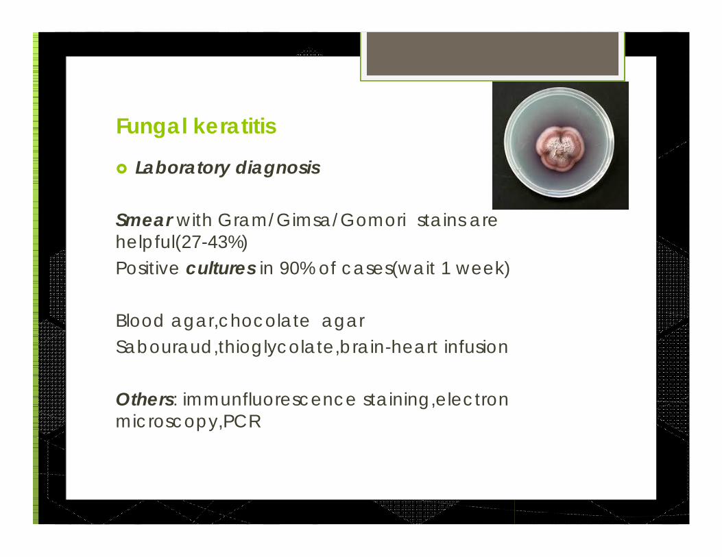
Laboratory diagnosis
Smear with Gram/Gimsa/Gomori stains are helpful(27-43%)Positive cultures in 90% of cases(wait 1 week)
Blood agar,chocolate agarSabouraud,thioglycolate,brain-heart infusion
Others: immunfluorescence staining,electronmicroscopy,PCR

Fungal keratitis
Laboratory diagnosis(cont...)
Corneal biopsy (lamellar keratectomy) is superior to scraping for recovering fungi in cases with negative smear/culture
AC tap
Confocal microscopy is promising

Classification of antifungals
othersPyrimidinesAzolePolyenes
BetadinNystatin 5%PHMBchlorhexidn
5-FCClotrimazolKetoconazolFluconazolvoriconazol
Amphotericin B(0.15%)
Natamycin (5%)
AspergillusCandidafusarium
?Almost allExceptfusarium
Almost allfungi

Fungal keratitis treatment
Natamycin 5% is recommended for initial treatment of most cases of filamentous(Fusarium)fungal ulcer
topical amphotericin B(0.15%- 0.30%) is the most effectice agent agianst Yeast keratitis &Aspergillus
Topical voriconazole(1%) is useful in some cases of refractory fungal keratitis( increasing resistance!)

Principles of Fungal keratitis treatment
1-Epith debridment especially early (every 1-2 day)*significantly enhance the topical antifungal penetration
2-Start with natamycin(every 5 min for 1 hour loading dose)If worsening of keratitis continues: in candida keratitis add amph-B 0.15% In aspergillus keratitis add an azole(fluconazole2%)
Don use amph-B and imidazole (antagonist)

Principles of Fungal keratitis treatment (cont…)
Treatmen lengh: 4-6 weeks(based on clinical response)
Sys antifungal or sub conj antifungals indicated in:1-Severe deep keratitis, 2-scleritis, 3- endophthalmitis
Sys voriconazole(200-400 mg),posaconazole has the better corneal penetration
intrastromal/intracameral Amph-B(5-10 mcg/0.1 cc) or voriconazole(50-100 mcg/0.1cc) are becoming more widely validated

Principles of Fungal keratitis treatment (cont…)
Corticosteroids can be used after 2 weeks of treatment & clear clinical evidence of inf control
The steroid drop is used in conjuction with antifungal and never without
Newer modalities of fungal keratitis Rx: Collagen sheilds impregnated with Ampho-B Excimer laser (surface infec ablation) Intra-cameral Ampho-B Cryotherapy(keratoscleritis)
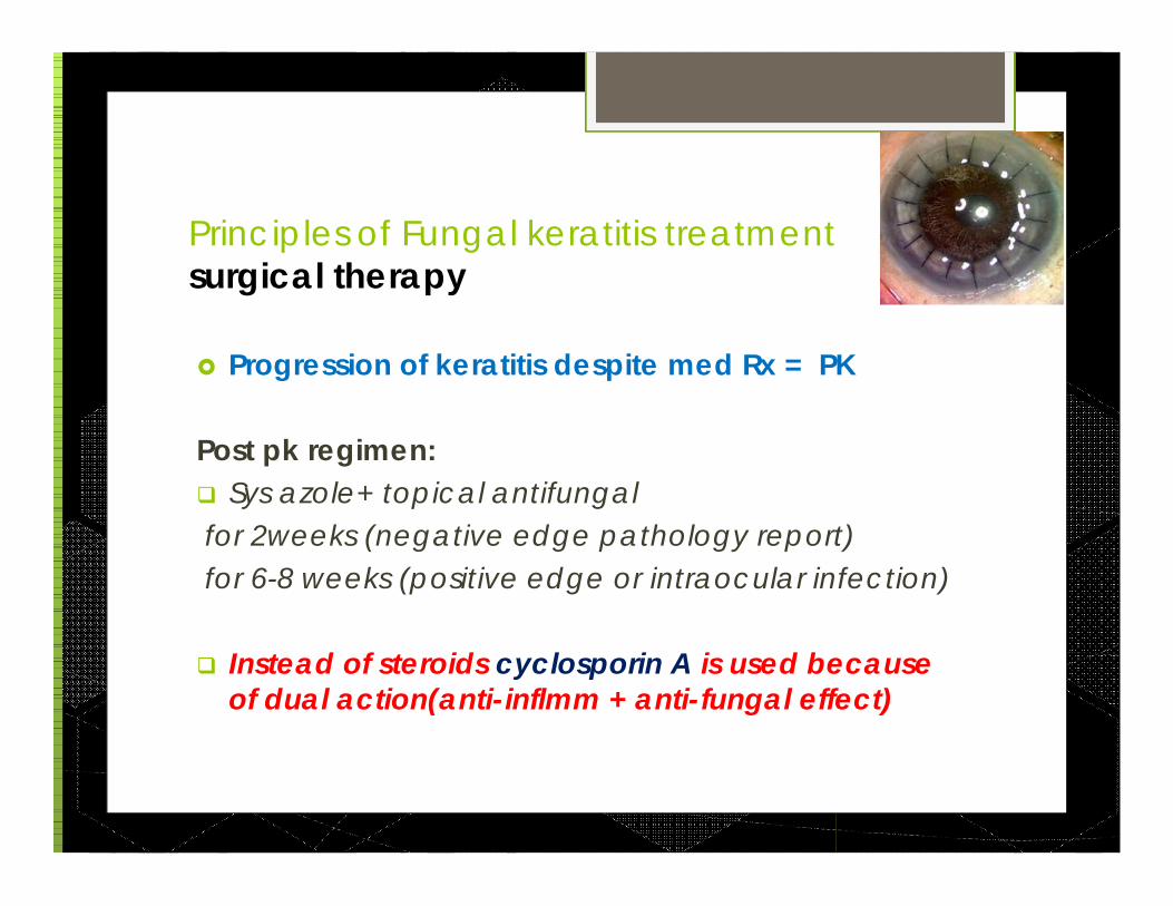
Principles of Fungal keratitis treatment surgical therapy
Progression of keratitis despite med Rx = PK
Post pk regimen: Sys azole+ topical antifungalfor 2weeks (negative edge pathology report)for 6-8 weeks (positive edge or intraocular infection)
Instead of steroids cyclosporin A is used because of dual action(anti-inflmm + anti-fungal effect)