inferior vena cava collapsibility and heart failure signs ... · inferior vena cava collapsibility...
TRANSCRIPT
Inferior Vena Cava Collapsibility and Heart Failure Signs and Symptoms. New Insights about Possible LinksRenato De Vecchis1, Antonio Ciccarelli2, Carmelina Ariano2
Cardiology Unit, Presidio Sanitario Intermedio Elena d Aosta1, Napoli; Neurorehabilitation Unit, Clinica S. Maria del Pozzo2, Somma Vesuviana, Italy
AbstractBackground: In chronic heart failure patients (CHF), ultrasound measurement of inferior vena cava collapsibility index (IVCCI) has been proposed to yield careful assessment and grading of the hemodynamic congestion.
Objective: The purpose of this study was to correlate the findings of physical examination with IVCCI in CHF patients.
Methods: According to a retrospective cohort design, we analyzed 54 CHF patients with right or biventricular CHF, belonging to III NYHA class. We planned to determine whether any basal IVCCI range would be able to predict persistent or worsening clinical congestion found at the end of subsequent follow up (i.e. after 1-2 months of oral optimized therapy). For this purpose, the patients were subdivided by three groups according to the basal IVCCI value: ≤ 15% (13 pts), 16 - 40% (21 pts) and > 40% (20 pts).Several clinical criteria of congestion were compared across the three groups and subsequently entered in the Cox multivariate model.
Results: Multivariate predictors of high congestion score were jugular venous distension (HR: 13,38 95% C.I.: 2,13 - 84 p = 0,0059) and rales (HR: 11 95% C.I : 1,45 - 83,8 p = 0,0213). IVCCI ≤ 15% was always associated with high congestion score at the second visit; but IVCCI ≤ 15% failed to predict high congestion score at the second visit.
Conclusions: In CHF setting, low IVCCI did not reliably predict high congestion score. Nevertheless, the cluster with IVCCI ≤ 15% was always found associated with signs and symptoms from both right and left-sided decompensated CHF. (Arq Bras Cardiol. 2012; [online].ahead print, PP.0-0)
Keywords: Cardiac output, low; heart failure; vena cava,inferior/abnormalities; ventricular dysfunction, left.
Correspondência: Renato De Vecchis • P. Gaurico 21 – 80125 - Napoli, ItalyE-mail: [email protected], [email protected] Artigo recebido em 28/08/11; revisado recebido em 30/08/11;aprovado em 06/02/12.
IntroductionHemodynamic congestion1 is thought to derive primarily
from impaired cardiac index - with or without reduction in left ventricular ejection fraction- .This in turn generates elevation in pulmonary capillary wedge pressure (PCWP)2, in the case of left chambers isolated decompensation, or combined increase in both PCWP and right atrial pressure, in the case of biventricular heart failure1. Impaired cardiac index, for left only ventricular heart failure, or the combination of impaired cardiac index plus elevated central venous pressure, in the case of right or biventricular heart failure, with or without patent fall in blood pressure, substantially reduce renal blood flow, the most important variable of kidney filtration gradient in patients presenting with congestive heart failure, thus generating neuro-hormonal stimulation and development of hydrosaline retention4-8. Hemodynamic worsening consisting in impaired cardiac index and increased filling pressures will further activate the renin-angiotensin and sympathetic nervous system, reduce nitric oxide in the endothelium, and induce inflammatory mediators, to aggravate the hypoperfusion
state of the glomeruli and so to further harm the renal function9. Reduction in water and salt clearance in turn propitiates persistence of hemodynamic congestion to create and maintain over time a condition of cardiac overload. Hemodynamic congestion contributes to the progression of HF by further activating neurohormones and by causing subendocardial ischaemia, resulting in myocardial necrosis/apoptosis and/or secondary mitral insufficiency by its effects on LV geometry (changing it from an ellipsoid to a sphere)10. In addition, elevated right atrial pressure may contribute to the cardio-renal syndrome through reduction of the perfusion gradient across the kidneys11-12.
Among the proposed methods for estimating and grading hemodynamic congestion in right or biventricular heart failure, a remarkable role is played by ultrasound evaluation of the inferior vena cava diameter (IVCD) and its collapsibility index (IVCCI)13. This technique is aimed to identify any changes in respiratory pattern of IVC indicating the presence of abnormalities in volume status (that is hemodynamic congestion or intravascular depletion). Based on well-established knowledge, wide respiratory fluctuations of IVCCI would indicate that intravascular depletion is likely, whereas low values of this parameter would mean high probability of hemodynamic congestion. These inferences have received exhaustive confirmation by many large well-conducted studies13-20.
Arq Bras Cardiol. 2012; [online].ahead print, PP.0-0
De Vecchis et al New insights about their possible relations
rest with orthopnea was selected as being a symptom usually associated with severe impairment in left ventricle pump function (in our case to be set in the context of biventricular cardiac decompensation, as right heart failure was present in all recruited cases by definite enrollment criterion ). Orthopnea was defined as any respiratory distress associated with lying down or the perceived need to use more than one pillow to avoid respiratory distress. Rales were considered owing to their well-known association with pulmonary capillary and venous hematic stagnation usually related to relatively high levels of capillary pulmonary wedge pressure (CPWP). Dyspnea on exertion was included in analysis as being a symptom that usually indicates lesser cardiac impairment compared to orthopnea, generated by right-sided as well as by combined biventricular heart failure. Besides, the other four signs (jugular venous distension, peripheral edema, congestive hepatomegaly and weight gain ≥ 1 kg per week) were evaluated in the study as markers of hydrosaline retention as usually found in systemic congestion from right or biventricular advanced CHF. Particularly, jugular venous distension was considered present if visualized at least 10 cm vertically above the right atrium. Weight gain was defined as ≥ 1kg mean increase per week.
In addition, a congestion score was set up and defined retrospectively for this analysis as the sum of the seven criteria for congestion, with a possible range from 0 to 7. A score ≥ 4 was termed “high congestion score” and used as outcome variable (“end – point”) in the subsequent statistical analysis. According to the customary approach applied at the two Centers, every patient in each of the day hospitals underwent a careful clinical examination along with a complete echocardiography including a thorough assessment of IVC with IVCCI calculation. Subsequently, a second scheduled visit was usually accomplished within one-two months in order to timely establish any appropriate pharmacologic and dosing change to be done based on the evolving clinical picture. Should any complaint or unexpected complication arise, the patients were recommended to bring forward the date of the visit .
In our retrospective study, we tried to ascertain whether any association could exist between IVCCI values, as detected at the first visit, and one or more of the abovementioned signs and symptoms of congestion, as noticed after 1-2 months of clinical follow up. We investigated this hypothesis by using the preliminary categorization of IVCCI values by three layers (IVCCI ≤ 15% , IVCC I 16-40%, IVCC I >40%) and by considering the clinical picture after adequate oral therapy kept for one-two months. We also verified the possible existence of any significant association between the cluster characterized by the lowest IVCCI (≤ 15%) at baseline and the finding of “high congestion score”, as found at the second visit after one-two months. The prescribed oral therapy usually included ACE-inhibitors, beta-blockers and diuretics; in addition, according to the physician’s judgment, aldosterone receptor antagonists, oral or transdermal nitrates, antiaggregant drugs, warfarin, amiodarone and digoxin were also administered if necessary. It was also established that every patient requiring intravenous diuretics or inotropes for worsening HF during follow up would have been excluded from the statistical analysis, as being considered to demonstrate treatment failure.
ObjectivesIn a number of patients with right or biventricular CHF,
we have tested the possible relationships between IVCCI and some signs and symptoms of CHF. Besides, we have tried to establish whether in these CHF patients a low IVCCI value, as found at the first visit, can be assumed as a reliable predictor of subsequent clinical congestion – after a clinical follow up of one-two months - .
Methods An observational retrospective study was carried out
by enrolling patients from two Centers (C.U. E. d’A. and N.R. S.M.d P.). For enrollment in the study , documented evidence of right or bi-ventricular chronic heart failure in II- III NYHA class was required. Exclusion criteria were: patients with pace-maker or treated by cardiac resynchronization therapy; myocardial infarction within 30 days, arrhythmia-related syncope, major cardiac surgery, unstable angina, uncontrollable hypertension, cor pulmonale, advanced pulmonary disease, major neurologic disease or cerebrovascular disease, suspected renal artery stenosis, advanced renal failure (i.e. serum creatinine >2,5 mg/dl at baseline), other life-threatening disease.
Retrospective chart review was performed to analyze characteristics of all eligible patients. For each patient, date of birth, sex, race, and weight and height were noted. Comorbidities, including diabetes, dyslipidemia, nicotine abuse, hypertension , ischemic heart disease, preexisting diabetic nephropathy or other chronic renal disease in addition to medical treatment at the time of hospital stay
also were extracted for each patient. IVCCI was defined as the difference between maximum expiratory diameter and minimum inspiratory diameter divided by the maximum expiratory diameter . IVCCI measurements were obtained 1 to 2 cm below the level of the hepatic veins, using a two-dimensional echographic sector (Vivid 7 ultrasound machine, GE Healthcare Systems, Milwaukee, WI).The IVC diameter recording was made on M-mode approximately 3 cm from the right atrium with patients in a 45° recumbent position. Subcostal or subxiphoid windows were used based on available views, patient habits, presence of external impediments (e.g., drains, surgical dressings), and preference of the sonologist.
The patients were divided into three groups according to IVCCI values: IVCCI ≤ 15%(13 pts), IVCC I 16-40%(21 pts), IVCC I >40% (20 pts). Among these, first and third layers were assumed to indicate the presence of systemic venous congestive status or of likely intravascular depletion , respectively; whereas an IVCCI in the intermediate range(0,16 to 0,40) was considered not helpful in discriminating CVP.
The following clinical criteria of congestion were compared across the three groups: dyspnea/orthopnea, dyspnea on exertion, jugular venous distension, peripheral edema, congestive hepatomegaly, pulmonary rales, mean weight gain ≥ 1 kg per week through the follow up.
These signs and symptoms were chosen due to their proven relationship with CHF syndrome. Particularly, dyspnea at
Arq Bras Cardiol. 2012; [online].ahead print, PP.0-0
De Vecchis et al New insights about their possible relations
Statistical analysisWe used the Statistical Package of Social Sciences (SPSS
Version 14 ) and Excel to perform the analysis. Baseline demographics, physical examination, and laboratory findings were compared between patients across the three IVCCI layers. The χ² (chi square) test and the unpaired t-test were respectively applied for comparison of dichotomous or continuous variables. One-way analysis of variance (ANOVA) and Student-Newman-Keuls test for all peer comparisons (“post hoc” analysis) were used for multiple comparisons . Kruskal Wallis test was also employed for comparison of data with asymmetric distribution.
For identifying the predictors of hemodynamic congestion, univariate and multivariate Cox proportional hazards regression analyses were used . The following variables were entered into the model as exposure variables:
IVCCI 15%mean weight gain ≥ 1 Kg per week edema orthopnea rales jugular venous distension dyspnea on exertion during optimized therapy congestive hepatomegaly
ResultsA total of 54 patients were retrospectively enrolled, by
drawing them from the charts of all CHF patients admitted for day-hospital stay between June 2009 and June 2010 at the two Centers. This group consisted of 28 women and 26 men, with mean age of 78 ±5,5 years. Their basal clinical and hematochemical characteristics are described in Table 1. In Table 2, the respective percentages of all of the investigated clinical signs and symptoms are listed.
Table 3 shows the results of the multivariate Cox proportional hazards regression analysis including 8 covariates entered in the model, with high congestion score being used as outcome variable.
Multivariate predictors of high congestion score were jugular venous distension (hazard ratio: 13,38 95% C.I.: 2,13 to 84 p = 0,0059) and rales (hazard ratio: 11 95% C.I : 1,45 to 83,8 p = 0,0213).
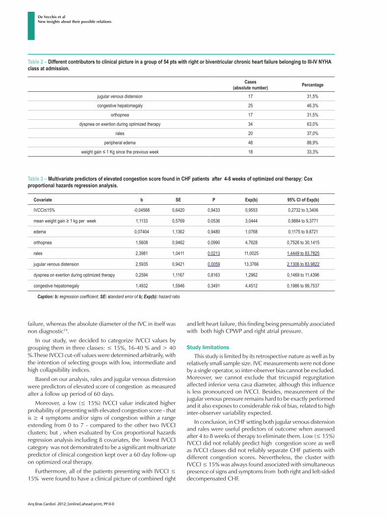
Figure 1 shows the distribution (%) of the cases (17 on the whole) of jugular venous distension (jvd) across the 3 IVCCI groups, as found at the second visit. It documents that in the IVCCI ≤15% group, occurrence of jugular venous distension was higher than in IVCCI=16-40% and IVCCI>40% groups: p (Kruskal Wallis ) <0.05 for both comparisons.
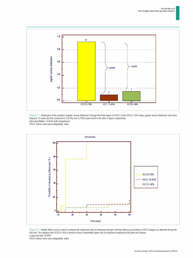
Furthermore, by considering the relation between every category of IVCCI and the probability of developing orthopnea (figure 2), a remarkable difference emerges: actually, the patients with IVCCI ≤ 15% were burdened with a much higher risk of being involved by orthopnea during the subsequent follow up. In addition, low venous collapsibility was found to entail high rate of early referral to heart failure
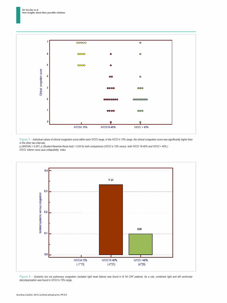
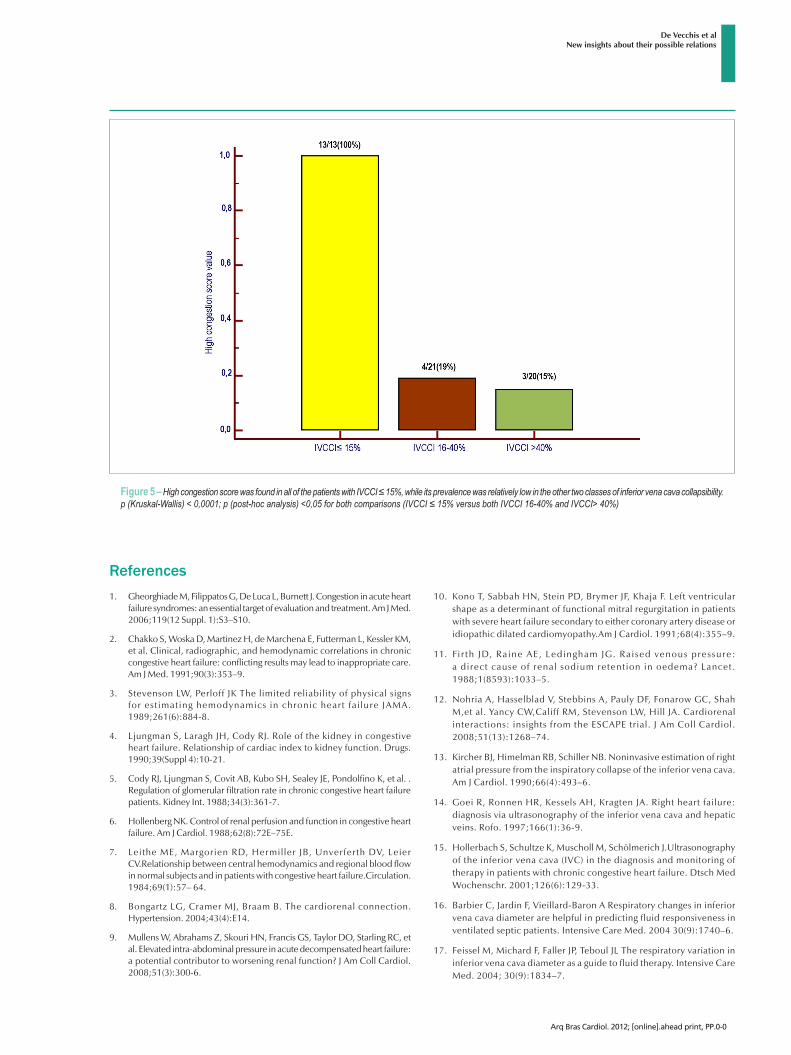
unit, due to unscheduled re-admission for worsening symptoms and signs of systemic and pulmonary congestion. By comparing the three classes of caval collapsibility, the most severe impairment of the clinical picture was exhibited by patients belonging to the lowest class of IVCCI values (figures 3-5) . Nevertheless, Cox multivariate proportional hazard model failed to identify IVCCI ≤ 15% as a significant multivariate predictor of worsening clinical congestion (Tab 3). In addition, it would be noted that this pattern was accompanied in any case by simultaneous involvement of signs and symptoms from both left-sided and right-sided cardiac failure; on the contrary, the patients belonging to the intermediate range of caval collapsibility(IVCCI 16-40%) exhibited the highest rate of isolated systemic - but not pulmonary - congestion at the end of the prescribed follow up (30-60 days after the first visit)(figure 5).
DiscussionBased on the pathophysiological concepts represented
above, prognostic importance of clinical congestion in HF patients is expected. However, clinical congestion may be the ‘tip of the iceberg’ of the hemodynamic derangements that precede symptoms21-22. For instance, in chronic biventricular HF, even severe hemodynamic congestion rarely causes rales and/or radiographic pulmonary edema. This may be related to several adaptive pathophysiological changes such as increases in alveolar capillary membrane thickness, increased lymphatic drainage, and/or pulmonary hypertension3,21 (3,21). Thus, in the opinion of several Authors13,22, it would be very interesting and useful to explore not only clinical but also hemodynamic congestion in order to achieve a really careful patient assessment. In truth, this would be opportune considering that patients with even only hemodynamic congestion were shown to have poor outcomes23-25.
Ultrasound assessment of IVC respiratory fluctuations has been proposed from a long while as a possible diagnostic tool for obtaining noninvasive reliable estimation of volume status and/or right atrial pressure in CHF patients. Briefly, in spontaneously breathing subjects, intrathoracic pressure decreases during inspiration, thereby increasing venous return and inducing collapse of the IVC; inversely, during expiration, venous return decreases, so causing an increase in the diameter of the IVC26. High right atrial pressures dilate the IVC and worsen this normal IVC collapsibility. According to these observations, congestion would be indicated by relatively small IVCCI values, while intravascular depletion would be revealed by wide fluctuations of IVC diameter, generating relatively high values of IVCCI. Therefore, small, collapsible IVCs as visualized by echocardiography represent low right atrial pressures, whereas large, non-collapsible IVCs reflect high right atrial pressures13. In the presence of marked volume overload, the respiratory cycle leads to minimal change in diameter of IVC, regardless of its absolute diameter27. This depend on the peculiar non-linear pressure-diameter relationship of the IVC so that, above a threshold pressure (i.e., CVP >20 mmHg), no further increase in IVC diameter can be observed28.This has been confirmed by a recent study in which a IVCCI ≤ 15% was highly sensitive and specific for the diagnosis of acute decompensated heart
Arq Bras Cardiol. 2012; [online].ahead print, PP.0-0
De Vecchis et al New insights about their possible relations
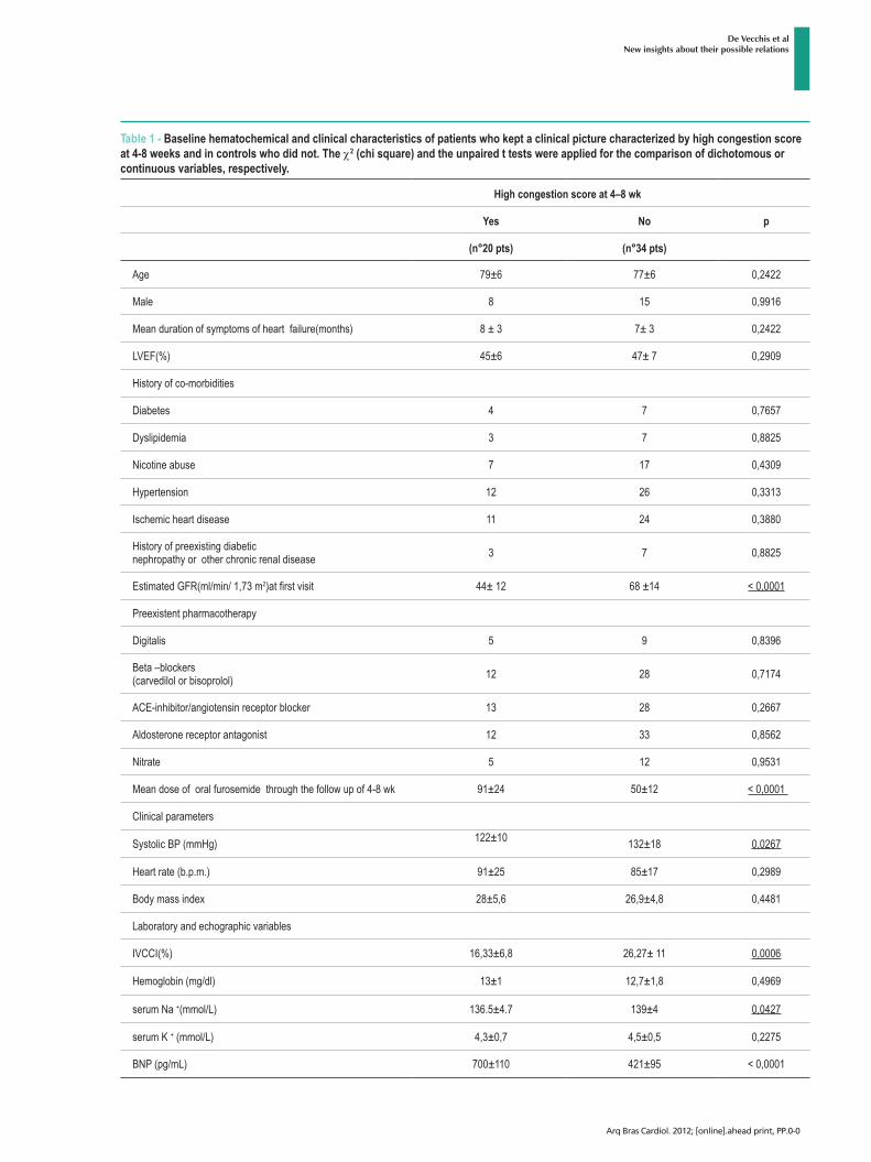
Table 1 - Baseline hematochemical and clinical characteristics of patients who kept a clinical picture characterized by high congestion score at 4-8 weeks and in controls who did not. The χ2 (chi square) and the unpaired t tests were applied for the comparison of dichotomous or continuous variables, respectively.
High congestion score at 4–8 wk
Yes No p
(n°20 pts) (n°34 pts)
Age 79±6 77±6 0,2422
Male 8 15 0,9916
Mean duration of symptoms of heart failure(months) 8 ± 3 7± 3 0,2422
LVEF(%) 45±6 47± 7 0,2909
History of co-morbidities
Diabetes 4 7 0,7657
Dyslipidemia 3 7 0,8825
Nicotine abuse 7 17 0,4309
Hypertension 12 26 0,3313
Ischemic heart disease 11 24 0,3880
History of preexisting diabetic nephropathy or other chronic renal disease 3 7 0,8825
Estimated GFR(ml/min/ 1,73 m2)at first visit 44± 12 68 ±14 < 0,0001
Preexistent pharmacotherapy
Digitalis 5 9 0,8396
Beta –blockers(carvedilol or bisoprolol) 12 28 0,7174
ACE-inhibitor/angiotensin receptor blocker 13 28 0,2667
Aldosterone receptor antagonist 12 33 0,8562
Nitrate 5 12 0,9531
Mean dose of oral furosemide through the follow up of 4-8 wk 91±24 50±12 < 0,0001
Clinical parameters
Systolic BP (mmHg) 122±10 132±18 0,0267
Heart rate (b.p.m.) 91±25 85±17 0,2989
Body mass index 28±5,6 26,9±4,8 0,4481
Laboratory and echographic variables
IVCCI(%) 16,33±6,8 26,27± 11 0,0006
Hemoglobin (mg/dl) 13±1 12,7±1,8 0,4969
serum Na +(mmol/L) 136.5±4.7 139±4 0,0427
serum K + (mmol/L) 4,3±0,7 4,5±0,5 0,2275
BNP (pg/mL) 700±110 421±95 < 0,0001
Arq Bras Cardiol. 2012; [online].ahead print, PP.0-0
De Vecchis et al New insights about their possible relations
Table 2 – Different contributors to clinical picture in a group of 54 pts with right or biventricular chronic heart failure belonging to III-IV NYHA class at admission.
Cases(absolute number) Percentage
jugular venous distension 17 31,5%
congestive hepatomegaly 25 46,3%
orthopnea 17 31,5%
dyspnea on exertion during optimized therapy 34 63,0%
rales 20 37,0%
peripheral edema 48 88,9%
weight gain ≤ 1 Kg since the previous week 18 33,3%
failure, whereas the absolute diameter of the IVC in itself was non diagnostic19.
In our study, we decided to categorize IVCCI values by grouping them in three classes: ≤ 15%, 16-40 % and > 40 %.These IVCCI cut-off values were determined arbitrarily, with the intention of selecting groups with low, intermediate and high collapsibility indices.
Based on our analysis, rales and jugular venous distension were predictors of elevated score of congestion as measured after a follow up period of 60 days.
Moreover, a low (≤ 15%) IVCCI value indicated higher probability of presenting with elevated congestion score - that is ≥ 4 symptoms and/or signs of congestion within a range extending from 0 to 7 - compared to the other two IVCCI clusters; but , when evaluated by Cox proportional hazards regression analysis including 8 covariates, the lowest IVCCI category was not demonstrated to be a significant multivariate predictor of clinical congestion kept over a 60 day follow-up on optimized oral therapy.
Furthermore, all of the patients presenting with IVCCI ≤ 15% were found to have a clinical picture of combined right
Table 3 – Multivariate predictors of elevated congestion score found in CHF patients after 4-8 weeks of optimized oral therapy: Cox proportional hazards regression analysis.
Covariate b SE P Exp(b) 95% CI of Exp(b)
IVCCI≤15% -0,04568 0,6420 0,9433 0,9553 0,2732 to 3,3406
mean weight gain ≥ 1 kg per week 1,1133 0,5769 0,0536 3,0444 0,9884 to 9,3771
edema 0,07404 1,1362 0,9480 1,0768 0,1175 to 9,8721
orthopnea 1,5608 0,9462 0,0990 4,7628 0,7526 to 30,1415
rales 2,3981 1,0411 0,0213 11,0025 1,4449 to 83,7825
jugular venous distension 2,5935 0,9421 0,0059 13,3766 2,1306 to 83,9822
dyspnea on exertion during optimized therapy 0,2594 1,1167 0,8163 1,2962 0,1469 to 11,4396
congestive hepatomegaly 1,4932 1,5946 0,3491 4,4512 0,1986 to 99,7537
Caption: b: regression coefficient; SE: standard error of b; Exp(b): hazard ratio
and left heart failure, this finding being presumably associated with both high CPWP and right atrial pressure.
Study limitationsThis study is limited by its retrospective nature as well as by
relatively small sample size. IVC measurements were not done by a single operator, so inter-observer bias cannot be excluded. Moreover, we cannot exclude that tricuspid regurgitation affected inferior vena cava diameter, although this influence is less pronounced on IVCCI. Besides, measurement of the jugular venous pressure remains hard to be exactly performed and it also exposes to considerable risk of bias, related to high inter-observer variability expected.
In conclusion, in CHF setting both jugular venous distension and rales were useful predictors of outcome when assessed after 4 to 8 weeks of therapy to eliminate them. Low (≤ 15%) IVCCI did not reliably predict high congestion score as well as IVCCI classes did not reliably separate CHF patients with different congestion scores. Nevertheless, the cluster with IVCCI ≤ 15% was always found associated with simultaneous presence of signs and symptoms from both right and left-sided decompensated CHF.
Arq Bras Cardiol. 2012; [online].ahead print, PP.0-0
De Vecchis et al New insights about their possible relations
Figure 1 – Distribution of the symptom “jugular venous distension” through the three layers of IVCCI. In the IVCCI ≤ 15% range, jugular venous distension was more frequent: 12 cases (92,3%) compared to 2 (9,5%) and 3 (15%) cases found in the other 2 layers, respectively.p(Kruskal-Wallis) < 0,05 for both comparisons IVCCI: inferior vena cava collapsibility index
Figure 2 – Kaplan Meier curve is used to compare the respective risks of orthopnea through a 60-day follow-up according to IVCCI category as detected during the first visit. The category with IVCCI ≤ 15% is shown to have a remarkably higher risk of orthopnea compared to the other two classes. p (log rank test) <0,0001IVCCI: inferior vena cava collapsibility index
Arq Bras Cardiol. 2012; [online].ahead print, PP.0-0
De Vecchis et al New insights about their possible relations
Figure 3 – Individual values of clinical congestion score within each IVCCI range. In the IVCCI ≤ 15% range, the clinical congestion score was significantly higher than in the other two intervals.p (ANOVA) < 0,001; p (Student-Newman-Keuls test) < 0,05 for both comparisons (IVCCI ≤ 15% versus both IVCCI 16-40% and IVCCI > 40% )IVCCI: inferior vena cava collapsibility index
Figure 4 – Systemic but not pulmonary congestion (isolated right heart failure) was found in 9/ 54 CHF patients. As a rule, combined right and left ventricular decompensation was found in IVCCI ≤ 15% range.
Arq Bras Cardiol. 2012; [online].ahead print, PP.0-0
De Vecchis et al New insights about their possible relations
Figure 5 – High congestion score was found in all of the patients with IVCCI ≤ 15%, while its prevalence was relatively low in the other two classes of inferior vena cava collapsibility.p (Kruskal-Wallis) < 0,0001; p (post-hoc analysis) <0,05 for both comparisons (IVCCI ≤ 15% versus both IVCCI 16-40% and IVCCI> 40%)
References1. Gheorghiade M, Filippatos G, De Luca L, Burnett J. Congestion in acute heart
failure syndromes: an essential target of evaluation and treatment. Am J Med. 2006;119(12 Suppl. 1):S3–S10.
2. Chakko S, Woska D, Martinez H, de Marchena E, Futterman L, Kessler KM, et al. Clinical, radiographic, and hemodynamic correlations in chronic congestive heart failure: conflicting results may lead to inappropriate care. Am J Med. 1991;90(3):353–9.
3. Stevenson LW, Perloff JK The limited reliability of physical signs for est imat ing hemodynamics in chronic heart fa i lure JAMA. 1989;261(6):884-8.
4. Ljungman S, Laragh JH, Cody RJ. Role of the kidney in congestive heart failure. Relationship of cardiac index to kidney function. Drugs. 1990;39(Suppl 4):10-21.
5. Cody RJ, Ljungman S, Covit AB, Kubo SH, Sealey JE, Pondolfino K, et al. . Regulation of glomerular filtration rate in chronic congestive heart failure patients. Kidney Int. 1988;34(3):361-7.
6. Hollenberg NK. Control of renal perfusion and function in congestive heart failure. Am J Cardiol. 1988;62(8):72E–75E.
7. Leithe ME, Margorien RD, Hermil ler JB, Unverferth DV, Leier CV.Relationship between central hemodynamics and regional blood flow in normal subjects and in patients with congestive heart failure.Circulation. 1984;69(1):57– 64.
8. Bongartz LG, Cramer MJ, Braam B. The cardiorenal connection. Hypertension. 2004;43(4):E14.
9. Mullens W, Abrahams Z, Skouri HN, Francis GS, Taylor DO, Starling RC, et al. Elevated intra-abdominal pressure in acute decompensated heart failure: a potential contributor to worsening renal function? J Am Coll Cardiol. 2008;51(3):300-6.
10. Kono T, Sabbah HN, Stein PD, Brymer JF, Khaja F. Left ventricular shape as a determinant of functional mitral regurgitation in patients with severe heart failure secondary to either coronary artery disease or idiopathic dilated cardiomyopathy.Am J Cardiol. 1991;68(4):355–9.
11. Fi r th JD, Ra ine AE, Ledingham JG. Ra i sed venous pressure: a direct cause of renal sodium retent ion in oedema? Lancet. 1988;1(8593):1033–5.
12. Nohria A, Hasselblad V, Stebbins A, Pauly DF, Fonarow GC, Shah M,et al. Yancy CW,Califf RM, Stevenson LW, Hill JA. Cardiorenal interactions: insights from the ESCAPE trial. J Am Coll Cardiol. 2008;51(13):1268–74.
13. Kircher BJ, Himelman RB, Schiller NB. Noninvasive estimation of right atrial pressure from the inspiratory collapse of the inferior vena cava. Am J Cardiol. 1990;66(4):493–6.
14. Goei R, Ronnen HR, Kessels AH, Kragten JA. Right heart failure: diagnosis via ultrasonography of the inferior vena cava and hepatic veins. Rofo. 1997;166(1):36-9.
15. Hollerbach S, Schultze K, Muscholl M, Schölmerich J.Ultrasonography of the inferior vena cava (IVC) in the diagnosis and monitoring of therapy in patients with chronic congestive heart failure. Dtsch Med Wochenschr. 2001;126(6):129-33.
16. Barbier C, Jardin F, Vieillard-Baron A Respiratory changes in inferior vena cava diameter are helpful in predicting fluid responsiveness in ventilated septic patients. Intensive Care Med. 2004 30(9):1740–6.
17. Feissel M, Michard F, Faller JP, Teboul JL The respiratory variation in inferior vena cava diameter as a guide to fluid therapy. Intensive Care Med. 2004; 30(9):1834–7.
Arq Bras Cardiol. 2012; [online].ahead print, PP.0-0
De Vecchis et al New insights about their possible relations
18. Stawicki SP, Braslow BM, Panebianco NL, Kirkpatrick JN, Gracias VH, Hayden GE,et al. Intensivist use of hand-carried ultrasonography to measure IVC collapsibility in estimating intravascular volume status: correlations with CVP. J Am Coll Surg. 2009;209(1):55-61.
19. Blehar DJ, Dickman E, Gaspari R Identification of congestive heart failure via respiratory variation of inferior vena cava diameter. Am J Emerg Med. 2009;27(1):71-5.
20. Guiotto G, Masarone M, Paladino F, Ruggiero E, Scott S, Verde S,et al. Inferior vena cava collapsibility to guide fluid removal in slow continuous ultrafiltration: a pilot study. Intensive Care Med. 2010;36(4):692-6.
21. Mahdyoon H, Klein R, Eyler W, Lakier JB, Chakko SC, Gheorghiade M. Radiographic pulmonary congestion in end-stage congestive heart failure. Am J Cardiol.1989;63(9):625-7.
22. Binanay C, Califf RM, Hasselblad V, O’Connor CM, Shah MR, Sopko G,et al. Evaluation study of congestive heart failure and pulmonary artery catheterization effectiveness: the ESCAPE trial. JAMA. 2005;294(13):1625–33.
23. Unverferth DV, Magorien RD, Moeschberger ML, Baker PB, Fetters JK, Leier CV. Factors influencing the one-year mortality of dilated cardiomyopathy. Am J Cardiol. 1984; 54(1):147–52.
24. Stevenson LW, Tillisch JH, Hamilton M, Luu M, Chelimsky-Fallick C, Moriguchi J,et al. Importance of hemodynamic response to therapy in predicting survival with ejection fraction less than or equal to 20% secondary to ischemic or nonischemic dilated cardiomyopathy. Am J Cardiol. 1990;66(19):1348–54.
25. Zile MR, Bennett TD, St John Sutton M, Cho YK, Adamson PB, Aaron MF,et al. Transition from chronic compensated to acute decompensated heart failure: pathophysiological insights obtained from continuous monitoring of intracardiac pressures. Circulation. 2008;118(14):1433–41.
26. Mintz G, Kotler M, Parry W, Iskandrian A, Kane S. Real-time inferior vena caval ultrasonography: normal and abnormal findings and its use in assessing right heart function. Circulation. 64(5):1018–25.
27. Moreno F, Hagan A, Holmen J, Pryor A, Strickland R, Castle H. Evaluation of size and dynamics of the inferior vena cava as an index of right-sided cardiac function. Am J Cardiol.1984; 53(4):579–85.
28. Jardin F, Vieillard-Baron A. Ultrasonographic examination of the venae cavae. Intensive Care Med. 2006;32(2):203-6.