inferior vena cava enteric fistula due to unresected ... · inferior vena cava enteric fistula due...
TRANSCRIPT
WORLD JOURNAL OF EMERGENCY SURGERY
Gilshtein et al. World Journal of Emergency Surgery (2015) 10:29 DOI 10.1186/s13017-015-0024-7
REVIEW Open Access
Inferior vena cava enteric fistula due tounresected colorectal metastasis
Hayim Gilshtein*, Offir Ben-Ishay, Karina Nascovica and Yoram KlugerAbstract
A 57 year old male presented to our department with recurrent attacks of sepsis and upper gastrointestinalbleeding due to colorectal cancer metastasis that resulted in a fistula involving the inferior vena cava and the thirdpart of the duodenum.Four and a half years ago he underwent laparoscopic right hemicolectomy due to colonic adenocarcinoma.A year prior to his recent hospitalization he underwent cytoreductive surgery followed by HIPEC due to peritonealmetastases in another hospital. During the operation a metastasis adherent to the inferior vena cava and the III partof the duodenum was revealed. The surgeon decided to mark the area with hemo- clips and after the patientrecovered from surgery he was sent for radiotherapy aimed at controlling the left over metastases.In his current hospitalization he underwent an en bloc resection of the III part of the duodenum, the adherent venacava and the right kidney.Gross pathology revealed a fistula between the vena cava and the duodenum with bile stained clot within theresected part of the vena cava.The patient recovered well with resolutions of his presenting symptoms.
IntroductionColon cancer metastases are not rare. Liver, lung andperitoneal metastases are the most common sites [1].Local recurrence, primarily in the site of a previousanastomosis is also well described [2]. The disease re-lapse pattern is related to both the cancer staging uponpresentation and biologic characteristics of the tumorand host, only partially elucidated thus far.The treatment of choice for resectable colon cancer is
surgery. Adjuvant chemotherapy is tailored in line withthe pathologic staging revealed after surgery. For localrecurrent colon cancer, complete surgical excision, whenapplicable, remains the only option for cure. Surgery isalso well proven for colorectal liver metastasis [3] withensuing significant impact on disease free and 5-yearsurvival. Moreover, in the last years surgery is offered inselected cases of colorectal peritoneal carcinomatosis inthe form of cytoreductive surgery with heated chemo-therapy (HIPEC) [4, 5].
* Correspondence: [email protected] of General Surgery, Rambam Health Care Campus, 8 Ha’AliyahSt, Haifa 35254, Israel
© 2015 Gilshtein et al. This is an Open Access(http://creativecommons.org/licenses/by/4.0),provided the original work is properly creditedcreativecommons.org/publicdomain/zero/1.0/
Palliative resection of colorectal metastasis is usuallylimited to lesions causing significant bleeding, obstruc-tion or perforation.As it was described in the past [6] cytoreductive sur-
gery with HIPEC might cause serious postsurgical com-plications such as anastomotic leaks, gastrointestinalbleeding and sepsis. Several other life threatening andrare complications were also reported. One of these is acase of colobronchial fistula [7].Herein we describe a rare case of gastrointestinal
bleeding and sepsis caused by a colon cancer metastasisinvolving the inferior vena cava, after cytoreductive sur-gery and HIPEC that was treated in our department.
Case reportA 57-year old male presented to the emergency roomwith high grade fever and chills.The patient was known to suffer from metastatic colon
cancer. Following positive occult blood in his stool hewas diagnosed in 2010 with colonic adenocarcinoma, in-volving the cecum, without any evidence of distant me-tastasis. A laparoscopic right hemicolectomy wasperformed. His final pathology revealed a T3N1 tumor.He received a FOLFOX adjuvant systemic chemotherapy.
article distributed under the terms of the Creative Commons Attribution Licensewhich permits unrestricted use, distribution, and reproduction in any medium,. The Creative Commons Public Domain Dedication waiver (http://) applies to the data made available in this article, unless otherwise stated.
Gilshtein et al. World Journal of Emergency Surgery (2015) 10:29 Page 2 of 3
The patient recovered well and proceeded with stand-ard regular oncologic follow up including interval ab-dominal CT and PET scans as required.About 3.5 years after the index operation several new
lesions, suspicious of secondary spread were revealed. Alesion in the upper lobe of his right lung was resectedthoracoscopically. Another lesion caused a significantobstruction of the right kidney that resulted in nephrost-omy tube insertion.The patient was offered a cytoreductive surgery with
heated chemotherapy (HIPEC) with curative intent. Atsurgery HIPEC was performed as planned after resectionand excision of all the abdominal load of metastases ex-cept for a solid lesion involving the IVC and the 3rd partof the duodenum, deemed unresectable. Due to its pre-sumed irresectability the site was marked with metallicclips for later irradiation.After surgery the patient underwent a targeted irradi-
ation to the marked site and received additional courseof systemic chemotherapy. He underwent further followup with PET scans, revealing three main lesions with ahigh uptake on PET, at the previous anastomosis of thetransverse colon with small bowel, in the omentum nextto the anastomtic site and the metastsasis revealed atprevious surgery involving the inferior vena cava, nearthe entrance of the right renal vein and the third part ofthe duodenum. There was no evidence of neither livernor other distant metastasis.The patient remained in a good general condition and
performance status up to his presentation with fever andfatigue on 6 month after the second operation. His bloodtests revealed anemia, high white blood count and CRP.Blood cultures were taken and the patient was admit-
ted for observation and empiric antibiotic treatment.
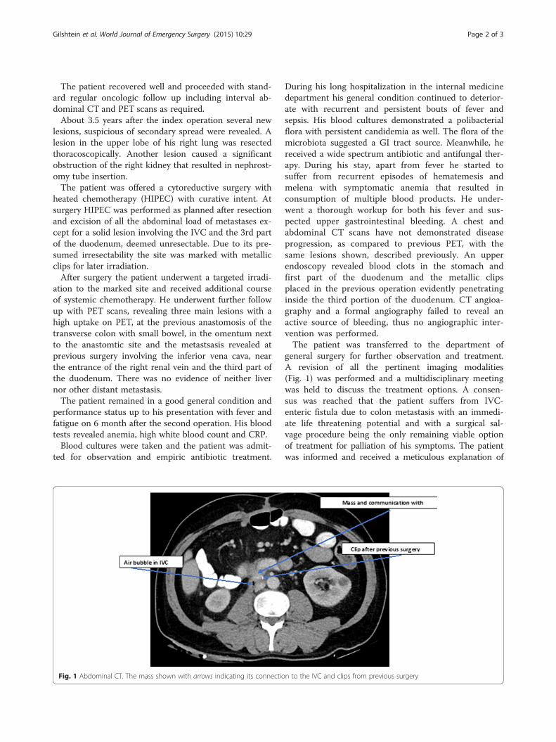
Fig. 1 Abdominal CT. The mass shown with arrows indicating its connectio
During his long hospitalization in the internal medicinedepartment his general condition continued to deterior-ate with recurrent and persistent bouts of fever andsepsis. His blood cultures demonstrated a polibacterialflora with persistent candidemia as well. The flora of themicrobiota suggested a GI tract source. Meanwhile, hereceived a wide spectrum antibiotic and antifungal ther-apy. During his stay, apart from fever he started tosuffer from recurrent episodes of hematemesis andmelena with symptomatic anemia that resulted inconsumption of multiple blood products. He under-went a thorough workup for both his fever and sus-pected upper gastrointestinal bleeding. A chest andabdominal CT scans have not demonstrated diseaseprogression, as compared to previous PET, with thesame lesions shown, described previously. An upperendoscopy revealed blood clots in the stomach andfirst part of the duodenum and the metallic clipsplaced in the previous operation evidently penetratinginside the third portion of the duodenum. CT angioa-graphy and a formal angiography failed to reveal anactive source of bleeding, thus no angiographic inter-vention was performed.The patient was transferred to the department of
general surgery for further observation and treatment.A revision of all the pertinent imaging modalities(Fig. 1) was performed and a multidisciplinary meetingwas held to discuss the treatment options. A consen-sus was reached that the patient suffers from IVC-enteric fistula due to colon metastasis with an immedi-ate life threatening potential and with a surgical sal-vage procedure being the only remaining viable optionof treatment for palliation of his symptoms. The patientwas informed and received a meticulous explanation of
n to the IVC and clips from previous surgery
Gilshtein et al. World Journal of Emergency Surgery (2015) 10:29 Page 3 of 3
the planned procedure with its pure palliative intent. Aninformed consent was obtained.At laparotomy a solid tumor lesion was found at the
third and fourth parts of the duodenum. The inferiorvena cava, at the level of the right renal vein appearedfriable and almost necrotic, with a large opening in itsanterior wall, covered by only a clot and fibrin, fistulaz-ing to the third portion of the duodenum. En bloc resec-tion of the tumors with the involved IVC, 3rd and 4thparts of the duodenum and right kidney (known as non-functional by DMSA and with previous nephrostomy)was completed.The second part of the duodenum was anasto-
mosed to the proximal jejunum. The tranverse colonanastomosis to the small bowel appeared intact. An-other lesion in the omentum, measuring about 2 cm,at the proximity of the previous anastomosis, asshown on PET, was also excised. No reconstructionof the vena cava was performed, in considerationwith avaibale collaterals. The surgery was completedwith no evidence of gross disease apparent in theperitoneal cavity.The final pathologic report revealed desmoplastic reac-
tion and fibrosis, free of tumor in the main specimenand the omentum excised separately. Well differentiatedcolon adenocarcinoma with mucus production wasfound only in the margins of resected duodenum.The patient underwent an uneventful recovery with
return to good functional status, and resolution of bothhis fever and GI bleeding. Of notice, only minor peroph-eral edema developed after IVC resection.
DiscussionColorectal cancer metastasis remains a challenge. Whilesurgery is well established for the treatment of liver me-tastasis, lung metastasis and also HIPEC gained accept-ability in high volume centers for groups of patientswith peritoneal carcinomatosis, there is still an ongoingdebate for other types of metastasis. We presented a rarecase of a symptomatic IVC-duodenal fistula treated suc-cessfully with surgical excision. It remains unclear whatwas the role of HIPEC and the later external beam ir-radiation on the development of this serious complica-tion. The intent of the procedure was palliative and itsinfluence on the patients’ overall survival is still ques-tionable. However, we believe that for some, selectedyoung patients a radical procedure might be undertakenprudently for either curative or palliative intent in casesof uncommon presentation of colorectal metastasis.Dealing with complications of colorectal metastasis
can be demanding with early involvement of experiencedsurgical team, in need of some specialized surgical pro-cedures, as described herein.
Consent statementWritten informed consent was obtained from the patientfor publication of this Case report and any accompany-ing images. A copy of the written consent is available forreview by the Editor-in-Chief of this journal.
Competing interestsThe authors declare that they have no competing interests.
Authors’ contributionsHG participated in conceiving and drafting the manuscript. OY and KNparticipated in the design. YK participated in drafting the manuscript.All authors read and approved the final manuscript.
AcknowledgementsThere is no one to acknowledge except for the co-authors listed.
Received: 9 May 2015 Accepted: 23 June 2015
References1. Thomassen I, van Gestel YR, Lemmens VE, de Hingh IH. Incidence,
prognosis, and treatment options for patients with synchronous peritonealcarcinomatosis and liver metastases from colorectal origin. Dis ColonRectum. 2013;56(12):1373–80. Review.
2. Tsikitis VL, Malireddy K, Green EA, Christensen B, Whelan R, Hyder J, et al.Postoperative surveillance recommendations for early stage colon cancerbased on results from the clinical outcomes of surgical therapy trial. J ClinOncol. 2009;27(22):3671–6. Epub 2009 Jun 29.
3. Akgül O, Cetinkaya E, Ersöz S, Tez M. Role of surgery in colorectal cancerliver metastasis. World J Gastroenterol. 2014;20(20):6113–22. Review.
4. Vallicelli C, Cavaliere D, Catena F, Coccolini F, Ansaloni L, Poiasina E, et al.Management of peritoneal carcinomatosis from colorectal cancer: review ofthe literature. Int J Colorectal Dis. 2014;29(8):895–8.
5. de Cuba EM, Kwakman R, Knol DL, Bonjer HJ, Meijer GA, Te Velde EA.Cytoreductive surgery and HIPEC for peritoneal metastases combined withcurative treatment of colorectal liver metastases: systematic review of allliterature and meta-analysis of observational studies. Cancer Treat Rev.2013;39(4):321–7. Epub 2012 Dec 12.
6. Kusamura S, Younan R, Baratti D, Costanzo P, Favaro M, Gavazzi C, et al.Cytoreductive surgery followed by intraperitoneal hyperthermic perfusion inthe treatment of peritoneal surface malignancies: analysis of morbidity andmortality in 209 cases treated with closed-abdomen technique. Cancer.2006;106:1144–53.
7. Laterza B, Baratti D, Cozzi G, Kusamura S, Oliva GD, Gavazzi C, et al.Colobronchial fistula: an unusual complication after peritonectomy andHyperthermic Intraperitoneal Chemotherapy (HIPEC). In Vivo. 2009;23:151–3.
Submit your next manuscript to BioMed Centraland take full advantage of:
• Convenient online submission
• Thorough peer review
• No space constraints or color figure charges
• Immediate publication on acceptance
• Inclusion in PubMed, CAS, Scopus and Google Scholar
• Research which is freely available for redistribution
Submit your manuscript at www.biomedcentral.com/submit