inflammation and n-formyl peptide receptors mediate the ... · the angiogenic activity of human...
TRANSCRIPT
ARTICLE
Inflammation and N-formyl peptide receptors mediatethe angiogenic activity of human vitreous humour in proliferativediabetic retinopathy
Sara Rezzola1 & Michela Corsini1 & Paola Chiodelli1 & Anna Cancarini2 &
Imtiaz M. Nawaz1 & Daniela Coltrini1 & Stefania Mitola1 & Roberto Ronca1 &
Mirella Belleri1 & Liliana Lista3 &Dario Rusciano4 &Mario DeRosa5 &Vincenzo Pavone3 &
Francesco Semeraro2 & Marco Presta1
Received: 3 September 2016 /Accepted: 13 December 2016 /Published online: 13 January 2017#
AbstractAims/hypothesis Angiogenesis and inflammation characteriseproliferative diabetic retinopathy (PDR), a major complicationof diabetes mellitus. However, the impact of inflammation onthe pathogenesis of PDR neovascularisation has not been elu-cidated. Here, we assessed the capacity of PDR vitreous fluidto induce pro-angiogenic/proinflammatory responses in endo-thelium and the contribution of the inflammation-related pat-tern recognition N-formyl peptide receptors (FPRs) in medi-ating these responses.Methods Pooled and individual pars plana vitrectomy-derivedPDR vitreous fluid (‘PDR vitreous’) samples were assessed inendothelial cell proliferation, motility, sprouting and
morphogenesis assays, and for the capacity to induce proin-flammatory transcription factor activation, reactive oxygenspecies production, intercellular junction disruption andleucocyte-adhesion molecule upregulation in these cells.In vivo, the pro-angiogenic/proinflammatory activity of PDRvitreous was tested in murine Matrigel plug and chick embryochorioallantoic membrane (CAM) assays. Finally, the FPRinhibitors Boc-Phe-Leu-Phe-Leu-Phe (Boc-FLFLF) andAc-L-Arg-Aib-L-Arg-L-Cα(Me)Phe-NH2 tetrapeptide(UPARANT) were evaluated for their capacity to affect thebiological responses elicited by PDR vitreous.Results PDR vitreous activates a pro-angiogenic/proinflamma-tory phenotype in endothelial cells. Accordingly, PDR vitreoustriggers a potent angiogenic/inflammatory response in vivo.Notably, the different capacity of individual PDR vitreous sam-ples to induce neovessel formation in the CAM correlates withtheir ability to recruit infiltrating CD45+ cells. Finally, the FPRinhibitor Boc-FLFLF and the novel FPR antagonistUPARANT inhibit neovessel formation and inflammatoryresponses triggered by PDR vitreous in the CAM assay.Conclusions/interpretation This study provides evidence thatinflammation mediates the angiogenic activity of PDR vitreousand paves the way for the development of FPR-targeting anti-inflammatory/anti-angiogenic approaches for PDR therapy.
Keywords Angiogenesis . Diabetic retinopathy . Endothelialcells . Inflammation .N-formyl peptide receptor . Vitreous
AbbreviationsBoc-FLFLF Boc-Phe-Leu-Phe-Leu-PheCAM Chorioallantoic membranefMLF Formyl-methionyl-leucyl phenylalanine
Electronic supplementary material The online version of this article(doi:10.1007/s00125-016-4204-0) contains peer-reviewed but uneditedsupplementary material, which is available to authorised users.
* Sara [email protected]
* Francesco [email protected]
* Marco [email protected]
1 Department of Molecular and Translational Medicine, University ofBrescia, Via Branze 39, Brescia 25123, Italy
2 Department of Ophthalmology, University of Brescia, PiazzaleSpedali Civili 1, Brescia 25123, Italy
3 Department of Chemical Sciences, ‘Federico II’ University ofNaples, Naples, Italy
4 Sooft Italia Spa, Montegiorgio, Italy5 Department of ExperimentalMedicine, Second University of Naples,
Naples, Italy
Diabetologia (2017) 60:719–728DOI 10.1007/s00125-016-4204-0
Springer-Verlag Berlin Heidelberg 2017
FPR N-formyl peptide receptorICAM-1 Intercellular adhesion molecule 1pCREB Phosphorylated cAMP-response element-
binding proteinPDR Proliferative diabetic retinopathyqPCR Quantitative PCRROS Reactive oxygen speciesuPA Urokinase-type plasminogen activatoruPAR Urokinase-type plasminogen activator
receptorUPARANT Ac-L-Arg-Aib-L-Arg-L-Cα(Me)Phe-NH2
tetrapeptideVCAM-1 Vascular cell adhesion molecule-1VE-CAD Vascular endothelial cadherinVEGF Vascular endothelial growth factorZO-1 Tight junction protein 1
Introduction
Diabetic retinopathy, the leading cause of visual impairment inthe working-age population [1], begins as non-proliferativeretinal abnormalities and progresses to proliferative diabeticretinopathy (PDR) characterised by neovascularisation and apersistent grade of inflammation [2–5]. Even though anti-angiogenic vascular endothelial growth factor (VEGF) inhib-itors are widely used in PDR therapy [2], several limitations toanti-VEGF interventions exist [6]. Furthermore, production ofother angiogenic factors and proinflammatory mediators maynullify and/or cause resistance to anti-VEGF therapies [6, 7].
Angiogenesis and inflammation are closely related pro-cesses that play a pivotal role in ocular diseases associatedwith retinal neovascularisation [5, 8, 9]. Thus, a tight crosstalk may exist between angiogenesis and inflammation inPDR. Accordingly, the therapeutic potential of intravitreal ad-ministration of anti-inflammatory corticosteroids (e.g. triam-cinolone acetonide) has been investigated in patients with di-abetic retinopathy. However, beneficial effects can be tran-sient and associated with steroid-related adverse events [10,11]. This calls for a better understanding of the cross talkbetween angiogenesis and inflammation in PDR to identifynovel anti-inflammatory approaches able to suppress retinalneovascularisation.
N-formyl peptide receptors (FPRs) are involved in therecruitment and activation of immune cells in response topathogen-associated molecular patterns. In addition, experi-mental evidence implicates FPRs in angiogenic responseslinked to inflammation [12], and FPR interaction with theurokinase-type plasminogen activator (uPA)/uPA receptor(uPAR) system may stimulate angiogenesis in a protease-independent manner [13–15]. Accordingly, the uPAR-derived tetrapeptide Ac-L-Arg-Aib-L-Arg-L-Cα(Me)Phe-NH2 (UPARANT) competes with formyl-methionyl-leucyl
phenylalanine (fMLF) peptide for binding to FPRs and isendowed with a significant anti-angiogenic activity in vitroand in vivo [16, 17]. In addition, UPARANT prevents ocularangiogenesis and reduces the levels of inflammatory media-tors in murine models of oxygen-induced retinopathy andlaser-induced choroidal neovascularisation [17, 18].
The study of the biological effects exerted by PDR vitreousfluid (‘PDR vitreous’) on endothelial cells may represent auseful tool to investigate the relationship between neovascularand inflammatory responses in preclinical experimentalmodels. PDR vitreous contains high levels of pro-angiogenicand proinflammatory mediators [19, 20] and stimulates angio-genesis in vitro and in vivo, whereas no angiogenic responseis elicited by vitreous fluid from patients with a macular hole[7, 17, 21–23]. However, no data are available about thecapacity of PDR vitreous to exert proinflammatory responsesin endothelium, their relationship with its ability to stimulateneovessel formation or the role of FPRs in this process. Thisstudy was designed to assess the pro-angiogenic and proin-flammatory potential of vitreous fluid obtained from PDRpatients after pars plana vitrectomy and to evaluate the impactof FPR inhibitors on their biological activity.
Methods
Reagents A list of reagents is included in the electronic sup-plementary material (ESM).
Human vitreous fluid samples Patients with PDR (Table 1)and age-matched patients with macular hole underwent parsplana vitrectomy at the Clinics of Ophthalmology (Universityof Brescia) during the period January 2014–June 2016.Collection and analysis of human samples were approved bythe internal review board of the Spedali Civili of Brescia andfollowed the principles of the Declaration of Helsinki.Informed consent was obtained from all participants.Samples were stored at −80°C. Their content of pro-angiogen-ic/proinflammatory mediators was evaluated using a semi-quantitative antibody-based RayBio Human AngiogenesisArray C1000 (Raybiotech, Norcross, GA, USA). See ESMmethods.
All assays were performed on vitreous samples pooledfrom 4-5 patients unless specified otherwise. Data are repre-sentative of at least three independent pools of vitreous fluidthat provided similar results.
In vitro and ex vivo endothelial cell studies PDR vitreouswas tested on HUVECs in different angiogenesis assays.Briefly, in the proliferation assay, HUVECs (17,000 cells/cm2) were treated with increasing amounts of vitreous fluiddiluted in culture medium plus 2.5% FCS and counted after24 h. In the migration assay, HUVECs were seeded at
720 Diabetologia (2017) 60:719–728
1.0×10⁶ cells/ml in the upper compartment of a Boyden cham-ber, and increasing amounts of vitreous fluid were placed inthe lower compartment. After 3 h at 37°C, cells that hadmigrated to the lower side of the filter were counted. In themorphogenesis assay, HUVECs were seeded on Matrigel at2.0×105 cells/ml in the absence or presence of vitreous fluidand the number of meshes was counted after 8 h. In the endo-thelial cell sprouting assay, fibrin gel-embedded HUVECspheroids were stimulated with vitreous fluid and endothelialcell sprouts were counted 24 h thereafter. In addition, murineretina tissue fragments were embedded in fibrin gel and stim-ulated with vitreous fluid. Retinal endothelial cell sproutingwas evaluated after 7 days. See ESM methods for details.
The same vitreous samples were assessed in HUVECs fortheir proinflammatory potential. Briefly, HUVECs (1.0×10⁶)suspended in serum-free medium in the presence of 1:4 vitre-ous fluid were incubated with 5 μmol/l dichloro-dihydro-fluorescein diacetate (DCFH-DA) for 5–30 min in the darkat 37°C and reactive oxygen species (ROS) production wasquantified by FACS analysis. Serum-starved confluentHUVECs were treated with 1:4 vitreous fluid for 0–30 min,fixed in 4% paraformaldehyde and immunostained with anti-bodies against anti-tight junction protein 1 (ZO-1), anti-vascu la r endothe l ia l cadher in (VE-CAD), an t i -phosphorylated-cAMP-response element-binding protein(pCREB) or anti-NF-κB p65 subunit. Endothelial barrierfunction was studied on confluent HUVEC monolayers by atrans-endothelial electrical resistance assay in which imped-ance was monitored every min for 3 h following PDR vitreous
treatment. Also, fluorescently labelled THP-1 cells(7.0×10⁵ cells/well) were added on the top of HUVECmono-layers that had been treated with 1:4 vitreous fluid for 18 h.After 30 min at 37°C, adherent THP-1 cells were quantified.See ESM methods for details.
Finally, HUVECs were evaluated for FPR1-3 expressionby RT-PCR, FACS and western blot analysis according tostandard procedures. See ESM methods.
Real-time quantitative PCR Real-time quantitative PCR(qPCR) was used to determine the relative expression levelsof mRNAs. See ESM methods.
Animal experiments The pro-angiogenic/proinflammatoryactivity of vitreous fluid was evaluated in vivo in the murineMatrigel plug [24] and chick embryo chorioallantoic mem-brane (CAM) [7] assays.
Matrigel was mixed at 4°C with 1:4 vitreous fluid andinjected subcutaneously (0.4 ml/mouse) into the flank of6–8 week old C57BL6 female mice (Charles River, Calco,Italy). One week after injection, the proinflammatory/pro-angiogenic response was quantified in harvested plugs byqPCR. See ESM methods.
Alginate beads (3 μl) containing 2 μl of vitreous fluidwere placed on top of the CAM of fertilised chicken eggs atday 11 of development. Microvessels converging towardsthe implant were counted under a stereomicroscope and theCD45+ infiltrate was assessed by immunofluorescenceanalysis. See ESM methods.
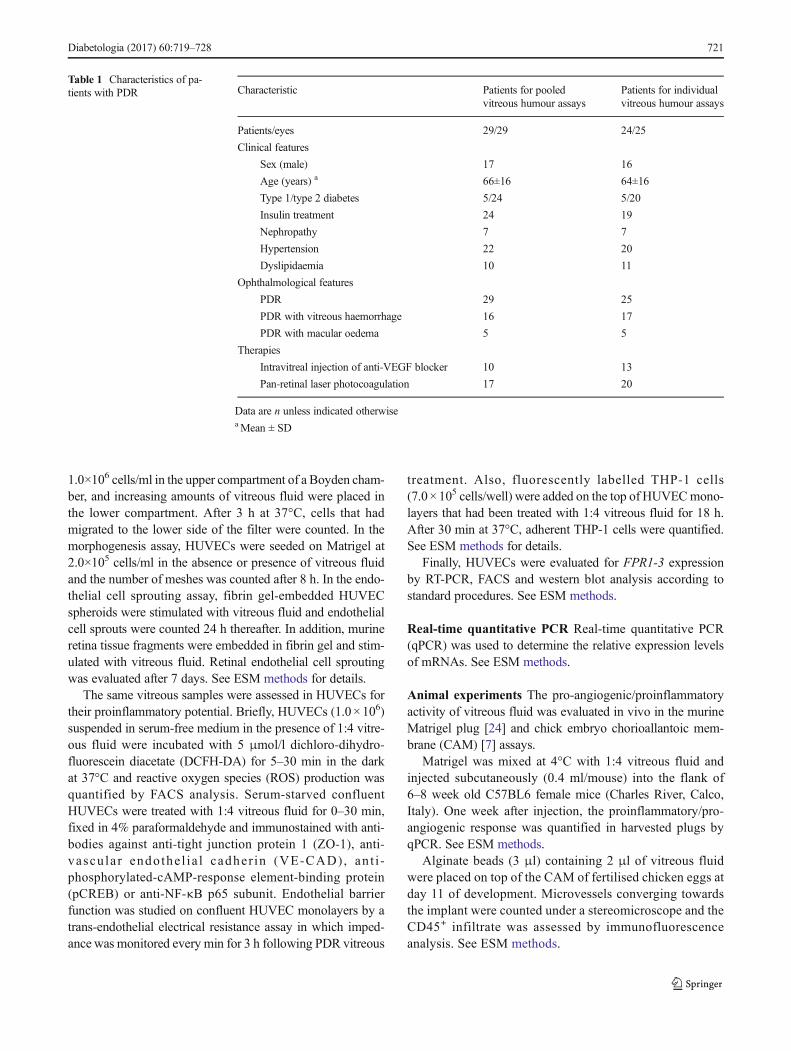
Table 1 Characteristics of pa-tients with PDR Characteristic Patients for pooled
vitreous humour assaysPatients for individualvitreous humour assays
Patients/eyes 29/29 24/25
Clinical features
Sex (male) 17 16
Age (years) a 66±16 64±16
Type 1/type 2 diabetes 5/24 5/20
Insulin treatment 24 19
Nephropathy 7 7
Hypertension 22 20
Dyslipidaemia 10 11
Ophthalmological features
PDR 29 25
PDR with vitreous haemorrhage 16 17
PDR with macular oedema 5 5
Therapies
Intravitreal injection of anti-VEGF blocker 10 13
Pan-retinal laser photocoagulation 17 20
Data are n unless indicated otherwiseaMean ± SD
Diabetologia (2017) 60:719–728 721
Procedures were carried out according to the Guide for thecare and use of laboratory animals, the animal care Italian guide-lines (DL 116/92) and the European Communities CouncilDirective (86/609/EEC) and were approved by the EthicalCommittee in Animal Experiments of the University of Brescia.
Statistics Data are mean ± SEM. Statistical significance wasevaluated with commercial software (GraphPad Prism 6; SanDiego, CA, USA) using Student’s t test or one-way ANOVAfollowed by Bonferroni multiple comparison post test.Differences were considered significant when p<0.05.
Results
Human PDR vitreous induces a pro-angiogenic/proin-flammatory phenotype in endothelial cells Three pools ofhuman PDR vitreous were evaluated for their content of pro-angiogenic/proinflammatory mediators using a semi-quantitative antibody-based array. As shown in ESMTable 2, a variety of cytokines, chemokines and angiogenicgrowth factors are detectable in PDR vitreous, supporting thehypothesis that this fluid may exert both angiogenic andinflammatory responses in endothelial cells.
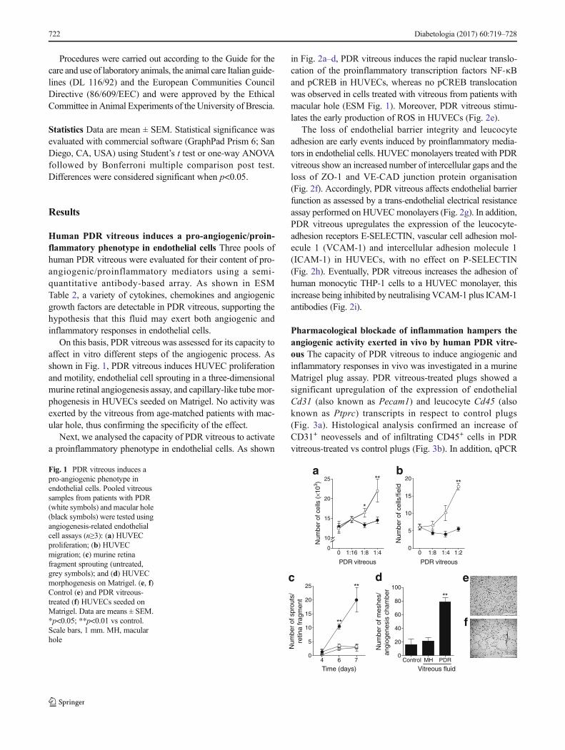
On this basis, PDR vitreous was assessed for its capacity toaffect in vitro different steps of the angiogenic process. Asshown in Fig. 1, PDR vitreous induces HUVEC proliferationand motility, endothelial cell sprouting in a three-dimensionalmurine retinal angiogenesis assay, and capillary-like tubemor-phogenesis in HUVECs seeded on Matrigel. No activity wasexerted by the vitreous from age-matched patients with mac-ular hole, thus confirming the specificity of the effect.
Next, we analysed the capacity of PDR vitreous to activatea proinflammatory phenotype in endothelial cells. As shown
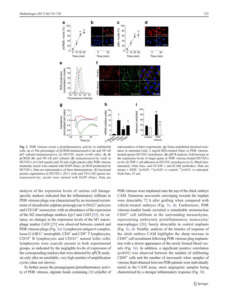
in Fig. 2a–d, PDR vitreous induces the rapid nuclear translo-cation of the proinflammatory transcription factors NF-κBand pCREB in HUVECs, whereas no pCREB translocationwas observed in cells treated with vitreous from patients withmacular hole (ESM Fig. 1). Moreover, PDR vitreous stimu-lates the early production of ROS in HUVECs (Fig. 2e).
The loss of endothelial barrier integrity and leucocyteadhesion are early events induced by proinflammatory media-tors in endothelial cells. HUVECmonolayers treated with PDRvitreous show an increased number of intercellular gaps and theloss of ZO-1 and VE-CAD junction protein organisation(Fig. 2f). Accordingly, PDR vitreous affects endothelial barrierfunction as assessed by a trans-endothelial electrical resistanceassay performed on HUVECmonolayers (Fig. 2g). In addition,PDR vitreous upregulates the expression of the leucocyte-adhesion receptors E-SELECTIN, vascular cell adhesion mol-ecule 1 (VCAM-1) and intercellular adhesion molecule 1(ICAM-1) in HUVECs, with no effect on P-SELECTIN(Fig. 2h). Eventually, PDR vitreous increases the adhesion ofhuman monocytic THP-1 cells to a HUVEC monolayer, thisincrease being inhibited by neutralising VCAM-1 plus ICAM-1antibodies (Fig. 2i).
Pharmacological blockade of inflammation hampers theangiogenic activity exerted in vivo by human PDR vitre-ous The capacity of PDR vitreous to induce angiogenic andinflammatory responses in vivo was investigated in a murineMatrigel plug assay. PDR vitreous-treated plugs showed asignificant upregulation of the expression of endothelialCd31 (also known as Pecam1) and leucocyte Cd45 (alsoknown as Ptprc) transcripts in respect to control plugs(Fig. 3a). Histological analysis confirmed an increase ofCD31+ neovessels and of infiltrating CD45+ cells in PDRvitreous-treated vs control plugs (Fig. 3b). In addition, qPCR
4 6 70
5
10
15
20
25
Time (days)
Num
ber
of s
prou
ts/
retin
a fr
agm
ent
**
**
0 1:16 1:8 1:40
10
15
20
25
PDR vitreous
Num
ber
of c
ells
(×1
03 )
*
**
c
0 1:8 1:4 1:20
5
10
15
20
PDR vitreous
Num
ber
of c
ells
/fiel
d **a b
Control MH PDR0
20
40
60
80
100
Num
ber
of m
eshe
s/an
giog
enes
is c
ham
ber
**
Vitreous fluid
d e
f
Fig. 1 PDR vitreous induces apro-angiogenic phenotype inendothelial cells. Pooled vitreoussamples from patients with PDR(white symbols) and macular hole(black symbols) were tested usingangiogenesis-related endothelialcell assays (n≥3): (a) HUVECproliferation; (b) HUVECmigration; (c) murine retinafragment sprouting (untreated,grey symbols); and (d) HUVECmorphogenesis on Matrigel. (e, f)Control (e) and PDR vitreous-treated (f) HUVECs seeded onMatrigel. Data are means ± SEM.*p<0.05; **p<0.01 vs control.Scale bars, 1 mm. MH, macularhole
722 Diabetologia (2017) 60:719–728
analysis of the expression levels of various cell lineage-specific markers indicated that the inflammatory infiltrate inPDR vitreous plugs was characterised by an increased recruit-ment of chondroitin sulphate proteoglycan 4 (NG2)+ pericytesand CD11B+ monocytes, with an abundance of the expressionof the M2 macrophage markers Egr2 and Cd83 [25]. At var-iance, no changes in the expression levels of the M1 macro-phage marker Cd38 [25] was observed between control andPDR vitreous plugs (Fig. 3a). Lymphocyte antigen 6 complex,locus G (GR1)+ neutrophils, CD4+ and CD8+ T lymphocytes,CD19+ B lymphocytes and CD161+ natural killer cells/lymphocytes were scarcely present in both experimentalgroups, as indicated by the negligible levels of expression ofthe corresponding markers that were detected by qPCR analy-sis only after an unreliable, very high number of amplificationcycles (data not shown).
To further assess the proangiogenic/proinflammatory activi-ty of PDR vitreous, alginate beads containing 2.0 μl/pellet of
PDR vitreous were implanted onto the top of the chick embryoCAM. Numerous neovessels converging towards the implantwere detectable 72 h after grafting when compared withvehicle-treated embryos (Fig. 3c, d). Furthermore, PDRvitreous-loaded beads recruited a remarkable mononuclearCD45+ cell infiltrate in the surrounding mesenchyme,representing embryonic proinflammatory monocytes/macrophages [26], barely detectable in control implants(Fig. 3c, d). Notably, analysis of the kinetics of response ofthe chick embryo CAM highlights the sharp increase inCD45+ cell recruitment following PDR vitreous plug implanta-tion with a slower appearance of the newly formed blood ves-sels (Fig. 3e). In addition, a significant positive correlation(p=0.01) was observed between the number of infiltratingCD45+ cells and the number of neovessels when samples ofvitreous fluid obtained from ten PDR patients were individuallytested in the CAM assay, more angiogenic samples beingcharacterised by a stronger inflammatory response (Fig. 3f).
24 25 260.7
0.8
0.9
1.0
1.1
1.2
Time (h)
Impe
denc
e(n
orm
alis
ed c
ell i
ndex
)
10-1 100 101 102 1030
20
40
60
80
100
Fluorescence intensity
RO
S p
rodu
ctio
n(n
orm
alis
ed to
mod
e)
0 10 20 300
20
40
60
80
100
Time (min)
p65+
nuc
lei (
%)
****
0 10 20 300
20
40
60
80
100
Time (min)
pCR
EB
+ nu
clei
(%
)
** **
*
e
Con
trol
PD
R v
itreo
usZO-1 VE-CAD Merge
f
ig h
a cb d
0 30
pCREB
DAPI
Time (min)
0 30
NF-κB p65
DAPI
Time (min)
P-Selectin
E-SelectinICA
M1
VCAM
10
5
10
15
20
25
mR
NA
(re
lativ
e un
its)
*
*
*
Control PDRvitreous
0
20
40
60
80
100
Adh
eren
t cel
ls/fi
eld
††
**
Fig. 2 PDR vitreous exerts a proinflammatory activity in endothelialcells. (a, c) The percentages of pCREB-immunoreactive (a) and NF-κBp65 subunit-immunoreactive (c) HUVEC nuclei (n=60 cells). (b, d)pCREB (b) and NF-κB p65 subunit (d) immunoreactivity (red) inHUVECs at 0 (left panels) and 30 min (right panels) after PDR vitreoustreatment; nuclei were stained with DAPI (blue). (e) ROS production byHUVECs. Data are representative of three determinations. (f) Junctionalprotein organisation in HUVECs. ZO-1 (red) and VE-CAD (green) im-munoreactivity; nuclei were stained with DAPI (blue). Data are
representative of three experiments. (g) Trans-endothelial electrical resis-tance in untreated (red), 2 mg/ml BSA-treated (blue) or PDR vitreous-treated (green) HUVEC monolayers. (h) qPCR analysis. Fold increase inthe expression levels of target genes in PDR vitreous-treated HUVECs(n=4). (i) THP-1 cell adhesion on HUVECmonolayers (n=3). Black bars,untreated; white bars, anti-VCAM + anti-ICAM antibodies. Data aremeans ± SEM. *p<0.05; **p<0.01 vs control; ††p<0.01 vs untreated.Scale bars, 25 μm
Diabetologia (2017) 60:719–728 723
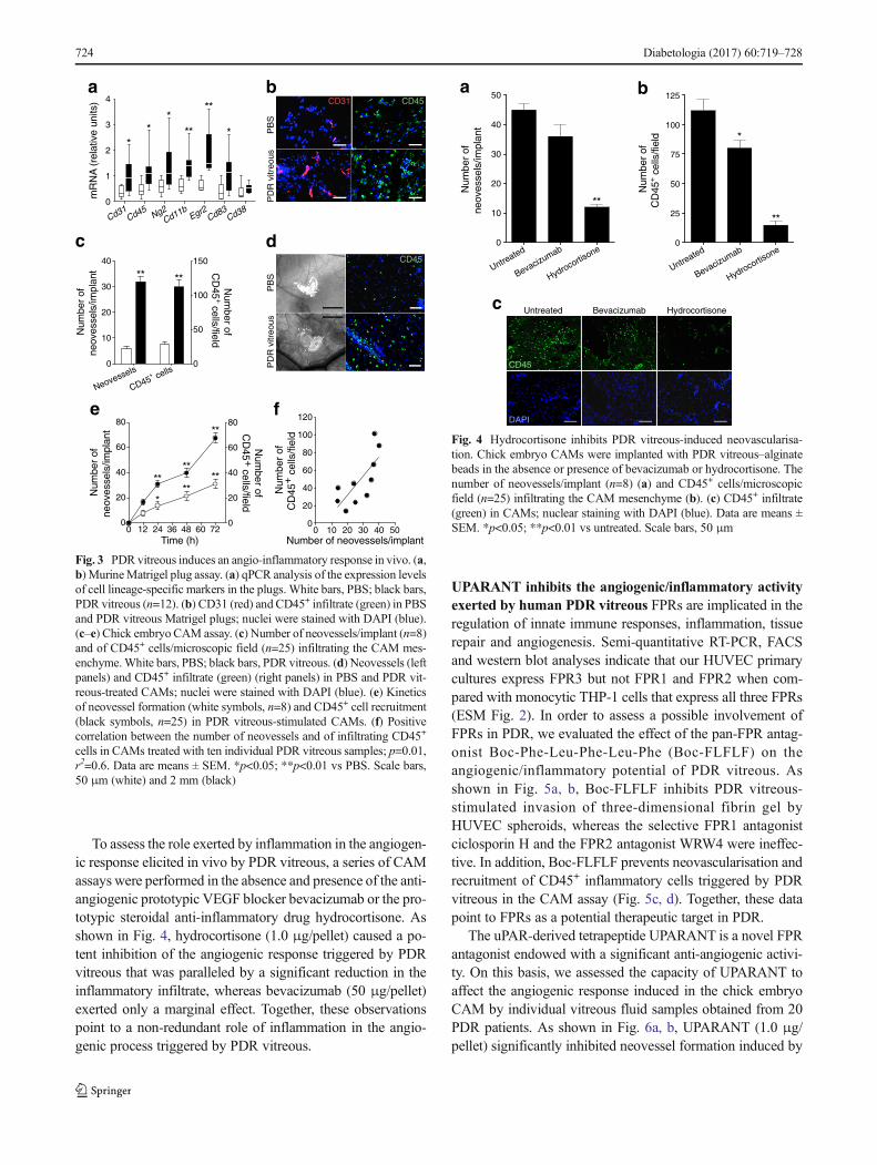
To assess the role exerted by inflammation in the angiogen-ic response elicited in vivo by PDR vitreous, a series of CAMassays were performed in the absence and presence of the anti-angiogenic prototypic VEGF blocker bevacizumab or the pro-totypic steroidal anti-inflammatory drug hydrocortisone. Asshown in Fig. 4, hydrocortisone (1.0 μg/pellet) caused a po-tent inhibition of the angiogenic response triggered by PDRvitreous that was paralleled by a significant reduction in theinflammatory infiltrate, whereas bevacizumab (50 μg/pellet)exerted only a marginal effect. Together, these observationspoint to a non-redundant role of inflammation in the angio-genic process triggered by PDR vitreous.
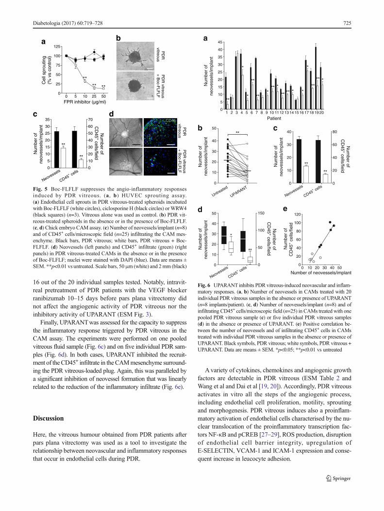
UPARANT inhibits the angiogenic/inflammatory activityexerted by human PDR vitreous FPRs are implicated in theregulation of innate immune responses, inflammation, tissuerepair and angiogenesis. Semi-quantitative RT-PCR, FACSand western blot analyses indicate that our HUVEC primarycultures express FPR3 but not FPR1 and FPR2 when com-pared with monocytic THP-1 cells that express all three FPRs(ESM Fig. 2). In order to assess a possible involvement ofFPRs in PDR, we evaluated the effect of the pan-FPR antag-onist Boc-Phe-Leu-Phe-Leu-Phe (Boc-FLFLF) on theangiogenic/inflammatory potential of PDR vitreous. Asshown in Fig. 5a, b, Boc-FLFLF inhibits PDR vitreous-stimulated invasion of three-dimensional fibrin gel byHUVEC spheroids, whereas the selective FPR1 antagonistciclosporin H and the FPR2 antagonist WRW4 were ineffec-tive. In addition, Boc-FLFLF prevents neovascularisation andrecruitment of CD45+ inflammatory cells triggered by PDRvitreous in the CAM assay (Fig. 5c, d). Together, these datapoint to FPRs as a potential therapeutic target in PDR.
The uPAR-derived tetrapeptide UPARANT is a novel FPRantagonist endowed with a significant anti-angiogenic activi-ty. On this basis, we assessed the capacity of UPARANT toaffect the angiogenic response induced in the chick embryoCAM by individual vitreous fluid samples obtained from 20PDR patients. As shown in Fig. 6a, b, UPARANT (1.0 μg/pellet) significantly inhibited neovessel formation induced by
Cd31
Cd45 Ng
2Cd
11b Egr2Cd
83Cd
380
1
2
3
4m
RN
A (
rela
tive
units
)
**
****
**
ba
PB
SP
DR
vitr
eous
CD45CD31
c dP
BS
PD
R v
itreo
usCD45
Neovessels
CD45+ cells
0
10
20
30
40
0
50
100
150
Num
ber
ofne
oves
sels
/impl
ant
Num
ber ofC
D45
+ cells/field
** **
0 12 24 36 48 60 720
20
40
60
80
0
20
40
60
80
Time (h)
Num
ber
ofne
oves
sels
/impl
ant
Num
ber ofC
D45 +
cells/field
**
****
****
*
e
0 10 20 30 40 500
20
40
60
80
100
120
Number of neovessels/implant
Num
ber
ofC
D45
+ c
ells
/fiel
d
f
Fig. 3 PDR vitreous induces an angio-inflammatory response in vivo. (a,b) MurineMatrigel plug assay. (a) qPCR analysis of the expression levelsof cell lineage-specific markers in the plugs. White bars, PBS; black bars,PDR vitreous (n=12). (b) CD31 (red) and CD45+ infiltrate (green) in PBSand PDR vitreous Matrigel plugs; nuclei were stained with DAPI (blue).(c–e) Chick embryo CAM assay. (c) Number of neovessels/implant (n=8)and of CD45+ cells/microscopic field (n=25) infiltrating the CAM mes-enchyme.White bars, PBS; black bars, PDR vitreous. (d) Neovessels (leftpanels) and CD45+ infiltrate (green) (right panels) in PBS and PDR vit-reous-treated CAMs; nuclei were stained with DAPI (blue). (e) Kineticsof neovessel formation (white symbols, n=8) and CD45+ cell recruitment(black symbols, n=25) in PDR vitreous-stimulated CAMs. (f) Positivecorrelation between the number of neovessels and of infiltrating CD45+
cells in CAMs treated with ten individual PDR vitreous samples; p=0.01,r2=0.6. Data are means ± SEM. *p<0.05; **p<0.01 vs PBS. Scale bars,50 μm (white) and 2 mm (black)
Untreated Bevacizumab Hydrocortisone
CD45
DAPI
Untreated
Bevacizumab
Hydrocortisone
0
10
20
30
40
50
Num
ber
ofne
oves
sels
/impl
ant
**
Untreated
Bevacizumab
Hydrocortisone
0
25
50
75
100
125
Num
ber
ofC
D45
+ ce
lls/fi
eld
**
*
a b
c
Fig. 4 Hydrocortisone inhibits PDR vitreous-induced neovascularisa-tion. Chick embryo CAMs were implanted with PDR vitreous–alginatebeads in the absence or presence of bevacizumab or hydrocortisone. Thenumber of neovessels/implant (n=8) (a) and CD45+ cells/microscopicfield (n=25) infiltrating the CAM mesenchyme (b). (c) CD45+ infiltrate(green) in CAMs; nuclear staining with DAPI (blue). Data are means ±SEM. *p<0.05; **p<0.01 vs untreated. Scale bars, 50 μm
724 Diabetologia (2017) 60:719–728
16 out of the 20 individual samples tested. Notably, intravit-real pretreatment of PDR patients with the VEGF blockerranibizumab 10–15 days before pars plana vitrectomy didnot affect the angiogenic activity of PDR vitreous nor theinhibitory activity of UPARANT (ESM Fig. 3).
Finally, UPARANTwas assessed for the capacity to suppressthe inflammatory response triggered by PDR vitreous in theCAM assay. The experiments were performed on one pooledvitreous fluid sample (Fig. 6c) and on five individual PDR sam-ples (Fig. 6d). In both cases, UPARANT inhibited the recruit-ment of the CD45+ infiltrate in the CAMmesenchyme surround-ing the PDR vitreous-loaded plug. Again, this was paralleled bya significant inhibition of neovessel formation that was linearlyrelated to the reduction of the inflammatory infiltrate (Fig. 6e).
Discussion
Here, the vitreous humour obtained from PDR patients afterpars plana vitrectomy was used as a tool to investigate therelationship between neovascular and inflammatory responsesthat occur in endothelial cells during PDR.
Avariety of cytokines, chemokines and angiogenic growthfactors are detectable in PDR vitreous (ESM Table 2 andWang et al and Dai et al [19, 20]). Accordingly, PDR vitreousactivates in vitro all the steps of the angiogenic process,including endothelial cell proliferation, motility, sproutingand morphogenesis. PDR vitreous induces also a proinflam-matory activation of endothelial cells characterised by the nu-clear translocation of the proinflammatory transcription fac-tors NF-κB and pCREB [27–29], ROS production, disruptionof endothelial cell barrier integrity, upregulation ofE-SELECTIN, VCAM-1 and ICAM-1 expression and conse-quent increase in leucocyte adhesion.
1 2 3 4 5 6 7 8 9 10 1112 13 14 15 16 17 18 19 200
5
10
15
20
25
30
35
40
45
Patient
Num
ber
ofne
oves
sels
/impl
ant
**
*
**
******
***
** *
*****
*
*
a
Untreated
UPARANT0
10
20
30
40
50
Num
ber
ofne
oves
sels
/impl
ant
**b
Neovessels
CD45+ cells
0
10
20
30
40
0
20
40
60
80
Num
ber
ofne
oves
sels
/impl
ant
Num
ber ofC
D45
+ cells/field
**
**
c
Neovessels
CD45+ cells
0
10
20
30
40
50
0
50
100
150
Num
ber
ofne
oves
sels
/impl
ant
Num
ber ofC
D45
+ cells/field
**
*
d
0 10 20 30 40 500
20
40
60
80
100
120
Number of neovessels/implant
Num
ber
ofC
D45
+ cel
ls/fi
eld
e
Fig. 6 UPARANT inhibits PDR vitreous-induced neovascular and inflam-matory responses. (a, b) Number of neovessels in CAMs treated with 20individual PDR vitreous samples in the absence or presence of UPARANT(n=8 implants/patient). (c, d) Number of neovessels/implant (n=8) and ofinfiltrating CD45+ cells/microscopic field (n=25) in CAMs treated with onepooled PDR vitreous sample (c) or five individual PDR vitreous samples(d) in the absence or presence of UPARANT. (e) Positive correlation be-tween the number of neovessels and of infiltrating CD45+ cells in CAMstreated with individual PDR vitreous samples in the absence or presence ofUPARANT. Black symbols, PDR vitreous; white symbols, PDR vitreous +UPARANT. Data are means ± SEM. *p<0.05; **p<0.01 vs untreated
a b
dCD45
PD
Rvitreous
PD
R vitreous
+ B
oc-FLF
LF
PD
Rvitreous
PD
R vitreous
+ B
oc-FLF
LF 0 5 10 25 50
0
25
50
75
100
125
FPR inhibitor (µg/ml)
Cel
l spr
outin
g(%
vs
cont
rol)
**** **
Neovessels
CD45+ cells
0
5
10
15
20
25
30
35
0
10
20
30
40
50
60
70
Num
ber
ofne
oves
sels
/impl
ant
**
**
Num
ber ofC
D45
+ cells/field
c
Fig. 5 Boc-FLFLF suppresses the angio-inflammatory responsesinduced by PDR vitreous. (a , b) HUVEC sprouting assay.(a) Endothelial cell sprouts in PDR vitreous-treated spheroids incubatedwith Boc-FLFLF (white circles), ciclosporine H (black circles) orWRW4(black squares) (n=3). Vitreous alone was used as control. (b) PDR vit-reous-treated spheroids in the absence or in the presence of Boc-FLFLF.(c, d) Chick embryo CAM assay. (c) Number of neovessels/implant (n=8)and of CD45+ cells/microscopic field (n=25) infiltrating the CAM mes-enchyme. Black bars, PDR vitreous; white bars, PDR vitreous + Boc-FLFLF. (d) Neovessels (left panels) and CD45+ infiltrate (green) (rightpanels) in PDR vitreous-treated CAMs in the absence or in the presenceof Boc-FLFLF; nuclei were stained with DAPI (blue). Data are means ±SEM. **p<0.01 vs untreated. Scale bars, 50μm (white) and 2mm (black)
Diabetologia (2017) 60:719–728 725
In keeping with its proinflammatory/pro-angiogenic poten-tial, PDR vitreous triggers a potent neovascular response andthe recruitment of a CD45+/CD11+ infiltrate in the in vivomurine Matrigel plug assay characterised by a predominantpro-angiogenic M2 polarisation. Accordingly, a rapid CD45+
leucocyte recruitment occurs when PDR vitreous is tested forits angiogenic capacity in the chick embryo CAM assay.Notably, a significant positive correlation was observed be-tween the number of neovessels and of infiltrating CD45+
cells when PDR vitreous samples from different patients wereindividually tested in the CAM assay. Together, these dataindicate that a strict correlation may exist between the pro-angiogenic and proinflammatory activity of PDR vitreous,the inflammatory environment playing a non-redundant rolein neovessel formation. The capacity of the anti-inflammatorydrug hydrocortisone to inhibit both inflammatory and angio-genic responses in PDR vitreous-treated CAMs supports thishypothesis. This is in keeping with previous observations in-dicating that inflammation plays a pivotal role in the angio-genesis process driven by various angiogenic growth factors,including VEGF [30], fibroblast growth factor 2 (FGF2) [31],placenta growth factor (PlGF) [32] and IL-1β [33].
At variance with hydrocortisone, the VEGF blockerbevacizumab was poorly effective in inhibiting the activityof PDR vitreous. This accords with previous observationsshowing that the K5-NOS(H) polysaccharide, a pan-inhibitor for heparin-binding proinflammatory/angiogenicfactors, was more effective than bevacizumab in inhibitingthe angiogenic activity of PDR vitreous [7]. Thus, the bestperformance of hydrocortisone vs bevacizumab suggests thatthe angio-inflammatory responses elicited by PDR vitreousmay represent the result of the synergistic action of variousmodulators, besides VEGF.
Notably, no difference in pro-angiogenic/proinflammatoryactivity was observed between haemorrhagic and non-haemorrhagic PDR vitreous samples when tested in theCAM assay (ESM Fig. 4), indicating that local production,rather than systemic inflow, is the relevant source of cytokineswithin the ocular PDR microenvironment. This is in keepingwith the high vitreous/plasma ratio measured for cytokinelevels in PDR patients [34–36]. Together, these data point tolocal eye inflammation as a driving force that sustains angio-genesis in PDR.
Intravitreal administration of corticosteroids is associatedwith possible adverse events [10, 11], leading to the require-ment for novel anti-inflammatory approaches in PDR therapyable to suppress retinal neovascularisation. FPRs are Gprotein-coupled receptors implicated in the regulation ofinnate immune responses, inflammation, tissue repair andangiogenesis [12]. FPRs exert a productive interaction withthe uPAR88–92 receptor region that modulates the biologicalresponse of leucocytes and endothelial cells to inflammatorymediators and angiogenic factors [37–39]. On this basis,
allosteric inhibitors related to the uPAR88–92 sequence andable to block the cross talk involving uPAR, FPRs andintegrins were developed [16, 40, 41]. Among them,UPARANT competes with fMLF peptide for the binding toFPRs and is endowed with a significant anti-angiogenic activ-ity in vitro and in vivo [16–18]. In addition, UPARANT sup-presses the angiogenic activity of pooled PDR vitreous sam-ples, pointing to this compound as a promising therapeutic forthe treatment of inflammatory diseases associated with ocularangiogenesis, including PDR [17, 18].
Here, we demonstrate that UPARANT inhibits theneovascular response elicited in the CAM assay by 16 out ofthe 20 individual vitreous samples obtained from PDRpatients. Inhibition of neovessel formation by UPARANTwent along with a significant reduction in the inflammatoryinfiltrate. Notably, intravitreal pretreatment of PDR patientswith the VEGF blocker ranibizumab before vitrectomy didnot affect the inhibitory effect exerted by UPARANT.Together, these data strongly support the hypothesis that theinflammatory response elicited by the PDR vitreous via FPRactivation plays a non-redundant role in neovessel formationand raise the question about the endothelial FPR subtype(s)and vitreous fluid mediator(s) responsible for such activation.
Our results demonstrate that HUVECs express FPR3, butnot FPR1 or FPR2. Accordingly, the pan-FPR antagonist Boc-FLFLF hampers the angio-inflammatory responses elicited byPDR vitreous in endothelial cells, whereas the FPR1 antago-nist ciclosporin H and the FPR2 antagonist WRW4 [12] wereineffective. However, VEGF-inducible expression of FPR2has been reported in endothelial cells [42], indicating thatdifferences in cell isolation and/or cell culture conditions, aswell as changes of the in vivo microenvironment, may affectthe pattern of FPR expression in endothelial cells. This callsfor further experiments aimed to assess the expression ofFPRs in retinal vessels of PDR patients.
Various danger-associated molecular pattern host-derivedpeptides can activate FPRs [12]. Notably, the FPR ligandsserum amyloid A, LL-37 and Hp(2-20) have been involvedin the regulation of neovascularisation under inflammatoryconditions [12]. High levels of serum amyloid A are detect-able in the vitreous and plasma of PDR patients [43] and ineyes with macular oedema [44, 45] whereas, to the best of ourknowledge, no data are available about the levels of other FPRligands in PDR vitreous.
Even though further studies will be required to identifyunambiguously the FPR subtypes and their natural ligandsacting as mediators of the angiogenic/inflammatory activityof PDR vitreous, our data indicate that anti-angiogenic strat-egies targeting FPR activation may be exploited in persistentocular inflammatory conditions, including PDR. In thisframe, UPARANT may represent the basis for the develop-ment of novel anti-inflammatory/anti-angiogenic approachesfor PDR therapy.
726 Diabetologia (2017) 60:719–728
Acknowledgements We thank S. Calza (Department of Molecular andTranslational Medicine, University of Brescia, Italy) for statistical analy-sis and S. Liekens (Rega Institute, Leuven, Belgium) for trans-endothelialelectrical resistance measurements.
Data availability The authors declare that the data supporting the find-ings of this study are available within the article and its supplementaryinformation file.
Funding This work was supported in part by grants from: Ministerodell’Istruzione, Università e Ricerca (FIRB project RBAP11H2R9 2011),Associazione Italiana per la Ricerca sul Cancro (AIRC grant no. 14395)and BIOOS Italia to MP; Fondi Europei per lo Sviluppo Regionale,Ministero dell’Istruzione, Università e Ricerca and Ministero delloSviluppo Economico (PON01 02464) to MDR and VP; AIRC IG 2015,grant 17276 to SM, SR and PC were supported by fellowships fromAIRC and Fondazione Italiana per la Ricerca sul Cancro (FIRC),respectively.
Duality of interest The authors declare that there is no duality of inter-est associated with this manuscript.
Contribution statement SR contributed to study design, acquisitionand analysis of data and drafted the manuscript. MC, PC, AC, IMN,DC, SM, RR, MB and LL contributed to study design, acquisition andanalysis of data and critical revision of the manuscript. LL performed thesynthesis of UPARANT and contributed to acquisition and analysis ofdata. DR, MDR, VP and FS contributed to the conception and design ofthe experiments, interpretation of data and critical revision of the manu-script. MP supervised the project, designed experiments, analysed dataand drafted the manuscript. All authors gave their approval to the finalversion of the manuscript. MP is responsible for the integrity of the workas a whole.
References
1. Congdon N, O’Colmain B, Klaver CC et al (2004) Causes andprevalence of visual impairment among adults in the UnitedStates. Arch Ophthalmol 122:477–485
2. Bandello F, Lattanzio R, Zucchiatti I, Del Turco C (2013)Pathophysiology and treatment of diabetic retinopathy. ActaDiabetol 50:1–20
3. Wilkinson CP, Ferris FL 3rd, Klein RE et al (2003) Proposed inter-national clinical diabetic retinopathy and diabetic macular edemadisease severity scales. Ophthalmology 110:1677–1682
4. Stitt AW, Curtis TM, Chen M et al (2016) The progress in under-standing and treatment of diabetic retinopathy. Prog Retin Eye Res51:156–186
5. Tang J, Kern TS (2011) Inflammation in diabetic retinopathy. ProgRetin Eye Res 30:343–358
6. Kwong TQ, Mohamed M (2014) Anti-vascular endothelial growthfactor therapies in ophthalmology: current use, controversies andthe future. Br J Clin Pharmacol 78:699–706
7. Rezzola S, Dal Monte M, Belleri M et al (2015) Therapeutic poten-tial of anti-angiogenic multitarget N, O-sulfated E. coli K5 polysac-charide in diabetic retinopathy. Diabetes 64:2581–2592
8. Tas SW, Maracle CX, Balogh E, Szekanecz Z (2016) Targeting ofproangiogenic signalling pathways in chronic inflammation. NatRev Rheumatol 12:111–122
9. Semeraro F, Cancarini A, dell’Omo R, Rezzola S, Romano MR,Costagliola C (2015) Diabetic retinopathy: vascular and inflamma-tory disease. J Diabetes Res 2015:582060
10. Ahmadieh H, Feghhi M, Tabatabaei H, Shoeibi N, Ramezani A,Mohebbi MR (2008) Triamcinolone acetonide in silicone-filledeyes as adjunctive treatment for proliferative vitreoretinopathy: arandomized clinical trial. Ophthalmology 115:1938–1943
11. Diabetic Retinopathy Clinical Research Network (2008) A random-ized trial comparing intravitreal triamcinolone acetonide and focal/grid photocoagulation for diabetic macular edema. Ophthalmology115:1447–1449
12. Prevete N, Liotti F, Marone G, Melillo RM, de Paulis A (2015)Formyl peptide receptors at the interface of inflammation, angio-genesis and tumor growth. Pharmacol Res 102:184–191
13. Waltz DA, Fujita RM, Yang X et al (2000) Nonproteolytic role forthe urokinase receptor in cellular migration in vivo. Am J RespirCell Mol Biol 22:316–322
14. Duru EA, Fu Y, Davies MG (2015) Role of formic receptors insoluble urokinase receptor-induced human vascular smooth musclemigration. J Surg Res 195:396–405
15. Gargiulo L, Longanesi-Cattani I, Bifulco K et al (2005) Cross-talkbetween fMLP and vitronectin receptors triggered by urokinasereceptor-derived SRSRY peptide. J Biol Chem 280:25225–25232
16. Carriero MV, Bifulco K, Minopoli M et al (2014) UPARANT: aurokinase receptor-derived peptide inhibitor of VEGF-driven an-giogenesis with enhanced stability and in vitro and in vivo potency.Mol Cancer Ther 13:1092–1104
17. Dal Monte M, Rezzola S, Cammalleri M et al (2015)Antiangiogenic effectiveness of the urokinase receptor-derived pep-tide UPARANT in a model of oxygen-induced retinopathy. InvestOphthalmol Vis Sci 56:2392–2407
18. Cammalleri M, Dal Monte M, Locri F et al (2016) The urokinasereceptor-derived peptide UPARANT mitigates angiogenesis in amouse model of laser-induced choroidal neovascularization.Invest Ophthalmol Vis Sci 57:2586–2597
19. Wang S, Park JK, Duh EJ (2012) Novel targets against retinal an-giogenesis in diabetic retinopathy. Curr Diab Rep 12:355–363
20. Dai Y, Wu Z, Wang F, Zhang Z, Yu M (2014) Identification ofchemokines and growth factors in proliferative diabetic retinopathyvitreous. Biomed Res Int 2014:486386
21. Aiello LP, Avery RL, Arrigg PG et al (1994) Vascular endothelialgrowth factor in ocular fluid of patients with diabetic retinopathyand other retinal disorders. N Engl J Med 331:1480–1487
22. Takagi H, Watanabe D, Suzuma K et al (2007) Novel role of eryth-ropoietin in proliferative diabetic retinopathy. Diabetes Res ClinPract 77(Suppl 1):S62–S64
23. Murugeswari P, Shukla D, Kim R, Namperumalsamy P, Stitt AW,Muthukkaruppan V (2014) Angiogenic potential of vitreous fromproliferative diabetic retinopathy and Eales’ disease patients. PLoSOne 9, e107551
24. Coltrini D, Di Salle E, Ronca R, Belleri M, Testini C, Presta M(2013) Matrigel plug assay: evaluation of the angiogenic responseby reverse transcription-quantitative PCR. Angiogenesis 16:469–477
25. Jablonski KA, Amici SA, Webb LM et al (2015) Novel markers todelineate murine M1 and M2 macrophages. PLoS One 10,e0145342
26. Deryugina EI, Quigley JP (2008) Chick embryo chorioallantoicmembrane model systems to study and visualize human tumor cellmetastasis. Histochem Cell Biol 130:1119–1130
27. Corsini M, Moroni E, Ravelli C et al (2014) Cyclic adenosinemonophosphate-response element-binding protein mediates the
Diabetologia (2017) 60:719–728 727
proangiogenic or proinflammatory activity of gremlin. ArteriosclerThromb Vasc Biol 34:136–145
28. Jeon SH, Chae BC, Kim HA et al (2007) The PKA/CREB pathwayis closely involved in VEGF expression in mouse macrophages.Mol Cells 23:23–29
29. Sehnert B, Burkhardt H, Wessels JT et al (2013) NF-κB inhibitortargeted to activated endothelium demonstrates a critical role ofendothelial NF-κB in immune-mediated diseases. Proc Natl AcadSci U S A 110:16556–16561
30. Cursiefen C, Chen L, Borges LP et al (2004) VEGF-A stimulateslymphangiogenesis and hemangiogenesis in inflammatory neovas-cularization via macrophage recruitment. J Clin Invest 113:1040–1050
31. Andres G, Leali D, Mitola S et al (2009) A pro-inflammatory sig-nature mediates FGF2-induced angiogenesis. J Cell Mol Med 13:2083–2108
32. Pipp F, Heil M, Issbrucker K et al (2003) VEGFR-1-selectiveVEGF homologue PlGF is arteriogenic: evidence for a monocyte-mediated mechanism. Circ Res 92:378–385
33. Nakao S, Kuwano T, Tsutsumi-Miyahara C et al (2005) Infiltrationof COX-2-expressing macrophages is a prerequisite for IL-1 beta-induced neovascularization and tumor growth. J Clin Invest 115:2979–2991
34. Gustavsson C, Agardh CD, Agardh E (2013) Profile of intraoculartumour necrosis factor-alpha and interleukin-6 in diabetic subjectswith different degrees of diabetic retinopathy. Acta Ophthalmol 91:445–452
35. Semeraro F, Cancarini A, Morescalchi F et al (2014) Serum andintraocular concentrations of erythropoietin and vascular endothe-lial growth factor in patients with type 2 diabetes and proliferativeretinopathy. Diabetes Metab 40:445–451
36. Takeuchi M, Sato T, Tanaka A et al (2015) Elevated levels of cy-tokines associated with Th2 and Th17 cells in vitreous fluid ofproliferative diabetic retinopathy patients. PLoS One 10, e0137358
37. Selleri C, Montuori N, Ricci P et al (2005) Involvement of theurokinase-type plasminogen activator receptor in hematopoieticstem cell mobilization. Blood 105:2198–2205
38. Resnati M, Pallavicini I, Wang JM et al (2002) The fibrinolyticreceptor for urokinase activates the G protein-coupled chemotacticreceptor FPRL1/LXA4R. Proc Natl Acad Sci U S A 99:1359–1364
39. Bifulco K, Longanesi-Cattani I, Liguori E et al (2013) A urokinasereceptor-derived peptide inhibiting VEGF-dependent directionalmigration and vascular sprouting. Mol Cancer Ther 12:1981–1993
40. Bifulco K, Longanesi-Cattani I, Gargiulo L et al (2008) An uroki-nase receptor antagonist that inhibits cell migration by blocking theformyl peptide receptor. FEBS Lett 582:1141–1146
41. Carriero MV, Longanesi-Cattani I, Bifulco K et al (2009) Structure-based design of an urokinase-type plasminogen activator receptor-derived peptide inhibiting cell migration and lung metastasis. MolCancer Ther 8:2708–2717
42. Lee MS, Ghim J, Kim SJ et al (2015) Functional interaction be-tween CTGF and FPRL1 regulates VEGF-A-induced angiogenesis.Cell Signal 27:1439–1448
43. Ma Y, Tao Y, Lu Q, Jiang YR (2011) Intraocular expression ofserum amyloid a and interleukin-6 in proliferative diabetic retinop-athy. Am J Ophthalmol 152(678-685), e672
44. Feng J, Zhao T, Zhang Y, Ma Y, Jiang Y (2013) Differences inaqueous concentrations of cytokines in macular edema secondaryto branch and central retinal vein occlusion. PLoS One 8, e68149
45. Wen J, Jiang Y, Zheng X, Zhou Y (2015) Six-month changes incytokine levels after intravitreal bevacizumab injection for diabeticmacular oedema and macular oedema due to central retinal veinocclusion. Br J Ophthalmol 99:1334–1340
728 Diabetologia (2017) 60:719–728