inflammation in retinal disease - downloads - hindawi publishing
TRANSCRIPT


Inflammation in Retinal Disease

International Journal of Inflammation
Inflammation in Retinal Disease
Guest Editors: Scott M. Whitcup, Robert B. Nussenblatt,Sue Lightman, and David A. Hollander

Copyright © 2013 Hindawi Publishing Corporation. All rights reserved.
This is a special issue published in “International Journal of Inflammation.” All articles are open access articles distributed under theCreative Commons Attribution License, which permits unrestricted use, distribution, and reproduction in any medium, provided theoriginal work is properly cited.

Editorial Board
Jonathan Steven Alexander, USAMadhav Bhatia, New ZealandIstvan Boldogh, USAJean-Marc Cavaillon, FranceKris Chadee, CanadaAndrew S. Day, New ZealandChristoph Gasche, AustriaDavid A. Hart, CanadaSarah Howie, UKK. Hu, USAKamyar Kalantar-Zadeh, USA
Juan Carlos Kaski, UKNorbert Leitinger, USAJian-Dong Li, USAHoward Maibach, USAHan Moshage, The NetherlandsYuji Naito, JapanKazuo Ohuchi, JapanMorikazu Onji, JapanIrfan Rahman, USARami Reifen, IsraelG. Rogler, Switzerland
D. Salvemini, USARosario Scalia, USAP. Sirois, CanadaB. L. Slomiany, USACai Song, CanadaNeil C. Thomson, UKStephen G. Ward, UKMichael J. Wargovich, USAMarla R. Wolfson, USA

Contents
Inflammation in Retinal Disease, Scott M. Whitcup, Robert B. Nussenblatt, Susan L. Lightman,and David A. HollanderVolume 2013, Article ID 724648, 4 pages
TheEvolving Treatment Options for Diabetic Macular Edema, Atul Jain, Neeta Varshney, and Colin SmithVolume 2013, Article ID 689276, 10 pages
TheRole of the Immune Response in Age-Related Macular Degeneration, Scott M. Whitcup, Akrit Sodhi,John P. Atkinson, V. Michael Holers, Debasish Sinha, Barbel Rohrer, and Andrew D. DickVolume 2013, Article ID 348092, 10 pages
Inflammation in Retinal Vein Occlusion, Avnish Deobhakta and Louis K. ChangVolume 2013, Article ID 438412, 6 pages
Peripheral Fluorescein Angiographic Findings in Fellow Eyes of Patients with Branch Retinal VeinOcclusion, Irena Tsui, Asima Bajwa, Valentina Franco-Cardenas, Carolyn K. Pan, Hanna Y. Kim,and Steven D. SchwartzVolume 2013, Article ID 464127, 4 pages
Infiltration of Proinflammatory M1 Macrophages into the Outer Retina Precedes Damage in a MouseModel of Age-Related Macular Degeneration, Fernando Cruz-Guilloty, Ali M. Saeed, Jose J. Echegaray,Stephanie Duffort, Asha Ballmick, Yaohong Tan, Michel Betancourt, Eduardo Viteri,Ghansham C. Ramkhellawan, Eric Ewald, William Feuer, DeQiang Huang, Rong Wen, Li Hong, Hua Wang,James M. Laird, Abdoulaye Sene, Rajendra S. Apte, Robert G. Salomon, Joe G. Hollyfield, and Victor L. PerezVolume 2013, Article ID 503725, 12 pages
Targeting Inflammation in EmergingTherapies for Genetic Retinal Disease, Ishaq A. Viringipurampeer,Abu E. Bashar, Cheryl Y. Gregory-Evans, Orson L. Moritz, and Kevin Gregory-EvansVolume 2013, Article ID 581751, 7 pages
Nonsteroidal Anti-Inflammatory Drugs for Retinal Disease, Scott D. Schoenberger and Stephen J. KimVolume 2013, Article ID 281981, 8 pages

Hindawi Publishing CorporationInternational Journal of InflammationVolume 2013, Article ID 724648, 4 pageshttp://dx.doi.org/10.1155/2013/724648
EditorialInflammation in Retinal Disease
Scott M. Whitcup,1 Robert B. Nussenblatt,2 Susan L. Lightman,3 and David A. Hollander1
1 Allergan, Inc., Irvine, CA 92623-9534, USA2 Laboratory of Immunology, National Eye Institute, National Institute of Health, Bethesda, MD 20892-2510, USA3UCL Institute of Ophthalmology, Moorfields Eye Hospital, London EC1V 2PD, UK
Correspondence should be addressed to Scott M. Whitcup; whitcup [email protected]
Received 25 July 2013; Accepted 25 July 2013
Copyright © 2013 Scott M. Whitcup et al. This is an open access article distributed under the Creative Commons AttributionLicense, which permits unrestricted use, distribution, and reproduction in any medium, provided the original work is properlycited.
1. Introduction
Ocular inflammation and its related complications are impor-tant causes of vision loss. Inflammatory processes have longbeen implicated in the pathogenesis and sequelae of non-infectious uveitis and understood to underlie the macu-lar edema which may arise following even uncomplicatedintraocular surgeries [1]. More recently, evidence has alsoarisen supporting a prominent role for inflammation under-lying the pathogenesis of a wide array of retinal diseases,including age-related macular degeneration (AMD) [2], dia-betic retinopathy (DR) [3], retinal vein occlusion (RVO)[4], and retinitis pigmentosa (RP) [5], and has suggested arole for anti-inflammatory therapies to potentially alter theseverity and course of these disorders.The goal of this specialissue is to highlight the latest understanding of the role ofinflammation in retinal diseases, to address current questionsand controversies, and to facilitate future research.
Traditionally, the eye has been considered an immuneprivileged site. Contributing to this immune privilege is theblood-retinal barrier which consists of both an inner barrierformed by the tight junctional complexes between retinalvascular endothelial cells and an outer barrier formed bythe tight junctions between the retinal pigment epithelium(RPE) cells. Research over the last 30 years has demonstratedthat mechanisms beyond tissue barriers contribute to ocularimmune privilege and an immunosuppressive intraocularenvironment. In fact, the pigment epithelial cells which linethe iris, ciliary body, and retina serve an immunomodulatoryrole through both the secretion of soluble immunosuppres-sive factors as well as contact-dependent mechanisms [6].
Vision is dependent on the exquisite and precise struc-ture of the retina, and any process which significantly dis-rupts retinal architecture can have a profound impact onvision. The immune response, when controlled, is an adap-tive response to restore homeostasis. Alterations in retinalhomeostasis secondary to aging, metabolic abnormalities,altered vascular perfusion, or degenerative genetic conditionsmay initiate various inflammatory cascades. In all of thesesettings, a prolonged, dysregulated immune response mayitself be pathologic, contributing to both the pathogenesis ofretinal diseases as well as vision threatening complications.
2. Age-Related Macular Degeneration
Age-related macular degeneration (AMD) is a leading causeof irreversible vision loss in the western world. AMD canmanifest as both a “dry” form (90% of cases) featuringgeographic atrophy of the RPE which currently has notreatment as well as an exudative “wet” form (10% of cases)which is responsible for the majority of cases of vision lossdue to choroidal neovascularization which may now respondto treatment with antivascular endothelial growth fac-tor (VEGF) agents. While genetic, environmental, and meta-bolic factors may all be contributing factors, recent evidencesupports a more central role for the immune system in thepathogenesis of AMD.
Aging is associated with a decrease in the number of RPEcells as well as the number of photoreceptors [7]. With aging,oxidative stress secondary to the accumulation of oxidizedlipoproteins and free radicals in retinal and choroidal tissuesmay trigger a tissue adaptive response, recently described as

2 International Journal of Inflammation
“para-inflammation,” in which cells of the innate immunesystem mount a low-grade inflammatory response in orderto restore tissue homeostasis [8]. Sustained injury or chronicinflammation may lead to an imbalance in the local inflam-matory response and contribute to AMD.
Drusen, extracellular deposits located between the RPEand Bruch’s membrane, are most commonly seen in indi-viduals over 60 years of age and represent the clinicalhallmark of AMD. Though once considered simply to bewaste products consisting of lipid and carbohydrate, drusenare now understood to also consist of byproducts of localactive inflammation and complement activation (C3, C5a,and C9) [9]. The etiology of drusen and the progression ofAMD are likely multifactorial, though a primary mechanismmay be related to RPE cell injury. Injured RPE cells releasecytokines and chemokines that recruit and activate choroidaldendritic cells. Dendritic cells may amplify the inflammatoryprocess via cell to cell contact, immune complex formation,and complement activation leading to additional RPE celldamage, potentially producing a state of chronic inflamma-tion [9].
In addition to elements of the complement system beingfound in drusen, both genetic and animal studies have alsostrongly supported a pivotal role of the complement systemin the pathogenesis of AMD. While complement is activeat a low basal level in the normal retina as a protectivemechanism, alterations in the regulation of the complementsystem can trigger significant pathology. Strong associationswith AMD have been identified in association with partic-ular mutations in the complement factor H (CFH) protein(Y402H), a key regulatory component of the alternativepathway in distinguishing self from nonself [10–13]. Thus, agrowing body of histopathological, preclinical, and epigeneticdata now supports a key role of inflammation in the patho-genesis of AMD, a disease which was not classically describedas inflammatory in origin.
3. Diabetic Retinopathy
Over 285million individuals worldwide are estimated to havediabetes mellitus [14]. Diabetic retinopathy (DR), a commoncomplication of diabetes, increases in prevalence with dura-tion of disease. DRhas traditionally been considered a diseaseof the retinal microvasculature and has been categorized intoan early nonproliferative stage and an advanced, proliferativestage based on the natural history. The most common causesof vision loss in diabetics are diabetic macular edema (DME),typically seen early in the course of DR, and proliferativeretinopathy.
The mechanisms by which high glucose levels directlylead to diabetic retinopathy have not been fully elucidated.Chronic hyperglycemia leads to a series of biochemicalchanges, including activation of protein kinase C, accu-mulation of polyols through the aldose reductase pathway,increased formation of advanced glycation end products(AGEs), and overproduction of free radicals.Thesemetabolicchanges increase proinflammatory cytokines, chemokines,and other inflammatory mediators that stimulate an influxof leukocytes and alter vascular permeability [15]. Elevated
levels of interleukin 6 (IL-6), IL-8, tumor necrosis factor-𝛼 (TNF𝛼), VEGF, interferon-induced protein-10 (IP-10),intercellular adhesion molecule 1 (ICAM-1), and monocytechemoattractant protein-1 (MCP-1) have been demonstratedin eyes with DR [15].
Inflammatory processes may underlie many of the func-tional retinal vasculature alterations observed histologicallyin early diabetic retinopathy, such as pericyte loss, saccularmicroaneurysms, and occluded and degenerated capillaries.An increase in the attraction and adhesions of leukocytes hasbeen observed in experimental models of diabetes within 1week of disease onset [16]. This leukostasis is a direct resultof the interactions between elevated expression of ICAM-1on retinal vessels and the CD18 adhesion molecule on mono-cytes and neutrophils [16]. Increased leukocyte stiffness mayalso contribute to capillary nonperfusion [17]. Experimentalmodels of diabetes inmice deficient in the genes encoding forICAM-1 and CD18 have revealed fewer adherent leukocytesin the retinal vasculature, a reduced number of damagedendothelial cells, and less vascular leakage [3].
In addition to the leukocyte-mediated endothelial celldamage, increased vascular permeability leading toDMEalsoarises due to conformational alterations in the tight junc-tional proteins. The tight junctions consist of over 40 dif-ferent proteins and various inflammatory mediators, includ-ing VEGF, TNF𝛼, protein-kinase-C, IL-1𝛽, and IL-6, alterparticular proteins via phosphorylation, redistribution, oralteration in content thereby reducing the endothelial barrier[18]. As inhibition of different inflammatory mediators hasbeen shown to limit the degeneration of retinal capillariescharacteristic of early stages of DR, continued investigationsinto the role of inflammation in the pathogenesis of DR arewarranted.
4. Retinal Vein Occlusion
Retinal vein occlusion is the second most common ocularvascular abnormality, following diabetic retinopathy, result-ing in vision loss. The occlusion may occur at or proximal tothe lamina cribrosa of the optic nerve involving the centralretinal vein or occur more commonly at an arteriovenousintersection involving a branch retinal vein. The origin ofthe occlusion likely stems from compression and local retinalvascular damage, followed by stasis and thrombosis. Insome patients, inflammatory conditions may play a role incontributing to the vascular injury and thrombus formation[4]. Increased hydrostatic pressure proximal to the occlusioncommonly leads to vascular leakage and subsequent macularedema, the most frequent cause of vision loss in the setting ofRVO.
Vascular endothelial damage in the occluded vein mayresult in a low-grade, chronic inflammation and the produc-tion of inflammatory mediators that exacerbate and prolongthe edema. A number of inflammatory cytokines and growthfactors may be elevated in RVO patients, including IL-1𝛼,IL-6, IL-8, MCP-1, platelet derived growth factor (PDGF-)AA, and VEGF relative to control eyes [19–21]. These factorscontribute to the transition from an acute to chronic inflam-mation, the recruitment of monocytes to the site of injury,

International Journal of Inflammation 3
an increase in vascular permeability, and the developmentof ocular neovascularization. The severity of macular edemasecondary to BRVO has been correlated with both elevatedvitreous and aqueous levels of VEGF and IL-6 [22].
5. Retinitis Pigmentosa
Retinitis pigmentosa is a heterogeneous group of inheritedretinal degenerative diseases which lead to photoreceptor celldeath and severe vision loss. Clinically, RP is characterizedby a pigmentary retinopathy, optic nerve pallor, progressivevisual field loss, and nyctalopia. Additional clinical findingsmay include vitreous cells, posterior subcapsular cataract,and macular edema. Lymphocytes have been detected in thevitreous gel of RP patients, further characterizing the inflam-matory nature of the vitreous cells [23]. While RP is nowknown to be primarily a hereditary disease caused by muta-tions in over 45 different genes, investigators have continuedto examine the role of the immune system in the pathogenesisand progression of the disease.
It has been suggested that the observed immune re-sponses are likely secondary to the release of retinal proteinsby the underlying degenerative disease [24]. First, majordifferences in immune responses have not been detectedacross different subtypes of RP [24]. Secondly, it has oftenbeen in those patients with severe vision loss that significantcellular immune responses have been shown [25]. In arecent clinical study of RP patients, greater inflammation inthe anterior vitreous correlated with worse VA as well aslower mean deviation on visual field testing [5]. Elevatedproinflammatory markers, most notably MCP-1, have beendetected in both the aqueous and vitreous [5]. MCP-1 isknown to activate microglia as well as recruit monocytes,memory T cells, and dendritic cells to sites of injury. Whilethe chronic inflammation in RP patients may be secondaryto a primary genetic mutation leading to photoreceptor loss,the immune response to the shed proteins may subsequentlyexacerbate the retinal destructive processes in RP and otherretinal degenerative diseases [26].
6. Conclusion
We believe that the papers included in this issue will offerreaders a greater appreciation for the role of inflammation ina variety of retinal diseases, many of which were not tradi-tionally considered to be inflammatory in nature.
Whitcup et al. summarize discussions from the 5th annu-al conference of the Arnold and Mabel Beckman Initiativefor Macular Research by the Inflammation and ImmuneResponse Task Force in which they review data supportingthe dysregulation of immune response as a contributingfactor to the pathogenesis of AMD and propose a series ofexperimental approaches to address unanswered questions.
In a mouse model of AMD, Cruz-Guilloty et al. demon-strate a link of AMD-like histopathological changes with thepresence of macrophages in the outer retina during earlystages of disease. The authors suggest that immune modula-tion may play a role in the future in either the prevention ortreatment of patients with early signs of AMD.
Jain et al. address the evolving pharmacologic treatmentoptions for DME, focusing on the multifactorial nature of thedisease in their reviewofmajor studies of both corticosteroidsand anti-VEGF therapies.
Deobhakta and Chang summarize the laboratory andclinical studies supporting the role of inflammation in thepathogenesis and clinical consequences of RVO. The authorsalso review the latest clinical studies of anti-inflammatorytreatments for patients with macular edema secondary toRVO. Using ultra wide field fluorescein angiography, Tsui etal. report late peripheral retinal leakage in the fellow eyesin patients with BRVO and suggest that these findings mayrepresent underlying systemic inflammation, hypertension,or bilateral BRVO.
Viringipurampeer et al. review the preclinical and clinicalevidence linking inflammatory mediators to genetic retinaldiseases, specifically RP and AMD, and summarize the lat-est anti-inflammatory interventional studies. The authorsconclude that anti-inflammatory agents are likely to playsignificant roles in the future treatment algorithms of thesediseases.
Schoenberger and Kim review the role of nonsteroidalanti-inflammatory drugs (NSAIDs) as inhibitors of thecyclooxygenase (COX) enzymes that catalyze the synthesisof prostaglandins. The authors review the scientific rationaleand provide an update on the interventional studies that havebeen conducted with NSAIDs in postoperative cystoid mac-ular edema, AMD, DME, and DR.
The ideas discussed in this issue should demonstratethat immune responses, while often beneficial in the acutesetting, can have undesirable effects if they result in a stateof chronic inflammation. Ultimately, as the roles of differentinflammatory pathways in retinal diseases become moreclearly elucidated, greater emphasis can be placed on newtargets for future treatment options.
We would like to dedicate this special issue to Stephen J.Ryan, MD, who passed away on April 29, 2013. Dr. Ryan wasan expert in retinal diseases and a leader in ophthalmology.He was the president of the Doheny Eye Institute from 1974to 2012, the first full-time chairman of the University ofSouthern California (USC) Department of Ophthalmology,and the dean of USC’s school of medicine from 1991 to2004 which later became the Keck School of Medicine. Dr.Ryan also was a member of the Institute of Medicine and amember of the National Advisory Eye Council and foundedthe National Alliance for Eye and Vision Research (NAEVR).Dr. Ryan devoted his career to understanding the pathogen-esis of diseases of the retina including age-related maculardegeneration. In addition to his own pioneering research, Dr.Ryan trained and educated countless scientists and cliniciansaround the world. It is therefore befitting that we dedicate thiscollection ofmanuscripts discussing the role of inflammationon the pathogenesis of retinal diseases to Dr. Ryan.
Scott M. WhitcupRobert B. Nussenblatt
Susan L. LightmanDavid A. Hollander
4 International Journal of Inflammation
References
[1] M. W. Johnson, “Etiology and treatment of macular edema,”American Journal of Ophthalmology, vol. 147, no. 1, pp. 11–21,2009.
[2] E. Buschini, A. Piras, R. Nuzzi, and A. Vercelli, “Age relatedmacular degeneration and drusen: neuroinflammation in theretina,” Progress in Neurobiology, vol. 95, no. 1, pp. 14–25, 2011.
[3] A. M. Joussen, V. Poulaki, M. L. Le et al., “A central role forinflammation in the pathogenesis of diabetic retinopathy,”FASEB Journal, vol. 18, no. 12, pp. 1450–1452, 2004.
[4] J. P. Ehlers and S. Fekrat, “Retinal vein occlusion: beyond theacute event,” Survey of Ophthalmology, vol. 56, no. 4, pp. 281–299, 2011.
[5] N. Yoshida, Y. Ikeda, S. Notomi et al., “Clinical evidence of sus-tained chronic inflammatory reaction in retinitis pigmentosa,”Ophthalmology, vol. 120, no. 1, pp. 100–105, 2013.
[6] S. Sugita, “Role of ocular pigment epithelial cells in immuneprivilege,”Archivum Immunologiae etTherapiae Experimentalis,vol. 57, no. 4, pp. 263–268, 2009.
[7] T. C. Nag and S. Wadhwa, “Ultrastructure of the human retinain aging and various pathological states,” Micron, vol. 43, no. 7,pp. 759–781, 2012.
[8] H. Xu, M. Chen, and J. V. Forrester, “Para-inflammation in theaging retina,” Progress in Retinal and Eye Research, vol. 28, no. 5,pp. 348–368, 2009.
[9] G. S. Hageman, P. J. Luthert, N. H. V. Chong, L. V. Johnson, D.H. Anderson, and R. F. Mullins, “An integrated hypothesis thatconsiders drusen as biomarkers of immune-mediated processesat the RPE-Bruch’smembrane interface in aging and age-relatedmacular degeneration,”Progress in Retinal and Eye Research, vol.20, no. 6, pp. 705–732, 2001.
[10] G. S. Hageman, D. H. Anderson, L. V. Johnson et al., “Acommon haplotype in the complement regulatory gene factorH (HF1/CFH) predisposes individuals to age-related maculardegeneration,” Proceedings of the National Academy of Sciencesof the United States of America, vol. 102, no. 20, pp. 7227–7232,2005.
[11] A. O. Edwards, R. Ritter III, K. J. Abel, A. Manning, C. Panhuy-sen, and L. A. Farrer, “Complement factorHpolymorphism andage-related macular degeneration,” Science, vol. 308, no. 5720,pp. 421–424, 2005.
[12] J. L. Haines, M. A. Hauser, S. Schmidt et al., “Complementfactor H variant increases the risk of age-related maculardegeneration,” Science, vol. 308, no. 5720, pp. 419–421, 2005.
[13] R. J. Klein, C. Zeiss, E. Y. Chew et al., “Complement factor Hpolymorphism in age-related macular degeneration,” Science,vol. 308, no. 5720, pp. 385–389, 2005.
[14] J. E. Shaw, R. A. Sicree, and P. Z. Zimmet, “Global estimates ofthe prevalence of diabetes for 2010 and 2030,”Diabetes Researchand Clinical Practice, vol. 87, no. 1, pp. 4–14, 2010.
[15] J. Tang and T. S. Kern, “Inflammation in diabetic retinopathy,”Progress in Retinal and Eye Research, vol. 30, no. 5, pp. 343–358,2011.
[16] A. M. Joussen, T. Murata, A. Tsujikawa, B. Kirchhof, S.-E.Bursell, andA. P. Adamis, “Leukocyte-mediated endothelial cellinjury and death in the diabetic retina,” American Journal ofPathology, vol. 158, no. 1, pp. 147–152, 2001.
[17] A. G. Harris, T. C. Skalak, and D. L. Hatchell, “Leukocyte-capil-lary plugging and network resistance are increased in skeletalmuscle of rats with streptozotocin-induced hyperglycemia,”
International Journal of Microcirculation, vol. 14, no. 3, pp. 159–166, 1994.
[18] N. Bhagat, R. A. Grigorian, A. Tutela, and M. A. Zarbin, “Dia-betic macular edema: pathogenesis and treatment,” Survey ofOphthalmology, vol. 54, no. 1, pp. 1–32, 2009.
[19] W. J. Lee, M. H. Kang, M. Seong, and H. Y. Cho, “Comparisonof aqueous concentrations of angiogenic and inflammatorycytokines in diabeticmacular oedema andmacular oedema dueto branch retinal vein occlusion,” British Journal of Ophthalmol-ogy, vol. 96, no. 11, pp. 1426–1430, 2012.
[20] M. J. Koss, M. Pfister, F. Rothweiler et al., “Comparison ofcytokine levels from undiluted vitreous of untreated patientswith retinal vein occlusion,” Acta Ophthalmologica, vol. 90, no.2, pp. e98–e103, 2012.
[21] H. Noma, H. Funatsu,M. Yamasaki et al., “Pathogenesis ofmac-ular edema with branch retinal vein occlusion and intraocularlevels of vascular endothelial growth factor and interleukin-6,”American Journal of Ophthalmology, vol. 140, no. 2, pp. 256–261,2005.
[22] H. Noma, H. Funatsu, M. Yamasaki et al., “Aqueous humourlevels of cytokines are correlated to vitreous levels and severityof macular oedema in branch retinal vein occlusion,” Eye, vol.22, no. 1, pp. 42–48, 2008.
[23] D.A.Newsome andR.G.Michels, “Detection of lymphocytes inthe vitreous gel of patients with retinitis pigmentosa,”AmericanJournal of Ophthalmology, vol. 105, no. 6, pp. 596–602, 1988.
[24] C. J. J. Brinkman, A. J. L. G. Pinckers, and R. M. Broekhuyse,“Immune reactivity to different retinal antigens in patients suf-fering from retinitis pigmentosa,” Investigative Ophthalmologyand Visual Science, vol. 19, no. 7, pp. 743–750, 1980.
[25] J. H. Yamamoto, O. Okajima, M. Mochizuki et al., “Cellularimmune responses to retinal antigens in retinitis pigmentosa,”Graefe’s Archive for Clinical and Experimental Ophthalmology,vol. 230, no. 2, pp. 119–123, 1992.
[26] S. A. Tamm, S. M. Whitcup, I. Gery, B. Wiggert, R. B. Nussen-blatt, and M. I. Kaiser-Kupfer, “Immune response to retinalantigens in patients with gyrate atrophy and other hereditaryretinal dystrophies,”Ocular Immunology and Inflammation, vol.9, no. 2, pp. 75–84, 2001.

Hindawi Publishing CorporationInternational Journal of InflammationVolume 2013, Article ID 689276, 10 pageshttp://dx.doi.org/10.1155/2013/689276
Review ArticleThe Evolving Treatment Options for Diabetic Macular Edema
Atul Jain,1 Neeta Varshney,2 and Colin Smith1
1 San Diego Retina Associates, 7695 Cardinal Court, Suite 100, San Diego, CA 92123, USA2 Jules Stein Eye Institute, UCLA School of Medicine, Los Angeles, CA 90095, USA
Correspondence should be addressed to Atul Jain; [email protected]
Received 26 February 2013; Revised 3 June 2013; Accepted 13 June 2013
Academic Editor: David A. Hollander
Copyright © 2013 Atul Jain et al.This is an open access article distributed under the Creative Commons Attribution License, whichpermits unrestricted use, distribution, and reproduction in any medium, provided the original work is properly cited.
Diabetic retinopathy (DR) is the leading cause of vision loss in working-age adults, and diabetic macular edema (DME) is themost common cause of visual impairment in individuals with DR.This review focuses on the pathophysiology, previous treatmentparadigms, and emerging treatment options in the management of DME.
1. Introduction
Diabetic retinopathy (DR) is the leading cause of vision loss inworking-age adults. In 2002, there were estimated to be justover 13.5 million individuals afflicted with diabetes mellitus(DM) in the USA, or about 6% of the population. Since then,revised estimates for 2011 indicate that 25.8 million peoplehave DM in the USA, of which 18.8 million are diagnosedand 7 million cases are undiagnosed [1, 2]. Approximately28.5%of individuals withDMhave some formof retinopathy;4.4% of individuals are at risk of severe vision loss secondaryto advanced disease. Present estimates indicate that theincidences of DM and DR are both significantly increasingwith as many as 50 million or more individuals in the USAhaving DM by the year 2050, of which half are expected tohave some form of retinopathy [1–5].
DR can be categorized into two broad groups: (1) nonpro-liferative diabetic retinopathy (NPDR) and (2) proliferativediabetic retinopathy (PDR). Within NPDR, patients are clas-sified as mild, moderate, or severe; severe NPDR is based onat least one of the following findings: diffuse intraretinal hem-orrhages in all quadrants, venous beading in at least 2 quad-rants, or the presence of intraretinalmicrovascular abnormal-ities. Of the two broad categories, proliferative disease, whileit is less common, results in more severe vision loss. In non-proliferative disease, the most common cause of vision loss isdue to diabetic macular edema (DME). At present, individu-als with DR in the USA have a prevalence of DME between 3and 5%, with this percentage increasing with age [6].
A recent meta-analysis of 35 population-based studiespooling data from the USA, Europe, Asia, and Australiafound that in individuals with DM the prevalence of any typeof DR is 35%, with DME present in 7.5% and PDR present in7.2% of individuals. These prevalence rates were found to besignificantly higher in individuals with type 1 DM comparedto type 2 DM [7]. In the USA, over 90% of individuals withDM are type 2 diabetics [8].
Summarizing the above data as it applies to the USA, atpresent, approximately 1.1 million individuals are at seriousrisk of sight-threatening vision loss from DR. Of these“at risk” individuals, DME is the major etiology of visualimpairment or loss with approximately 900,000 individualswith active DME in the USA. A decrease in visual acuity (VA)is commonly used to assess the severity of DME. Fluoresceinangiography (FA) has been used extensively to image andassess diabetic eye disease and is useful in the identificationof specific areas to treat when using targeted macular laserphotocoagulation.More recently, optical coherence tomogra-phy (OCT) has become the gold standard used to objectivelyassess and quantifyDME; centralmacular thickness (CMT) isthe most common OCT measurement used for comparativepurposes in recent clinical trials. VA outcomes are the focusof this paper.
2. Inflammation and DME
DME is due to extracellular swelling typically in Henle’s layerof the macula caused by breakdown of the blood-retinal

2 International Journal of Inflammation
barriers [3]. Previously, DMEwas defined as clinically signifi-cantmacular edema (CSME) or not, and focal laser treatmentwas initiated only for CSME (defined as thickening of theretina at or within 500 microns of the center of the macula,hard exudates at or within 500 microns of the center of themacula, if associated with thickening of adjacent retina, ora zone or zones of retinal thickening 1 disc area or larger ofwhich any part is within 1 disc diameter of the center of themacula) [9]. More recently, DME has been subcategorizedinto two main categories: (1) focal diabetic macular edema(fDME) and (2) diffuse diabetic macular edema (dDME).With advancements in retinal imaging and an increasedarmamentarium of treatment options, the terms fDME anddDME may be more clinically relevant. Center-involvingdiabeticmacular edema (ciDME) is also now commonly usedto describe DME in which the central macula is involved.
As our knowledge of DME has advanced, we nowknow that the cause is multifactorial. Blood vessel dam-age plays a significant role in diabetics, both systemicallyand as related to the development of DME. Long-termhyperglycemia leads to vascular basement membrane thick-ening, nonenzymatic glycosylation, free radical formation,and pericyte death. These changes ultimately compromisethe retinal vascular autoregulatory functioning leading tovascular dilation, increased capillary hydrostatic pressure,and microaneurysm formation [10]. The already weakenedcapillaries are further compromised due to the inflammatorychanges known to occur in diabetics. The retinal vasculatureof individuals with DM contains an increased density ofleukocytes, which coincides with an increase in expressionof ICAM-1 (intercellular adhesion molecule 1), also knownas CD54 (cluster of differentiation 54) [11]. ICAM-1 can beinduced by interleukin-1 (IL-1) and tumor necrosis factoralpha (TNF-𝛼). ICAM-1 activation leads to proinflammatorychanges and increased vascular permeability due to damageof vascular endothelial cells via a FasL-mediated mechanismleading to further breakdownof the blood-retinal barrier [12].Numerous cytokines and proinflammatory factors have alsobeen implicated as having a role in DME, the most studiedof which is vascular endothelial growth factor (VEGF) [13,14]. Table 1 lists the inflammatory factors which have beensuggested to play a role in DME [15–23].
It is now well known that breakdown of the blood-retinal barrier results from compromised endothelial cellintegrity. Osmotic fluctuations, due to hypertension andvarying glycemic levels, increased vascular permeability,and capillary dropout, create an environment of inadequateblood flow to the retina. This retinal ischemia leads to theupregulation of VEGF, one of the most potent moleculesin causing vascular permeability in humans [11]. VEGFmediates retinal vasculature hyperpermeability by openingendothelial tight junctions and inducing fenestrations. Acompromised vascular endothelium secondary to ICAM-1pathways in conjunction with damage caused by VEGF andother factors in the alreadyweakened diabetic retinal vascula-ture precipitates a vicious cycle resulting in the inappropriateextravasation of intravascular contents.
While there is significant upregulation of proinflam-matory factors in individuals with DME, there is also
downregulation of antiinflammatory factors, in particularpigment epithelium derived growth factor (PEDF). Vitreouslevels of the following proinflammatory molecules: VEGF,ICAM-1, interleukin-6 (IL-6), and monocyte chemoattrac-tant protein 1 (MCP-1) increase in individuals with DME,while vitreous levels of the antiinflammatory molecule PEDFmay be significantly lower in diabetics with severe DMEcompared to those with only minimal or no DME [24].Interleukin-8 (IL-8) levels are elevated in the aqueous ofindividuals with macular edema secondary to diabetes, butnot retinovascular occlusive disease. Furthermore, IL-8 levelsare not affected by the administration of intravitreal anti-VEGF or corticosteroid agents, indicating it could represent anew target in the management of DME [20].
3. Systemic Conditions and DME
Duration and control of DM play a major role in the devel-opment of DME. Individuals with a longer history of DM areat higher risk of developing DME as well as individuals withpoor DM control (higher hemoglobin A
1C concentrations)
[3, 25]. Optimal hypertensive and DM control can delay andeven prevent the onset of DME and vision loss.
The Diabetes Control and Complications Trial (DCCT)evaluated patients with type 1 (insulin dependent) DM for6.5 years and demonstrated that intensive glycemic controlreduced the risk of developing retinopathy by 76% (10.7%versus 33.2%, intensive versus conventional control groups,resp.) in those with no previous retinopathy and slowedthe progression of retinopathy by 54% in those who hadmild DR. The conventional group had a hemoglobin A
1C
of 9.1 versus 7.2 in the intensive control group. At thecloseout of the DCCT study, 3.9% (intensive group) versus7.7% (conventional group) developed CSME [26–28]. TheEpidemiology of Diabetes Interventions and Complications(EDIC) Research Group followed patients for 4 years afterconclusion of the DCCT and found that the benefits ofintensive diabetes control persisted even with increasinghyperglycemia (hemoglobin A
1C increased to 7.9 in the
intensive group, compared with a reduction to 8.2 in theconventional group). After four years of follow-up in theEDIC study, 18% of the patients in the intensive-therapygroup had a progression in DR compared to 49% of thepatients in the conventional-therapy group. At the closeoutof the EDIC study, 3.8% (intensive group) versus 13.3%(conventional group) developed CSME [29]. At 10 yearsafter the conclusion of the DCCT study, both intensiveand conventional groups had a hemoglobin A
1C of 8, with
36% of patients in the intensive group demonstrating aprogression of DR compared to 61% in the conventionalgroup. In the intensive group, 9% developed CSME and 8.9%developed PDR compared to 19% developing CSME and24.7% developing PDR in the conventional group [30].
The United Kingdom Prospective Diabetes Study(UKPDS) studied the effects of glycemic control on type 2(non-insulin dependent) diabetics and found that intensiveglycemic control was associated with a 25% decrease inmicrovascular complications and a reduction in the needfor macular laser photocoagulation. The UKPDS also found

International Journal of Inflammation 3
Table 1: Inflammatory factors suggested to play a role in DME.
Reference Factor Abbreviation Clinical relevance[15] Angiopoietin-1 and 2 Ang1/Ang2 Angiogenesis and neovascularization[16] Erythropoietin Epo Stimulates retinal endothelial cell proliferation
[17] Hepatocyte growth factor HGF Stimulate: proliferation, migration, and invasiveness of retinalendothelial cells
[18] High-sensitivity C-reactiveprotein hsCRP Possibly related to CSME and hard exudation
[19] Insulin-like growth factor-1 IGF-1 Angiogenesis
[18] Intercellular adhesionmolecule 1 ICAM-1 Possibly related to CSME and hard exudation
[20] Interleukin 6 IL-6 Vascular permeability
[20] Interleukin 8 IL-8 Mechanism unknown, upregulated in DME but not macularedema from vascular occlusive disease
[20] Monocyte chemoattractantprotein 1 MCP-1 Leukostasis leading to hypoxia
[21] Pigmentepithelium-derived factor PEDF Antiangiogenic and antiinflammatory
[22] Protein kinase C PKC Increases vascular permeability and contractility[19] Stromal-derived factor 1 SDF-1 Angiogenesis
[23] Thrombospondins 1 and 2 TSP-1 and 2 Anti-angiogenic; inhibit endothelial cell proliferation andapoptosis
[20] Vascular endothelialgrowth factor VEGF Angiogenesis and vascular permeability
that intensive control of blood pressure (BP) had a 34%reduction in the risk of DR progression and a 37% reductionin diabetic microvascular endpoints, such as the need forretinal photocoagulation [31, 32].
4. Laser DME Treatment Paradigms
Until the early 1980s, there was no intervention availablefor the treatment of DME. A landmark prospective ran-domized study performed by the Early Treatment DiabeticRetinopathy Study (ETDRS) group found that grid macularphotocoagulation decreased the risk of moderate to severevision loss fromDMEby 50% compared to untreated controlsover 3 years [33]. This was the standard of care for over2 decades. Since the original ETDRS study, there has beenevidence to support that a modified ETDRS laser techniquehas slightly better visual outcomes than a grid pattern oflaser alone. In the modified technique, a light maculargrid is performed in addition to the targeted treatment ofmicroaneurysms with laser photocoagulation [34].
There is some pieces of evidence that very short dura-tion focal macular laser photocoagulation and subthresholdmicropulse diode laser treatments are just as effective as themodified ETDRS method of laser treatment for DME, butwith less collateral damage, a lower risk of inducing choroidalneovascularization, and less likelihood of laser wound creepinto the central fovea [35–37].
The goal of focal macular laser photocoagulation is pres-ervation of VA and prevention of severe VA loss (≥15 ETDRSletters, or 3 Snellen lines of VA) over the long term. Visualacuity gains from focal laser treatment are frequently modest
with most studies reporting that 40% of eyes gain between 0and 5 ETDRS letters over a two-year period [38–41].
5. Pharmacological DMETreatment Paradigms
Corticosteroids were the first pharmacologic intravitrealtreatment to be used for DME. Corticosteroids reduce vascu-lar permeability of the retina; while their exact mechanism ofaction is not completely understood, they reduce productionof arachidonic acid derivatives such as prostaglandins as wellas inhibiting ICAM-1, TNF-𝛼, and VEGF [3, 11, 37].
Triamcinolone acetonide has been the most widely usedand studied corticosteroid in the treatment of DME [39,42–44]. More recently, other formulations of corticosteroidshave been studied and found to be effective in the reduc-tion of DME, including a biodegradable dexamethasoneimplant (Ozurdex; Allergan, Irvine, CA), a time-releasednonbioerodible surgically implantable reservoir of fluoci-nolone (Retisert; Bausch & Lomb, Rochester, NY), and anon-bioerodible injectable fluocinolone polymer (Iluvien;Alimera Sciences, Alpharetta, GA) [45–49]. None of thecorticosteroids mentioned are currently Food and DrugAdministration (FDA) approved for the treatment of DME.Table 2 lists the results of the major studies evaluatingcorticosteroids for the treatment of DME [39, 43, 46–48, 50].
Intravitreal triamcinolone acetonide has been used forthe treatment of DME for a number of years. The effectsare often short-lived, requiring frequent retreatment with themain side effects being cataract and glaucoma. In eyes withDME, use of both 2mg and 4mg doses resulted in over 50%

4 International Journal of Inflammation
Table 2: Summary of major studies evaluating corticosteroids for DME.
Reference Study name Follow-up Type of DME Type of study Studymethodology
Number oftreatments
Mean ETDRSletter gains
Numberof eyes
[39]DRCR protocol B:triamcinoloneversus laser
36 months CMT OCT ≥250 𝜇m ciDME
Prospective,multicenter
Laser alone 3.1 5 1151mg
triamcinolone 4.2 IVI 0 93
4mgtriamcinolone 4.1 IVI 0 98
[43]Triamcinoloneversus placebo forrefractory DME
24 monthsciDME after ≥ 1previous lasertreatment
Prospective,multicenter
Placebo (shamIVI) N/A −2.9 29
4mgTriamcinolone 2.6 3.1 31
[46]
Flucinoloneacetonide
36 months CSME after ≥ 1previous laser
Prospective,multicenter,Phase 2
0.59mgflucinoloneacetonide
surgical implant
1 31% ≥ 15 lettergain 127
Intravitreal implantfor DME (Retisert)
Standard of care(observation or
laser)Not stated 20% ≥ 15 letter
gain 69
Note: rescuemacular laserfor both groups
[47]∗∗FAME∧(Iluvien) 36 months
CMT OCT ≥250 𝜇m after ≥ 1previous laser
Prospective,multicenter
0.5𝜇gfluocinoloneacetonideintravitreal
insert
1.3 IVI; ≥3laser in 3.3% 7.1 270
0.2 𝜇gfluocinoloneacetonideintravitreal
insert
1.2 IVI; ≥3laser in 6.6% 8.1 276
Sham ≥3 laser in11.9% 3.1 126
Note: rescuemacular laserafter week 6
[49]
∗∗∗DexamethasoneDrug
6 months CSME after ≥ 1previous laser
Prospective,multicenter,Phase 2
700 𝜇gdexamethasonesurgical implant
1 33.3% ≥ 10letter gain∧∧ 57
Delivery system inDME (Ozurdex)
350 𝜇gdexamethasonesurgical implant
1 21.1% ≥ 10 lettergain 57
Observation N/A 12.3% ≥ 10 lettergain 57
[50]
DexamethasonedrugDelivery system invitrectomizedpatients
6 months
CMT OCT ≥275 𝜇m withhistory ofvitrectomy
Prospective,multicenter,Phase 2
0.7mgdexamethasone
IVI1 3 56
∗
IVI: intravitreal injection.∗∗Specific number of laser treatments not stated.∗∗∗Specific letter gains not stated.∧Trade name of medication used is indicated in parentheses ().∧∧Primary endpoint was day 90 and 10 letter gain.

International Journal of Inflammation 5
of eyes gaining ≥10 ETDRS letters (2 lines of Snellen VA),with the effects lasting for 16 and 20 weeks, respectively [42].In 2-year follow-up of eyes with DME refractory to macularlaser, eyes that received 4mg of intravitreal triamcinoloneacetonide gained 3.1 ETDRS letters compared to a loss of 2.9ETDRS letters in the placebo group [43].When comparing 2-year VA outcomes of focal macular laser alone to 1mg versus4mg intravitreal injections of triamcinolone acetonide, it wasfound that laser was superior. Eyes treated with macularlaser photocoagulation gained a mean of 2 ETDRS letterscompared to a loss of 2 and 4 ETDRS letters in the 1mgand 4mg triamcinolone groups, respectively. At 3 years, thelaser only group continued to fare better with a gain of 5ETDRS letters compared to a 0 letter gain in both 1 and 4mgtriamcinolone groups [39, 44].
A Phase 2 clinical trial evaluating the safety and efficacyof a 0.59mg surgically implanted fluocinolone acetonideintravitreal implant (Retisert) in eyes with DME found thatVAgains of≥15 ETDRS letters occurred in 16.8%of implantedeyes at 6 months and 31.1% of eyes at 3 years, comparedto 1.4% at 6 months and 20% at 3 years in the macularlaser group. The results were significant at the 6 monthtime point (𝑃 = 0.002) but not at 3 years (𝑃 = 0.16).The incidence of elevated intraocular pressure and cataractformation was much higher in eyes receiving the implantwith 33.8% requiring incisional glaucoma surgery and 91%requiring cataract extraction compared to 0% and 20% in thestandard of care group (observation or laser), respectively.Retisert is FDA approved for use in chronic, noninfectiousuveitis [46].
A Phase 3 clinical trial evaluating the efficacy and safety ofan intravitreally injected fluocinolone acetonide insert (Ilu-vien) in eyes with DME at low (0.2 𝜇g/d) and high (0.5𝜇g/d)doses found VA gains at 3-years of ≥15 ETDRS letters in 33%and 31.9% of study eyes, respectively, while 21% of eyes inthe sham injection group had a ≥15 ETDRS letter gain at 3years (𝑃 = 0.030). Of treated eyes, 26% required more thanone treatment over the 3 year period. Cataract surgery wasrequired in 83.8% of eyes in the treatment groups comparedto 27.3% in the sham group. The incidence of elevatedintraocular pressurewasmuchhigher in the treatment groupswith 4.8% (low dose) and 8.1% (high dose) of eyes requiringincisional glaucoma surgery compared to 0.5% in the shamgroup [47, 48]. While the 0.2 𝜇g/d dose of Iluvien is approvedfor use in many European countries (Austria, the UnitedKingdom, Portugal, France, Germany and Spain), it has yetto be approved for use in the United States.
A Phase 2 clinical trial evaluating the efficacy and safetyof a surgically implanted intravitreal dexamethasone deliverysystem in eyes with DME found that a 700𝜇g dose resulted inVA gains of ≥10 ETDRS letters at 90 days after implantationin 33.3% of eyes and 30% of eyes at 180 days. In the 350 𝜇ggroup, ≥10 ETDRS letter gains were seen in 21.1% and 19%at 90 and 180 days after implantation, respectively. In thecontrol (observation) group, ≥10 ETDRS letter gains wereseen in 12.3% and 23% of eyes at 90 and 180 days, respectively.The only statistically significant difference between treatmentversus control groups at day 90 was in the 700𝜇g treatmentgroup (𝑃 = 0.007). There was no significant increase in
cataract development between treatment and control groups.The treatment group did have a higher incidence of elevatedintraocular pressure compared to the control group, butno incisional glaucoma surgery was required in any eyesstudy [49]. A Phase 3 study of an injectable form of thisbiodegradable implant (Ozurdex) is currently ongoing.
VEGF-A is believed to be one of the major mediatingfactors associated with the development of DR and DME.VEGF is a proinflammatory mediator and plays a pivotalrole in vascular permeability. It is well known that VEGFlevels are higher in diabetic eyes than in normal eyes [51]. Atpresent, there are 4 medications available that target VEGF-A: pegaptanib (Macugen; Eyetech Pharmaceuticals, PalmBeachGardens, FL,USA), bevacizumab (Avastin,Genentech,San Francisco, CA, US), ranibizumab (Lucentis; Genentech,San Francisco, CA, US), and aflibercept (Eylea; Regeneron,Tarrytown, NY) [40, 52, 53]. Table 3 lists the results of themajor studies evaluating anti-VEGF agents for the treatmentof DME [40, 41, 53–59].
Pegaptanib, a pegylated aptamer that targets the VEGF-165 isoform, when administered intravitreally every 6 weekswas found to be more efficacious than macular laser at 24months, with ETDRS letter gains of 6.1 and 1.3, respectively[52]. Intravitreal bevacizumab, a full-length recombinanthumanized antibody against all isoforms of VEGF-A, wasfound to be more effective than macular laser for persistentciDME at 24 months, with ETDRS letter gains of 8.5 and−0.5, respectively [40]. Neither pegaptanib nor bevacizumabis approved by the FDA for the treatment of DME thoughbevacizumab is widely used for this indication. Pegaptanib isFDA approved for the treatment of neovascular age-relatedmacular degeneration (AMD).
In August 2012, ranibizumab, a recombinant humanizedmonoclonal antibody fragment that binds all isoforms ofVEGF-A, was approved by the FDA for the treatment of DMEat the 0.3mg dose, administered monthly via intravitrealinjection. Treatment with ranibizumab resulted in over 39%of eyes with visually significant DME gaining ≥15 ETDRSletters or more of vision compared to only 18% of controleyes (which were eligible for macular laser photocoagulationbased on protocol specific criteria). The overall gain inVA with monthly ranibizumab injections was 10.9 and 12ETDRS letters in the 0.3mg and 0.5mg groups, respectively,compared to a 2.3 letter gain in the control group. Individualswith a hemoglobin A
1C level ≤8 had a higher likelihood of a
≥15 letter gain than individuals with higher hemoglobin A1C
levels. Results were sustained for 24 months with continuedtreatment [53].
The most recent anti-VEGF agent which has been intro-duced is aflibercept, previously known as the VEGF-Trap-Eyeand is currently approved in the USA for the treatment ofneovascular AMD and macular edema secondary to centralretinal venous obstruction. Aflibercept binds both VEGF-A and placental growth factors 1 and 2, is delivered viaintravitreal injection and is currently under study for thetreatment of DME. Initial one year results demonstrate thatover 40% of eyes with visually significant DME gained atleast 3 lines of vision compared to 11.4% in the macular lasercontrol group [58].

6 International Journal of Inflammation
Table 3: Summary of major studies evaluating anti-VEGF medications for DME.
Reference Study name Follow-up Type of DME Type of study Studymethodology
Number oftreatments
Mean ETDRSletter gains
Numberof eyes
[53] RIDE 24 months CMT OCT ≥275 𝜇m
Prospective,multicenter,Phase 3
Sham 1.6 laser 2.3 130
0.3mg lucentis 20.5 IVI; 0.7laser 10.9 125
0.5mg lucentis 21.9 IVI; 0.3laser 12 127
Note: rescuelaser aftermonth 3
[53] RISE 24 months CMT OCT ≥275 𝜇m
Prospective,multicenter,Phase 3
Sham 1.8 laser 2.6 127
0.3mg lucentis 21.5 IVI; 0.8laser 12.5 125
0.5mg lucentis 20.9 IVI; 0.8laser 11.9 125
Note: rescuelaser aftermonth 3
[54] RESTORE 12 months fDME anddDME
Prospective,multicenter,Phase 3
lucentis + shamlaser 7 IVI 6.1 116
Lucentis + laser 6.8 IVI; 1.7laser 5.9 118
Sham lucentis +laser 2.1 laser 0.8 111
[55] READ-2 6 months
CMT OCT≥ 250𝜇m Prospective,
multicenter,Phase 2
Lucentis alone 4 7.2 42
dDME and fDME Laser alone 1.8 −0.4 42Lucentis + laser 2 IVI; 2 laser 3.8 42
[56] READ-2
24 months CMT OCT ≥250 𝜇m Lucentis alone 9.3 7.7 33
Abovestudy [55]
+ 18months
dDME and fDMEProspective,multicenter,
Phase 2
Laser alone;delayed lucentis
4.4 IVI; 1.8laser 5.1 34
+18 months Lucentis + laser 4.9 IVI; 2laser 6.8 34
[57] RESOLVE 12 months CMT OCT ≥300 𝜇m
Prospective,multicenter,phase 2
Lucentis 10.2 10.3 102Sham (nomedicationinjected)
8.9 (shamtreatments) −1.4 49
Note: rescuelaser for both
groups
[58] DA-VINCI 12 months CMT OCT ≥250 𝜇m
Prospective,multicenter,Phase 2
Eylea (all armscombined)
9.3 IVI; 0.7laser 9.7 to 13.1 175
Laser alone 2.5 −1.3 44Note: rescuelaser aftermonth 6
[59]DRCR ProtocolI: lucentis versusprompt ordeferred laser
36 months ciDME Prospective,multicenter
0.5mg lucentis+ prompt laser
12 IVI; ≥ 1laser, 100% 6.8 144
0.5mg lucentis+ deferred laser
15 IVI; ≥ 1laser, 46% 9.7 147

International Journal of Inflammation 7
Table 3: Continued.
Reference Study name Follow-up Type of DME Type of study Studymethodology
Number oftreatments
Mean ETDRSletter gains
Numberof eyes
[40] BOLT 24 monthsCMT OCT ≥
270 𝜇m persistentciDME
Prospective,single center
Avastin alone 13 IVI 8.6 37
Laser alone 4 laser −0.5 28
[41] PACORS 24 months dDMERetrospective,multicenter
Avastin alone 5.8 11.8 141Laser alone 2.2 4.8 120
Avastin + laser 6.2 IVI∗; 1laser 8.2 157
∗
IVI: Intravitreal injection.
Given the results from studies with both corticosteroidsand anti-VEGF agents, the goal in treatment of DME isnow preservation and improvement in VA instead of justmaintenance or reduction in the amount of vision loss as wasthe case with macular laser photocoagulation, the previousstandard of care.
6. Combination Therapy for DME
Intravitreal pharmacotherapy has replaced macular laserphotocoagulation as the gold standard in the care of DME.While it is quite successful in preventing vision loss fromDME, and allowing for a significant number of people torealize a gain in VA, the burden of monthly intravitrealinjections can become quite an encumbrance for patients,physicians, and the healthcare system as a whole due to highcosts of medications, multiple physician visits, and potentialcomplications froman invasive procedure.This has promptedstudies to evaluate if combination therapies with both laserand intravitreal injections can bemore efficacious than eithertreatment alone or if combination therapy allows for fewertreatments while maintaining VA gains. A large prospective,randomized, double-blinded study conducted by theDiabeticRetinopathy Clinical Research Network (DRCR) sought toanswer this specific question. Eyes with DME were treatedwith focal macular laser photocoagulation alone, 0.5mg ofmonthly ranibizumab + prompt focal macular laser, 0.5mgof monthly ranibizumab + deferred focal macular laser (afterweek 24), or 4mg of quarterly triamcinolone acetonide +prompt focal macular laser. After the first year, intravitrealmedications were only administered as needed based on clin-ical examination. At the end of the 2-year study, it was foundthat ranibizumab + deferred focal macular laser was thesuperior treatment algorithm for eyeswith visually significantDME. In the ranibizumab + deferred laser group 28% of eyesgained≥15 ETDRS (mean gain= 9 letters); in the ranibizumab+ prompt laser group 29% of eyes gained ≥15 ETDRS letters(mean gain = 8 letters); a median of 2 and 3 ranibizumabinjections were required the second year for the deferred ver-sus prompt groups, respectively. In the laser only group, 18%of eyes gained ≥15 ETDRS letters with a mean VA gain of 3letters. In the triamcinolone + laser group, 22% of eyes gained≥15 ETDRS letters, with a mean VA gain of 2 letters [60].
A 2-year retrospective study evaluating bevacizumabversus bevacizumab + macular laser versus macular laser
alone for eyes with DME found that the bevacizumab onlygroup did better than the other groups with gains of 11.8ETDRS letters compared to 8.2 and 4.8 ETDRS letter gains,respectively. There was no statistically significant differencebetween the bevacizumab and bevacizumab + macular lasergroup, but both these groups were statistically superior tothe macular laser only group [61].The retrospective nature ofthis study limits the conclusions that can be drawn, and thenumber of intravitreal treatments in the bevacizumab groupswas not indicated.
Anti-VEGF agents have changed how DME is managedproviding patients with significant VA gains that are sus-tainable with repeat injections. Combination therapy is anevolving field and further research is needed to determinehow best to care for patients with DME. Given the multi-factorial nature of DME, additional studies are necessary toevaluate the role of combination therapy of anti-VEGF agentswith corticosteroids in an effort to alleviate the treatmentburden of monthly dosing and to assess the efficacy in thoseindividuals with persistent DME despite repeated anti-VEGFtherapy. Macular laser photocoagulation still has a role inDME, particularly fDME; however, the optimal timing ofwhen to initiate treatment needs to be further elucidated.
7. Other and Emerging Treatments for DME
The vitreous humor has been implicated as a cause of DMEdue to an increase in the concentration of factors affectingvascular permeability as well as the exertion of tractionalforces on the macula [62]. The role of pars plana vitrectomyhas been evaluated in the management of DME with mixedresults with slightly more eyes gaining ≥10 ETDRS lettersthan losing the same amount (38 and 22%, resp.). The bestoutcomes were seen in eyes in which starting VA was lowerand had an epiretinal membrane present prior to surgery(which was removed at the time of vitrectomy) [63, 64].
Use of pharmacologic therapy after vitrectomy in patientswith persistent DME remains challenging as clearance ofdrugs is more rapid in vitrectomized eyes. In a retrospectivestudy of 11 vitrectomized eyes with DME, 3 monthly injec-tions of bevaacizumb had no effect on mean VA or meanfoveal thickness [65]. A single intravitreal injection of 0.7mgdexamethasone (Ozurdex) in previously vitrectomized eyeswith persistent DME demonstrated a VA gain of 6 ETDRSletters at week 8 and 3 ETDRS letters at week 26 [50]. In

8 International Journal of Inflammation
a small prospective study evaluating vitrectomy + intrav-itreal bevacizumab and triamcinolone acetonide versusvitrectomy + intravitreal bevacizumab and triamcinoloneacetonide followed by focalmacular laser 2 weeks later in eyeswith intractable dDME, VA gains of approximately 10 ETDRSletters were realized in both groups 1 year after treatment [66].
Due to the tractional component of the vitreous on themacula, induction of a posterior vitreous detachment (PVD)has shown some modest benefit in those with DME [67].Ocriplasmin (Jetrea; ThromboGenics, Belgium) has beenapproved by the FDA for the treatment of vitreomacularadhesion and has some efficacy in inducing a PVD [67]. It isa serine protease which is injected into the vitreous and mayhave a beneficial role in the treatment for DME. Prospectivestudies to evaluate this are currently underway.
8. Conclusion
There has been an incredible advancement in the treat-ment of DME over the past 2 decades with the treatmentparadigm changing from observation and macular laserphotocoagulation to intravitreal pharmacologic therapies ofcorticosteroids and anti-VEGF agents. Physician and patientsare now pursuing gains in VA instead of maintenance orreduction in rate of visual loss from DME.
The future of DME has numerous treatment optionsavailable for physicians and patients to not only maintainvision but also improve and maintain sustained VA gains.The future is promising and will likely be comprised of acombination approach utilizing anti-VEGF agents, laser, andcorticosteroids designed to address the multifactorial natureof the disease. Thanks to advances in our understandingand increased treatment options for DME, we are now ableto better manage this condition for affected patients. WhileDME was often blinding in the past, we now are able toprovide many of our patients with excellent and sustainedvision, thereby allowing them to continue to be a partof the workforce. The future is promising, but it must bekept in mind that DM is a systemic disease and optimalglycemic and BP control are of paramount importance inboth preventing and delaying the progression of bothDR andDME. Communication and a team approach among primarycare physicians, endocrinologists, and ophthalmologists willallow patients with DME to achieve and maintain long-termsustained VA gains.
References
[1] Centers for Disease Control and Prevention, National DiabetesFact Sheet: National Estimates and General Information onDiabetes and pre Diabetes in the United States, 2011, DepartmentofHealth andHuman Services, Centers for Disease Control andPrevention, Atlanta, Ga, USA, 2011.
[2] A. Jain, D. Sarraf, andD. Fong, “Preventing diabetic retinopathythrough control of systemic factors,” Current Opinion in Oph-thalmology, vol. 14, no. 6, pp. 389–394, 2003.
[3] M. S. Blumenkranz, “Optimal current and future treatments fordiabetic macular oedema,” Eye, vol. 24, no. 3, pp. 428–434, 2010.
[4] J. H. Kempen, B. J. O’Colmain, M. C. Leske et al., “Theprevalence of diabetic retinopathy among adults in the United
States,” Archives of Ophthalmology, vol. 122, no. 4, pp. 552–563,2004.
[5] K. M. V. Narayan, J. P. Boyle, L. S. Geiss, J. B. Saaddine, and T.J. Thompson, “Impact of recent increase in incidence on futurediabetes burden: U.S., 2005–2050,” Diabetes Care, vol. 29, no. 9,pp. 2114–2116, 2006.
[6] J. Ding and T. Y. Wong, “Current epidemiology of diabeticretinopathy and diabetic macular edema,” Current DiabetesReports, vol. 12, no. 4, pp. 346–354, 2012.
[7] J. W. Y. Yau, S. L. Rogers, R. Kawasaki et al., “Global prevalenceand major risk factors of diabetic retinopathy,” Diabetes Care,vol. 35, no. 3, pp. 556–564, 2012.
[8] K. M. V. Narayan, J. P. Boyle, T. J. Thompson, S. W. Sorensen,and D. F. Williamson, “Lifetime risk for diabetes mellitus in theUnited States,” Journal of the AmericanMedical Association, vol.290, no. 14, pp. 1884–1890, 2003.
[9] Photocoagulation for Diabetic Macular Edema, “Early treat-ment diabetic retinopathy study report number 1. Early Treat-ment Diabetic Retinopathy Study Research Group,” Archives ofOphthalmology, vol. 103, no. 12, pp. 1796–1806, 1985.
[10] I. Klaassen, C. J. Van Noorden, and R. O. Schlingemann,“Molecular basis of the inner blood-retinal barrier and itsbreakdown in diabetic macular edema and other pathologicalconditions,” Progress in Retinal and Eye Research, vol. 34, pp. 19–48, 2013.
[11] A. M. Joussen, V. Poulaki, M. L. Le et al., “A central role forinflammation in the pathogenesis of diabetic retinopathy,” TheFASEB Journal, vol. 18, no. 12, pp. 1450–1452, 2004.
[12] A. M. Joussen, V. Poulaki, N. Mitsiades et al., “Suppression ofFas-FasL-induced endothelial cell apoptosis prevents diabeticblood-retinal barrier breakdown in a model of streptozotocin-induced diabetes,” The FASEB Journal, vol. 17, no. 1, pp. 76–78,2003.
[13] R. J. Antcliff and J. Marshall, “The pathogenesis of edema indiabetic maculopathy,” Seminars in Ophthalmology, vol. 14, no.4, pp. 223–232, 1999.
[14] M. J. Tolentino, J.W.Miller, E. S. Gragoudas et al., “Intravitreousinjections of vascular endothelial growth factor produce retinalischemia and microangiopathy in an adult primate,” Ophthal-mology, vol. 103, no. 11, pp. 1820–1828, 1996.
[15] P. Carmeliet and R. K. Jain, “Molecularmechanisms and clinicalapplications of angiogenesis,”Nature, vol. 473, no. 7347, pp. 298–307, 2011.
[16] D. Watanabe, K. Suzuma, S. Matsui et al., “Erythropoietin as aretinal angiogenic factor in proliferative diabetic retinopathy,”The New England Journal of Medicine, vol. 353, no. 8, pp. 782–792, 2005.
[17] W. Cai, S. L. Rook, Z. Y. Jiang, N. Takahara, and L. P. Aiello,“Mechanisms of hepatocyte growth factor-induced retinalendothelial cell migration and growth,” Investigative Ophthal-mology and Visual Science, vol. 41, no. 7, pp. 1885–1893, 2000.
[18] R. H. Muni, R. P. Kohly, E. Q. Lee et al., “rospective study ofinflammatory biomarkers and risk of diabetic retinopathy inthe diabetes control and complications trial,” JAMA Ophthal-mology, vol. 131, no. 4, pp. 514–521, 2013.
[19] S. Wang, J. K. Park, and E. J. Duh, “Novel targets against retinalangiogenesis in diabetic retinopathy,” Current Diabetes Reports,vol. 12, no. 4, pp. 355–363, 2012.
[20] L. A. Owen and M. E. Hartnett, “Soluble mediators of diabeticmacular edema: the diagnostic role of aqueous VEGF andcytokine levels in diabetic macular edema,” Current DiabetesReports, vol. 13, no. 4, pp. 476–480, 2013.

International Journal of Inflammation 9
[21] H. Funatsu, H. Noma, T. Mimura, S. Eguchi, and S. Hori,“Association of vitreous inflammatory factors with diabeticmacular edema,”Ophthalmology, vol. 116, no. 1, pp. 73–79, 2009.
[22] M. Brownlee, “The pathobiology of diabetic complications: aunifying mechanism,” Diabetes, vol. 54, no. 6, pp. 1615–1625,2005.
[23] Y. Wang, S. Wang, and N. Sheibani, “Enhanced proangiogenicsignaling in thrombospondin-1-deficient retinal endothelialcells,”Microvascular Research, vol. 71, no. 3, pp. 143–151, 2006.
[24] H. Funatsu, H. Noma, T. Mimura, S. Eguchi, and S. Hori,“Association of vitreous inflammatory factors with diabeticmacular edema,”Ophthalmology, vol. 116, no. 1, pp. 73–79, 2009.
[25] The Diabetes Control and Complications Trial/Epidemiologyof Diabetes Interventions and Complications Research Group,“Retinopathy and nephropathy in patients with type 1 diabetesfour years after a trial of intensive therapy,” The New EnglandJournal of Medicine, vol. 342, no. 6, pp. 381–389, 2000.
[26] TheDiabetes Control and Complications Trial Research Group,“The effect of intensive treatment of diabetes on the develop-ment and progression of long-term complications in insulin-dependent diabetes mellitus,” The New England Journal ofMedicine, vol. 329, no. 14, pp. 977–986, 1993.
[27] The Diabetes Control and Complications Trial, “The effect ofintensive diabetes treatment on the progression of diabeticretinopathy in insulin-dependent diabetesmellitus: the diabetescontrol and complications trial,”Archives of Ophthalmology, vol.113, no. 1, pp. 36–51, 1995.
[28] TheDiabetes Control and Complications Trial Research Group,“Progression of retinopathy with intensive versus conventionaltreatment in the Diabetes Control and Complications Trial,”Ophthalmology, vol. 102, no. 4, pp. 647–661, 1995.
[29] The Diabetes Control and Complications Trial and Epidemi-ology of Diabetes Interventions and Complications ResearchGroup, “Retinopathy and nephropathy in patients with type 1diabetes four years after a trial of intensive therapy,” The NewEngland Journal of Medicine, vol. 342, no. 6, pp. 381–389, 2000.
[30] N. H. White, W. Sun, P. A. Cleary et al., “Prolonged effect ofintensive therapy on the risk of retinopathy complications inpatients with type 1 diabetes mellitus: 10 years after the DiabetesControl and Complications Trial,” Archives of Ophthalmology,vol. 126, no. 12, pp. 1707–1715, 2008.
[31] R. Turner, “Intensive blood-glucose control with sulphony-lureas or insulin compared with conventional treatment andrisk of complications in patients with type 2 diabetes (UKPDS33),”The Lancet, vol. 352, no. 9131, pp. 837–853, 1998.
[32] The United Kingdom Prospective Diabetes Study Group,“ETight blood pressure control and risk of macrovascular andmicrovascular complications in type 2 diabetes: UKPDS 38,”British Medical Journal, vol. 317, no. 7160, pp. 703–713, 1999.
[33] Early Treatment Diabetic Retinopathy Study Group, “Photoco-agulation for diabetic macular edema. Early treatment diabeticretinopathy study report number 1,” Archives of Ophthalmology,vol. 103, no. 12, pp. 1796–1806, 1985.
[34] D. S. Fong, S. F. Strauber, L. P. Aiello et al., “Comparison ofthe modified early treatment diabetic retinopathy study andmildmacular grid laser photocoagulation strategies for diabeticmacular edema,” Archives of Ophthalmology, vol. 125, no. 4, pp.469–480, 2007.
[35] A. Jain, J. Collen, A. Kaines, J.-P. Hubschman, and S. Schwartz,“Short-duration focal pattern grid macular photocoagulationfor diabetic macular edema: four-month outcomes,”Retina, vol.30, no. 10, pp. 1622–1626, 2010.
[36] S. Sivaprasad, M. Elagouz, D. McHugh, O. Shona, and G.Dorin, “Micropulsed diode laser therapy: evolution and clinicalapplications,” Survey of Ophthalmology, vol. 55, no. 6, pp. 516–530, 2010.
[37] A. J. Witkin and G. C. Brown, “Update on nonsurgical therapyfor diabetic macular edema,” Current Opinion in Ophthalmol-ogy, vol. 22, no. 3, pp. 185–189, 2011.
[38] M. Soheilian, K. H. Garfami, A. Ramezani, M. Yaseri, and G. A.Peyman, “Two-year results of a randomized trial of intravitrealbevacizumab alone or combined with triamcinolone versuslaser in diabetic macular edema,” Retina, vol. 32, no. 2, pp. 314–321, 2012.
[39] Diabetic Retinopathy Clinical Research Network, “Three-yearfollow-up of a randomized trial comparing focal/grid photo-coagulation and intravitreal triamcinolone for diabetic macularedema,” Archives of Ophthalmology, vol. 127, no. 3, pp. 245–251,2009.
[40] R. Rajendram, S. Fraser-Bell, A. Kaines et al., “A 2-year prospec-tive randomized controlled trial of intravitreal bevacizumab orlaser therapy (BOLT) in the management of diabetic macularedema: 24-month data: report 3,” Archives of Ophthalmology,vol. 130, no. 8, pp. 972–979, 2012.
[41] Pan-American Collaborative Retina Study Group (PACORES),“Intravitreal bevacizumab plus grid laser photocoagulation orintravitreal bevacizumab or grid laser photocoagulation fordiffuse diabetic macular edema: results of the Pan-americanCollaborative Retina Study Group at 24 months,” Retina, vol.33, no. 2, pp. 403–413, 2013.
[42] F. Audren, A. Lecleire-Collet, A. Erginay et al., “Intravitrealtriamcinolone acetonide for diffuse diabetic macular edema:phase 2 trial comparing 4mg vs 2mg,” American Journal ofOphthalmology, vol. 142, no. 5, pp. 794.e8–799.e8, 2006.
[43] M. C. Gillies, F. K. P. Sutter, J. M. Simpson, J. Larsson, H. Ali,and M. Zhu, “Intravitreal triamcinolone for refractory diabeticmacular edema. Two-year results of a double-masked, placebo-controlled, randomized clinical trial,” Ophthalmology, vol. 113,no. 9, pp. 1533–1538, 2006.
[44] Diabetic Retinopathy Clinical Research Network, “A random-ized trial comparing intravitreal triamcinolone acetonide andfocal/grid photocoagulation for diabetic macular edema,” Oph-thalmology, vol. 115, no. 9, pp. 1447–1449, 2008.
[45] B. D. Kuppermann, M. S. Blumenkranz, J. A. Haller et al.,“Randomized controlled study of an intravitreous dexametha-sone drug delivery system in patients with persistent macularedema,” Archives of Ophthalmology, vol. 125, no. 3, pp. 309–317,2007.
[46] P. A. Pearson, T. L. Comstock, M. Ip et al., “Fluocinoloneacetonide intravitreal implant for diabetic macular edema:a 3-year multicenter, randomized, controlled clinical trial,”Ophthalmology, vol. 118, no. 8, pp. 1580–1587, 2011.
[47] FAME StudyGroup, “Sustained delivery fluocinolone acetonidevitreous inserts provide benefit for at least 3 years in patientswith diabetic macular edema,” Ophthalmology, vol. 119, no. 10,pp. 2125–2132, 2012.
[48] P. A. Campochiaro, G. Hafiz, S. M. Shah et al., “Sustained oculardelivery of fluocinolone acetonide by an intravitreal insert,”Ophthalmology, vol. 117, no. 7, pp. 1393–1399, 2010.
[49] J. A. Haller, B. D. Kuppermann, M. S. Blumenkranz et al., “Ran-domized controlled trial of an intravitreous dexamethasonedrug delivery system in patients with diabetic macular edema,”Archives of Ophthalmology, vol. 128, no. 3, pp. 289–296, 2010.
[50] Ozurdex CHAMPLAIN Study Group, “Dexamethasone intrav-itreal implant for treatment of diabetic macular edema invitrectomized patients,” Retina, vol. 31, no. 5, pp. 915–923, 2011.
10 International Journal of Inflammation
[51] L. P. Aiello, R. L. Avery, P. G. Arrigg et al., “Vascular endothelialgrowth factor in ocular fluid of patients with diabetic retinopa-thy and other retinal disorders,” The New England Journal ofMedicine, vol. 331, no. 22, pp. 1480–1487, 1994.
[52] Macugen 1013 Study Group, “A phase 2/3, multicenter, random-ized, double-masked, 2-year trial of pegaptanib sodium for thetreatment of diabetic macular edema,” Ophthalmology, vol. 118,no. 6, pp. 1107–1118, 2011.
[53] RISE and RIDE Research Group, “Ranibizumab for diabeticmacular edema: results from 2 phase iii randomized trials: RISEand RIDE,” Ophthalmology, vol. 119, no. 4, pp. 789–801, 2012.
[54] RESTORE Study Group, “The restore study: ranibizumabmonotherapy or combined with laser versus laser monotherapyfor diabetic macular edema,” Evidence-Based Ophthalmology,vol. 12, no. 4, pp. 206–207, 2011.
[55] READ-2 Study Group, “Primary end point (Six Months) resultsof the ranibizumab for edema of themacula in diabetes (READ-2) study,” Ophthalmology, vol. 116, no. 11, pp. 2175–2181, 2009.
[56] READ-2 Study Group, “Two-year outcomes of the ranibizumabfor edema of the macula in diabetes (READ-2) study,”Ophthal-mology, vol. 117, no. 11, pp. 2146–2151, 2010.
[57] P. Massin, F. Bandello, J. G. Garweg et al., “Safety and efficacyof ranibizumab in diabetic macular edema (RESOLVE study): a12-month, randomized, controlled, double-masked, multicen-ter phase II study,” Diabetes Care, vol. 33, no. 11, pp. 2399–2405,2010.
[58] DAVINCI StudyGroup, “One-year outcomes of theDAVINCIstudy of VEGF trap-eye in eyes with diabetic macular edema,”Ophthalmology, vol. 119, no. 8, pp. 1658–1665, 2012.
[59] Diabetic Retinopathy Clinical Research Network, “Intravitrealranibizumab for diabetic macular edema with prompt versusdeferred laser treatment: three-year randomized trial results,”Ophthalmology, vol. 119, no. 11, pp. 2312–2318, 2012.
[60] Diabetic Retinopathy Clinical ResearchNetwork, “Expanded 2-year follow-up of ranibizumab plus prompt or deferred laser ortriamcinolone plus prompt laser for diabetic macular edema,”Ophthalmology, vol. 118, no. 4, pp. 609–614, 2011.
[61] Pan-American Collaborative Retina Study Group (PACORES),“Comparison of two doses of primary intravitreal bevacizumab(Avastin) for diffuse diabetic macular edema: results from thePan-American Collaborative Retina Study Group (PACORES)at 12-month follow-up,” Graefe’s Archive for Clinical and Exper-imental Ophthalmology, vol. 247, no. 6, pp. 735–743, 2009.
[62] J. W. Harbour, W. E. Smiddy, H. W. Flynn Jr., and P. E. Rub-samen, “Vitrectomy for diabeticmacular edema associated witha thickened and taut posterior hyaloid membrane,” AmericanJournal of Ophthalmology, vol. 121, no. 4, pp. 405–413, 1996.
[63] Diabetic Retinopathy Clinical ResearchNetworkWriting Com-mittee, “Vitrectomy outcomes in eyes with diabetic macularedema and vitreomacular traction,”Ophthalmology, vol. 117, no.6, pp. 1087.e3–1093.e3, 2010.
[64] C. J. Flaxel, A. R. Edwards, L. P. Aiello et al., “Factors associ-ated with visual acuity outcomes after vitrectomy for diabeticmacular edema: diabetic retinopathy clinical research network,”Retina, vol. 30, no. 9, pp. 1488–1495, 2010.
[65] A. Yanyali, B. Aytug, F. Horozoglu, and A. F. Nohutcu, “Beva-cizumab (Avastin) for diabetic macular edema in previouslyvitrectomized eyes,” American Journal of Ophthalmology, vol.144, no. 1, pp. 124–126, 2007.
[66] A. M. Saeed, “Combined vitrectomy and intravitreal injectionversus combined laser and injection for treatment of intractablediffuse diabetic macular edema,” Journal of Clinical Ophthal-mology, vol. 7, pp. 283–297, 2013.
[67] F. Lopez-Lopez, M. Rodriguez-Blanco, F. Gomez-Ulla, and J.Marticonera, “Enzymatic vitreolysis,”Current Diabetes Reviews,vol. 5, no. 1, pp. 57–62, 2009.

Hindawi Publishing CorporationInternational Journal of InflammationVolume 2013, Article ID 348092, 10 pageshttp://dx.doi.org/10.1155/2013/348092
Review ArticleThe Role of the Immune Response inAge-Related Macular Degeneration
Scott M. Whitcup,1 Akrit Sodhi,2 John P. Atkinson,3 V. Michael Holers,4
Debasish Sinha,2 Bärbel Rohrer,5 and Andrew D. Dick6,7
1 Allergan, Inc., 2525 Dupont Drive, Irvine, CA 92612, USA2The Wilmer Eye Institute, The Johns Hopkins University School of Medicine, Baltimore, MD 21287, USA3Division of Rheumatology, Washington University School of Medicine, St. Louis, MO 63110, USA4Division of Rheumatology, University of Colorado School of Medicine, Aurora, CO 80045, USA5Department of Ophthalmology, Medical University of South Carolina, Charleston, SC 29425, USA6 School of Clinical Sciences and School of Cellular and Molecular Medicine,University of Bristol and Bristol Eye Hospital and NIHR, Bristol BS1 2LX, UK
7 Biomedical Research Centre at Moorfields Eye Hospital, NHS Foundation Trust, and UCL Institute of Ophthalmology,London EC1V 2PD, UK
Correspondence should be addressed to Scott M. Whitcup; whitcup [email protected]
Received 7 March 2013; Accepted 9 April 2013
Academic Editor: Robert B. Nussenblatt
Copyright © 2013 Scott M. Whitcup et al. This is an open access article distributed under the Creative Commons AttributionLicense, which permits unrestricted use, distribution, and reproduction in any medium, provided the original work is properlycited.
Age-related macular degeneration (AMD) is the leading cause of blindness in developed countries; with the aging population, thenegative health impacts and costs of the disease will increase dramatically over the next decade. Although the exact cause of AMDis unknown, genetic studies have implicated the complement system as well as other immune responses in disease pathogenesisand severity. Furthermore, histologic studies have shown the presence of macrophages, lymphocytes, and mast cells, as well asfibroblasts, in both atrophic lesions and with retinal neovascularization. This review summarizes discussions from the fifth annualconference of the Arnold and Mabel Beckman Initiative for Macular Research by the Inflammation and Immune ResponseTask Force. These deliberations focused on the role of inflammatory immune responses, including complement, inflammasomes,adaptive immune responses, and para-inflammation, unanswered questions and studies to address these questions, and potentialimmune-related therapeutic targets for AMD.
1. Introduction
Age-related macular degeneration (AMD) is the leadingcause of central vision loss in developed countries. The mostrecent data suggest that more than 3 million people in theUnited States will be affected by the disease by 2020 [1]. Thedisease affects the choriocapillaris, Bruch’smembrane and theretinal pigment epithelium, with dysfunction and death ofoverlying photoreceptors. In addition to age, risk factors forthe disease include both environmental and epidemiologicfactors. Specific disease associations include smoking, lightexposure, obesity, and race [2]. Recent genetic studies have
implicated roles for the immune system, particularly abnor-malities in the complement system, in disease pathogenesis,and severity. Although patients with AMD do not have signsof overt ocular inflammation, histologic studies have shownthe presence of macrophages, lymphocytes, and mast cells, aswell as fibroblasts, associated with both atrophic lesions andwith neovascularization of the retina [3].
Importantly, the retina is a highlymetabolically active tis-sue, with requirements tomediate photoreceptor turnover. Asthe retina ages, it may be less able to handle these metabolicrequirements. Immunologically active deposits called drusenthat contain lipids, complement, and other potentially

2 International Journal of Inflammation
immune activating substances may act as additional trig-gers for immune responses in the eye. Other inflammatoryinitiators include oxidative stress and secondary mediatorsof inflammation such as cytokines. On the other hand, theretina performs well until late in life despite constant stress,suggesting that at least some of the inflammatory responsesobserved may be beneficial. Equally intriguing, althoughperhaps less well understood, is a renewed appreciation forthe role of the adaptive immune response in the pathogenesisof AMD. Collectively, as a result of previous studies showinginflammatory cells associated with AMD and newer geneticstudies implicating the innate immune system in developingthe disease, there is heightened interest in studying the role ofthe immune response in AMD and in determining whethermodulating the immune response could help treat the dis-ease.
The extent towhich innate and adaptive immune respons-es play roles in the pathogenesis of AMD, and the ability totarget these pathways to effectively treat the disease, remainsdebatable. This may in part be due to the complexity ofthe immune response, the number of different inflammatorycell types and cytokines involved, and the kinetics of theinflammatory response. Further, it is as yet difficult to knowwhether immune responses are driven and controlled locallyin the retina, or operate systemically, further complicatinginterpretations and the development of useful therapeuticapproaches.
One key question, however, is whether this immune acti-vation is always pathologic inAMD, orwhether it can actuallyhelp preserve function andmoderate damage at certain stagesof the disease. The data support the idea that activated statesconfer protection. Resident CD200R myeloid cells in theretina are under tonic control by cognate interaction withCD200 [4, 5].The tissue consequence of microglial activationis context dependent [6, 7]. For example, in photoreceptorneurodegenerativemodels,microglia do not contribute to theprogression of disease despite being activated [8]. In moreinflammatory scenarios, a recognized consequence of acti-vated response is contributing toward immune regulationin an attempt to contain further retinal damage [9]. Achronic inflammatory state has also been identified in anumber of nonocular diseases, including type 2 diabetes andcardiovascular disease. Could a low-grade immune responsebe helpful in some circumstances?The intriguing concept hasbeen distilled and developed to infer that tissue stress or mal-function can induce an advantageous response, and has beenreferred to as para-inflammation [10]. Medzhitov hypoth-esized that a well-controlled “para-inflammatory” responsecould be beneficial by either protecting against infection orpreserving function in diseased tissues. The experimentalevidence and now the concept of para-inflammation havebeen further articulated and illuminated experimentally byXu et al., who discuss the potential role of para-inflammationin the aging retina elsewhere [11]. Briefly, and discussed inmore detail below, immune activation and recruitment ofmacrophages may be required to help process photoreceptorand RPE byproducts, thus controlling overt inflammation,tissue dysfunction, and cell death.
In January 2013, the fifth annual conference of the Arnoldand Mabel Beckman Initiative for Macular Research wasparticularly focused on a common form of AMD, namely,atrophic macular degeneration. Meeting participants weredivided into task groups devoted to discussing and brain-storming particular aspects of AMD, including one respon-sible for considering the role of inflammation and immuneresponses. This review arose in part from the discussions ofthat task group.Here, therefore, the role of immune responsesin regulating or promoting tissue damage, including com-plement, inflammasomes, and para-inflammation, will bediscussed, followed by a summary of the group’s thinking onpotential research approaches and therapeutic targets.
2. The Complement System and AMD
The complement system is the most widely accepted path-ogenic pathway of the immune system implicated in AMD.The genetic evidence from genome wide association studies(GWAS) and rare variant analyses indicate an overactivealternative pathway (AP). Multiple outstanding reports havedetailed and reviewed this evidence at the genetic, RNAand protein levels [13, 14, 16–25]. Therefore, these data willprimarily be summarized here—the underlying thesis beingthat excessive engagement of the alternative pathway is a keycomponent in AMD pathogenesis.
In 2005, four GWAS demonstrated that approximately50% of the inheritance in AMD could be accounted for by asingle nucleotide polymorphism (SNP) in an exon encodingthe regulator complement factor H (CFH) [26–29]. More-over, this SNP in CFH at amino acid position 402—a tyrosine(Y) (major allele) or a histidine (H) (minor allele)—has afunctional consequence. At sites of tissue injury, the risk vari-ant 402H does not dampen the alternative pathway (AP) ofcomplement activation as efficiently as 402Y [30–34]. Whilethe complement system had been previously implicated inAMD [35–38], it was the GWAS-derived genetic data thatcemented the relationship [26–29].
In Caucasian populations of European ancestry, the riskallele (402H) has a gene frequency of 0.3 to 0.4, and the morecommon allele (Y402) 0.6 to 0.7. The 402H allele is likelyreplacing the major one because in early life it provides asurvival advantage against streptococcal infections [13, 39,40]. Multiple bacteria and several groups of viruses impairthe complement system by hijacking the host’s regulators(reviewed in [40]); for example, microbes bind CFH to theirsurface to inhibit complement activation. The CFH bindingprotein of group A beta hemolytic streptococcus has a loweraffinity for 402H than for Y402. Consequently, the host’scomplement system has greater activity against the pathogenif the host expresses 402H, thereby reducing the microbes’ability to counteract theAP.CFHadheres to damaged eukary-otic cells and tissue debris via the same anionic (heparin)binding sites that microorganisms employ to attach it totheir surface. Two and possibly as many as four such cellularand tissue binding sites are positioned along the linear CFHprotein (Figure 1). An unintended consequence later in lifeof carrying 402H is that it does not bind as well as Y402to debris in the retina. Differential binding of 402H versus

International Journal of Inflammation 3
Y402HAMD risk SNP
60% aHUSmutants
DAA/CA
C3bC3bC3b
HepHepHepCRP
2019181716151413121110987654321
Figure 1: Schematic diagram of complement factor H (CFH). Theprotein consists entirely of 20 repeating homologous units (comple-ment control repeats or CCPs), each ∼60 amino acids in length (likebeads on a string). The N-terminal portion houses the regulatorydomains (repeats 1–4). The surface-binding recognition motifs arelocated in repeats 6–8, 12–14 and 19 and 20. They are also knownas anionic- or heparin-binding sites. Both Y402H in repeat 7, anda rare variant in repeat 20 [12], are associated with AMD; theseregions mediate the binding of factor H to cellular debris such asdrusen or damaged retinal cells/tissues. Atypical hemolytic uremicsyndrome (aHUS) has been compared to AMD because multiplevariants leading to haploinsufficiency of factor H allow for excessivecomplement activation in this thrombomicroangiopathy [13–15].Specifically, about 60% of the mutations in aHUS occur in repeats19 and 20, which decrease factor H’s ability to bind to damagedendothelium. DAA, decay accelerating activity; CA, cofactor activ-ity; Hep, heparin binding; CRP, C-reactive protein. Modified fromRichards et al. [13].
Y402 tomultiple constituents of a damaged retina [30–34, 41–44] has been demonstrated for DNA, RNA, lipids, C-reactiveprotein (CRP), necrotic and apoptotic cells, heparin andotherglycosaminoglycans, lipofuscin, bisretinoids, photooxidationbyproducts, and amyloid beta. The common finding is thatthe 402H protein binds with a lower affinity than Y402.Therefore, in the retina of an individual carrying this riskvariant, there is a greater degree of AP activation as retinaldebris accumulates in AMD patients.
Thus, the complement hypothesis for the etiopathogen-esis of AMD centers on the concept of an “overreaction”to injury and debris in the retina by individuals carrying a“complement hyperinflammatory phenotype” [13, 14, 45].TheAP becomes engaged on a target site if there is a relativelack of inhibitors. To regulate the AP that is continuouslyturning over, CFH must first transfer from plasma to theforeign material or altered self. To maintain homeostasis andto prevent excessive AP activation, it binds to the target usingbothC3b and anionicmaterial as ligands for itsmultiple bind-ing sites (Figure 1). It then serves as a cofactor for the serineprotease (Factor I) to cleave C3b.This results in a C3 fragmentthat does not support amplification via the AP’s feedbackloop. A host carrying the 402H allele or other risk factorsmay deposit undesirable quantities of C3b and release C3a inthe retina, as well as the downstream effectors C5a and C5b-C9. This scenario for AMD pathogenesis does not precludetriggering of the classical or lectin complement pathways byautoantibodies or lectins, which could then be followed byexcessive amplification through the AP. Also, environmental
(e.g., smoking) and endogenous (e.g., increased body mass)factors further tip the balance in favor of more inflammation[46–52].
Multiple other CFH variants, both common and rare,influence risk of developing AMD [18, 20, 21, 53–55]. Forexample, the CFH 62I variant is protective, as is a 84 kbdeletion of two CFH-related genes, FHR-1 and FHR-3. Thesimplest and most likely interpretation of these data is thatthese genetic changes enhance regulation of the AP byCFH. In contrast, a rare and defective CFH variant conferssubstantial risk (with high penetrance) for AMD [12]. Thisrecent discovery of a rare variant in CFH with a large effectis probably just the beginning in terms of identification bytargeted deep sequencing of highly penetrant mutations inregulators and components of the AP in AMD. Further,haploinsufficiency of C9 conferred a nearly 5-fold reductionin neovascular AMD in the Japanese population, where anonsense mutation in the C9 gene is frequently found [56].The interpretation here is that membrane attack complex isless active and thus is protective against retinal damage.
In addition to risk and protective variants of CFH andCFH-related genes, polymorphisms in AP components C3[57, 58] and factor B [30, 58] are also associated with AMD.A consistent observation is that the protective variants resultin less AP activity, whereas risk variants result in more APactivity. Genetic variants in Factor I, the protease employedby CFH to inactivate C3b, have also been associated withAMD by GWAS [59]. Taken together, these findings providepowerful evidence implicating overactivation of the APpredisposing to AMD; thus, common and rare variants inmultiple members of a proinflammatory pathway of innateimmunity—the AP—are associated with the same disease.Those that decrease function of the pathway are protective,and those that increase function create risk. Moreover, thevariants have both independent and additive effects on therisk of developing AMD [25, 47, 48, 52, 58].
Other inhibitors of the AP include membrane cofac-tor protein (MCP; CD46), decay accelerating factor (DAF;CD55), and complement receptor one (CR1; CD35, theC3b/C4b or immune adherence receptor). MCP and DAFare widely expressed, whereas CR1 has a more limited dis-tribution. DAF and MCP are expressed on the cell surface,where they protect healthy cells from complement attack.MCP is expressed at a high level by RPE cells (particularlyat the basal surface) and endothelial cells [24]. A decreasein MCP expression at this RPE location was observed inearly AMD. CR1 also has potent regulatory activity for APC3 and C5 convertases. A surprising recent observation isCR1 expression on the apical surface of RPE cells [24]. Theseobservations concerning the expression and function of DAF,MCP, and CR1 in the retina require further investigation.Although GWAS have not implicated DAF, MCP, or CR1 insusceptibility to AMD, results of targeted next generationdeep sequencing of these genes have not been reported.
A role for complement is further evident specifically in“wet” AMD.This severe condition is associated with choroid-al neovascularization (CNV) [53], a process characterizedby newly formed and leaky vessels invading the sub-retinalspace. CNV is associated with fluid accumulation and retinal

4 International Journal of Inflammation
detachment with loss of the underlying photoreceptors. Oneanimal model of wet AMD is the laser-induced CNV modelin rodents. The model is initiated by argon laser photoco-agulation, which ruptures Bruch’s membrane and triggerscomplement activation [60]. In mice, the key role of thecomplement system in the development of CNV is well estab-lished. Using knockout and specific inhibitor approaches, itappears that the alternative pathway of complement is thekey driver of CNV, in that the removing of the classical orlectin pathway has no protective effect [61, 62]. However,the alternative pathway alone is not sufficient to drive CNV,confirming its importance in the amplification loop [61].With regard to effector functions, the anaphylatoxins C3aand C5a [60] are important in developing injury. In addition,the membrane attack complex (MAC) contributes to thedevelopment of CNV, as CD59−/− mice lacking the MACregulator CD59 develop CNV at a higher level than controlmice [63], and treatment with recombinant soluble CD59a-IgG2a fusion-protein [64] or gene therapy expressing solubleCD59 [65] both reduce CNV. The CNV model has also beensuccessfully treated with the targeted murine CR2-factor H(muCR2-fH) protein, which consists of a domain whichdirects the regulatory domain of CFH to sites of complementactivation [66], as demonstrated by systemic administrationand evaluation of local CNV development [67]. Importantly,in each model evaluated, complement activation amplifiesthe generation of vascular endothelial growth factor (VEGF),which is strongly implicated in fueling the development ofCNV and AMD [68].
3. Inflammasome Activation in AMD
The maintenance of the delicate balance between self andnonself regulates cellular homeostasis. However, during theaging process this systemmay bemore vulnerable to a varietyof noxious challenges that may activate host defense systems.The inflammasome is responsible for activation of manyinflammatory processes.The inflammasome is amultiproteincomplex, comprising of a sensor protein, the adaptor proteinASC (apoptosis-associated speck-like domain containing acaspase recruitment domain), and the inflammatory proteasecaspase-1. The assembly of the inflammasome signalingplatform occurs due to conformational changes in the sensorprotein, which in turn recruits caspase-1 to the complexand subsequently promotes the activation of caspase-1. Onceactivated, caspase-1 cleaves the inactive precursors of twoproinflammatory cytokines, interleukin 1𝛽 (IL-1𝛽) and IL-18, thereby generating mature forms which are then secretedfrom cells [69]. The inflammasome forming sensors aredifferent receptor molecules, such as nucleotide-bindingdomain and leucine-rich repeat containing family pyrin(NLRP), which belong to the Nod-like receptor family ofproteins. These include NLRP1, NLRP3, and NLRC4; orAbsent In Melanoma (AIM 2), a receptor of the HIN (IFN-inducible nuclear proteins) family of proteins [70]. A growingbody of evidence suggests that the NLRP3 inflammasome isclearly involved in host defense and autoinflammatory con-ditions, and is an integrator of cell damage and stress signals[71].
Activation of IL-1𝛽 by an inflammasome is required toefficiently control viral, bacterial, and fungal pathogen infec-tions. However, excess IL-1𝛽 activity contributes to a varietyof diseases [72]. The NLRP3 inflammasome has been shownto play a central role in the pathogenesis of autoinflammatorydisorders; its activity has also been implicated in diseasessuch as Alzheimer’s disease, cancer, type II diabetes, andmost recently AMD [71, 73, 74]. The classic pathology ofAMD ismultiple small or intermediate drusen in themaculararea. In a recent study, drusen isolated from donor AMDeyes were shown to activate NLRP3 inflammasome, causingsecretion of IL-1𝛽 and IL-18 [73]. The authors postulated thatNLRP3 may be a sensor for drusen-induced inflammasomes,as NLRP3 has been shown previously to act as a receptorfor “danger” signals such as amyloid-like structures. Becauselaser-induced CNV was considerably greater in NLRP3knockout mice, but not IL-1R knockout mice, NLRP3 and IL-18 may have a protective role in the progression of AMD [73].Further, CEP (carboxyethylpyrrole), a biomarker of AMD,was thought to prime the inflammasome. Interestingly, whileC1q, another complement component known to contributeto the inflammation and the pathophysiology of AMD [61],can also act as a danger signal that is, sensed by the NLRP3inflammasome [75], C1q knockout mice develop CNV ofsimilar size to control mice [61]. In addition, a recent studyreported that lysosomal destabilization can activate theNLRP3 inflammasome in RPE cells [76].
Regulation of the NLRP3 inflammasome is poorly under-stood but probably involves the integration of signals froma number of stimuli, such as cellular damage and stress. Itis now appreciated that inflammasome-dependent biologicaleffects may be mediated not only by IL-1𝛽 and IL-18, butalso by the multifaceted activities of caspase-1.Therefore, it isimportant to determine themechanisms by which inflamma-somes inRPE cells directly or indirectlymodulate IL-1𝛽 activ-ity thatmay lead toAMD. In chronically stressed states, whereautophagy is increased, there may be secondary effects ofprotecting against inflammasome activation [77, 78]. Furtherunderstanding in context of drusen and RPE behavior mayprovide pathways to interrogate to maintain RPE functionand health and attenuate inflammatory activation. Futurestudies to better understand how inflammasomes may beactivated in AMD, and the molecular mechanisms involvedin the assembly of the inflammasome signaling platform,may therefore lead to the development of novel therapeuticapproaches for AMD.
4. Para-Inflammation in AMD
Inflammation, both acute and chronic, functions to controldanger signals or to respond to pathogens to safeguard ahost andmaintain tissue health. Disturbances of homeostasis(e.g., infection, tissue injury, foreign bodies, but may alsoinclude stresses from aging) trigger inflammatory responses,the purpose of which are to remove or sequester the source ofthe disturbance and to allow the host to adapt to the abnormalconditions and return to a state of homeostasis. However,the spectrum of inflammation is broad. When appropriate,
International Journal of Inflammation 5
inflammation can be both adaptive and protective. Con-versely, the immune response also has significant patholog-ical potential and can promote tissue damage and facilitatedisease progression.
Medzhitov first introduced the idea of para-inflammationas a tissue adaptive response to noxious stress or malfunc-tion that has characteristics intermediate between basal andinflammatory states [10]. Briefly, in the basal state, tissue-resident macrophages (principally retinal microglia and reti-nal perivascular macrophages or choroidal macrophages)may play a role to promote an adaptive change with short-term benefits, promoting tissue homeostasis. However, if theabnormal conditions are sustained, or if the tissue receivesa “danger signal,” this can result in immune cell infiltration,which in turn can become maladaptive. Para-inflammationhas characteristics that are intermediate between basal andinflammatory states. The purpose of normal para-inflamma-tion is presumably to maintain tissues homeostasis and torestore tissue function. Nonetheless, if a tissue is exposedto prolonged stress or malfunction, para-inflammation canbecome chronic and promote disease progression. Dysreg-ulated para-inflammation has been proposed to play animportant role in the progression of diabetes, atherosclerosis,and obesity.
Similarly, dysregulated para-inflammation, which is espe-cially relevant in aging tissues dependent on nonproliferativecells and characterized by very high metabolism and otheroxidative stress (e.g., the macula), has also been postulatedto contribute to the development of AMD [11]. In the agingretina, oxidized lipoproteins and free radicals are majorcauses of tissue stress and serve as local triggers for retinalpara-inflammation. Para-inflammatory responses in the neu-roretinamay be reflected inmicroglial activation and subreti-nal migration, and (potentially) breakdown of blood-retinalbarrier. At the retinal/choroidal interface, para-inflammationmanifests as complement activation in Bruch’s membraneand RPE cells, and accumulation of microglia (and myeloidcells that have recently immigrated) in the subretinal space.In the choroid, para-inflammation may be characterizedby increased thickness of choroid, increased macrophages,morphological abnormalities in choroidal melanocytes, mastcell activation and fibrosis.
Recent evidence, derived from the cybridmodels ofmito-chondrial haplotypes into a mitochondrial DNA-null RPEcell line (ARPE19), showed that mitochondrial dysfunctionmay promote the progression and AMD [79]. The observeddistinct polarization of energy cellular energy source andproduction suggest an approach with promise in furtherinterrogating the influence on immune responses, includingpara-inflammation. The notion is that switching energysources, which may be dependent on haplotype, influencesthe signaling pathways and thus phenotype of any subsequentimmune activation of the cell. Further studies may increaseour understanding of potential switch of energy sourcing,and the influence on immune activation of RPE that in turnwill direct immune responses in cells (i.e., macrophages andchoroidal mast cells) to deliver a trigger for progression ofdisease.
5. Adaptive Immunity in AMD
The role of adaptive immunity in AMD has received increas-ing attention. Whether adaptive immune responses relaypathogenic or regulatory functions, or are simply bystandereffects, remains elusive. In support, there have been numer-ous reports suggesting involvement due to finding of autoan-tibodies in AMD patients, not least with the detection ofanti-retinal autoantibodies [80, 81]. Whether they have a roleas potential pathogenic mediators, or occur as bystanders,it remains to be determined if autoantibodies can act asa prognosticator or biomarker in AMD patients [82]. Thesearch has been driven further with utilization of serumantigen arrays and 2-D gel electrophoresis. Specific targetssuch as RPB-3, aldolase C and pyruvate kinase IgG havebeen derived, and altered IgG/IgM ratios of anti-phophsat-idylserine are associated with patients with AMD [83, 84].Autoantibodies have been observed even when investigatingresponses to complement regulators, such as CFH [85]. Thelatter finding is enticing in that autoantibodies to CFH wereunexpectedly lower in AMD patients, inferring a protectiveeffect. Nevertheless, together there is increasing evidenceof the presence of autoantibodies in AMD. The spectrumsuggests secondary effects, and indeed also infers the poten-tial of adaptive immune engagement. Consequently furthersearches for autantibodies, albeit possibly in only a smallsubset of patients, may be justified to determine whetherthere is a prevalent autoantibody signature.
More compelling data arises from mouse work. The datafrom Hollyfield et al. [43, 86] demonstrated that carboxy-ethylpyrrole (CEP) is present in AMD eye tissue, and miceimmunized with this adducted oxidated product generatedantibodies and exhibited pathology with some similaritieswith human AMD. Moreover, in experiments in RAG-deficient animals which lack B and T cells, no anti-CEPantibody was detected. Given the cell infiltrate noted aroundlesions, both T cell engagement and complement fixationwere thought to contribute in this model to the loss of RPEand photoreceptors, and thus progression of AMD.
Most recently, novel observations of cytokine and T cellsignatures from AMD patients have been published. First, anintriguing increase in IL-22 and IL-17 levels in serum fromAMD patients was shown, supported by the further findingthat C5a stimulated IL-22 and IL-17 fromT cells [87]. Second,studies of twins and siblings found that the IL-17RC promoteris hypomethylated in AMD patients [88], further suggestingthe involvement of adaptive immunity and TH17 cells, as wellas potential effect on macrophages. Consequently, a testablehypothesis is that autoantibodies are present early in subsetsof AMDpatients, and are pathogenic.Thenotion that autoan-tibodies may create further complement-mediated damage,or activate myeloid cells to switch from protective para-inflammatory to pathogenic responses, may also be tested.Generation of autoantibodies (i.e., engagement of adaptiveimmune responses that are pathogenic) may tip the balancefrom para-inflammatory control, and create an environmentthat induces further loss of cells, angiogenesis, and anunremitting walk to late stage AMD.

6 International Journal of Inflammation
6. Summary of BIMR Conference Discussions
Recent data suggest that dysregulation of immune responsecould contribute to the pathogenesis of AMD.However, thereare a number of questions that remain unanswered. First, ifpara-inflammation is involved in the pathogenesis of AMD,when and how does a dysregulation of the immune responsechange from a protective role to a harmful process? Second,although genetic studies point to a role for the complementsystem and innate immunity in AMD, what role if any doesadaptive immunity play in the disease?The group discussed anumber of experimental approaches that could help addressthese questions.
6.1. Human Tissue Studies. Existing tissue banksmay be usedto interrogate immune response in AMD, with a particularfocus on early events. Diseased and fellow eye tissue mightfirst be graded using an established system [89], and thencomprehensively characterized in terms of inflammatory cellcontents and patterns, presence of complement and autoan-tibodies, and gene and protein expression profiles. It may beespecially useful to compare tissue from different areas of anindividual retina (e.g., in the fovea, adjacent to drusen, and“normal” tissue away from drusen).
6.2. Retrospective Clinical Studies. Existing medical recordsfrom large databases, such as those available from Medicarein the USA or anti-TNF-𝛼 treatment registries in the UK,could be mined to gain insight into selected questions. Forexample, do patients on immunosuppressive therapies forrheumatologic diseases have a lower prevalence of AMD?
6.3. Prospective Clinical Trials. There are a number of ran-domized clinical trials examining immunomodulation as atherapy for retinal diseases including AMD. Results fromthese trials will help guide our knowledge about the role of theimmune system in the disease. Examples of such informativetrials include prior trials studying treatment with immun-omodulators in other diseases, and trials in AMD usingimmunomodulators such as those that target C5 and othercomplement components, mTOR, or TNF-𝛼.
6.4. Biomarkers. Identification of direct and/or surrogatebiomarkers that are predictive or prognostic for diseasesusceptibility, disease progression, or treatment response willbe beneficial to the study of AMD. Serum, plasma, PBLs,platelets, and aqueous humor could be obtained frompatientsenrolled in natural history studies, and at the same time,patients encouraged to consent to eventually donate eyes.Samples could be used to assess complement components,cytokines, carboxyethylpyrrole (CEP), and autoantibodies.Collectively, such studies may yield clinical/pathological cor-relations and genotype/phenotype relationships in individualpatients.
6.5. Imaging Modalities. The development of new imagingmodalities can detect the trafficking and function of immunecells in the retina and choroid (which may be transient).
These tools, if designed to provide quantitative analysesof immune system functions such as the presence in thehuman eye of ongoing complement activation or specificcellular infiltration could then be applied to both directimaging studies, particularly immunomodulatory treatmentsand bioenergetic evaluations, and to studies using transfer ofex vivo labeled cells.
6.6. Animal Models. Although there is no perfect animalmodel for AMD, preclinical models that reproduce specificaspects of early AMD (e.g., drusen, low grade chronic inflam-mation, and GA) need to be developed to ask specific ques-tions about the role of inflammation, and to probe specificdisease mechanisms. Although eyes in mice do not havemacular structures and do have a distinct RPE morphology,an example of a model for para-inflammation may includedeliberately inducing low grade inflammation in ob/ob orsenescentmice, followed by addition of a systemic insult (e.g.,light toxicity) to reproduce the hypothesized dysregulation ofpara-inflammation.
6.7. In Vitro Cell Biology Studies. Finally, there is interest instudies designed question whether changes in aging RPE andphotoreceptors make them more susceptible to damage bydysregulated para-inflammation in AMD. For example, invitro cultures of photoreceptors could be used to evaluateearly changes in rods inAMD, or resistance to injury by conesinAMD.Cultures of RPE could be used to examinemetabolicdysfunctions (e.g., mitochondrial dysfunction, haplotypes),and the responses of retinal and choroidal cells to cytokinesreleased by such metabolic change. Of particular interest areepigenetic changes in such cells that may promote or protectcells in aging and AMD.
7. Conclusions
The prevalence of AMD will continue to increase as thepopulation ages. Although we do not know the exact etiologyof the disease, recent genetic studies have implicated thecomplement system in disease pathogenesis and severity.Other studies further support the hypothesis that the immunesystem is involved in the disease, in concert with or in addi-tion to other factors such as environmental conditions andproducts of photooxidation (Figure 2). Importantly, under-standing how immune responses initiate or exacerbate AMDwill allow us to identify novel therapeutic approaches to thedisease.
Conflict of Interests
Scott M. Whitcup is an employee of Allergen, Inc. JohnP. Atkinson is funded by Alexion Pharmaceuticals, andBarbel Rohrer has received royalty payments as well as asponsored research Grant from Alexion Pharmaceuticals.Michael Holers has received royalty payments from TaligenTherapeutics and Alexion Pharmaceuticals and has receivedsponsored research grants and served as a consultant to bothcompanies.
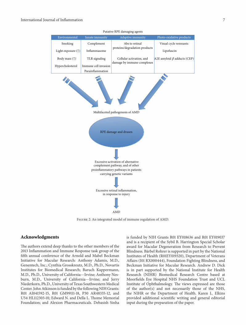
International Journal of Inflammation 7
Putative RPE damaging agents
Environmental
Smoking
Light exposure (↑)
Body mass (↑)
Hypercholesterol
Innate immunity
Complement
Inflammasome
TLR signaling
Immune cell invasionParainflammation
Adaptive immunity
Abs to retinalproteins/degradation products
Cellular activation, anddamage by immune-complexes
Photo-oxidative products
Visual cycle remnants
Lipofuscin
A2E amyloid 𝛽 adducts (CEP)
Multifaceted pathogenesis of AMD
RPE damage and drusen
Excessive activation of alternativecomplement pathway, and of other
proinflammatory pathways in patientscarrying genetic variants
Excessive retinal inflammation,in response to injury
AMD
Figure 2: An integrated model of immune regulation of AMD.
Acknowledgments
The authors extend deep thanks to the other members of the2013 Inflammation and Immune Response task group of thefifth annual conference of the Arnold and Mabel BeckmanInitiative for Macular Research: Anthony Adamis, M.D.,Genentech, Inc.; Cynthia Grosskreutz, M.D., Ph.D., NovartisInstitutes for Biomedical Research; Baruch Kuppermann,M.D., Ph.D., University of California—Irvine; Anthony Nes-burn, M.D., University of California—Irvine; and JerryNiederkorn, Ph.D., University of Texas SouthwesternMedicalCenter. JohnAtkinson is funded by the followingNIHGrants:R01 AI041592-15, R01 GM99111-18, P30 AR48335-12, andU54 HL112303-01; Edward N. and Della L. Thome MemorialFoundation; and Alexion Pharmaceuticals. Debasish Sinha
is funded by NIH Grants R01 EY018636 and R01 EY019037and is a recipient of the Sybil B. Harrington Special Scholaraward for Macular Degeneration from Research to PreventBlindness. Barbel Rohrer is supported in part by the NationalInstitutes of Health (R01EY019320), Department of VeteransAffairs (I01 RX000444), Foundation Fighting Blindness, andBeckman Initiative for Macular Research. Andrew D. Dickis in part supported by the National Institute for HealthResearch (NIHR) Biomedical Research Centre based atMoorfields Eye Hospital NHS Foundation Trust and UCLInstitute of Ophthalmology. The views expressed are thoseof the author(s) and not necessarily those of the NHS,the NIHR or the Department of Health. Karen L. Elkinsprovided additional scientific writing and general editorialinput during the preparation of the paper.

8 International Journal of Inflammation
References
[1] D. S. Friedman, B. J. O’Colmain, B. Munoz et al., “Prevalence ofage-relatedmacular degeneration in theUnited States,”Archivesof Ophthalmology, vol. 122, no. 4, pp. 564–572, 2004.
[2] L. S. Lim, P. Mitchell, J. M. Seddon, F. G. Holz, and T. Y. Wong,“Age-related macular degeneration,” The Lancet, vol. 379, no.9827, pp. 1728–1738, 2012.
[3] P. L. Penfold, M. C. Madigan, M. C. Gillies, and J. M. Provis,“Immunological and aetiological aspects of macular degener-ation,” Progress in Retinal and Eye Research, vol. 20, no. 3, pp.385–414, 2001.
[4] D. A. Carter and A. D. Dick, “CD200 maintains microglialpotential to migrate in adult human retinal explant model,”Current Eye Research, vol. 28, no. 6, pp. 427–436, 2004.
[5] C. Broderick, R.M.Hoek, J. V. Forrester, J. Liversidge, J. D. Sedg-wick, and A. D. Dick, “Constitutive retinal CD200 expressionregulates resident microglia and activation state of inflamma-tory cells during experimental autoimmune uveoretinitis,” TheAmerican Journal of Pathology, vol. 161, no. 5, pp. 1669–1677,2002.
[6] K. Saijo and C. K. Glass, “Microglial cell origin and phenotypesin health and disease,” Nature Reviews Immunology, vol. 11, no.11, pp. 775–787, 2011.
[7] C. Broderick, L. Duncan, N. Taylor, and A. D. Dick, “IFN-𝛾 and LPS-mediated IL-10-dependent suppression of retinalmicroglial activation,” Investigative Ophthalmology and VisualScience, vol. 41, no. 9, pp. 2613–2622, 2000.
[8] E. H. Hughes, F. C. Schlichtenbrede, C. C. Murphy et al.,“Minocycline delays photoreceptor death in the rds mousethrough a microglia-independent mechanism,” ExperimentalEye Research, vol. 78, no. 6, pp. 1077–1084, 2004.
[9] A. D. Dick, D. Carter, M. Robertson et al., “Control of myeloidactivity during retinal inflammation,” Journal of LeukocyteBiology, vol. 74, no. 2, pp. 161–166, 2003.
[10] R.Medzhitov, “Origin and physiological roles of inflammation,”Nature, vol. 454, no. 7203, pp. 428–435, 2008.
[11] H. Xu, M. Chen, and J. V. Forrester, “Para-inflammation in theaging retina,” Progress in Retinal and Eye Research, vol. 28, no. 5,pp. 348–368, 2009.
[12] S. Raychaudhuri, O. Iartchouk, K. Chin et al., “A rare penetrantmutation in CFH confers high risk of age-related maculardegeneration,” Nature Genetics, vol. 43, no. 12, pp. 1232–1236,2011.
[13] A. Richards, D. Kavanagh, and J. P. Atkinson, “Inherited com-plement regulatory protein deficiency predisposes to humandisease in acute injury and chronic inflammatory statestheexamples of vascular damage in atypical hemolytic uremic syn-drome and debris accumulation in age-related macular degen-eration,” Advances in Immunology, vol. 96, pp. 141–177, 2007.
[14] T. H. J. Goodship, “Factor H genotype-phenotype correlations:lessons from aHUS,MPGN II, andAMD,”Kidney International,vol. 70, no. 1, pp. 12–13, 2006.
[15] P. J. Lachmann and R. A. G. Smith, “Taking complement tothe clinic—has the time finally come?” Scandinavian Journal ofImmunology, vol. 69, no. 6, pp. 471–478, 2009.
[16] J. Tuo, S.Grob,K. Zhang, andC.C.Chan, “Genetics of immuno-logical and inflammatory components in age-related maculardegeneration,” Ocular Immunology and Inflammation, vol. 20,no. 1, pp. 27–36, 2012.
[17] J. R. Sparrow, K.Ueda, and J. Zhou, “Complement dysregulationin AMD: RPE-Bruch’s membrane-choroid,” Molecular Aspectsof Medicine, vol. 33, no. 4, pp. 436–445, 2012.
[18] P. F. Zipfel, N. Lauer, and C. Skerka, “The role of complementin AMD,” Advances in Experimental Medicine and Biology, vol.703, pp. 9–24, 2010.
[19] K. M. Gehrs, J. R. Jackson, E. N. Brown, R. Allikmets, and G.S. Hageman, “Complement, age-related macular degenerationand a vision of the future,” Archives of Ophthalmology, vol. 128,no. 3, pp. 349–358, 2010.
[20] D. H. Anderson, M. J. Radeke, N. B. Gallo et al., “The pivotalrole of the complement system in aging and age-relatedmaculardegeneration: hypothesis re-visited,” Progress in Retinal and EyeResearch, vol. 29, no. 2, pp. 95–112, 2010.
[21] S. Khandhadia, V. Cipriani, J. R. Yates, and A. J. Lotery, “Age-related macular degeneration and the complement system,”Immunobiology, vol. 217, no. 2, pp. 127–146, 2012.
[22] I. Bhutto and G. Lutty, “Understanding age-related maculardegeneration (AMD): relationships between the photorecep-tor/retinal pigment epithelium/Bruch’s membrane/choriocap-illaris complex,” Molecular Aspects of Medicine, vol. 33, no. 4,pp. 295–317, 2012.
[23] D. D. G. Despriet, C. C. W. Klaver, J. C. M. Witteman et al.,“Complement factor H polymorphism, complement activators,and risk of age-related macular degeneration,” Journal of theAmericanMedical Association, vol. 296, no. 3, pp. 301–309, 2006.
[24] A. L. Fett, M. M. Hermann, P. S. Muether, B. Kirchhof, andS. Fauser, “Immunohistochemical localization of complementregulatory proteins in the human retina,”Histol Histopathol, vol.27, no. 3, pp. 357–364, 2012.
[25] R. Sofat, J. P. Casas, A. R. Webster et al., “Complement factorH genetic variant and age-related macular degeneration: effectsize, modifiers and relationship to disease subtype,” Interna-tional Journal of Epidemiology, vol. 41, no. 1, pp. 250–262, 2012.
[26] J. L.Haines,M.A.Hauser, S. Schmidt et al., “Complement factorH variant increases the risk of age-related macular degenera-tion,” Science, vol. 308, no. 5720, pp. 419–421, 2005.
[27] A. O. Edwards, R. Ritter, K. J. Abel, A. Manning, C. Panhuysen,and L. A. Farrer, “Complement factor H polymorphism andage-related macular degeneration,” Science, vol. 308, no. 5720,pp. 421–424, 2005.
[28] R. J. Klein, C. Zeiss, E. Y. Chew et al., “Complement factor Hpolymorphism in age-related macular degeneration,” Science,vol. 308, no. 5720, pp. 385–389, 2005.
[29] G. S. Hageman, D. H. Anderson, L. V. Johnson et al., “A com-mon haplotype in the complement regulatory gene factorH (HF1/CFH) predisposes individuals to age-related maculardegeneration,” Proceedings of the National Academy of Sciencesof the United States of America, vol. 102, no. 20, pp. 7227–7232,2005.
[30] T. Montes, A. Tortajada, B. P. Morgan, S. R. De Cordoba, andC. L. Harris, “Functional basis of protection against age-relatedmacular degeneration conferred by a common polymorphismin complement factor B,” Proceedings of the National Academyof Sciences of the United States of America, vol. 106, no. 11, pp.4366–4371, 2009.
[31] A. P. Sjoberg, L. A. Trouw, S. J. Clark et al., “The factor H variantassociated with age-related macular degeneration (His-384)and the non-disease-associated form bind differentially to C-reactive protein, fibromodulin, DNA, and necrotic cells,” Jour-nal of Biological Chemistry, vol. 282, no. 15, pp. 10894–10900,2007.

International Journal of Inflammation 9
[32] R. J. Ormsby, S. Ranganathan, J. C. Tong et al., “Functionaland structural implications of the complement factor H Y402Hpolymorphism associated with age-related macular degenera-tion,” Investigative Ophthalmology and Visual Science, vol. 49,no. 5, pp. 1763–1770, 2008.
[33] S. J. Clark, R. Perveen, S. Hakobyan et al., “Impaired binding ofthe age-related macular degeneration-associated complementfactor H 402H allotype to Bruch’s membrane in human retina,”The Journal of Biological Chemistry, vol. 285, no. 39, pp. 30192–30202, 2010.
[34] M. Laine, H. Jarva, S. Seitsonen et al., “Y402H polymorphismof complement factor H affects binding affinity to C-reactiveprotein,” The Journal of Immunology, vol. 178, no. 6, pp. 3831–3836, 2007.
[35] G. S. Hageman, P. J. Luthert, N. H. Victor Chong, L. V.Johnson, D. H. Anderson, and R. F. Mullins, “An integratedhypothesis that considers drusen as biomarkers of immune-mediated processes at the RPE-Bruch’s membrane interface inaging and age-relatedmacular degeneration,”Progress in Retinaland Eye Research, vol. 20, no. 6, pp. 705–732, 2001.
[36] D. H. Anderson, R. F. Mullins, G. S. Hageman, and L. V.Johnson, “A role for local inflammation in the formation ofdrusen in the aging eye,” American Journal of Ophthalmology,vol. 134, no. 3, pp. 411–431, 2002.
[37] R. F. Mullins, N. Aptsiauri, and G. S. Hageman, “Structureand composition of drusen associated with glomerulonephritis:implications for the role of complement activation in drusenbiogenesis,” Eye, vol. 15, no. 3, pp. 390–395, 2001.
[38] L. V. Johnson, W. P. Leitner, M. K. Staples, and D. H. Anderson,“Complement activation and inflammatory processes in drusenformation and age related macular degeneration,” ExperimentalEye Research, vol. 73, no. 6, pp. 887–896, 2001.
[39] R. D. Horstmann, H. J. Sievertsen, J. Knobloch, and V. A.Fischetti, “Antiphagocytic activity of streptococcal M protein:selective binding of complement control protein factor H,”Proceedings of the National Academy of Sciences of the UnitedStates of America, vol. 85, no. 5, pp. 1657–1661, 1988.
[40] J. D. Lambris, D. Ricklin, and B. V. Geisbrecht, “Complementevasion by human pathogens,” Nature Reviews Microbiology,vol. 6, no. 2, pp. 132–142, 2008.
[41] N. Lauer, M. Mihlan, A. Hartmann et al., “Complement reg-ulation at necrotic cell lesions is impaired by the age-relatedmacular degeneration-associated factor-H His402 risk variant,”The Journal of Immunology, vol. 187, no. 8, pp. 4374–4383, 2011.
[42] S. J. Clark, V. A. Higman, B. Mulloy et al., “His-384 allotypicvariant of factor H associated with age-related macular degen-eration has different heparin binding properties from the non-disease-associated form,” The Journal of Biological Chemistry,vol. 281, no. 34, pp. 24713–24720, 2006.
[43] J. G. Hollyfield, V. L. Bonilha, M. E. Rayborn et al., “Oxidativedamage-induced inflammation initiates age-related maculardegeneration,”NatureMedicine, vol. 14, no. 2, pp. 194–198, 2008.
[44] P. T. Johnson, K. E. Betts, M. J. Radeke, G. S. Hageman, D.H. Anderson, and L. V. Johnson, “Individuals homozygous forthe age-related macular degeneration risk-conferring variantof complement factor H have elevated levels of CRP in thechoroid,” Proceedings of the National Academy of Sciences of theUnited States of America, vol. 103, no. 46, pp. 17456–17461, 2006.
[45] P. J. Lachmann, “The amplification loop of the complementpathways,” Advances in Immunology, vol. 104, pp. 115–149, 2009.
[46] J. M. Seddon, R. Reynolds, J. Maller, J. A. Fagerness, M. J. Daly,and B. Rosner, “Prediction model for prevalence and incidence
of advanced age-relatedmacular degeneration based on genetic,demographic, and environmental variables,” Investigative Oph-thalmology and Visual Science, vol. 50, no. 5, pp. 2044–2053,2009.
[47] R. Reynolds, M. E. Hartnett, J. P. Atkinson, P. C. Giclas, B.Rosner, and J. M. Seddon, “Plasma complement componentsand activation fragments: associations with age-relatedmaculardegeneration genotypes and phenotypes,” InvestigativeOphthal-mology and Visual Science, vol. 50, no. 12, pp. 5818–5827, 2009.
[48] H. P.N. Scholl, P. C. Issa,M.Walier et al., “Systemic complementactivation in age-relatedmacular degeneration,” PLoSONE, vol.3, no. 7, Article ID e2593, 2008.
[49] J. Wang, K. Ohno-Matsui, T. Yoshida et al., “Amyloid-𝛽 up-regulates complement factor B in retinal pigment epithelial cellsthrough cytokines released from recruited macrophages/mi-croglia: another mechanism of complement activation in age-related macular degeneration,” Journal of Cellular Physiology,vol. 220, no. 1, pp. 119–128, 2009.
[50] I. Droz, I. Mantel, A. Ambresin, M. Faouzi, D. F. Schorderet,and F. L. Munier, “Genotype-phenotype correlation of age-related macular degeneration: influence of complement factorH polymorphism,” British Journal of Ophthalmology, vol. 92, no.4, pp. 513–517, 2008.
[51] T. Sepp, J. C. Khan, D. A. Thurlby et al., “Complement factorH variant Y402H is a major risk determinant for geographicatrophy and choroidal neovascularization in smokers and non-smokers,” Investigative Ophthalmology and Visual Science, vol.47, no. 2, pp. 536–540, 2006.
[52] J. M. Seddon, S. George, B. Rosner, and N. Rifai, “Progressionof age-related macular degeneration: prospective assessmentof C-reactive protein, interleukin 6, and other cardiovascularbiomarkers,”Archives of Ophthalmology, vol. 123, no. 6, pp. 774–782, 2005.
[53] K.M.Gehrs, D.H.Anderson, L. V. Johnson, andG. S.Hageman,“Age-related macular degeneration—emerging pathogeneticand therapeutic concepts,” Annals of Medicine, vol. 38, no. 7, pp.450–471, 2006.
[54] G. S. Hageman, L. S. Hancox, A. J. Taiber et al., “Extendedhaplotypes in the complement factorH (CFH) andCFH-related(CFHR) family of genes protect against age-related maculardegeneration: characterization, ethnic distribution and evolu-tionary implications,”Annals ofMedicine, vol. 38, no. 8, pp. 592–604, 2006.
[55] J. Maller, S. George, S. Purcell et al., “Common variation inthree genes, including a noncoding variant in CFH, stronglyinfluences risk of age-related macular degeneration,” NatureGenetics, vol. 38, no. 9, pp. 1055–1059, 2006.
[56] K. M. Nishiguchi, T. R. Yasuma, D. Tomida et al., “C9-R95X polymorphism in patients with neovascular age-relatedmacular degeneration,” Investigative Ophthalmology & VisualScience, vol. 53, no. 1, pp. 508–512, 2012.
[57] J. B. Maller, J. A. Fagerness, R. C. Reynolds, B. M. Neale, M.J. Daly, and J. M. Seddon, “Variation in complement factor 3is associated with risk of age-related macular degeneration,”Nature Genetics, vol. 39, no. 10, pp. 1200–1201, 2007.
[58] M. Heurich, R. Martınez-Barricarte, N. J. Francis et al., “Com-mon polymorphisms in C3, factor B, and factor H collaborateto determine systemic complement activity and disease risk,”Proceedings of the National Academy of Sciences of the UnitedStates of America, vol. 108, no. 21, pp. 8761–8766, 2011.
[59] J. A. Fagerness, J. B. Maller, B. M. Neale, R. C. Reynolds, M. J.Daly, and J. M. Seddon, “Variation near complement factor I

10 International Journal of Inflammation
is associated with risk of advanced AMD,” European Journal ofHuman Genetics, vol. 17, no. 1, pp. 100–104, 2009.
[60] M. Nozaki, B. J. Raisler, E. Sakurai et al., “Drusen complementcomponents C3a and C5a promote choroidal neovasculariza-tion,” Proceedings of the National Academy of Sciences of theUnited States of America, vol. 103, no. 7, pp. 2328–2333, 2006.
[61] B. Rohrer, B. Coughlin, K. Kunchithapautham et al., “The alter-native pathway is required, but not alone sufficient, for retinalpathology in mouse laser-induced choroidal neovasculariza-tion,”Molecular Immunology, vol. 48, no. 6-7, pp. e1–e8, 2011.
[62] N. S. Bora, S. Kaliappan, P. Jha et al., “Complement activationvia alternative pathway is critical in the development of laser-induced choroidal neovascularization: role of factor B andfactor H,” The Journal of Immunology, vol. 177, no. 3, pp. 1872–1878, 2006.
[63] N. S. Bora, S. Kaliappan, P. Jha et al., “CD59, a complementregulatory protein, controls choroidal neovascularization in amouse model of wet-type age-related macular degeneration,”The Journal of Immunology, vol. 178, no. 3, pp. 1783–1790, 2007.
[64] N. S. Bora, P. Jha, V. V. Lyzogubov et al., “Recombinant mem-brane-targeted form of CD59 inhibits the growth of choroidalneovascular complex in mice,” The Journal of Biological Chem-istry, vol. 285, no. 44, pp. 33826–33833, 2010.
[65] S. M. Cashman, K. Ramo, and R. Kumar-Singh, “A non mem-brane-targeted human soluble CD59 attenuates choroidal neo-vascularization in amodel of age relatedmacular degeneration,”PLoS ONE, vol. 6, no. 4, Article ID e19078, 2011.
[66] Y. Huang, F. Qiao, C. Atkinson, V. M. Holers, and S. Tomlinson,“A novel targeted inhibitor of the alternative pathway of com-plement and its therapeutic application in ischemia/reperfusioninjury,” The Journal of Immunology, vol. 181, no. 11, pp. 8068–8076, 2008.
[67] B. Rohrer, Q. Long, B. Coughlin et al., “A targeted inhibitor ofthe alternative complement pathway reduces angiogenesis in amouse model of age-related macular degeneration,” Investiga-tive Ophthalmology and Visual Science, vol. 50, no. 7, pp. 3056–3064, 2009.
[68] S. B. Bressler, “Introduction: understanding the role of angio-genesis and antiangiogenic agents in age-related macular de-generation,” Ophthalmology, vol. 116, no. 10, pp. S1–S7, 2009.
[69] O. Gross, C. J.Thomas, G. Guarda, and J. Tschopp, “The inflam-masome: an integrated view,” Immunological Reviews, vol. 243,no. 1, pp. 136–151, 2011.
[70] J. K. Dowling and L. A. O’Neill, “Biochemical regulation of theinflammasome,” Critical Reviews in Biochemistry andMolecularBiology, vol. 47, no. 5, pp. 424–443, 2012.
[71] P. Menu and J. E. Vince, “The NLRP3 inflammasome in healthand disease: the good, the bad and the ugly,” Clinical &Experimental Immunology, vol. 166, no. 1, pp. 1–15, 2011.
[72] A. K. Mankan, A. Kubarenko, and V. Hornung, “Immunologyin clinic review series, focus on autoinflammatory diseases:inflammasomes: mechanisms of activation,” Clinical & Exper-imental Immunology, vol. 167, no. 3, pp. 369–381, 2012.
[73] S. L. Doyle, M. Campbell, E. Ozaki et al., “NLRP3 has a pro-tective role in age-related macular degeneration through theinduction of IL-18 by drusen components,” Nature Medicine,vol. 18, no. 5, pp. 791–798, 2012.
[74] V. Tarallo, Y. Hirano, B. D. Gelfand et al., “DICER1 loss and AluRNA induce age-related macular degeneration via the NLRP3inflammasome and MyD88,” Cell, vol. 149, no. 4, pp. 847–859,2012.
[75] M. E. Benoit, E. V. Clarke, P. Morgado, D. A. Fraser, and A. J.Tenner, “Complement protein C1q directs macrophage polar-ization and limits inflammasome activity during the uptake ofapoptotic cells,”The Journal of Immunology, vol. 188, no. 11, pp.5682–5693, 2012.
[76] W. A. Tseng, T. Thein, K. Kinnunen et al., “NLRP3 inflamma-some activation in retinal pigment epithelial cells by lysosomaldestabilization: implications for age-related macular degenera-tion,” Investigative Ophthalmology & Visual Science, vol. 54, no.1, pp. 110–120, 2013.
[77] K. Nakahira, J. A. Haspel, V. A. Rathinam et al., “Autophagyproteins regulate innate immune responses by inhibiting therelease of mitochondrial DNA mediated by the NALP3 inflam-masome,” Nature Immunology, vol. 12, no. 3, pp. 222–230, 2011.
[78] C. S. Shi, K. Shenderov, N. N. Huang et al., “Activation ofautophagy by inflammatory signals limits IL-1𝛽 production bytargeting ubiquitinated inflammasomes for destruction,”NatureImmunology, vol. 13, no. 3, pp. 255–263, 2012.
[79] M. C. Kenney, M. Chwa, S. R. Atilano et al., “MitochondrialDNA variants mediate energy production and expression levelsfor CFH, C3 and EFEMP1 genes: implications for age-relatedmacular degeneration,” PLoS ONE, vol. 8, no. 1, Article IDe54339, 2013.
[80] N. Patel, M. Ohbayashi, A. K. Nugent et al., “Circulating anti-retinal antibodies as immune markers in age-related maculardegeneration,” Immunology, vol. 115, no. 3, pp. 422–430, 2005.
[81] S. Cherepanoff, P.Mitchell, J. J.Wang, andM.C.Gillies, “Retinalautoantibody profile in early age-related macular degeneration:preliminary findings from the blue mountains eye study,”Clinical and Experimental Ophthalmology, vol. 34, no. 6, pp.590–595, 2006.
[82] K. Morohoshi, A. M. Goodwin, M. Ohbayashi, and S. J. Ono,“Autoimmunity in retinal degeneration: autoimmune retinopa-thy and age-related macular degeneration,” Journal of Autoim-munity, vol. 33, no. 3-4, pp. 247–254, 2009.
[83] K. Morohoshi, M. Ohbayashi, N. Patel, V. Chong, A. C. Bird,and S. J. Ono, “Identification of anti-retinal antibodies inpatients with age-related macular degeneration,” Experimentaland Molecular Pathology, vol. 93, no. 2, pp. 193–199, 2012.
[84] K. Morohoshi, N. Patel, M. Ohbayashi et al., “Serum autoan-tibody biomarkers for age-related macular degeneration andpossible regulators of neovascularization,” Experimental andMolecular Pathology, vol. 92, no. 1, pp. 64–73, 2012.
[85] B. Dhillon, A. F. Wright, A. Tufail et al., “Complement factorH autoantibodies and age-related macular degeneration,” Inves-tigative Ophthalmology and Visual Science, vol. 51, no. 11, pp.5858–5863, 2010.
[86] J. G. Hollyfield, V. L. Perez, and R. G. Salomon, “A haptengenerated from an oxidation fragment of docosahexaenoicacid is sufficient to initiate age-related macular degeneration,”Molecular Neurobiology, vol. 41, no. 2-3, pp. 290–298, 2010.
[87] B. Liu, L. Wei, C. Meyerle et al., “Complement component C5apromotes expression of IL-22 and IL-17 from human T cells andits implication in age-related macular degeneration,” Journal ofTranslational Medicine, vol. 9, p. 111, 2011.
[88] L. Wei, B. Liu, J. Tuo et al., “Hypomethylation of the IL17RCpromoter associates with age-related macular degeneration,”Cell Reports, vol. 2, no. 5, pp. 1151–1158, 2012.
[89] T. W. Olsen and X. Feng, “Theminnesota grading system of eyebank eyes for age-related macular degeneration,” InvestigativeOphthalmology and Visual Science, vol. 45, no. 12, pp. 4484–4490, 2004.
Hindawi Publishing CorporationInternational Journal of InflammationVolume 2013, Article ID 438412, 6 pageshttp://dx.doi.org/10.1155/2013/438412
Review ArticleInflammation in Retinal Vein Occlusion
Avnish Deobhakta and Louis K. Chang
Department of Ophthalmology, Columbia University Medical Center, New York, NY 10032, USA
Correspondence should be addressed to Louis K. Chang; [email protected]
Received 31 January 2013; Accepted 5 March 2013
Academic Editor: David A. Hollander
Copyright © 2013 A. Deobhakta and L. K. Chang. This is an open access article distributed under the Creative CommonsAttribution License, which permits unrestricted use, distribution, and reproduction in any medium, provided the original work isproperly cited.
Retinal vein occlusion is a common, vision-threatening vascular disorder.The role of inflammation in the pathogenesis and clinicalconsequences of retinal vein occlusion is a topic of growing interest. It has long been recognized that systemic inflammatorydisorders, such as autoimmune disease, are a significant risk factor for this condition. A number of more recent laboratory andclinical studies have begun to elucidate the role inflammation may play in the molecular pathways responsible for the vision-impairing consequences of retinal vein occlusion, such asmacular edema.This improved understanding of the role of inflammationin retinal vein occlusion has allowed the development of new treatments for the disorder, with additional therapeutic targets andstrategies to be identified as our understanding of the topic increases.
1. Introduction
Retinal vein occlusions (RVOs) are the secondmost commonvisually disabling disease affecting the retina, after diabeticretinopathy [1]. Obstruction of retinal venous flow leads todamage of the vasculature, hemorrhage, and tissue ischemia[2]. Occlusions affecting the central retinal vein, or centralretinal vein occlusion (CRVO), affect the entire retina, whilethose affecting lesser tributaries of the venous circulation,the so-called branch retinal vein occlusion (BRVO), affect aportion of the retina. Despite the fact that the disease entityhas been known to exist for over 100 years, current treatmentoptions often still leave patients with clinically problematicvisual disturbances and overall increased morbidity. RVOgenerally affects patients in middle age and the elderlypopulation [2], and several studies have identified systemicrisk factors, such as hypertension, diabetes, systemic vasculardisease, glaucoma, and hypercoagulable states [3, 4].
Although proliferative vascular changes can cause sig-nificant morbidity (particularly due to subsequent vitreoushemorrhage and neovascular glaucoma), the main reason fordecreased visual acuity in both CRVO and BRVO is macularedema [5]. As a result, elucidation of the causes of, as wellas treatment for, macular edema has been at the center oflarge-scale studies on patients with RVO.While the causes forRVO are multifactorial, with local and systemic factors being
identified as etiologic, most of the literature generally impli-cates vascular and inflammatory mediators as being partic-ularly salient [6–8]. Prior to the advent of intravitreal drugdelivery, treatment for macular edema for CRVO and BRVOwas observation and grid laser photocoagulation, respec-tively, the latter of which resolvedmacular edema slowly evenunder optimal circumstances [9]. The subsequent creation ofintravitreal medicines that block vascular endothelial growthfactor (VEGF) and the intravitreal delivery of corticosteroidsfor RVOhas led to better clinical outcomes overall [10].Whilethe focus of much of the literature is currently on the role ofanti-VEGF medications in the treatment of RVO, the role ofinflammation in both pathogenesis and treatment of RVO isequally exigent.
2. Pathogenesis of Inflammation in RVO
Both systemic and local inflammations have been hypothe-sized to play a significant role in the etiology of RVO. Thepredisposing systemic risk factors for RVO include hyper-tension, diabetes, dyslipidemia, and elevated plasma levels ofhomocysteine [11–13]. Atherosclerosis, a chronic, low-gradeinflammatory condition, has been studied extensively inrelation to RVO. Indeed, the systemic risk factors that predis-pose patients to RVO are also independently associated with

2 International Journal of Inflammation
atherosclerosis [11, 13].The initial pathological findings of thiscondition are composed of monocyte-derived macrophagesand T-lymphocytes (purely inflammatory lesions) whichlater progress to thrombus and clot formation [14]. Resultspertaining to the hypothesis of atherosclerosis as a risk factorfor RVO have been mixed. Large population-based cross-sectional studies have found that, while the prevalence ofRVO is fairly similar across ethnic groups, atheroscleroticdisease and markers of inflammation, such as C-reactiveprotein, were not associated with the disease [15]. In addition,certain genetic polymorphisms that had been previouslyimplicated in atherogenesis, inflammation, and coagulationdid not show association with BRVO or CRVO [16, 17].However, other reports have shown potential links betweenatherosclerosis (and by extension, systemic inflammation)and RVO. In particular, recent studies have shown thatpatients with RVO have an increased risk of asymptomaticipsilateral carotid artery plaques, and those with BRVOoften also have decreased aortic distensibility and elasticity,a finding frequently found in patients with atherosclerosis[18, 19]. In addition, pathological studies have shown anatherosclerotic retinal artery at the lamina cribrosa in somepatients with CRVO [20].
Another mechanism by which systemic inflammation isproposed to lead to RVO is through the induction of sys-temic hypercoagulability. Many inflammatory chemokines/cytokines are prothrombogenic; for example, interleukin-1 beta, interleukin-6, and tumor necrosis factor Alpha allsimultaneously upregulate tissue factor, which is a majoractivator of the extrinsic coagulation cascade pathway, anddownregulate tissue type plasminogen activator, which dis-rupts fibrinolysis [21–23]. In particular, homocysteine, aplasma element found elevated in patients with chronicinflammatory conditions, such as atherosclerosis, as well asin patients with errors of proteinmetabolism (homocysteine-mia/homocystinuria), can cause adverse systemic thromboticevents. Patients suffering from grossly elevated plasma lev-els of homocysteine often develop deep vein thromboses,myocardial infarctions, carotid atherosclerosis, and stroke[24]. In a similar fashion to other inflammatory-mediatedprocesses, proposedmechanisms of thrombosis include inhi-bition of plasminogen activator, inhibition of protein Cactivation, activation of Factor V, and the inducement ofendothelial cell dysfunction [25–27]. Perhaps unsurprisingly,given the strong possible link between hyperhomocysteine-mia and hypercoagulation, subsequent case control studiesbetween patients with andwithout CRVOhave demonstrateda robust correlation between CRVO and elevated plasmalevels of homocysteine [28, 29]. However, other studies haverightfully pointed out that, given that elevated levels ofplasma homocysteine are found in various other chronicinflammatory states, such as atherosclerosis, the associationof homocysteinemia with RVO is likely multifactorial [30].
Local inflammation within the eye has also been impli-cated in the pathogenesis of RVO. In vivo assessment of thevitreous fluid in patients with RVO has demonstrated ele-vated levels of proinflammatorymediators and lower levels ofanti-inflammatory cytokines [31, 32]. In particular, in amajorstudy on inflammatory immune mediators in a group of
vitreoretinal diseases, patients with RVO had elevated levelsof interleukin-6, interleukin-8, and monocyte chemoattrac-tant protein-1, and patients with CRVO had elevated levels ofVEGF, all of which are considered highly proinflammatory[33]. In follow-up studies, patients with macular edema fromboth BRVOandCRVOwere shown to have increased levels ofsoluble intercellular adhesion molecule-1 (proinflammatory)and decreased levels of pigment epithelium derived factor(anti-inflammatory) [34, 35]. Unsurprisingly, the literaturesuggests that for larger order vessel disruptions, such asthose affecting the central retinal vein or a larger branchretinal vein (“major” BRVO), there are even higher elevationsand reductions of the aforementioned pro-inflammatory andanti-inflammatory cytokines, respectively, as compared tosmaller branch vessel disruptions [32, 36]. Of particular noteis the fact that VEGF is classified as a pro-inflammatorycytokine; while VEGF is famously known for its central rolein retinal angiogenesis, recent studies have revealed its role inpermitting leukocyte infiltration into the retina—a key initialstep in the inflammatory pathway [37, 38].
Macular edema itself has been shown to result from pro-longed inflammatory states, such as those seen in uveitis [39].While the exact mechanism for how inflammation actuallycauses macular edema is still unclear, the prevailing theoryincludes the instigation of pro-inflammatory cytokines thatsubsequently damage retinal cells, particularly retinal pig-ment epithelial cells, which leads to fluid leakage into theretina [15]. In addition, the retinal ischemia seen with RVOhas also been postulated to lead to a pro-inflammatorymilieu,with the added insult of increased vascular permeabilitypartially due to a breakdown of the blood-retinal barrier[40]. Given these conditions, treatment options for RVOpreventing inflammation were developed.
3. Treatment of Inflammation in RVO
While the mainstay of treatment for systemic inflammatorystates has been oral or intravenous corticosteroids, thismethod of administration precluded their effective use forocular conditions given the side effect profile of long-termsteroid use. In addition, topical steroids do not penetrate theposterior segment of the eye in an efficacious manner [5].However, injecting corticosteroids directly into the vitreouscavity allows for a targeted, high dose use of the medicationsfor ocular inflammatory conditions with a low side effectprofile. Currently, the major anti-inflammatory medicationsin use for the treatment of RVO are intravitreal triamcinoloneacetonide (IVTA) and the newly developed dexamethasoneintravitreal implant.
Triamcinolone acetonide is a synthetic glucocorticoidthat has a potency that is five times that of cortisol andhas been reported to remain in the eye for months toyears after its initial injection [41, 42]. Initial use of IVTAfor treatment of CRVO resulted in significantly improvedanatomical changes within the macula [8, 43, 44]. As a result,the SCORE (Standard Care versus Corticosteroid for RetinalVein Occlusion) trial was launched by the National EyeInstitute.The study consisted of twomulticenter, randomized

International Journal of Inflammation 3
controlled clinical trials comparing the efficacy of IVTAversus standard of care for both BRVO and CRVO [45, 46].The SCORE-BRVO arm placed patients in cohort groupswhich received 1mg of IVTA, 4mg of IVTA, or standardof care (macular grid laser photocoagulation). The resultsdemonstrated no difference between the three groups interms of visual outcome; however, there was an increasedincidence of adverse side effects such as glaucoma, cataract,and injection-related problems in the IVTA groups relativeto the laser group [46]. Expectantly, the adverse side effectswere more pronounced in patients receiving the higherdosage of IVTA. As a result, the study concluded that forBRVO, macular grid laser photocoagulation should remainthe gold standard for treatment. The SCORE-CRVO armplaced patients in cohorts similar to the SCORE-BRVO arm;however, the results demonstrated that both IVTA groupswere superior to observation (standard of care for CRVO)in both visual acuity and anatomic resolution of macularedema [45]. These beneficial changes occurred as early as 4months into treatment and persisted for 24months.The studyalso demonstrated a reduced incidence of adverse side effectsin the 1mg IVTA group; as a result, this dosage has beenpreferred by some in the treatment of CRVO.
Given the partial success of temporary intravitreal cor-ticosteroids, a method of delivering corticosteroids in amanner that obviated the need for multiple injections wasdeveloped. The dexamethasone implant is a biodegradablecopolymer of both lactic and glycolic acids with micronizeddexamethasone that gradually releases the dose of the steroidover a period of months via the pars plana [5]. The GENEVAtrials were two phase III trials that tested the effect ofdexamethasone implants (in the 0.35mg and 0.7mg dosages)versus sham injections in patients with BRVO and CRVO[47, 48]. The results for the BRVO study group were mixed;while there was a trend towards better visual acuity inthe dexamethasone implant groups after 6 months, therewas a statistically significant improvement of acuity in thedexamethasone implant groups after 3 months. A similarfinding, though less in magnitude, was seen in the CRVOgroup. Patients tolerated the implant well, with a minorityof patients developing medically manageable glaucoma andcataract [47]. Given the results of the GENEVA trials, someadvocate use of the implant for patients with a relatively shortduration of macular edema [48]. Others have suggested thatthe dexamethasone implant may be useful for less frequentoccurrences of macular edema secondary to RVO, suchas those occurring in postvitrectomized eyes with CRVO,and those with long-standing BRVO and chronic edema[49, 50].
However, considering that the pathogenesis of inflam-mation in RVO also includes VEGF as a key mediatingcytokine, the advent of intravitreal anti-VEGF medicationsand their role in the treatment of RVO are especially salient.Ranibizumab is amonoclonal, humanized antibody fragmentthat binds to all VEGF isomers. Two randomized controlledtrials were established to determine the efficacy and safety ofranibizumab in the treatment of RVO: BRAVO (BRVO) andCRUISE (CRVO) [51, 52]. In both BRAVO and CRUISE stud-ies, patients with fovea involving macular edema within the
prior 12 months were given monthly ranibizumab injectionsof either 0.3mg, 0.5mg, or sham injections. In the BRAVOstudy, patients who were not responding to treatment wereeligible to receive rescue laser photocoagulation (standard ofcare) after 3months. At 6months of treatment, patients in theranibizumab groups in both studies had significantly higheraverage gains in visual acuity, significantly higher proportionsof patients gaining at least 15 letters of vision, and significantlylower mean foveal thicknesses relative to the sham injectiongroup. In addition, patients maintained this vision with con-tinued injections through 12 months; intriguingly, patients inthe sham group who were subsequently given ranibizumabinjections after the 6-month period enjoyed beneficial visualand anatomic changes—however, their final visual acuitieswere generally less than those in the ranibizumab groups,engendering a discussion on whether there was a visualpenalty resulting from a delay in treatment [53, 54]. Similarlybeneficial effects in smaller studies have been noted withanother anti-VEGF antibody, bevacizumab; however,many ofthe studies also mention a high recurrence rate and relativelyshort-term-efficacy [55–60].
Given the beneficial treatment outcomes of both intrav-itreal steroid and intravitreal anti-VEGF medications, a fewreports have attempted to ascertain whether a synergisticeffect might exist. One study found no significant differencein outcome between patients with CRVO who only receivedbevacizumab versus patients who received both bevacizumaband triamcinolone [61]. Another study attempted to assesswhether patients with RVO who received both bevacizumaband a dexamethasone implant (0.7mg) had significantlybetter outcomes than those who received only the dexam-ethasone implant [62].The patients in the combination groupwere given the dexamethasone implant 2 weeks after the firstinjection of bevacizumab. Most patients (65 percent) werebeing treated for BRVO. The primary outcome was the timerequired for reinjection based on existing OCT and visualdata. While most patients gained vision, a small minority didnot require a retreatment with an additional bevacizumabinjection during the 6-month study. While the data suggeststhat there may be a synergy between anti-VEGF medicationsand steroids, further study is required.
4. Conclusion
RVO is a highly prevalent cause of vision loss in the world.While the causes for RVO are multifactorial, both local andsystemic inflammations have been found to be highly con-tributory factors. Along with photocoagulation, medicationsthat reduce the level of inflammation in the eye, specificallytriamcinolone and the dexamethasone implant, have beenshown to provide beneficial results for patients with certainforms of RVO. Coupled with the explosion of anti-VEGFmedications, such as ranibizumab and bevacizumab, thetreatment of RVO is destined to change. Further study of therole of inflammation in the pathogenesis and propagationof RVO will aid in the identification of therapeutic targetsand the development of new treatment modalities for thisdisease.

4 International Journal of Inflammation
References
[1] H. Shahid, P. Hossain, and W. M. Amoaku, “The managementof retinal vein occlusion: is interventional ophthalmology theway forward?” British Journal of Ophthalmology, vol. 90, no. 5,pp. 627–639, 2006.
[2] S. S. Hayreh,M. B. Zimmerman, and P. Podhajsky, “Incidence ofvarious types of retinal vein occlusion and their recurrence anddemographic characteristics,” American Journal of Ophthalmol-ogy, vol. 117, no. 4, pp. 429–441, 1994.
[3] H. Koizumi, D. C. Ferrara, C. Brue, and R. F. Spaide, “Centralretinal vein occlusion case-control study,” American Journal ofOphthalmology, vol. 144, no. 6, pp. 858–863, 2007.
[4] The Eye Disease Case-Control Study Group, “Risk factors forcentral retinal vein occlusion the eye disease case-control studygroup,” Archives of Ophthalmology, vol. 114, no. 5, pp. 545–554,1996.
[5] M. L. Shahsuvaryan, “Therapeutic potential of intravitrealpharmaco-therapy in retinal vein occlusion,” Indian Journal ofOphthalmology, vol. 5, no. 6, pp. 759–770, 2012.
[6] R. D. Sperduto, R. Hiller, E. Chew et al., “Risk factors forhemiretinal vein occlusion: comparison with risk factors forcentral and branch retinal vein occlusion: the eye disease case-control study,”Ophthalmology, vol. 105, no. 5, pp. 765–771, 1998.
[7] S. Jain, J. R. Hurst, J. R. Thompson, and T. Eke, “UK nationalsurvey of current practice and experience of intravitreal triam-cinolone acetonide,” Eye, vol. 23, no. 5, pp. 1164–1167, 2009.
[8] A. Ladjimi, H. Zeghidi, S. Ben Yahia et al., “Intravitreal injec-tion of triamcinolone acetonide for the treatment of macularedema,” Journal Francais d’Ophtalmologie, vol. 28, no. 7, pp. 749–757, 2005.
[9] The Branch Vein Occlusion Study Group, “Argon laser pho-tocoagulation for macular edema in branch vein occlusion,”American Journal of Ophthalmology, vol. 98, no. 3, pp. 271–282,1984.
[10] P. A. Campochiaro, J. S. Heier, L. Feiner et al., “Ranibizumabfor macular edema following branch retinal vein occlusion:six-month primary end point results of a phase III study,”Ophthalmology, vol. 117, no. 6, pp. 1102.e1–1112.e1, 2010.
[11] S. J. McGimpsey, J. V. Woodside, C. Cardwell, M. Cahill, andU. Chakravarthy, “Homocysteine, methylenetetrahydrofolatereductase C677T polymorphism, and risk of retinal vein occlu-sion: a meta-analysis,” Ophthalmology, vol. 116, no. 9, pp. 1778–1787, 2009.
[12] J. B. Blice andG. C. Brown, “. Retinal vascular occlusive disease,”in Diseases of the Retina and the Vitreous, R. F. Spaide, Ed., pp.109–127, WB Saunders, Philadelphia, Pa, USA, 1999.
[13] R. Ross, “Atherosclerosis-an inflammatory disease,” New Eng-land Journal of Medicine, vol. 340, pp. 115–126, 1999.
[14] H. C. Stary, A. B. Chandler, S. Glagov et al., “A definition ofinitial, fatty streak, and intermediate lesions of atherosclerosis: areport from the committee on vascular lesions of the council onarteriosclerosis, American Heart Association,” Circulation, vol.89, no. 5, pp. 2462–2478, 1994.
[15] N. Cheung, R. Klein, J. W. Jie et al., “Traditional and novelcardiovascular risk factors for retinal vein occlusion: the mul-tiethnic study of atherosclerosis,” Investigative Ophthalmologyand Visual Science, vol. 49, no. 10, pp. 4297–4302, 2008.
[16] I. Steinbrugger, A. Haas, R. Maier et al., “Analysis ofinflammation- and atherosclerosis-related gene polymor-phisms in branch retinal vein occlusion,”Molecular Vision, vol.15, pp. 609–618, 2009.
[17] R. Maier, I. Steinbrugger, A. Haas et al., “Role of inflammation-related gene polymorphisms in patients with central retinal veinocclusion,” Ophthalmology, vol. 118, no. 6, pp. 1125–1129, 2011.
[18] Y.-J. Song, K.-I. Cho, S.-M. Kim et al., “The predictive value ofretinal vascular findings for carotid artery atherosclerosis: arefurther recommendations with regard to carotid atherosclerosisscreening needed?” Heart and Vessels, 2012.
[19] A. Aydin Kaderli, B. Kaderli, S. Gullulu, and R. Avci, “Impairedaortic stiffness and pulse wave velocity in patients with branchretinal vein occlusion,” Graefe’s Archive for Clinical and Experi-mental Ophthalmology, vol. 248, no. 3, pp. 369–374, 2010.
[20] W. R. Green, C. C. Chan, G. M. Hutchins, and J. M. Terry,“Central retinal vein occlusion: a prospective histopathologicstudy of 29 eyes in 28 cases,” Retina, vol. 1, no. 1, pp. 27–55, 1981.
[21] F. J. Neumann, I. Ott, N. Marx et al., “Effect of humanrecombinant interleukin-6 and interleukin-8 onmonocyte pro-coagulant activity,” Arteriosclerosis, Thrombosis, and VascularBiology, vol. 17, pp. 3399–3405, 1997.
[22] T. van der Poll, H. R. Buller, H. Ten Cate et al., “Activationof coagulation after administration of tumor necrosis factor tonormal subjects,”New England Journal of Medicine, vol. 322, no.23, pp. 1622–1627, 1990.
[23] E. Ulfhammer, P. Larsson, L. Karlsson et al., “TNF-𝛼 mediatedsuppression of tissue type plasminogen activator expressionin vascular endothelial cells is NF-𝜅B- and p38 MAPK-dependent,” Journal of Thrombosis and Haemostasis, vol. 4, no.8, pp. 1781–1789, 2006.
[24] C. J. Boushey, S. A. A. Beresford, G. S. Omenn, and A. G.Motulsky, “A quantitative assessment of plasma homocysteineas a risk factor for vascular disease: probable benefits ofincreasing folic acid intakes,” Journal of the American MedicalAssociation, vol. 274, no. 13, pp. 1049–1057, 1995.
[25] J. C. Chambers, A. McGregor, J. Jean-Marie, and J. S. Kooner,“Acute hyperhomocysteinaemia and endothelial dysfunction,”The Lancet, vol. 351, no. 9095, pp. 36–37, 1998.
[26] K. A. Hajjar, “Homocysteine-induced modulation of tissueplasminogen activator binding to its endothelial cell membranereceptor,” Journal of Clinical Investigation, vol. 91, no. 6, pp.2873–2879, 1993.
[27] G. M. Rodgers andM. T. Conn, “Homocysteine, an atherogenicstimulus, reduces protein C activation by arterial and venousendothelial cells,” Blood, vol. 75, no. 4, pp. 895–901, 1990.
[28] M. Weger, O. Stanger, H. Deutschmann et al., “Hyperhomo-cyst(e)inemia and MTHFR C677T genotypes in patients withcentral retinal vein occlusion,” Graefe’s Archive for Clinical andExperimental Ophthalmology, vol. 240, no. 4, pp. 286–290, 2002.
[29] M. Cahill, M. Karabatzaki, R. Meleady et al., “Raised plasmahomocysteine as a risk factor for retinal vascular occlusivedisease,”British Journal ofOphthalmology, vol. 84, no. 2, pp. 154–157, 2000.
[30] A. Kesler, V. Shalev, O. Rogowski et al., “Comparative analysisof homocysteine concentrations in patients with retinal veinocclusion versus thrombotic and atherosclerotic disorders,”Blood Coagulation and Fibrinolysis, vol. 19, no. 4, pp. 259–262,2008.
[31] H. Noma, H. Funatsu, T. Mimura, S. Harino, S. Eguchi,and S. Hori, “Pigment epithelium-derived factor and vascularendothelial growth factor in branch retinal vein occlusion withmacular edema,” Graefe’s Archive for Clinical and ExperimentalOphthalmology, vol. 248, no. 11, pp. 1559–1565, 2010.

International Journal of Inflammation 5
[32] H. Noma, H. Funatsu, T. Mimura, S. Eguchi, and K. Shimada,“Inflammatory factors inmajor andmacular branch retinal veinocclusion,” Ophthalmologica, vol. 227, no. 3, pp. 146–152, 2012.
[33] T. Yoshimura, K. H. Sonoda, M. Sugahara et al., “Comprehen-sive analysis of inflammatory immune mediators in vitreoreti-nal diseases,” PLoS ONE, vol. 4, no. 12, Article ID e8158, 2009.
[34] H. Noma, H. Funatsu, T. Mimura, and S. Eguchi, “Vitreousinflammatory factors and serous retinal detachment in centralretinal vein occlusion: a case control series,” Journal of Inflam-mation, vol. 8, article 38, 2011.
[35] H. Noma, H. Funatsu, T. Mimura, M. Tatsugawa, K. Shimada,and S. Eguchi, “Vitreous inflammatory factors and serousmacular detachment in branch retinal vein occlusion,” Retina,vol. 32, no. 1, pp. 86–91, 2012.
[36] J. W. Lim, “Intravitreal bevacizumab and cytokine levels inmajor and macular branch retinal vein occlusion,” Ophthalmo-logica, vol. 225, no. 3, pp. 150–154, 2011.
[37] A. P. Adamis, “Is diabetic retinopathy an inflammatory dis-ease?” British Journal of Ophthalmology, vol. 86, no. 4, pp. 363–365, 2002.
[38] S. Nakao, M. Arima, K. Ishikawa et al., “Intravitreal anti-VEGF therapy blocks inflammatory cell infiltration and re-entry into the circulation in retinal angiogenesis,” InvestigativeOphthalmology & Visual Science, vol. 53, no. 7, pp. 4323–4328,2012.
[39] A. Ossewaarde-Van Norel and A. Rothova, “Clinical review:update on treatment of inflammatory macular edema,” OcularImmunology and Inflammation, vol. 19, no. 1, pp. 75–83, 2011.
[40] C. Kaur, W. S. Foulds, and E. A. Ling, “Blood-retinal barrier inhypoxic ischaemic conditions: basic concepts, clinical featuresand management,” Progress in Retinal and Eye Research, vol. 27,no. 6, pp. 622–647, 2008.
[41] J. B. Jonas, “Intraocular availability of triamcinolone acetonideafter intravitreal injection,”American Journal of Ophthalmology,vol. 137, no. 3, pp. 560–562, 2004.
[42] J. B. Jonas, “Concentration of intravitreally injected triam-cinolone acetonide in aqueous humour,” British Journal ofOphthalmology, vol. 86, no. 9, p. 1066, 2002.
[43] M. Ip, A. Kahana, and M. Altaweel, “Treatment of centralretinal vein occlusion with triamcinolone acetonide: an opticalcoherence tomography study,” Seminars in Ophthalmology, vol.18, no. 2, pp. 67–73, 2003.
[44] J. B. Jonas, I. Akkoyun, B. Kamppeter, I. Kreissig, and R. F.Degenring, “Intravitreal triamcinolone acetonide for treatmentof central retinal vein occlusion,” European Journal of Ophthal-mology, vol. 15, no. 6, pp. 751–758, 2005.
[45] M. S. Ip, I. U. Scott, P. C. vanVeldhuisen et al., “A randomizedtrial comparing the efficacy and safety of intravitreal triam-cinolone with observation to treat vision loss associated withmacular edema secondary to central retinal vein occlusion:the standard care vs corticosteroid for retinal vein occlusion(SCORE) study report 5,” Archives of Ophthalmology, vol. 127,no. 9, pp. 1101–1114, 2009.
[46] I. U. Scott, M. S. Ip, P. C. vanVeldhuisen et al., “A randomizedtrial comparing the efficacy and safety of intravitreal triamci-nolone with standard care to treat vision loss associated withmacular edema secondary to branch retinal vein occlusion:the standard care vs corticosteroid for retinal vein occlusion(SCORE) study report 6,” Archives of Ophthalmology, vol. 127,no. 9, pp. 1115–1128, 2009.
[47] J. A. Haller, F. Bandello, R. Belfort et al., “Randomized,sham-controlled trial of dexamethasone intravitreal implant inpatients with macular edema due to retinal vein occlusion,”Ophthalmology, vol. 117, no. 6, pp. 1134–1146, 2010.
[48] W. S. Yeh, J. A. Haller, P. Lanzetta et al., “Effect of theduration of macular edema on clinical outcomes in retinal veinocclusion treated with dexamethasone intravitreal implant,”Ophthalmology, vol. 119, no. 6, pp. 1190–1198, 2012.
[49] M. Reibaldi, A. Russo, M. Zagari et al., “Resolution of persistentcystoidmacular edema due to central retinal vein occlusion in avitrectomized eye following intravitreal implant of dexametha-sone 0.7mg,” Case Reports in Ophthalmology, vol. 3, no. 1, pp.30–34, 2012.
[50] S. Kiss, “Moving beyond laser in treatment of DME, BRVO,”Retina Today, pp. 48–50, 2012.
[51] P. A. Campochiaro, J. S. Heier, L. Feiner et al., “Ranibizumabfor macular edema following branch retinal vein occlusionsix-month primary end point results of a phase III study,”Ophthalmology, vol. 117, no. 6, pp. 1102–1112, 2010.
[52] D. M. Brown, P. A. Campochiaro, R. P. Singh et al.,“Ranibizumab for macular edema following central retinal veinocclusion six-month primary end point results of a phase IIIstudy,” Ophthalmology, vol. 117, no. 6, pp. 1124–1133, 2010.
[53] P. A. Campochiaro, D. M. Brown, C. C. Awh et al., “Sustainedbenefits from ranibizumab formacular edema following centralretinal vein occlusion: twelve-month outcomes of a phase IIIstudy,” Ophthalmology, vol. 118, no. 10, pp. 2041–2049, 2011.
[54] D. M. Brown, P. A. Campochiaro, R. B. Bhisitkul et al., “Sus-tained benefits from ranibizumab for macular edema followingbranch retinal vein occlusion: 12-month outcomes of a phase IIIstudy,” Ophthalmology, vol. 118, no. 8, pp. 1594–1602, 2011.
[55] E. J. Chung, Y. T. Hong, S. C. Lee, O. W. Kwon, and H. J.Koh, “Prognostic factors for visual outcome after intravitrealbevacizumab for macular edema due to branch retinal veinocclusion,” Graefe’s Archive for Clinical and Experimental Oph-thalmology, vol. 246, no. 9, pp. 1241–1247, 2008.
[56] F. Prager, S. Michels, K. Kriechbaum et al., “Intravitreal beva-cizumab (Avastin) for macular oedema secondary to retinalvein occlusion: 12-month results of a prospective clinical trial,”British Journal of Ophthalmology, vol. 93, no. 4, pp. 452–456,2009.
[57] M. Kondo, N. Kondo, Y. Ito et al., “Intravitreal injection ofbevacizumab for macular edema secondary to branch retinalvein occlusion: results after 12 months and multiple regressionanalysis,” Retina, vol. 29, no. 9, pp. 1242–1248, 2009.
[58] L. Wu, J. F. Arevalo, M. H. Berrocal et al., “Comparison oftwo doses of intravitreal bevacizumab as primary treatment formacular edema secondary to branch retinal vein occlusions:results of the Pan American collaborative retina study group at24 months,” Retina, vol. 29, no. 10, pp. 1396–1403, 2009.
[59] T. Ach, A. E. Hoeh, K. B. Schaal, A. F. Scheuerle, and S. Dithmar,“Predictive factors for changes in macular edema in intravitrealbevacizumab therapy of retinal vein occlusion,” Graefe’s Archivefor Clinical and Experimental Ophthalmology, vol. 248, no. 2, pp.155–159, 2010.
[60] D. L. J. Epstein, P. V. Algvere, G. vonWendt, S. Seregard, and A.Kvanta, “Bevacizumab formacular edema in central retinal veinocclusion: a prospective, randomized, double-masked clinicalstudy,” Ophthalmology, vol. 119, no. 6, pp. 1184–1189, 2012.
[61] H. Y. Wang, X. Li, Y. S. Wang et al., “Intravitreal injec-tion of bevacizumab alone or with triamcinolone acetonide

6 International Journal of Inflammation
for treatment of macular edema caused by central retinal veinocclusion,” International Journal of Ophthalmology, vol. 4, no. 1,pp. 89–94, 2011.
[62] M. A. Singer, D. J. Bell, P. Woods et al., “Effect of combinationtherapy with bevacizumab and dexamethasone intravitrealimplant in patients with retinal vein occlusion,” Retina, vol. 32,no. 7, pp. 1289–1294, 2012.

Hindawi Publishing CorporationInternational Journal of InflammationVolume 2013, Article ID 464127, 4 pageshttp://dx.doi.org/10.1155/2013/464127
Research ArticlePeripheral Fluorescein Angiographic Findings in Fellow Eyes ofPatients with Branch Retinal Vein Occlusion
Irena Tsui, Asima Bajwa, Valentina Franco-Cardenas, Carolyn K. Pan,Hanna Y. Kim, and Steven D. Schwartz
Retina Division, Jules Stein Eye Institute, University of California, 100 Stein Plaza, Los Angeles, CA 90095, USA
Correspondence should be addressed to Irena Tsui; [email protected]
Received 4 December 2012; Accepted 8 March 2013
Academic Editor: David A. Hollander
Copyright © 2013 Irena Tsui et al.This is an open access article distributed under theCreativeCommonsAttributionLicense,whichpermits unrestricted use, distribution, and reproduction in any medium, provided the original work is properly cited.
Introduction. Branch retinal vein occlusion (BRVO) is a common retinal vascular condition that results in intraocular inflammatorychanges. Ultra wide field fluorescein angiography (UWFFA) is a retinal imaging device that can capture peripheral retinal findings.The purpose of this study was to look for peripheral findings in the fellow eye of patients with BRVO using UWFFA. Methods.Retrospective imaging review of patients diagnosed with BRVO that had both eyes imaged with UWFFA. Images were graded forperipheral findings in other quadrants of the same eye as well as in all quadrants of the fellow eye. Results. Of 81 patients, 14 (17%)patients had late vascular leakage in a quadrant other than the BRVO distribution. Five (6%) findings were in the same eye, 8 (10%)findings were in the fellow eye, and 1 (1%) finding was in both the same eye and the fellow eye. Of these 14 patients, 11 (80%) patientshad hypertension. Conclusion. Late peripheral retinal leakage in the fellow eye of patients with BRVO was detected in this cohortof patients with UWFFA. This novel finding may represent underlying systemic inflammation, hypertension, or bilateral BRVOs.
1. Introduction
Branch retinal vein occlusion (BRVO) affects approximately1% of the population and can cause severe vision lossthroughmacular edema, retinal neovascularization, and reti-nal detachment [1–4]. The disease is estimated to be bilateralin 5% of patients at presentation and become bilateral in 15%of patients over time [3, 5]. When bilateral and/or multiple,systemic vasculitis such as sarcoidosis, systemic lupus erythe-matosus, or Behcet’s disease may be the underlying etiology.
Atherosclerosis risk factors such as hypertension andhypercholesterolemia are thought to contribute to BRVO for-mation by causing arterial wall hardening and inflammationat arteriovenous crossing sites [6]. In theory, systemic riskfactors should put patients at similar risk for BRVO in botheyes and it is unclear why the disease is typically unilateral.One explanation could be due to random and individualvariation in arteriovenous crossing patterns [7].
There are several studies examining the vitreous of eyesthat have suffered BRVO showing that there are increasedinflammatory mediators when compared with control sur-gical patients. For example, vascular endothelial growth
factor (VEGF), soluble intercellular adhesion molecule-1,interleukin-8, and interleukin-6 were among the inflamma-tory markers found to be elevated in the vitreous of BRVOeyes [8–10]. In these studies inflammatory markers werecorrelated to macular edema [10] and the size of BRVO[8]. However, vitreous samples were not taken before BRVOwas detected nor was the vitreous of the fellow eye assessedas vitreous removal is invasive and there was no clinicalindication. Therefore, it is not certain if the increase ininflammatory markers was a cause or effect of the disease.
Ultra wide field fluorescein angiography (UWFFA,Optos, Marlborough, MA, USA) is a retinal imaging tech-nology that captures up to 200 degrees of the retina in asingle picture and has been useful in detecting peripheralfindings in a variety of retinal vascular diseases includingdiabetes, vein occlusions, and uveitis [11–13]. Prior work byour group studying peripheral retinal findings in BRVOusingUWFFA found that peripheral nonperfusion and vascularleakage were detectable [11]. In a cohort of 80 patients,peripheral nonperfusion was associated with angiographicmacular edema, while vascular leakage was not. The purposeof this current study was to assess peripheral findings on

2 International Journal of Inflammation
Figure 1: Ultra wide field fluorescein angiogram of an eye witha superotemporal branch retinal vein occlusion. There is also lateperipheral leakage in the inferotemporal quadrant.
fluorescein angiography in other quadrants of the same eyeand in fellow eyes of patients with BRVO.
2. Materials and Methods
This retrospective imaging study was Institutional ReviewBoard approved and carried out at the Jules Stein Eye Institutein Los Angles, CA, USA. An imaging database was searchedfor patients with the diagnosis of BRVO who underwentUWFFA (Optos, Marlborough, MA, USA). Patients withanother diagnosis or with concurrent other retinal dis-eases (i.e., age-related macular degeneration) were excluded.Patients with poorly controlled diabetes or signs of diabeticretinopathy such as intraretinal hemorrhage and venousbeading were excluded. Patients with known inflammatorydisorders such as sarcoidosis, systemic lupus erythematosus,and Behcet’s disease were excluded.
Demographic data such as patient gender and age werecollected. Significant past medical history such as hyperten-sion, high cholesterol, and diabetes mellitus were noted. Allimages were reviewed with Vantage Review Software (Optos,Marlborough, MA, USA) and adjusted with zoom, gamma,and contrast to optimize image quality.The quadrant affectedby a BRVO was recorded. Other quadrants of the same eyewere assessed for peripheral findings. Similarly, all quadrantsof the fellow eye were graded for peripheral findings.
3. Results
A total of 84 patients with BRVO were included in this study.Forty-nine (58%) patientswere female and 55 (65%) eyeswereOD. Average patient age was 60 years old (range 34–94 years;SD 13 years). Sixteen (19%) patients had no significant pastmedical history. Thirty-eight (45%) patients had hyperten-sion only.Nineteen (23%) patients had hypertension and highcholesterol only. Five (6%) patients had hypertension anddiabetes mellitus only. Four (5%) patients had hypertension,high cholesterol, and diabetesmellitus. Two (2%) patients haddiabetes mellitus only.
Thirteen (15%) BRVOs were macular and 71 (85%) werequadrantic. Of the macular BRVOs, 10 (77%) were superior
(a)
(b)
Figure 2: (a) Ultra wide field angiogram of a patient with branchretinal vein occlusion in the right eye. (b) The fellow eye has lateperipheral leakage temporally.
and 3 (23%) were inferior. Of the quadrantic BRVOs, 40(56%) were superotemporal, 25 (35%) were inferotemporal, 3(4%) were inferonasal, and 1 (1%) was superonasal. Two (3%)patients had two quadrantic BRVOs in the same eye.
Three (4%) patients were known to have bilateral BRVOsat the time of presentation. Of the remaining 81 patients, 14(17%) patients had late vascular leakage in a quadrant otherthan the BRVO distribution. Five (6%) findings were in thesame eye, 8 (10%) findings were in the fellow eye, and 1 (1%)finding was in both the same eye and the fellow eye. The sixeyes (7%) with leakage in the same eye all had leakage in aquadrant contiguous to the BRVO. Of the 14 patients withleakage in a quadrant other than the BRVO, 8 (57%) patientshad hypertension only; 2 (14%) patients had hypertension,high cholesterol, and diabetes mellitus; 1 (7%) patient hadhypertension and diabetes mellitus only; 1 (7%) patient hadhigh cholesterol only; and 2 (14%) patients had no knownsystemic diseases.
4. Discussion
This study examined peripheral retinal changes in patientswith BRVO in other quadrants of the same eye and in thefellow eye using UWFFA. In 81 patients, 14 (17%) patientshad late vascular leakage in a quadrant other than the BRVOdistribution. Two examples from our study are shown inFigures 1 and 2.

International Journal of Inflammation 3
There are three explanations for detecting late peripheralleakage in 11% (𝑛 = 9) of fellow eyes. One possibilityis that inflammation is not only a consequence of veinocclusion, but it is also a part of the pathogenesis of BRVOand patients with BRVO have higher levels of inflammationsystemically. This hypothesis has been tested by examininggene polymorphisms related to inflammatory cytokines inBRVO patients and normal control patients [14]. In a studyof almost 400 patients, 10 single nucleotide polymorphismswere assayed but were not found to be independent riskfactors for BRVO suggesting that inflammation did notcontribute to the pathogenesis of BRVO.
A multiethnic epidemiological study of over 6000 sub-jects considered the association of retinal vein occlusion(RVO) to traditional cardiovascular risk factors [1]. RVO wasnot associated with systemic inflammation, hematologicalabnormalities, or atherosclerosis but it was associated withhypertension and dyslipidemia. Given the high prevalence ofhypertension in our group of patients, a second explanationfor late peripheral leakage in the fellow eye is that it was amanifestation of hypertension.
A third explanation of bilateral leakage is that BRVOmaybe underrecognized as a bilateral disease and these areas oflate peripheral leakage are actually small peripheral BRVOsthat are otherwise undetectable with clinical exam andtraditional imaging. This would be supported by the fact thatthe distribution of leakage was often in a venous distribution.Without coexisting macular edema or peripheral ischemia,small incidentally found BRVOs do not require treatmentbut the finding does warrant further observation to detectprogression of disease.
Lastly, it is possible that these small areas of peripheralvascular leakage are not pathological andmay be found in thegeneral population. To our knowledge, there is no prior studyestablishing UWFFA findings in healthy patients withoutknown ocular or systemic disease. In 1985, a study usingtraditional fluorescein angiography in 25 patients did notdetect peripheral leakage in normal patients [15]. However,this was before UWFFAwas available and likely incompletelycaptured the periphery.
Limitations of our study are related to its retrospectivenature, sample size, and lack of a control group. Some patientshad anti-VEGF treatment within two months of havingUWFFA and this may have masked leakage in the treatedeye. It is possible that patients with unexpected peripheralleakage do in fact have a coexisting systemic illness thatwas not documented in the chart. Furthermore, patientsat a tertiary referral center may not represent the generalpopulation. Nonetheless, a subset of patients were foundto have unexpected late peripheral leakage on UWFFA ofpossible clinical significance.
In conclusion, 11% of patients with BRVO in our popu-lation had unexpected late peripheral leakage in fellow eyeson UWFFA. It may represent an underlying inflammatorycondition, hypertensive changes, or bilateral BRVO. Furtherstudies to evaluate a larger population of BRVO patients ina protocol manner should be considered to further elucidatethe significance of these findings.
Conflict of Interests
Authors have no conflict of interests to declare.
Acknowledgment
This work was presented in part in a meeting at the Asso-ciation for Research and Vision in Ophthalmology, FortLauderdale, FL, USA; May 2012.
References
[1] N. Cheung, R. Klein, J. J. Wang et al., “Traditional and novelcardiovascular risk factors for retinal vein occlusion: the mul-tiethnic study of atherosclerosis,” Investigative Ophthalmologyand Visual Science, vol. 49, no. 10, pp. 4297–4302, 2008.
[2] J. P. Ehlers and S. Fekrat, “Retinal vein occlusion: beyond theacute event,” Survey of Ophthalmology, vol. 56, no. 4, pp. 281–299, 2011.
[3] S. L. Rogers, R. L. McIntosh, L. Lim et al., “Natural historyof branch retinal vein occlusion: an evidence-based systematicreview,”Ophthalmology, vol. 117, no. 6, pp. 1094.e5–1101.e5, 2010.
[4] R. Klein, S. E. Moss, S. M. Meuer, and B. E. K. Klein, “The 15-year cumulative incidence of retinal vein occlusion: the BeaverDam Eye Study,” Archives of Ophthalmology, vol. 126, no. 4, pp.513–518, 2008.
[5] S. S. Hayreh, P. A. Podhajsky, and M. B. Zimmerman, “Branchretinal artery occlusion. natural history of visual outcome,”Ophthalmology, vol. 116, no. 6, pp. 1188.e4–1194.e4, 2009.
[6] P. R. A. O’Mahoney, D. T. Wong, and J. G. Ray, “Retinalvein occlusion and traditional risk factors for atherosclerosis,”Archives of Ophthalmology, vol. 126, no. 5, pp. 692–699, 2008.
[7] D. V. Weinberg, K. M. Egan, and J. M. Seddon, “Asymmetricdistribution of arteriovenous crossings in the normal retina,”Ophthalmology, vol. 100, no. 1, pp. 31–36, 1993.
[8] H. Noma, H. Funatsu, T. Mimura, S. Eguchi, and K. Shimada :,“Inflammatory factors inmajor andmacular branch retinal veinocclusion,” Ophthalmologica, vol. 227, no. 3, pp. 146–152, 2012.
[9] T. Yoshimura, K. H. Sonoda, M. Sugahara et al., “Comprehen-sive analysis of inflammatory immune mediators in vitreoreti-nal diseases,” PLoS ONE, vol. 4, no. 12, Article ID e8158, 2009.
[10] Y. Okunuki, Y. Usui, N. Katai et al., “Relation of intraocularconcentrations of inflammatory factors and improvement ofmacular edema after vitrectomy in branch retinal vein occlu-sion,” The American Journal of Ophthalmology, vol. 151, no. 4,pp. 610.e1–616.e1, 2011.
[11] P. S. Prasad, S. C. N. Oliver, R. E. Coffee, J. P. Hubschman, andS. D. Schwartz, “UltraWide-Field Angiographic Characteristicsof Branch Retinal and Hemicentral Retinal Vein Occlusion,”Ophthalmology, vol. 117, no. 4, pp. 780–784, 2010.
[12] S. C. N. Oliver and S. D. Schwartz, “Peripheral vessel leakage(PVL): a new angiographic finding in diabetic retinopathy iden-tifiedwith ultrawide-field fluorescein angiography,” Seminars inOphthalmology, vol. 25, no. 1-2, pp. 27–33, 2010.
[13] I. Tsui, A. Kaines, and S. Schwartz, “Patterns of periphlebitis inintermediate uveitis using ultra wide field fluorescein angiogra-phy,” Seminars in Ophthalmology, vol. 24, no. 1, pp. 29–33, 2009.
[14] I. Steinbrugger, A. Haas, R. Maier et al., “Analysis of inflam-mation- and atherosclerosis-related gene polymorphisms inbranch retinal vein occlusion,” Molecular Vision, vol. 15, pp.609–618, 2009.

4 International Journal of Inflammation
[15] H. J. Zenker, “Fluorescein angiography in inflammation of theperipheral fundus: the normal fluorescein angiographic pattern.I,” Ophthalmologica, vol. 190, no. 2, pp. 77–82, 1985.
Hindawi Publishing CorporationInternational Journal of InflammationVolume 2013, Article ID 503725, 12 pageshttp://dx.doi.org/10.1155/2013/503725
Research ArticleInfiltration of Proinflammatory M1 Macrophages intothe Outer Retina Precedes Damage in a Mouse Model ofAge-Related Macular Degeneration
Fernando Cruz-Guilloty,1,2 Ali M. Saeed,1 Jose J. Echegaray,1 Stephanie Duffort,1
Asha Ballmick,1 Yaohong Tan,1 Michel Betancourt,1 Eduardo Viteri,1
Ghansham C. Ramkhellawan,1 Eric Ewald,1 William Feuer,1 DeQiang Huang,1
Rong Wen,1 Li Hong,3 Hua Wang,3 James M. Laird,3 Abdoulaye Sene,4 Rajendra S. Apte,4
Robert G. Salomon,3 Joe G. Hollyfield,5 and Victor L. Perez1,2,6
1 Department of Ophthalmology, Bascom Palmer Eye Institute, University of Miami Miller School of Medicine, Miami, FL 33136, USA2Department of Microbiology and Immunology, University of Miami Miller School of Medicine, Miami, FL 33136, USA3Department of Chemistry, Case Western Reserve University, Cleveland, OH 44106, USA4Department of Ophthalmology and Visual Sciences, Washington University School of Medicine, St. Louis, MO 63110, USA5Department of Ophthalmology, Cole Eye Institute, Cleveland Clinic Lerner College of Medicine, Cleveland, OH 44195, USA6Ophthalmology, Immunology & Microbiology, University of Miami Miller School of Medicine, Bascom Palmer Eye Institute,Miami, FL 33136, USA
Correspondence should be addressed to Victor L. Perez; [email protected]
Received 17 October 2012; Revised 20 December 2012; Accepted 24 January 2013
Academic Editor: Robert B. Nussenblatt
Copyright © 2013 Fernando Cruz-Guilloty et al.This is an open access article distributed under theCreativeCommonsAttributionLicense, which permits unrestricted use, distribution, and reproduction in anymedium, provided the originalwork is properly cited.
Age-related macular degeneration (AMD) is a major cause of blindness in the developed world. Oxidative stress and inflammationare implicated in AMD, but precise mechanisms remain poorly defined. Carboxyethylpyrrole (CEP) is an AMD-associated lipidperoxidation product. We previously demonstrated that mice immunized with CEP-modified albumin developed AMD-likedegenerative changes in the outer retina. Here, we examined the kinetics of lesion development in immunized mice and thepresence of macrophages within the interphotoreceptor matrix (IPM), between the retinal pigment epithelium and photoreceptorouter segments. We observed a significant and time-dependent increase in the number of macrophages in immunized micerelative to young age-matched controls prior to overt pathology. These changes were more pronounced in BALB/c mice thanin C57BL/6 mice. Importantly, IPM-infiltrating macrophages were polarized toward the M1 phenotype but only in immunizedmice. Moreover, when Ccr2-deficient mice were immunized, macrophages were not present in the IPM and no retinal lesions wereobserved, suggesting a deleterious role for these cells in our model. This work provides mechanistic evidence linking immuneresponses against oxidative damage with the presence of proinflammatory macrophages at sites of future AMD and experimentallydemonstrates that manipulating immunity may be a target for modulating the development of AMD.
1. Introduction
Age-related macular degeneration (AMD) is the most com-mon cause of legal blindness in the elderly population ofdeveloped countries with over 300,000 newly diagnosedpatients per year in Europe and North America [1, 2]. It iswidely believed that AMD starts with the insidious, slowlyprogressing “dry” form (dry AMD) and can later develop into
themore severe “wet” AMD,which advances very rapidly andis characterized by abnormal development of blood vessels, aprocess called choroidal neovascularization (CNV) [3, 4] thataffects the macular region of the retina and leads to loss ofcentral vision. In turn, dry AMD without CNV can proceedto focal loss of the retinal pigment epithelium (RPE), termedgeographic atrophy (GA), which is accompanied by loss ofvision over these slowly expanding areas of RPE atrophy

2 International Journal of Inflammation
[5]. RPE loss or dysfunction renders the surrounding tissuevulnerable to damage by reactive oxygen species and loss ofphotoreceptor density ensues [6]. It has been recognized thataccumulation of debris below the RPE in the macula, alsoknown as drusen, is a risk factor for AMD. Identification ofcomplement factor proteins in drusen from AMD eyes [7],coupled with genetic variation in several complement factorgenes in AMD patients [8–13], collectively implicates inflam-mation as an important component in the pathophysiology ofthis disease [14, 15]. For instance, it was recently demonstratedthat complement factor H (CFH) serves a protective rolein AMD by binding and inhibiting the inflammatory effectsof the lipid peroxidation product malondialdehyde (MDA)[16].Therefore, dissecting the initiating events of outer retinalinflammation during the early stages of AMD can lead to abetter understanding of its pathogenesis. Such findings willenable the development of innovative immunotherapies thatcan prevent inflammatory damage to retinal tissue and lossof vision in a disease process that is estimated to increase inprevalence by 50% by the year 2020 [17].
In the past decade, several novel therapeutic agents havebeen identified as effective drugs to treat wet AMD, whichdelay new blood vessel formation and improve vision [18].However, there is no effective treatment for dry AMD to date.In the pursuit of identifying the signals from the outer retinathat initiate inflammation and possibly involve the immunesystem in AMD pathogenesis, we have evaluated immuneresponses to carboxyethylpyrrole (CEP), a protein modifica-tion that forms from an oxidation fragment of docosahex-aenoic acid (DHA) [19], the most oxidizable of all long chainpolyunsaturated fatty acids. Studies have shown that AMDdonor eyes contain more CEP-modified proteins in the outerretina and drusen than in age-matched controls [7]. CEP-modified proteins and CEP autoantibodies are also moreabundant in AMD plasma than in control samples [19, 20].
Since DHA is abundant in the outer retina [21], wherethe amalgamation of high oxygen tension and light pro-vides an environment suitable for oxidative damage, our labhas previously developed a murine model in which miceimmunizedwithCEP-modifiedmouse serum albumin (CEP-MSA) develop a CEP-specific immune response which corre-lates with dry AMD-like pathology when compared to age-matched controls, including CEP autoantibodies, comple-ment deposition in the Bruch’s membrane, and RPE damage[22, 23]. To our knowledge, this is the first immune-mediatedmouse model of dry AMD in genetically unmanipulated ani-mals and stems directly fromobservations in human patients.
Another component of the immune system that hasbeen implicated in AMD is the macrophage lineage [24, 25],although the specific role of these innate immune cells atdifferent stages of AMDdisease progression is still controver-sial. Within the retina, there are two sources of macrophages:(1) microglia, bone marrow-derived resident macrophagesthat are recruited to neural tissue during retinal developmentand provide immunosurveillance in the inner retina, and(2) circulating monocytes that can be recruited from theblood vessels to sites of inflammation when needed by spe-cific chemokines and cytokines. Independent of the source,these cells can undergo a diverse program of differentiation
depending on the microenvironment that ultimately dictatestheir effector functions [26, 27].Macrophages activated in thepresence of interferon-gamma (IFN-𝛾) become proinflam-matory M1 macrophages, characterized by their productionof tumor necrosis factor alpha (TNF-𝛼) and interleukin-12(IL-12) and are associated with tissue damage. On the otherhand, macrophages activated in the presence of IL-4 differ-entiate into M2-type, marked by production of the immuno-suppressive cytokine IL-10 and involved in tissue remodeling.The CEP-MSA-induced changes in the outer retina thatprovide a model for AMD afford the unique opportunity todirectly test the role of these cells in the disease process.
Our current study aims to characterize both the mag-nitude and kinetics of development of retinal lesions andmacrophage involvement in the BALB/c and C57BL/6 (B6)mouse strains at various intervals postimmunization (p.i.)in young mice compared to age-matched controls, beforeextensive retinal lesions are observed. Here we extend ouroriginal study [22] by showing that CEP-MSA immunizationleads, in aged (old) BALB/c mice, to the end stage cardinalfeature of human dry AMD: loss of photoreceptor cells. Thismajor damage is the result of a low-grade but significantinflammatory response in the retina prior to overt tissuedamage, which can be quantified in young mice. We haveidentified M1 macrophages localized to the interphotore-ceptor matrix (IPM) surrounding the photoreceptor outersegments in close proximity to the RPE. These changesoccur in both BALB/c and B6 strains, but the kinetics aredifferent; BALB/c mice are more susceptible at a youngerage. We also detected elevated levels of the monocyte che-moattractant Ccl2 in the retinas of CEP-immunized mice.Moreover, 𝐶𝑐𝑟2−/− B6 mice immunized with CEP-MSA lackmacrophage recruitment, and retinal lesion development isreduced or prevented. Since AMD is an age-related disease,defining the progression of inflammatory cell recruitmentand development of AMD-like lesions at earlier stages of thedisease is essential in order to map the character and timingof immune mechanisms that take place in our model andcorrelate with development of pathology. This work clarifiesa long-standing question by defining a clear mechanisticpath that explains the role of inflammation in AMD: M1macrophages are key factors in dry AMD pathogenesis. Wealso provide experimental demonstration for the idea thatregulation of immune responses (in this case, inhibition ofmacrophage recruitment) can be a target of therapy to preventthe development of AMD.
2. Results
2.1. Magnitude and Tempo of Lesion Development andInflammatory Cell Recruitment. We have previouslydescribed AMD-like lesions in wild-type (WT) B6 miceimmunized with CEP-MSA [22]. We now describe in detaillesions in CEP-MSA-immunized mice on the BALB/c back-ground, which are inherently albino. Importantly, but alsotechnically challenging, the severity of lesions increaseswith time after immunization, and it can take for up to12–24 months to observe signs of geographic atrophyand photoreceptor cell loss, cardinal features of AMD

International Journal of Inflammation 3
Early
In
term
edia
te
Naive CEP-MSA
BALB/c
Naive CEP-MSA
C57BL/6 La
te (o
ld)
INL
ONL
RPE ROS
ONL
RPE
ROS
ONL
RPE
ROS
20 𝜇m
Figure 1: Immunization with CEP-MSA leads to overt retinal degeneration in aged mice that resembles geographic atrophy with loss ofphotoreceptor cells, particularly in the BALB/c background. Histology of CEP-MSA versus naıve BALB/c or C57BL/6 eyes at different timepoints postimmunization.TheRPE is located at the lower part of each image.The dark arrows showmacrophage-like cells. CEP-MSABALB/cmice show strong pathology since the early time points, starting with swollen RPE cells and leading to massive geographic atrophy at the latetime point, including complete loss of the photoreceptor cells. The kinetics of pathology in CEP-MSA B6 mice is slower, but eventually thereare focal lesions of the RPE such as vesiculation, as previously reported [21]. Representative images are shown (from 3–5 mice per group perexperiment; two or three independent experiments were performed for each strain). INL: inner nuclear layer; ONL: outer nuclear layer; ROS:rod outer segment; RPE: retinal pigment epithelium. Images were obtained using a 63x oil objective, and the scale marker represents a 20𝜇mlength.
(Figure 1, Supplemental Figure S1 available online athttp://dx.doi.org/10.1155/2013/503725). The main purpose ofthe current study was to determine the earliest time at whichsignificant differences between CEP-immunized and controlmice could be detected. The reasoning behind this approachis to define the molecular and cellular mechanisms that takeplace at the initial stages of disease, before there are grosschanges to the retina that could alter its function.These earlytime points could also be critical for therapeutic intervention.
Eyes harvested at 40–90 days p.i. were defined as of earlyrecovery times, those harvested at 100–200 days p.i. were con-sidered of intermediate recovery times, while those obtainedafter day 200 p.i. were considered of late recovery times. Focallesions in the RPE and IPM consisting of vacuolization ofindividual or groups of cells, pyknotic RPE cells, hypertrophicRPE cells, and melanin engulfment by macrophages (onlypossible in B6 mice), as well as darkly stained nuclei ofinflammatory (macrophage-like) cells in the RPE and IPMwere counted (refer to Methods for detailed scoring parame-ters) (Figure 1). While significant damage can be observed inthe retina of aged (12–24 months old) control mice (mainlythinning of the photoreceptor layer, Figure 1, SupplementaryFigure 1), noGApathology is seen inmice that did not receiveCEP immunization or in younger CEP-immunized mice.
Our data was analyzed with a three-factor analysis ofvariance; the factors are (1) strain of mice (BALB/c versusB6), (2) immunization status (naıve versus immunized), and(3) time of recovery of tissue (early versus intermediate)(Figure 2). Pathology scores (defined as the summationof retinal lesions and cellular infiltration) were higher inBALB/c CEP-immunizedmice at both early and intermediaterecovery times but only higher in B6 CEP-immunized miceat the intermediate recovery time (Figure 2(a)). To have arepresentative and objective pathological score throughoutthe retina, we focused on the number of IPM-infiltratingcells present in close proximity to the RPE. Using routinehistopathology, it is not possible to distinguish different typesof monocyte lineage cells present in the outer retina (forexample, resident microglia versus macrophages recruitedfrom the circulation). For this reason in the description ofthe results to follow, we will simply refer to the nonneu-ral cells in the IPM as macrophages, since microglia aredistinguished mainly for their location within the centralnervous system. Careful examination of the lesions at theseearly and intermediate recovery times demonstrated that adistinct population of macrophages is present in the IPMcompartment near the RPE in these animals. Quantificationin plastic sections showed that CEP-immunized BALB/cmice

4 International Journal of Inflammation
Path
olog
y pe
r sec
tion
EarlyIntermediate
∗10
8
6
4
2
0
BALB
/c n
aive
BALB
/c C
EP
B6 n
aive
B6 C
EP
∗
(a)In
filtr
ate c
ells
per s
ectio
n
EarlyIntermediate
BALB
/c n
aive
BALB
/c C
EP
B6 n
aive
B6 C
EP
8
6
4
2
0
∗
∗
(b)
Infil
trat
e cel
ls pe
r sec
tion
Intermediate time point
8
6
4
2
0
BALB
/c n
aive
BALB
/c C
EP
H&E stains
(c)
Figure 2: Quantification of retinal pathology and macrophage infiltration in CEP-MSA-immunized mice. (a) Combined pathology scoreper section (all kinds of lesions and IPM-infiltrating cells). The significant changes due to CEP-MSA immunization occur at the early timepoint in the BALB/c background, whereas they occur at intermediate time points for B6 mice. (b) CEP-MSA immunization acceleratesIPM-infiltrating cell recruitment at early stages of AMD development, prior to major retinal damage. Mean values are shown; error barsrepresent S.D. (∗) denotes statistically significant differences (𝑃 < 0.05). Data from one representative experiment was used for this analysis;similar results were obtained in additional repeat experiments. (c) Haematoxylin & Eosin (H&E) staining of frozen sections confirmed theaccelerated macrophage recruitment associated with CEP-MSA immunization. 5 sections were scored per mouse, with 3 mice per group atthe intermediate time point (d 100–200 p.i.). Mean values are shown; error bars represent S.D.
have a significantly higher number of macrophages thanage-matched naıve animals harvested at 40–100 days p.i.(𝑃 = 0.023) as well as those harvested at 100–200 days p.i.(𝑃 = 0.023) (Figure 2(b)). The same cellular quantification isobserved with H&E staining of frozen sections (Figure 2(c)).When comparing IPM macrophages between the two timepoints, BALB/c mice showed an increase in the number ofthese cells in the early to intermediate recovery times inboth immunized and naıve mice, but the magnitude was
significantly higher in immunized mice (𝑃 = 0.012). WhileCEP-immunized B6 mice harvested at early recovery timesshowed no significant differences in IPMmacrophages whencompared to naıve B6 mice, immunized mice harvested atintermediate time points had more IPM macrophages thanage-matched B6 naıve controls (𝑃 = 0.023). Therefore,the number of monocyte-lineage cells present in the IPMincreases over time in CEP-immunized B6 mice to reachsignificant numbers by the intermediate recovery times.
International Journal of Inflammation 5
2.2. Enhanced Immune-Mediated AMD-Like Pathology Is Spe-cific to Genetic Background rather than Light Damage. NaıveBALB/c mice contained more macrophages in the IPM thanage-matched naıve B6 mice (𝑃 < 0.01), and immunizedBALB/cmice show higher number of IPMmacrophages thanimmunized B6 mice as well (𝑃 < 0.01). This differenceis also evident in the number of RPE lesions, which arehigher in BALB/c mice than in B6 mice, regardless ofimmunization status. To test the possibility that photosensi-tivity in the BALB/c albino mice [28] could be responsiblefor the differences in outer retinal lesion development andmacrophage presence when compared to B6 mice, we immu-nized albino B6 mice (B6(Cg)-𝑇𝑦𝑟𝑐−2𝐽/𝐽 or 𝑇𝑦𝑟−/− mice,which lack the tyrosinase enzyme) and compared them toage-matched naıve 𝑇𝑦𝑟−/− mice as well as to correspondingBALB/c and WT-B6-immunized and naıve mice at 40–90days p.i. The results from these comparisons showed thatboth macrophages in the outer retina and development ofretinal lesions observed in𝑇𝑦𝑟−/−mice are comparable toWTB6 and do not show the enhanced cellular recruitment, RPEhypertrophy, and ROS vacuolization present in BALB/c miceat the early time points, regardless of immunization status(Figure 3(a)). Quantification analysis of pathological changesper section supports these observations (Figure 3(b)). Thissuggests that the differences in inflammatory cells present inthe outer retina and AMD-like pathology observed betweenthe BALB/c and the B6 strains are not due to photosensitivityin the albino mice because of lack of pigmentation, butinstead they are specific to genetic background, possibly dueto differences in the immune responses against CEP-MSAbetween the mice or to inherent differences in RPE functionand/or local oxidative damage responses in the retina.
2.3. Inflammatory Cells Surround the Retina of CEP-MSA-Immunized Mice. Because inflammation associated withmacrophages present in the IPM seems to play a role in theretinal pathology observed in CEP-MSA-immunized mice,we performed immunohistochemical (IHC) analysis usingcell surface immune markers for the identification of thespecific macrophages present in the retina, generally, and tothe IPM region, specifically. A large number of CD45+ (apan-leukocyte marker) cells were observed in the choroid ofimmunized mice compared to controls (Supplementary Fig-ure 2), indicating that CEP immunization leads to increasedocular inflammation. In terms of specific cell subsets, weobserved only a few CD3+ and CD19+ cells in the outerretina, suggesting that both T cells and B cells are largelyabsent from the site of retinal lesions (data not shown).In contrast, there were substantial numbers of choroidalCD11b+, F4/80+, and CD68+ cells surrounding the retinasof BALB/c CEP-MSA-immunized mice and not of naıvecontrols (data not shown). Similar findings were observed inB6 immunized animals at late recovery times when lesionsare present.
2.4. Proinflammatory M1Macrophages Are Present in the IPMat Early Recovery Times in Mice Immunized with CEP-MSA.Immunofluorescence staining identified retinal CD11b+,
F4/80+, and CD68+ cells in mice immunized with CEP-MSA, suggesting the potential role of macrophages in thedevelopment of disease in thismodel.Wewere able to localizea significant number of these macrophages in the RPE andIPM using nuclear counterstaining with DAPI (Figure 4(a),Supplementary Figure 3). This correlates with the location ofthemacrophages identified by histopathology using toluidineblue and H&E staining that are mostly observed in proximitywith vacuolization and lesions of RPE and photoreceptors.In order to determine the specific class and activation statusof these macrophages, we performed intracellular stainsfor Tumor Necrosis Factor-alpha (TNF-𝛼) and Interleukin-12 (IL-12) production, which identify M1 macrophages,versus IL-10 production, a hallmark of M2 macrophagedifferentiation. We observed both TNF+ and IL-12+ cellswithin the IPM of CEP-MSA-immunized mice, but IL-10staining was negative, indicating the presence of activatedM1 macrophages in the pathological regions of immunizedmice only (Figure 4(b)). To substantiate the IHC results,we performed mRNA quantification on IPM-infiltratingmacrophages isolated by laser capture. These data confirmedthat detectable levels of M1 marker genes (IL-6, TNF-𝛼,and IL-1𝛽) were observed only in CEP-immunized mice,whereas IL-10 expression was not detected (Figure 4(c)).Expression of Arg1, another M2 marker, was not elevated inCEP-immunized mice. Interestingly, we also observed CEP-associated increased expression of Ccl2, a monocyte che-moattractant that has been implicated in AMD, suggestingthat the Ccl2/Ccr2 axis may play a role in CEP-inducedpathology. These data strongly suggest that M1 macrophagesare primarily associated with the lesions we observe in theouter retina and may be the main effectors of the inflamma-tory response observed in this model.
2.5. Macrophage Recruitment into the Outer Retina of CEP-MSA-Immunized Mice Is Necessary for the Induction of Dis-ease. To directly test the role of macrophage recruitmentinto the IPM and development of outer retinal lesions weimmunizedCcr2-deficientmice on the B6 background.Thesemice have a defect in chemokine signaling and show poorrecruitment of inflammatory cells into sites of inflammation.In addition, it has been previously shown that disruption ofchemokine signaling to macrophages in these Ccr2 knockoutmice can have a deleterious effect on the integrity of theretina in aged mice (2 years and older) [29]. While Ccr2deficiency did not affect the levels of anti-CEP antibody titersin young immunized mice (Figure 5(a)), 𝐶𝑐𝑟2−/− animalsshowed no increase of IPM-infiltratingmacrophages or outerretinal lesions in immunizedmice compared to those in naiveage-matched controls at late recovery times (Figure 5(b)).This observation strongly suggests that macrophages, mainlyrecruited by Ccl2, play a causative role in the induction oftissue damage in this model.
3. Discussion
Immunization of mice with CEP-MSA provides a valuablemodel to study dry AMD from an immunological per-spective, helping to dissect the immune system’s role in

6 International Journal of Inflammation
BALB
/c
WT
B6
Alb
ino
B6
Naive CEP-MSA
Early time point
INL
ONL
RPE ROS
ONL
RPE
ROS
ONL
RPE
ROS
20 𝜇m
(a)
Path
olog
y pe
r sec
tion
6
4
2
0
Alb
ino
B6 n
aive
Alb
ino
B6 C
EP
WT
B6 C
EP
BALB
/c C
EP
(b)
Figure 3: Immune-mediated cellular infiltration and development of outer retinal lesions are specific to genetic background and pigmentindependent. (a) Histology of CEP-MSA albino B6 mice compared toWT BALB/c andWT B6 at the early time point. Representative imagesare shown (from 3–5 mice per group per experiment; two or three independent experiments were performed for each strain). INL: innernuclear layer; ONL: outer nuclear layer; ROS: rod outer segment; RPE: retinal pigment epithelium. Images were obtained using a 63x oilobjective, and the scale marker represents a 20 𝜇m length. (b) Quantification shows the lack of pathology at early time points in albino B6mice, similar to WT B6 and in contrast to WT BALB/c mice. Mean values are shown; error bars represent S.D.
the development of disease. Studies presented here linkthe AMD-like histopathological changes with the presenceof macrophages in the outer retina during early stages ofdisease, suggesting that macrophages are involved in theunderlying pathology. Notably, our data suggest that BALB/cmice tend to be more sensitive to immunization with CEP-MSA than B6mice by having a greater magnitude and earliersignificant difference of inflammatory cells in the IPM whencompared to age-matched controls. In addition, dry AMD-like pathology, such as RPE cell hypertrophy, vacuolizationof RPE and ROS, and RPE cell pyknosis, is also found atgreater magnitude and arises earlier following immunizationin the BALB/c mice. We also show that old CEP-immunizedBALB/c mice develop photoreceptor cell loss. This suggeststhat future studies using this model would benefit froma more rapid and amplified immunopathological effect inBALB/c mice than in B6 mice, yielding results as early as 40–100 days postimmunization.
Even if BALB/c mice show an earlier significant responseto immunization with CEP-MSA than age-matched B6 mice,it is important to stress that CEP-immunized mice containlarger numbers of macrophages in the IPM and AMD-likepathology than naıve controls in both strains. Furthermore,
our data showed no statistically significant differencesbetween the two strains through time. This suggests thatany age-related changes seen in immunized mice observedduring the early stages of disease are of comparable magni-tude regardless of strain, but that the higher number ofmacrophages present in the IPM of BALB/c mice makes ittechnically easier for quantification of disease onset. In otherwords, the reason that there seems to be no early differencesin naıve versus CEP-MSA mice on the B6 background isbecause the actual number of IPM-infiltrating cells is toolow at that point to achieve statistical significance.
Differences between these two strains could also beattributed to background-specific (genetic and/or immune)mechanisms or to the reduced melanin levels in BALB/cmice. B6 mice are prone to develop T helper type 1 (Th1)responses, whereas BALB/c are Th2-prone. On the otherhand, it has been shown that melanin in the RPE providesprotection from light damage [30]. By showing that albino B6(𝑇𝑦𝑟−/−) mice have comparable inflammatory cell numbersin the IPM and AMD-like pathology with WT B6 mice in amuch less robust form than BALB/c mice, the possibility thatlight damage largely contributes to pathology is less likely.Indeed, it has been previously shown that B6 (𝑇𝑦𝑟−/−) are

International Journal of Inflammation 7
BALB/c, CEP, intermediate
BALB
/c, C
EPin
term
edia
teC5
7BL/
6, C
EPlat
eBA
LB/c
, nai
vein
term
edia
te
F4/80 CD68
IL-12 IL-10
IgG2b
INL
ONL
RPE ROS
CHO 20 𝜇m
TNF-𝛼
CFACEP-MSA
IL6 TNF𝛼 IL1𝛽 Ccl2 IL10 Arg1
Relat
ive m
RNA
4 × 10−02
3 × 10−02
1 × 10−02
2 × 10−02
6 × 10−05
4 × 10−05
2 × 10−05
0
(a)
(b)
(c)
Figure 4: CEP-MSA immunization leads to M1 (TNF-𝛼, IL-12 producing) macrophage recruitment and activation in the subretinal space.(a) Frozen sections followed by immunostaining and confocal microscopy. Surface marker stains were used to identify macrophages as theinfiltrate cells. F4/80+ and CD68+ cells are absent from the RPE of naive mice, but they are found in CEP-MSA-immunized BALB/c andB6 mice. (b) Intracellular stains for TNF-𝛼, IL-12, and IL-10 production were used to determine the phenotype of the activated macrophagesobserved in CEP-MSA BALB/c mice at the intermediate recovery time. IgG2b was used as isotype control. Representative images are shown(from 3–5 mice per group per experiment; two or three independent experiments were performed for each strain). INL: inner nuclear layer;ONL: outer nuclear layer; ROS: rod outer segment; RPE: retinal pigment epithelium; CHO: choroid. The scale marker represents a 20𝜇mlength. (c) Infiltrating macrophages from B6 mice were isolated by laser microdissection, and RNA was obtained for qPCR analysis of geneexpression. Relative mRNA (in arbitrary units) was calculated using the 2−ΔCt method with Actin as the calibrator gene. Transcripts for M1marker genes (IL-1𝛽, TNF, and Ccl2) were detectable in 3 out of 5 CEP-MSA-immunized mice but were not present in CFA age-matchedcontrols (𝑛 = 4). M2 marker expression did not correlate with CEP immunization: IL-10 was completely absent, whereas the levels of Arg-1did not increase. Results are representative of at least two independent laser capture experiments.

8 International Journal of Inflammation
2
1.5
1
0.5
0
CEP
Ab ti
ter
WT B6 Ccr2−/−
(a)
5
4
3
2
1
0
NaiveCEP-MSA
ns
Late time point (d200+ p.i.)
∗
WT B6
Path
olog
y pe
r sec
tion
Ccr2−/−
(b)
Figure 5: Ccr2 chemokine receptor signaling is required for macrophage recruitment to the outer retina after CEP-MSA immunization. (a)Anti-CEP antibody titers were examined following immunization ofWT versus𝐶𝑐𝑟2−/− B6mice (𝑛 = 5). Naıve mice have no anti-CEP titers.Mean values are shown; error bars represent S.D. (b) Retinal pathology scores for the indicated groups at the late time point (day 200+ p.i.;𝑛 = 3). (∗) denotes statistically significant difference (𝑃 < 0.05); ns: not significant. Data from one representative experiment was used forthis analysis; similar results were obtained in a separate independent experiment.
not vulnerable to light damage [31]. Therefore, we believethat at least one major reason for the observed kinetic andquantitative differences is the number of inflammatory cellsin the outer retinas of BALB/c mice. Whether there are sig-nificant differences in endogenous CEP levels in the retinas ofthese mice, inherent differences in RPE function and/or localoxidative damage responses in the retina, or the particularcontribution of specific adaptive immunity pathways, is anaspect under current investigation in our laboratory.
This work also describes in detail the differences in sub-retinal macrophages between these two widely used mousestrains. While many macrophage-like cells are present in thesubretinal space of young naıve BALB/c mice, we have notbeen able to successfully identify these cells based on surfacemarker expression. The true nature of these baseline retinalmacrophages in BALB/c mice remains unknown. Impor-tantly, we only found subretinal CD11b+/F4/80+/CD68+macrophages in CEP-MSA-immunized but not naive miceof either strain. A previous study has shown the presence ofthese macrophage-like cells in WT B6 mice but only after 20months of age [32]. Because CEP-MSA immunization leadsto the presence of these macrophages in younger mice, webelieve that this is additional confirmation of the validityof our model in accelerating an endogenous aging-relatedprocess. Thus, the CEP model provides an ideal setting tostudy different subpopulations of retinal macrophages.
The controversial role suggested for macrophages inAMD stems primarily from the use of gene knockout miceas well as an acute model for choroidal neovascularization(CNV) that has been widely (and successfully) used tomimicwet AMD. For instance, the assertion that macrophagesare antiangiogenic comes mostly from studies using laser-induced CNV [33], which is actually an acute wound healingresponse, not a chronic pathological state progressing from apreviously established disease state, such as humanwetAMD.
A further complication involves the two different forms ofAMD: macrophages could have different roles in dry versuswetAMD. It is important to stress that the laser-inducedCNVmodel is a completely different system from our CEP modelof dry AMD, and findings in one model will not necessarilybe directly comparable to the other.
The initial evidence linking macrophages with AMDcame from the analysis of mice deficient in macrophage che-mokine signaling components (𝐶𝑐𝑙2−/− and 𝐶𝑐𝑟2−/− mice)which show retinal defects similar to AMD with advancedage (2-year-old mice or older), including spontaneous CNVand “drusen” formation [29]. However, subsequent work byLuhmann et al. (2009) [32] revealed that these findings werein fact an artifact due subretinal macrophage accumulationand that any AMD-like pathology in 𝐶𝑐𝑙2−/− mice wasmost likely due to aging alone. An additional problem withthe knockout mice mentioned previously and their use asAMD models is the fact that these strains were found toinclude a known mutation (rd8) that by itself results inretinal degeneration [34, 35]. Therefore most, if not all, thepreviously published papers using these strains must bereevaluated in that context.
However, there is still acceptable evidence associatingmacrophages with AMD. For example, young macrophagesinhibit CNV in the laser-induced model of wet AMD,but their antiangiogenic potential is reduced with age asthey switch to an M2 phenotype [33]. More recently, ithas been shown that microglia can induce RPE cells toproduce proinflammatory cytokines and chemokines [36].However, information is lacking to clarify the pathologicalrole of macrophages at different stages of the AMD diseaseprocess, particularly at the time of onset of dry AMDbefore the transition to CNV. The presence of subretinalCD11b+/F4/80+/CD68+ macrophages in CEP-MSA immu-nized mice we show here is similarly reported in a recent

International Journal of Inflammation 9
paper by a different group [37]. In addition, we showed thatthese macrophages were M1 polarized. This suggests a strongcausal link between the M1 macrophages and outer retinallesions.
In the original publication of our model, it was suggestedthat macrophages were present as a result of tissue damageand were not likely to cause disease [22]. The rationale forthis conclusion was the fact that many lesions occurred inthe absence of these cells. However, that original paper didnot go into detail on the characterization of these cells.Missing from the first study and addressed in this paperare three key parameters that now lead to the interpretationthat there is a causal relationship between M1 macrophagesand dry AMD-like pathology: (i) kinetics and magnitude(quantification) of macrophage infiltration into the IPMrelative to lesion development; (ii) activation status of theobservedmacrophages; (iii) how are the cells being recruited?This current study provides evidence for the first time that theearly involvement of M1 macrophages occurs in animals thatare predisposed to develop retinal lesions.We also provide themechanism for recruitment of these cells, as Ccl2 is elevatedin retinas of CEP-immunized mice, and its receptor, Ccr2, isrequired for macrophage infiltration into the IPM.
While we cannot completely rule out at this time thatthe M1 macrophages present in the IPM of CEP immunizedmice are actually microglia migrating from the inner retina,it is likely that these cells come from the blood because ofthe systemic nature of our immunization protocol; retinalmicroglia are present at their normal inner retina locationin Rag-deficient mice that do not develop lesions upon CEP-MSA immunization [22]. Furthermore, this model relies onthe endogenous accumulation of CEP adducts in the outerretina, which should occur at equivalent rates in immunizedversus naıve mice, allowing resident microglia an equalaccess to the CEP antigen. A more definitive distinction ofthe original source of these cells awaits the developmentof microglia-specific and/or macrophage-specific markers.Regardless, our work confirms the critical role of bone-marrow-derived macrophages in the development of retinaldegeneration and provides an excellent platform to furthercharacterize this process.
As mentioned previously, we are aware that both 𝐶𝑐𝑟2−/−
and𝐶𝑐𝑙2−/−mice develop AMD-like pathology with age [29],even though the recent work by Luhmann et al. [32, 34] haschallenged this notion, at least for 𝐶𝑐𝑙2−/− mice. A majordifference between these other studies and ours is that ourmodel allows us to focus on the evaluation of relatively younganimals following immunization with CEP-MSA, in contrastto the retinal lesions described previously that develop in theolder knockout animals; we analyzed mice before 12 monthsof age, the naıve𝐶𝑐𝑟2−/−mice develop retinal pathology after18–20months.Therefore, it would be difficult tomake a directcomparison with our study, but it provides the opportunityto explore new mechanisms that link immunity to AMD.𝐶𝑐𝑟2−/− mice do not lack ocular macrophages, just defective
(or delayed) age-related recruitment (to the choroid). In fact,as shown by Luhmann et al. 2009 [32], “old” 𝐶𝑐𝑙2−/− mice(which closely resemble the Ccr2 macrophage phenotype)
have increased macrophage recruitment to the subretinalspace (the same area in which we observe macrophageinfiltration in CEP-MSA mice) when compared to wild type,showing that a defective Ccl2/Ccr2 axis does not necessarily,by itself, preclude retinal infiltration of macrophages. Whilethere is certainly a possibility that the observed pathology inCEP-immunized WT mice may not be due to macrophages,we think the𝐶𝑐𝑟2−/− data in this paper answers that question:if macrophages did not play a detrimental role in our model(if the retinal lesions in CEP-MSA mice were macrophageindependent) then we should have observed some pathologyin the immunized 𝐶𝑐𝑟2−/− mice, which we did not. Becausethe 𝐶𝑐𝑟2−/− mice still develop CEP antibodies similar to WT(indicative of an effective adaptive immune response), theM1phenotype of subretinal macrophages as well as the temporalrelationship between macrophage infiltration and retinallesions (macrophage recruitment precedes lesion develop-ment), we believe that our interpretation that macrophagesare detrimental in our model is justified.
It is tempting to hypothesize that there could be twodifferent populations of macrophages involved in the AMDdisease process: one being early “harmful” M1 and theother being the “protective” late M2, which in turn maycontribute to CNV (once disease has progressed sufficiently).We believe that our data is representative of the role ofearly M1 macrophages and provides a nice platform to studyearly events in the development of AMD. This does notexclude the idea that later cellular involvement may includeM2 macrophages that could be important for resolution ofdisease, suggested in the published studies looking at agedCcr2/Ccl2 knockout mice [29, 32]. In fact, it was recentlyshown in a retinal neuropathy injury model that IL-10-producing (M2)macrophages have a protective role [38].Thebalance between M1 and M2 at different ages may actuallydictate the damaged versus repaired tissue status of the retina.To support this notion, a recent paper analyzing humanAMDeyes showed that AMD correlated with increased M1/M2ratios, whereas normal aging eyes hadmoreM2macrophages[39]. In the context of the retina, CEP tilts the balance towardthe M1 pathway for its role in inflammation-induced GA.
The enhanced presence of proinflammatorymacrophagesin our model offers new opportunities to investigate theirrole and function in AMD pathogenesis, as well as theimmunological signals and inflammatory agents behind theiractivation and recruitment to the outer retina, a tissue histor-ically thought of as an immunosuppressive environment. Webelieve that innovative immunotherapies that target the low-grade inflammatory responses at the early stages of ourmodelcan yield further promising information on the immunemechanisms that take place in response to oxidative damagein the retina.
4. Conclusions
An incomplete understanding of AMD pathogenesis pre-vents the development of effective therapies. Current under-standing of AMD recognizes oxidative stress and chronicretinal inflammation as possible causative factors. Retinal
10 International Journal of Inflammation
macrophages have been recognized to have a role in AMD,but their precise role (whether protective, damaging, or inci-dental) remains controversial. Using our AMDmousemodel,we observed significant macrophage retinal infiltration thattemporally preceded the onset of overt retinal pathology,suggesting a causative role for macrophages in retinaldegeneration. Interestingly, mice with defective macrophagerecruitment (Ccr2-deficient mice) lack macrophage retinalinfiltrates and are devoid of AMD-like retinal pathology.This work uncovers an important and detrimental role formacrophages in the development of AMD. Such an under-standing raises the possibility of exploring immune-modu-lating therapy for the treatment or prevention of retinaldegeneration, especially in patients exhibiting early signs ofdisease.
5. Materials and Methods
5.1. Mice. BALB/c wild-type mice, C57BL/6 wild-type mice,and 𝐶𝑐𝑟2−/− and B6(Cg)-𝑇𝑦𝑟𝑐−2𝐽/J (B6-albino) mice wereobtained fromThe Jackson Laboratory. All mice were housedin a room exposed to 300 lux (outside the cage) in a 12 hrdark/light cycle. Protocols for use of experimental animals inthis study adhered to the ARVOStatement for theUse of Ani-mals in Ophthalmic and Vision Research and were approvedby the Institutional Animal Care and Use Committee of theUniversity of Miami Miller School of Medicine.
5.2. Antigen. CEP-MSA was prepared from commerciallyavailable mouse serum albumin (Sigma-Aldrich), which wasconverted to CEP-modified MSA following previously pub-lished procedures [40].
5.3. Immunizations. The CEP-MSA immunization protocolhas been described previously [22]. In summary, mice wereprimed by hind leg injections of 200 𝜇g CEP-MSA in com-plete Freund’s adjuvant (CFA; from DIFCO) at 6–10 weeksof age. At day 10 postimmunization (p.i.), the mice werechallenged in the neck with 100 𝜇g CEP-MSA in incompleteFreund’s adjuvant (IFA; from DIFCO), followed by a finalboost with 100 𝜇g CEP-MSA in CFA in the neck seven daysbefore harvest. Anti-CEP antibody titers at days 40–60 p.i.were quantified by ELISA as previously described [22] andused to determine efficiency of immunization. All immu-nized mice were compared with age-matched naıve, sham-MSA, or CFA controls. There are no significant differencesamong the control mice (with low to undetectable anti-CEPtiters) in terms of retinal pathology and are therefore usedinterchangeably, depending on experimental setup.
5.4. Histology. Eyes were harvested at early (40–90 days),intermediate (100–200 days), and late (over 200 days) recov-ery times postimmunization (p.i.). Right eyes were used forhistology and were fixed in 2% paraformaldehyde and 2.5%glutaraldehyde in 0.1M PO
4buffer (pH = 7.4) overnight
and dehydrated in graded ethanol and propylene oxide.After polymerization in a resin mixture containing Polybed812 (Polysciences) and Araldite 502 (Polysciences), semithin(0.7 𝜇m) sagittal sections of each eye were stained with
toluidine blue and analyzed for histopathology with lightmicroscopy using a Zeiss microscope (equipped with anAxioCam digital camera) using a 63x oil-immersion lens.
5.5. Quantification of Lesions and Inflammatory Cells in theIPM. Each individual mouse in this study was scored forretinal pathology on a masked fashion, using 10 sectionsof the right eye with at least 25–30𝜇m intervals betweeneach section. Scoring was divided in 2 subclasses: (1) theretinal lesion count represents the sum of RPE areas showingabnormal vesiculation, swelling, thinning, pyknosis, and celllysis; (2) inflammatory cells were defined as dark nuclearstains of macrophage-like cells observed and counted onlywithin the interphotoreceptor matrix compartment at thelevel of the photoreceptor outer segments and the apicalborder of the RPE.The overall pathology score for each eye isthe sum of the two subclasses.The data is always presented aspathology (cells or lesions or combined) per section.
5.6. Statistical Analysis of Retinal Pathology. Our data wasanalyzed in the Biostatistics Department at the BascomPalmer Eye Institute with a three factor analysis of variance;the factors are: (1) strain of mice (BALB/c versus B6), (2)immunization status (naıve versus immunized), and (3)recovery time (early versus intermediate). A total of 3–5mice were used in the analysis at each recovery time. Atleast two independent experiments were performed foreach strain reported in this study. Repeat experiments withsimilar results were analyzed separately because of the use ofindependent batches of CEP-MSA.
5.7. Immunohistochemistry. Identification of inflammatorycells in the IPM was done by immunostaining of frozensagittal sections from the corresponding left eye for eachanimal. Following enucleation, eyes were embedded in OCTcompound (Sakura Finetek USA), frozen on dry ice, and8 𝜇m sections were cut using a cryostat (−20∘C). Frozensections were collected on microscope slides. The sectionswere fixed with 3% formaldehyde for 25min then pretreatedwith a blocking solution containing 0.05% tween 20 and 3%bovine serum albumin in PBS for 1 h at room temperatureto saturate nonspecific binding sites. The sections were thenincubated 1 h at room temperature with the primary antibodydiluted in PBS tween and 1% BSA. The following antibodieswere used for surface stains: rat anti-mouse CD11b, F4/80,CD68, and CD45 (all from eBioscience). For intracellularstaining, we diluted the primary antibodies in saponin buffer.The following antibodies were used for intracellular stains:rat anti-mouse TNF-𝛼 (BD Bioscience), IL-12p70 (Endogen),and IL-10 (BD Bioscience). Sections were then rinsed for10min in PBS tween and incubated with 1 : 2000 goat anti-rat Alexa Fluor 594 (Invitrogen) for 1 h at room temperature.The sections were washed three times for 10min in PBStween and for 10min in PBS, coverslipped with Vectashieldwith DAPI for nuclear counterstaining (Vector Laboratory)and photographed in a Zeiss universal microscope (CarlZeiss, Oberkochen, Germany) equipped for incident-lightfluorescence and confocal microscopy.
International Journal of Inflammation 11
5.8. Laser Capture Microdissection of Outer RetinaMacropha-ges. CFA or CEP-MSA-immunized mice were euthanizedin a CO
2chamber, and their eyes were harvested for tissue
processing. Eyes were cryoprotected in 1.5% sucrose,embedded in Tissue-Tek OCT compound (Sakura Finetek,USA), and frozen. Cryostat sections, 12𝜇m thick, weremounted on PEN-membrane slides (Leica). Sections werethen incubated in absolute ethanol and briefly stained forH&E. Single infiltrating macrophages in interphotoreceptormatrix were collected using Laser Microdissection SystemLMD6000 (Leica). Total RNA was extracted using theRNeasy mini kit (Qiagen) and reversely transcribed withHigh-Capacity cDNA Archive Kit (Applied Biosystems).cDNA was preamplified with Taqman PreAmp Master MixKit followed by PCR amplifications of cDNA, using Taqmanprobe-based gene expression assay (Applied Biosystems).Relative mRNA (in arbitrary units) was calculated using thecomparative quantitationmethod of relative quantity (2−ΔCt),with actin as the calibrator for each gene of interest. Primerand probe sets were as follows: ActB, Mm00607939 s1,IL-1𝛽, and Mm01336189 m1; TNF-𝛼, Mm00443258 m1;IL-6, Mm00446190 m1; Ccl2, Mm00441242 m1; IL-10,Mm99999062 m1; Arg1, Mm00475988 M1.
Authors’ Contribution
V. L. Perez and J. G. Hollyfield initiated the research. F.Cruz-Guilloty and V. L. Perez designed the experiments andanalyzed all the data. F. Cruz-Guilloty, A. M. Saeed, J. J.Echegaray, S. Duffort, A. Ballmick, Y. Tan, M. Betancourt,E. Viteri, G. C. Ramkhellawan, and E. Ewald performed theresearch and analyzed data. W. Feuer performed statisticalanalysis. L. Hong, H. Wang, J. M. Laird, and R. G. Salomonprepared theCEP reagents. D.Huang andR.Wen contributedwith analytical tools for histology. A. Sene and R.S. Apteperformed the laser capture microdissection experiments. F.Cruz-Guilloty andV. L. Perezwrote the paperwith input fromJ. G. Hollyfield and R. G. Salomon.
Conflict of Interest
The mouse model for dry AMD described in this studyis protected for commercialization by SKS Ocular. J. G.Hollyfield, R. G. Salomon, and V. L. Perez are the inventors.
Acknowledgments
The authors thank T. Malek and Z. Chen for valuable dis-cussions, L. Buffa for critical comments on the paper, E.Hernandez for expert animal care, W. Shi for statisticalanalysis, and G. Gaidosh for help with confocal microscopy.This workwas supported byTheEdwardN. &Della L.ThomeMemorial Foundation, Bank of America N. A. Trustee AwardProgram in Macular Degeneration Research (VLP), NIHP30EY14801 (Center Grant), Research to Prevent Blindness(institutional grant), NIH R01-GM21249 (RGS), NIH R01-EY018586 (RW), NIH Grant R01-EY019287 (RSA), NIHVision Core Grant P30EY02687, Carl Marshall Reeves andMildred Almen Reeves Foundation Award (RSA), Research
to Prevent Blindness Career Development Award (RSA),International Retina Research Foundation (RSA), AmericanHealth Assistance Foundation (RSA), Thome Foundation(RSA), Research to Prevent Blindness (Unrestricted Grantto Washington University), NIH R01-EY014240 (JGH), theMacular Vision Research Foundation (JGH), FoundationFighting Blindness (JGH), and the Llura and Gordon GundFoundation (JGH).A.M. Saeed acknowledges partial supportand assistance from the Sheila and David Fuente GraduateProgram inCancer Biology, Sylvester Comprehensive CancerCenter. F. Cruz-Guilloty is a Howard Hughes Medical Insti-tute Fellow of the Life Sciences Research Foundation.
References
[1] C. A. Augood, J. R. Vingerling, P. T. V. M. De Jong et al., “Prev-alence of age-related maculopathy in older Europeans: theEuropean Eye Study (EUREYE),” Archives of Ophthalmology,vol. 124, no. 4, pp. 529–535, 2006.
[2] J. C. Javitt, Z. Zhou, M. G. Maguire, S. L. Fine, and R. J. Will-ke, “Incidence of exudative age-related macular degenerationamong elderly Americans,” Ophthalmology, vol. 110, no. 8, pp.1534–1539, 2003.
[3] S. B. Bressler, M. G. Maguire, N. M. Bressler, and S. L. Fine,“Relationship of drusen and abnormalities of the retinal pig-ment epithelium to the prognosis of neovascular maculardegeneration,” Archives of Ophthalmology, vol. 108, no. 10, pp.1442–1447, 1990.
[4] S. H. Sarks, D. Van Driel, L. Maxwell, and M. Killingsworth,“Softening of drusen and subretinal neovascularization,” Trans-actions of the Ophthalmological Societies of the United Kingdom,vol. 100, no. 3, pp. 414–422, 1980.
[5] F. G. Holz, C. Bellman, S. Staudt, F. Schutt, and H. E. Volcker,“Fundus autofluorescence and development of geographic atro-phy in age-relatedmacular degeneration,” InvestigativeOphthal-mology and Visual Science, vol. 42, no. 5, pp. 1051–1056, 2001.
[6] Y. Imamura, S. Noda, K. Hashizume et al., “Drusen, choroidalneovascularization, and retinal pigment epithelium dysfunc-tion in SOD1-deficient mice: a model of age-related maculardegeneration,” Proceedings of the National Academy of Sciencesof the United States of America, vol. 103, no. 30, pp. 11282–11287,2006.
[7] J. W. Crabb, M. Miyagi, X. Gu et al., “Drusen proteome analy-sis: an approach to the etiology of age-related macular degen-eration,” Proceedings of the National Academy of Sciences of theUnited States of America, vol. 99, no. 23, pp. 14682–14687, 2002.
[8] A. O. Edwards, R. Ritter, K. J. Abel, A. Manning, C. Panhuysen,and L. A. Farrer, “Complement factor H polymorphism andage-related macular degeneration,” Science, vol. 308, no. 5720,pp. 421–424, 2005.
[9] B. Gold, J. E. Merriam, J. Zernant et al., “Variation in factor B(BF) and complement component 2 (C2) genes is associatedwith age-related macular degeneration,” Nature Genetics, vol.38, no. 4, pp. 458–462, 2006.
[10] G. S. Hageman, D. H. Anderson, L. V. Johnson et al., “A com-mon haplotype in the complement regulatory gene factorH (HF1/CFH) predisposes individuals to age-related maculardegeneration,” Proceedings of the National Academy of Sciencesof the United States of America, vol. 102, no. 20, pp. 7227–7232,2005.

12 International Journal of Inflammation
[11] J. L.Haines,M.A.Hauser, S. Schmidt et al., “Complement factorH variant increases the risk of age-related macular degenera-tion,” Science, vol. 308, no. 5720, pp. 419–421, 2005.
[12] R. J. Klein, C. Zeiss, E. Y.Chew et al., “Complement factorHpol-ymorphism in age-related macular degeneration,” Science, vol.308, no. 5720, pp. 385–389, 2005.
[13] J. R. W. Yates, T. Sepp, B. K. Matharu et al., “Complement C3variant and the risk of age-related macular degeneration,” NewEngland Journal of Medicine, vol. 357, no. 6, pp. 553–561, 2007.
[14] D. H. Anderson, R. F. Mullins, G. S. Hageman, and L. V. John-son, “A role for local inflammation in the formation of drusenin the aging eye,” American Journal of Ophthalmology, vol. 134,no. 3, pp. 411–431, 2002.
[15] R. B. Nussenblatt and F. Ferris III, “Age-related macular degen-eration and the immune response: implications for therapy,”American Journal of Ophthalmology, vol. 144, no. 4, pp. 618–626,2007.
[16] D.Weismann, K. Hartvigsen, N. Lauer et al., “Complement fac-tor H binds malondialdehyde epitopes and protects from oxi-dative stress,” Nature, vol. 478, no. 7367, pp. 76–81, 2011.
[17] D. S. Friedman, B. J. O’Colmain, S. C. Tomany et al., “Prevalenceof age-related macular degeneration in the United States,”Archives of Ophthalmology, vol. 122, no. 4, pp. 564–572, 2004.
[18] P. J. Rosenfeld, D. M. Brown, J. S. Heier et al., “Ranibizumab forneovascular age-related macular degeneration,” New EnglandJournal of Medicine, vol. 355, no. 14, pp. 1419–1431, 2006.
[19] X. Gu, S. G. Meer, M. Miyagi et al., “Carboxyethylpyrrole pro-tein adducts and autoantibodies, biomarkers for age-relatedmacular degeneration,” Journal of Biological Chemistry, vol. 278,no. 43, pp. 42027–42035, 2003.
[20] J. Gu, G. J. T. Pauer, X. Yue et al., “Proteomic and genomicbiomarkers for age-related macular degeneration,” Advances inExperimental Medicine and Biology, vol. 664, pp. 411–417, 2010.
[21] R. E. Anderson, “Lipids of ocular tissues. IV. A comparison ofthe phospholipids from the retina of six mammalian species,”Experimental Eye Research, vol. 10, no. 2, pp. 339–344, 1970.
[22] J. G. Hollyfield, V. L. Bonilha, M. E. Rayborn et al., “Oxidativedamage-induced inflammation initiates age-related maculardegeneration,”NatureMedicine, vol. 14, no. 2, pp. 194–198, 2008.
[23] J. G. Hollyfield, V. L. Perez, and R. G. Salomon, “A hapten gen-erated from an oxidation fragment of docosahexaenoic acid issufficient to initiate age-related macular degeneration,”Molecu-lar Neurobiology, vol. 41, no. 2-3, pp. 290–298, 2010.
[24] R. B. Nussenblatt, B. Iu, and Z. Li, “Age-related macular degen-eration: an immunologically driven disease,” Current Opinionin Investigational Drugs, vol. 10, no. 5, pp. 434–442, 2009.
[25] P. S. Mettu, A. R.Wielgus, S. S. Ong, and S.W. Cousins, “Retinalpigment epithelium response to oxidant injury in the patho-genesis of early age-related macular degeneration,” MolecularAspects of Medicine, vol. 33, no. 4, pp. 376–398, 2012.
[26] S. K. Biswas and A. Mantovani, “Macrophage plasticity andinteraction with lymphocyte subsets: cancer as a paradigm,”Nature Immunology, vol. 11, no. 10, pp. 889–896, 2010.
[27] A. Sica and A.Mantovani, “Macrophage plasticity and polariza-tion: in vivo veritas,” Journal of Clinical Investigation, vol. 122,no. 3, pp. 787–795, 2012.
[28] Y. Wang, M. Burnier, B. Detrick, and J. J. Hooks, “Genetic pre-disposition to coronavirus-induced retinal disease,” Investiga-tive Ophthalmology and Visual Science, vol. 37, no. 1, pp. 250–254, 1996.
[29] J. Ambati, A. Anand, S. Fernandez et al., “An animal model ofage-related macular degeneration in senescent Ccl-2- or Ccr-2-deficient mice,” Nature Medicine, vol. 9, no. 11, pp. 1390–1397,2003.
[30] S. Y. Schmidt and R. D. Peisch, “Melanin concentration in nor-mal human retinal pigment epithelium. Regional variation andage-related reduction,” Investigative Ophthalmology and VisualScience, vol. 27, no. 7, pp. 1063–1067, 1986.
[31] T. F. Ng and J. W. Streilein, “Light-induced migration of retinalmicroglia into the subretinal space,” Investigative Ophthalmol-ogy and Visual Science, vol. 42, no. 13, pp. 3301–3310, 2001.
[32] U. F. O. Luhmann, S. Robbie, P.M.G.Munro et al., “The drusen-like phenotype in aging Ccl2-knockout mice is caused by anaccelerated accumulation of swollen autofluorescent subretinalmacrophages,” Investigative Ophthalmology and Visual Science,vol. 50, no. 12, pp. 5934–5943, 2009.
[33] J. Kelly, A.A.Khan, J. Yin, T.A. Ferguson, andR. S.Apte, “Senes-cence regulates macrophage activation and angiogenic fate atsites of tissue injury in mice,” Journal of Clinical Investigation,vol. 117, no. 11, pp. 3421–3426, 2007.
[34] C. A. Lange, S. Robbie, P. M. G.Munro et al., “Differential mod-ulation of retinal degeneration by Ccl2 and Cx3cr1 chemokinesignalling,” PLoS ONE, vol. 7, no. 4, Article ID e35551, 2012.
[35] M. J. Mattapallil, E. F. Wawrousek, C. C. Chan et al., “The Rd8mutation of the Crb1 gene is present in vendor lines of C57BL/6N mice and embryonic stem cells, and confounds ocularinduced mutant phenotypes,” Investigative Ophthalmology &Visual Science, vol. 53, no. 6, pp. 2921–2927, 2012.
[36] W. Ma, L. Zhao, A. M. Fontainhas, R. N. Fariss, and W. T.Wong, “Microglia in the mouse retina alter the structure andfunction of retinal pigmented epithelial cells: a potential cellularinteraction relevant to AMD,” PLoS ONE, vol. 4, no. 11, ArticleID e7945, 2009.
[37] S. L. Doyle, M. Campbell, E. Ozaki et al., “NLRP3 has a pro-tective role in age-related macular degeneration through theinduction of IL-18 by drusen components,” Nature Medicine,vol. 18, no. 5, pp. 791–798, 2012.
[38] A. London, E. Itskovich, I. Benhar et al., “Neuroprotectionand progenitor cell renewal in the injured adult murine retinarequires healing monocyte-derived macrophages,” Journal ofExperimental Medicine, vol. 208, no. 1, pp. 23–39, 2011.
[39] X. Cao, D. Shen, M.M. Patel et al., “Macrophage polarization inthe maculae of age-related macular degeneration: a pilot study,”Pathology International, vol. 61, no. 9, pp. 528–535, 2011.
[40] L. Lu, X. Gu, L. Hong et al., “Synthesis and structural charac-terization of carboxyethylpyrrole-modified proteins: mediatorsof age-related macular degeneration,” Bioorganic and MedicinalChemistry, vol. 17, no. 21, pp. 7548–7561, 2009.

Hindawi Publishing CorporationInternational Journal of InflammationVolume 2013, Article ID 581751, 7 pageshttp://dx.doi.org/10.1155/2013/581751
Review ArticleTargeting Inflammation in Emerging Therapies forGenetic Retinal Disease
Ishaq A. Viringipurampeer, Abu E. Bashar, Cheryl Y. Gregory-Evans,Orson L. Moritz, and Kevin Gregory-Evans
Eye Care Centre, Department of Ophthalmology and Visual Science, University of British Columbia,2550 Willow Street, Vancouver, BC, Canada V5Z 3N9
Correspondence should be addressed to Kevin Gregory-Evans; [email protected]
Received 5 November 2012; Accepted 20 January 2013
Academic Editor: David A. Hollander
Copyright © 2013 Ishaq A. Viringipurampeer et al. This is an open access article distributed under the Creative CommonsAttribution License, which permits unrestricted use, distribution, and reproduction in any medium, provided the original work isproperly cited.
Genetic retinal diseases such as age-related macular degeneration and monogenic diseases such as retinitis pigmentosa account forsome of the commonest causes of blindness in the developed world. Diverse genetic abnormalities and environmental causes havebeen implicated in triggering multiple pathological mechanisms such as oxidative stress, lipofuscin deposits, neovascularisation,and programmed cell death. In recent years, inflammation has also been highlighted although whether inflammatory mediatorsplay a central role in pathogenesis or a more minor secondary role has yet to be established. Despite this, numerous interventionalstudies, particularly targeting the complement system, are underway with the promise of novel therapeutic strategies for theseimportant blinding conditions.
1. Introduction
Inherited retinal diseases include some of the commonestcauses of blindness in the developed world [1, 2]. Prominentexamples included age-relatedmacular degeneration (AMD),diabetic retinopathy, and the numerous monogenic condi-tions such as retinitis pigmentosa (RP), Stargardt’s disease,and X-linked retinoschisis. As well as causative/predisposinggenetic abnormalities [3], in some cases (such as AMD),environmental factors such as smoking and diet have beenhighlighted [4, 5]. These aetiological factors have beenassociated with diverse abnormal biochemical pathways inthe degenerating retina, for instance, oxidative stress [6];lipofuscin accumulation in the retinal pigment epithelium[7]; abnormalities of the extracellular matrix [8]; mitochon-drial abnormalities [9]; ischaemia with neovascularisation[10]; programmed cell death [11]. In particular in AMD andRP, inflammation has recently become a prominent memberof this list of abnormal pathological pathways triggered bygenetic retinal disease [12, 13].
2. Established Links between Inflammationand Genetic Retinal Disease
Early studies showed that autoantibodies can be detectedin blood of AMD patients [14, 15] and that macrophagesalso accumulate in the choroid [16] which suggested thatimmune-mediated processes were involved in the pathogen-esis of AMD. Renewed interest in inflammation in geneticretinal disease was, however, more recently triggered by thediscovery of elements of the immune system and multipleproteins of the complement pathway within the drusenseen in AMD [17]. Although the exact pathophysiology ofAMD remains largely unknown, accumulation of drusen isacknowledged as an early and major pathological hallmarkof the disease, preempting damage in the retinal pigmentepithelium, photoreceptors, and choroid leading to atrophicor neovascular complications.
Immunohistochemical studies of human retina high-lighted that amongst other components, AMD drusencontained inflammatory mediators such as vitronectin,

2 International Journal of Inflammation
immunoglobulin light chains, factor X, and complementproteins (C5 and C5b-9 complex). Importantly, it wasalso demonstrated that drusen displays intense HLA-DRimmunoreactivity [17, 18]. This later finding complementsother independent work suggesting and association betweenAMD andHLA-genotype [19]. It is, however, now recognisedthat the composition of drusen deposits is extremely het-erogeneous amongst different patients and that many othercomponents are also found in addition to inflammatorymediators. For instance, other studies have highlighted theappearance of oxidative protein modifications within AMDdrusen including cross-linked species of tissue metallopro-teinase inhibitor 3, vitronectin, and carboxyethyl pyrroleprotein adducts suggesting oxidative stress as an etiologicalfactor in AMD drusen formation [20].
The feasibility of linking immunity to AMD pathophysi-ology has also been suggested by the central role the retinalpigment epithelium (RPE) plays in both AMD pathogenesis[21] and ocular immunemodulation [22]. Both in vivo and invitro experiments have demonstrated that the RPE expressesboth innate and adaptive immune receptors [22, 23]. Inaddition, RPE cells are known to secrete numerous cytokines,chemokines, and adhesive molecules including interleukin-6, interleukin-8, and immunosuppressive factors includingtissue-necrosis factor-𝛽, interleukin-11, and interferon-𝛽 [22].
Evidence suggesting a role for inflammation in AMDhas,however, been strongly supported by molecular genetic stud-ies. In particular, genes encoding components of the com-plement pathways have been associated with AMD. Strongassociations have been demonstrated for alleles of genesencoding complement factor H (CFH) [24–26] which is aregulator of the alternate complement pathway, complementcomponent C2/complement factor B (C2/CFB) [27], andCFH-related genes CFHR1 and CFHR3 [28]. Weaker associ-ations have been linked to complement factor 1; complementC3 and complement component 1 inhibitor (SERPING1), aregulator of the classic complement pathway; chemokineC-X3-C receptor 1 (CX3Cr1); and toll-like receptor genesTLR3 and TLR4 (with a role in the innate immune system)[29, 30]. It should be noted, however, that strong geneticassociations between loci andAMDalso exist, for example, tothe PLEKHA1/ARMS2/HTRA1 region of chromosome 10q26[29].
Most recently, however, there has been remarkable worklinking nucleotide-binding domain and leucine-rich-repeat-protein 3 (NLRP3) and the “inflammasome” with the aeti-ology of ARMD [31–33]. The inflammasome is a term usedto identify a collection of proteins that work together withincells with a common purpose, more specifically, a caspase-1 dependant multiprotein complex that has a key role ininnate immunity. The inflammasome can be triggered by anumber of stimuli including microbial pathogen-associatedmolecular patterns, bacterial toxins and most relevant togenetic retinal disease “damage-associated molecular pat-terns” (denatured nuclear or cytosolic proteins released fromdying cells). This results in the upregulation of proinflamma-tory cytokines interleukin-1𝛽 and interleukin-18 [34, 35].Theinflammasome can be activated by three classes of immunesensors including the toll-like receptors, RIG-I-like helicases,
and NLR proteins [36]. Currently, four inflammasomesbased on NLRP3 have been characterised: NLRP1/NLRP1b[37]; NLRC4/IPAF [38]; NLRP3/NALP3 [39]; AIM2 [40].Activation of the NLRP3 inflammasome has recently beenreported in dry AMD drusen, by complement componentC1Q or by carboxyethylpyrrole (during oxidative stress) [31].In wet ARMD, activation of the NLRP3 inflammasome hasbeen seen triggered by Alu RNA molecules (short chainsRNA) [32]. Intriguingly, reflecting the diversity of immuneresponses, the most recent in vivo and in vitro studieshave suggested that the NLRP3 inflammasomes may have abeneficial role in wet ARMD but a harmful effect leading toRPE cell death in dry AMD [31, 32].
Linking inflammatory mediators to the choroidal neo-vascularization seen in complicated AMD is also firmlyestablished. In both preclinical and clinical studies, cellularcomponents of immunity including macrophages, lympho-cytes and neutrophils have been found to be significantcomponents of choroidal neovascular complexes [16, 41, 42].Additionally, inflammatory cytokines such as interleukin-6and interleukin-8 have also been identified in the aqueoushumor of AMD patients suffering from choroidal neovascu-larization [43].
While the term “retinitis pigmentosa” as coined byDonders in 1857 is generally considered a misnomer, arole for inflammation and immunity in the pathogenesisof the disease has significant merit. The earliest studies tosuggest this showed elevated IgM in six out of ten RPpatients [44]. Other early studies also suggested that retinal(probably photoreceptor) autoantibodies could be found inthe systemic circulation of RP patients [45–48]. Immunereactivity in RPwas also established by exposing lymphoctyesand leuckocytes from blood samples of RP patients to humanand bovine retinal antigens [49]. However, results have beencomplicated by the fact that immune reactivity appears tovary amongst RP patients possibly reflecting the genetic het-erogeneity of the disease. Studies suggested that circulatingimmune complexes could be detected in less than 50% ofRP patients [50]. This was, however, reported as significantsince it correlated with statistically significant reductions ofcirculating complement factors C3 and C4 and a significantreduction in time taken for RP patient sera to achieve 50%hemolysis of sheep red blood cells [50]. A link with HLAstatus has also been reported in RP patients [51], and vitreoussamples from RP patients have been shown to contain manyimmune system cells such as various types of lymphocytes[52].
More recent studies have highlighted the activation ofmicroglia in RP retina preceding photoreceptor cell death.Activation of microglia results in many biochemical eventsincluding the release of cytokines and chemokines [53]. Inrd mice (a homozygous nonsense phosphodiesterase-betasubunit gene mutant), it has been shown that prior to peakphotoreceptor cell death, there is an upregulation of mRNAof proinflammatory factors: monocyte chemoattractant pro-teins 1 and 3; macrophage inflammatory proteins 1alphaand 1beta; regulated on activation normal T-cell expressedand secreted (RANTES); tumor necrosis factor-alpha [54].Microglia activation and upregulation of proinflammatory
International Journal of Inflammation 3
markers has also been demonstrated to precede peak pho-toreceptor cell death in rd10 mice (homozygous missensephosphodiesterase beta subunit mutant) [55].
Although direct biochemical assessment of retina fromhuman RP patients is difficult, more detailed recent studieshave searched for signs of inflammatory cells and humoralinflammatory factors in aqueous and vitreous humor fromRP patients [13]. Slit lamp examination revealed that cellscould be visualised in the anterior chamber 37% (190) andthe vitreous of 61% (313) of 509 RP eyes (up to 30 cellsidentified in a 1 × 9mm vertical slit-lamp field). It was notexplained however how it was concluded that these wereinflammatory cells (and not for instance, pigmentary cells).The study did, however, also report multiplex ELISA datasuggesting significantly increased protein levels of cytokines(interleukin 6) and chemokines (interleukin-8, monocytechemoattractant protein 1, and thymus activation-regulatedchemokine) in the aqueous. Much more notably, in thevitreous of these RP patients, significantly elevated levels ofcytokines (interleukin 1𝛼, 1𝛽, 2, 4, 6, and 10, interferon-𝛾and tissue necrosis factor-𝛼) along with chemokines (suchas interleukin-8, monocyte chemoattractant proteins 1 and2, interferon-𝛾 inducible protein-10, and thymus activation-regulated chemokine) were found [13].
3. Therapeutic Targets
Numerous therapeutic strategies are emerging in the treat-ment of genetic retinal disease.These include cell-based ther-apies [56]; gene therapy [57]; electronic retinal replacements[58]; molecular-based approaches such as neuroprotection[59] and antiangiogenesis [60]. With an established rolefor inflammatory mediators in the pathogenesis of bothAMD and RP, it would therefore be rational to investigateanti-inflammatory approaches. However, despite numerousanimal models for RP [61], no universally recognised ani-mal model for AMD yet exists, and only approximationsmodeling aspects of the disease are available [62]. This haslimited preclinical research into AMD treatment. In addition,it is as yet unclear what role relative to other pathogenicmechanisms inflammation plays in disease pathogenesis.Anti-inflammatory approaches, for example, might have littleimpact on disease if inflammation is a secondary effector a minor contributor to pathogenesis. To some extent,therefore, the importance of inflammation in AMD and RPcan be validated through quantification of the effect of anti-inflammatory therapeutics.
There are currently 846 listed clinical trials (http://www.clinicaltrials.gov/) focused on AMD of which 51 are specifi-cally targeting inflammation. There are also 78 clinical trialslisted forRP, althoughnone are focused on anti-inflammatorytherapeutics. Studies targeting inflammation in AMD and RPmay be subdivided into broad approaches targeting multiplecomponents of inflammation or more specific targeting ofcomplement activation, for instance.
3.1. Broad-Based Anti-Inflammatory Studies in AMD. Corti-costeroids have been used in retinal disease for many years
because of their general anti-inflammatory effect, generallyup-regulating the expression of anti-inflammatory proteinsand suppressing the expression of proinflammatory factors[63, 64]. Although their use in uveitis and retinitis is wellestablished, corticosteroids are now being considered inAMD. Many studies have looked at the use of intravitrealdexamethasone in the treatment of neovascular complica-tions of AMD, mostly as an adjuvant therapy in combinationwith photodynamic therapy, intravitreal anti-VEGF therapy,or in multitherapy approaches [65, 66]. A sustained releasedexamethasone (such as Ozurdex, Allergan, Inc., Irvine, CA)would seem to be the logical option in such an adjuvantapproach. One single-masked, randomized control study in243 eyes reported work comparing intravitreal ranibizumabplus sustained release dexamethasone with ranibizumab plussham. Results suggested a reduction in the need for multipleranibizumab injections and an increasing interval betweeninjections although combination therapy was associated withraised intraocular pressure requiring treatment in 16% [67,68]. A number of other clinical trials are now assessingdexamethasone as an adjunct in the treatment of wet AMD,and full reports are awaited (http://www.clinicaltrials.gov/,NCT01162746; NCT00793923; NCT00390208).
Other approaches include Iluvien, an intravitrealimplant containing fluocinolone acetonide packed in anonbiodegradable polyamide tube [69]. An ongoing phase2 clinical trial is currently recruiting patients to look at theefficacy of this intravitreal implant in inhibiting geographicatrophy progression in AMD (http://www.clinicaltrials.gov/,NCT00695318).
Rapamycin, a drug originally designed as an antifungalagent, has recently been shown to be a potent immuno-suppressive, anti-inflammatory, and antiangiogenesis agentby inhibiting the mammalian target of rapamycin (mTOR),a serine/threonine protein kinase. Recent work in thesenescence-accelerated OXYS rat has shown some inhibi-tion of the spontaneous retinopathy phenotype seen whichmodels age-related macular degeneration in some respects[70]. A phase 2 study has been launched by NationalEye Institute to determine whether repeated intravitrealrapamycin can slow progression of geographic atrophy(http://www.clinicaltrials.gov/, NCT01445548). Glatirameracetate is another broad immunomodulatory agent thatupregulates specific suppressor T-cells and suppresses inflam-matory cytokines. Intravenous glatiramer acetate in dryAMD patients has been shown to reduce drusen load onoptical coherence tomography imaging [71]. In the “wet”form of AMD, anti-inflammatory agents have been found tobe moderately successful in controlling choroidal neovascu-larization. For example, intravitreal triamcinolone acetonideand infliximab, an antibody of tumor necrosis factor 𝛼 (TNF-𝛼), have shown positive effects in treating CNV in patientsand animal models [72, 73].
3.2. Focused Targeting of the Complement System in AMD.The area in which inflammatory mediators in genetic reti-nal disease is being most intensively investigated, however,is in inhibiting activation of the complement system inAMD patients [74]. These studies have focused on both

4 International Journal of Inflammation
antibody dependent and independent complement inhibitiontrials.
FCFD4514S is a recombinant, humanized monoclonalantibody Fab fragment antibody targeted to block the com-plement factor D, an early rate-limiting enzyme in the activa-tion of the alternative complement pathway. Current phase2 clinical trials are assessing its use in geographic atrophy(http://www.clinicaltrials.gov/, NCT01602120). Eculizumab(Soliris) is a humanized complement factor 5 monoclonalantibody that binds to complement factor 5 to block subse-quent downstream anaphylatoxin activation and the forma-tion of membrane attack complexes. It is considered by someto be the most likely approach since it preserves productionof C3a anaphylatoxin and C3b production required foropsonisation and clearing of harmful immune complexes[64]. Intravenous eculizumab is currently under trial for dryAMD (http://www.clinicaltrials.gov/, NCT00935883).
An example of studies focusing on nonantibodyapproaches to modify complement activation is POT-4,the first complement inhibitor to be assessed in AMD.This molecule is a cyclic peptide that reversibly binds toC3 preventing its activation to C3a and C3b, thus blockingthe classic pathway and the lecithin pathway as well asthe alternative complement pathway. Intravitreal depotinjections are being assessed in patients with wet and dryAMD (http://www.clinicaltrials.gov/, NCT00473928).
Other nonantibody approaches include small moleculepeptidomimetic C5a receptor antagonists currently beingconsidered for AMD [75]. An alternative approach that mayavoid the general consequences of inhibiting complementactivation is studies focusing on replacing abnormal comple-ment factor H alleles. TT30 is a recombinant fusion proteinbeing used to replace defective complement factor H [76] andis being considered for use in AMD trials. Modification ofcomplement activation may also be of benefit in wet AMD.In a laser-inducedmousemodel of choroidal neovasculariza-tion, intravenous administration of “CR2-fH,” a recombinantform of complement factor H linked to complement receptor2, inhibited neovascular growth [77].
3.3. Broad-Based Anti-Inflammatory Studies in RP. Corticos-teroids have been routinely used to treat the macular edemaseen in late RP with mixed success [78]. Most recently, how-ever, a sustained-release dexamethasone implant (Ozurdex)has been used in a small cohort of RP patients with macularoedema, suggesting some structural and functional benefits[79]. Also recently, N-acetylcysteine, an orally bioavailableantioxidant, has been used in rd10 mice [55]. It was shownby TUNEL staining that a reduction in photoreceptor celldeath was associated with a strong suppression of expres-sion of cytokines interleukin 1𝛽 and tissue necrosis factor-𝛼 and chemokines monocyte chemoattractant proteins 1and thymus activation-regulated chemokine [55]. In anotherstudy, fluocinolone acetonide has been conjugated with den-drimer particles (a hydroxyl-terminated polyamidoaminedendrimer-drug conjugate nanodevice) to target outer retinaactivated microglia [80]. They showed that after intravitrealadministration in the Royal College of Surgeons rat model ofRP (homozygous Mertk mutant), four weeks later, there had
been significant preservation of outer nuclear layer thickness(indicative of photoreceptor survival) and in electroretino-gram b-wave response. In addition, it was shown that thiswas associated with a reduction of activated microglia in theretina [80].
4. Conclusion
Considerable evidence now exists linking inflammatorymediators to genetic retinal diseases such as AMD and RP.It is, however, still unclear whether this is a central or pivotalrole [81]. Preclinical and clinical trials suggest that inhibitinginflammatory mediators can have some therapeutic benefit,but further ongoing trials are needed to demonstrate thetrue impact of this approach. The benefits of inhibitinginflammation in genetic retinal disease might, for instancenot be so clear-cut. In recent clinical trials, ciliary-derivedneurotrophic factor (CNTF, a neuroprotective growth factor)has been shown to provide some inhibition of degenerationin both dry AMD [82] and RP [83]. Studies in mouse retina,however, have suggested that CNTF induces expression ofproinflammatory genes in retinal Muller cells [84]. Thiswould seem counterintuitive, and its clinical relevance hasyet to be determined. A role for anti-inflammatory agents,as stand-alone monotherapies or as adjuvants (for instance,in combination with neuroprotective strategies or anti-VEGFtherapies), is certain to be prominent feature of futureresearch into the treatment of retinal disease.
Conflict of Interests
There is no conflict of interests related to this paper.
Acknowledgment
This work was supported by funding from the CanadianInstitutes of Health Research (Team Grant no. 222728).
References
[1] D. Pascolini and S. P. Mariotti, “Global estimates of visualimpairment 2010,” British Journal of Ophthalmology, vol. 96, no.5, pp. 614–618, 2010.
[2] S. Resnikoff, D. Pascolini, D. Etya’ale et al., “Global data onvisual impairment in the year 2002,”Bulletin of theWorldHealthOrganization, vol. 82, no. 11, pp. 844–851, 2004.
[3] A. F. Wright, C. F. Chakarova, M. M. Abd El-Aziz, and S. S.Bhattacharya, “Photoreceptor degeneration: genetic and mech-anistic dissection of a complex trait,” Nature Reviews Genetics,vol. 11, no. 4, pp. 273–284, 2010.
[4] S. C. Tomany, J. J. Wang, R. Van Leeuwen et al., “Risk factorsfor incident age-related macular degeneration: pooled findingsfrom 3 continents,”Ophthalmology, vol. 111, no. 7, pp. 1280–1287,2004.
[5] J. S. L. Tan, J. J. Wang, V. Flood, E. Rochtchina, W. Smith, andP. Mitchell, “Dietary antioxidants and the long-term incidenceof age-related macular degeneration: the Blue Mountains EyeStudy,” Ophthalmology, vol. 115, no. 2, pp. 334–341, 2008.

International Journal of Inflammation 5
[6] S. M. Plafker, G. B. O’Mealey, and L. I. Szweda, “Mechanismsfor countering oxidative stress and damage in retinal pigmentepithelium,” International Review of Cell and Molecular Biology,vol. 298, pp. 135–177, 2012.
[7] N. P. Boyer, D. Higbee, M. B. Currin et al., “Lipofuscinand N-retinylidene-N-retinylethanolamine (A2E) accumulatein retinal pigment epithelium in absence of light exposure: theirorigin is 11-cis-retinal,” Journal of Biological Chemistry, vol. 287,no. 26, pp. 22276–22286, 2012.
[8] J. C. Booij, D. C. Baas, J. Beisekeeva, T. G. M. F. Gorgels, andA. A. B. Bergen, “The dynamic nature of Bruch’s membrane,”Progress in Retinal and Eye Research, vol. 29, no. 1, pp. 1–18, 2010.
[9] A. Swaroop, E. Y. Chew, C. B. Rickman, and G. R. Abecasis,“Unraveling a multifactorial late-onset disease: from geneticsusceptibility to disease mechanisms for age-related maculardegeneration,” Annual Review of Genomics and Human Genet-ics, vol. 10, pp. 19–43, 2009.
[10] L. S. Lim, P. Mitchell, J. M. Seddon, F. G. Holz, and T. Y. Wong,“Age-related macular degeneration,” The Lancet, vol. 379, no.9827, pp. 1728–1738, 2012.
[11] F. Doonan, G. Groeger, and T. G. Cotter, “Preventing retinalapoptosis–is there a common therapeutic theme?” Experimen-tal Cell Research, vol. 318, no. 11, pp. 1278–1284, 2012.
[12] K.M.Gehrs, D.H.Anderson, L. V. Johnson, andG. S.Hageman,“Age-related macular degeneration—emerging pathogeneticand therapeutic concepts,” Annals of Medicine, vol. 38, no. 7, pp.450–471, 2006.
[13] N. Yoshida, Y. Ikeda, S. Notomi et al., “Clinical evidence of sus-tained chronic inflammatory reaction in retinitis pigmentosa,”Ophthalmology, vol. 120, no. 1, pp. 100–105, 2013.
[14] P. L. Penfold, J. M. Provis, J. H. Furby, P. A. Gatenby, and F.A. Billson, “Autoantibodies to retinal astrocytes associated withage-related macular degeneration,” Graefe’s Archive for Clinicaland Experimental Ophthalmology, vol. 228, no. 3, pp. 270–274,1990.
[15] D. H. Gurne, M. O. M. Tso, D. P. Edward, and H. Ripps,“Antiretinal antibodies in serum of patients with age-relatedmacular degeneration,” Ophthalmology, vol. 98, no. 5, pp. 602–607, 1991.
[16] M. C. Killingsworth, J. P. Sarks, and S. H. Sarks, “Macrophagesrelated to Bruch’s membrane in age-related macular degenera-tion,” Eye, vol. 4, no. 4, pp. 613–621, 1990.
[17] R. F. Mullins, S. R. Russell, D. H. Anderson, and G. S.Hageman, “Drusen associated with aging and age-related mac-ular degeneration contain proteins common to extracellulardeposits associated with atherosclerosis, elastosis, amyloidosis,and dense deposit disease,”FASEB Journal, vol. 14, no. 7, pp. 835–846, 2000.
[18] G. S. Hageman, P. J. Luthert, N. H. V. Chong, L. V. Johnson, D.H. Anderson, and R. F. Mullins, “An integrated hypothesis thatconsiders drusen as biomarkers of immune-mediated processesat the RPE-Bruch’smembrane interface in aging and age-relatedmacular degeneration,”Progress in Retinal and Eye Research, vol.20, no. 6, pp. 705–732, 2001.
[19] S. V. Goverdhan, S. I. Khakoo, H. Gaston, X. Chen, and A. J.Lotery, “Age-relatedmacular degeneration is associatedwith theHLA-cw∗0701 genotype and the natural killer cell receptor AAhaplotype,” Investigative Ophthalmology and Visual Science, vol.49, no. 11, pp. 5077–5082, 2008.
[20] J. W. Crabb, M. Miyagi, X. Gu et al., “Drusen proteomeanalysis: an approach to the etiology of age-related macular
degeneration,” Proceedings of the National Academy of Sciencesof the United States of America, vol. 99, no. 23, pp. 14682–14687,2002.
[21] I. Bhutto and G. Lutty, “Understanding age-related maculardegeneration (AMD): relationships between the photorecep-tor/retinal pigment epithelium/Bruch’s membrane/choriocap-illaris complex,” Molecular Aspects of Medicine, vol. 33, no. 4,pp. 295–317, 2012.
[22] B. Detrick and J. J. Hooks, “Immune regulation in the retina,”Immunologic Research, vol. 47, no. 1-3, pp. 153–161, 2010.
[23] J. R. Sparrrow, D. Hicks, and C. P. Hamel, “The retinal pigmentepithelium in health and disease,” Current Molecular Medicine,vol. 10, no. 9, pp. 802–823, 2010.
[24] R. J. Klein, C. Zeiss, E. Y. Chew et al., “Complement factor Hpolymorphism in age-related macular degeneration,” Science,vol. 308, no. 5720, pp. 385–389, 2005.
[25] J. L. Haines, M. A. Hauser, S. Schmidt et al., “Complementfactor H variant increases the risk of age-related maculardegeneration,” Science, vol. 308, no. 5720, pp. 419–421, 2005.
[26] A. O. Edwards, R. Ritter III, K. J. Abel, A. Manning, C. Panhuy-sen, and L. A. Farrer, “Complement factorHpolymorphism andage-related macular degeneration,” Science, vol. 308, no. 5720,pp. 421–424, 2005.
[27] B. Gold, J. E. Merriam, J. Zernant et al., “Variation in factor B(BF) and complement component 2 (C2) genes is associatedwith age-related macular degeneration,” Nature Genetics, vol.38, no. 4, pp. 458–462, 2006.
[28] A. E. Hughes, N. Orr, H. Esfandiary, M. Diaz-Torres, T.Goodship, and U. Chakravarthy, “A common CFH haplotype,with deletion of CFHR1 and CFHR3, is associated with lowerrisk of age-related macular degeneration,” Nature Genetics, vol.38, no. 10, pp. 1173–1177, 2006.
[29] Y. Chen, M. Bedell, and K. Zhang, “Age-related maculardegeneration: genetic and environmental factors of disease,”Molecular Interventions, vol. 10, no. 5, pp. 271–281, 2010.
[30] A. M. Newman, N. B. Gallo, L. S. Hancox et al., “Systems-level analysis of age-related macular degeneration revealsglobal biomarkers and phenotype-specific functional net-works?” Genome Medicine, vol. 4, no. 2, p. 16, 2012.
[31] S. L. Doyle, M. Campbell, E. Ozaki E et al., “NLRP3 has aprotective role in age-relatedmacular degeneration through theinduction of IL-18 by drusen components,” Nature Medicine,vol. 18, no. 5, pp. 791–798, 2012.
[32] V. Tarallo, Y. Hirano, B. D. Gelfand et al., “DICER1 loss and AluRNA induce age-related macular degeneration via the NLRP3inflammasome and MyD88,” Cell, vol. 149, no. 4, pp. 847–859,2012.
[33] H. Kaneko, S. Dridi, V. Tarallo et al., “DICER1 deficit inducesAlu RNA toxicity in age-related macular degeneration,”Nature,vol. 471, no. 7338, pp. 325–332, 2011.
[34] C. A. Dinarello, “Interleukin-1𝛽, interleukin-18, and theinterleukin-1𝛽 converting enzyme,” Annals of the New YorkAcademy of Sciences, vol. 856, pp. 1–11, 1998.
[35] F. Martinon, K. Burns, and J. Tschopp, “The inflammasome:a molecular platform triggering activation of inflammatorycaspases and processing of proIL-𝛽,”Molecular Cell, vol. 10, no.2, pp. 417–426, 2002.
[36] S. Akira, S. Uematsu, and O. Takeuchi, “Pathogen recognitionand innate immunity,” Cell, vol. 124, no. 4, pp. 783–801, 2006.
[37] E. D. Boyden and W. F. Dietrich, “Nalp1b controls mousemacrophage susceptibility to anthrax lethal toxin,” NatureGenetics, vol. 38, no. 2, pp. 240–244, 2006.

6 International Journal of Inflammation
[38] Y. Zhao, J. Yang J, J. Shi et al., “The NLRC4 inflammasomereceptors for bacterial flagellin and type III secretion apparatus,”Nature, vol. 477, no. 7366, pp. 596–600, 2011.
[39] F. Martinon, V. Petrilli, A. Mayor, A. Tardivel, and J. Tschopp,“Gout-associated uric acid crystals activate the NALP3 inflam-masome,” Nature, vol. 440, no. 7081, pp. 237–241, 2006.
[40] T. Fernandes-Alnemri, J. W. Yu, P. Datta, J. Wu, and E. S.Alnemri, “AIM2 activates the inflammasome and cell death inresponse to cytoplasmic DNA,” Nature, vol. 458, no. 7237, pp.509–513, 2009.
[41] L. L. Jones and D. A. Vignali, “Molecular interactions withinthe IL-6/IL-12 cytokine/receptor superfamily,” ImmunologyResearch, vol. 52, pp. 5–14, 2011.
[42] T. Tanaka, M. Narazaki, and T. Kishimoto, “Therapeutic target-ing of the interleukin-6 receptor,” Annual Reviews in Pharma-cologic Toxicolgy, vol. 52, pp. 199–219, 2012.
[43] H. Miao, Y. Tao, and X. X. Li, “Inflammatory cytokines inaqueous humor of patients with choroidal neovascularization,”Molecular Vision, vol. 18, pp. 574–580, 2012.
[44] W. J. Fessel, “Serum protein disturbance in retinitis pigmentosa.And the association of retinitis pigmentosa with psychosis,”American Journal of Ophthalmology, vol. 53, no. 4, pp. 640–642,1962.
[45] A. H. Rahi, “Autoimmunity and the retina. I. Antigenic speci-ficity of photoreceptor cells,” British Journal of Ophthalmology,vol. 54, no. 7, pp. 441–444, 1970.
[46] A.H. Rahi, “Autoimmunity and the retina. II. Raised serum IgMlevels in retinitis pigmentosa,” British Journal of Ophthalmology,vol. 57, no. 12, pp. 904–909, 1973.
[47] D. J. Spalton, A. H. S. Rahi, and A. C. Bird, “Immunologicalstudies in retinitis pigmentosa associated with retinal vascularleakage,”British Journal ofOphthalmology, vol. 62, no. 3, pp. 183–187, 1978.
[48] V. G. Wong, W. R. Green, T. Kuwabara, P. R. McMaster, and T.P. Cameron, “Homologous retinal outer segment immunizationin primates: a clinical and histopathological study,”Transactionsof the American Ophthalmological Society, vol. 72, pp. 184–195,1974.
[49] C. J. J. Brinkman, A. J. L. G. Pinckers, and R. M. Broekhuyse,“Immune reactivity to different retinal antigens in patients suf-fering from retinitis pigmentosa,” Investigative Ophthalmologyand Visual Science, vol. 19, no. 7, pp. 743–750, 1980.
[50] C. D. Heredia, J. Huguet, N. Cols, P. Engel, and P. A. Garcia-Calderon, “Immune complexes in retinitis pigmentosa,” BritishJournal of Ophthalmology, vol. 68, no. 11, pp. 811–814, 1984.
[51] B. Detrick, M. Rodrigues, C. C. Chan, M. O. Tso, and J. J.Hooks, “Expression of HLA-DR antigen on retinal pigmentepithelial cells in retinitis pigmentosa,” American Journal ofOphthalmology, vol. 101, no. 5, pp. 584–590, 1986.
[52] D.A.Newsome andR.G.Michels, “Detection of lymphocytes inthe vitreous gel of patients with retinitis pigmentosa,”AmericanJournal of Ophthalmology, vol. 105, no. 6, pp. 596–602, 1988.
[53] N. Gupta, K. E. Brown, and A. H. Milam, “Activated microgliain human retinitis pigmentosa, late-onset retinal degenera-tion, and age-related macular degeneration,” Experimental EyeResearch, vol. 76, no. 4, pp. 463–471, 2003.
[54] H. Y. Zeng, X. A. Zhu, C. Zhang, L. P. Yang, L. M. Wu,and M. O. M. Tso, “Identification of sequential events andfactors associated with microglial activation, migration, andcytotoxicity in retinal degeneration in rd mice,” InvestigativeOphthalmology andVisual Science, vol. 46, no. 8, pp. 2992–2999,2005.
[55] N. Yoshida, Y. Ikeda, S. Notomi et al., “Laboratory evidence ofsustained chronic inflammatory reaction in retinitis pigmen-tosa,” Ophthalmology, vol. 120, no. 1, pp. e5–e12, 2013.
[56] R. A. Pearson, A. C. Barber,M. Rizzi et al., “Restoration of visionafter transplantation of photoreceptors,” Nature, vol. 485, no.7396, pp. 99–103, 2012.
[57] A. M. Maguire, K. A. High, A. Auricchio et al., “Age-dependenteffects of RPE65 gene therapy for Leber’s congenital amaurosis:a phase 1 dose-escalation trial,” The Lancet, vol. 374, no. 9701,pp. 1597–1605, 2009.
[58] A. Kusnyerik, U. Greppmaier, R. Wilke et al., “Positioningof electronic subretinal implants in blind retinitis pigmentosapatients through multimodal assessment of retinal structures,”Investigative Ophthalmology andVisual Science, vol. 53, no. 7, pp.3748–3755, 2012.
[59] D. Trifunovic, A. Sahaboglu, J. Kaur et al., “Neuroprotectivestrategies for the treatment of inherited photoreceptor degen-eration,” Current Molecular Medicine, vol. 12, no. 5, pp. 598–612,2012.
[60] D. R. Lally, A. T. Gerstenblith, and C. D. Regillo, “Preferredtherapies for neovascular age-related macular degeneration,”Current Opinion in Ophthalmology, vol. 23, no. 3, pp. 182–188,2012.
[61] E. L. Fletcher, A. I. Jobling, K. A. Vessey, C. Luu, R. H. Guymer,and P. N. Baird, “Animal models of retinal disease,” Progress inMolecular Biology and Translational Science, vol. 100, pp. 211–286, 2011.
[62] M. E. Pennesi, M. Neuringer, and R. J. Courtney, “Animalmodels of age related macular degeneration,”Molecular Aspectsof Medicine, vol. 33, no. 4, pp. 487–509, 2012.
[63] A. Sallam, S. R. Taylor, and S. Lightman, “Review and update ofintraocular therapy in noninfectious uveitis,” Current Opinionin Ophthalmology, vol. 22, no. 6, pp. 517–522, 2011.
[64] Z. Yehoshua, P. J. Rosenfeld, and T. A. Albini, “Current clinicaltrials in dry AMD and the definition of appropriate clinicaloutcome measures,” Seminars in Ophthalmology, vol. 26, no. 3,pp. 167–180, 2011.
[65] D. Ehmann and R. Garcıa, “Triple therapy for neovascularage-related macular degeneration (verteporfin photodynamictherapy, intravitreal dexamethasone, and intravitreal beva-cizumab),” Canadian Journal of Ophthalmology, vol. 45, no. 1,pp. 36–40, 2010.
[66] S. Sivaprasad, S. Patra, J. Dacosta et al., “A pilot study onthe combination treatment of reduced-fluence photodynamictherapy, intravitreal ranibizumab, intravitreal dexamethasoneand oral minocycline for neovascular age-related maculardegeneration,” Ophthalmologica, vol. 225, no. 4, pp. 200–206,2011.
[67] N. J. S. London, A. Chiang, and J. A. Haller, “The dexametha-sone drug delivery system: indications and evidence,” Advancesin Therapy, vol. 28, no. 5, pp. 351–366, 2011.
[68] B. Kupperman, “Safety and efficacy of dexamethasone intravit-real implant as adjunctive therapy to Lucentis in patients withchoroidal neovascularization secondary to age-related maculardegeneration,” inProceedings of the 33rdAnnualMacular SocietyMeeting, Tucson, Ariz, USA, February 2010.
[69] F. E. Kane, J. Burdan, A. Cutino, andK. E. Green, “Iluvien: a newsustained delivery technology for posterior eye disease,” ExpertOpinion on Drug Delivery, vol. 5, no. 9, pp. 1039–1046, 2008.
[70] N. G. Kolosova, N. A. Muraleva, A. A. Zhdankina, N. A.Stefanova, A. Z. Fursova, and M. V. Blagosklonny, “Preven-tion of age-related macular degeneration-like retinopathy by

International Journal of Inflammation 7
rapamycin in rats,” American Journal of Pathology, vol. 181, no.2, pp. 472–477, 2012.
[71] G. Landa, R. B. Rosen, A. Patel et al., “Qualitative spectralOCT/SLO analysis of drusen change in dry age-relatedmaculardegeneration patients treated with copaxone,” Journal of OcularPharmacology andTherapeutics, vol. 27, no. 1, pp. 77–82, 2011.
[72] R. S. Kadam, P. Tyagi, H. F. Edelhauser, and U. B. Kompella,“Influence of choroidal neovascularization and biodegradablepolymeric particle size on transscleral sustained delivery of tri-amcinolone acetonide,” International Journal of Pharmacology,vol. 434, pp. 140–147, 2012.
[73] J. L. Olson, R. J. Courtney, and N. Mandava, “Intravitreal inflix-imab and choroidal neovascularization in an animal model,”Archives of Ophthalmology, vol. 125, no. 9, pp. 1221–1224, 2007.
[74] R. Troutbeck, S. Al-Qureshi, and R. H. Guymer, “Therapeutictargeting of the complement system in age-related maculardegeneration: a review,” Clinical Experimental Ophthalmology,vol. 40, no. 1, pp. 18–26, 2012.
[75] K. Schnatbaum, E. Locardi, D. Scharn et al., “PeptidomimeticC5a receptor antagonists with hydrophobic substitutions at theC-terminus: increased receptor specificity and in vivo activity,”Bioorganic and Medicinal Chemistry Letters, vol. 16, no. 19, pp.5088–5092, 2006.
[76] M. Fridkis-Hareli, M. Storek, I. Mazsaroff et al., “Design anddevelopment of TT30, a novel C3d-targeted C3/C5 conver-tase inhibitor for treatment of human complement alternativepathway-mediated diseases,” Blood, vol. 118, no. 17, pp. 4705–4713, 2011.
[77] B. Rohrer, Q. Long, B. Coughlin et al., “A targeted inhibitor ofthe alternative complement pathway reduces angiogenesis in amouse model of age-related macular degeneration,” Investiga-tive Ophthalmology and Visual Science, vol. 50, no. 7, pp. 3056–3064, 2009.
[78] H. Ozdemir, M. Karacorlu, and S. Karacorlu, “Intravitreal tri-amcinolone acetonide for treatment of cystoidmacular oedemain patients with retinitis pigmentosa,” Acta OphthalmologicaScandinavica, vol. 83, no. 2, pp. 248–251, 2005.
[79] M. Srour, G. Querques, N. Leveziel et al., “Intravitreal dexam-ethasone implant (Ozurdex) for macular edema secondary toretinitis pigmentosa,” Graefe’s Archive for Clinical and Experi-mental Ophthalmology, 2012.
[80] R. Iezzi, B. R. Guru, I. V. Glybina, M. K. Mishra, A. Kennedy,and R. M. Kannan, “Dendrimer-based targeted intravitrealtherapy for sustained attenuation of neuroinflammation inretinal degeneration,” Biomaterials, vol. 33, pp. 979–988, 2012.
[81] M. A. Zarbin and P. J. Rosenfeld, “Pathway-based therapiesfor age-related macular degeneration: an integrated survey ofemerging treatment alternatives,”Retina, vol. 30, no. 9, pp. 1350–1367, 2010.
[82] W. Xue, R. I. Cojocaru, V. J. Dudley, M. Brooks, A. Swaroop,and V. P. Sarthy, “Ciliary neurotrophic factor induces genesassociated with inflammation and gliosis in the retina: a geneprofiling study of flow-sorted, muller cells,” PLoS ONE, vol. 6,no. 5, Article ID e20326, 2011.
[83] K. Zhang, J. J. Hopkins, J. S. Heier et al., “Ciliary neurotrophicfactor delivered by encapsulated cell intraocular implants fortreatment of geographic atrophy in age-related macular degen-eration,” Proceedings of the National Academy of Sciences of theUnited States of America, vol. 108, no. 15, pp. 6241–6245, 2011.
[84] K. E. Talcott, K. Ratnam, S. M. Sundquist et al., “Longitudinalstudy of cone photoreceptors during retinal degeneration and in
response to ciliary neurotrophic factor treatment,” InvestigativeOphthalmology and Visual Science, vol. 52, no. 5, pp. 2219–2226,2011.

Hindawi Publishing CorporationInternational Journal of In�ammationVolume 2013, Article ID 281981, 8 pageshttp://dx.doi.org/10.1155/2013/281981
Review Article�on�teroida� �nti�In�ammator� Drug� �or �etina� Di�ea�e
Scott D. Schoenberger and Stephen J. Kim
Vanderbilt Eye Institute, Vanderbilt University Medical Center, Nashville, TN 37232, USA
Correspondence should be addressed to Stephen J. Kim; [email protected]
Received 22 September 2012; Accepted 12 December 2012
Academic Editor: David A. Hollander
Copyright © 2013 S. D. Schoenberger and S. J. Kim. is is an open access article distributed under the Creative CommonsAttribution License, which permits unrestricted use, distribution, and reproduction in any medium, provided the original work isproperly cited.
Nonsteroidal anti-in�ammatory drugs (NSAIDs) are used extensively in ophthalmology for pain and photophobia a�erphotorefractive surgery and to reduce miosis, in�ammation, and cystoid macular edema following cataract surgery. In recentyears, the US Food and Drug Administration has approved new topical NSAIDs and previously approved NSAIDs have beenreformulated. ese changes may allow for greater drug penetration into the retina and thereby offer additional therapeuticadvantages. For example, therapeutic effects on diabetic retinopathy and age-related macular degeneration may now be achievable.We provide an updated review on the scienti�c rationale and clinical use of NSAIDs for retinal disease.
1. Introduction
Nonsteroidal anti-in�ammatory drugs (NSAIDs) are one ofthemost commonly prescribed classes ofmedications and areroutinely employed for their analgesic, antipyretic, and anti-in�ammatory properties. NSAIDs are potent inhibitors ofcyclooxygenase (COX) enzymes and thereby the synthesis ofpro-in�ammatory prostaglandins (PGs). In ophthalmology,topical NSAIDs are used to stabilize pupillary dilation duringintraocular surgery and to treat allergic conjunctivitis andpostoperative in�ammmation, pain and cystoid macularedema (CME) [1].e therapeutic efficacy of topical NSAIDsfor these aforementioned conditions has been well estab-lished [1, 2]. ere is also increasing evidence that PGs playa role in the pathogenesis of diabetic retinopathy and age-related macular degeneration (AMD) and recent years haveseen more studies examining the therapeutic role of NSAIDsfor these disorders [1]. e intent of this paper is to focus onthe potential application of NSAIDs to treat retinal disease.
�. �on�teroida� �nti�In�ammator� Drug�
NSAIDs are a class of medications that lack a steroid nucleusand inhibit COX enzymes [1]. COX enzymes catalyze theproduction of �ve classes of PGs: PGE2, PGD2, PGF2𝛼𝛼, PGI2,and romboxane A2. Two main isoforms of COX, COX-1
and COX-2, exist [3], and a third (COX-3) remains largelyuncharacterized [4]. COX-1 contributes to normal physiolog-ical processes and is expressed in the gastrointestinal tract,kidneys, platelets, and vascular endothelium [1]. COX-2 isan inducible enzyme that is upregulated during pain, fever,and in�ammatory responses, but is also expressed in somesystems under normal conditions. COX-2 is the predominateisoform in retinal pigment epithelium (RPE) cells and isup-regulated in the presence of proin�ammatory cytokines[5]. COX-2 has an important role in angiogenesis and hasbeen implicated in choroidal neovascularization (CNV) andproliferative diabetic retinopathy (PDR) [1].
PGs are an important class of in�ammatory mediatorsthat are biosynthesized from membrane bound arachidonicacid. Within the eye, PGs disrupt the blood-ocular barrier,increase vasodilation, and facilitate leukocyte migration [1].ey also interact with and amplifymany other solublemedi-ators including vascular endothelial growth factor (VEGF)[1, 6, 7]. As a result, their inhibition has favorable effects onintraocular in�ammation and retinal edema [8].
2.1. Formulations. Several topical NSAIDs are commerciallyavailable for ophthalmic use, including ketorolac, diclofenac,nepafenac, bromfenac, and �urbiprofen. Dosing varies fromdaily (Bromday, bromfenac 0.09%, ISTA Pharmaceuticals)to four times daily (Acular, ketorolac 0.5%, Allergan, Inc).

2 International �ournal of In�ammation
Ketorolac is reported to be the most potent inhibitor ofCOX-1, while bromfenac and amfenac are the most potentinhibitors of COX-2 [9–13]. Bromfenac may be 3 to 18fold more potent of an inhibitor of COX-2 than diclofenac,ketorolac and amfenac (the active metabolite of nepafenac)[9, 12], but this attribute has not been consistently reported[13]. Furthermore, the relative importance of COX-1 versusCOX-2 inhibition in ocular disease remains unproven [1].
2.2. Aqueous Levels. Several studies have measured intraoc-ular NSAID levels in humans aer topical use. Aer asingle application, peak aqueous drug levels are detectablefor: diclofenac 0.1% (82 ng/mL; 2.4 hour peak), �urbipro-fen 0.03% (60 ng/mL; 2.0 hour peak), nepafenac 0.1%(205.3 ng/mL; peak 30 minutes), amfenac (70.1 ng/mL),ketorolac 0.4% (57.5 ng/mL; 60 minutes), and bromfenac0.09% (25.9 ng/mL) [13, 14]. Acuvail (Allergan, Inc) is anewer preservative-free formulation (0.45%) of ketorolacdosed twice daily that has been reported to achieve a muchhigher peak aqueous concentration aer a single applicationthan older formulations but as of yet has not been testedin humans [15]. More frequent and continued dosing leadsto even higher aqueous levels. Twelve doses over two daysof ketorolac 0.4% and nepafenac 0.1% result in reportedaqueous levels of 1079 ng/mL of ketorolac and 353.4 ng/mLof amfenac [16], which far exceed reported inhibitory con-centration 50 (IC50) for COX-1 and COX-2 enzymes for bothNSAIDs: ketorolac (COX-1, 5.3 to 7.5 ng/mL; COX-2, 33.9 to45.2 ng/mL) and amfenac (COX-1, 35.6 to 63.6 ng/mL; COX-2, 0.51 to 38.1 ng/mL).
2.3. Vitreous Levels. In contrast to aqueous drug levels, thereis a paucity of human studies measuring NSAID levels inthe vitreous aer topical application. A single study mea-sured vitreous drug levels in patients who received ketorolac0.4% four times daily, bromfenac 0.09% two times daily,or nepafenac 0.1% three times daily for three days beforevitrectomy surgery [17]. Vitreous levels of ketorolac, brom-fenac, and amfenac were reported as 2.8 ng/mL, 0.96 ng/mL,and 2.0 ng/mL, respectively, but only ketorolac resulted insigni�cantly lower vitreous PGE2 levels compared to placebo.Aqueous and vitreous concentrations of NSAID would likelyhave a direct effect on anterior (ciliary body and iris) andposterior (retina and choroid) PG production, respectively.
3. Postoperative CystoidMacular Edema
Cystoid macular edema is the accumulation of extracellular�uid within the retina due to leakage from dilated capillaries.It is the most common cause of vision loss aer cataractsurgery [1], and was �rst described over a half-century ago[18]. Its incidence has been reported to be as high as 9–19%on �uorescein angiography (FA) and 41% on optical coher-ence tomography (OCT), but clinically important CME is farless common [19–21]. In�ammation has been implicated as amain cause of postoperative CME [1] and numerous studieshave examined the role of NSAIDs for the treatment of acuteand chronic CME and its prophylaxis.
3.1. Acute and Chronic CME. CME associated with cataractsurgery may be treated early (less than 6 months) or late (6months ormore) following its diagnosis [1].ese two groupsare distinguished as acute and chronic CME. e efficacyof topical NSAIDs in treating both conditions has beenreviewed in great detail elsewhere with general consensus,despite the paucity of well-designed studies, that treatmentwith NSAIDs is bene�cial (reduces macular edema and mayimprove vision) at least over the short-term [1]. Recently,Warren et al. evaluated the adjunctive use of nepafenac0.1%, diclofenac 0.1%, ketorolac 0.4%, bromfenac 0.09%, orplacebo in 39 patients for 16 weeks in addition to intravitrealtriamcinolone and bevacizumab for treatment of chronicCME [22]. Both adjunctive use of nepafenac and bromfenacresulted in greater reduction of retinal thickness at 12 and 16weeks but only nepafenac led to a signi�cant improvementin vision. Similarly, in a retrospective, uncontrolled study,nepafenac 0.1% improved retinal thickness and visual acuityin patients with chronic, recalcitrant CME [23].
3.2. Prophylaxis of CME. Numerous studies have evaluatedNSAIDs for prevention of postoperative CME followingcataract surgery. Only pertinent well-designed studies arereviewed here. A randomized, double-masked, placebo-controlled trial by Flach et al. reported that prophylacticuse of ketorolac 0.5% was effective in reducing angiographicCME in aphakic patients without the use of corticosteroids[24]. Amulticenter, prospective study compared the effects oftopical diclofenac 0.1% versus �uorometholone (FML) 0.1%on prevention of CME in eyes undergoing modern, small-incision phacoemulsi�cation [25]. Five weeks aer surgery,angiographic CME was present in 5.7% of diclofenac-treatedeyes and 54.7% of FML-treated eyes. FML has limitedintraocular penetration; therefore, these results may approx-imate the effectiveness of diclofenac as compared to placebo.A more recent randomized, masked comparison of topicalketorolac 0.4% plus corticosteroid versus corticosteroid alonedemonstrated a signi�cantly reduced rate of CME withcombination treatment in low-risk patients aer cataractsurgery [26]. �owever, the absolute incidence of de�niteor probable CME was low in both groups (2.4% for corti-costeroid group; 0% for ketorolac/corticosteroid group) andthere was no difference reported in visual outcomes. eresults of this latter study question the cost-effectiveness ofroutine prophylactic treatment with both a corticosteroidand NSAID for patients at low risk for CME. On the otherhand, routine use in patients with diabetes or uveitis whoare at higher risk of developing postoperative CME may bewarranted [27].
e use of a topical NSAID and corticosteroid togetheris sometimes reported to be “synergistic” in the literature.is clinical impression of synergy remains unproven andwould seem unlikely given the fact that both drug classeshave overlapping mechanisms of action [8]. Synergy isde�ned as two or more agents working in combination toproduce an effect that could not be obtained by either agentalone. A classic example of synergy involves penicillin andaminoglycoside antibiotics where use of both antibiotics in

International �ournal of In�ammation 3
combination signi�cantly lowers the IC50 of each antibioticfor a given microorganism. Although a large, randomized,prospective study demonstrated that ketorolac 0.5% wasmore effective than dexamethasone sodium phosphate 0.1%solution in facilitating reestablishment of the blood-aqueousbarrier aer surgery, differences in drug formulation andintraocular concentration preclude any conclusions aboutsynergy [28]. Furthermore, although many prospective stud-ies have con�rmed that the combination use of a NSAIDand corticosteroid is superior to a corticosteroid alone forCME and visual improvement aer intraocular surgery, these�ndings can be explained by an additive effect of a secondanti-in�ammatory agent.
3.3. CME aer Vitreoretinal Surgery. Several studies haveassessed the therapeutic bene�t of NSAIDs for the pre-vention of CME aer vitreoretinal surgery. A prospective,randomized, placebo-controlled trial reported that topicalketorolac 0.4% reduced both retinal thickness (9%) and totalmacular volume (6%) but neither outcome reached statisticalsigni�cance [29]. Schoenberger et al. reported that topicalnepafenac more rapidly reduced macular volume in patientsundergoing epiretinal membrane surgery, but this effect wasnot observed by another study using nepafenac [30, 31].
4. Age-RelatedMacular Degeneration
CNV is the most common cause of severe vision loss inpatients with the wet (neovascular) form of age-relatedmacular degeneration (AMD) [32–34]. AMD is the leadingcause of blindness in the United States and will affect nearly 8million Americans by 2020 [32]. Many patients with AMDhave moderate vision loss (20/50 to 20/100) in the bettereye that results in quality-of-life measurements that are 32%below normal and similar to patients with severe angina orhip fractures [33]. An increasing percentage of patients withAMD suffer severe vision loss (20/800) which results in a 60%reduction in quality of life and is similar to a patient who isbedridden due to a catastrophic stroke.
It is now �rmly established that VEGF is a principlemediator of CNV. While VEGF inhibitors have been animportant advance in treating neovascular AMD, they donot slow down the underlying disease process. Moreover,VEGF is essential for normal homeostasis of retinal cellsand its chronic inhibition may therefore be undesirable [35].Consequently, it is clear that strictly inhibiting VEGF neitheraddresses the multifactorial pathogenesis of CNV nor theunderlying cause of VEGF induction. Instead, a growingbody of scienti�c evidence indicates that in�ammation playsa central role in CNV [36, 37]. A better understanding ofin�ammatory mediators of VEGF induction may thereforeprovide an opportunity to develop preventative strategies.
In this regard, COX-2 can be detected in human choroidalneovascular membranes [38] and considerable scienti�cevidence indicates that COX is a promoter of angiogen-esis [39, 40]. Patients who regularly take NSAIDs havea 40–50% reduction in mortality from colorectal cancerand a distinguishing feature of colorectal tumors is high
expression of COX [41]. Pharmacologic inhibition of COXappears to reduce VEGF expression in cultured human RPEcells and suppresses VEGF in both trauma- and ischemia-induced models of retinal angiogenesis [42–44]. In a varietyof experimental systems, inhibition of COX-2 suppressesangiogenesis. In vitro studies have demonstrated that PGE2increases VEGF expression in cultured Müller cells andagonism or antagonism of the PGE2 receptor EP4 increasesor decreases VEGF production, respectively [42].
4.1. Animal Studies. Animal studies have consistently shownthat NSAIDs reduce or inhibit CNV. Kim et al. havedemonstrated that both topical and intravitreal ketorolacsigni�cantly reduces angiographic leakage and retinal levelsof PGE2 and VEGF in an animal model of CNV [45,46]. Furthermore, CNV was signi�cantly reduced in COX-2 null mice aer laser-induction, an effect that could beexplained by reduced retinal VEGF [47]. Other investigatorshave also independently reported similar observations withadministration of topical or oral NSAIDs [48, 49].
4.2. Clinical Studies. In contrast to more robust evidence inanimal studies, clinical evidence demonstrating a consistenttherapeutic bene�t of NSAIDs for AMD is lacking. A cohortof patients with rheumatoid arthritis was prospectively fol-lowed and found to have a low prevalence of AMD [50],presumed to be due to long-term administration of anti-in�ammatory medications, and a large retrospective studyreported decreased rates of CNV among AMD patients tak-ing aspirin [51]. In contrast, no association between systemicNSAIDs and �ve-year incidence of age-related maculopathywas observed in the Blue Mountains Eye Study [52].
Studies investigating topical NSAIDs for exudative AMD(Table 1) [53–58] have also reported con�icting results. Arandomized, controlled study reported no additional bene�tin regards to vision or lesion size with combination treatmentwith diclofenac and photodynamic therapy for subfovealCNV [55]. Two retrospective studies also showed no bene�twith the addition of topical bromfenac or nepafenac tointravitreal anti-VEGF agents in patients with persistentlyactive exudative AMD [53, 54]. In contrast, two prospective,randomized, controlled clinical studies reported favorableeffects of topical bromfenac with respect to retinal thicknessand reduced number of anti-VEGF treatments. Flaxel et al.investigated combination treatment with topical bromfenac0.09% for new or recurrent exudative AMD [57]. Patientsreceived monthly intravitreal ranibizumab (IVR) for fourmonths, followed by as needed treatment and were ran-domized to either combination treatment with bromfenac ormonotherapy. ere was no observed difference in regardsto vision or number of injections between groups, but therewas a signi�cant difference in favor of combination treatmentin reduction of central macular thickness (−81.56 microns,combination group; −42.50 microns, IVR group). In anindependent study by Gomi et al., combination treatmentwith bromfenac 0.1% and IVR signi�cantly reduced thenumber of anti-VEGF injections needed compared to IVRmonotherapy [58].

4 International �ournal of In�ammation
T 1: Studies that investigated topical NSAIDs for exudative AMD.
Study Design, sample size andstudy duration NSAID Treatment group(s) Outcomes Author conclusions
Boyer et al.(2007) [55]
Randomized, prospective,placebo-controlled
57 eyes3 months
Diclofenac0.1%
Diclofenac with PDT (C)versus PDT for subfovealclassic CNV
No improvement in VA,lesion area, GLD,�uorescein leakage, orCMT
No added bene�t ofdiclofenac to PDT forsubfoveal classic CNV
Grant(2008) [56]
Retrospective, comparative60 eyes
6 months
Bromfenac0.09%
Bromfenac with IVR (C)versus IVR for wet AMD
VA increased more in Cgroup (𝑃𝑃 𝑃 𝑃𝑃𝑃𝑃𝑃)Fewer injections in Cgroup (𝑃𝑃 𝑃 𝑃𝑃𝑃𝑃𝑃𝑃)
Combination therapy withbromfenac may be moreefficacious than IVR alone
Zweifel et al.(2009) [53]
Retrospective, uncontrolled22 eyes
2 months
Bromfenac0.09%
Bromfenac withIVR/IVB for persistentSRF/IRF
VA and CMT unchangedat end of study
No added bene�t ofbromfenac to standard ofcare
Chen et al.(2010) [54]
Retrospective, uncontrolled25 eyes
3 months
Nepafenac0.1%
Nepafenac withIVR/IVB for persistentSRF/IRF/PED
VA and CMT unchangedat end of study
No signi�cant change inVA or OCT with theaddition of nepafenac
Flaxel et al.(2012) [57]
Randomized, prospective,controlled,30 eyes
12 months
Bromfenac0.09%
Bromfenac with IVR (C)versus IVR fornew/recurrent exudativeAMD
No difference for VA andno. of injections, butCMT decreased more inC group (𝑃𝑃 𝑃 𝑃𝑃𝑃𝑃)
Combination therapy withbromfenac may be moreefficacious than IVR alone
Gomi et al.(2012) [58]
Randomized, prospective,placebo-controlled,
38 eyes6 months
Bromfenac0.1%
Bromfenac with IVR (C)versus IVR for exudativeAMD
Fewer injections in Cgroup (𝑃𝑃 𝑃 𝑃𝑃𝑃𝑃)VA similar (𝑃𝑃 𝑃 𝑃𝑃𝑃𝑃)CMT tended to be lowerin C group (𝑃𝑃 𝑃 𝑃𝑃𝑃𝑃)
Bromfenac may reducethe need for intravitrealinjections
NSAID: nonsteroidal anti-in�ammatory drug; AMD: age-related macular degeneration; C: combination; PDT: photodynamic therapy; CNV: choroidalneovascularization; VA: visual acuity; GLD: greatest linear dimension; CMT: central macular thickness; IVR: intravitreal ranibizumab; IVB: intravitrealbevacizumab; SRF: subretinal �uid; IRF: intraretinal �uid; PED: pigment epithelial detachment; OCT: optical coherence tomography.
5. Diabetic Macular Edema andDiabetic Retinopathy
Diabetic retinopathy (DR) is the most frequent cause of legalblindness among working-aged individuals in developedcountries [59]. Diabetic macular edema (DME) is the mostcommon cause of vision loss in diabetic patients, affectingabout 75,000 newpatients in theUnited States every year [60].Proven preventablemeasures forDR include lowering of highblood pressure and strict control of blood glucose [61, 62] buta growing body of scienti�c evidence supports a pathogenicrole of in�ammation [63].
In support of this, a number of pro-in�ammatorycytokines are consistently elevated in the vitreous of patientswith advanced stages of DR [64–66] and treatment withNSAIDs prevents or delays its progression in animal models.Recentwork fromour group has demonstrated elevated levelsof PGE𝑃 in vitreous samples taken from patients with PDRwhich correlate with vitreous levels of VEGF and providessupport for a pathogenic role of PGs in DR [67].
5.1. Experimental and Animal Studies. In both experimentaland animal models, PGs induce VEGF production [45, 68]with subsequent development of vascular leakage and retinalneovascularization [69]. In cultured Müller cells, agonism orantagonism of the PGE𝑃 receptor EP4 increases or decreasesVEGF production, respectively, in a dose-dependent manner[42]. Retinal cells consistently upregulate COX and PGs [43,
70] in DR and PGE𝑃 is increased by 40% in the retinalvasculature of diabetic rats [70]. Topical nepafenac 0.1%signi�cantly inhibits diabetes-induced retinal microvasculardisease and treatment with celecoxib reduces retinal VEGFexpression and vascular leakage in streptozotocin-induceddiabetic rats [71, 72]. Administration of other NSAIDs(nepafenac, aspirin, meloxicam) has also been reported toinhibit diabetes-induced retinal microvascular disease andprevent early DR [71, 73].
5.2. Systemic erapy. e therapeutic bene�t of systemicNSAIDs for DR has been evaluated in a few clinical studies.It was �rst observed a half century ago that rheumatoidarthritis patients taking salicylates had a reduced incidenceof DR [74]. is observation was later examined in twolarge multicenter clinical trials, the Early Treatment DiabeticRetinopathy Study (ETDRS), which examined the effect of650mg aspirin on advanced DR [75], and the DipyridamoleAspirin Microangiopathy of Diabetes (DAMAD) Study [76],which tested the impact of 990mg aspirin in patients withearly DR. �hile no bene�t was found in patients with moreadvanced DR in ETDRS, a signi�cant effect was seen in theDAMAD study, where higher doses of aspirin were foundto slow the development of retinal microaneurysms. islatter observation is supported by a randomized 3-year pilotstudy where the NSAID sulindac prevented developmentand progression of DR [77]. Similarly, a recent prospective,controlled trial conducted by the National Eye Institute
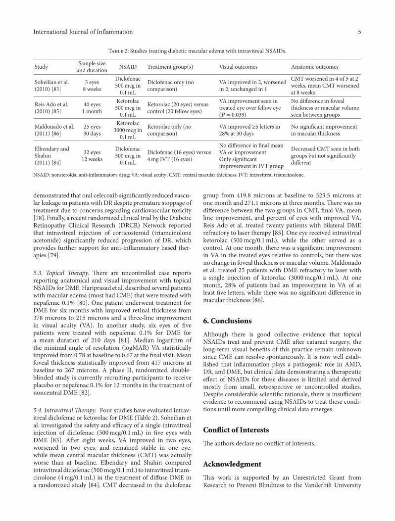
International �ournal of In�ammation 5
T 2: Studies treating diabetic macular edema with intravitreal NSAIDs.
Study Sample sizeand duration NSAID Treatment group(s) Visual outcomes Anatomic outcomes
Soheilian et al.(2010) [83]
5 eyes8 weeks
Diclofenac500mcg in
0.1mL
Diclofenac only (nocomparison)
VA improved in 2, worsenedin 2, unchanged in 1
CMT worsened in 4 of 5 at 2weeks, mean CMT worsenedat 8 weeks
Reis Ado et al.(2010) [85]
40 eyes1 month
Ketorolac500mcg in
0.1mL
Ketorolac (20 eyes) versuscontrol (20 fellow eyes)
VA improvement seen intreated eye over fellow eye(𝑃𝑃 𝑃 𝑃𝑃𝑃𝑃𝑃)
No difference in fovealthickness or macular volumeseen between groups
Maldonado et al.(2011) [86]
25 eyes30 days
Ketorolac3000mcg in
0.1mL
Ketorolac only (nocomparison)
VA improved ≥5 letters in28% at 30 days
No signi�cant improvementin macular thickness
Elbendary andShahin(2011) [84]
32 eyes12 weeks
Diclofenac500mcg in
0.1mL
Diclofenac (16 eyes) versus4mg IVT (16 eyes)
No difference in �nal meanVA or improvementOnly signi�cantimprovement in IVT group
Decreased CMT seen in bothgroups but not signi�cantlydifferent
NSAID: nonsteroidal anti-in�ammatory drug� VA: visual acuity� CMT: central macular thickness� IVT: intravitreal triamcinolone.
demonstrated that oral celecoxib signi�cantly reduced vascu-lar leakage in patients with DR despite premature stoppage oftreatment due to concerns regarding cardiovascular toxicity[78]. Finally, a recent randomized clinical trial by theDiabeticRetinopathy Clinical Research (DRCR) Network reportedthat intravitreal injection of corticosteroid (triamcinoloneacetonide) signi�cantly reduced progression of DR, whichprovides further support for anti-in�ammatory based ther-apies [79].
5.3. Topical erapy. ere are uncontrolled case reportsreporting anatomical and visual improvement with topicalNSAIDs forDME.Hariprasad et al. described several patientswith macular edema (most had CME) that were treated withnepafenac 0.1% [80]. One patient underwent treatment forDME for six months with improved retinal thickness from378 microns to 215 microns and a three-line improvementin visual acuity (VA). In another study, six eyes of �vepatients were treated with nepafenac 0.1% for DME fora mean duration of 210 days [81]. Median logarithm ofthe minimal angle of resolution (logMAR) VA statisticallyimproved from 0.78 at baseline to 0.67 at the �nal visit. Meanfoveal thickness statistically improved from 417 microns atbaseline to 267 microns. A phase II, randomized, double-blinded study is currently recruiting participants to receiveplacebo or nepafenac 0.1% for 12 months in the treatment ofnoncentral DME [82].
5.4. Intravitreal erapy. Four studies have evaluated intrav-itreal diclofenac or ketorolac for DME (Table 2). Soheilian etal. investigated the safety and efficacy of a single intravitrealinjection of diclofenac (500mcg/0.1mL) in �ve eyes withDME [83]. Aer eight weeks, VA improved in two eyes,worsened in two eyes, and remained stable in one eye,while mean central macular thickness (CMT) was actuallyworse than at baseline. Elbendary and Shahin comparedintravitreal diclofenac (500mcg/0.1mL) to intravitreal triam-cinolone (4mg/0.1mL) in the treatment of diffuse DME ina randomized study [84]. CMT decreased in the diclofenac
group from 419.8 microns at baseline to 323.5 microns atone month and 271.1 microns at three months. ere was nodifference between the two groups in CMT, �nal VA, meanline improvement, and percent of eyes with improved VA.Reis Ado et al. treated twenty patients with bilateral DMErefractory to laser therapy [85]. One eye received intravitrealketorolac (500mcg/0.1mL), while the other served as acontrol. At one month, there was a signi�cant improvementin VA in the treated eyes relative to controls, but there wasno change in foveal thickness ormacular volume.Maldonadoet al. treated 25 patients with DME refractory to laser witha single injection of ketorolac (3000mcg/0.1mL). At onemonth, 28% of patients had an improvement in VA of atleast �ve letters, while there was no signi�cant difference inmacular thickness [86].
6. Conclusions
Although there is good collective evidence that topicalNSAIDs treat and prevent CME aer cataract surgery, thelong-term visual bene�ts of this practice remain unknownsince CME can resolve spontaneously. It is now well estab-lished that in�ammation plays a pathogenic role in AMD,DR, and DME, but clinical data demonstrating a therapeuticeffect of NSAIDs for these diseases is limited and derivedmostly from small, retrospective or uncontrolled studies.Despite considerable scienti�c rationale, there is insufficientevidence to recommend using NSAIDs to treat these condi-tions until more compelling clinical data emerges.
Con�ict o� �nte�ests
e authors declare no con�ict of interests.
Acknowledgment
is work is supported by an Unrestricted Grant fromResearch to Prevent Blindness to the Vanderbilt University
6 International Journal of In�ammation
School of Medicine, Department of Ophthalmology andVisual Sciences.
References
[1] S. J. Kim, A. J. Flach, and L. M. Jampol, “Nonsteroidal anti-in�ammatory drugs in ophthalmology,” Survey of Ophthalmol-ogy, vol. 55, no. 2, pp. 108–133, 2010.
[2] H. N. Shelsta and L. M. Jampol, “Pharmacologic therapy ofpseudophakic cystoidmacular edema: 2010 update,”Retina, vol.31, no. 1, pp. 4–12, 2011.
[3] D. L. Simmons, R. M. Botting, and T. Hla, “Cyclooxygenaseisozymes: the biology of prostaglandin synthesis and inhibi-tion,” Pharmacological Reviews, vol. 56, no. 3, pp. 387–437,2004.
[4] N. M. Davies, R. L. Good, K. A. Roupe, and J. A. Yáñez, “Cyclo-oxygenase-3: axiom, dogma, anomaly, enigma or spliceerror?—not as easy as 1, 2, 3,” Journal of Pharmacy andPharmaceutical Sciences, vol. 7, no. 2, pp. 217–226, 2004.
[5] M. S. Chin, C. N. Nagineni, L. C. Hooper, B. Detrick, and J.J. Hooks, “Cyclooxygenase-2 gene expression and regulation inhuman retinal pigment epithelial cells,” Investigative Ophthal-mology and Visual Science, vol. 42, no. 10, pp. 2338–2346, 2001.
[6] S. Shinomiya, H. Naraba, A. Ueno et al., “Regulation ofTNF𝛼𝛼 and interleukin-10 production by prostaglandins I2 andE2: studies with prostaglandin receptor-de�cient mice andprostaglandin E-receptor subtype-selective synthetic agonists,”Biochemical Pharmacology, vol. 61, no. 9, pp. 1153–1160, 2001.
[7] T. Cheng, W. Cao, R. Wen, R. H. Steinberg, and M. M. LaVail,“Prostaglandin E2 induces vascular endothelial growth factorand basic �broblast growth factormRNAexpression in culturedratMuller cells,” Investigative Ophthalmology andVisual Science,vol. 39, no. 3, pp. 581–591, 1998.
[8] R. Reddy and S. J. Kim, “Critical appraisal of ophthalmic keto-rolac in the treatment of pain and in�ammation followingcataract surgery,” Clinical Ophthalmology, vol. 5, pp. 751–758,2011.
[9] M. Ahuja, A. S. Dhake, S. K. Sharma, and D. K. Majumdar,“Topical ocular delivery of NSAIDs,”eAAPS Journal, vol. 10,no. 2, pp. 229–241, 2008.
[10] D. Riendeau, S. Charleson, W. Cromlish, J. A. Mancini, E.Wong, and J. Guay, “Comparison of the cyclooxygenase-1inhibitory properties of nonsteroidal anti-in�ammatory drugs(NSAIDs) and selective COX-2 inhibitors, using sensitivemicrosomal and platelet assays,”Canadian Journal of Physiologyand Pharmacology, vol. 75, no. 9, pp. 1088–1095, 1997.
[11] D. A. Gamache, G. Graff, M. T. Brady, J. M. Spellman, and J. M.Yanni, “Nepafenac, a unique nonsteroidal prodrug with poten-tial utility in the treatment of trauma-induced ocular in�am-mation: I. Assessment of anti- in�ammatory e�cacy,” In�am-mation, vol. 24, no. 4, pp. 357–370, 2000.
[12] L. D. Waterbury, D. Silliman, and T. Jolas, “Comparisonof cyclooxygenase inhibitory activity and ocular anti-in�ammatory effects of ketorolac tromethamine and bromfenacsodium,” Current Medical Research and Opinion, vol. 22, no. 6,pp. 1133–1140, 2006.
[13] T. Walters, M. Raizman, P. Ernest, J. Gayton, and R. Lehmann,“In vivo pharmacokinetics and in vitro pharmacodynamicsof nepafenac, amfenac, ketorolac, and bromfenac,” Journal ofCataract and Refractive Surgery, vol. 33, no. 9, pp. 1539–1545,2007.
[14] P. P. Ellis, D. S. Pfoff, D. C. Bloedow, and M. Riegel, “Intraoculardiclofenac and �urbiprofen concentrations in human aqueoushumor following topical application,” Journal of Ocular Phar-macology, vol. 10, no. 4, pp. 677–682, 1994.
[15] M. Attar, R. Schiffman, L. Borbridge, Q. Farnes, and D. Welty,“Ocular pharmacokinetics of 0.45% ketorolac tromethamine,”Clinical Ophthalmology, vol. 4, no. 1, pp. 1403–1408, 2010.
[16] F. A. Bucci Jr., L. D. Waterbury, and L. M. Amico, “Pros-taglandin E2 inhibition and aqueous concentration of ketorolac0.4% (Acular LS) and nepafenac 0.1% (Nevanac) in patientsundergoing phacoemulsi�cation,” American Journal of Oph-thalmology, vol. 144, no. 1, pp. 146–147, 2007.
[17] J. S. Heier, C. C. Awh, B. G. Busbee et al., “Vitreous nonsteroidalantiin�ammatory drug concentrations and prostaglandin E2levels in vitrectomy patients treated with ketorolac 0.4%, brom-fenac 0.09%, and nepafenac 0.1%,” Retina, vol. 29, no. 9, pp.1310–1313, 2009.
[18] S. R. Irvine, “A newly de�ned vitreous syndrome followingcataract surgery,” American Journal of Ophthalmology, vol. 36,no. 5, pp. 499–619, 1953.
[19] S. J. Kim, R. Equi, and N. M. Bressler, “Analysis of macularedema aer cataract surgery in patients with diabetes usingoptical coherence tomography,” Ophthalmology, vol. 114, no. 5,pp. 881–889, 2007.
[20] C. L. Lobo, P. M. Faria, M. A. Soares, R. C. Bernardes, and J. G.Cunha-Vaz, “Macular alterations aer small-incision cataractsurgery,” Journal of Cataract and Refractive Surgery, vol. 30, no.4, pp. 752–760, 2004.
[21] P. G. Ursell, D. J. Spalton, S. M. Whitcup, and R. B. Nussenblatt,“Cystoidmacular edema aer phacoemulsi�cation: relationshipto blood- aqueous barrier damage and visual acuity,” Journal ofCataract and Refractive Surgery, vol. 25, no. 11, pp. 1492–1497,1999.
[22] K. A. Warren, H. Bahrani, and J. E. Fox, “NSAIDs in combina-tion therapy for the treatment of chronic pseudophakic cystoidmacular edema,” Retina, vol. 30, no. 2, pp. 260–266, 2010.
[23] S. M. Hariprasad, L. Akduman, J. A. Clever, M. Ober, F. M.Recchia, and W. F. Mieler, “Treatment of cystoid macularedema with the new-generation NSAID nepafenac 0.1%,” Clin-ical Ophthalmology, vol. 3, no. 1, pp. 147–154, 2009.
[24] A. J. Flach, R. C. Stegman, J. Graham, and L. P. Kruger, “Pro-phylaxis of aphakic cystoid macular edema without corti-costeroids: a paired-comparison, placebo-controlled double-masked study,” Ophthalmology, vol. 97, no. 10, pp. 1253–1258,1990.
[25] K. Miyake, K. Masuda, S. Shirato et al., “Comparison ofdiclofenac and �uorometholone in preventing cystoid macularedema aer small incision cataract surgery: a multicenteredprospective trial,” Japanese Journal of Ophthalmology, vol. 44,no. 1, pp. 58–67, 2000.
[26] J. R. Wittpenn, S. Silverstein, J. Heier, K. R. Kenyon, J. D.Hunkeler, and M. Earl, “A randomized, masked comparison oftopical ketorolac 0.4% plus steroid vs. steroid alone in low-riskcataract surgery patients,” American Journal of Ophthalmology,vol. 146, no. 4, pp. 554–560, 2008.
[27] S. J. Kim and N. M. Bressler, “Optical coherence tomographyand cataract surgery,” Current Opinion in Ophthalmology, vol.20, no. 1, pp. 46–51, 2009.
[28] A. J. Flach, M. C. Kraff, D. R. Sanders, and L. Tanenbaum, “equantitative effect of 0.5% ketorolac tromethamine solution and

International Journal of In�ammation 7
0.1% dexamethasone sodium phosphate solution on postsurgi-cal blood-aqueous barrier,”Archives of Ophthalmology, vol. 106,no. 4, pp. 480–483, 1988.
[29] S. J. Kim, W. R. Lo, G. B. Hubbard et al., “Topical ketorolacin vitreoretinal surgery: a prospective, randomized, placebo-controlled, double-masked trial,” Archives of Ophthalmology,vol. 126, no. 9, pp. 1203–1208, 2008.
[30] S. D. Schoenberger, D. M. Miller, M. R. Petersen, R. E. Fos-ter, C. D. Riemann, and R. A. Sisk, “Nepafenac for epireti-nal membrane surgery,” Ophthalmology, vol. 118, no. 7, pp.1482.e1–1482.e3, 2011.
[31] P. Naithani, S. Puranik, N. Vashisht, S. Khanduja, S. Kumar, andS. Garg, “Role of topical nepafenac in prevention and treatmentofmacular edema aer vitreoretinal surgery,”Retina, vol. 32, no.2, pp. 250–255, 2012.
[32] D. S. Friedman, B. J. O’Colmain, B. Munoz et al., “Prevalence ofage-relatedmacular degeneration in theUnited States,”Archivesof Ophthalmology, vol. 122, no. 4, pp. 564–572, 2004.
[33] G. C. Brown,M.M. Brown, S. Sharma et al., “e burden of age-relatedmacular degeneration: a value-basedmedicine analysis,”Transactions of the AmericanOphthalmological Society, vol. 103,pp. 173–186, 2005.
[34] J. C. Folk and E. M. Stone, “Ranibizumab therapy for neovascu-lar age-related macular degeneration,” New England Journal ofMedicine, vol. 363, no. 17, pp. 1648–1655, 2010.
[35] P. A. Yourey, S. Gohari, J. L. Su, and R. F. Alderson, “Vascularendothelial cell growth factors promote the in vitro develop-ment of rat photoreceptor cells,” Journal of Neuroscience, vol. 20,no. 18, pp. 6781–6788, 2000.
[36] M. A. Zarbin, “Current concepts in the pathogenesis of age-related macular degeneration,” Archives of Ophthalmology, vol.122, no. 4, pp. 598–614, 2004.
[37] M. Patel and C. C. Chan, “Immunopathological aspects of age-related macular degeneration,” Seminars in Immunopathology,vol. 30, no. 2, pp. 97–110, 2008.
[38] S. C. Maloney, B. F. Fernandes, E. Castiglione et al., “Expressionof cyclooxygenase-2 in choroidal neovascular membranes fromage-related macular degeneration patients,” Retina, vol. 29, no.2, pp. 176–180, 2009.
[39] Y. Monnier, J. Zaric, and C. Rüegg, “Inhibition of angiogenesisby non-steroidal anti-in�ammatory drugs: from the bench tothe bedside and back,” Current �rug Targets� In�ammation andAllergy, vol. 4, no. 1, pp. 31–38, 2005.
[40] S. Gately and R. Kerbel, “erapeutic potential of selectivecyclooxygenase-2 inhibitors in the management of tumorangiogenesis,” Progress in Experimental Tumor Research, vol. 37,pp. 179–192, 2003.
[41] W. K. K. Wu, J. J. Sung, C. W. Lee, J. Yu, and C. H. Cho,“Cyclooxygenase-2 in tumorigenesis of gastrointestinal cancers:an update on the molecular mechanisms,” Cancer Letters, vol.295, no. 1, pp. 7–16, 2010.
[42] S. E. Yanni, J. M. Barnett, M. L. Clark, and J. S. Penn, “erole of PGE2 receptor EP4 in pathologic ocular angiogenesis,”Investigative Ophthalmology and Visual Science, vol. 50, no. 11,pp. 5479–5486, 2009.
[43] A. C. Amrite, S. P. Ayalasomayajula, N. P. S. Cheruvu, and U.B. Kompella, “Single periocular injection of celecoxib-PLGAmicroparticles inhibits diabetes-induced elevations in retinalPGE2, VEGF, and vascular leakage,” Investigative Ophthalmol-ogy and Visual Science, vol. 47, no. 3, pp. 1149–1160, 2006.
[44] K. Takahashi, Y. Saishin, Y. Saishin et al., “Topical nepafenacinhibits ocular neovascularization,” Investigative Ophthalmol-ogy and Visual Science, vol. 44, no. 1, pp. 409–415, 2003.
[45] S. J. Kim, H. S. Toma, J. M. Barnett, and J. S. Penn, “Ketorolacinhibits choroidal neovascularization by suppression of retinalVEGF,” Experimental Eye Research, vol. 91, no. 4, pp. 537–543,2010.
[46] S. J. Kim and H. S. Toma, “Inhibition of choroidal neovascu-larization by intravitreal ketorolac,” Archives of Ophthalmology,vol. 128, no. 5, pp. 596–600, 2010.
[47] K. A. Rezaei, H. S. Toma, J. Cai, J. S. Penn, P. Sternberg, and S. J.Kim, “Reduced choroidal neovascular membrane formation incyclooxygenase-2 null mice,” Investigative Ophthalmology andVisual Science, vol. 52, no. 2, pp. 701–707, 2011.
[48] W. Hu, M. H. Criswell, A. Ottlecz et al., “Oral administration oflumiracoxib reduces choroidal neovascularmembrane develop-ment in the rat laser-trauma model,” Retina, vol. 25, no. 8, pp.1054–1064, 2005.
[49] H. Takahashi, Y. Yanagi, Y. Tamaki, S. Uchida, and K. Muran-aka, “COX-2-selective inhibitor, etodolac, suppresses choroidalneovascularization in a mice model,” Biochemical and Biophysi-cal Research Communications, vol. 325, no. 2, pp. 461–466, 2004.
[50] P. L. McGeer and J. Sibley, “Sparing of age-related maculardegeneration in rheumatoid arthritis,” Neurobiology of Aging,vol. 26, no. 8, pp. 1199–1203, 2005.
[51] H. L. Wilson, D. M. Schwartz, H. R. F. Bhatt, C. E. McCulloch,and J. L. Duncan, “Statin and aspirin therapy are associated withdecreased rates of choroidal neovascularization among patientswith age-related macular degeneration,” American Journal ofOphthalmology, vol. 137, no. 4, pp. 615–624, 2004.
[52] J. J. Wang, P. Mitchell, W. Smith, M. Gillies, F. Billson, andBlue Mountains Eye Study, “Systemic use of anti-in�ammatorymedications and age-related maculopathy: the Blue MountainsEye Study,” Ophthalmic Epidemiology, vol. 10, no. 1, pp. 37–48,2003.
[53] S. A. Zweifel, M. Engelbert, S. Khan, and K. B. Freund, “Retro-spective review of the efficacy of topical bromfenac (0.09%) asan adjunctive therapy for patients with neovascular age-relatedmacular degeneration,” Retina, vol. 29, no. 10, pp. 1527–1531,2009.
[54] E. Chen, M. S. Benz, R. H. Fish et al., “Use of nepafenac(Nevanac)in combination with intravitreal anti-VEGF agentsin the treatment of recalcitrant exudative macular degenerationrequiring monthly injections,” Clinical Ophthalmology, vol. 4,no. 1, pp. 1249–1252, 2010.
[55] AdjunctiveDiclofenacwithVertepor�n (ADD-V) StudyGroup,D. S. Boyer, P.M. Beer et al., “Effect of adjunctive diclofenacwithvertepor�n therapy to treat choroidal neovascularization due toage-related macular degeneration: phase II study,” Retina, vol.27, no. 6, pp. 693–700, 2007.
[56] C. A. Grant, “Combination therapy: lucentis (ranibizumabinjection) and xibrom (bromfenac ophthalmic solution) 0. 09%in the treatment of chroidal neovascular membrane secondaryto age-related macular degeneration,” Investigative Ophthalmol-ogy & Visual Science, vol. 49, 2008, E-abstract 563.
[57] C. Flaxel,M. B. Schain, S. C. Hamon, and P. J. Francis, “Prospec-tive randomized controlled trial of combination ranibizumab(Lucentis) and bromfenac (Xibrom) for neovascular age-relatedmacular degeneration,” Retina, vol. 32, no. 3, pp. 417–423, 2012.
[58] F. Gomi, M. Sawa, M. Tsujikawa, and K. Nishida, “Topi-cal bromfenac as an adjunctive treatment with intravitreal
8 International Journal of In�ammation
ranibizumab for exudative age-related macular degeneration,”Retina, vol. 32, no. 9, pp. 1804–1810, 2012.
[59] D. A. Antonetti, R. Klein, and T. W. Gardner, “Diabetic ret-inopathy,” New England Journal of Medicine, vol. 366, no. 13,pp. 1227–1239, 2012.
[60] N. Bhagat, R. A. Grigorian, A. Tutela, and M. A. Zarbin,“Diabetic macular edema: pathogenesis and treatment,” Surveyof Ophthalmology, vol. 54, no. 1, pp. 1–32, 2009.
[61] “Progression of retinopathy with intensive versus conventionaltreatment in the Diabetes Control and Complications Trial.Diabetes Control and Complications Trial Research Group,”Ophthalmology, vol. 102, no. 4, pp. 647–661, 1995.
[62] “Tight blood pressure control and risk of macrovascular andmicrovascular complications in type 2 diabetes: UKPDS 38.United Kingdom Prospective Diabetes Study Group,” BritishMedical Journal, vol. 317, no. 7160, pp. 703–713, 1998.
[63] A. P. Adamis and A. J. Berman, “Immunological mecha-nisms in the pathogenesis of diabetic retinopathy,” Seminars inImmunopathology, vol. 30, no. 2, pp. 65–84, 2008.
[64] C. A. Lange, P. Stavrakas, U. F. Luhmann et al., “Intraocularoxygen distribution in advanced proliferative diabetic retinopa-thy,” American Journal of Ophthalmology, vol. 152, no. 3, pp.406–412, 2011.
[65] Y. Suzuki, M. Nakazawa, K. Suzuki, H. Yamazaki, and Y.Miyagawa, “Expression pro�les of cytokines and chemokinesin vitreous �uid in diabetic retinopathy and central retinal veinocclusion,” Japanese Journal of Ophthalmology, vol. 55, no. 3, pp.256–263, 2011.
[66] J. Zhou, S. Wang, and �. �ia, “Role of intravitreal in�amma-tory cytokines and angiogenic factors in proliferative diabeticretinopathy,” Current Eye Research, vol. 37, no. 5, pp. 416–420,2012.
[67] S. D. Schoenberger, S. J. Kim, J. Sheng, K. A. Rezaei, M.Lalezary, and E. Cherney, “Increased prostaglandin E2 (PGE2)levels in proliferative diabetic retinopathy and correlation withVEGF and in�ammatory cytokines,” Investigative Ophthalmol-ogy & Visual Science, vol. 53, no. 9, pp. 5906–5911, 2012.
[68] T. Cheng, W. Cao, R. Wen, R. H. Steinberg, and M. M. LaVail,“Prostaglandin E2 induces vascular endothelial growth factorand basic �broblast growth factormRNAexpression in culturedratMuller cells,” Investigative Ophthalmology andVisual Science,vol. 39, no. 3, pp. 581–591, 1998.
[69] L. P. Aiello, R. L. Avery, P. G. Arrigg et al., “Vascular endothe-lial growth factor in ocular �uid of patients with diabeticretinopathy and other retinal disorders,” New England Journalof Medicine, vol. 331, no. 22, pp. 1480–1487, 1994.
[70] E. I. M. Johnson, M. E. Dunlop, and R. G. Larkins, “Increasedvasodilatory prostaglandin production in the diabetic rat retinalvasculature,” Current Eye Research, vol. 18, no. 2, pp. 79–82,1999.
[71] T. S. Kern, C. M. Miller, Y. Du et al., “Topical administrationof nepafenac inhibits diabetes-induced retinal microvasculardisease and underlying abnormalities of retinalmetabolism andphysiology,” Diabetes, vol. 56, no. 2, pp. 373–379, 2007.
[72] S. P. Ayalasomayajula and U. B. Kompella, “Celecoxib, aselective cyclooxygenase-2 inhibitor, inhibits retinal vascularendothelial growth factor expression and vascular leakage in astreptozotocin-induced diabetic ratmodel,”European Journal ofPharmacology, vol. 458, no. 3, pp. 283–289, 2003.
[73] A. M. Joussen, V. Poulaki, N. Mitsiades et al., “Nonsteroidalanti-in�ammatory drugs prevent early diabetic retinopathy via
TNF-alpha suppression,” e FASEB Journal, vol. 16, no. 3, pp.438–440, 2002.
[74] E. D. Powell andR. Field, “Diabetic retinopathy and rheumatoidarthritis,” e Lancet, vol. 284, no. 7349, pp. 17–18, 1964.
[75] “Effects of aspirin treatment on diabetic retinopathy. ETDRSreport number 8. Early Treatment Diabetic Retinopathy StudyResearchGroup,”Ophthalmology, vol. 98, no. 5, supplement, pp.757–765, 1991.
[76] “Effect of aspirin alone and aspirin plus dipyridamole in earlydiabetic retinopathy. A multicenter randomized controlledclinical trial. e DAMAD Study Group,” Diabetes, vol. 38, no.4, pp. 491–498, 1989.
[77] Y. Hattori, K. Hashizume, K. Nakajima, Y. Nishimura, M. Naka,and K. Miyanaga, “e effect of long-term treatment withsulindac on the progression of diabetic retinopathy,” CurrentMedical Research and Opinion, vol. 23, no. 8, pp. 1913–1917,2007.
[78] E. Y. Chew, J. Kim, H. R. Coleman et al., “Preliminary assess-ment of celecoxib and microdiode pulse laser treatment ofdiabetic macular edema,” Retina, vol. 30, no. 3, pp. 459–467,2010.
[79] N. M. Bressler, A. R. Edwards, R. W. Beck et al., “Exploratoryanalysis of diabetic retinopathy progression through 3 years ina randomized clinical trial that compares intravitreal triamci-nolone acetonide with focal/grid photocoagulation,”Archives ofOphthalmology, vol. 127, no. 12, pp. 1566–1571, 2009.
[80] S. M. Hariprasad, D. Callanan, S. Gainey, Y. G. He, and K.Warren, “Cystoid and diabetic macular edema treated withnepafenac 0.1%,” Journal of Ocular Pharmacology and era-peutics, vol. 23, no. 6, pp. 585–589, 2007.
[81] D. Callanan and P. Williams, “Topical nepafenac in the treat-ment of diabetic macular edema,” Clinical Ophthalmology, vol.2, no. 4, pp. 689–692, 2008.
[82] July 2012, http://clinicaltrials.gov/ct2/show/NCT01331005.[83] M. Soheilian, S. Karimi, A. Ramezani, and G. A. Peyman, “Pilot
study of intravitreal injection of diclofenac for treatment ofmacular edema of various etiologies,” Retina, vol. 30, no. 3, pp.509–515, 2010.
[84] A. M. Elbendary and M. M. Shahin, “Intravitreal diclofenacversus intravitreal triamcinolone acetonide in the treatment ofdiabetic macular edema,” Retina, vol. 31, no. 10, pp. 2058–2064,2011.
[85] C. Reis Ado, R. N. Vianna, R. S. Reis, and G. P. Cardoso,“Intravitreal injection of ketorolac tromethamine in patientswith diabetic macular edema refractory to retinal photocoag-ulation,” Arquivos Brasileiros de Oalmologia, vol. 73, no. 4, pp.338–342, 2010.
[86] R. M. Maldonado, R. N. G. Vianna, G. P. Cardoso, A. V. deMagalhães, and M. N. Burnier Jr., “Intravitreal injection ofcommercially available ketorolac tromethamine in eyes withdiabetic macular edema refractory to laser photocoagulation,”Current Eye Research, vol. 36, no. 8, pp. 768–773, 2011.