influence dichloromethylene vitro phagocytosis rat ... · thein vitro phagocytosis...
TRANSCRIPT

Annals of the Rheumatic Diseases 1992; 51: 203-209
Influence of dichloromethylene bisphosphonate on
the in vitro phagocytosis of hydroxyapatite particlesby rat peritoneal exudate cells: an electronmicroscopic and chemiluminescence study
P M Hyvonen, M J Kowolik
AbstractTransmission electron microscopy andstandard chemiluminescence assays wereused to investigate the in vivo effect ofdichloromethylene bisphosphonate (clodron-ate) on the phagocytosis ofpure hydroxyapatiteparticles by rat peritoneal macrophages andthe production of chemiluminescence by theperitoneal exudate celis. Hydroxyapatite(control) and a hydroxyapatite/clodronate sus-pension (28 imol clodronate per gram ofhydroxyapatite, experimental) were injectedinto the peritoneum of rats, the clodronatedose being 10 ,tg/kg. Macrophages were har-vested at 12, 24, 48, and 96 hours afterinjection and the particle phagocytosis wasassessed by transmission electron microscopy.Hydroxyapatite alone was completely phago-cytosed by 24 hours and hydroxyapatite reactedwith clodronate was completely phagocytosedby 48 hours. From 48 hours onwards hydroxy-apatite particle dissolution was observed inthe phagosomes of cells in the two groups. At48 hours the chemiluminescence produced bythe peritoneal exudate cells was also measured.Clodronate and clodronate/hydroxyapatiteenhanced cell activity on subsequent challengewith phorbol myristate acetate or zymosan.Clodronate seemed to exhibit an inhibitoryeffect on the phagocytic activity and anenhancement of the chemiluminescenceproduction by the cells in this model, indicatingthat it was modifying the inflammatory cellresponse.
Faculty of Dentistry,University of Kuopio,FinlandP M HyvonenUniversity of LeedsDental School, Leeds,United KingdomM J Kowolik
Correspondence to:Dr Pekka M Hyvonen,Faculty of Dentistry,University of Kuopio,POB 1627, SF-70211 Kuopio,Finland.
Accepted for publication12 February 1991
Geminal bisphosphonates are a group of drugsof particular interest because of their ability tomodify calcium metabolism. This effect resultsfrom a decrease in the solubility ofhydroxyapatitein vitro and by cell mediated mechanisms invivo.' 2 In clinical treatment, bisphosphonateshave been used to treat metastatic bone disease,3hypercalcaemia due to malignancy,4 5 andPaget's disease.6 At the cellular level, the modeof action is not fully understood, but it is knownthat one of the target cells is the osteoclast,towards which dichloromethylene bisphos-phonate (clodronate), at least, is toxic.2 Clod-ronate is highly soluble in water and shows astrong affinity for calcified tissue.7 In an attemptto clarify the pharmacological mechanismsoperating, a number of experimental modelshave been used.8 It has been shown in an in vivoexperiment that clodronate also has anti-inflammatory properties.9
Hydroxyapatite crystals have been found inthe joint effusions of patients with rheumatoidarthritis,'0 and macrophages remove thesecrystals from the joint cavities." Macrophages,in general, are immunoregulatory and inflam-matory cells, the latter function being charac-terised by their phagocytic capacity. Phago-cytosis is the primordial function of neutrophilgranulocytes and the two cell types comprise thehost's phagocytes. They have been found in theperitoneal cell exudate of many laboratoryanimals.'2 The membrane stimulation ofinflammatory phagocytes can be measured bychemiluminescence, that is, the light generatedby the oxygen dependent microbicidal system.The chemiluminescence assay is a measure ofthe rate of liberation of reactive oxygen speciesby macrophages and granulocytes; these reactiveoxygen species may contribute to tissue damagein inflammatory diseases.'3 Different pathwaysof light production can be identified: themyeloperoxide mediated reaction can be identi-fied by stimulating cells with zymosan andusing luminol as an intermediate; superoxideanion dependent chemiluminescence can bemeasured by stimulating cells with phorbolmyristate acetate and augmenting the lightoutput with lucigenin.'4 Neutrophil granulo-cytes in most animal species can be stronglyactivated through these two pathways, whereasmonocytes and macrophages have little or nomyeloperoxidase.On the basis of the reports summarised
above, we decided to investigate, by transmissionelectron microscopy, whether the simultaneouspresence of clodronate with pure hydroxyapatiteparticles influences the phagocytosis of these byrat peritoneal macrophages in vivo. We alsowanted to see whether, in this experimentalsystem, there would be an effect on the liberationof reactive oxygen species by these cells, asmeasured by chemiluminescence.
Materials and methodsHYDROXYAPATITE AND CLODRONATEChemically pure, synthetically manufacturedhydroxyapatite (British Charcoals and Mac-Donald) with a mean particle size of 5 ,tm wasused.
Clodronate (Leiras Pharmaceuticals) was dis-solved in calcium and magnesium free phosphatebuffered saline (PBS), pH 7-2, at a workingconcentration of 2 mg/ml. A hydroxyapatite/clodronate combination was prepared by allow-
203
on January 30, 2020 by guest. Protected by copyright.
http://ard.bmj.com
/A
nn Rheum
Dis: first published as 10.1136/ard.51.2.203 on 1 F
ebruary 1992. Dow
nloaded from
Hyvonen, Kowolik
ing the two compounds to react in the ratio of 1g hydroxyapatite to 28 Ftmol clodronate for aminimum of 42 hours.7
EXPERIMENTAL ANIMALSHealthy, adult Wistar SPF rats, in groups of 25(males, transmission electron microscopyexperiments) and 22 (eight males and 14 females,chemiluminescence experiments) animals wereused.
EXPERIMENTAL PROTOCOLThe peritoneal cavity of each experimentalanimal was injected according to one of thefollowing protocols: (a) PBS, control group; (b)clodronate alone, drug concentration as above,dose 10 mg/kg body weight; (c) hydroxyapatitesuspension alone at 84-55 mg/ml; (d) hydroxy-apatite/clodronate combination giving the sameconcentrations of agents as in b and c.
Intraperitoneal incubation was allowed toproceed for 12, 24, 48, 72, or 94 hours.The chemiluminescence experiments were
performed at 48 hours after injection only. Inaddition, the drug at 30 mg/kg alone and thehydroxyapatite/clodronate combination weretested for their influence on chemiluminescence.The volume injected was consistently 10 mg/kg.
HARVESTING AND COLLECTION OF PERITONEALCELLSThe animals were anaesthetised with 6 1%thiopental solution (Hypnostan, Leiras), theminimum dose being 2 ml. Abdominal skin andmuscles were reflected and the peritoneal wallexposed. One catheter (Venflon No 2, Viggo)was inserted into the peritoneal cavity at thegroin and another symmetrically on the oppositeside. Via one catheter, 20 ml of sterile PBS wasinjected into the peritoneum and after twominutes of gentle massage the stopper wasremoved from the opposite catheter. The fluidwas allowed to drip into a 50 ml polyethylenetube (Costar No 3350) in ice. PBS was addedcontinuously via the first catheter and a total of50-100 ml of lavage fluid was collected. Thiswas centrifuged at 380 g for 10 minutes, afterwhich any red blood cells were removed byhypotonic lysis with 0-87% ammonium chloridesolution for 10 minutes. After further centri-fugation the cells were resuspended in PBS andcounted in a Coulter counter prior to theassessment of cell viability by trypan blue dyeexclusion.The animals were killed after cell harvesting
by cervical dislocation. All animal procedureswere in compliance with the Declaration ofHelsinki.
PREPARATION OF SPECIMENS FOR TRANSMISSION
ELECTRON MICROSCOPYA cell pellet was formed by centrifugation at 380g for 10 minutes, and prefixed in 2% glutaralde-hyde solution for two hours. The pellet wasthen washed for 10 minutes in PBS andpostfixed in 1% osmium tetroxide for one hour.
Thereafter the pellet was washed and dehydratedin increasing concentrations of alcohol andfinally in propylene oxide. The specimen wasthen embedded in epoxy resin (LX-1 12, LaddResearch Industries) by a standard procedure.Ultrathin sections were cut with an LKBUltramicrotome and stained with Ultrostain 1(9001 3710) for 20 minutes and Ultrostain 2(9001 3711) for two minutes, at room tempera-ture (both stains from LKB). Finally, thespecimens were mounted on copper grids,sputtered with carbon and viewed with a JeolJEM- 1200EX transmission electron microscope.For each experiment at each time point a
minimum of 50 fields was examined to assesscell morphology and phagocytosis.
MEASUREMENT OF CHEMILUMINESCENCEThe concentration of cells was adjusted to1 x 107 cells per ml for the chemiluminescenceexperiments.To test the activation of the superoxide anion
pathway, 500 ,ul of cell suspension was placed inthe reaction cuvette, followed by 200 RI lucigenin(I X 10-3 M, Sigma Chemicals) and 200 ,ulphorbol myristate acetate as stimulant (2 5[ig/ml, Sigma Chemicals).The resultant chemiluminescence was
measured at 37°C in an LKB 1250 luminometer,recorded as millivolts and calculated per 1 x 10⁶cells. From the curves generated the maximumpotential was recorded. In addition, the areaunder the curve was quantitated between thestart of the experiment and the point at whichthe potential had declined to 50% of the peakvalue. This was achieved by digitising the datafrom the curves using a Kontron image analysissystem (IBAS 1).To assess any contribution to the production
of chemiluminescence by the myeloperoxidasemediated pathway, 200 RI luminol (5 x 10⁻⁴ M)and 200 1t serum treated zymosan (10 mg/ml)were added to the reaction vial.The two chemiluminescence systems were
studied alone and consecutively in the cell batchexperiment. For example, luminol and zymosanwere added to the reaction cuvette after thechemiluminescence produced by the lucigenin/phorbol myristate acetate regimen had subsidedto 50% of the peak potential and vice versa. Atleast one duplicate experiment was conductedon cells collected from each animal.
STATISTICAL ANALYSISStudent's t test was used for statistical analysis.
ResultsCell viability in the lavage fluid was consistentlygreater than 96%, as determined by the trypanblue dye exclusion test.
TRANSMISSION ELECTRON MICROSCOPYEXPERIMENTSPhagocytosis and morphology were assessedonly for the macrophages. These made upapproximately 90% of the exudate cells, the restof the cells being mainly granulocytes.
204
on January 30, 2020 by guest. Protected by copyright.
http://ard.bmj.com
/A
nn Rheum
Dis: first published as 10.1136/ard.51.2.203 on 1 F
ebruary 1992. Dow
nloaded from

Dichloromethylene bisphosphonateandphagocytosis ofhydroxyapatite
Figure I Control cell at 12 hours after injection.Bar= I mm.
Figure 2 (A) Rat peritoneal macrophages after intraperitoneal clodronate injection at 48hours. Bar= IMm. (B) Rat peritoneal macrophages after intraperitoneal clodronate injectionat 96 hours. Bar=l-m.
Figure 1 shows that the cells collected fromthe control animals showed good general mor-phology and cell organelles.
Effect of clodronateInitially, the cell morphology seemed to beunaffected by the presence of clodronate. Figure2A shows that until 24 hours after injection thecells from the control and injected animalslooked similar. Figure 2B shows that macro-phages collected later showed increasing hetero-geneity of the cytoplasm, but mitochondriawere seen.
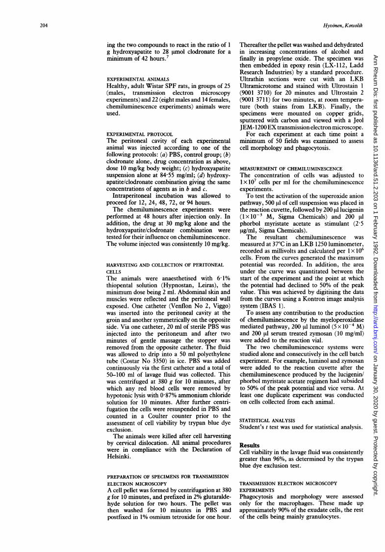
Phagocytosis of hydroxyapatite particlesAfter 12 hours of incubation there was evidenceof intracellular hydroxyapatite, but at this timeparticles were also regularly observed extra-cellularly. All hydroxyapatite particles had beenphagocytosed by 24 hours as none was seenextracellularly. Figure 3 shows that in several 72and 96 hour specimens disintegration of intra-vacuolar hydroxyapatite particles was observed.This was accompanied by vacuolisation of thecytoplasm and degeneration of cellular mor-phology (fig 3B).
Phagocytosis of hydroxyapatite particles reactedwith clodronatePhagocytosis was frequently seen from 12 hoursonwards. By 24 hours most but not all hydroxy-apatite particles were intracellular, and by 48hours no particles remained outside the cells.Hydroxyapatite particle disintegration apdcrystal dissolution were seen only rarely at 48hours, but in numerous cells in 72 and 96 hourexperiments (figs 4A and B). The frequency orextent of particle dissolution was not quanti-tated. In the presence of phagocytosis, good cellmorphology was preserved during the incu-bation. Figure 4B shows that at 96 hourscytoplasm had degenerated less than in cellscontaining plain hydroxyapatite, and morpho-logically distinct mitochondria were seen. Thesituation was similar at 72 hours. Figure 4Cshows an example of more advanced hydroxy-apatite particle dissolution at 96 hours.
CHEMILUMINESCENCE EXPERIMENTSChemiluminescence assays were performed forthe peritoneal exudate cells, which consisted oftwo cell types capable of light generation:macrophages and polymorphonuclear neutrophilgranulocytes.
Pattern of chemiluminescence productionIn all experimental systems chemiluminescencewas produced, showing cell viability and acti-vation. A total of 45 experiments was conducted,and a minimum of four tests was performed perexperimental group. Figures 5-8 show examplesof the pattern of chemiluminescence kinetics,the voltage values on the vertical axis being for5x 106 cells.Figure 5 shows a typical example of phorbol
205
on January 30, 2020 by guest. Protected by copyright.
http://ard.bmj.com
/A
nn Rheum
Dis: first published as 10.1136/ard.51.2.203 on 1 F
ebruary 1992. Dow
nloaded from
Hyvonen, Kowolik
Figure 3 (A) Rat peritoneal macrophages after intraperitoneal tnjectwn of hydroxyapatttesuspension at 12 hours. Bar=I1tm. Phagosome membrane seen surrounding hydroxyapatiteparticle (arrow). (B) Rat peritoneal macrophages after intraperitoneal injection ofhydroxyapatite suspension at 72 hours. Bar= ]pm. Phagosome membrane seen surroundinghydroxyapatite particle (arrow).
Table I Companrson of peak chemiluminescence potential and area under the curve forexudate cells assayed in the phorbol myristate acetatellucigenin system. n=Number of assaysperformed in each group. There was no significant difference between the drug and controlgroups (t test)
Clodronate concentration (mglkg) Phosphatebuffered
10 30 saline(n=4) (n=4) (control)
(n=7)
Mean (SD) potential per lx 106 cells 0-34 (0-31) 0-38 (0 22) 0-29 (0-10)Mean (SD) area under curve
per lx106 cells (mm2) 187 (137) 330 (183) 158 (88)
myrnstate acetate stimulated, lucigenin aug-mented chemiluminescence. Figure 6 illustratesthat of a zymosan/luminol experiment. A com-
parison of the two curves shows a fasterresponse in the phorbol myristate acetate/lucigenin system. This pattern of kinetics was
also observed in the combination experiments as
shown in figs 7 and 8. Similar chemiluminescencecurve shapes were obtained in combinationexperiments with hydroxyapatite/clodronateincubation.
Effect of clodronate on chemiluminescenceTable 1 summarises the results obtained for thephorbol myristate acetate/lucigenin system,comparing the influence of clodronate with thePBS control. The chemiluminescence outputwas similar in terms of potential to the control,but the area under the curve was greater at highclodronate concentrations than the control.
Effect of hydroxyapatite and hydroxyapatitelclodronate on chemiluminescenceTable 2 compares assays performed withhydroxyapatite/clodronate using hydroxyapatiteas the drug free control and PBS as the apatitefree control. The difference between the areasunder the curve reflects the difference in curve
shape, which in turn denotes a more sustainedchemiluminescence response with a higher totalenergy output.
In addition to the results shown in the tables,there was a significant difference in both thepotential and the area under the curve between10 mg/kg clodronate and 10 mg/kg hydroxy-apatite/clodronate (p<OO1).
DiscussionThe mechanism(s) by which bisphosphonatedrugs exert their therapeutic effects in themanagement of osteolytic bone diseases havebeen extensively studied, but only partly eluci-dated. It is now known that, in bone, theosteoclast is the primary biological target ofclodronate.2 15
In this study we have attempted to apply twomethods of studying the effects of bisphospho-nate on inflammatory cells. This approach was
used to aid the understanding of the proposedanti-inflammatory action of clodronate9 at thecellular level. This drug has been studiedextensively and used in treatment for many
years and data have accumulated on its effect onhard tissue metastases,'6 17 hypercalcaemia dueto malignancy,4 18 Paget's disease of bone,6 8and osteoporosis.'9 This drug affects not only
Table 2 Comparison ofpeak chemtiluminescence potential and area under the curve for exudate cells assayed in the phorbolmyristate acetatellucigenin system in the presence of hydroxyapatite. n=Number of assays performed in each group
Clodronatelhydroxyapatite Hydroxyapatite Phosphateconcentration (mglkg) (control) at buffered
84 55 mglml saline10 30 (n=4) (control)(n=4) (n=4) (n= 7)
Mean (SD) potential per lx 106 cells 1-16 (0-38)*t 0-83 (0-45) 0 47 (0-22)* 0 21 (0 l0)*tMean (SD) area under curve per lx 106 cells (mm2) 426 (258)§ 508 (173)4 407 (258) 158 (88)#5
-'tfSignificant difference (p<0O05) between marked groups (t test).SSignificant difference (p<001) between marked groups (t test).
206
on January 30, 2020 by guest. Protected by copyright.
http://ard.bmj.com
/A
nn Rheum
Dis: first published as 10.1136/ard.51.2.203 on 1 F
ebruary 1992. Dow
nloaded from

Dichloromethylene bisphosphonateandphagocytosisofhydroxyapatite
ivEArM'"'''ilt; ' <;F;f'w A?'>BR4S° ';v t~KX <. A
Figure 4 (A) Rat peritoneal macrophages after intraperitonealinjection ofhydroxyapatite combined with clodronate at 12 hours. Bar=Jgm. Hydroxyapatite surrounded by phagosome membrane (arrow).(B) Rat peritoneal macrophages after intraperitoneal injection ofhydroxyapatite combined with clodronate at 96 hours. Bar=1Mm.Hydroxyapatite surrounded by phagosome membrane (arrow). (C)Typical view ofadvanced dissolution ofhydroxyapatite particle in thepresence ofclodronate. Bar=500 nm.
bone calcium metabolism, but has been shownto have anti-inflammatory properties in anadjuvant arthritis animal model9 and to inhibitthe excretion of lysosomal enzymes in conjunc-tion with parathyroid hormone induced resorp-
10[
mvj A
0 15 30TIME min
FigureS Phorbol myristate acetate (PMA)Ilucigeningenerated chemiluminescence (CL)from peritoneal exudatecells after in vtvo tncubation with clodronate/hydroxyapatite(10 mglkg). Lucigenin added at 0 min, phorbol myristateacetate addition shown by arrow.
tion20 and prostaglandin E221 in vitro. It was ofinterest to determine the effect of reactinghydroxyapatite with clodronate on the subse-quent phagocytosis of particles, and chemi-luminescence production, by rat peritonealmacrophages.The intracellular location of clodronate, when
given to cells in culture in the absence ofmineral crystals, appears to be in the cytosol.22As clodronate has a high affinity for hydroxy-apatite,7 binding with the mineral could give it
0 30 60
TIME min
Figure 6 Zymosan (ZYM)/luminol generatedchemiluminescence (CL)from peritoneal exudate cells after invivo incubation with clodronatelhydroxyapatite (10 mg/kg).Luminol added at 0 min, zymosan addition shown by arrow.
207
on January 30, 2020 by guest. Protected by copyright.
http://ard.bmj.com
/A
nn Rheum
Dis: first published as 10.1136/ard.51.2.203 on 1 F
ebruary 1992. Dow
nloaded from

Hyvonen, Kowolik
30
CLmV
20
10ZYM
.PMA LUMIIZ
30 60
TIME min
Figure 7 Phorbol myristate acetate (PMA)/lucigeningenerated chemiluminescence (CL)followed by zymosan(ZYM)Iluminol (LUM) chemiluminescence from the sameperitoneal exudate cells, after 48 hours in vivo incubationwith hydroxyapatite 84 55 mglml. Lucigenin added at 0 minphorbol myristate acetatel and zymosanlluminol additionsshown by arrows.
30
TIME min
Figure 8 Zymosan (ZYM)Iluminol generatedchemiluminescence (CL)followed by phorbol myristateacetatellucigenin (LUCIG) chemiluminescence from thesame peritoneal exudate cells, after 48 hours in vivo
incubation with hydroxyapatite 84-55 mglml. Luminoladded at 0 min, zymosan and phorbol myristateacetatellucigenin additions shown by arrows.
an effective means of entry into the cell, once
the particle is phagocytosed. Its high solubilityin water, which prevents it passing throughphysiological membranes and thus entering thecell in large amounts (only 1-2%, concludedfrom the kinetic study of Yakatan et al23), mayfacilitate its intracellular pharmacological actionafter having gained entry at a higher concentra-tion in combination with the mineral.When hydroxyapatite particles were pre-
incubated with clodronate, phagocytosis was
observed less frequently at 12 and 24 hours thanfor hydroxyapatite particles alone. This findingis in agreement with the impaired attachment ofmacrophages to bone in the presence of bis-phosphonate, observed by Chambers using lightmicroscopy.24 The hypothesis that the action ofclodronate on the macrophage is mediatedintracellularly is naturally valid, at least in thepresence of hydroxyapatite. The observed delayin phagocytosis of the hydroxyapatite/clodronateparticles may be a result of extracellular mem-brane irritation due to the clodronate bound tothe crystal surface. Cellular toxicity has beenshown towards the macrophage.' 24 and theosteoclast.2 1 2 The results presented heremay be a reflection of similar cytotoxicity. Theconclusions are not surprising if these two cellstypes share a common bone haematopoieticprecursor.2129 Minkin and Shapiro30 have,however, voiced a word of caution about the
assumption that peripheral blood monocytes orelicited peritoneal macrophages might be theactual osteoclast precursors.
In considering rheumatoid arthritis, theintracellular activation of macrophages byhydroxyapatite may inadvertently contribute totissue damage.'0 11 The anti-inflammatory effectof clodronate may therefore result in macro-phages being impaired in their ability to contri-bute to the destruction of surrounding tissues.In this in vivo model there was no doubt thatthe cells were able to ingest the hydroxyapatiteparticles whereby the drug was able to enter thecell. This can also occur in the affected joint inrheumatoid arthritis, where the macrophage isfunctioning as a scavenger of the crystals andother debris.'"Once phagocytosed, crystal degradation and
dissolution occurs with time in the phagocyticvacuoles containing both pure hydroxyapatiteand hydroxyapatite/clodronate particles. Theconcentration of clodronate used in thisexperiment did not significantly alter this lyticactivity compared with the clodronate free,crystal containing cells during the observationperiod. The finding does not conflict withreports of some in vitro studies on mononuclearcells being capable of mineral particle phago-cytosis24 or bone resorption.3"34 A fundamentaldifference between the above mentioned in vitrostudies, which have been conducted withpowdered bone, and this work is the lack oforganic matrix in the hydroxyapatite used. Asthere is no foreign protein component in themineral, the cells could regard this substance asnon-immunogenic.
Cell morphology showed progressive de-generation with time. This seemed to be moreextensive with hydroxyapatite alone than withhydroxyapatite/clodronate. Clodronate isknown to be cytotoxic in the presence ofcalcified material,2 and yet in this experimentalsystem good cell morphology was preserved.Throughout the study mitochondria were clearlyseen in most cells, indicating the potential formetabolism.The chemiluminescence results support those
of the transmission electron microscopy experi-ments. Firstly, under all in vivo incubationconditions the cells were viable and capable ofactivation in the standardised chemilumine-scence assays, the least activation occurring inthe PBS control group. Secondly, the resultsreinforce the assumption that clodronate gainsminimal access to the interior of the cell withouthydroxyapatite, but does so in combinationwith it. Finally, it appeared that incubationwith hydroxyapatite and with hydroxyapatite/clodronate enhanced the subsequent response tostimulus, i.e. the incubation had primed thecells, a phenomenon thought to be of consider-able importance in biological homeostasis.35This effect was seen in the higher peak potentialwith the hydroxyapatite/clodronate group,denoting a more rapid release of energy by thecells than in the hydroxyapatite group. Incontrast, the areas under the curves weresimilar, suggesting a more sustained response inthe hydroxyapatite group. It may be relevantthat clodronate is a chlorinated molecule and as
O' - m -L.^L
Il
208
on January 30, 2020 by guest. Protected by copyright.
http://ard.bmj.com
/A
nn Rheum
Dis: first published as 10.1136/ard.51.2.203 on 1 F
ebruary 1992. Dow
nloaded from
209Dichloromethylenebisphosphonateandphagocytosisofhydroxyapatite
such may be able to interact directly withchemiluminescence production.36The finding that cell activation occurred in
the two chemiluminescence systems suggeststhat macrophages and neutrophils were presentin the exudate; this was supported by lightmicroscopic and transmission electron micro-scopy examination. The consecutive activationof chemiluminescence pathways in the same
cells (figs 7 and 8) is also suggestive of a mixedcell population but, as neutrophils in particularare able to respond strongly through the twosystems, the results may also illustrate the highresponse levels in this exudate. This has beenshown previously in human gingival crevicularexudate cells.37 In the course of this study,neither the earliest stages of phagocytosis nor
the late lysis of the hydroxyapatite containingcells was seen. Further studies will be necessary
to observe these stages.This animal model allows the study of some
cellular effects of the bisphosphonate drugs invivo, and thereby facilitates the interpretationof their pharmacological action in relation tobone metabolism and anti-inflammatory func-tion. As the hydroxyapatite crystals are takeninto the cells, and clodronate thereby gainsaccess to the cell interior, the inflammatory cellfunction is modified, as indicated by thechemiluminescence assays. These results sup-
port the use of clodronate in the clinicalmanagement of inflammatory bone and jointdiseases.
We are grateful to Aria Korhonen and Pekka Alakuijala for theirtechnical expertise and Eija Voutilainen, Eija Antikainen, andAlpo Pelttari for their assistance and expert advice with theelectron microscopic work. The authors also thank Dr ANaukkarinen for her advice on the interpretation of the trans-mission electron microscopy photographs. The hydroxyapatitewas kindly donated by British Charcoals and McDonald(Greenock, United Kingdom) and the dichloromethylene bis-phosphonate by Dr Hannu Hanhijarvi (Leiras Pharmaceuticals,Turku, Finland). Financial support was also provided by LeirasPharmaceuticals. This study was supported by a grant by theFinnish Cultural Foundation to P M Hyvonen. Our gratitude isexpressed.
1 Reitsma P H, Teitelbaum S L, Bijvoet 0 L M, Kahn A J.Differential action of the bisphosphonates (3-amino-l-hydroxypropylidene)-1,1-bisphosphonate (APD) anddisodium dichloromethylidene bisphosphonate (Cl2MDP)on rat macrophage-mediated bone resorption in vitro.JClin Invest 1982; 70: 927-33.
2 Flanagan A M, Chambers T J. Dichloromethylenebisphos-phonate(Cl2MBP)inhibits bone resportion through injuryto osteoclasts that resorb Cl2MBP-coated bone. BoneMiner 1989; 6: 33-43.
3 Jung A, Chantraine A, Donath A, et al. Use of dichloro-methylene diphosphonate in metastatic bone disease.N Engl J Med 1983; 308: 1499-1501.
4 Cohen AI, Koeller J, Davis T E, Citrin D L. Ivdichloromethylene diphosphonate in cancer-associatedhypercalcemia: a phase I-II evaluation. Cancer TreatmentReport 1981; 65: 651-3.
5 Jung A, van Ouwenaller C, Chantraine-A, Courvoisier B.Parenteral diphosphonates for treating malignant hyper-calcemia. Cancer 1981; 48: 1922-5.
6 Delmas P D, Chapuy M C, Vignon E, et al. Long term effectsof dichloromethylene diphosphonate in Paget's disease ofbone. JClGn Endocrmnol Metab 1982;54: 83744.
7 Jung A, BisazS, Fleisch H. The binding of pyrophosphateand two diphosphonates by hydroxyapatite crystals. CakifTissue Res 1973; 11: 269-80.
8 Fleisch H. Experimental basis for the use of bisphosphonatesin Paget's disease of bone. Clin Orthop 1987; 21: 72-8.
9 Flora L. Comparative antiinflammatory and bone protectiveeffects of two diphosphonates in adjuvant arthritis. ArthritisRheum 1979; 22: 340-6.
10 Gibilisco P A, Schumacher H R Jr, Hollander J L, SoperK A. Synovial fluid crystals in osteoarthritis. ArthritisRheum 1985; 28: 511-5.
11 Hirsch R S, Smith K, Vernon-Roberts B. A morphologicalstudy of macrophage and synovial cell interactions withhydroxyapatite crystals. Ann Rheum Dis 1985; 44: 176-80.
12 Tsunawaki S, Nathan C F. Enzymatic basis of macrophageactivation. J Biol Chem 1984; 259: 4305-12.
13 Rossi F, Bellavite P, Berton G, Grzeskowiak M, Papini E.Mechanism of production of toxic oxygen radicals bygranulocytes and macrophages and their function in theinflammatory process. Pathol Res Pract 1985; 180: 136-42.
14 Dahlgren C, Stendahl 0. Role of myeloperoxidase onluminol-dependent chemiluminescence of polymorpho-nuclear leukocytes. Infect Immun 1983; 39: 736-41.
15 Rowe D J, Hausmann E. The alteration of osteoclastmorphology by diphosphonates in bone organ culture.Calcif Tissue Res 1976; 20: 53-60.
16 ElomaaI, Blomqvist C, Gr6hn P, et al. Long term controlledtrial with diphosphonate in patients with osteolytic bonemetastases. Lancet 1983; i: 146-8.
17 Elte J W, Bijvoet 0 L M, Cleton F J, VanOosterom A T,Sleebom H P. Osteolytic bone metastases in breastcarcinoma, pathogenesis, morbidity and bisphosphonatetreatment. EurJ CancerClin Oncol 1986; 22: 493-500.
18 Jung A. Comparison of two parenteral diphosphonates inhypercalcemia of malignancy. AmJ Med 1982; 72: 221-6.
19Jowsey J, Holley K E. Influence of diphosphonates onprogress of experimentally induced osteoporosis. J LabClin Med 1973; 82: 567-75.
20 Delaisse J-M, Eeckhout Y, Vaes G. Bisphosphonates andbone resorption: effects on collagenase and lysosomalenzyme excretion. Life Sci 1985; 37: 2291-6.
21 Felix R, Bettex J-B, Fleisch H. Effect of diphosphonates onthe synthesis of prostaglandins in cultured calvaria cells.Calcif Tissue Int 1981; 33: 549-52.
22 Felix R, Guenther H L, Fleisch H. The subcellular distribu-tion of '4C-dichloromethylenebisphosphonate and'4C-1-hydroxyethylidene-1,l-bisphosphonate in cultured calvariacells.Calcif TissueInt 1984; 36: 108-13.
23 Yakatan G J, Poynor W J, Talbert R L, et al. Clodronatekinetics and bioavailability.Clin Pharnacol Therap 1982;31: 402-10.
24 Chambers T J. Diphosphonates inhibit bone resorption bymacrophages in vitro. J Pathol 1980; 132: 255-62.
25 Miller S C, Jee W S S. The effect of dichloromethylenediphosphonate, a pyrophosphate analogue, on bone andbone cell structure in the growing rat. Anat Rec 1979; 193:439-62.
26 Ash P, Loutit J F, Townsend K M S. Osteoclasts derivedfrom haematopoietic stem cells. Nature 1980; 238: 669-70.
27 Baron R, Vignery A, Horowitz M. Lymphocytes, macro-phages and the regulation of bone remodeling. In: Peck WA, ed. Bone and mineral research, Annual 2. Amsterdam:Excerpta Medica, 1983: 175-243.
28 Burger E H, van der Meer J W M, van de Gevel JS, GribnauJ C, Thesingh C W, van Furth R. In vitro formation ofosteoclasts from long term cultures of bone marrowmononuclear phagocytes. JExp Med 1982; 156: 1604-14.
29 Chambers T J. The cellular basis of bone resorption.ClinOrthop 1980;151: 282-93.
30 Minkin C, ShapiroI M. Osteoclasts, mononuclear phagocytes,and physiological bone resorption. Cakif TissueInt 1986;39: 357-9.
31 Chambers T J. Resorption of bone by mouse peritonealmacrophages. J Pathol 1981; 135: 295-9.
32 Kahn A J, Stewart C C, Teitelbaum S L. Contact-mediatedbone resorption by human monocytes in vitro. Science1978;19: 988-90.
33 McArthur W, Yaari A M, Shapiro I M. Bone solubilisationby mononuclearcells. Lab Invest 1980; 42: 450-6.
34 Teitelbaum S L, Stewart C C, Kahn A. Rodent peritonealmacrophages as bone resorbing cells. Calcif TissInt 1979;27: 255-61.
35 Guthrie L A, McPhail L C, Henson P M, Johnston R B Jr.Priming of neutrophils for enhanced release of oxygenmetabolites by bacterial lipopolysaccharide: evidence forincreased activity of the superoxide-producing eznyme. JExp Med 1984; 160: 1656-71.
36 Kowolik M J. The oxygen-dependent microbicidal system inhuman gingival crevicular neutrophils. In: Mauri C, RizzoSC, Ricevuti G, eds. The biology of phagocytes in health anddisease. Advances in the biosciences. Vol 66. Oxford:Pergamon Press, 1987, 175-83.
37 Kowolik M J, Hyvonen PM. The effect of dichloromethylenebisphosphonate on human gingival crevicular myeloperoxi-dase activity. Arch Oral Biol 1990; 35 (suppl): 20IS-3S.
on January 30, 2020 by guest. Protected by copyright.
http://ard.bmj.com
/A
nn Rheum
Dis: first published as 10.1136/ard.51.2.203 on 1 F
ebruary 1992. Dow
nloaded from