influence of physical activity, sedentary lifestyle...
TRANSCRIPT
INFLUENCE OF PHYSICAL ACTIVITY, SEDENTARY LIFESTYLE AND
BONE BIOMARKERS ON BONE HEALTH AMONG ADOLESCENTS IN
KOTA BHARU, KELANTAN
by
TEO PEY SZE
Thesis submitted in fulfilment of the requirements
for the degree of
Master of Science
May 2013

DEDICATION
This dissertation is lovingly dedicated to my family, particularly my parents, Teo
Chok Joo and Lee Kah Heng. Their endless love, support and encouragement has
sustained me throughout my life. A special feeling of gratitude to my lovely sister,
Teo Pey Huey, who has always being there for me throughout my entire postgraduate
study. I also dedicate this work to my uncles and aunties, especially to Teo Kuek
Chong, Low Gek Luang and Teo Kiok Long who believe in the pursuit of dreams
and have been my best cheerleaders.
ii
ACKNOWLEDGEMENTS
First and foremost, I would like to express my utmost gratitude to Universiti Sains
Malaysia (USM) for providing the USM PRGS grant (1001/PPSK/8143002) for my
research work on a validation study of physical activity and the USM Postgraduate
Fellowship Scheme throughout my postgraduate study.
My sincere thanks to my main supervisor, Dr. Foo Leng Huat for his guidance and
mentorships throughout my postgraduate study. I am fortunate to have acquired a
wealth of knowledge and skills under his supervision. My sincere gratitude for his
time, efforts, and numerous opportunities he have provided throughout my
postgraduate training. I also would like to thank my co-supervisor, Associate
Professor Dr. Mohd Ezane Aziz from Department of Radiology, School of Medical
Sciences, USM. He has shared his knowledge and experience in the clinical
radiology research in which he has provided important dimensions to our research
group. I would like also to thank Dr. Chan Chee Keong from the Sport Science Unit
for his helpful guidance on the assessment of isokinetic muscle strength.
Special thanks also goes to my research study colleague, Ms Nurul Fadhilah
Abdullah for his excellent contribution working together as very effective team
throughout the study. This thesis also would not have been possible without the
supports provided from all staffs from the School of Health Sciences and School of
Medical Sciences, in particularly Mr. Mohd Hafezi Mat Zain, Mr. Mohd Roslan Mod,
Ms Malisa Yoong, Ms. Nur Hidayah Yahya Anuar, Ms. Che Rohaida Che Mohd, Mr.
Arrifin Harun, Mr. Koh Chun Haw, Ms Noor Salwah, Mr. Mohammad Fadzil Ismail,

iii
Ms. Normayazi Mohemed Ali, Ms Jamaayah Meor Osman, Mr. Nawawi Yasin and
Ms Norlida Azalan @ Zed as well as community leaders and person in-charge that
had helping us in facilitating the study participations during the recruitment process.
My sincere gratitude and appreciation also would like to go to all wonderful
participants and their parents or guardians for allowing their children to participate
and committed in the study.
Lastly, I wish to sincerely thank my parents, sister, uncles and aunties for their full
encouragements and continuous supports to me throughout my education and
postgraduate study.
iv
TABLES OF CONTENTS
ACKNOWLEDGEMENTS ......................................................................................... ii
TABLES OF CONTENTS .......................................................................................... iv
LIST OF TABLES ..................................................................................................... vii
LIST OF FIGURES .................................................................................................... ix
LIST OF ABBREVIATIONS ...................................................................................... x
ABSTRAK .................................................................................................................. xi
ABSTRACT .............................................................................................................. xiv
CHAPTER 1 INTRODUCTION ................................................................................ 1
1.0 Introduction of the osteoporosis..................................................................... 1
1.0.1 Aetiology of osteoporosis development .................................................... 3 1.1 Peak bone mass (PBM) .................................................................................. 3
1.1.1 Factors associated with PBM- Physical activity ........................................ 4 1.1.2 Factors associated with PBM- Sedentary practices ................................... 5 1.1.3 Factors associated with PBM- Blood bone markers .................................. 7
1.2 Significance of the study ................................................................................ 8
1.3 Objectives ...................................................................................................... 9
1.3.1 Main objective ........................................................................................... 9 1.3.2 Specific objectives ..................................................................................... 9
1.4 Research null hypotheses ............................................................................. 11
1.5 Conceptual framework ................................................................................. 11
1.6 Definition of terminology ............................................................................ 12
CHAPTER 2 LITERATURE REVIEW .................................................................... 14
2.0 Overview of osteoporosis ............................................................................ 14
2.0.1 Aetiology of osteoporosis ........................................................................ 17
2.1 Bone structure and physiology..................................................................... 18
2.1.1 Bone remodeling process through the life spans ..................................... 21 2.2 Bone growth throughout the lifespan ........................................................... 25
2.3 Peak Bone Mass (PBM) ............................................................................... 26
2.3.1 Factors affecting peak bone mass ............................................................ 27 2.4 Non-modifiable factors on PBM .................................................................. 27
2.4.1 Genetic factors ......................................................................................... 27 2.4.2 Pubertal status .......................................................................................... 28
2.4.3 Sex factors................................................................................................ 30 2.5 Modifiable environmental factors on bone mass ......................................... 30
2.5.1 Lifestyle Physical activity (PA) on musculoskeletal health .................... 30 2.5.2 The effect of muscular strength on bone mass ........................................ 43 2.5.3 Sedentary practices .................................................................................. 47 2.5.4 Other modifiable factors such as body composition, dietary practices ... 49
2.6 Methodological issues of assessment of bone mass status in children
and adolescents ............................................................................................ 55

v
2.6.1 Dual energy X-ray absorptiometry (DXA) .............................................. 56 2.6.2 Quantitative computed tomography (QCT) ............................................. 58 2.6.3 Quantitative ultrasound (QUS) ................................................................ 59 2.6.4 Magnetic resonance imaging (MRI) ........................................................ 60
2.7 Assessment of biochemical markers of bone remodeling levels ................. 61
2.8 Summary of the literature review ................................................................ 65
CHAPTER 3 METHODOLOGY ............................................................................. 67
3.0 Study design ................................................................................................. 67
3.1 Study protocols and methodology ............................................................... 69
3.2 Research methodology ................................................................................. 72
3.2.1 Exposure measurements .......................................................................... 72 3.2.2 Outcome measurements ........................................................................... 85
3.2.3 Confounder variables in the study ........................................................... 86 3.3 Statistical analysis ........................................................................................ 88
CHAPTER 4 RESULTS ........................................................................................... 92
4.0 General characteristics of the participants ................................................... 92
4.1 Physical activity and sedentary lifestyle status ............................................ 94
4.2 Dietary intakes and dietary behaviours ........................................................ 96
4.3 Blood biochemical markers of bone remodeling profiles .......................... 100
4.4 Body composition and muscle strength profiles ........................................ 102
4.5 Bone mass parameters................................................................................ 104
4.6 Distribution of physical activity level and sedentary SSR practices ......... 107
4.7 Socio-demographic and dietary behaviour determinants on physical
activity levels ............................................................................................. 109
4.8 Socio-demographic and dietary behaviour determinants on sedentary
SSR practice ............................................................................................... 111
4.9 Influence of physical activity status on body composition profiles ........... 113
4.10 Influence of sedentary SSR practices on body composition profiles ........ 116
4.11 Influences of physical activity status on prevalence of obesity assessed
by percent body fat..................................................................................... 118
4.12 Influences of sedentary SSR on prevalence of obesity assessed by
percent body fat.......................................................................................... 120
4.13 Relationship between physical activity and bone mass parameters........... 122
4.14 Relationship between sedentary SSR levels and bone mass parameters ... 125
4.15 Relationships between muscle strength profiles and bone mass
parameters .................................................................................................. 127
4.16 Relationships between blood biochemical markers of bone remodeling
and bone mass parameters ......................................................................... 131
4.17 Multivariate analyses of physical activity and sedentary SSR practices
on musculoskeletal parameters .................................................................. 140
vi
CHAPTER 5 DISCUSSIONS ................................................................................. 155
5.0 General characteristics of the participants ................................................. 155
5.1 Lifestyle physical activity pattern of the adolescents ................................ 156
5.2 Socio-demographic and lifestyle correlates of PA status .......................... 158
5.3 Sedentary screen-based behaviour practices of the adolescents ................ 161
5.4 Socio-demographic and lifestyle correlates of sedentary screen-based
behaviour.................................................................................................... 162
5.5 The influences of PA on musculoskeletal health ....................................... 165
5.6 The relationship between muscle strength and bone mass ........................ 173
5.7 The influences of sedentary lifestyle practices on muscle strength and
bone mass ................................................................................................... 174
5.8 The influences of PA and sedentary lifestyle practices on body
adiposity levels........................................................................................... 176
5.9 Relationships between blood biochemical markers of bone remodeling,
pubertal growth status and bone mass status ............................................. 180
5.10 Strengths and limitations of the study ........................................................ 183
CHAPTER 6 CONCLUSION AND RECOMMENDATIONS ............................. 186
6.0 Summary of key findings ........................................................................... 186
6.1 Recommendations and suggestions ........................................................... 191
6.2 Recommendations for the future studies.................................................... 195
REFERENCES ......................................................................................................... 197
APPENDICES
Appendix A Photos of the study protocol
Appendix B Human ethical approval letter
Appendix C Information sheet and written consent forms
Appendix D Advertisement for the recruitment purpose
Appendix E Sample of questionnaires
Appendix F List of scientific publications and conference presentations
Appendix G Selected journal publications

vii
LIST OF TABLES
Table 2.1 The incidence of hip fracture in four Asian countries with
United States and Oxford, England (per 100,000 populations).
....................................................................................................... 17
Table 2.2 Summary of published studies on the relationships between
physical activity and bone mass in children and adolescents ........ 32
Table 2.3 Summary of published studies on the relationships between
physical activity (PA), and bone and muscular strength
profiles in children and adolescents ............................................... 40
Table 2.4 Summary of published studies on the relationships between
muscular strength profiles and bone mass in children and
adolescents ..................................................................................... 45
Table 2.5 Summary of the advantages and disadvantages of each bone
assessment techniques ................................................................... 57
Table 2.6 Commonly used biochemical markers of bone remodeling .......... 63
Table 3.1 Precision of calcium homeostasis indicators expressed as
within assay (intra) and between-assay (inter) coefficient of
variation ......................................................................................... 82
Table 3.2 Precision of biochemical markers of bone remodeling
expressed as within assay (intra) and between-assay (inter)
coefficient of variation ................................................................... 84
Table 4.1 General characteristics of participant boys and girls by
ethnicity ......................................................................................... 93
Table 4.2 Physical activity (PA) levels and sedentary practices patterns
of participant boys and girls by ethnicity ...................................... 95
Table 4.3 Dietary intakes and nutritional behaviours of participant boys
and girls by ethnicity ..................................................................... 97
Table 4.4 Dietary nutrients intake of boys and girls of combined
ethnicity ......................................................................................... 99
Table 4.5 Blood biochemical markers of bone remodeling profiles of
participant boys and girls by ethnicity ......................................... 101
Table 4.6 Body composition profiles and muscle strength measures of
participant boys and girls by ethnicity ......................................... 103
Table 4.7 Bone mass profiles of participant boys and girls by ethnicity ..... 105
Table 4.8 Determinants of active PA status, as determined by the
moderate-to-vigorous physical activity (MVPA) levels in
adolescentsa .................................................................................. 110
Table 4.9 Determinants of sedentary status, as determined by the small
screen recreation (SSR) levels in adolescentsa ............................ 112

viii
Table 4.10 Multivariate analyses of total PA and MVPA duration on
body fatness profiles of boys and girlsa ....................................... 114
Table 4.11 Multivariate analyses of total SSR duration on body fatness
profiles of boys and girlsa ............................................................ 117
Table 4.12 Multiple linear logistics models of odd ratios (OR) for being
overweight or obese compared with normal weight based on
total PA and MVPA time groups in boys and girls ..................... 119
Table 4.13 Multiple logistics models of odd ratios (OR) for being
overweight or obese compared with normal weight based on
total SSR time groups in boys and girls ....................................... 121
Table 4.14 Sex-specific partial correlation between physical activity
status and bone mass profiles in participants1 ............................. 123
Table 4.15 Sex-specific partial correlation between daily sedentary SSR
levels and bone mass profiles in participants1 ............................. 126
Table 4.16 Sex-specific partial correlation between muscle strength
measures and bone mass profiles in participants1 ........................ 128
Table 4.17 Sex-specific partial correlation between blood biochemical
markers of bone remodeling and bone mass profiles in
participants1 ................................................................................. 136
Table 4.18 Multivariate analyses of total PA and MVPA duration on
muscle strength profiles of boys and girlsa .................................. 141
Table 4.19 Multivariate analyses of sedentary SSR duration on muscle
strength profiles of boys and girlsa .............................................. 143
Table 4.20 Sex-specific multivariate analysis of influences of PA status
of adolescents on size-adjusted BMC, BMD and BA of
skeletal sites measured ................................................................. 146
Table 4.21 Sex-specific multivariate analyses of influences of sedentary
SSR of adolescents on size-adjusted BMC, BMD and BA of
skeletal sites measured ................................................................. 152
ix
LIST OF FIGURES
Figure 1.1 A conceptual framework of influences of physical activity on
bone health status in adolescents ................................................... 13
Figure 2.1 Bone modeling and remodeling. Osteoclasts break down old
bone tissue and followed by osteoblasts activity, in which
osteoblasts form new collagen and other matrix proteins.
Then, the collagenous matrix undergoes mineralization ............... 22
Figure 2.2 Bone growth and bone loss process throughout the lifespan in
male and female ............................................................................. 26
Figure 3.1 Flow chart of study methodology and participants recruitment .... 71
Figure 4.1 Distribution of total daily moderate-to-vigorous physical
activity practices .......................................................................... 108
Figure 4.2 Distribution of total daily Small Screen Recreation (SSR)
sedentary practices ....................................................................... 108
Figure 4.3a(i) Relationships between pubertal growth status and calcium
homeostasis markers (serum calcium) in boys and girls ............. 132
Figure 4.3a(ii) Relationships between pubertal growth status and calcium
homeostasis markers (serum inorganic phosphorus) in boys
and girls ....................................................................................... 132
Figure 4.3a(iii) Relationships between pubertal growth status and calcium
homeostasis markers (serum creatinine) in boys and girls .......... 133
Figure 4.3b(i) Relationships between pubertal growth status and bone
formation markers (serum total ALP) in boys and girls .............. 133
Figure 4.3b(ii) Relationships between pubertal growth status and bone
formation markers (serum total P1NP) in boys and girls ............ 134
Figure 4.3b(iii) Relationships between pubertal growth status and bone
formation markers (serum osteocalcin) in boys and girls ............ 134
Figure 4.3c Relationships between pubertal growth status and bone
resorption markers (serum CTx) in boys and girls ...................... 135
Figure 4.3d Relationships between pubertal growth status and
calciotropic hormone of PTH in boys and girls ........................... 135

x
LIST OF ABBREVIATIONS
PA Physical activity
MVPA Moderate-to-vigorous physical activity
SSR Small screen recreation
Ca Calcium
Phos Inorganic phosphorus
Cr Creatinine
ALP Alkaline phosphatase
P1NP Procollagen type I amino-terminal propeptide
OC Osteocalcin
CTx Collagen type I cross-linked C-telopeptide
PTH Parathyroid hormone
BMI Body mass index
WC Waist circumference
WHR Waist-hip ratio
TBF Total body fat
%BF Percent body fat
ARF Android regional fat
TLM Total lean mass
BMC Bone mineral content
BMD Bone mineral density
BA Bone area
DXA Dual energy X-ray absorptiometry
PBM Peak bone mass

xi
PENGARUH DI ANTARA AKTIVITI FIZIKAL, GAYA HIDUP SEDENTARI
DAN PETUNJUK BIOKIMIA TULANG TERHADAP KESIHATAN
TULANG DALAM KALANGAN REMAJA DALAM KOTA BHARU
KELANTAN
ABSTRAK
Pencapaian jisim tulang puncak pada masa pertumbuhan adalah sangat penting untuk
mengurangkan risiko keretakan osteoporosis pada kemudian hari. Pemahaman dan
pengenalan faktor-faktor gaya hidup seperti aktiviti fizikal dan gaya hidup yang
berkaitan dengan pertumbuhan dan pemeliharaan jisim tulang yang tinggi dalam
kalangan kanak-kanak dan remaja adalah amat penting bagi memaksimakan jisim
tulang puncak sewaktu tempoh pertumbuhan yang kritikal. Oleh itu, objektif utama
kajian ini adalah untuk mengkaji pengaruh aktiviti fizikal, gaya hidup sedentari dan
petunjuk biokimia darah tulang terhadap status kesihatan tulang yang ditentukan
dengan pengukuran tenaga dwi sinar-X absorptiometri (DXA) dalam kalangan 455
orang remaja lelaki dan perempuan berbangsa Melayu dan Cina yang berumur
lingkungan di antara 12 hingga 19 tahun di Kota Bharu, Kelantan. Borang soal-
selidik yang telah divalidasi digunakan untuk menentukan amalan aktiviti fizikal,
amalan sedentari yang berunsurkan skrin (SSR) dan corak pemakanan, manakala
komposisi tubuh badan dan kekuatan otot bagi anggota atas dan bawah dinilai
dengan menggunakan pengukuran antropometri, dinamometer tangan dan isokinetik.
Bagi penilaian status kesihatan tulang, kandungan mineral tulang (BMC) dan
kepadatan mineral tulang (BMD) bagi keseluruhan badan (TB), tulang paha
proksimal (PF), tulang belakang lumbar L2-L4 (LS) dan bahagian tulang tertentu

xii
diukur dengan menggunakan DXA. Purata umur peserta kajian adalah 15.4 1.9
tahun, dan kebanyakannya (72.5%) mempunyai indek jisim tubuh (BMI) yang
normal. Perbandingan mengikut jantina menunjukkan remaja lelaki mengamalkan
jumlah harian aktiviti fizikal (PA) (1.5 vs. 1.0jam/hari; P<0.001) dan aktiviti fizikal
sederhana kepada aktivit lasak (MVPA) (1.2 vs. 0.4jam; P<0.001) yang lebih tinggi
berbanding dengan perempuan; manakala pengamalan masa SSR adalah sama di
antara jantina (3.1 vs. 3.3jam/hari). Secara umumnya, terdapat dua-pertiga daripada
remaja (63.3%) mengamalkan amalan aktiviti fizikal aktif yang rendah, ia ditentukan
oleh amalan MVPA harian yang kurang daripada sejam, di mana peratusan adalah
tinggi dalam kalangan perempuan (80%) berbanding dengan remaja lelaki (34%).
Analisis regresi linear berganda menunjukkan umur (P=0.012) dan jantina (P<0.001)
merupakan penentu secara negatif terhadap MVPA harian model yang digunakan
setelah penyelarasan bagi etnik, status sosio-demografi dan pengamalan pemakanan
yang diuji. Tambahan pula, umur (P<0.001) dan amalan sarapan pagi seharian
(P<0.05) muncul sebagai penentu secara negatif, manakala kumpulan etnik (P<0.01)
sebagai penentu secara positif terhadap sedentari SSR model yang digunakan.
Pengaruh di antara faktor-faktor gaya hidup dengan status kesihatan tulang diuji
untuk menggunakan kumpulan berdasarkan jumlah dan intensiti aktiviti fizikal
harian dan sedentari SSR. Ia menunjukkan bahawa remaja lelaki yang mengamalkan
jumlah dan intensiti akviviti fizikal harian yang tinggi iaitu PA>1.5jam/hari dan
MVPA>1jam/hari mempunyai BMC yang lebih tinggi pada TB, bahagian inter-
trochanter, dan kaki (semuanya dengan P<0.05), dan BMD yang lebih tinggi pada
bahagian TB, PF, LS, lengan dan kaki (perkaitannya sekurang-kurangnya P<0.01),
berbanding mereka yang mengamalkan PA dan MVPA yang rendah. Pengaruh
positif secara signifikan hanya dikenalpasti di antara jumlah PA dan MVPA tinggi

xiii
dengan BMC dan BMD pada TB dan bahagian kaki dalam kalangan perempuan.
Tambahan pula, lelaki yang mempunyai MVPA yang tinggi juga menunjukkan
kekuatan otot genggaman tangan (P<0.01), quadriceps (P<0.05) dan hamstring
(P<0.001) yang lebih tinggi berbanding dengan mereka yang mengamalkan MVPA
harian yang rendah. Perkaitan secara signifikan hanya ditunjukkan di antara amalan
MVPA dan kekuatan otot hamstring dalam kalangan remaja perempuan (P<0.01).
Sebaliknya, jumlah gaya hidup sedentari hanya menunjukkan perkaitan secara
signifikan dan negatif terhadap profil komposisi tulang bagi perempuan. Remaja
perempuan yang mengamalkan tempoh masa SSR yang tinggi mempunyai BMD
yang lebih rendah di LS (P<0.05) dan saiz tulang pada TB (P<0.05), bahagian lengan
(P<0.01) dan kaki (P<0.05) berbanding dengan mereka yang mengamalkan SSR
yang rendah. Petunjuk biokimia darah tulang juga menunjukkan perkaitan secara
negatif dengan profil komposisi tulang bagi semua bahagian tulang yang diukur,
setelah mengambil kira faktor-faktor lain seperi status pertumbuhan pubertal dan
etnik. Kajian ini menunjukkan bahawa penyertaan masa dan intensiti aktiviti fizikal
yang tinggi akan menghasilkan kesan yang positif terhadap profil komposisi tulang
dalam kalangan remaja serta mempunyai pengaruh yang berkesan dan baik terhadap
bahagian tulang yang sangat sensitif dengan loading mekanikal. Selain itu, gaya
hidup sedentari juga menunjukkan pengaruh yang negatif terhadap status kesihatan
tulang dalam kalangan perempuan. Oleh itu, galakan secara berterusan terhadap
pengamalan gaya hidup yang aktif perlu dititikberatkan dalam kalangan remaja bagi
memaksimakan pertumbuhan dan pemeliharaan jisim tulang puncak sewaktu tempoh
pertumbuhan kritikal.

xiv
INFLUENCE OF PHYSICAL ACTIVITY, SEDENTARY LIFESTYLE AND
BONE BIOMARKERS ON BONE HEALTH AMONG ADOLESCENTS IN
KOTA BHARU, KELANTAN
ABSTRACT
Maximum attainment of peak bone mass (PBM) during the growing years is
ultimately important to reduce the risk of osteoporotic fracture later in life.
Understanding and identification of lifestyle factors such as physical activity (PA)
and other lifestyle practices that are associated with higher bone mass accruals in
children and adolescents is important in order to optimize the PBM during these
critical years of growth. Therefore, the main objective of the study was to determine
the influence of PA, sedentary behavioural practice and blood biomarkers of bone
remodeling on bone health status, as assessed by a dual energy X-ray absorptiometry
(DXA) in 455 adolescent boys and girls of Malay and Chinese-origins aged 12 to 19
years of age in Kota Bharu, Kelantan. Validated questionnaires were used to assess
PA, sedentary small screen recreation (SSR) practice assessments, and dietary food
intakes, whereas body composition and muscular strength of the upper and lower
extremities were determined using anthropometry measurements, handgrip and
isokinetic-dynamometers. For the bone health status, bone mineral content (BMC),
bone area (BA) and bone mineral density (BMD) were assessed for total body (TB),
at the lumbar spine (L2-L4), proximal femur and specific regions of interest using
the DXA device. Mean age of the adolescents were 15.4 1.9 years, with majority
(72.5%) had a normal ranges of body mass index (BMI). Sex-specific comparisons
on lifestyle practices showed that adolescent boys had significantly higher levels of
xv
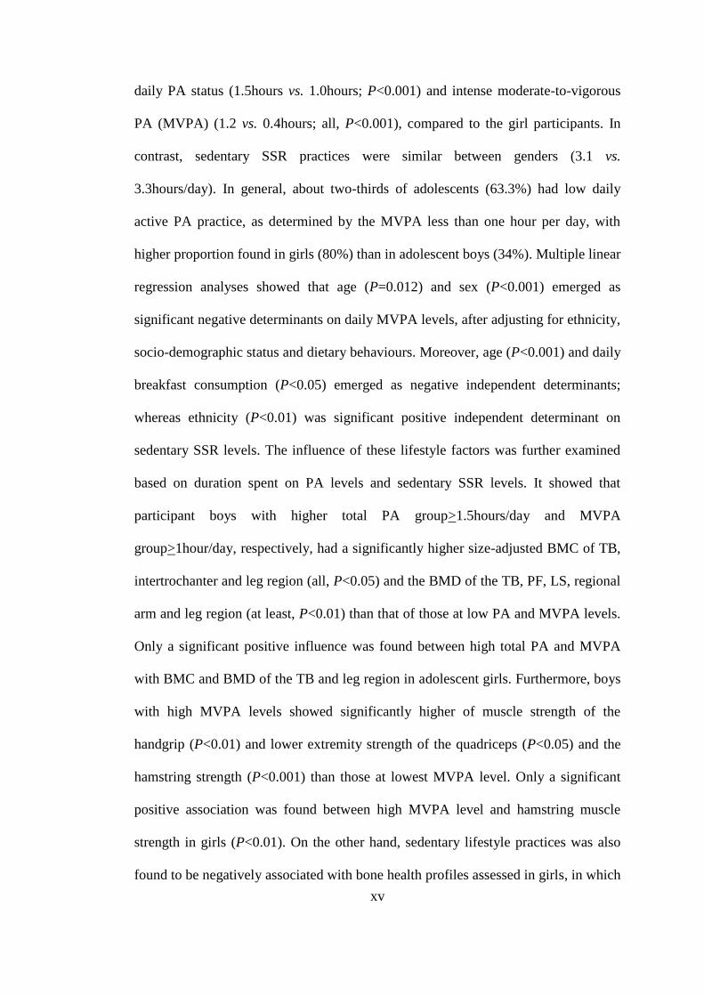
daily PA status (1.5hours vs. 1.0hours; P<0.001) and intense moderate-to-vigorous
PA (MVPA) (1.2 vs. 0.4hours; all, P<0.001), compared to the girl participants. In
contrast, sedentary SSR practices were similar between genders (3.1 vs.
3.3hours/day). In general, about two-thirds of adolescents (63.3%) had low daily
active PA practice, as determined by the MVPA less than one hour per day, with
higher proportion found in girls (80%) than in adolescent boys (34%). Multiple linear
regression analyses showed that age (P=0.012) and sex (P<0.001) emerged as
significant negative determinants on daily MVPA levels, after adjusting for ethnicity,
socio-demographic status and dietary behaviours. Moreover, age (P<0.001) and daily
breakfast consumption (P<0.05) emerged as negative independent determinants;
whereas ethnicity (P<0.01) was significant positive independent determinant on
sedentary SSR levels. The influence of these lifestyle factors was further examined
based on duration spent on PA levels and sedentary SSR levels. It showed that
participant boys with higher total PA group>1.5hours/day and MVPA
group>1hour/day, respectively, had a significantly higher size-adjusted BMC of TB,
intertrochanter and leg region (all, P<0.05) and the BMD of the TB, PF, LS, regional
arm and leg region (at least, P<0.01) than that of those at low PA and MVPA levels.
Only a significant positive influence was found between high total PA and MVPA
with BMC and BMD of the TB and leg region in adolescent girls. Furthermore, boys
with high MVPA levels showed significantly higher of muscle strength of the
handgrip (P<0.01) and lower extremity strength of the quadriceps (P<0.05) and the
hamstring strength (P<0.001) than those at lowest MVPA level. Only a significant
positive association was found between high MVPA level and hamstring muscle
strength in girls (P<0.01). On the other hand, sedentary lifestyle practices was also
found to be negatively associated with bone health profiles assessed in girls, in which

xvi
high SSR practice showed a significantly lower lumbar spine BMD (P<0.05) and
bone area of the TB (P<0.05), arm (P<0.01) and leg region (P<0.05) compared to
those who only practiced low SSR level. Blood biomarkers of bone remodeling were
significantly and negatively associated with all skeletal sites assessed, after
adjustments for pubertal growth and ethnicity. These present results showed that
higher habitual total and intense PA level could contributed to positive bone mass
profiles in these adolescents, and it has profoundly influence on weight-loaded
skeletal region assessed. In addition, sedentary lifestyle practices also exert a
negative influence on bone health assessed in adolescent girls. Therefore,
encouragement of active lifestyle practices in children and adolescents should be
promoted to optimize the peak bone mass accretion during the critical years of
growth.

1
CHAPTER 1
INTRODUCTION
1.0 Introduction of the osteoporosis
Osteoporosis is defined as a systematic skeletal disorder disease that characterized by
low bone mass, deterioration of skeletal architecture and compromised bone strength,
which consequently increased risk of bone fragility and susceptibility to fracture
(Consensus Development Conference, 1993). Several skeletal sites such as proximal
femur (hip), vertebrae (spine), and distal forearm (wrist) are regarded as most
commonly reported fractures attributed to osteoporosis, in which it exerts significant
clinical consequences to affected individual such as morbidity and prolonged
medical healthcare and medical expenses and to some extent may cause death due to
serious complications from osteoporotic-related fractures (Riggs and Melton III,
1995; Reginster and Burlet, 2006; Leboime et al., 2010).
An increasing prevalence of osteoporosis is considered as emerging serious public
health challenges worldwide. It is estimated that the prevalence of osteoporosis is
expected to rise to threefold in next 60 years from 1990 to 2050 worldwide (Prins et
al., 1998; Gullberg et al., 1997) and the healthcare cost is expecting to escalate
rapidly. For instance, hip fractures alone accounting for almost two third of the total
annual fractures expenses in USA (Ray et al., 1997) and it is reported that the annual
expenditures for medical treatment for osteoporotic-related fractures in 1995 is
estimated to cost around $13.8 billion in the United States, which the amount was far
exceeding the total expenses spent for breast and gynaecological cancers (Ray et al.,
1997; Hoerger et al., 1999). Moreover, it is found that approximately 10% to 20%

2
women whom sustained a hip fracture become totally dependent on their daily living
and more than 20% required a long-term nursing care for their normal daily activities
(Salkeld et al., 2000).
Osteoporotic fracture, especially the hip fracture, is considered to be a major public
health problem and challenge in Asian countries (Lau et al., 2001), with more than
half of the hip fractures (3.2 million) were expected to occur in Asia in 2050 (Cooper
et al., 1992). The trend and prevalence of osteoporosis is expecting to increase
dramatically due to increasing of the numbers of aging populations aged 65 years and
above from 145 million in 1990 to 894 million in 2050 (Cummings and Melton,
2002). In a recent data by Lau (2010), it is reported that approximately 17% and 29%
of all types of osteoporotic fractures, respectively, are found in the regions of the
Southeast Asia and West Pacific, as compared with 35% prevalence rates in Europe
regions (Lau, 2010), suggesting that osteoporosis is becoming as an alarming
healthcare burden, which could lead to serious adverse clinical consequences to an
affected individual and the nations (Cummings and Melton, 2002; Melton, 2000).
Based on the study of the Asian Osteoporosis Study (AOS) in four countries namely,
Hong Kong, Singapore, Malaysia and Thailand found that a significant increase in
the prevalence of hip fracture in these countries, i.e. 639, 606, 306 and 383 per
100,000 population respectively which approaching the hip fractures incidence in
Caucasian countries (Lau et al., 2001), whereby the prevalence of osteoporosis is
higher among women than that of their men counterparts.
3
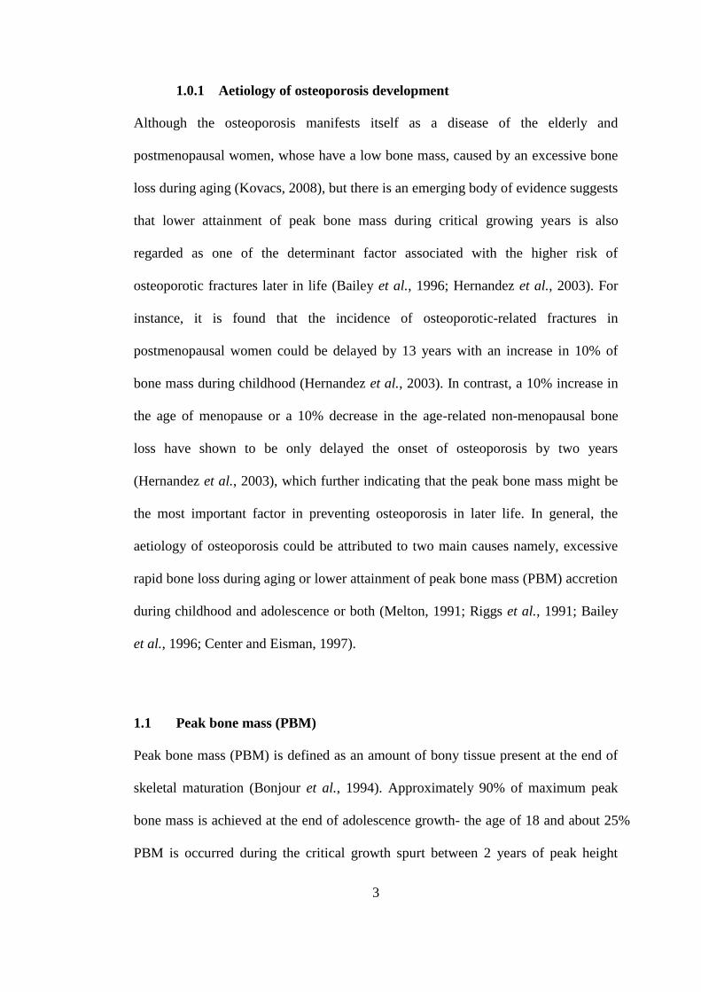
1.0.1 Aetiology of osteoporosis development
Although the osteoporosis manifests itself as a disease of the elderly and
postmenopausal women, whose have a low bone mass, caused by an excessive bone
loss during aging (Kovacs, 2008), but there is an emerging body of evidence suggests
that lower attainment of peak bone mass during critical growing years is also
regarded as one of the determinant factor associated with the higher risk of
osteoporotic fractures later in life (Bailey et al., 1996; Hernandez et al., 2003). For
instance, it is found that the incidence of osteoporotic-related fractures in
postmenopausal women could be delayed by 13 years with an increase in 10% of
bone mass during childhood (Hernandez et al., 2003). In contrast, a 10% increase in
the age of menopause or a 10% decrease in the age-related non-menopausal bone
loss have shown to be only delayed the onset of osteoporosis by two years
(Hernandez et al., 2003), which further indicating that the peak bone mass might be
the most important factor in preventing osteoporosis in later life. In general, the
aetiology of osteoporosis could be attributed to two main causes namely, excessive
rapid bone loss during aging or lower attainment of peak bone mass (PBM) accretion
during childhood and adolescence or both (Melton, 1991; Riggs et al., 1991; Bailey
et al., 1996; Center and Eisman, 1997).
1.1 Peak bone mass (PBM)
Peak bone mass (PBM) is defined as an amount of bony tissue present at the end of
skeletal maturation (Bonjour et al., 1994). Approximately 90% of maximum peak
bone mass is achieved at the end of adolescence growth- the age of 18 and about 25%
PBM is occurred during the critical growth spurt between 2 years of peak height
4
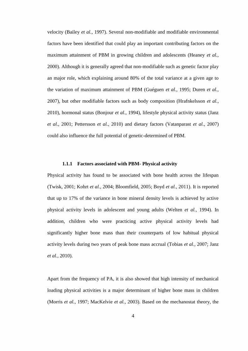
velocity (Bailey et al., 1997). Several non-modifiable and modifiable environmental
factors have been identified that could play an important contributing factors on the
maximum attainment of PBM in growing children and adolescents (Heaney et al.,
2000). Although it is generally agreed that non-modifiable such as genetic factor play
an major role, which explaining around 80% of the total variance at a given age to
the variation of maximum attainment of PBM (Guéguen et al., 1995; Duren et al.,
2007), but other modifiable factors such as body composition (Hrafnkelsson et al.,
2010), hormonal status (Bonjour et al., 1994), lifestyle physical activity status (Janz
et al., 2001; Pettersson et al., 2010) and dietary factors (Vatanparast et al., 2007)
could also influence the full potential of genetic-determined of PBM.
1.1.1 Factors associated with PBM- Physical activity
Physical activity has found to be associated with bone health across the lifespan
(Twisk, 2001; Kohrt et al., 2004; Bloomfield, 2005; Boyd et al., 2011). It is reported
that up to 17% of the variance in bone mineral density levels is achieved by active
physical activity levels in adolescent and young adults (Welten et al., 1994). In
addition, children who were practicing active physical activity levels had
significantly higher bone mass than their counterparts of low habitual physical
activity levels during two years of peak bone mass accrual (Tobias et al., 2007; Janz
et al., 2010).
Apart from the frequency of PA, it is also showed that high intensity of mechanical
loading physical activities is a major determinant of higher bone mass in children
(Morris et al., 1997; MacKelvie et al., 2003). Based on the mechanostat theory, the

5
direct effect of physical activity or exercise on bone mass is mainly based on the
association between the intensity of strain on bone and adaptation of bone to that
particular stimulus (van Der Meulen et al., 1993).
However, most studies of the influence of physical activity on bone mass accretion
among apparently healthy children and adolescents are largely focused on
Caucasian-origin populations, in which the intensity and duration of the habitual
physical activity level may different between Caucasian children and Asian children.
It is reported that the frequency and intensity of PA level of children and adolescents
in the Asian regions are relatively lower than the PA levels reported in Caucasian
populations (Rhodes et al., 2006; Foo et al., 2009). In addition, the precise
elucidation of the duration, frequency, intensity as well as the magnitude of physical
activity that is augmented greater bone mass accretion in growing children and
adolescents still remains uncertain among Asian populations. Therefore,
comprehensive understanding of various physical activity measures such as
magnitude, frequency and intensity of habitual physical activity is highly required in
order to determine the potential contributing factors associated with greater bone
mass in these growing Asian populations.
1.1.2 Factors associated with PBM- Sedentary practices
Apart from the active habitual PA status, there is an increasing evidence suggesting
that sedentary practices, also known as sedentary physiology, is associated with an
adverse effects on general well-being and health status (Hamilton et al., 2004; Healy
et al., 2008). It has been defined as the low end of physical activity continuum where
6
most of the changes in disease occur, in which it exerts a unique, independent and
qualitatively different effects on human physiological response and health
consequences, which is totally different than the physical activity (Hamilton et al.,
2004; Healy et al., 2008).
Several epidemiological studies found that sedentary lifestyles is associated
significantly with greater risk of non-communicable diseases such as type 2 diabetes
(Zimmet et al., 2001), coronary heart disease (Owen et al., 2009), obesity (Lakerveld
et al., 2011) and colon cancer (Powell and Blair, 1994). In addition, sedentary livings
are also found to be responsible for about one-third of deaths of those non-
communicable diseases (Powell and Blair, 1994). Those study findings also stated
that association between sedentary behaviour and bone mass accrual is mediated by
bone resorption without any concomitant changes in bone formation (Kim et al.,
2003; Smith et al., 2003). There is very limited information pertaining to sedentary
behaviour practices on the bone health, with only a study found out that the sedentary
behaviours associated with the bone mass attainment during the prepuberty among
adolescents when growth may relatively independently from the sex hormones
(Wang et al., 2003). Therefore apart from determine the influence of physical
activity and sedentary practices with bone mass accrual among adolescents, this
study is also aimed to elucidate the mechanism involved between physical activity
and sedentary with bone mass, whether this effect is mediated by the mechanism of
bone remodeling markers of bone formation and bone resorption and/ or parathyroid
hormones.
7
1.1.3 Factors associated with PBM- Blood bone markers
Biochemical markers of bone remodeling are also associated with the bone health
especially in the osteoporosis aspect. Biochemical markers of bone remodeling is
defined as the fragments of skeletal tissue proteins or enzymes specific to osteocytes
released into the circulation during the bone turnover or remodeling process (Carey
John et al., 2006), which is divided into two different groups namely, bone resorption
markers and bone formation markers. The resorption markers reflect its activity on
the osteoclast which is also known as the bone resorption cells and/or collagen
degradation, while the bone formation marker is an indicator involve in osteoblastic
synthetic activity or postrelease metabolism of procollagen (Christenson, 1997). The
occurrence of osteoporotic fractures in the elderly occurs is attributed to the higher
rate of bone turnover exceeds the bone formation. Thus, bone biochemical markers
for both bone formation and bone resorption are important to indicate and predict the
risk of occurrence of osteoporosis among elderly and postmenopausal women
(Seeman, 1994). Moreover, parathyroid hormone (PTH) is an 84-amino acid
polypeptide secreted by parathyroid glands which acts as the principal modulator by
response the small changes of Ca2+
(Rosen, 2004). Hence, PTH is important in
activating the interactions between osteoclasts (bone resorption) and osteoblasts
(bone formation). However, the use of biochemical markers of bone remodeling and
PTH in growing children and adolescents is still limited, which mainly focused on
illness children. Understanding the association between bone remodeling markers,
PTH and bone mass accretion as well as whether these effects are mediated by other
external factor such as physical activity and sedentary is particularly important in
bone homeostasis in growing children and adolescents. To our knowledge, none of

8
the studies have been investigated this mechanism in Asian population especially in
populations of diverse ethnicity.
1.2 Significance of the study
Several studies in last decade found out that around one-quarter of the total
adulthood bone mass are gained during the 2 years peak bone mass accrual period
(girls: 12.5 years and boys: 14.1 years) and almost same with the amount bone mass
that people will loss in their entire adulthood (Bailey et al., 1999; Bailey et al., 2000).
Therefore, maximum attainment of peak bone mass during growing years by
increasing engagement of physical activity and reducing the sedentarism should be
emphasized since the subsequent gain of bone mass will be minimal after achieving
peak bone mass. As known that most of the studies of lifestyle factors on bone health
are widely focused on the elderly and postmenopausal women (Lewiecki, 2008). In
addition, study of Ginty et al. (2005) stated that most studies of bone health
assessments in growing children and adolescents have not been carried-out at the
very specific age group such as pre-pubertal, pubertal and post pubertal-aged of
adolescents and mainly focused on adolescent girls (Ginty et al., 2005). Hence, the
present study subjects comprise whole range of adolescence period from early
pubertal to the late stage adolescent period. In addition, other important factors such
as race and sex differences are also taking into account.
There are many factors associated with physical activity and sedentary behaviour.
According to the reviews of Sallis et al. (2000) there are five groups which found
correlates of physical activity and sedentary behaviours, including demographic and

9
biological; psychchological, cognitive and emotional; behaviour attributes and skills;
and social and cultural (Sallis et al., 2000). Therefore, factor determinants
contributing physical activity and sedentary practices also investigated in this present
study to provide a more holistic picture and make up the important policy for
promoting active lifestyles and reducing sedentary behaviour, for instance by
implementing extra co-curriculum and physical education class in schools.
To date, there is limited studies have investigated the influence of physical activity,
sedentary practices, parathyroid hormone and bone biochemical markers on bone
mass accretion in growing children and adolescents. Therefore, the present
population-based study is formulate to understand the mechanism of these factors
involved in maximum attainment of bone mass accretion among these growing
adolescents in Kelantan, Malaysia.
1.3 Objectives
1.3.1 Main objective
To investigate the influence of physical activity, sedentary lifestyles, and blood
biochemical markers of bone remodeling on bone health status among adolescent
boys and girls of Malay and Chinese aged 12 to 19 years in Kota Bharu, Kelantan.
1.3.2 Specific objectives
i. To design and develop the newly past one-year computerized Physical
Activity Questionnaire (cPAQ) to assess the habitual physical activity pattern
among adolescent boys and girls.
10
ii. To compare the anthropometric measurements, dietary intakes, physical
activity, sedentary lifestyle, blood biochemical markers of bone remodelling,
muscle strength and bone mass profiles between adolescent boys and girls.
iii. To examine the socio-demographic and dietary behaviours determinants of
physical activity and sedentary practices.
iv. To examine the influences of physical activity on muscle strengths of
adolescent boys and girls.
v. To examine the influences of physical activity on bone mass status of
adolescent boys and girls.
vi. To examine the relationship between muscle strengths and bone mass status
of adolescent boys and girls.
vii. To examine the influences of sedentary practices on muscle strengths of
adolescent boys and girls.
viii. To examine the influences of sedentary practices on bone mass status of
adolescent boys and girls.
ix. To examine the influences of physical activity and sedentary practices on
body composition profiles of adolescent boys and girls.
x. To examine the relationship between biochemical markers of bone
remodeling, pubertal growth status and bone mass status of adolescent boys
and girls.

11
1.4 Research null hypotheses
i. There are no differences of the anthropometric measurements, dietary intakes,
physical activity, sedentary lifestyle, blood biochemical markers of bone
remodelling, muscle strength and bone mass profiles between adolescent
boys and girls.
ii. There is no influence of physical activity on muscle strengths of adolescent
boys and girls.
iii. There is no influence of physical activity on bone mass status of adolescent
boys and girls.
iv. There is no relationship between muscle strengths and bone mass status of
adolescent boys and girls.
v. There is no influence of sedentary practices on muscle strengths of adolescent
boys and girls.
vi. There is no influence of sedentary practices on bone mass status of adolescent
boys and girls.
vii. There is no influence of physical activity and sedentary lifestyle practices on
body composition profiles of adolescent boys and girls.
viii. There is no relationship between biochemical markers of bone remodeling,
pubertal growth status and bone mass status of adolescent boys and girls.
1.5 Conceptual framework
Physical activity and sedentary behaviour practices during childhood and
adolescence are considered as one of the major determinants that positively
associated with better health outcomes. In the present study, the main focus was to
12
determine the relationship of physical activity and sedentary behaviour practices and
its correlate factors with general characteristics and socio-demographic status, as
well as its association with bone health and body composition status in population-
based adolescents aged 12 to 19 years, as illustrated by the study framework in
Figure 1.1. Physical activity and sedentary practices in adolescents are associated
with five main factors such as demographic, psychological, behavioural, social and
cultural as well as inter-mediated with significant and important determinants of
muscular strength, blood biochemical bone markers and parathyroid hormone by
further influencing the quality, size and mass of bone accrual when peak bone mass
achieved during the critical growing periods.
1.6 Definition of terminology
Osteoporosis. It is defined as the bone mineral density (BMD) as assessed by
dual energy X-ray absorptiometry (DXA) as less than a 2.5 standard deviation
below the normal mean (T-score < -2.5).
Bone health. It is determined by the level bone mass status, bone quality, bone
size as well as the presence of skeletal deterioration of bone tissue.
Adolescents. They are defined as the young people between the ages of 10 and
19 years who are often thought as a healthy group by World Health Organization.
Physical activity. It is defined as any bodily movement produced by skeletal
muscle that results in energy expenditure with more than or equal to 2.0

13
metabolic equivalents (METs) in which the low or light PA is defined as <3
METs and active or moderate-to-vigorous PA is 3 METs.
Sedentary practice. It refers to the low energy expenditure above the resting
metabolic rate but below the expenditure of light PA with 1.0-1.5 METs.
Figure 1.1 A conceptual framework of influences of physical activity on bone
health status in adolescents
Lifestyle
Practices
Adolescents
Physical
activity
Sedentary
behaviour
1) Mechanical
loading
forces
2) Human
physiologic
al response
Suppress/
activate
Suppress/
activate
1) High/ low
bone mass
2) High/low
bone quality
3) High/low
bone size
4) Skeletal
deterioration
Bone
Health
Inter Mediators
- Muscle strength
- Bloodbiochemical
markers/ PTH
Moderators
- Demographic
- Psychological
- Behavioral
- Social
- Cultural
14
CHAPTER 2
LITERATURE REVIEW
2.0 Overview of osteoporosis
Osteoporosis is defined as “a systemic skeletal disorder characterized by low bone
mass, micro-architectural deterioration of bone tissue and compromised bone
strength, consequently increased risk of bone fragility and fractures” (WHO, 1994).
Osteoporosis is considered as a “silent disease” because it is usually known when it
is a bone fractures occurred and it happened without any symptoms (WHO, 1994;
Lin and Lane, 2004). Operationally, it is defined as the bone mineral density (BMD),
as assessed by the dual energy X-ray absorptiometry (DXA) as less than a 2.5
standard deviation (SD) below the normal mean (T-score <-2.5), whereas person
with BMD more than 1.0 SD but less than or equal to 2.5SD below the mean (-1.0>
T-score >-2.5) is classified as “osteopenia”, or also known as low bone mass, and
those with the BMD more than or equal to 1.0 SD of the normal mean T-score of the
BMD is considered as normal (WHO, 1994). It has been well established that low
BMD status is significantly associated with increased risk of osteoporotic fractures
among postmenopausal women and elderly. For instance, a decreased in 1SD of
BMD, the risk of having hip fractures is increased by 2.6-fold (Cummings et al.,
1993), and the two times higher risk of vertebrae fracture (Wasnich, 1993), compared
to those who have their BMD of more or equal to 1SD T-score.
Osteoporosis affects over 200 million people worldwide, with significant clinical
consequences, not only restricted to the immediate pain as a result of osteoporotic
fracture, but it also contributes to significant increase risk of morbidity and mortality

15
(Riggs and Melton III, 1995; Reginster and Burlet, 2006; Leboime et al., 2010). A
worldwide incidence of hip fracture alone in men and women is projected to increase
by 2 to 3 folds by the year of 2050 (Gullberg et al., 1997). It is estimated the lifetime
risk of osteoporotic fractures of the hip, vertebral and the radius are estimated to
about 40%, which is equivalent to the risk of coronary heart disease (WHO, 2003). In
addition, the one-in-six lifetime risk of sustaining a hip fracture in women of
Caucasian-origin, which is greater than that of the risk of cancer risk (one-in-nine
risk of developing breast cancer) (Melton, 2000).
The prevalence of osteoporosis is increasing among populations in Asia region,
making it as one of the major public health challenges and problems in Asian
populations due to advancing of aging populations. It is estimated that by 2050, the
aging populations aged 65years and above will reach to approximately 894 million
elderly living in Asia countries (Cummings et al., 2002). Moreover, it is projected
that more than 50% of total hip fracture is projected to occur in Asia (3.2 million per
year) by the year 2050 (Cooper et al., 1992). Furthermore, 46% of all types of
osteoporotic fractures are reported in the Southeast Asia and West Pacific. In
addition, the estimated disability-adjusted life years lost was 24.7 million in
Southeast Asia and West Pacific (34% of the world figure) (Lau, 2010). Moreover, in
a regional study of Asian Osteoporosis study, it showed that Hong Kong and
Singapore were among the highest rates of hip fracture, followed by Malaysia and
Thailand. In addition, the prevalence of hip fracture of some Asia countries was
reported to be similar to that of Caucasian populations in the United States and in
United Kingdom (Lau et al., 2001) (Table 2.1), in which elderly women and

16
postmenopausal women is considered at high risk of osteoporotic fracture compared
to their male counterpart (Lau et al., 2001; Cummings et al., 2002).
The common sites of osteoporotic fractures are hip, vertebra and wrist. Of all these
skeletal sites, hip fractures is considered as a most serious form of osteoporotic
fracture compared to fractures of any other skeletal sites such as at vertebral or wrist
fractures. For instance, hip fracture increases the mortality risk by 20% during the
first year of fracture. In addition, the rate of mortality within 90days among patients
with osteoporotic fractures aged65years and above is unexpectedly high, in which
the risk of early lethality could increase to about 6times and 4times in men and
women , respectively compared to those elderly without any osteoporotic fracture
(Roberts and Goldacre, 2003).
Osteoporotic fractures is required a long term health care and high health care
expenses. For instance, it is estimated that approximately one-quarter of affected
people are require a long-term healthcare due to long-term disability to perform their
normal daily life after fractures (Jensen and Bagger, 1982; Clayer and Bauze, 1989).
High cost and healthcare expenses are required such as hospitalization and long term
nursing home care (Melton et al., 2004; U.S Department of Health and Human
Services, 2004). Similar healthcare cost burden is also reported in Asian countries.

17
Table 2.1 The incidence of hip fracture in four Asian countries with United
States and Oxford, England (per 100,000 populations).
Hong
Konga
Singaporea Malaysia
a Thailanda US
b Oxford,
Englandc
Women (F) 459 442 218 269 535 603
Men (M) 180 164 88 114 189 114
Total 639 606 306 383 724 717
Ratio (F: M) 2.55 2.70 2.48 2.36 2.83 5.29
Source: aLau et. al. (2001),
bHo et. al. (1993) and
cVilla et. al. (2001)
For instance, it is reported that costs related to treatment and rehabilitation attributed
to hip fracture is exceeded 1% of the total hospital services cost, which is
approximately about HK$130 million in Hong Kong (Lau, 1997). In addition,
approximately 500,000 hospital bed-nights have been occupied by osteoporotic
patients annually in European Union in 1998 and it is expecting to increase to double
by the year of 2050 (Delmas and Fraser, 1998).
2.0.1 Aetiology of osteoporosis
Osteoporosis is generally regarded as a disease that is manifested in postmenopausal
women and elderly, as a result of excessive bone loss, leading to risk of osteoporotic
fractures (Ensrud et al., 1995). However, there is growing body of evidence also
indicates that osteoporosis may have its origin during early stage of lifespan. it has
documented that low attainment of peak bone mass (PBM) during the critical

18
growing years is significantly associated with greater risk of osteoporotic fractures
later in life (Bailey et al., 1996; Hernandez et al., 2003).
In general, the aetiology of osteoporosis could be attributed to two main determinants,
which is an excessive or rapid bone loss in ageing or/and low peak bone mass (PBM)
accretion during childhood and adolescence (Melton, 1991; Bailey et al., 1996;
Center and Eisman, 1997). A growing body of evidence showing that maximum
attainment of the PBM during first two decades of lifespan is regarded as an
important strategy to prevent the risk of osteoporotic fractures in later life, apart from
the prevention of the rapid bone loss during ageing (Bachrach and Smith, 1996;
Dombrowski, 2000). It is estimated that the risk of osteoporotic-related fractures in
postmenopausal women could be delayed by 13 years with an 10% increases in bone
mass accrued during childhood and adolescence, whereas a 10% increase in the age
of menopause or a 10% decrease in the age-related bone loss could only help to delay
the onset risk of osteoporotic fractures only by two years (Hernandez et al., 2003).
This suggests that maximum attainment of PBM during the growing years is
ultimately important factor that could help to prevent the risk of osteoporotic
fractures in later life.
2.1 Bone structure and physiology
Bone is a unique and complex tissue with its main functions is to provide mechanical
support for the weight bearing and locomotive activity. Skeletons is also serves as
attachment sites for the muscles, ligaments and tendons, central reservoir for calcium,
a protector of vulnerable internal organs and a site for haematopoiesis (Baron, 2003;
Pearson and Lieberman, 2004). By fulfilling these requirements, skeletons
19
continually adapts to the mechanical and physiological demands placed upon it.
During growth process, the skeleton maintain these functions while undergoes a
dramatic changes in size and shape. Skeleton could be divided into the following five
types based on the basis of shape, namely flat, short, irregular, sesamoid, and long
bones. Flat bones consists of skull, scapula, mandibula, and ileum are formed by the
ossification of membranes (intramembranous bone formation) independently of
cartilage. Long bones such as tibia, femur and humerus are formed by the deposition
of mineralised tissue preceded by cartilage analogue (endochondral bone formation
or ossification) during the modelling process (Raisz, 1999). The long bones consists
of two epiphyses with a midshaft (diaphysis) and a metaphysis (developmental zone)
(Buckwalter and Cooper, 1995a). In growing bone, the epiphysis and metaphysis are
separated by cartilage or growth plate that responsible for a longitudinal bone growth
and becomes calcified at the end of the longitudinal growth after pubertal growth
during adolescence (Ganong, 2003).
Bone tissue in general can be divided into two different bone components namely,
cortical (or compact) and trabecular (spongy or cancellous) bone. Cortical bone is
arranged in cylinders that align with the long axis and made up of dense and calcified
tissue, with approximately 80 to 90% of its bone volume is made up of calcium.
Trabecular bone exists as a three-dimensional lattice structure composed of an inner
network of thin calcified trabeculae, with only 15 to 25% of its bone volume are
made by calcium. Trabecular bone is present at the end of long bones, in vertebral
bodies and nearby joint surface, and in flat bones (Khan et al., 2001). In general,
approximately 80% of the skeleton is cortical bone and only 20% is attributed to the
20
trabecular bone. Cortical bone primarily serves mechanical and protective functions;
whereas trabecular bone is involved in metabolic activity (Watkins and Seifert, 2000).
Bone is a connective tissue composed of cells and extracellular matrix having
inorganic and organic components. On the basic level by weight, bone tissue consists
of approximately 70% of mineral (or inorganic matter), about 20 to 25% of organic
matrix, and remaining 5% as water. However, the major variation in the degree of
mineralization in bone is depending on function within the skeletons. Major mineral
(inorganic) component in bone extracellular matrix is the mixture of calcium and
phosphorus in crystalline hydroxyapatite [Ca10 (PO4)6 (OH)2], which composing of
95%, whereas the rest is composed of calcium carbonate, calcium citrate and
magnesium (Seeman and Delmas, 2006). These crystals are found within the
collagen fibres, that giving the rigidity and compressive strength to the skeletons
(Currey, 2002).
Osteoblasts, osteoclasts and bone-lining cells are found on bone surface, whereas
osteocytes permeate the mineralized interior. Osteoblasts are the fully differentiated
bone-forming cells which originate from mesenchymal stem cells. The osteoblasts
synthesis and secrete collagen and non-collagenous proteins that comprise the
organic matrix of bone (osteoid) and subsequently mineralize the organic bone
matrix (Parfitt, 2002; Baron, 2003; Seeman and Delmas, 2006). Subsequently, some
osteoblasts disappear through a process of apoptosis (cell death); others differentiate
into flat cells lining the bone surface (bone-lining cells) or are embedded in lacunae
in the bone matrix after morphologic changes (osteocytes) (Burger and Klein-Nulend,
1999; Parfitt, 2002). On the other hand, osteocytes are osteoblasts that stopped
matrix synthesis and are embedded deep within small bone cavities (osteocytic

21
lacunae). Osteocytes are connecting to adjacent osteocytes as well as with osteoblasts
and bone-lining cells through the cytoplasmic network projecting through small
canals between lacunae (as called canaliculi) in the mineralized bone matrix. This
connection or communication is critical for mechno-transduction also known as
mechanosensitivity of the bone (Mosekilde, 1995; Burger and Klein-Nulend, 1999).
Osteoclast is a large multinucleated bone-resorbing cell that derived from the
haematopietic stem cells, in which it is responsible for bone resorption and usually
found in cavities on bone surfaces that known as the resorption pit (Mosekilde, 1995;
Watkins and Seifert, 2000). During bone resorption process, osteoclasts secrete
lysosomal enzymes, hydrogen protons and free radicals into a confined space next to
bone and dissolve or degrade the bone matrix (Frost, 1987; Watkins and Seifert,
2000).
2.1.1 Bone remodeling process through the life spans
The cellular mechanisms responsible for the adaptation of bone tissue are modeling
(construction) and remodeling (reconstruction) (Figure 2.1). Initially, modeling
forms mineralized bone at developmentally determined skeletal sites during growth
or acts as an adaptive response to external mechanical loading by simultaneous
processes of the resorption and formation at different sites (Frost, 1987; Seeman,
2003). In growing skeleton, modeling is dominant and during this period of life, a
significant change in bone shape that leading to greater bone strength (Frost, 1990).
In mature skeleton during adulthood, bone remodeling process is constantly
remodeled by the resorption followed by bone formation in the bone tissues, in
which osteoblastic bone formation is coupled together with osteoclastic bone

22
resorption to restore bone loss (Buckwalter and Cooper, 1995b). Normally, the
osteoblastic and osteoclastic activities are balanced in adults and remodeling.
Figure 2.1 Bone modeling and remodeling. Osteoclasts break down old bone
tissue and followed by osteoblasts activity, in which osteoblasts form new collagen
and other matrix proteins. Then, the collagenous matrix undergoes mineralization
(Adapted from Baron, 2003)
However, during aging and postmenopausal periods, processes of bone remodeling is
occurs dramatically, that consequently resulting in trabecular thinning, disappearance
and loss of connectivity, cortical thinning and increased intracortical porosity
(Ahlborg et al., 2003; Seeman, 2003).
23
Several blood biochemical metabolite components such as fragments of skeletal
tissue proteins or enzymes specific to osteocytes are released into circulation system
when bone matrix is formed and degraded during the bone remodeling process.
These bone metabolites provide dynamic information on skeletal remodeling status
through its metabolic activity, in which this metabolite analysis could be used to
assess the biochemical markers of bone remodeling (Carey et al., 2006). In general,
these metabolites resulted from the bone turnover can be generally grouped into bone
formation markers and bone resorption markers. Biochemical bone formation marker
is used as indicator of osteoblast synthetic activity and also by-products of collagen
synthesis, matrix proteins or osteoblastic enzymes. There are several bone formation
markers that are commonly used such as blood osteocalcin (OC), total alkaline
phosphatase (total ALP), bone-specific alkaline phosphatase (BALP) and total
procollagen type I amino-terminal propeptide (P1NP). On the contrary, the bone
resorption marker is used to assess the activity of osteoclast. Majority of these
resorption markers are used to assess the degradation products of bone collagen and
some non-collagenous proteins and osteoclast-specific enzymes (Christenson, 1997;
Seibel, 2005; Vasikaran et al., 2011). Several bone resorption markers that are used
such as the C-terminal telopeptide of type I collagen (CTx) and N-terminal
telopeptide of type I collagen (NTx) (Seibel, 2005).
Apart from the bone biochemical markers of bone remodeling, calcitropic hormones
of vitamin D and parathyroid hormone (PTH) are also play a critical mediator role on
skeletal development and remodeling. Vitamin D is fat-soluble and can promote
calcium absorption from the intestine. Adequate vitamin D concentrations can
prevent bone loss and reduce the fracture risk among the elderly (Dawson-Hughes et
24
al., 1995; Dawson-Hughes et al., 1997) and musculoskeletal health and functions in
growing children and adolescents (El-Hajj Fuleihan et al., 2006; Foo et al., 2009).
The assessment of vitamin D is based on the measurement of serum 25-
hydroxyvitamin D, which is considered as the main circulating metabolite form of
vitamin D in the body (Lips et al., 1999). Whereas PTH is defined as an 84-amino
acid polypeptide that secreted by parathyroid glands in response to relatively small
changes in serum Ca2+
, which is responsible for calcium homeostasis (Rosen, 2004).
PTH synthesis is increased when low serum calcium level is sensed parathyroid
glands, in which it is crucial for maintaining enzymatic process, cell membranes’
stability and permeability and mineralization of newly formed bone (Kraenzlin and
Meier, 2011). It acts as a potent stimulator of bone resorption during states of
reduced ionized calcium availability in the blood and has biphasic effects on bone
formation (Raisz, 1999). A high PTH concentrations result an acute inhibition of
collagen synthesis but prolonged intermittent administration of PTH produces
increased bone formation; thus, it is being explored clinically as anabolic agent
(Dempster et al., 1993; Frolik et al., 2003). The anabolic effects of PTH are appeared
to be partly site-specific, and this might due to the ability of PTH to activate
mechanical loading pathways (Bakker et al., 2003a). In general, process of bone
modeling and remodeling is influenced by several factors such as sex, physiological
growth factors, nutritional and lifestyle physical activity status, and also other
hormonal status such as vitamin D, PTH, cytokines, prostaglandins and
glucocortisords (Seeman and Delmas, 2006; Shapiro, 2008).