influence of root conditioning prior to emd application on...
TRANSCRIPT

543
IntroductionAberrant healing after replantation of avulsed teeth often resulted from irreversible damage to the periodontal ligament caused by traumatic injury, long extraoral dry time or microbial infection [1-7]. It is suggested that the remaining periodontal ligament cells near severely damaged periodontium have the potential to populate on the necrotic area of the root surface and enhance normal periodontium. We reported that using tissue culture, the healthy periodontal ligament cells could populate on the area severely damaged by trauma during tooth extraction in an animal model [8]. Aberrant healing such as root resorption and ankylosis could then be suppressed after teeth implantation into the bone cavity [9].
The enamel matrix proteins (EMD) were used to commercially develop a product called Emdogain for regeneration of injured periodontal tissues. It was reported that EMD promoted periodontal ligament cells to populate and make new cementum [10-12]. Following application of EMD on the root surface of teeth before implantation, healthy periodontal ligament cells can populate on the area severely damaged by trauma during tooth extraction [13]. In 2001 and 2003, EMD was suggested for treatment of avulsed teeth replanted after 60 min in dry extraoral conditions by the International Association of Dental Traumatology (IADT) and the American Association of Endodontists [14,15]. For advanced effects of EMD, it was recommended that root conditioning be performed prior to application of EMD on the
root surface. Root conditioning is aimed to remove the smear layer, demineralize acellular cementum and expose collagen. Some studies recommended the use of 36% ortho-phosphoric acid [16], 3% citric acid [17], enzymes [18], sodium fluoride [19], sodium hypochlorite, and 24% EDTA [20,21] for root conditioning. However, some studies do not support the use of root conditioning for enhancing the effect of EMD in vivo [22]. There has been no study that directly shows the influence of root conditioning just before application of EMD on the migration, proliferation, attachment and Alkaline Phosphatase (ALP) activity of periodontal ligament cells. The purpose of this study was to investigate the influence of root conditioning on cell proliferation and ALP activity increased by application of EMD in vitro.
Materials and MethodsPreparation of teethThirty maxillary incisors of five beagle dogs (female, 1 year old; mean weight, 10.3 kg) were used. This study protocol followed the guidelines for care and use of laboratory animals of Hokkaido University Graduate School of Medicine.
The surgical procedures were performed under anesthesia by an intramuscular injection of medetomidine hydrochloride (5 μg/kg, Domitor®; Meiji Seika, Tokyo, Japan) and ketamine hydrochloride (2.9 mg/kg, Ketaral 50®; Sankyo, Tokyo, Japan) and local infiltration (xylocaine 2% with 1:80,000 epinephrine, Xylocaine®; DENTSPLY SANKIN, Tokyo,
Influence of Root Conditioning Prior to EMD Application on Periodontal Ligament Cells of Extracted Teeth
Akira Saito1, Emiko Saito2
1Department of Oral Rehabilitation, Division of Oral Functional Science, Hokkaido University Graduate School of Dental Medicine, Sapporo, Japan. 2Department of Periodontology and Endodontology, Division of Oral Health Science, Hokkaido University Graduate School of Dental Medicine, Sapporo, Japan.
AbstractBackground: Root conditioning prior to the application of Enamel Matrix Derivative (EMD) on the root surface was suggested for treatment of replanted avulsed teeth. However, some studies do not support the use of root conditioning. The purpose of this study was to investigate the effects of root conditioning on cell proliferation and Alkaline Phosphatase (ALP) activity on application of EMD in vitro. Methods: Thirty incisors of five beagle dogs were used. The roots were hemisectioned axially and the periodontal ligament and cementum were removed from the coronal half of the roots. The specimens were treated with or without root conditioning (24% EDTA for 2 minutes or 36% ortho-phosphoric acid for 60 seconds) and EMD were or was not applied. Then all specimens were observed using SEM or cultured for 4 weeks. After culture, the cell migration and alkaline phosphatase activity of migrated and proliferated cells on the root planed surface from the remaining periodontal ligament was histometrically observed.Results: The cell population in EMD-applied groups was significantly higher than that without EMD application groups. There was no significant difference among the two root conditioning protocols and EMD alone. ALP activity of migrated and proliferated cells in the EMD in combination with EDTA group was significantly higher than the other groups. However, 36% ortho-phosphoric acid group showed the lowest value. Conclusion: The findings in this study showed that EMD in combination with 24% EDTA showed additional benefit in terms of population of cells expressed APL activity on the root surface compared with application of EMD alone.
Key words: Enamel matrix derivative, EDTA, Root conditioning, Periodontal ligament, Replantation, Alkaline phosphatase activity
Corresponding author: Emiko Saito, DDS, PhD, Department of Periodontology and Endodontology, Division of Oral Health Scien-ce, Hokkaido University Graduate School of Dental Medicine Kita-13, Nishi-7, Kita-ku, Sapporo 060-8586, Japan, Tel: +81 11 706 4266; Fax: +81 11 706 4334; e-mail: [email protected]
544
OHDM - Vol. 13 - No. 2 - June, 2014
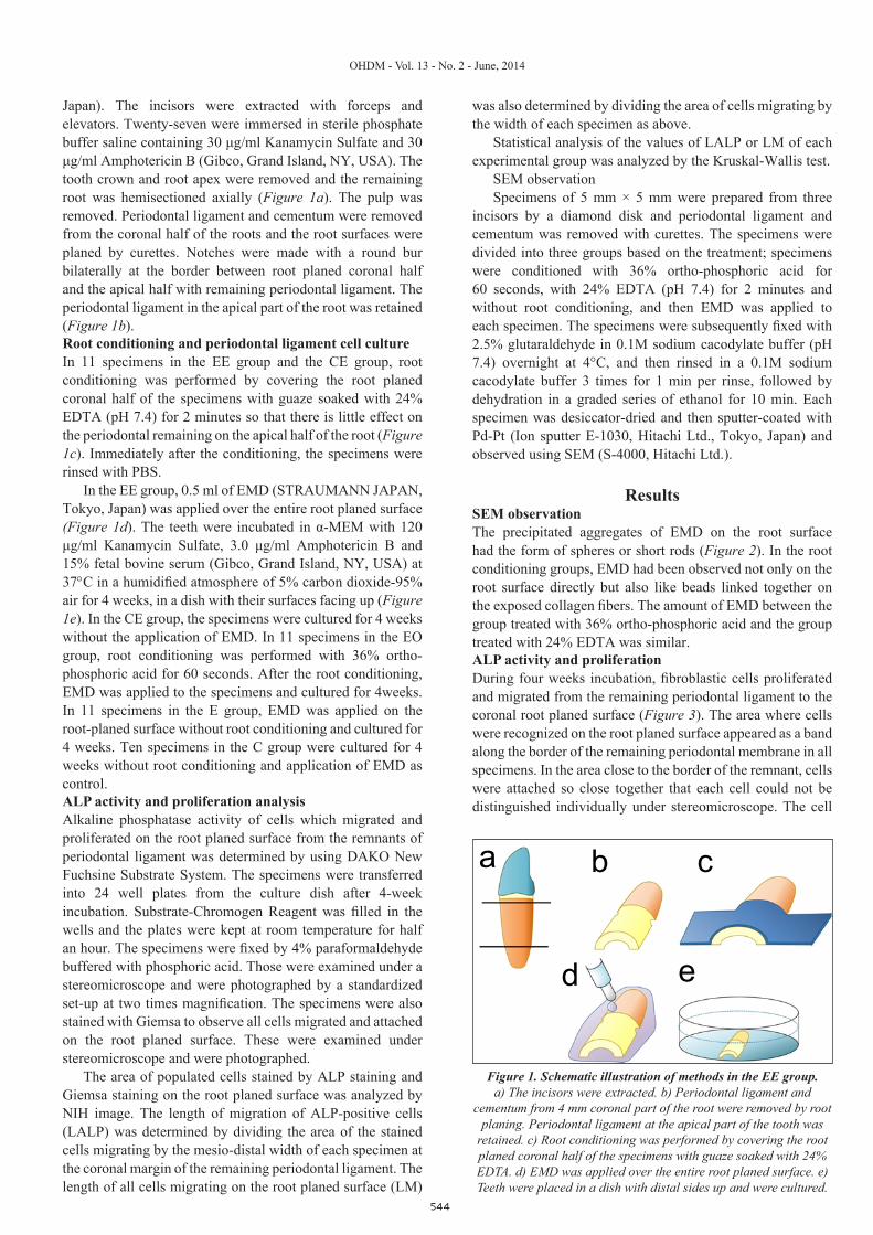
Japan). The incisors were extracted with forceps and elevators. Twenty-seven were immersed in sterile phosphate buffer saline containing 30 μg/ml Kanamycin Sulfate and 30 μg/ml Amphotericin B (Gibco, Grand Island, NY, USA). The tooth crown and root apex were removed and the remaining root was hemisectioned axially (Figure 1a). The pulp was removed. Periodontal ligament and cementum were removed from the coronal half of the roots and the root surfaces were planed by curettes. Notches were made with a round bur bilaterally at the border between root planed coronal half and the apical half with remaining periodontal ligament. The periodontal ligament in the apical part of the root was retained (Figure 1b). Root conditioning and periodontal ligament cell cultureIn 11 specimens in the EE group and the CE group, root conditioning was performed by covering the root planed coronal half of the specimens with guaze soaked with 24% EDTA (pH 7.4) for 2 minutes so that there is little effect on the periodontal remaining on the apical half of the root (Figure 1c). Immediately after the conditioning, the specimens were rinsed with PBS.
In the EE group, 0.5 ml of EMD (STRAUMANN JAPAN, Tokyo, Japan) was applied over the entire root planed surface (Figure 1d). The teeth were incubated in α-MEM with 120 μg/ml Kanamycin Sulfate, 3.0 μg/ml Amphotericin B and 15% fetal bovine serum (Gibco, Grand Island, NY, USA) at 37°C in a humidified atmosphere of 5% carbon dioxide-95% air for 4 weeks, in a dish with their surfaces facing up (Figure 1e). In the CE group, the specimens were cultured for 4 weeks without the application of EMD. In 11 specimens in the EO group, root conditioning was performed with 36% ortho-phosphoric acid for 60 seconds. After the root conditioning, EMD was applied to the specimens and cultured for 4weeks. In 11 specimens in the E group, EMD was applied on the root-planed surface without root conditioning and cultured for 4 weeks. Ten specimens in the C group were cultured for 4 weeks without root conditioning and application of EMD as control.ALP activity and proliferation analysisAlkaline phosphatase activity of cells which migrated and proliferated on the root planed surface from the remnants of periodontal ligament was determined by using DAKO New Fuchsine Substrate System. The specimens were transferred into 24 well plates from the culture dish after 4-week incubation. Substrate-Chromogen Reagent was filled in the wells and the plates were kept at room temperature for half an hour. The specimens were fixed by 4% paraformaldehyde buffered with phosphoric acid. Those were examined under a stereomicroscope and were photographed by a standardized set-up at two times magnification. The specimens were also stained with Giemsa to observe all cells migrated and attached on the root planed surface. These were examined under stereomicroscope and were photographed.
The area of populated cells stained by ALP staining and Giemsa staining on the root planed surface was analyzed by NIH image. The length of migration of ALP-positive cells (LALP) was determined by dividing the area of the stained cells migrating by the mesio-distal width of each specimen at the coronal margin of the remaining periodontal ligament. The length of all cells migrating on the root planed surface (LM)
was also determined by dividing the area of cells migrating by the width of each specimen as above.
Statistical analysis of the values of LALP or LM of each experimental group was analyzed by the Kruskal-Wallis test.
SEM observationSpecimens of 5 mm × 5 mm were prepared from three
incisors by a diamond disk and periodontal ligament and cementum was removed with curettes. The specimens were divided into three groups based on the treatment; specimens were conditioned with 36% ortho-phosphoric acid for 60 seconds, with 24% EDTA (pH 7.4) for 2 minutes and without root conditioning, and then EMD was applied to each specimen. The specimens were subsequently fixed with 2.5% glutaraldehyde in 0.1M sodium cacodylate buffer (pH 7.4) overnight at 4°C, and then rinsed in a 0.1M sodium cacodylate buffer 3 times for 1 min per rinse, followed by dehydration in a graded series of ethanol for 10 min. Each specimen was desiccator-dried and then sputter-coated with Pd-Pt (Ion sputter E-1030, Hitachi Ltd., Tokyo, Japan) and observed using SEM (S-4000, Hitachi Ltd.).
ResultsSEM observationThe precipitated aggregates of EMD on the root surface had the form of spheres or short rods (Figure 2). In the root conditioning groups, EMD had been observed not only on the root surface directly but also like beads linked together on the exposed collagen fibers. The amount of EMD between the group treated with 36% ortho-phosphoric acid and the group treated with 24% EDTA was similar.ALP activity and proliferationDuring four weeks incubation, fibroblastic cells proliferated and migrated from the remaining periodontal ligament to the coronal root planed surface (Figure 3). The area where cells were recognized on the root planed surface appeared as a band along the border of the remaining periodontal membrane in all specimens. In the area close to the border of the remnant, cells were attached so close together that each cell could not be distinguished individually under stereomicroscope. The cell
a b c
ed
Figure 1. Schematic illustration of methods in the EE group.a) The incisors were extracted. b) Periodontal ligament and
cementum from 4 mm coronal part of the root were removed by root planing. Periodontal ligament at the apical part of the tooth was
retained. c) Root conditioning was performed by covering the root planed coronal half of the specimens with guaze soaked with 24% EDTA. d) EMD was applied over the entire root planed surface. e) Teeth were placed in a dish with distal sides up and were cultured.

545
OHDM - Vol. 13 - No. 2 - June, 2014
density seemed to decrease from the border to coronal part.Means of LMs in the Group E, EO, and EE were
significantly higher than in the CE group and the C group (Table 1). There were no significant differences among the three groups with root conditioning. Mean LM in the CE group was larger than in the C group but it was insignificant.
The area in which cells were stained with DAKO New Fuchsin Substrate System appeared as a narrower band continuously to the border of the remaining periodontal ligament. Mean LALP in the EE group was highest of all experimental groups, and was significantly higher than in the Group EO, E, CE and Control. Mean LALP in the E group was second highest, and was significantly higher than the
Group EO, CE and C. The values in the Groups EO and CE did not show significant difference with the control group.
DiscussionIn this study, the influence of root conditioning beforehand application of EMD on cell proliferation and ALP activity was examined histologically in vitro. The cell culture in this study was performed using previously published techniques [8]. These cells populating from remnants of periodontal ligament had formed a single layer on the root planed surface initially and changed into multilayer with time. Cells staining positive for Proliferating Cell Nuclear Antigen (PCNA) were markedly observed not only in the remaining periodontal
Figure 2. SEM observation of root surfacea) Root surface in the EMD-application without root conditioning group. b) Root surface in the EMD-application with EDTA conditioning group.
c) Root surface in the EMD-application with phosphoric acid conditioning group.
EMD only EDTA + EMD ortho-phosphoric acid + EMD
Figure 3. Proliferated cells and ALP positive cells on the planed root surface. a) Giemsa staining in the C group. b) Giemsa staining in the CE group. c) Giemsa staining in the E group. d) Giemsa staining in the EO group.
e) Giemsa staining in the EE group. f) ALP staining in the C group. g) ALP staining in the CE group. h) ALP staining in the E group. i) ALP staining in the EO group. j) ALP staining in the EE group.

546
OHDM - Vol. 13 - No. 2 - June, 2014
ligament near the border of the root planed surface but also in a single layer on the coronal root planed surface [8]. Herr et al. reported that periodontal ligament fibroblasts in the coronal portion of the remaining periodontal ligament close to wounds proliferated actively along the root surface and formed fibrous connective tissue using Bromodeoxyuridine (BrdU) labeling in beagle dogs [23]. These findings seemed similar to the present culture system. However, the findings in this study need to be distinguished from reactions in vivo because this culture system lacked vascular formation and interaction of the periodontal ligament cells with the surrounding tissue. So the data from this study cannot be used clinically. However, it is an advantage that this culture system provides a very effective way to visually evaluate the distance of cell populating/migration from the remaining periodontal ligament. We investigated the association between root conditioning and the effect of EMD using this culture system in this study.
The precipitated aggregates of EMD on the root surface had the form of spheres or short rods. In the root conditioning groups, EMD had been observed not only on the root surface directly but also like beads linked together on the exposed collagen fibers. This finding was in agreement with the findings of a previous report which suggested that EMD adsorbed both to hydroxyapatite and collagen [24]. The amount of adsorbed EMD in the root conditioning groups seemed greater than the groups without root conditioning. The amount of adsorbed EMD seemed similar between root conditioning with 36% ortho-phosphoric acid and root conditioning with 24% EDTA.
In this study, EMD stimulated migration/proliferation of the periodontal ligament cells. These findings supported the previous reports that the adsorbed EMD enhanced the proliferation of periodontal ligament cells [10,11]. However, a previous study reported that the increase of adsorbed EMD by root conditioning did not affect the proliferation rate of periodontal ligament cells [20-22]. These findings suggest that root conditioning could not supplement the effect of EMD application.
A high ALP level is one of the characteristics of periodontal ligament cells. The increase in cells expressed ALP activity by EMD was in agreement with previous reports [11]. Tokiyasu reported that the proliferation of cementoblasts was EMD concentration-dependent [25]. In the EE group, the increase of adsorbed EMD on the root planed surface with conditioning of 24% EDTA might promote the proliferation of cementoblasts and change them into osteoblastic cells. On the other hand, 36% ortho-phosphoric acid might affect the remnants of periodontal ligament and decrease of proliferation of cells expressed ALP activity. The mechanism of the effect on periodontal ligament by root conditioning treatment is not fully clear and further investigations are required. The EMD on the root planed surface conditioned with 24% EDTA might promote the proliferation of cells expressed ALP activity on the root surface with damaged periodontal ligament. These cells may prevent or delay replacement osseous root resorption.
Some studies reported that EMD therapy does not depend on the use of EDTA gel root conditioning. However, the findings in this study showed Emdogain in combination with 24% EDTA could enhance the outcome in terms of population of cells on expressed ALP activity the root surface compared with application of EMD alone.
ConclusionThis study demonstrated that EMD in combination with 24% EDTA showed additional benefit in terms of population of cells expressed ALP activity on the root surface compared with application of EMD alone.
Conflicts of Interest and Source of Funding StatementThe authors declare that they have no conflict of interests. This study was supported by Grants-in-Aid for Encouragement of Scientific Research (22592307 and 25463211) from the Ministry of Education, Science, Sports and Culture of Japan.
Parameter C group (n=10) CE group (n=11) E group (n=11) EO group (n=11) EE group (n=11)Cell Proliferation 2.55 ± 0.54 3.98 ± 0.41a 5.38 ± 0.24ab 5.39 ± 0.49ab 5.32 ± 0.34ab
ALP activity 0.31 ± 0.13cd 0.42 ± 0.29cd 1.35 ± 0.42e 0.08 ± 0.34cd 3.13 ± 1.04
Table 1. Cell proliferation and ALP activity (Group mean ± SD in mm).
SD = standard Deviation; n = number of sites; P value by Kruskal-Wallis test.aSignificantly different from C group (P<0.01).bSignificantly different from CE group (P<0.01).c Significantly different from E group (P<0.01).dSignificantly different from EE group (P<0.01).eSignificantly different from EE group (P<0.05).
References1. Andreasen JO, Andreasen FM. Avulsions: Textbook and color
atlas of traumatic injuries to the teeth. (4th edn.) Oxford, United Kingdom: Blackwell/Munksgaard Publishing Company; 2007.
2. Krasner P, Rankow HJ. New philosophy for the treatment of avulsed teeth. Oral Surgery, Oral Medicine, Oral Pathology, Oral Radiology and Endodontology. 1995; 79: 616-623.
3. Yang ZP, Chan CC, Yang SF, Lee G, Yang SF. The interrelationship between the root surface and alveolar bone of the
replanted avulsed tooth after etching. Zhonghua Yi Xue Za Zhi. 1989; 44: 298-303.
4. Lindskog S, Pierce AM, Blomlof L, Hammarstrom L. The role of the necrotic periodontal membrane in cementum resorption and ankylosis. Endodontics & Dental Traumatology. 1985; 1: 96-101.
5. Sae-Lim V, Wang CY, Trope M. Effect of systemic tetracycline and amoxicillin on inflammatory root resorption of replanted dogs’ teeth. Endodontics & Dental Traumatology. 1998; 14: 216-220.
6. Sae-Lim V, Wang CY, Choi GW, Trope M. The effect of

547
OHDM - Vol. 13 - No. 2 - June, 2014
systemic tetracycline on resorption of dried replanted dogs’ teeth. Endodontics & Dental Traumatology. 1998; 14: 127-132.
7. Hammarstrom L, Blomlof L, Feiglin B, Andersson L, Lindskog S. Replantation of teeth and antibiotic treatment. Endodontics & Dental Traumatology. 1986; 2: 51-57.
8. Shimada A, Saito A, Kato H. A study of proliferation of periodontal ligament cells to exposed root surface from remaining periodontal ligament on the extracted teeth (in Japanese). Journal of the Japanese Society of Periodontology. 1997; 39: 432-442.
9. Saito E, Shimada A, Saito A, Kawanami M. Healing following implantation of root with remaining periodontal ligament cultured in vitro (in Japanese). Journal of Conservative Dentistry. 2005; 48: 546-552.
10. Gestrelius S, Andersson C, Lidström D, Hammarström L, Sommerman M. In vitro studies on periodontal ligament cells and enamel matrix derivative. Journal of Clinical Periodontology. 1997; 24: 685-692.
11. Van der Pauw MT, Van Den Bos T, Everts V, Beertsen W. Enamel matrix-derived protein stimulates attachment of periodontal ligament fibroblast and enhances alkaline phosphatase activity and transforming growth factor 1 release of periodontal ligament and gingival fibroblasts. Journal of Clinical Periodontology. 2007; 71: 31-43.
12. Cattaneo V, Rota C, Silvestri M. Effect of enamel matrix derivative on human periodontal fibroblasts: proliferation, morphology and root surface colonization - an in vitro study. Journal of Periodontal Research. 2003; 38: 568-574.
13. Hamamoto Y, Kawasaki N, Jarnbring F, Hammarström L. Effects and distribution of the enamel matrix derivative Emdogain in the periodontal tissues of rat molars transplanted to the abdominal wall. Dental Traumatology. 2002; 18: 12-33.
14. AAE. Recommended guidelines of the American Association of Endodontists for the treatment of traumatic dental injuries. Chicago: American Association of Endodontists; 2003.
15. Flores MT, Andreasen JO, Bakland LK, Feiglin B, Gutmann JL, et al. Guidelines for the evaluation and management of traumatic dental injuries. Dental Traumatology. 2001; 17: 193-198.
16. Nordenram A, Bang G, Anneroth G. A histopathologic study of replanted teeth with superficially demineralized root surfaces in Java monkeys. Scandinavian Journal of Dental Research.1973; 81: 294-302.
17. Sakallioglu U, Acikgoz G, Ayas B, Kirtiloglu T, Sakallioglu E. Healing of periodontal defects treated with enamel matrix proteins and root surface conditioning - an experimental study in dogs. Biomaterials. 2004; 25: 1831-1840.
18. Nevins AJ, LaPorta RF, Borden BG, Lorenzo P. Replantation of enzymatically treated teeth in monkeys. Part I. Oral Surgery, Oral Medicine, Oral Pathology. 1980; 50: 277-281.
19. Coccia CT. A clinical investigation of root resorption rates in reimplanted young permanent incisors: a five-year study. Journal of Endodontics. 1980; 6: 413-420.
20. Sculean A, Berakdar M, Willershausen B, Arweiler NB, Becker J, et al. Effect of EDTA root conditioning on the healing of intrabony defects treated with an enamel matrix protein derivative. Journal of Periodontology. 2006; 77: 1167-1172.
21. Parashis AO, Tsiklakis K, Tatakis DN. EDTA gel root conditioning: lack of effect on clinical and radiographic outcomes of intrabony defect treatment with enamel matrix derivative. Journal of Periodontology. 2006; 77: 103-110.
22. Guzmán-Martínez N, Silva-Herzog FD, Méndez GV, Martín-Pérez S, Cerda-Cristerna BI, et al. The effect of Emdogain and 24% EDTA root conditioning on periodontal healing of replanted dog's teeth. Dental Traumatology. 2009; 25: 43-50.
23. Herr Y, Matsuura M, Lin WL, Genco RJ, Cho MI. The origin of fibroblasts and their role in the early stages of horizontal furcation defect healing in the beagle dog. Journal of Periodontology. 1995; 66: 915-923.
24. Gestrelius S, Andersson C, Johansson AC, Persson E, Brodin A, et al. Formulation of enamel matrix derivative for surface coating. Kinetics and cell colonization. Journal of Clinical Periodontology. 1997; 24: 678-684.
25. Tokiyasu Y, Takata T, Saygin E, Somerman MJ. Enamel factors regulate expression of genes associated with cementoblasts. Dental Traumatology. 2000; 71: 1829-1839.