inhibition of dystrophin breakdown and endothelial … · muscle. dystrophin is attached to the...
TRANSCRIPT
Inhibition of Dystrophin Breakdown and Endothelial Nitric-Oxide Synthase Uncoupling Accounts for Cytoprotection by3-[2-[4-(3-Chloro-2-methylphenyl)-1-piperazinyl]ethyl]-5,6-dimethoxy-1-(4-imidazolylmethyl)-1H-indazole Dihydrochloride3.5 Hydrate (DY-9760e) in Left Ventricular Hypertrophied Mice
Feng Han, Ying-Mei Lu, Hideyuki Hasegawa, Hiroshi Kanai, Erika Hachimura,Yasufumi Shirasaki, and Kohji FukunagaInstitute of Pharmacology, Toxicology, and Biochemical Pharmaceutics, Zhejiang University, Hangzhou, China (F.H.);Department of Pharmacology, Graduate School of Pharmaceutical Sciences, Tohoku University, Sendai, Japan (Y.-M.L., E.H.,K.F.); Graduate School of Biomedical Engineering, Tohoku University, Sendai, Japan (H.H.); Graduate School of Engineering,Tohoku University, Sendai, Japan (H.K.); and Daiichi-Sankyo Co., Ltd., Tokyo, Japan (Y.S.)
Received September 15, 2009; accepted November 2, 2009
ABSTRACTUsing a heart ischemia/reperfusion model in rats, we recently dem-onstrated that 3-[2-[4-(3-chloro-2-methylphenyl)-1-piperazinyl]ethyl]-5,6-dimethoxy-1-(4-imidazolylmethyl)-1H-indazole dihydrochloride3.5 hydrate (DY-9760e), a calmodulin inhibitor, is a cardioprotectivedrug. Here, we examined cardioprotective mechanisms of DY-9760ein hypertrophy and heart failure using a mouse transverse aorticconstriction (TAC) model. Mice were subjected to TAC and 2 weekslater they were administered DY-9760e for another 6 weeks (at 10 or20 mg/kg/day p.o.). Chronic administration inhibited TAC-inducedincreased heart-to-body weight ratio dose-dependently. Consistentwith inhibition of hypertrophy, fraction shortening, an indicator ofheart contractile function, assessed by echocardiography was com-pletely restored by DY-9760e (20 mg/kg/day) administration. Inhibi-tion of TAC-induced atrial natriuretic peptide (ANP) up-regulationfurther confirmed an antihypertrophic effect of DY-9760e. It is note-
worthy that we found that breakdown of dystrophin and spectrin bycalpain was associated with heart failure in TAC mice. Caveolin-3breakdown was closely associated with endothelial nitric-oxide syn-thase (eNOS) dissociation from the plasma membrane and its sub-sequent uncoupling. Uncoupled monomeric eNOS formation wasassociated with increased protein tyrosine nitration, suggesting per-oxynitrite production and NO and superoxide formation. It is impor-tant to note that 6 weeks of DY-9760e treatment significantly blockedhypertrophic responses, such as increased heart weight and ANPinduction. Overall, we show that inhibition of both dystrophin/spectrinbreakdown and uncoupling of eNOS probably underlies the cardio-protective mechanisms of DY-9760e. The observed protection ofsarcolemmal proteins and eNOS by DY-9760e during pressure over-load suggests a novel therapeutic strategy to rescue the heart fromhypertrophy-induced failure.
Cardiac hypertrophy is an adaptive response to prolongedincreases in hemodynamic pressure overload and humoralstresses. Although this compensatory process is initially aphysiological event to reduce wall stress and oxygen con-
sumption, progressive left ventricular (LV) hypertrophy sig-nificantly increases the risk of developing heart failure andsudden death in humans (Levy et al., 1996; Haider et al.,1998; Brown et al., 2000; Meijs et al., 2007). During contin-uous pressure overload, degradation of sarcolemma andcaveolae components, including dystrophin, spectrin, andcaveolin-3, in cardiomyocytes triggers cardiomyocyte injury(Weber, 1989; Jugdutt, 2003; Spinale, 2007).
Dystrophin, a 430-kDa rod-shaped protein, is a componentof the sarcolemma cytoskeleton in both cardiac and skeletal
This work was supported in part by the Ministry of Education, Science, Sportsand Culture of Japan [Grant 19390150] (to K.F.); and by the Uehara MemorialFoundation (to Y.-M.L.) and the Smoking Research Foundation (to K.F.).
F.H. and Y.-M.L. contributed equally to this work.Article, publication date, and citation information can be found at
http://jpet.aspetjournals.org.doi:10.1124/jpet.109.161646.
ABBREVIATIONS: LV, left ventricle; eNOS, endothelial nitric-oxide synthase; CaM, calmodulin; ET, endothelin; DY-9836, 3-(2-(4-(3-chloro-2-methylphenyl)-1-piperazinyl)ethyl)-5,6-dimethoxyindazole; DY-9760e, 3-[2-[4-(3-chloro-2-methlphenyl)-1-piperazinyl]ethyl]-5,6-dimethoxy-1-(4-imidazolylmethyl)-1H-indazole dihydrochloride 3.5 hydrate; TAC, transverse aortic constriction; PO, pressure overload; HW, heart weight; BW,body weight; LVEDD, left ventricle end-diastolic diameter; LVESD, left ventricle end-systolic diameter; FS, fraction shortening; PCR, polymerasechain reaction; ANP, atrial natriuretic peptide; PBS, phosphate-buffered saline; BH4, tetrahydrobiopterin.
0022-3565/10/3322-421–428$20.00THE JOURNAL OF PHARMACOLOGY AND EXPERIMENTAL THERAPEUTICS Vol. 332, No. 2Copyright © 2010 by The American Society for Pharmacology and Experimental Therapeutics 161646/3550644JPET 332:421–428, 2010 Printed in U.S.A.
421
at ASPE
T Journals on A
pril 3, 2017jpet.aspetjournals.org
Dow
nloaded from

muscle. Dystrophin is attached to the �-subunit of dystrogly-can, a member of a complex of dystrophin-associated glyco-proteins. Dystrophins are also associated with other cytoskel-etal elements, such as spectrin, through filamentous actin,thereby supporting the submembranous cytoskeletal struc-ture to maintain heart function. Thus, disruption of myocar-dial dystrophin is a critical event in progression to advancedheart failure (Toyo-oka et al., 2004). Both calpains 1 and 2account for degradation of dystrophin (Yoshida et al., 2003)and spectrin (Saido et al., 1994).
Caveolin-3 is a component of caveolae in cardiac and skel-etal muscles. Caveolin-3 is localized to the sarcolemma andfunctions in formation of caveolae membranes, serving as ascaffolding protein to interact with and organize lipid andprotein constituents, including endothelial nitric-oxide syn-thase (eNOS), in caveolae. As in endothelial cells, eNOS istargeted to sarcolemmal caveolae through both post-transla-tional myristoylation and later palmitoylation in cardiomyo-cytes. Caveolin-3 binds to eNOS in caveolae, thereby inhib-iting its NO-generating activity. When Ca2� is mobilized intocardiomyocytes through stimulation of calcium channels orGTP-binding protein-coupled receptors localized in caveolae,eNOS is efficiently activated by Ca2�/calmodulin (CaM)(Feron et al., 1996).
We have documented that prolonged exposure of culturedcardiomyocytes to endothelin (ET)-1 causes biphasic changesin dystrophin levels (Lu et al., 2007). We subsequently foundthat dystrophin levels were markedly elevated during thefirst 48 h of ET-1 exposure, whereas dystrophin was signifi-cantly degraded after 96 h of ET-1 treatment, resulting ininstability of sarcolemmal structures, dissociation of caveo-lin-3 and eNOS from caveolae and eNOS uncoupling, andultimately apoptosis (Lu et al., 2009a). These in vitro studiesprompted us to seek novel cardioprotective drugs to preventdystrophin breakdown and eNOS dysregulation.
We have found that the novel calmodulin antagonist DY-9760e can protect the heart from ischemic/reperfusion injury(Hashimoto et al., 2005) and that its active metabolite DY-9836 prevents phenylephrine-induced injury to cultured car-diomyocytes (Lu et al., 2009a). It is more important thatDY-9836 treatment totally inhibited aberrant NO productionand superoxide generation by uncoupled eNOS after phenyl-ephrine treatment of these cells (Lu et al., 2009a). We alsofound that DY-9760e treatment of cultured cardiomyocytesprevents ET-1-induced hypertrophy (Lu et al., 2009b). How-ever, potential antihypertrophic and cardioprotective effectsof DY-9760e had not been tested in vivo on pressure overload-induced hypertrophy.
Here, we used a mouse heart failure model induced bytransverse aortic constriction (TAC)-induced hypertrophy toanalyze potential antihypertrophy and cardioprotective ef-fects of DY-9760e in vivo. DY-9760e markedly inhibited TAC-induced cardiac hypertrophy with concomitant restoration ofcontractile function. Our findings strongly suggest that pro-tection of dystrophin and spectrin from calpain-induced deg-radation as well as inhibition of eNOS uncoupling probablymediates these cardioprotective effects in vivo.
Materials and MethodsAnimals. Adult male DDY mice weighing 35 to 40 g were obtained
from Nippon SLC (Hamamatsu, Japan). Ten-week-old males were
acclimated to the local environment for 1 week, which includedhousing in polypropylene cages at 23 � 1°C in a humidity-controlledenvironment maintained on a 12-h light/dark schedule (lights on8:00 AM–8:00 PM). Mice were provided food and water ad libitum.Animal experiments were in accordance with the Guide for the Careand Use of Laboratory Animals at Tohoku University. The MedicalExperimental Animal Administrative Committee of Tohoku Univer-sity approved all experiments. All efforts were made to minimizeanimal suffering and reduce the number of animals used.
Surgical Procedures and Drug Treatment. Transverse aorticconstriction (TAC) was performed on males as described previously(Hu et al., 2003). After acclimatization for 7 days, animals wereanesthetized with tribromoethanol (0.25–0.3 g/kg i.p.). The chestcavity was opened with scissors by a small incision at the level of thesecond intercostal space. After isolating the aortic arch, a 6-0 silksuture was placed around the aorta with a 27-gauge needle. Theneedle was immediately removed to produce an aorta with a stenoticlumen. The chest cavity was then closed with one 6-0 nylon suture,during which negative pressure in the thorax was re-established byremoving air with a polyethylene-50 chest tube attached to a syringe.Sham-operated animals, which underwent surgery without the finaltightening of the constrictive suture, served as controls.
After 2 weeks of TAC or sham surgery, mice were randomlydivided into five groups: 1) sham receiving vehicle (0.5% methylcellulose; n � 6), 2) sham receiving DY-9760e at 20 mg/kg (n � 6),3) a pressure overload (PO) group receiving vehicle (n � 6), 4) a POgroup receiving DY-9760e at 10 mg/kg (n � 6), and 5) a PO groupreceiving DY-9760e at 20 mg/kg (n � 6). Mice were orally adminis-tered vehicle or DY-9760e daily for another 6 weeks, as indicated.
Measurement of Cardiac Hypertrophy. After 6 weeks of DY-9760e or vehicle administration, animals were subjected to terminalsurgery. Mice were weighed and anesthetized with tribromoethanol(0.25–0.3 g/kg i.p.). The thoracic cavity was opened, and hearts wereimmediately harvested and weighed. Cardiac indices, expressed asthe ratio of heart (in milligrams) to body (in grams) weight (HW/BW),were used to estimate the degree of cardiac hypertrophy.
Transthoratic Echocardiography. Noninvasive echocardio-graphic measurements were performed using ultrasonic diagnosticequipment (SSD-6500; Aloka, Tokyo, Japan). In motion-mode imagesobtained using a 10-Hz linear type ultrasonic probe (UST-5545;Aloka), the following parameters, including LV end-diastolic diame-ter (LVEDD) and LV end-systolic diameter (LVESD), were used tocalculate fraction shortening (FS), as follows: %FS � ([LVEDD �LVESD]/LVEDD) � 100. Animals (n � 4 in each group) were sub-jected to echocardiographic analyses after sham operation or TAC at8 weeks with or without DY-9760e administration.
Reverse Transcription-Polymerase Chain Reaction. Cardiacgene expression was analyzed by reverse transcription-PCR. TotalRNA was extracted from heart after 8 weeks of TAC using TRIzolReagent (Invitrogen, Carlsbad, CA). Two micrograms of total RNAwas reverse-transcribed using the Reverse Transcription System(Promega, Madison, WI) according to the manufacturer’s protocol.Primer sequences for ANP were as follows: 5�-GTCCAACACA-GATCTGATGG-3� and 5�-GATTTGGCTGTTATCTTCGG-3�, gener-ating a 377-base pair product. Primer sequences for �-actin were asfollows: 5�-CGTCCACCCGCGAGTACAAC-3� and 5�-TCCTTCTGAC-CCATACCCAC-3�, generating a 220-base pair product. PCR ampli-fication was performed using the GeneAmp PCR system 9700 (Ap-plied Biosystems Japan, Chiba, Japan). PCR conditions (ANP: 26cycles of 94°C for 30 s, 53°C for 30 s, and 72°C for 30 s; brainnatriuretic peptide �-actin: 30 cycles of 94°C for 30 s, 55°C for 30 s,and 72°C for 30 s) were determined within the linear amplificationrange. After separation of PCR products on a 2% agarose gel con-taining ethidium bromide, products were quantified using an imag-ing analyzer (ChemiDoc XRS; Bio-Rad Laboratories, Hercules, CA),and expression levels were normalized to those of �-actin.
Western Blot Analysis. All animals were sacrificed after 6 weeksof drug administration. After weighing the heart, the left ventricle
422 Han et al.
at ASPE
T Journals on A
pril 3, 2017jpet.aspetjournals.org
Dow
nloaded from

was isolated from each heart (n � 6) and stored at �80°C untilimmunoblotting analyses were performed as described previously(Lu et al., 2007). In brief, equal amounts of protein were separated on7.5 to 10% SDS-polyacrylamide gel electrophoresis gels and trans-ferred to polyvinylidene difluoride membranes (Millipore, Billerica,MA). Blots were then stained with 0.1% Ponceau S solution to visu-alize protein bands and confirm equal protein loading among groups.After blocking in 5% nonfat milk, blots were incubated overnight at4°C with antibodies against dystrophin (mouse monoclonal antibody,1:1000; Millipore Bioscience Research Reagents, Temecula, CA),spectrin, caveolin-3, eNOS, and nitrotyrosine (rabbit polyclonal an-tibody, 1:500; Hashimoto et al., 2005; Lu et al., 2009a). Immunore-active proteins on membranes were visualized with an enhancedchemiluminescence (ECL) detection system (GE Healthcare, LittleChalfont, Buckinghamshire, UK). Images were scanned and ana-lyzed semiquantitatively using Image (National Institutes of Health,Bethesda, MD) and Image Gauge software (Fujifilm, Tokyo, Japan).
Immunohistochemistry. For immunohistochemical studies, anadditional three mice in each experimental condition were anesthe-tized with pentobarbital and perfused via the ascending aorta with0.1 M phosphate-buffered saline, pH 7.4, until the outflow becameclear followed by 0.1 M phosphate buffer, pH 7.4, containing 4%paraformaldehyde for 15 min. The left ventricle was removed andpostfixed in the same solution for 24 h at 4°C and then sliced at 35�m using a Vibratome sectioning system (Dosaka EM Co. Ltd.,Kyoto, Japan). Sections were incubated at room temperature with0.01% Triton X-100 in phosphate-buffered saline for 30 min and foranother hour in 3% bovine serum albumin in phosphate-bufferedsaline. For immunolabeling, slices were probed with anti-dystrophinantibody (1:500) overnight at 4°C. After washing, sections wereincubated with biotinylated anti-rabbit IgG (1:5000) in TNB buffer(ECL detection system) for 1 h, followed by streptavidin-horseradishperoxidase (1:5000) labeling for 2 h. Sections were then stained withtetramethylrhodamine tyramide for 10 min using the TSA-Direct kit(PerkinElmer Life and Analytical Sciences, Boston, MA). Immuno-fluorescent images were acquired using a confocal laser scanningmicroscope (TCS SP; Leica Microsystems, Inc., Deerfield, IL).
Statistical Analyses. All values are expressed as means �S.E.M. Multiple comparisons between experimental groups weremade by analysis of variance followed by Dunnett’s test. P 0.05was considered significant.
ResultsInhibitory Effect of DY-9760e on Pressure Overload-
Induced Myocardial Hypertrophy in Vivo and Echo-cardiography Analysis. After 2 weeks of TAC, the heartwas slightly enlarged and the wall of ventricle was thickened(Fig. 1A). After 8 weeks of TAC, the ventricular cavity waslarger than that seen at 2 weeks. DY-9760e administration(10 or 20 mg/kg orally) for 6 weeks beginning after the first 2weeks of TAC dose-dependently inhibited heart weight in-creases, as indicated by the HW/BW ratio, and decreased thesize of the left ventricular cavity (Fig. 1B), suggesting thatDY-9760e inhibits TAC-induced hypertrophy in vivo.
Next, we used echocardiography to determine whether inhi-bition of hypertrophy is accompanied by functional recovery ofheart contraction. After 8 weeks of TAC, two-dimensional im-aging using LV echocardiography showed a significant increaseboth in LVEDD and LVESD in animals not treated with DY-9760e. By contrast, animals administered DY-9760e (20 mg/kg)showed values comparable with the sham-operated group (Fig.1C). TAC also resulted in a significant decrease in FS% com-pared with the sham group in untreated animals. However, thisimpairment was rescued by 6 weeks of DY-9760e treatment (20mg/kg) beginning after 2 weeks of TAC (Fig. 1C).
Inhibition of TAC-Induced ANP Expression by DY-9760e Administration. We next confirmed antihypertro-phic action of DY-9760e in vivo by measuring ANP mRNAlevels in the LV. Although DY-9760e treatment had no effecton baseline ANP expression in sham-operated animals, TAC-induced ANP up-regulation was totally blocked by DY-9760e(20 mg/kg) treatment (Fig. 2).
Inhibitory Effect of DY-9760e on TAC-Induced Degen-eration of Dystrophin. We showed previously that DY-9760etreatment inhibits dystrophin breakdown seen in cultured car-diomyocytes after prolonged ET-1 treatment (Lu et al., 2007).Therefore, we evaluated dystrophin levels in LV after TACin vivo using immunohistochemical analyses of LV cardiomyo-cytes at 2, 4, and 8 weeks after TAC surgery (Fig. 3A). Com-pared with strong dystrophin immunoreactivity seen in theplasma membrane of sham-operated animals, membrane dys-trophin immunoreactivity in experimental animals was weakand accompanied by increased cardiomyocyte size during theentire period after TAC surgery. Notably, cytoplasmic dystro-phin immunoreactivity was weak in cardiomyocytes at 2 and 4weeks but strong in some cells by 8 weeks (Fig. 3A). We thentested the effect of DY-9760e treatment on dystrophin levels byWestern blotting using LV extracts prepared after 8 weeks ofTAC. As shown in Fig. 3B, TAC alone markedly reduced dys-trophin levels (50 � 3% of the sham group), whereas DY-9760etreatment (20 mg/kg) blocked TAC-induced dystrophin break-down (75 � 3% of sham group; p 0.01 versus the PO group)(Fig. 3B). These findings indicate that inhibition of dystrophinbreakdown by DY-9760e treatment could protect cardiomyo-cytes in conditions of heart failure.
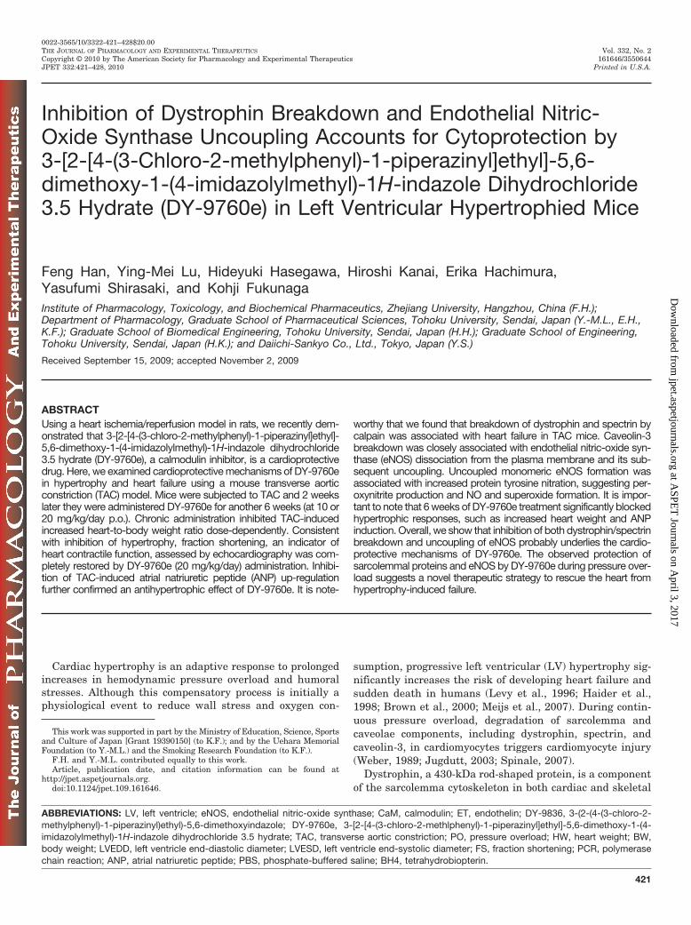
Effects of DY-9760e on TAC-Induced Spectrin andCaveolin-3 Breakdown. To confirm that TAC-induced dys-trophin breakdown is mediated by calpain activity, we inves-tigated calpain activity by assessing a specific, calpain-de-pendent 150-kDa spectrin breakdown product. As expected,after 8 weeks of TAC, levels of full-length, 240-kDa spectrinwere significantly reduced in the absence of DY-9760e (Fig. 4,A and B). These levels recovered dose-dependently after DY-9760e treatment. Inversely, levels of the TAC-induced 150-kDa breakdown product were markedly increased by 270%(p 0.05) and reduced dose-dependently by DY-9760e treat-ment (Fig. 4, A and C).
Caveolin-3 breakdown by phenylephrine-induced hyper-trophy is associated with injury in cultured cardiomyocytes(Lu et al., 2009a). As expected, we observed significant TAC-induced caveolin-3 breakdown in the LV compared withsham-operated animals. Significantly, DY-9760e (20 mg/kg)treatment strongly inhibited this effect (Fig. 4, D and E),suggesting that dystrophin breakdown is accompanied withbreakdown of spectrin and caveolin-3.
TAC-Induced eNOS Redistribution after TransverseAortic Constriction. Because eNOS is localized to caveolaethrough caveolin-3 in cardiomyocytes (Michel and Feron,1997), we asked whether dystrophin and caveolin-3 break-down triggers eNOS dissociation from caveolae. Althoughtotal eNOS protein levels in LV extracts remained un-changed in TAC versus sham-operated animals (Fig. 5A),membrane-associated eNOS levels were markedly reducedby TAC, whereas DY-9760e treatment dose-dependently re-stored plasma membrane eNOS levels (Fig. 5B). We hypoth-esized that caveolin-3 breakdown in caveolae and concomi-tant eNOS dissociation from the plasma membrane might
DY-9760e Inhibits Dystrophin Breakdown in Cardiac Hypertrophy 423
at ASPE
T Journals on A
pril 3, 2017jpet.aspetjournals.org
Dow
nloaded from

induce eNOS uncoupling and generate superoxide ratherthan nitric oxide. As expected, reduced levels of eNOS dimersand reciprocally increased levels of eNOS monomers wereobserved in LV extracts 8 weeks after TAC (Fig. 5C). DY-9760e treatment dose-dependently restored the dimeric formof eNOS, suggesting formation of physiological coupled eNOS(Fig. 5C). Likewise, peroxynitrite formation, as assessed byprotein tyrosine nitration in LV extracts, was markedly in-creased by TAC and blocked by DY-9760e dose-dependently(Fig. 5D). Increased peroxynitrite formation suggests gener-ation of superoxide by uncoupled eNOS, as we have shown incultured cardiomyocytes (Lu et al., 2009a).
DiscussionCalpain activation triggers cleavage of several mem-
brane-associated cytoskeletal proteins such as dystrophin
and spectrin. Disruption of the submembranous structureof cardiomyocytes triggered by dystrophin breakdowncauses contractile dysfunction in the muscle of dystrophicpatients (Toyo-oka et al., 2004). Here, we demonstratedthat calpain activation upon pressure overload leads todystrophin breakdown. In addition, disruption of eNOSlocalization by caveolin-3 breakdown in caveolae struc-tures probably induces aberrant eNOS uncoupling andsuperoxide generation, contributing to cardiomyocyte in-jury and contractile dysfunction. Thus, inhibiting the cal-pain/dystrophin cascade and eNOS uncoupling could rep-resent a novel therapeutic approach to inhibit heartdysfunction and heart failure. A critical finding of thepresent study is that DY-9760e, a novel calmodulin antag-onist, effectively blocked both dystrophin breakdown andeNOS uncoupling in vivo in a mouse TAC model. DY-9760e
A Sham TAC(2W)+
vehicle
TAC(8W) TAC(8W)+
DY(20)
B
TAC+
DY(20)
TAC+
DY(10)
DY(20)
TAC+
vehicle)
LVESD LVEDD
Sham
Sham TAC(2W)+
vehicle
TAC(8W) TAC(8W)+
DY(20)0
4
8
12
16
LW
/BW
(mg/
g)
**
##
#
Sham
C TAC+vehicle TAC+DY20
1
2
3
4
5
LV
end
-sys
tol i
c d
i am
e ter
(m
m)
*#
LV
fra
ctio
n sh
orte
ning
(%
)
1
2
3
4
5
LV
en
d-d
iast
olic
dia
met
e r (
mm
)
* #
TAC+
DY(20)
TAC+
vehicle)Sham
TAC+
DY(20)
TAC+
vehicle)Sham
10
20
30
40
50
**
#
TAC+
DY(20)
TAC+
vehicle)Sham
Fig. 1. DY-9760e treatment effectively inhibits pressure overload-induced cardiomyocyte hypertrophy. After 2 weeks (W) of TAC, DY-9760e [10 or 20mg/kg (DY10 or DY20)] was orally administered daily for the next 6 weeks of TAC. A, after isolation of whole heart, the sections are fixed inparaformaldehyde solution. B, HW/BW ratio was used as an index of cardiac hypertrophy. Data are expressed as percentage of values of sham-operated animals (mean � S.E.M.; n � 6). ��, P 0.01 versus sham; #, P 0.05; ##, P 0.01 versus vehicle. C, top, representative motion-modeechocardiographic images were obtained in conscious mice 8 weeks after aortic banding with or without DY-9760e (20 mg/kg) treatment. Bottom, dataare expressed as percentage of values of sham-operated animals (mean � S.E.M.; n � 4). �, P 0.05 versus sham; #, P 0.05 versus vehicle.
424 Han et al.
at ASPE
T Journals on A
pril 3, 2017jpet.aspetjournals.org
Dow
nloaded from

protective activity was further evidenced by inhibition ofspectrin breakdown and protein tyrosine nitration in theLV after TAC.
Our previous in vitro study demonstrated that DY-9760einhibits ET-1-induced cardiomyocyte hypertrophy and dys-trophin breakdown in cultured cardiomyocytes (Lu et al.,2007). In addition, DY-9760e significantly inhibits activa-tion of Ca2�/CaM-dependent nitric-oxide synthase afterbrain and heart ischemia (Hashiguchi et al., 2004; Hashi-moto et al., 2005; Han et al., 2006). The antihypertrophiceffect of DY9760e is mediated primarily by inhibition ofCa2�/CaM-dependent activation of CaMKII in cardiomy-ocytes (Lu et al., 2009b). This pathway, together withdystrophin/spectrin breakdown and eNOS uncoupling, un-derlies cardiomyocyte injury after cardiac hypertrophy.
Dystrophin is essential to maintain membrane integrityin cardiac and skeletal myocytes. Its disruption in thesubmembranous structure triggers heart failure after car-diac hypertrophy (Toyo-Oka et al., 2004; Kawada et al.,2005). The idea that membrane dystrophin is lost to thecytosol during the early weeks after TAC was supported byimmunohistochemistry after prolonged TAC. Calpains arecalcium- and thiol-dependent proteases whose overactiva-tion is implicated in several diseases, including cardiovas-cular disease (Matsumura et al., 1996) and ischemic stroke(Yoshida et al., 1995). Calpain activation followed by dys-trophin breakdown leads to myocardial injury. Indeed,breakdown of dystrophin and spectrin correlates with myo-cardial injury after cardiac ischemia (Armstrong et al.,2001; Hashimoto et al., 2005). In cardiac ischemia, calpainactivity mediates impaired Na�/K�-ATPase activity withconcomitant spectrin breakdown (Inserte et al., 2005). Be-cause Na�/K�-ATPase interacts with the cytoskeletal proteinspectrin through ankyrin, spectrin breakdown probably medi-ates impairment of Na�/K�-ATPase after cardiac ischemia. It is
interesting to note that overexpression of calpastatin, an endog-enous calpain inhibitor, blunts angiotensin II-induced cardiachypertrophy and perivascular fibrosis in heart (Letavernieret al., 2008). However, the precise mechanism underlyingDY-9760e-induced inhibition of spectrin breakdown remainsunclear.
Spectrin contains a calmodulin binding site, and calmod-ulin/�II-spectrin interaction regulates cleavage efficacy bycalpains and caspases (Rotter et al., 2004). Accumulatingevidence also demonstrates that Ca2�/calmodulin stimu-lates degradation of brain spectrin by calpain (Seubert etal., 1987; Harris et al., 1989). Thus, as a CaM antagonist,DY-9760e probably indirectly inhibits calpain-mediated
+DY(20)DY(20)TAC
TACSham
0
50
100
150
200
250
AN
P m
RN
A (
% o
f S
ham
)
**
#
ANP
DY(20) TACSham +DY(20)
TAC
β β-actin
377 bp
220 bp
Fig. 2. DY-9760e treatment inhibits TAC-induced ANP expression. After2 weeks of sham or TAC treatment, DY-9760e (20 mg/kg) was orallyadministered for 6 weeks to sham or experimental groups. Top, reversetranscription-PCR products of ANP and �-actin are shown. Bottom, quan-titation of ANP and �-actin mRNA levels shown as a percentage of valuesof sham-operated animals (mean � S.E.M.; n � 6). ��, P 0.01 versussham-operated mice; #, P 0.05 versus vehicle-treated TAC mice.
B
+DY(10)
+DY(20)
(20)
TAC
( - ) DY(20) DY(10) DY(20)( - )
Sham
Dystrophin
-tubulin
430 kDa
50 kDa
A
Sham 2W
4W 8W
0
50
100
150
Sham DY TAC TAC TAC
Dys
trop
hin
(%
of
Sh
am)
** **
##
Fig. 3. DY-9760e treatment blocks TAC-induced dystrophin degradation.A, immunohistochemical localization of dystrophin in sham- and TAC-operated animals. Localization of dystrophin in the plasma membrane ofcardiomyocytes indicates enlargement of cardiomyocytes 2 weeks (2W)after TAC and redistribution of dystrophin at 4 and 8 weeks after TAC.Scale bar, 40 �m. B, immunoblotting analyses of LV extracts (top) showmarkedly reduced levels of dystrophin 8 weeks after TAC and indicatethat 6 weeks of DY-9760e treatment (20 mg/kg) began after 2 weeks ofTAC significantly inhibit dystrophin breakdown. Bottom, quantitativeanalyses are shown in the bar graph as percentage of values of sham-operated animals (mean � S.E.M.; n � 6). ��, P 0.01 versus sham-operated mice; #, P 0.01 versus vehicle-treated TAC mice.
DY-9760e Inhibits Dystrophin Breakdown in Cardiac Hypertrophy 425
at ASPE
T Journals on A
pril 3, 2017jpet.aspetjournals.org
Dow
nloaded from
spectrin cleavage by inhibiting calmodulin-binding tospectrin.
DY-9760e cardioprotection also includes protection ofeNOS/caveolin-3 signaling in caveolae. In cardiac myo-cytes, the eNOS isoform mostly localizes in caveolae whereit associates with caveolin-3 (García-Cardena et al., 1996;Feron et al., 1996). Caveolin-3 inhibits eNOS activity,whereas calmodulin binding to eNOS after Ca2� mobiliza-tion disrupts caveolin-3 binding, leading to eNOS activa-tion and nitric oxide production (Michel and Feron, 1997).Our previous study documented that dystrophin break-down was closely associated with translocation of caveo-lin-3 from caveolae and decreased Ca2�/CaM-dependentnitric oxide generation by eNOS in cultured cardiomyo-cytes (Lu et al., 2009a). Likewise, dystrophin knockout miceshow impaired nitric oxide-dependent and endothelium-dependent vasodilation in arteries (Loufrani et al., 2004). In thepresent study, we demonstrated in vivo that disruption of eNOSassociation with caveolin-3 is associated with eNOS uncoupling,thereby increasing peroxynitrite formation, as assessed by pro-tein tyrosine nitration. Indeed, superoxide generation isclosely associated with eNOS uncoupling in cultured cardio-myocytes exposed to phenylephrine (Lu et al., 2009a). Fur-thermore, treatment with DY-9836, an active metabolite of
DY-9760e, largely inhibited elevation both of superoxide andnitric oxide generation after prolonged exposure to phenyl-ephrine in cultured cardiomyocytes. Under the same condi-tions, generation of monomeric eNOS was also inhibited byDY-9836 treatment (Lu et al., 2009a). It is noteworthy thatTempol (a radical scavenger), DY-9836, and N�-nitro-L-arginine methyl ester (a NO synthase inhibitor) all com-pletely inhibited apoptosis of cardiomyocytes exposed tophenylephrine (Lu et al., 2009a).
Accumulating evidence confirms the pathophysiologicalrelevance of eNOS uncoupling and peroxynitrite genera-tion to TAC-induced heart failure. Tetrahydrobiopterin(BH4) treatment in TAC-induced hypertrophy inhibitedpressure overload-induced oxidative stress through rever-sal of eNOS uncoupling, thereby ameliorating cardiac hy-pertrophy/dysfunction in mice (Takimoto et al., 2005;Moens et al., 2008). The reversal of eNOS uncoupling byexogenous BH4 was more effective than Tempol in pre-venting heart failure (Moens et al., 2008). Likewise, infu-sion of peroxynitrite or a peroxynitrite donor such as3-morpholino sydnonimine impaired contractile function by al-tering Ca2� handling and/or �-adrenergic responsiveness inisolated myocytes (Katori et al., 2006; Kohr et al., 2008). Takentogether, in addition to dystrophin/spectrin breakdown, de-
240 kDa
150 kDa
TAC
DY(10) DY(20)( - )Sham
A Spectrin
+DY(10)
+DY(20)
0
50
100
150
200
Sp
ectr
in 2
40 k
Da
(% o
f S
ham
)
**
##
*
TACTACTAC
Sham
0
100
200
300
400
Sp
ectr
in 1
5 0k
Da
(% o
f S
ham
)
**
**##
##
+DY(10)
+DY(20)
TACTAC TACSham
D
TAC
( - ) DY(20) DY(10) DY(20)( - )
Sham
0
50
100
150
Cav
-3 (
% o
f S
ham
)**
**
##
+DY(10)
+DY(20)
(20)TACTAC TACDYSham
Caveolin-3
18 kDa
B
C
E
Fig. 4. DY-9760e treatment antagonizesTAC-induced spectrin and caveolin-3 break-down. A, after 8 weeks of TAC, immunoblot-ting of LV extracts with anti-spectrin anti-body showed that levels of 240-kDa spectrinwere markedly reduced in untreated (�) an-imals compared with sham-operated ani-mals. Six weeks of DY-9760e [10 and 20mg/kg (DY10 or DY20)] treatment after 2weeks of TAC significantly and dose-depen-dently inhibited TAC-induced spectrinbreakdown. Inversely, the level of calpain-cleaved 150-kDa spectrin was increased byTAC and reduced by DY-9760e treatmentdose-dependently. B and C, quantitativeanalyses of levels of 240- and 150-kDa spec-trin are summarized, respectively, as a per-centage of values seen in sham-operatedanimals (mean � S.E.M.; n � 6). D, Immu-noblotting of LV extracts with an anti-caveolin-3 antibody indicates that levels of18-kDa caveolin-3 were significantly re-duced 8 weeks after TAC (�) compared withsham-operated animals. Six weeks of DY-9760e treatment (20 mg/kg) began after 2weeks of TAC significantly inhibited TAC-induced caveolin-3 breakdown. E, quantita-tive analyses are presented as the percent-age of values seen in sham-operated animals(mean � S.E.M.; n � 6). �, P 0.05; ��, P 0.05 versus sham-operated mice; ##, P 0.01 versus vehicle-treated TAC mice.
426 Han et al.
at ASPE
T Journals on A
pril 3, 2017jpet.aspetjournals.org
Dow
nloaded from

creased BH4 levels in cardiomyocytes probably underlies eNOSuncoupling, leading to heart failure by oxidant stress, includingperoxynitrite generation.
In summary, we report that a CaM antagonist, DY-9760e,inhibits in vivo the progression of cardiac hypertrophy-inducedcardiac injury by inhibiting calpain-mediated dystrophin/spec-trin degeneration and loss of caveolae integrity in cardiomyo-
cyte plasma membranes (Fig. 6). Protein tyrosine nitrationthrough superoxide generation by uncoupled eNOS was alsoameliorated by DY-9760e treatment. Loss of caveolae integrityprobably triggers eNOS uncoupling by dissociation from theplasma membranes. Taken together, evidence gathered usingthe TAC mouse model is consistent with our previous resultsobtained in cultured cardiomyocytes, supporting the idea that
A
135 kDa
B
Sham
TAC
(-) DY(10) DY(20)
0
50
100
150
mem
bra
ne e
NO
S (
% o
f Sh
am)
**
*##
##
+DY(10)
+DY(20)
TACTAC TACSham
eNOS
dimermonomer
Membrane fractions
eNOS
( - ) DY(20) DY(10) DY(20)( - )
Sham TAC
( - ) DY(20) DY(10) DY(20)( - )
Sham
C
eNOS
(-) DY(10) DY(20)Sham
TAC
+DY(10)
TACTAC +
DY(20)
TACSham
0
50
100
150
eNO
S (
% o
f Sh
am d
imer
) Dimer
Monomer
**
**#
##
$$
$$
§§
0
100
200
300
400
Nit
roty
rosi
ne
(% o
f S
ham
)
**
**
##
##
45 kDa
TAC
DY(10) DY(20)( - )Sham
+DY(10)
+DY(20)
TACTAC TACSham
D Nitrotyrosine
Total extracts
0
50
100
150
eNO
S (%
of
Sh
am)
+DY(10)
+DY(20)
TACTAC
TACSham DY(20)
Fig. 5. Effects of DY-9760e on TAC-induced membrane eNOS levels, eNOS uncoupling, and nitrotyrosine formation. A, representative image (top) andbar graph (bottom) showing results of immunoblotting of total heart extracts with anti-eNOS antibody. Analysis indicates no apparent changes in totaleNOS levels among conditions. B, representative image (top) and bar graph (bottom) showing results of immunoblotting with anti-eNOS antibody.Analysis of cell membrane extracts indicates marked reduction in eNOS levels in TAC animals and rescue of eNOS levels after DY-9760e treatmentdose-dependently. Data are expressed as a percentage of values seen in sham-operated animals (mean � S.E.M.; n � 6). �, P 0.05; ��, P 0.01 versussham-operated mice; ##, P 0.01 versus vehicle-treated mice. C, levels of uncoupled (monomer) eNOS are shown as analyzed by low-temperatureSDS-polyacrylamide gel electrophoresis (top). Quantitation of dimeric and monomeric eNOS (lower) was performed by densitometric analysis. DimericeNOS from sham-operated animals was arbitrarily set at 100%. Data are expressed as percentage of values seen in sham-operated animals (mean �S.E.M.; n � 6). ��, P 0.01 versus sham-operated mice (dimer); #, P 0.05 and ##, P 0.01 versus vehicle-treated mice (dimer); $$, P 0.01 versussham-operated mice (monomer); and §§, P 0.01 versus vehicle-treated mice (monomer). D, representative image (top) and bar graph (bottom) ofimmunoblots of total heart extracts with anti-nitrotyrosine antibody. Analysis indicates that protein tyrosine nitration was markedly increased afterTAC compared with sham-operated animals and decreased by DY-9760e treatment dose-dependently. ��, P 0.01 versus sham-operated mice; ##, P 0.01 versus vehicle-treated TAC mice.
DY-9760e Inhibits Dystrophin Breakdown in Cardiac Hypertrophy 427
at ASPE
T Journals on A
pril 3, 2017jpet.aspetjournals.org
Dow
nloaded from

DY-9760e treatment could provide a novel therapeutic strategyto treat hypertrophy and heart failure.
ReferencesArmstrong SC, Latham CA, Shivell CL, and Ganote CE (2001) Ischemic loss of
sarcolemmal dystrophin and spectrin: correlation with myocardial injury. J MolCell Cardiol 33:1165–1179.
Brown DW, Giles WH, and Croft JB (2000) Left ventricular hypertrophy as apredictor of coronary heart disease mortality and the effect of hypertension. AmHeart J 140:848–856.
Feron O, Belhassen L, Kobzik L, Smith TW, Kelly RA, and Michel T (1996) Endothelialnitric oxide synthase targeting to caveolae. Specific interactions with caveolin iso-forms in cardiac myocytes and endothelial cells. J Biol Chem 271:22810–22814.
García-Cardena G, Oh P, Liu J, Schnitzer JE, and Sessa WC (1996) Targeting ofnitric oxide synthase to endothelial cell caveolae via palmitoylation: implicationsfor nitric oxide signaling. Proc Natl Acad Sci U S A 93:6448–6453.
Haider AW, Larson MG, Benjamin EJ, and Levy D (1998) Increased left ventricularmass and hypertrophy are associated with increased risk for sudden death. J AmColl Cardiol 32:1454–1459.
Han F, Shirasaki Y, and Fukunaga K (2006) Microsphere embolism-induced endo-thelial nitric oxide synthase expression mediates disruption of the blood-brainbarrier in rat brain. J Neurochem 99:97–106.
Harris AS, Croall DE, and Morrow JS (1989) Calmodulin regulates fodrin suscepti-bility to cleavage by calcium-dependent protease I. J Biol Chem 264:17401–17408.
Hashiguchi A, Yano S, Morioka M, Hamada J, Shirasaki Y, Kochi M, and FukunagaK (2004) The post-ischemic administration of 3-[2-[4-(3-chloro-2-methylphenyl)-1-piperazinyl]ethyl]-5,6-dimethoxy-1-(4-imidazolylmethyl)-1H-indazole dihydro-chloride 3.5 hydrate (DY-9760e), a novel calmodulin antagonist, prevents delayedneuronal death in gerbil hippocampus. J Pharmacol Sci 96:65–72.
Hashimoto M, Takada Y, Takeuchi Y, Kasahara J, Hisa H, Shirasaki Y, and Fuku-naga K (2005) Cytoprotective effect of 3-[2-[4-(3-chloro-2-methylphenyl)-1-piperazinyl]ethyl]-5,6-dimethoxy-1-(4-imidazolylmethyl)-1H-indazole dihydro-chloride 3.5 hydrate (DY-9760e) against ischemia/reperfusion-induced injury inrat heart involves inhibition of fodrin breakdown and protein tyrosine nitration.J Pharmacol Sci 98:142–150.
Hu P, Zhang D, Swenson L, Chakrabarti G, Abel ED, and Litwin SE (2003) Minimallyinvasive aortic banding in mice: effects of altered cardiomyocyte insulin signalingduring pressure overload. Am J Physiol Heart Circ Physiol 285:H1261–H1269.
Inserte J, Garcia-Dorado D, Hernando V, and Soler-Soler J (2005) Calpain-mediatedimpairment of Na�/K�-ATPase activity during early reperfusion contributes tocell death after myocardial ischemia. Circ Res 97:465–473.
Jugdutt BI (2003) Remodeling of the myocardium and potential targets in thecollagen degradation and synthesis pathways. Curr Drug Targets CardiovascHaematol Disord 3:1–30.
Katori T, Donzelli S, Tocchetti CG, Miranda KM, Cormaci G, Thomas DD, KetnerEA, Lee MJ, Mancardi D, Wink DA, et al. (2006) Peroxynitrite and myocardialcontractility: in vivo versus in vitro effects. Free Radic Biol Med 41:1606–1618.
Kawada T, Masui F, Kumagai H, Koshimizu M, Nakazawa M, and Toyo-Oka T(2005) A novel paradigm for therapeutic basis of advanced heart failure–assessment by gene therapy. Pharmacol Ther 107:31–43.
Kohr MJ, Wang H, Wheeler DG, Velayutham M, Zweier JL, and Ziolo MT (2008)Biphasic effect of SIN-1 is reliant upon cardiomyocyte contractile state. Free RadicBiol Med 45:73–80.
Levy D, Larson MG, Vasan RS, Kannel WB, and Ho KK (1996) The progression fromhypertension to congestive heart failure. Jama 275:1557–1562.
Letavernier E, Perez J, Bellocq A, Mesnard L, de Castro Keller A, Haymann JP, and BaudL (2008) Targeting the calpain/calpastatin system as a new strategy to prevent cardio-vascular remodeling in angiotensin II-induced hypertension. Circ Res 102:720–728.
Loufrani L, Dubroca C, You D, Li Z, Levy B, Paulin D, and Henrion D (2004) Absenceof dystrophin in mice reduces NO-dependent vascular function and vascular den-sity: total recovery after a treatment with the aminoglycoside gentamicin. Arte-rioscler Thromb Vasc Biol 24:671–676.
Lu YM, Han F, Shioda N, Moriguchi S, Shirasaki Y, Qin ZH, and Fukunaga K(2009a) Phenylephrine-induced cardiomyocyte injury is triggered by superoxidegeneration through uncoupled endothelial nitric-oxide synthase and amelioratedby 3-[2-[4-(3-chloro-2-methylphenyl)-1-piperazinyl]ethyl]-5,6-dimethoxyindazole(DY-9836), a novel calmodulin antagonist. Mol Pharmacol 75:101–112.
Lu YM, Shioda N, Han F, Kamata A, Shirasaki Y, Qin ZH, and Fukunaga K (2009b)DY-9760e inhibits endothelin-1-induced cardiomyocyte hypertrophy through inhi-bition of CaMKII and ERK activities. Cardiovasc Ther 27:17–27.
Lu YM, Shioda N, Han F, Moriguchi S, Kasahara J, Shirasaki Y, Qin ZH, andFukunaga K (2007) Imbalance between CaM kinase II and calcineurin activitiesimpairs caffeine-induced calcium release in hypertrophic cardiomyocytes. BiochemPharmacol 74:1727–1737.
Matsumura Y, Saeki E, Inoue M, Hori M, Kamada T, and Kusuoka H (1996)Inhomogeneous disappearance of myofilament-related cytoskeletal proteins instunned myocardium of guinea pig. Circ Res 79:447–454.
Meijs MF, de Windt LJ, de Jonge N, Cramer MJ, Bots ML, Mali WP, and DoevendansPA (2007) Left ventricular hypertrophy: a shift in paradigm. Curr Med Chem14:157–171.
Michel T and Feron O (1997) Nitric oxide synthases: which, where, how, and why?J Clin Invest 100:2146–2152.
Moens AL, Takimoto E, Tocchetti CG, Chakir K, Bedja D, Cormaci G, Ketner EA,Majmudar M, Gabrielson K, Halushka MK, et al. (2008) Reversal of cardiac hyper-trophy and fibrosis from pressure overload by tetrahydrobiopterin: efficacy of recou-pling nitric oxide synthase as a therapeutic strategy. Circulation 117:2626–2636.
Rotter B, Kroviarski Y, Nicolas G, Dhermy D, and Lecomte MC (2004) AlphaII-spectrin is an in vitro target for caspase-2, and its cleavage is regulated bycalmodulin binding. Biochem J 378:161–168.
Saido TC, Sorimachi H, and Suzuki K (1994) Calpain: new perspectives in moleculardiversity and physiological-pathological involvement. FASEB J 8:814–822.
Seubert P, Baudry M, Dudek S, and Lynch G (1987) Calmodulin stimulates thedegradation of brain spectrin by calpain. Synapse 1:20–24.
Spinale FG (2007) Myocardial matrix remodeling and the matrix metalloproteinases:influence on cardiac form and function. Physiol Rev 87:1285–1342.
Takimoto E, Champion HC, Li M, Ren S, Rodriguez ER, Tavazzi B, Lazzarino G,Paolocci N, Gabrielson KL, Wang Y, et al. (2005) Oxidant stress from nitric oxidesynthase-3 uncoupling stimulates cardiac pathologic remodeling from chronicpressure load. J Clin Invest 115:1221–1231.
Toyo-Oka T, Kawada T, Nakata J, Xie H, Urabe M, Masui F, Ebisawa T, Tezuka A,Iwasawa K, Nakajima T, et al. (2004) Translocation and cleavage of myocardialdystrophin as a common pathway to advanced heart failure: a scheme for theprogression of cardiac dysfunction. Proc Natl Acad Sci U S A 101:7381–7385.
Weber KT (1989) Cardiac interstitium in health and disease: the fibrillar collagennetwork. J Am Coll Cardiol 13:1637–1652.
Yoshida H, Takahashi M, Koshimizu M, Tanonaka K, Oikawa R, Toyo-oka T, andTakeo S (2003) Decrease in sarcoglycans and dystrophin in failing heart followingacute myocardial infarction. Cardiovasc Res 59:419–427.
Yoshida K, Inui M, Harada K, Saido TC, Sorimachi Y, Ishihara T, Kawashima S, andSobue K (1995) Reperfusion of rat heart after brief ischemia induces proteolysis ofcalspectin (nonerythroid spectrin or fodrin) by calpain. Circ Res 77:603–610.
Address correspondence to: Dr. Kohji Fukunaga, Department of Phar-macology, Graduate School of Pharmaceutical Sciences, Tohoku University,Aramaki-Aoba Aoba-ku, Sendai 980-8578, Japan. E-mail: [email protected]
Ca2+
Pressure overloadShear stress
Calpain activation eNOS dysregulation
Protein tyrosine nitrationSuperoxide generation
Dystrophin/spectrin breakdownLoss of caveolae integrity
Progression to heart failure
DY-9760e DY-9760e
Fig. 6. Potential cardioprotective mechanism of DY-9760 onTAC-induced heart injury. DY-9760e in vivo inhibits progres-sion of cardiac hypertrophy-induced cardiac injury by inhibit-ing calpain-mediated dystrophin/spectrin degeneration andloss of caveolae integrity in cardiomyocyte plasma membranes.Protein tyrosine nitration through superoxide generation byuncoupled eNOS is also ameliorated by DY-9760e treatment.Loss of caveolae integrity probably triggers eNOS uncouplingby dissociation from plasma membranes.
428 Han et al.
at ASPE
T Journals on A
pril 3, 2017jpet.aspetjournals.org
Dow
nloaded from