inhibition of metabolism and induction of apoptosis in
TRANSCRIPT

INHIBITION OF METABOLISM AND INDUCTION OF APOPTOSIS IN
TRIPLE NEGATIVE BREAST CANCER CELLS BY LIPPIA ORIGANOIDES
PLANT EXTRACTS.
by
Vishak Raman
A Dissertation
Submitted to the Faculty of Purdue University
In Partial Fulfillment of the Requirements for the degree of
Doctor of Philosophy
Department of Biological Sciences
West Lafayette, Indiana
May 2019

2
THE PURDUE UNIVERSITY GRADUATE SCHOOL
STATEMENT OF COMMITTEE APPROVAL
Dr. Ignacio G. Camarillo, Chair
Department of Biological Sciences, Purdue University, Indiana, USA
Dr. Claudio Aguilar
Department of Biological Sciences, Purdue University, Indiana, USA
Dr. Jorge Luis Fuentes Lorenzo
Escuela de Biología, Universidad Industrial de Santander, Bucaramanga,
Colombia
Dr. Morris Levy
Department of Biological Sciences, Purdue University, Indiana, USA
Approved by:
Dr. Stephen Konieczny
Head of the Graduate Program, Biological Sciences

3
To Mum, Dad, Guha, Lavy, and Anika.

4
ACKNOWLEDGMENTS
As I look back at the journey I’ve taken to arrive here, I realize it quite simply would not
have been possible without some very special people pushing me along throughout the
way.
To begin with, I would like to thank my wonderful parents, who’ve taught me
infinitely more than any academic institution could. I am the person I am today because
of their love, support, and guidance. Knowing that they were there for me allowed me to
keep my head up and remain motivated throughout my time in grad school.
Learning to live in a new country is never easy, and is made even harder when
pursuing a PhD during that time. Having my brother, Guha, living in Indiana made my
move here much easier to handle. During my time at Purdue, he met and married my (now)
sister-in-law, Lavy, and they’ve even had a beautiful daughter (A lot has happened in five
years!). With them around, I was never too far from family, and all the time spent with
them has made Indiana feel like a second home.
I’d like to give a shout out to my best friends, Ish and Manoj, for the unconditional
support they’ve given me, and for the incredibly fun times we’ve shared traveling around
the United States. I want to thank the amazing friends I’ve made here at Purdue, especially
Alix and Swetha. The weekly coffee-breaks with them where we discussed everything
from failed experiments to career ambitions kept me cheerful and relaxed, and ready to
take on the next research challenge.
I’ve had the amazing experience of working alongside and mentoring some
astonishingly talented and motivated graduate and undergraduate students. With this in
mind, I’d like to thank Lakshya and Rodrigo. Their hard work has pushed the lab’s science
in new directions and taken it to another level. Mentoring them has been a massive
privilege.
I express my sincerest thanks and gratitude to my committee members, Dr. Aguilar,
Dr. Levy, and Dr. Fuentes, and collaborating professors, Dr. Uma Aryal and Dr. Raji
Sundararajan, whose useful feedback and advice throughout my PhD has added so much

5
to my research experience and to the content of my publications. I also want to extend a
special thank-you to Dr. Fuentes, Dr. Morry Levy, and Maria (Mechas) Levy for
entrusting me with this research project, which is part of an important on-going
collaboration between Purdue and the Universidad Industrial de Santander, Bucaramanga;
and I also thank them for their unlimited kindness and hospitality over the years.
Last but not the least, I want to thank my mentor, Dr. Ignacio Camarillo. Words
cannot fully express the gratitude I feel for everything he has done for me. His positive
influence has shaped me in a professional capacity, by improving and motivating me as a
researcher, and on a personal level, by making me a more-rounded person. His guidance
and support has given me the confidence to think and work independently, to foster
collaborations with other labs and core facilities, and to mentor new students. His trust in
my research and writing skills have given me the belief to pursue my own research
questions, and have made me a better grant- and manuscript-writer. Outside of the lab, I
will always remember the memorable concerts we attended, the long conversations over
a couple of beers at the local pubs, and the Thanksgiving lunches at his home with his
wife and children. Finally, I can never forget how supportive he was during my job hunt,
and now in preparing me for my defense. I hope to emulate his work ethic and principles
as I move on to the next phase of my life.

6
TABLE OF CONTENTS
LIST OF TABLES ............................................................................................................ 10
LIST OF FIGURES .......................................................................................................... 11
ABSTRACT ...................................................................................................................... 16
INTRODUCTION .................................................................................. 23
1.1 Breast Cancer Overview ....................................................................................... 23
1.1.1 Breast Cancer Epidemiology ......................................................................... 23
1.1.2 Mammary Histology and Origins of Breast Cancer Subtypes ....................... 25
1.2 Triple-Negative Breast Cancer Overview ............................................................. 29
1.2.1 Epidemiology and Behavioral/Genetic Risk Factors for TNBC ................... 29
1.2.2 Clinical Outcomes and Treatment Options for TNBC .................................. 31
1.3 Molecular Heterogeneity of TNBC ...................................................................... 34
1.3.1 Overview of Major Signaling Pathways in TNBC ........................................ 38
1.3.2 NF-B Signaling in TNBC ............................................................................ 40
1.4 Metabolism in Triple-Negative Breast Cancer ..................................................... 44
1.4.1 Amino Acid Metabolism in TNBC ................................................................ 45
1.4.2 Mitochondrial metabolism in TNBC ............................................................. 48
1.5 An Important Role for Plant-derived Metabolites in Cancer Therapy ................. 51
1.5.1 Cancer Drug Discovery from Plant Sources: A Historical Perspective ........ 51
1.5.2 Mechanisms of Anticancer Action of Plant-derived Compounds ................. 53
1.5.2.1 Interference with microtubule assembly ................................................... 53
1.5.2.2 Inhibition of DNA topoisomerases ........................................................... 53
1.5.2.3 Interference with multiple proliferative and survival pathways ............... 54
1.5.3 Lippia species as a source of anti-cancer compounds ................................... 56
1.6 Quantitative LC-MS-based Proteomics for Cancer Drug Discovery ................... 56
1.7 Research Questions ............................................................................................... 58
1.8 References ............................................................................................................. 60

7
LIPPIA ORIGANOIDES EXTRACT (LOE) INDUCES CELL CYCLE
ARREST AND APOPTOSIS, AND SUPPRESSES NF-B SIGNALING IN TNBC
CELLS ................................................................................................................. 84
2.1 Abstract ................................................................................................................. 84
2.2 Introduction ........................................................................................................... 84
2.3 Materials and Methods .......................................................................................... 87
2.3.1 Plant material and extract .............................................................................. 87
2.3.2 Cell culture ..................................................................................................... 87
2.3.3 Assessment of metabolic activity via MTT assay ......................................... 88
2.3.4 Assessment of cell cycle arrest via flow cytometry ....................................... 88
2.3.5 Assessment of apoptosis via flow cytometry ................................................. 89
2.3.6 Caspase-3/7 activation assay ......................................................................... 89
2.3.7 Western Blotting ............................................................................................ 89
2.3.8 Statistical Analysis ......................................................................................... 90
2.4 Results ................................................................................................................... 90
2.4.1 LOE decreases viability of TNBC cells. ........................................................ 90
2.4.2 LOE induces cell cycle arrest in TNBC cells. ............................................... 93
2.4.3 LOE induces apoptosis in TNBC cells. ......................................................... 95
2.4.4 LOE impacts critical regulators of cell cycle progression. ............................ 97
2.4.5 LOE induces apoptosis via caspase-8/-3 activation. ...................................... 97
2.4.6 LOE reduces RIP1 protein expression levels. ............................................. 100
2.5 Discussion ........................................................................................................... 103
2.5.1 LOE is a source of TNBC-specific cytotoxic compounds ........................... 103
2.5.2 LOE is a source of cell cycle inhibitors ....................................................... 104
2.5.3 LOE is a source of apoptotic compounds .................................................... 104
2.5.4 LOE is a source of compounds targeting NF-B signaling ......................... 104
2.6 Summary ............................................................................................................. 106
2.7 References ........................................................................................................... 106
PROTEOMIC ANALYSIS REVEALS LIPPIA ORIGANOIDES
EXTRACT (L42) TARGETS MITOCHONDRIAL METABOLISM IN TRIPLE-
NEGATIVE BREAST CANCER CELLS...................................................................... 113
8
3.1 Abstract ............................................................................................................... 113
3.2 Introduction ......................................................................................................... 113
3.3 Materials and Methods ........................................................................................ 115
3.3.1 Cell culture ................................................................................................... 115
3.3.2 L42 extract preparation ................................................................................ 116
3.3.3 Assessment of cell viability using MTT assay ............................................ 116
3.3.4 Western blotting ........................................................................................... 116
3.3.5 LC-MS/MS: sample preparation and analysis ............................................. 117
3.3.6 Proteomics data analysis: ............................................................................ 119
3.3.7 Validation experiment: mitochondrial membrane potential assay .............. 121
3.3.8 Statistical analysis ........................................................................................ 121
3.3.9 Data availability ........................................................................................... 121
3.4 Results ................................................................................................................. 122
3.4.1 Effect of L42 on TNBC cell viability. ......................................................... 122
3.4.2 Overview of proteome analysis and LC-MS reproducibility. ..................... 122
3.4.3 Functional classification of identified proteins. ........................................... 126
3.4.4 L42 disrupts metabolism in MDA-MB-231 TNBC cells. ........................... 128
3.4.5 L42 impacts expression of TCA cycle enzymes. ......................................... 131
3.4.6 L42 dysregulates Complex I of the Electron Transport Chain. ................... 131
3.4.7 L42 disrupts mitochondrial membrane potential in TNBC cells ................. 134
3.5 Discussion ........................................................................................................... 136
3.5.1 L42 inhibits survival and induces apoptosis in TNBC cells ........................ 136
3.5.2 L42 inhibits mitochondrial function and metabolism in TNBC cells. ......... 136
3.5.3 L42 extensively inhibits the TCA Cycle in TNBC cells. ............................ 137
3.5.4 L42 induces loss of NADH dehydrogenase (Complex I) expression .......... 138
3.6 Summary ............................................................................................................. 139
3.7 References ........................................................................................................... 140
ANALYSIS OF COMPOSITION AND IN VIVO TOXICITY OF
LIPPIA EXTRACTS ....................................................................................................... 146
4.1 Abstract ............................................................................................................... 146
4.2 Introduction ......................................................................................................... 146

9
4.3 Materials and Methods ........................................................................................ 148
4.3.1 Plant material and extract ............................................................................ 148
4.3.2 GC/MS analysis of L. origanoides extracts ................................................. 148
4.3.3 MTT cell viability assay .............................................................................. 149
4.3.4 In vivo toxicity study ................................................................................... 149
4.3.5 Histological analysis of mouse mammary gland ......................................... 149
4.4 Results ................................................................................................................. 150
4.4.1 GC/MS reveals compositional differences in L42 and LOE ....................... 150
4.4.2 Major components of LOE and L42 do not induce loss of viability in TNBC
cells. ..................................................................................................................... 150
4.4.3 L42 is well-tolerated in vivo upon IP administration ................................... 156
4.4.4 IP injection of L42 is non-toxic to mouse mammary glands ....................... 159
4.5 Summary ............................................................................................................. 161
4.6 References ........................................................................................................... 162
CONCLUSIONS AND FUTURE DIRECTIONS ................................ 166

10
LIST OF TABLES
Table 1.1 Classification of breast tumors into intrinsic molecular subtypes. Source:
Dai, X., Li, T., Bai, Z., Yang, Y., Liu, X., Zhan, J., & Shi, B. (2015). American Journal of
Cancer Research, 5(10), 2929–2943. [Ref. 216] .............................................................. 27
Table 4.1 Composition of L42. Peaks from GC/MS spectra of non-derivatized and
derivatized L42 (Figure 4.1) were searched against the NIST compound library based on
retention times, and the respective components were identified. Components in red are
shared between L42 and LOE (See Table 4.2). .............................................................. 153
Table 4.2 Composition of LOE. Peaks from GC/MS spectra of non-derivatized LOE.
Components in red are shared between L42 and LOE (See Table 4.1). (Taken with
permission from Castellanos et. al [17]) ........................................................................ 154
Table 4.3 L42 does not induce adverse effects in C57BL/6 mice. Female virgin
C57BL/6 mice were intraperitoneally injected with L42 at 100 mg/kg BW (High dose), or
50 mg/kg BW (Medium dose), or 25 mg/kg BW (Low dose) or Control, and monitored
for adverse effects over the course of 2 weeks. BAR: Bright, alert, and responsive. Mouse
number reflects ID number assigned to mouse at the start of experiment; numbers are not
sequential because mice were grouped based on body weight, not assigned number. .. 157
11
LIST OF FIGURES
Figure 1.1 Hormone receptor status correlates with luminal phenotype and degree
of differentiation. Fluorescence-assisted cell sorting (FACS) of mammary epithelial cells
based on surface markers including Estrogen Receptor (ER) and luminal and basal
markers allows for fractionation into distinct populations. Fractions enriched for ER+ cells
contain a low frequency of stem-like cells and progenitors capable of mammary
repopulation upon transplantation, and colony formation in vitro. Conversely, fractions
enriched for ER- and basal-like cells exhibit enhanced colony-formation and mammary
repopulation abilities, and are thus considered to have a greater population of stem and
progenitor cells.................................................................................................................. 28
Figure 1.2 TNBC subtypes. Classification of TNBCs on the basis of gene expression and
signaling pathways involved. The subclasses show varying rates of pathological complete
response (pCR) to standard chemotherapy, with promising targeted therapies suggested
for each subclass. .............................................................................................................. 37
Figure 1.3 NF-κB signaling: Interplay between Complex I and Complex II
determines cell fate upon TNF-R1 activation. .............................................................. 43
Figure 1.4 Glutamine transport and metabolism. Extracellular glutamine is transported
into the cell through transporters such as alanine, serine, cysteine-preferring transporter 2
(SLC1A5/ASCT2). Transport into the mitochondria is carried out through a currently
unknown mechanism but is theorized to occur based on the mitochondrial localization of
glutaminase (GLS), and glutamate dehydrogenase (GDH). The end product of GDH
action on glutamate results in the TCA Cycle intermediate, -ketoglutarate (-KG).
Glutamate and the TCA Cycle intermediate citrate can also be further utilized in amino
acid and lipid synthesis. .................................................................................................... 47
Figure 1.5 Waves of metabolic reprogramming during carcinogenesis. Wave 1:
Oncogenic transformation leads to enhanced glycolytic activity at the expense of
OXPHOS and mitochondrial biogenesis, leading to rapid proliferation. Wave 2:
Promotion of cell survival through HIF and NFB signaling, with sustained glycolytic
activity and inhibition of mitochondrial biogenesis. Wave 3: Activation of AMPK and Akt
signaling leads to inhibition of protein synthesis. PGC1 activation leads to mitochondrial
biogenesis followed by OXPHOS initiation, while MYC activation increases
glutaminolysis. Wave 4: Mitochondrial biogenesis and activity leads to mitochondrial
signaling activation through multiple effectors including NAD+/NADH ratio, AMPK,
Ca2+ and lipid signaling, and NO production. In addition, changes in mitochondrial inner
membrane potential and morphology may lead to downstream signaling
activation/inhibition. ......................................................................................................... 50
Figure 2.1 LOE impacts the viability of triple-negative breast cancer cells to a greater
extent than normal-like cells. MDA-MB-231, MCF10A-H, CRL-2321 and MCF10A
cells were seeded in 96-well plates and treated with indicated concentrations of LOE,
Methanol (Veh) or left untreated (NT) for 24h and subjected to MTT assay. This was

12
followed by absorbance reading at 570nm. n=10 replicates from 2 separate experiments;
*significantly different from vehicle-treated p < 0.0001. ................................................. 92
Figure 2.2 LOE induces G0/G1 phase arrest in MDA-MB-231 cells. MDA-MB-231
cells treated with indicated concentrations of LOE for 36h were stained with Propidium
Iodide (PI) and differential staining was measured using an FC500 flow cytometer
(Bindley Flow Cytometry Facility). (A) Representative plots of raw data indicating cell
cycle stages from treatment groups. (B) Average distribution across different cell cycle
stages from various treatment groups. SF: serum-free treatment. n=3; * significantly
different from vehicle treatment p<0.05 ★ significantly different from serum free treatment
p<0.05 ............................................................................................................................... 94
Figure 2.3 LOE induces apoptosis but not necrosis in MDA-MB-231 triple-negative
breast cancer cells. MDA-MB-231 cells were treated with indicated concentrations of
LOE for 24h and stained with Annexin-V/7-Aminoactinomycin D (7-AAD). This was
followed by measurement of differential Annexin-V/7-AAD staining using a Muse™ Cell
Analyzer. (A) Representative plots of raw data showing distribution of cells as Live, Early
Apoptotic (EA), Late Apoptotic (LA) or Necrotic. (B) Quantified graph showing effect of
LOE on apoptosis in MDA-MB-231 cells. N=3; Significant difference between LOE-
treated cells and control (Vehicle-treated) cells is indicated as *p<0.05 **p<0.005. ...... 96
Figure 2.4. LOE inhibits markers of cell cycle progression and survival. MDA-MB-
231 cells were treated with 0.06 and 0.15mg/ml of LOE for various time intervals. Cells
were then lysed in RIPA buffer. Lysates were subjected to Western Blotting and probed
for indicated proteins. (A) Representative blots of cytostatic markers Cyclin D1 and
cIAP2. (B) Densitometry quantification of blots using ImageJ. Protein levels were
normalized against β-actin and plotted as shown. n=3; Significant difference from
untreated (NT) is indicated as *p<0.05 or **p<0.005; significant difference from Vehicle-
treated (Veh) is indicated as ★p<0.05 or ★★p<0.005. ....................................................... 98
Figure 2.5 LOE induces apoptosis accompanied by caspase-8 activation and PARP
cleavage. MDA-MB-231 cells treated with LOE for various time intervals were lysed and
subjected to Western Blotting and probed for indicated proteins (A) Representative blots
of apoptotic markers cleaved caspase-8, cleaved caspase-3 and cleaved PARP (B)
Densitometry quantification of blots using ImageJ. Protein levels were normalized against
β-actin and plotted as shown. n=3. Significant difference from untreated (NT) is indicated
as *p<0.05 or **p<0.005; significant difference from Vehicle-treated (Veh) is indicated
as ★p<0.05 or ★★p<0.005. ................................................................................................. 99
Figure 2.6 LOE induces executioner caspase-3/-7 activity in TNBC cells. (A) MDA-
MB-231 cells incubated with full growth medium containing indicated treatments and
IncuCyte™ Caspase-3/7 apoptosis assay reagent were imaged periodically over 24h in an
IncuCyte® ZOOM live-cell analyzer to look at changes in caspase-3/-7 activity. (B)
Activity was quantified as the mean Green Fluorescent Object Count/Image for each
treatment group and plotted as shown. n=5; * significantly different from control (Vehicle-
treated) cells; p<0.005. .................................................................................................... 101
Figure 2.7 RIP1 protein levels are reduced upon treatment of TNBC cells with LOE.
MDA-MB-231 cells were treated with 0.15 mg/ml LOE for 9 h and cell lysates were

13
immunoblotted for RIP1. (A) Representative blots of RIP1 (B) Densitometry
quantification of blots using ImageJ. Protein levels were normalized against β-tubulin and
plotted as shown. N = average of 3 replicates run twice. Significant difference from
Vehicle-treated cells is indicated as *p<0.05. ................................................................. 102
Figure 3.1 L42 inhibits survival and induces apoptosis in MDA-MB-231 cells. A.
MDA-MB-231 and MCF10A cells were seeded in 96-well plates and treated with
indicated concentrations of L42 or vehicle control (VEH) for 24, 48 and 72 h and subjected
to MTT assay. This was followed by absorbance reading at 570 nm. N = 5 replicates;
Significance was determined using Tukey’s HSD test (see Supplementary Table 3.2), p <
0.0001. B. MDA-MB-231 cells were treated with 0.15 mg/ml of L42 for various time
intervals and lysates were probed for indicated proteins by Western Blot. (Upper panel)
Representative blots of Cyclin D1 and Caspase-8. (Lower panel) Densitometry
quantification of blots using ImageJ. Protein levels were normalized against β-actin and
plotted as shown. N = 3; * significant difference from VEH, p < 0.05. ......................... 124
Figure 3.2 Experimental design for proteomics analysis. A. Proteomics workflow. B.
CV % plot of peptide intensity from technical replicates of an E. coli tryptic digest. N =
3. C. Correlation plot of 519 significantly changed proteins (Supplementary Table 3.1C).
R2-values are consistent, and values between replicates within treatments is noticeably
higher than those between replicates across treatments, indicating good experimental
reproducibility. N = 3 biological replicates from each treatment group. ........................ 125
Figure 3.3 GO enrichment analysis of L42-regulated proteins. Significantly
upregulated (Red font, Supplementary Table 3.1C) and down-regulated (Blue font,
Supplementary Table 3.1C) proteins were compared to the pool of 2,407 identified
proteins to identify enrichment of Localization, Function, and Biological process. A-C.
GO enrichment for significant downregulation. D. GO enrichment for significant
upregulation (only observed for biological process). Enrichment analysis was performed
using GOrilla software and average protein fold-change/replicate/GO term was calculated
and visualized using heatmaps generated by Heatmapper. ............................................. 127
Figure 3.4 Hierarchical clustering of differentially-expressed proteins. Significantly
changed (Supplementary Table 3.1C) proteins were compared to the pool of 2,407
identified proteins to identify pathway enrichment using DAVID 6.8 software, with the
pathways matched to the KEGG database. A. Heatmaps of Lipid Metabolism (Top, GO
terms included: “Fatty Acid Metabolism”, “Fatty Acid Elongation”, and “Fatty Acid
Degradation”) and Amino Acid Metabolism (Bottom, GO terms included: “Amino Acid
Biosynthesis”, “Valine, Leucine and Isoleucine Degradation”, “Tryptophan Metabolism”,
and “Lysine Metabolism”). B. STRING interaction analysis of proteins from Lipid
Metabolism (Top) and Amino Acid Metabolism (Bottom), with coloring indicating
localization to mitochondria (Red: Mitochondrial matrix, Blue: Mitochondrial inner
membrane, Green: Mitochondrial unspecified). Shown are interactions at highest
confidence i.e. confidence score ≥ 0.9. C. Validation of GLS levels. (Left) Densitometry
quantification of blots using ImageJ. GLS levels were normalized against β-actin and
plotted as shown. N = 3. Significant difference from VEH is indicated as *p<0.05. (Right)
Representative western blot showing expression of glutaminase (GLS) in MDA-MB-231
cells treated with L42 (0.15 mg/ml) or vehicle control (VEH) for 12 h. ...................... 1291

14
Figure 3.5. Identification of TCA cycle proteins inhibited by L42 in MDA-MB-231
cells. A. Bar graph showing relative protein fold-change between treatment groups for the
GO term “Citrate cycle” from KEGG pathway analysis. B. Significantly changed
(Supplementary Table 3.1C) proteins were uploaded onto Cytoscape software (v3.6.0) and
matched to the TCA cycle using the WikiPathways app (v3.3.1), with degree of shading
of matching proteins set as a measure of fold-change (see key). .................................... 132
Figure 3.6 Identification of ETC proteins inhibited by L42 in MDA-MB-231 cells. A.
Bar graph showing relative protein fold-change between treatment groups for the GO term
“Oxidative Phosphorylation” from KEGG pathway analysis. B. Significantly changed
(Supplementary Table 3.1C) proteins were uploaded onto Cytoscape software (v3.6.0) and
matched to the Electron Transport Chain pathway using the WikiPathways app (v3.3.1),
with degree of shading of matching proteins set as a measure of fold-change (see
key). ................................................................................................................................ 133
Figure 3.7 Assessment of mitochondrial membrane potential upon L42 treatment. A.
MDA-MB-231 cells were treated with L42 (0.15 mg/ml) or vehicle for 8 h and stained
using 200 nM TMRE. Samples were imaged using a Zeiss Axiovert 200m fluorescence
microscope. B. Mitochondrial membrane potential was measured as the relative TMRE
fluorescent staining intensity between L42 and Vehicle-treated cells, quantified using
ImageJ. Statistical analysis was performed using an unpaired, two-tailed Mann-Whitney
Test. A minimum of 250 cells were analyzed from a total of 4 replicates. *p <
0.00001............................................................................................................................ 135
Figure 4.1 GC/MS spectra of L42. (upper panel) Spectra of L42 run without TMS
derivatization using N-Methyl-N-(trimethylsilyl) trifluoroacetamide (MSTFA). (lower
panel) Spectra of L42 run after derivatization using MSTFA. Numbers on major peaks
indicate component identified in Table 4.1 ..................................................................... 152
Figure 4.2. Effect of carvacrol on TNBC cell viability. MDA-MB-231 cells were treated
with 60 – 85 μM carvacrol for 24 h, then tested for viability using the MTT assay. Plot
shows viability relative to vehicle (methanol)-treated cells. .......................................... 155
Figure 4.3 C57BL/6 mice do not exhibit weight loss upon L42 administration. Female
virgin C57BL/6 mice were intraperitoneally injected with L42 at 100 mg/kg BW (High
dose), or 50 mg/kg BW (Medium dose), or 25 mg/kg BW (Low dose) or Control, and
weighed at indicated days post-treatment over the course of 2 weeks. .......................... 158
Figure 4.4 Mammary glands from C57BL/6 mice do not show pathological
differences upon L42 administration. Mammary glands from control- and L42-treated
mice were fixed and H&E stained. (Left) Representative mammary glands from control
(CTRL)- and L42 (100 mg/kg BW, HIGH dose)-treated mice showing intact lactiferous
glands (boxed), adipocytes with peripheral nuclei, and no sign of inflammation. (Right)
Magnified lactiferous glands showing normal phenotype fron both groups, i.e. a single
layer of luminal epithelial cells attached to a stroma-rich basement membrane,
surrounding the ductal lumen.......................................................................................... 160
Figure 5.1 Mechanism of apoptosis induced in TNBC cells by L42. Treatment with
Lippia leads to a significant reduction in the levels of several mitochondrial proteins
involved in metabolism, including the rate-limiting enzyme of the TCA cycle, -

15
ketoglutarate dehydrogenase complex (KGDHC) (1). Halting the TCA cycle would lead
to a loss in NADH and succinate levels (2, 3). This, in combination with the decrease in
levels of multiple subunits of Complex I of the electron transport chain (4), would result
in reduced H+ ions pumped into the intermembrane space (5), preventing the synthesis of
ATP by ATP Synthase (Complex V) (6), and also depolarizing the mitochondrial
membrane, activating cellular apoptosis. ........................................................................ 167
Figure 5.2 Establishment of in vivo model of TNBC. 2.5 x 10⁶ MDA-MB-231 cells
suspended in 200 μl of saline were injected into the inguinal fat pads of female athymic
nu/nu mice (n = 6). Tumor dimensions were measured using calipers, and tumor volume
was calculated as length x breadth x height. Following injections, 1 mouse was sacrificed
due to loss of weight, and 1 mouse was sacrificed due to slow tumor growth. No adverse
effects were observed in remaining mice. ....................................................................... 167

16
LIST OF ABBREVIATIONS
ABBREVIATION TERM
7-AAD 7-Aminoactinomycin D
AAT Aspartate aminotransferase
ABC Ammonium bicarbonate
ACAT1 Acetyl CoA acetyltransferase
ACN Acetonitrile
AE Adverse effect
-KG -ketoglutarate
-KGHDC -ketoglutarate dehydrogenase complex
Akt/AKT Protein kinase B
ALCAM Activated Leukocyte Cell Adhesion Molecule
ALDHA1 Aldehyde dehydrogenase 1 family, member A1
ALK Anaplastic lymphoma kinase
AML Acute myeloid leukemia
AMPK AMP-activated protein kinase
ANOVA Analysis of Variance
AOA Amino oxyacetate
AP1 Activating protein-1
APC Adenomatous polyposis coli (protein)
APOD Apolipoprotein D
AR Androgen receptor
ATM Ataxia-telangiectasia mutated (ser/thr kinase)
ATP Adenosine triphosphate
AURKA Aurora kinase A
AURKB Aurora kinase B
BAC Bioactive component
BAR Bright, Alert, Responsive
BARD1 BRCA1-associated RING domain protein 1
BCA Bicinchoninic acid
BCAA Branched chain amino acid
BCL2 B-cell lymphoma 2
Bcl-xL B-cell lymphoma-extra large
BMI Body Mass Index
BMP2 Bone morphogenetic protein 2
BRCA1 Breast cancer type 1 susceptibility protein
BRCA2 Breast cancer type 2 susceptibility protein
BRIP1 BRCA1 Interacting Protein C-Terminal Helicase 1
BSA Bovine Serum Albumin
BW Body weight

17
CBR1 Carbonyl reductase 1
CD24 Cluster of differentiation 24 (cell surface protein)
CD44 Cluster of differentiation 44 (cell surface protein)
CF1 Cleavage factor 1
cFLIP Cellular FLICE-inhibitory protein
CHEK1 Checkpoint kinase 1
CI Confidence interval
CIAP1 Cellular inhibitor of apoptosis 1
CIAP2 Cellular inhibitor of apoptosis 2
CMF Cyclophosphamide, methotrexate, 5-fluorouracil
CML Chronic myeloid leukemia
c-Myc Cellular myelocytomatosis
CNS Central nervous system
COX Cyclooxygenase
CPT Camptothecin
c-Rel Proto-oncogene REL
CSC Cancer stem cell
CSL CBF1, Suppressor of Hairless, Lag-1
CTRL Control
CV Coefficient of variation
DFS Disease-free survival
DLD Dihydrolipoamide dehydrogenase
DLST Dihydrolipoamide S-succinyltransferase
DMEM Dulbecco’s modified essential medium
DMSO Dimethyl sulfoxide
DRFS Distant-recurrence free survival
DTT Dithiothreitol
ECM Extracellular matrix
EDTA Ethylenediaminetetraacetic acid
EGFR Epidermal growth factor receptor
ELISA Enzyme-linked immunosorbent assay
EMT Epithelial-mesenchymal transition
ER Estrogen receptor
ETC Electron transport chain
EZH2 Enhancer Of Zeste 2 Polycomb Repressive Complex 2
Subunit
FA Formic acid
FAS Fatty acid synthase
Fas First apoptosis signal
FBS Fetal bovine serum
FDR False discovery rate
FEC 5’-fluorouracil, epirubicin and cyclophosphamide

18
FEC-P 5’-fluorouracil, epirubicin and cyclophosphamide-
paclitaxel
Fz Frazzled
GC/MS Gas chromatography/Mass spectrometry
GDH Glutamate dehydrogenase
GDP Guanosine diphosphate
GLI Glioma-associated oncogene
GLS Glutaminase
GLUL Glutamine synthetase
GLUT1/SLC2A1 Glucose transporter 1
GO Gene ontology
GPCR G-protein coupled receptor
GSK3 Glycogen synthase kinase 3
H&E Hematoxylin eosin
HDAC Histone deacetylase
HER2/neu/erbb2 Human epidermal growth factor receptor 2
Hh Hedgehog
HIF-1α Hypoxia-inducible factor 1α
HOX Homeobox
HOXA10 Homeobox protein A10
HOXA5 Homeobox protein A5
HPLC-ESI-
MS/MS
High performance liquid chromatography-electrospray
ionization-tandem mass spectrometry
HR Hazard ratio
IB NF-B inhibitor
IKK Inhibitor of B kinase
IL-6 Interleukin-6
JAK Janus kinase
L42 Lippia origanoides extract from chemotype 42, COL
560267
LFQ Label-free quantitation
LKB1 Liver kinase B1
LOE Lippia origanoides extract from chemotype 08, COL
560259
LTA4H Leukotriene-A4 hydrolase
MAPK Mitogen-activated protein kinase
ME Malic enzyme
MMP Matrix metalloprotease
MpBC Metaplastic breast cancer
MTD Maximum Tolerated Dose
mTOR Mammalian target of rapamycin
mTORC1 Mammalian target of rapamycin, complex 1

19
MTT 3-(4,5-dimethylthiazol-2-yl)-2,5-diphenyltetrazolium
bromide
NACT Neoadjuvant chemotherapy
NADH Nicotinamide adenine dinucleotide, reduced
NADPH Nicotinamide adenine dinucleotide phosphate, reduced
NCI National Cancer Institute
NF-B Nuclear factor B
NGFR Neuronal growth factor receptor
NICD Notch receptor intracellular domain
NIH National Institutes of Health
NQO2 NAD(P)H dehydrogenase, quinone 2
NT Non-treated/untreated
OC Oral contraceptive
OCR Oxygen consumption rate
OGDH Oxoglutarate dehydrogenase
OR Odds ratio
ORR Objective response rate
OS Overall survival
OXPHOS Oxidative phosphorylation
p50/p105 NFKB1 - Nuclear factor NF-kappa-B p105 subunit
p52/p100/NFKB2 NFKB2 - Nuclear factor NF-kappa-B p100 subunit
p65/RELA Nuclear factor NF-kappa-B p65 subunit
PALB2 Partner and localizer of BRCA2
PARP Poly-ADP ribose polymerase
PBS Phosphate buffered saline
pCR Pathological complete response
PCR Polymerase chain reaction
PDGF Platelet-derived growth factor
PDX Patient-derived xenograft
PFS Progression-free survival
PI Propidium iodide
PI3K Phosphatidylinositol-3-kinase
PIK3CA Phosphatidylinositol 3-kinase, catalytic, alpha
PIP Phosphatidylinositol phosphate
PKC Protein kinase C alpha
PKD1 Polycystin 1
PMN Polymorphonuclear neutrophil
Pol DNA polymerase alpha
PP2A Protein phosphatase 2
PR Progesterone receptor
PTCH1 Patched 1
PTEN Phosphatase and tensin homolog
PVDF Polyvinyl difluoride

20
RAD51C RAD51 homolog C
RAD51D RAD51 homolog D
RIP1 Receptor-interacting protein 1
RIPA Radioimunnoprecipitation assay
ROS Reactive oxygen species
RPMI Rosewell Park Memorial Institute
RTI Research Triangle Institute
SCID Severe combined immunodeficient
SDS-PAGE Sodium dodecyl sulfate-polyacrylamide gel
electrophoresis
SHH Sonic hedgehog
SLC1A5/ASCT2 Solute carrier family 1 member 5
SMO Smoothened
SNAI2 Snail Family Transcriptional Repressor 1
SP1 Specificity protein 1
SPE Solid-phase extraction
STAT3 Signal transducer and activator of transcription 3
TCA Tricarboxylic acid
TCF/LEF Transcription factor/Lymphoid enhancer-binding factor
1
TDLU Terminal duct lobular units
TGF- Transforming growth factor-
THY1 Thy-1 Cell Surface Antigen
TMOD1 Tropomodulin 1
TMRE Tetramethylrhodamine, Ethyl Ester, Perchlorate
TNF Tumor necrosis factor
TNFR1 Tumor necrosis factor receptor 1
TP53 Tumor protein 53
TRAIL TNF-related apoptosis-inducing ligand
TRAILR2 TNF-related apoptosis-inducing ligand receptor 2
TWIST1 Twist Family BHLH Transcription Factor 1
VCAM1 Vascular cell adhesion molecule 1
VCR Vincristine
VEGF-A Vascular endothelial growth factor-A
VEGFR2 Vascular endothelial growth factor receptor 2
VEH Vehicle (methanol)
VLB Vinblastine
VM-26 Teniposide
VP-16 Etoposide
WHR Waist-to-hip ratio
Wnt Wingless
ZEB1 Zinc Finger E-Box Binding Homeobox 1

21
ABSTRACT
Author: Raman, Vishak. PhD
Institution: Purdue University
Degree Received: May 2019
Title: Inhibition of Metabolism and Induction of Apoptosis in Triple Negative Breast
Cancer Cells by Lippia origanoides Plant Extracts
Committee Chair: Ignacio G. Camarillo
According to the Global Cancer Incidence, Mortality, and Prevention (GLOBOCAN)
study for 2018, 2,089,000 women will have been diagnosed with breast cancer
worldwide, with 627,000 breast cancer-related mortalities. It is estimated that between 15
– 20 % of breast cancer diagnoses are of the triple-negative subtype. Triple-negative
breast cancers (TNBCs) do not express the receptors for estrogen, progesterone, and
human epidermal growth factor 2, and hence cannot be treated using hormone receptor-
targeted therapy.
TNBCs are commonly of the basal-like phenotype, with high expression levels of
proteins involved in epithelial-mesenchymal transition, extracellular-matrix (ECM)
remodeling, cell cycle progression, survival and drug resistance, invasion, and metastasis.
5-year survival rates are significantly lower for TNBC patients, and the disease is
characterized by poorer grade at the time of diagnosis as well as higher 5-year distant
relapse rates, with a greater chance of lung and CNS metastases. Current treatments for
TNBC take the form of aggressive cytotoxic chemotherapy regimens with multiple
adverse side-effects. An important goal of on-going studies is to identify new compounds
with significant TNBC-specificity, in order to improve patient survival outcomes while
preserving a high quality of life during treatment.
For several decades, compounds originally isolated from bioactive natural
extracts, such as the taxanes and vinca alkaloids, have been at the forefront of
chemotherapy. However, due to their non -specific mechanisms of action, treatment with
these compounds eventually leads to significant toxicity to normal cells and tissues.
Modern transcriptomics, metabolomics, and proteomics tools have greatly improved our
understanding of the mechanisms governing cancer initiation and progression, and

22
revealed the considerable heterogeneity of tumor cells. This has allowed for the
identification of potential vulnerabilities in multiple cancers, including TNBCs. By
leveraging these new technologies and insights with the tremendous diversity of bioactive
compounds from organisms that remain unstudied, new classes of onco-drugs targeting
pathways specific to TNBC cells could be identified in the near future.
Here, we describe the cytotoxic effects of extracts from Lippia origanoides a
species of medicinal shrub native to Central and South America on TNBC cells. We
report that these extracts induce rapid, sustained, and irreversible apoptosis in TNBC
cells in vitro, with significantly reduced cytotoxicity against normal mammary epithelial
cells. The L. origanoides extracts LOE and L42 exploited two TNBC-specific
characteristics to induce apoptosis in these cells: i) inhibiting the constitutively active
survival and inflammatory NF-B signaling pathway, and ii) significantly dysregulating
the expression levels of mitochondrial enzymes required to maintain the TCA cycle and
oxidative phosphorylation; metabolic pathways that are required for the maintenance of
TNBC cell growth and proliferation.
Finally, to lay the foundations for future studies on the abilities of these extracts
to prevent tumor initiation and inhibit tumor growth in vivo, we also show that the L.
origanoides extract, L42, is non-toxic to immunocompetent C57BL/6 mice, and have
developed an in vivo model of human TNBC in athymic nu/nu mice.
Collectively, our studies are the first to identify the anti-TNBC-specific properties of
bioactive extracts from the Lippia species, and reveal that targeting NF-B signaling and
mitochondrial metabolism are potential avenues to new therapeutics against this subtype
of breast cancer. Future work in our lab will focus on identifying the bioactive
components (BACs) of the extract mediating its apoptotic effects, and shedding light on
their protein binding partners within the cell.

23
INTRODUCTION
1.1 Breast Cancer Overview
Breast cancer is a major health concern worldwide - and a leading cause of cancer-related
deaths among women in the United States where over 245,000 new cases are diagnosed
and 45,000 deaths occur each year [1]. In about 80 % of cases, this disease presents as
hormone-sensitive breast cancer with over-expression of Estrogen Receptor (ER),
Progesterone Receptor (PR) and/or Human Epidermal Growth Factor Receptor 2
(HER2/neu). These cancers typically depend on hormone-receptor signaling for tumor
growth and progression. Consequently, conventional hormone therapy against the
majority of breast cancers targets these receptors (e.g. tamoxifen, a selective Estrogen
Receptor modulator and herceptin, an antibody that inhibits HER2 activity) or attempts to
inhibit enzymes involved in estrogen synthesis (e.g. aromatase inhibitors such as
letrozole) [2-4]. In these scenarios, therapies targeting hormone receptors or hormone
production have provided clinical benefit.
In contrast, the deadliest subtype of this disease known clinically as triple-
negative breast cancer (TNBC) and which constitutes approximately 15% of invasive
breast cancers is not dependent on hormonal signaling for progression and
consequently does not respond to conventional hormone therapy [5]. TNBC treatment
options often take the form of cytotoxic chemotherapy, which is unable to fully
distinguish tumor cells from normal cells, thereby leading to serious side-effects such as
severe nausea and vomiting, peripheral neuropathy, anemia, myelosuppression, extreme
fatigue, and kidney toxicity. Hence, a critical need of the hour is the identification of
TNBC-specific molecular markers, and small molecule inhibitors to target them.
1.1.1 Breast Cancer Epidemiology
For 2018, the Global Cancer Incidence, Mortality, and Prevention (GLOBOCAN) study
estimates approximately 2,000,000 women will be diagnosed with breast cancer
worldwide [6]. The American Cancer Society predicts 266,120 new female breast cancer
diagnoses in the United States alone, along with 40,920 deaths [7]. The disease remains

24
the leading cancer-type diagnosed in women (30% of cancer-related diagnoses), and is
the second-leading cause of cancer-related death in women (14% of cancer-related
mortalities, second only to lung cancer at 25%) [7]. Lifestyle factors including high-fat
and high-sugar diets, reduced physical activity, increased alcohol intake, and smoking
have all been shown to contribute to heightened breast cancer risk [8-11]. In Western
countries in particular, high-calorie diets and lack of exercise leading to higher rates of
obesity is held to be a major preventable cause of the growing number of breast cancer
cases [12].
Additional factors influencing breast cancer incidence, subtype, and mortality are
age at menarche, parity, breastfeeding, genetics, race, and environmental factors such as
exposure to pollution and second-hand smoke [13-19]. Specifically, a lower age at
menarche and higher age at menopause increased the risk of breast cancer incidence,
while a greater number of birth (>3 births) was associated with increased breast cancer-
specific mortality [13, 14]. Race has been found to influence the subtype of breast cancer,
with a study of over 40,000 women from the state of California, diagnosed with breast
cancer between 2006-2007, finding that African-American women more likely to be
diagnosed with TNBC compared to white or Hispanic women, while Hispanic women
were at greatest risk of developing luminal breast cancer [17].
Multiple studies have confirmed the link between passive smoking and breast
cancer risk, while evidence linking active smoking with breast cancer risk is more
contentious. For example, a 2011 study by Luo et al. of ~ 80,000 women aged 50-79
enrolled in the Women’s Health Initiative Observational Study between 1993-1998,
found that breast cancer risk was significantly elevated among former and current
smokers, with the highest risk among women who had smoked for 50 y. [hazard ratio
1.45 (1.06 to 1.98) compared with lifetime non-smokers with no exposure to passive
smoking]. In addition, women with extensive exposure to passive smoking had a 32 %
increased risk of breast cancer compared to women who had never been exposed to
passive smoking [11]. In contrast, a meta-analysis of 51 studies (3 cohort and 48 case-
control studies) looking at the association between smoking and breast cancer in Chinese
women found that passive smoking, but not active smoking, was significantly associated

25
with breast cancer incidence [odds ratio (OR): 1.62; 95% confidence interval (CI): 1.39-
1.85, P < 0.001; n = 26] [18].
Overall, while a number of breast cancer cases could be prevented by improving
lifestyle choices; race, genetics, and hormonal changes influence a significant portion of
incidences, and will therefore require novel treatment options to improve survival
outcomes.
1.1.2 Mammary Histology and Origins of Breast Cancer Subtypes
Histopathological analysis of the mammary gland has shown it is a branching ductal
network embedded in a stromal environment rich with adipose. Ductal structures are
primarily composed of two types of epithelial cells – an inner luminal layer for milk
production and transport, and an outer myoepithelial basal layer for milk ejection [20]. At
the end of each duct are lobular units known as terminal duct lobular units (TDLUs),
which have been identified as the locational origin for most breast tumors [21]. This is
perhaps unsurprising, given that TDLUs are the products of branching morphogenesis in
the breasts; a complex process involving rapid cell growth, proliferation, and
differentiation throughout the female reproductive period [22]. Based on current
understanding of the differentiation process in the mammary gland, it is commonly
acknowledged that most primitive mammary stem and progenitor cells are ER-negative
and localize to the basal layer [23]. These differentiate into ER-positive and ER-negative
oligo- and bi-potent progenitor cells predominantly localized to the luminal compartment,
which can then eventually give rise to differentiated luminal and myoepithelial cells
(Figure 1.1). This model was supported by Gudjonsson et al, who identified progenitor
cells in the luminal epithelial compartment and showed they were capable of
differentiating into either cell type in vitro, and into entire TDLU-like structures in 3D
culture [24].
Stem and progenitor cells are commonly distinguished by their expression levels
of the cell surface adhesion receptors CD44 and CD24. Specifically, mammary
stem/progenitor cells are characterized by high levels of CD44 and low levels of CD24
(CD44high/CD24lo), a characteristic shared by breast cancer stem cells (CSCs) [23]. CSCs
are a subpopulation of tumor cells capable of self-renewal and differentiation into non-

26
tumorigenic progeny that contribute to overall tumor growth. Genetic alterations in
mammary stem and progenitor cells could produce CSCs expressing hormone receptor-,
stemness-, and lineage-specific markers that reflect their cell-of-origin [25]. However, as
the CSCs continue to proliferate and differentiate, they form highly heterogeneous tumors
comprised of cells characterized by distinct phenotypes and gene expression profiles [26].
These resulting differing phenotypes within a single tumor contribute to the substantial
complexity of developing effective treatment options for breast cancer.
Breast cancer is therefore an extremely heterogeneous disease, currently classified
on the basis of molecular stratification using gene signatures into “intrinsic” molecular
subtypes (Luminal A, Luminal B, HER-2 enriched, basal-like, and normal-like), and also
through histopathological features and patient clinical information. The intrinsic
molecular subtype classification was developed through pioneering studies by Sørlie and
Perou in the early 2000s [27]. The expression levels of 496 ‘intrinsic’ genes (genes with
significantly higher variability in expression across different tumors than between paired
samples from the same tumors) were compared across 65 tumor tissues, and used to order
the samples into subtypes. Five subtypes emerged from their analysis: Luminal-A and
Luminal-B, basal-like, ERBB2-overexpressing, and normal-like. These subtypes were
found to correlate strongly with hormone receptor status, i.e. with the expression levels of
receptors for estrogen (ER), progesterone (PR), and human epidermal growth factor
(HER2). The identification of intrinsic subtypes of patient tumors has both prognostic
and diagnostic value, and is currently utilized extensively in the clinical setting (See
Table 1.1).

27
Table 1.1 Classification of breast tumors into intrinsic molecular subtypes. Source:
Dai, X., Li, T., Bai, Z., Yang, Y., Liu, X., Zhan, J., & Shi, B. (2015). American Journal of
Cancer Research, 5(10), 2929–2943. [Ref. 216]

28
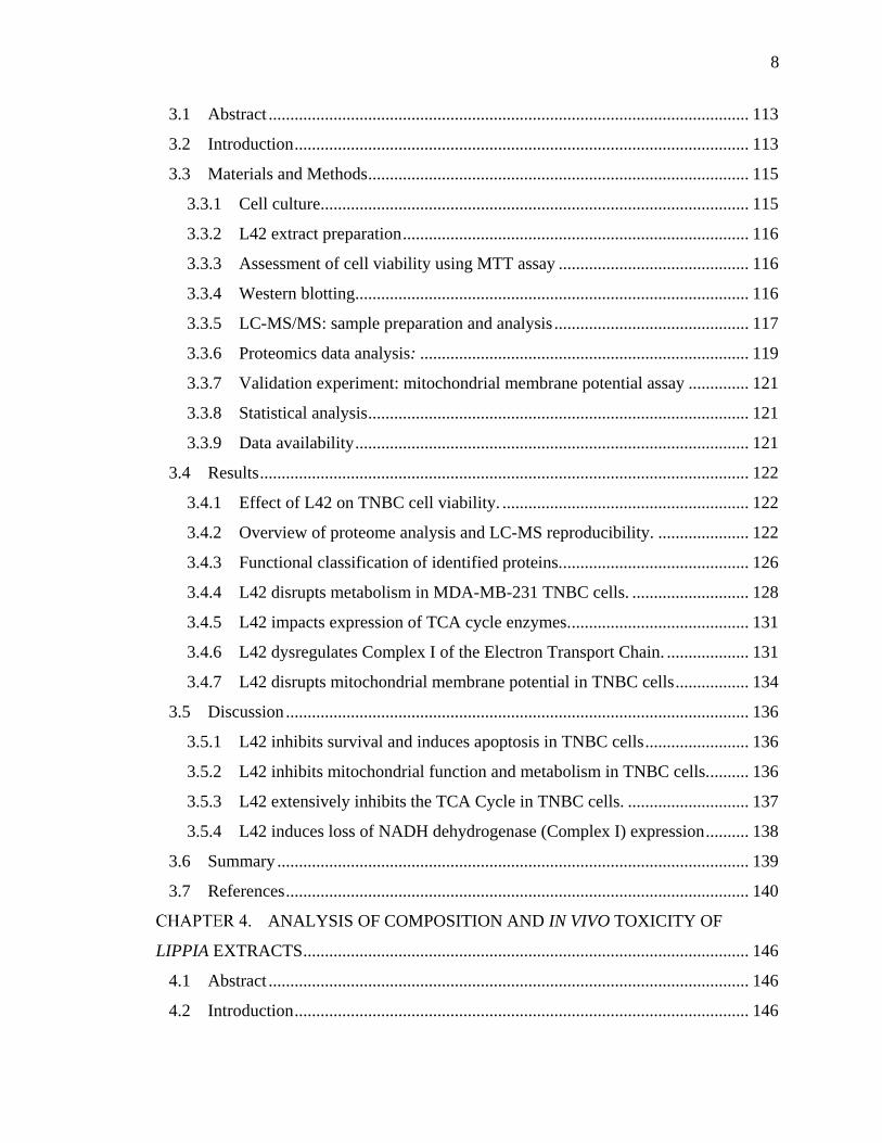
Figure 1.1 Hormone receptor status correlates with luminal phenotype and degree of
differentiation. Fluorescence-assisted cell sorting (FACS) of mammary epithelial cells based
on surface markers including Estrogen Receptor (ER) and luminal and basal markers allows
for fractionation into distinct populations. Fractions enriched for ER+ cells contain a low
frequency of stem-like cells and progenitors capable of mammary repopulation upon
transplantation, and colony formation in vitro. Conversely, fractions enriched for ER- and
basal-like cells exhibit enhanced colony-formation and mammary repopulation abilities, and
are thus considered to have a greater population of stem and progenitor cells.
Source: Tornillo, G., & Smalley, M. J. (2015). Journal of Mammary Gland Biology and
Neoplasia, 20(1-2), 63–73. [Ref. 23]

29
1.2 Triple-Negative Breast Cancer Overview
A particularly aggressive form of breast cancer is triple-negative breast cancer (TNBC),
identified as having < 1% of tumors cells expressing the three hormone receptors
commonly utilized for subtyping [28]. Pathologically, TNBC tumors are associated with
higher grade at diagnosis, the presence of unfavorable mitotic features such as higher
mitotic index, marked nuclear pleomorphisms, pushing borders of invasion, geographic
tumor necrosis, and poor overall differentiation [29]. 71 % of TNBCs are found to be
basal-like, and 77 % of basal-like breast cancers are triple-negative [30]. Basal-like breast
cancer are characterized by high expression levels of basal epithelial markers including
keratin 5 and 17, laminin, and fatty acid binding protein 7, and low levels of luminal
markers [31, 32]. Patients diagnosed with TNBC have lower overall median survival
rates compared to those diagnosed with hormone receptor-positive (HR+)/HER2- tumors
[overall survival: adjusted hazard ratio, 2.72; 95% confidence interval, 2.39-3.10; P <
.0001], with a dramatically increased risk of death within the first 2 years of diagnosis
[overall survival for 0-2 years: OR, 6.10; 95% confidence interval, 4.81-7.74] [33].
1.2.1 Epidemiology and Behavioral/Genetic Risk Factors for TNBC
Triple-negative/basal-like cancers make up 10-20 % of invasive breast cancers diagnoses.
Between 2010-2014, the incidence rate of TNBC was observed to be twice as high in
African American women (24/100,000) as in non-Hispanic white women (12/100,000),
and thrice as high compared to Asian/Pacific Islander women (8/100,000) [34]. A genetic
explanation for this difference has not been identified, but a 2010 study by Stark et al.
showed African ancestry strongly correlated with younger age of cancer diagnoses (48.0
y.o. for Ghanaian women, as opposed to 60.8 y.o. for African Americans, and 62.4 y.o.
for white Americans.), and higher proportion of TNBC (82.2 % for Ghanaians, 26.4 %
for African Americans, and 16 % for white Americans) [35].
Several reproductive factors have been shown to influence risk of TNBC
incidence. In a 2010 study looking at 1197 breast cancer cases and 2015 controls, Ma et
al. determined that women aged 45-64 y.o. who had a history of oral contraceptive (OC)
usage before age 18 had a 2.9-fold increased risk of TNBC [36]. In a follow-up 2017
study, Ma et al. performed a pooled analysis of 2658 white and African American

30
patients with breast cancer (of which 554 were diagnosed with TNBC) along with 2448
controls, with all patients aged between 20-64 years, and found that TNBC risk in parous
women who breast fed for at least 1 year was 31 % lower than in parous women who
never breast fed [37]. Remarkably, parous African American women between 20-44 y.o.
who breast fed for 6 months had an 82 % lowered risk of TNBC than those that did not
breast feed.
In addition to early OC use and non-breast feeding, TNBC risk has been linked to
premenopausal status as women under 40 y.o. are disproportionately diagnosed with ER-
breast cancers (33.8 %) compared to older women (21.9 %) [38]. A 2006 analysis of 496
samples from the Carolina Breast Cancer Study found that premenopausal African
American women had a higher prevalence of TNBC (39 %) compared to postmenopausal
African American women (14 %) [39].
In 2014, 40. 4 % of American women were defined as obese (BMI 30 kg/m2),
with 9.9 % considered severely obese (BMI 40 kg/m2) [40]. A 2008 study by Millikan
et al., using the Carolina Breast Cancer Study samples, showed that TNBC risk was
increased significantly for women with higher waist-to-hip ratio (WHR) [OR = 2.3; 95%
CI, 1.4–3.6], with a similar effect described for premenopausal ER-/PR- breast cancer in
the Women’s Circle of Health Study in 2009 [41, 42]. Mechanisms by which obesity may
support TNBC initiation, progression, and metastasis include i) influencing the
Akt/mTOR pathway and glycolysis through insulin signaling, ii) secretion of obesity-
related inflammatory cytokines such as leptin, as well as promotion of a general
inflammatory state characterized by heightened circulating levels of pro-inflammatory
cytokines which can in-turn stimulate STAT3, NFB, and Wnt/EZH2 signaling, and iii)
promote breast tumor aggressiveness and progression through the influence of the
adipose tissue microenvironment, including elevation of estrogen levels and promotion of
infiltration of immune cells [43-45].
Finally, specific genetic risk factors are known to be major contributors to TNBC
such as BRCA1 and BRCA2 mutations. BRCA1 and BRCA2 are proteins involved in the
error-free repair of double-strand DNA breaks, with germline BRCA mutations
commonly associated with risk of development of breast cancer. The Consortium of

31
Investigators of Modifiers of BRCA1/2 (CIMBA) utilized data from 4,325 BRCA1 and
2,568 BRCA2 mutation carriers, and found that the proportion of TNBCs decreased with
age at diagnosis for BRCA1 mutation carriers and increased with age at diagnosis for
BRCA2 mutation carriers [46]. In addition, 69 % of breast cancers in women with BRCA1
mutations were triple-negative. Also, 20 % of TNBCs carry BRCA1 mutations [47]. In a
2018 study, multigene panel testing methods utilized two TNBC patient cohorts – one in
which 21 breast cancer predisposition genes in 8753 patients were tested in Ambry
Genetics clinical laboratory, and another in which 17 genes from 2148 patients were
tested by a Triple-Negative Breast Cancer Consortium (TNBCC) – and found that
BARD1, BRCA1, BRCA2, PALB2, and RAD51D germline variants correlated with high
risk (OR > 5.0) of TNBC, while pathogenic variants for BRIP1, RAD51C, and TP53 were
associated with moderate risk (OR = 2.0 - 5.0) in Caucasian women [48].
1.2.2 Clinical Outcomes and Treatment Options for TNBC
Cancer ‘staging’ often utilizes the TNM system, where ‘T’ refers to the size of the
tumor, ‘N’ to the node status, i.e. if the cancer has invaded regional lymph nodes, and
‘M’ to the degree of metastasis. In comparison to other subtypes of breast cancer, TNBCs
have a worse prognosis, a more aggressive trajectory, and are more commonly diagnosed
in younger and obese women, with an average age of disease onset at 53 years. The poor
prognosis of TNBC is likely correlated with later stage at diagnosis, as most TNBCs are
T2 and T3 at the time of diagnosis (i.e. present with larger tumors), and are more likely to
be positive for lymphovascular invasion [49]. TNBCs also display preferential metastasis
to the brain and lungs, with a lower incidence of metastasis to the bone [50]. Lin et al.
conducted a study of 15,204 women who presented at the National Comprehensive
Cancer Network centers with Stage I-III breast cancer [33]. The study, which included
2,569 TNBCs, 2,602 HER2+, and 10,033 HR+/HER2- diagnoses, reported decreased
overall survival (OS) for TNBC compared to other HR+/HER2- cancers (HR 2.72 [2.39-
3.10], p <0.0001). Notably, the TNBC group had a dramatically increased risk of death
within the first 2 years of diagnosis (HR for OS for 0 - 2 y 6.10 [95% CI 4.81, 7.74]),
with the risk declining markedly over time [33].

32
Curiously, early-stage TNBCs have a better response rate than HR+/HER2-
tumors to anthracycline-based neoadjuvant chemotherapy (NACT) (85 % vs 47 %) [51].
The chemosensitivity of early stage TNBCs to NACT also leads to higher rates of
pathologic complete response (pCR), which is an important marker for improved survival
across all breast cancer subtypes. Unfortunately, patients with TNBCs also have the
highest 3-year relapse rates, with an increased risk of visceral relapse (i.e. metastatic
tumor relapse sites at lungs, pleura, liver, brain, or other thoracoabdominal organs). The
lower OS for TNBC patients is due to extremely limited treatment options post-
recurrence. Following recurrence, survival for TNBC patients rarely extends past 12
months, with British Columbia Cancer Agency data showing that median duration of
survival for distant metastatic basal-like breast cancer is just 0.5 years [52].
Presently, NACT protocols for TNBC advise anthracycline (such as epirubicin
and doxorubicin)- and taxane (such as paclitaxel and docetaxel)-based chemotherapy
[53]. Anthracyclines work by intercalating between DNA and RNA strands, preventing
replication and inhibiting cell division of rapidly proliferating cells such as those found in
tumors [54]. Similarly, taxanes also inhibit cell division, but do so by disrupting
microtubule formation through stabilizing GDP-bound tubulin molecules, preventing
microtubule depolymerization [55]. Unfortunately, anthracyline and taxane treatments are
non-specific, and are accompanied by major adverse side-effects which can themselves
drive fatality.
Both anthracyclines as well as taxanes were first discovered from natural sources,
with the anthracyline doxorubicin isolated from Streptomyces, and paclitaxel from the
Taxus brevifolia (Pacific Yew) conifer tree. Aside from the standard NACT regimen,
drugs with indications for TNBC include 5’-fluorouracil (currently hypothesized to be a
thymidylate synthase inhibitor preventing DNA formation) and bevacizumab (a
monoclonal antibody to VEGF-A, acting as an angiogenesis inhibitor) [53, 56, 57]. Meta-
analysis results from several clinical trials have supported the use of platinum
(carboplatin or cisplatin)-based chemotherapy regimens to improve pathological
complete response rates (pCR, i.e. the percentage of patients with lack of all signs of
cancer in tissue biopsies post-therapy), and objective response rates (ORR, i.e. percentage

33
of patients with tumors that shrink or disappear upon therapy), at the cost of greater
adverse events (AEs) due to toxicity [58]. However, there is currently no evidence that
platinum-based chemotherapy can increase overall survival (OS) or progression-free
survival (PFS), and its clinical use remains controversial.
The current adjuvant chemotherapy regimens for TNBC commonly utilizes
anthracycline and taxane-based combination treatments, which have been shown to
marginally improve 3-year disease-free survival (DFS) rates from 68 % to 73.5 % (HR =
0.50; 95% CI, 0.29 to 1.00; P = .051) [59]. In addition, sequential anthracycline-based
and taxane-based treatment regimens such as FEC (5’-fluorouracil, epirubicin and
cyclophosphamide) followed by paclitaxel/docetaxel (a sequential regimen known as
FEC-P) are commonly used for medium-to-high risk TNBCs, and show significantly
better DFS rates compared to FEC alone [60]. Another treatment option is the
combination of cyclophosphamide, methotrexate, and 5’-flurouracil (CMF) which causes
lower toxicity but requires a longer treatment duration. Patients with TNBC were shown
to have a significantly lower cumulative relapse rate with CMF treatment versus no
chemotherapy (21 % v 36 %), as well as longer time to relapse (HR: 0.46; 95% CI, 0.29
to 0.73; P = .009 relative to endocrine receptor–present subtype) [61].
Current areas of clinical research for TNBC treatment include poly-ADP ribose
(PARP)-inhibitors such as veliparib which interfere with base excision repair
mechanisms and are thought to be particularly lethal to BRCA1-mutated tumors; often
associated with TNBC [62]. Also, VEGF-inhibitors such as the monoclonal antibody
bevacizumab were evaluated at the level of phase III trials but unfortunately, did not
improve major patient outcomes in the adjuvant setting [63].
Taken together, TNBCs respond well to pre-operative (neoadjuvant) chemotherapy
compared to other subtypes, but conversely, have a much worse response to post-
operative (adjuvant) chemotherapy following relapse. Unfortunately, due to their higher
3-year relapse rates, greater propensity for CNS-directed metastasis, and poor chemo-
sensitivity in the adjuvant setting, metastatic TNBCs have the worst median survival time
(< 6 months) among breast cancer subtypes [52], and highlights the pressing need to

34
identify novel TNBC-specific signaling pathways and vulnerabilities that can be targeted
for therapy.
1.3 Molecular Heterogeneity of TNBC
Several studies have attempted to provide a mechanistic basis for the poor differentiation
status, aggressiveness, and drug resistance of TNBC tumors. In 2011, Lehmann et. al
provided a seminal description of the considerable heterogeneity of TNBCs through
expression profiling of 13,060 unique genes from 587 TNBC samples [64]. Clustering
analysis of these genes identified the 6 TNBC subtypes described below (Basal-like 1,
BL1; Immunomodulatory, IM; Mesenchymal, M; Mesenchymal stem-like, MSL; and
Luminal Androgen Receptor, LAR) classified on the basis of expression of gene clusters
from specific cellular pathways:
The Basal-like 1 (BL1) TNBC subtype is characterized by overexpression of
genes involved in proliferation (e.g. AURKA and AURKB) and DNA damage response
(e.g. CHEK1 and RAD51), while the Basal-like 2 (BL2) subtype overexpresses genes
regulating growth factor signaling, glycolysis and gluconeogenesis. Unsurprisingly,
basal-like (BL1 and BL2) TNBC has been found to be significantly more susceptible to
taxane-based therapies targeting cell division compared to the mesenchymal-like and
Luminal Androgen Receptor (LAR) subtypes (63 % pathological complete response
(PCR) for BL-TNBC, vs. 31% and 14 % for M-TNBC and LAR-TNBC respectively, P =
0.042).
The Immunomodulatory (IM) subtype is characterized by enrichment of immune
cell process gene ontologies (GOs). Several genes regulating events specific to immune
cells such as T cell receptor, B cell receptor, and NK cell signaling were shown to be
overexpressed, and immune signaling genes regulating NFB, TNF, and JAK/STAT
pathways are also enriched in the IM subtype.
Both the Mesenchymal (M) and Mesenchymal Stem-like (MSL) subtypes share
enrichment for GOs involving cell motility (Rho pathway), differentiation and growth
(Wnt, ALK, and TGF- pathways). Both subtypes are enriched for epithelial-

35
mesenchymal transition (EMT) genes (e.g. MMP2, TWIST1, ZEB1, and SNAI2), and
often have high rates of aberrations in the PI3K/AKT/mTOR pathway. However, the
MSL subtype also includes overexpression of genes functioning in growth factor
signaling such as EGFR, calcium signaling, and PDGF, and interestingly, has lower
expression of proliferation genes and a greater enrichment for stem-like genes, HOX
genes, and mesenchymal markers (e.g. BCL2, BMP2, ALDHA1, THY1, HOXA5,
HOXA10, NGFR, and VCAM1).
Further, using the GSE-10890 and ETABM-157 breast cancer cell line data sets,
the authors identified 30 non-overlapping TNBC cell lines, and correlated their gene
expression profiles to the 6 identified TNBC subtypes (Figure 1.2) [64]. Of note, the
MDA-MB-231 cell line, utilized as the primary model of aggressive TNBC in this
dissertation, was found to correlate with the mesenchymal stem-like (MSL) subtype.
Approximately 10 – 30 % of mesenchymal TNBCs have been morphologically
determined to fall within a class of rare, malignant form of drug-resistant breast cancer
known as metaplastic breast cancer (MpBC) [217, 218]. In addition, both mesenchymal
and metaplastic breast cancers contain aberrations in the mTOR pathway, and a phase I
trial of combination chemotherapy using doxorubicin, bavcizumab, and the mTOR
inhibitors temsirolimus or everolimus to treat MpBC patients (n = 52), showed an overall
response rate (ORR) of 21 % and a clinical benefit rate of 40 % [219].
The Luminal Androgen Receptor (LAR)-type, while also ER-, is characterized by
enrichment for hormone regulated pathways including steroid synthesis and
androgen/estrogen metabolism. Driving these pathways in this subtype is an
overabundance of androgen receptor (AR), expressed at 9-fold greater levels than other
subtypes, with downstream AR targets and coactivators also expressed (e.g. ALCAM,
PIP, and APOD). IHC staining also showed a significant, >10-fold increase in AR protein
expression in this TNBC subtype over others, making AR signaling an exciting potential
target against LAR-subtype TNBC tumors. The LAR subtype makes up roughly 11 % of
all TNBCs [64], and respond poorly to sequential taxane- and anthracycline-based
cytotoxic neoadjuvant chemotherapy, with patients diagnosed with LAR-TNBC showing
only 10 % pathological complete response (PCR) compared to 52 % for those with BL1-

36
TNBC [220]. However, treatment of advanced AR-positive TNBC with the antiandrogen
enzalutamide in a phase II trial showed a clinical benefit rate (CBR) of 29 % at 24 weeks
of treatment, and a median progression-free survival (PFS) of 14 weeks.
In summary, expression profiling of TNBC tumors and cell lines has helped
distinguish subtypes of clinical importance, and has provided a molecular basis for tumor
behavior and response to therapy, while also identifying potentially targetable
vulnerabilities.

37
Figure 1.2 TNBC subtypes. Classification of TNBCs on the basis of gene expression and
signaling pathways involved. The subclasses show varying rates of pathological complete
response (pCR) to standard chemotherapy, with promising targeted therapies suggested for
each subclass.
Source: Reproduced with permission from Omarini, C., et al (2017). Cancer Management
and Research, 10; 91-103 [Ref 222].

38
1.3.1 Overview of Major Signaling Pathways in TNBC
Several molecular pathways, including those involved in development (Wnt/-catenin,
Notch, and Hedgehog), survival (PI3K/Akt) and inflammation (NF-B signaling), have
been implicated in the oncogenesis, growth, invasion, metastasis, and development of
drug-resistance of triple-negative breast cancers. The evidence supporting the
involvement of these molecular pathways in TNBC are highlighted in this section below,
with an extended overview of the influence of NF-B signaling on TNBC covered in
section 1.3.2.
Wnt Signaling: Aberrations in Wnt signaling, a highly conserved developmental
pathway, have been shown to promote proliferation and survival [65], and epithelial-
mesenchymal transition [66] in cancer cells. Normally, -catenin, a dual function cell-cell
adhesion and transcriptional co-activator protein, is routinely cleared from the cytoplasm
by a destruction complex composed of APC, PP2A, GSK3, and CF1. However, ligand-
binding of Wnt to the G-protein coupled receptor (GPCR), Frazzled (Fz), induces
recruitment of the destruction complex to the cell membrane and subsequent de-
activation, leading to an accumulation of -catenin within the cytoplasm. -catenin
eventually translocates to the nucleus, where it binds to and co-activates TCF/LEF-family
transcription factors, thereby inducing transcription of genes involved in proliferation,
EMT, and migration, such as Cyclin D1 and c-myc.
The importance of Wnt signaling in promoting tumor growth in TNBC was
revealed by Xu et al. [67], who demonstrated that siRNA-mediated knockdown of -
catenin expression in TNBC cells implanted in mammary fat pads significantly reduced
the size and growth rate of tumors in mice. In addition, analysis of expression of Wnt
signaling genes in multiple patient tumor cohorts showed that an increase in Wnt/-
catenin signaling correlated with higher grade, poorer prognosis, and increase in lung and
brain metastasis in triple-negative breast cancer [68].
Notch Signaling: The highly conserved Notch developmental pathway is also
linked to breast cancer, with elevated Notch-1 and its ligand, Jagged-1, strongly
correlating with low overall survival in patients [69]. Notch signaling involves binding of

39
transmembrane Notch ligands (named Delta-like and Jagged-like in mammalian cells) to
Notch receptors which are usually expressed on neighboring cells. Ligand binding leads
to cleavage of the Notch receptor intracellular domain NICD, which translocates to the
nucleus and regulates gene expression by activating the transcription factor CSL. Notch
signaling heavily influences the determination of cell fate, for example, in hematopoiesis
and mammary gland development, and is also involved in angiogenesis, and in neuronal
function and development.
In 2006, Hu et al. showed that constitutive overexpression of Notch-1 (N1IC) and
Notch-4 (N4IC) could form spontaneous mammary tumors in a murine model [70]. Later
studies revealed TNBC cells have increased nuclear localization of Notch-4, and SCID
mice injected with MDA-MB-231 cells silenced for Notch-4 had reduced tumorigenesis
as well as tumor volume [71].
Hedgehog Signaling: The Hedgehog (Hh) developmental pathway begins with
the binding of a secreted ligand protein, Sonic hedgehog (SHH), to the Patched-1
receptor (PTCH1), which usually inhibits the activity of a transmembrane GPCR known
as Smoothened (SMO). However, SHH-binding to PTCH1 inactivates it, thereby leading
to SMO activation. SMO subsequently activates the GLI transcription factors, which are
responsible for regulating the expression of genes involved in proliferation and
differentiation.
Studies have shown Hedgehog signaling can promote self-renewal in breast
cancer stem cells [72], and chemotherapy-resistant tumor growth [73, 74]. In the latter
two studies, inhibiting Hh signaling was shown to reduce tumor growth and prevent
clonogenic expansion of resistant cell lines respectively, providing a mechanistic
explanation for the development of resistance in breast cancer cells. The GLI family of
transcription factors, which include Gli1, Gli2, and Gli3, are the primary effectors of Hh
signaling, and Gli1 overexpression and nuclear localization have been demonstrated as
poor prognostic markers in women diagnosed with TNBC [75]. Gli1 overexpression can
promote migration, invasion and metastasis in MDA-MB-231 TNBC cells by up-
regulating matrix mettaloproteinase 11 (MMP-11), and inhibiting Gli1 can reduce the
number of pulmonary metastases in mice injected with Gli1-silenced MDA-MB-231 cells

40
via tail vein [76]. Also of note is that Hh signaling is implicated in promoting
angiogenesis in TNBC cells; Gli1 levels correlate strongly with vasoendothelial growth
factor receptor 2 (VEGFR2) expression in tumor microarray studies, and silencing of
Gli1 decreased protein levels of VEGFR2 in MDA-MB-468 TNBC cells [77].
PI3K/AKT/mTOR Signaling: PI3K signaling plays an important role in
promoting cell survival, and anomalies in the PI3K/AKT/mTOR pathway are commonly
found across breast cancer subtypes [78]. In TNBCs, aberrant PI3K signaling occurs
primarily due to overexpression of epidermal growth factor receptor (EGFR), an
upstream PI3K regulator, or due to activating mutations in PI3K catalytic subunit
(PIK3CA), or loss of function/expression mutations in downstream regulators such as
phosphatase and tensin homolog (PTEN) and proline-rich inositol phosphate [79-81]. The
inhibition of PI3K/Akt/mTOR in cisplatin-resistant HCC38 and MDA-MB-231 TNBC
cells using the dual inhibitor NVP-BEZ235 was shown to re-sensitize the cells to
cisplatin, and synergism between NVP-BEZ235 and carboplatin treatment has also been
shown in TNBCs [82, 83].
1.3.2 NF-B Signaling in TNBC
The NF-κB family of transcription factors includes five genes: p65 (RelA), RelB, c-Rel,
p50/p105 (NF-κB1), and p52/p100 (NF-κB2). Most commonly, the heterodimer
p50/RelA is sequestered to the perinuclear cytoplasm by IκB proteins. Canonical NF-κB
signaling begins with TNF- binding to its membrane receptor TNFR1, followed by
TNFR1 trimerization [84, 85]. TNFR1 then recruits Complex I, which eventually leads to
translocation of p50/RelA to the nucleus, where it acts as a transcription factor for several
pro-survival genes [86-89]. If Complex I or downstream canonical signaling is inhibited,
TNFR1 stimulation leads instead to recruitment of Complex II which recruits and
activates caspase-8, leading to apoptosis [87]. Caspase-8 also cleaves RIP1, a key kinase
of Complex I, thereby halting pro-survival canonical signaling [90]. Conversely,
signaling through Complex I leads to increased expression of the Complex II inhibitor,
cFLIP [89]. Thus, the activation states of Complex I and Complex II can determine cell
fate in response to TNFR1 stimulation (Figure 1.3).

41
Over the last three decades, a mounting body of evidence has implicated aberrant
NF-κB signaling in promoting TNBC progression. In 1997, Nakshatri et al. showed that
NF-κB was constitutively activated during the advancement of RM22-F5 rat mammary
carcinoma cells from a hormone-dependent, non-malignant phenotype to hormone-
independent, malignant growth [91]. Constitutive activation of NF-κB has also been
associated with human TNBC models such as MDA-MB-231 cells [91, 92].
Studies have indicated that targeting the NF-kB pathway can be useful for
suppressing TNBC. For example, the chemical compound Bay-11-7082 ((E)-3-(4-
Methylphenylsulfonyl)-2-propenenitrile) can prevent IκB phosphorylation by IKK,
thereby preventing NF-κB translocation to the nucleus. Smith et al. showed that
inhibition of NF-κB signaling by Bay-11-7082 induced CD44 repression and inhibition
of proliferation and invasiveness of MDA-MB-231 and SUM159 TNBC cells [93].
Inhibiting NF-κB signaling directly or preventing the positive feedback mechanism via
TNF-α knockout has also been shown to induce apoptosis in both ER- breast cancer cells
as well as other cancer cell lines [94-98].
NF-κB signaling maintains an autocrine loop of pro-inflammatory cytokines like
IL-6 and TNF-a, promotes expression of pro-survival factors such as Bcl-xL, cIAP1 and
cIAP2 and proteins involved in cell cycle progression, for example Cyclin D1, as well as
invasion and metastasis markers such as MMP-9 and Vimentin [84, 99-104].
Tropomodulin-1 (TMOD1) is a protein that inhibits elongation and depolymerization of
actin filaments, and shows elevated expression in TNBC cells constitutively expressing
NF-κB. TMOD1 was shown to be an NF-κB target gene capable of enhancing in vivo
tumor growth of MDA-MB-231 cells in a mouse xenograft model [105].
Finally, the gene expression of several NF-κB-family members including RELA,
RELB, and NFKB2 are controlled by SP1 and STAT3 transcription factors, with
increased expression of RELA and SP1 correlating with lower 5-year distant recurrence-
free survival (DRFS) in TNBC patients treated with adjuvant doxorubicin chemotherapy
[106].
Overall, constitutive activation of NF-κB in cancer contributes to poor
differentiation, aggressiveness and ability to survive and maintain hormone-independent

42
growth. Finding new NF-κB inhibitors is therefore an exciting avenue to therapeutics
against TNBCs.

43
Figure 1.3 NF-κB signaling: Interplay between Complex I and Complex II determines
cell fate upon TNF-R1 activation.
44
1.4 Metabolism in Triple-Negative Breast Cancer
As part of their sequel paper to their seminal review “The Hallmarks of Cancer”,
Hanahan and Weinburg described metabolic reprogramming as one of the fundamental
traits of cancer cells required to sustain their continuous growth and division [107]. In
brief, cancer cells have been observed to shift their utilization of glucose towards
‘aerobic’ glycolysis (i.e. glycolysis in the presence of oxygen), and away from
mitochondrial metabolism, a phenomenon known as the ‘Warburg effect’, in honor of
Otto Warburg who first described this effect in the 1920’s - 50’s through a series of
investigations into tumor metabolism [108, 109]. Subsequent studies highlighted below
reveal TNBCs in particular are characterized by extensive metabolic reprogramming;
with the identification of druggable targets from these pathways becoming a major focus
of current research.
In TNBCs, EGFR signaling has been shown to elevate aerobic glycolysis, leading
to the build-up of intermediates which can promote tumor growth, and local immune
suppression of cytotoxic T-cells [110]. The transport of glucose into the cell is facilitated
by the GLUT family of transporters, and in a study of 55 patient tumors of TNBC,
GLUT1 (SLC2A1) was expressed in 42 (76.4 %) cases [111]. Metformin, a
mitochondrial Complex I inhibitor commonly used as an anti-diabetic, has been shown to
down-regulate the expression of several GLUT-family transporters, and can suppress
proliferation and reduce the cancer stem cell population in TNBCs [112-114]. Indeed, a
meta-analysis of 11 studies of over 5000 breast cancer patients found that the use of
metformin among diabetic breast cancer patients has been associated with a 47 %
decreased risk of death from all causes (HR: 0.53; 95% CI: 0.39-0.71) and significantly
improved cancer-specific survival times (HR: 0.89; 95% CI: 0.79-1.00) [221].
Recent studies have described the complexity of metabolic reprogramming in
TNBC cells as extending far beyond aerobic glycolysis. TNBCs are now understood to
have a fundamentally altered metabolic profile with higher oxidative stress through
increased reactive oxygen species (ROS) levels partially attributed to lower glutathione
biosynthesis [115], and elevated uptake and utilization of glucose [116], glutamine [117-

45
119], and TCA cycle intermediates [120], as well as increased fatty acid β-oxidation
[121].
1.4.1 Amino Acid Metabolism in TNBC
Amino acids serve as important precursors for TCA cycle entry in TNBC cells. In a
comprehensive metabolomics study comparing the metabolite profiles of 15 TNBC vs 15
ER+ breast cancer tissue samples from African-American patients, Kanaan et al. found
that 8 of 20 amino acids detected (aspartate, asparagine, glutamate, phenylalanine,
methionine, and the branched-chain amino acids (BCAAs) leucine, isoleucine and valine)
were significantly elevated in TNBC samples [120]. Transport of amino acids into cells is
hypothesized to occur via a catalytic reaction with glutathione that converts them to -
glutamyl amino acids, which can then traverse the plasma membrane. Following a
reverse catalytic reaction that regenerates glutathione via a 5-oxoproline intermediate, the
original amino acids are released into the intracellular space [122]. 6 of 7 -glutamyl
amino acids identified in the study were significantly increased in TNBC samples,
implying increased amino acid transportation in TNBC cells [120].
All 20 amino acids enter the TCA cycle via conversion to intermediates.
Methionine and the BCAAs valine and isoleucine are first converted to Succinyl CoA
which is rapidly converted to succinate and subsequently fumarate, while phenylalanine
and aspartate are converted directly to fumarate [122]. TNBCs were shown to have
elevated levels of the TCA intermediates fumarate, succinate, and malate, further
supporting that TNBCs amplify the utilization of amino acid metabolism to meet their
energy requirements [120].
The increase in amino acid metabolites in TNBCs has been validated in
subsequent studies as well, with glutamine metabolism emerging as a therapeutic target
[123]. “Glutamine addiction” is a term used to describe the dependence of many cancers,
including TNBCs, on glutamine as a carbon and nitrogen source, and their inability to
survive upon glutamine deprivation. Glutamine is shuttled into the cell through several
transporters including alanine, serine, cysteine-preferring transporter 2
(SLC1A5/ASCT2), and subsequently converted to glutamate via glutaminase (GLS1),

46
before conversion to -ketoglutarate by glutamate dehydrogenase (GDH) which marks
the entry into the TCA cycle (Figure 1.4).
A 2016 study by van Geldermalsen et al. revealed mRNA expression of
glutamine-metabolism genes SLC1A5, GLS, and GLUL (glutamine synthetase, which
catalyzes de novo glutamine synthesis) were uniformly elevated across a cohort of 90
TNBC patient tumor samples [119]. The authors also showed pharmaceutical inhibition
of SLC1A5 inhibited mTORC1 signaling, growth and progression of TNBC cells
specifically in vitro, and SLC1A5 knockdown repressed TNBC mammary tumor growth,
and improved survival in an in vivo mouse xenograft model. Suppression of GLS by both
shRNA-mediated knockdown as well as pharmacological inhibition led to TNBC cell
growth inhibition in vitro and in vivo [117, 124]. GLS inhibition by the chemical agent
CB-839 was also associated with reduction in glutamine consumption and glutamate
production, as well as decreases in oxygen consumption, glutathione levels, and TCA
cycle intermediates including fumarate, malate, and citrate [124]. Finally, in a 2018
study, Mauro-Lizcano and Lopez-Rivas revealed that glutamine deprivation sensitized
TNBC cells to tumor necrosis factor-related apoptosis-inducing ligand (TRAIL)-
induced apoptosis through upregulation of TRAIL Receptor 2 (TRAILR2) [125].
Collectively, these studies highlight the importance of glutamine metabolism in
maintaining energy homeostasis in TNBC cells, and supports investigations into targeting
glutamine transporters and GLS as an avenue to TNBC therapy.

47
Figure 1.4 Glutamine transport and metabolism. Extracellular glutamine is transported into
the cell through transporters such as alanine, serine, cysteine-preferring transporter 2
(SLC1A5/ASCT2). Transport into the mitochondria is carried out through a currently
unknown mechanism but is theorized to occur based on the mitochondrial localization of
glutaminase (GLS), and glutamate dehydrogenase (GDH). The end product of GDH action on
glutamate results in the TCA Cycle intermediate, -ketoglutarate (-KG). Glutamate and the
TCA Cycle intermediate citrate can also be further utilized in amino acid and lipid synthesis.

48
1.4.2 Mitochondrial metabolism in TNBC
Until recently, it was widely accepted that the ‘Warburg effect’, or increased aerobic
glycolysis, was accompanied by a suppression of mitochondrial biogenesis in neoplastic
cells. Beginning in the early 2000s however, a number of studies have shown that
mitochondria are not only functionally active in many different cancer types, but that
oncogenic activation of mitochondrial function is associated with increased malignancy
and a dependence on oxidative phosphorylation (OXPHOS) for energy production, with
breast cancer cells producing ~ 80 % of their ATP through mitochondrial metabolism
[126-128].
In order to explain these two observations seemingly at odds with one another, in
2011, Smolkova et al. advanced the hypothesis that multiple “waves of gene expression”
promote metabolic adaptations to the transforming tumor microenvironment and
changing metabolic requirements [129]. The initial waves promote rapid proliferation by
upregulating glycolysis at the expense of mitochondrial OXPHOS, leading to aglycemia
and nutrient shortages. To compensate for this, and to promote tumor cell survival and
clonal selection, subsequent waves re-establish glutaminolysis-driven TCA cycle and
OXPHOS through oncogenic activation of c-Myc, and upregulation of PI3K-Akt-mTOR
and LKB1-AMPK-p53 pathways [130-132]. As mentioned previously, glutamine uptake
and utilization is upregulated in many cancers, including TNBC. Mazurek et al. revealed
that when OXPHOS proceeds unencumbered, glutamine can fully compensate for a lack
of glucose in cancer cells [133]. A summary of these findings are portrayed in Figure 1.5.
The oncogene c-Myc drives cell cycle progression, metabolic pathways, and
survival under oxidative stress in cancer cells, with 54 % of chemotherapy-resistant
TNBCs harboring c-Myc amplifications [134]. Indeed, cancer stem cells (CSCs) derived
from TNBC cell lines have elevated c-Myc levels that correlate with increased ROS
production and mitochondrial OXPHOS activity [135]. Treatment of SUM159PT and
MDA-MB-436 TNBC cells (which express elevated levels of c-Myc) with oligomycin A
(an ATP synthase inhibitor), or metformin (a Complex I inhibitor) can reduce the oxygen
consumption rate (OCR) and mammosphere formation in these cell lines [135]. In
another study, c-Myc was shown to inhibit mitochondrial glutaminase (GLS) repression
49
in P-493B lymphoma PC3 prostate cancer cells, thereby upregulating glutamine
catabolism for ATP production through TCA cycle activity [136]. The siRNA-mediated
silencing of GLS inhibited cell proliferation, and addition of the TCA intermediate
oxaloacetate could partially rescue proliferation, further underlining the importance of
mitochondrial metabolism in the growth and proliferation of cancer cells [136].
The advantages of glutamine metabolism to cancer cells was highlighted in a
2009 review by Weinberg and Chandel [137]. By serving as a substrate for the TCA
cycle, glutamine drives ATP production via OXPHOS and also satisfies the proliferating
cell’s demand for lipid, nucleotide, and protein synthesis. Citrate produced in the TCA
cycle, for example, is transported to the cytosol and converted to Acetyl-CoA required in
lipid biosynthesis, while malate, another TCA intermediate, is instead converted to
oxaloacetate in the cytosol, and then to aspartate via aspartate aminotransferase (AAT);
this is required for synthesis of pyrimidines that make up DNA. Malate can also be
decarboxylated in the cytosol by malic enzyme (ME) to produce pyruvate and NADPH,
the latter of which is shuttled toward lipid biosynthesis and regeneration of reduced
glutathione. Thus, targeting enzymes utilizing TCA intermediates for cellular
biosynthesis may prove useful against aggressive cancers with elevated mitochondrial
metabolism. Indeed, inhibiting AAT activity in MDA-MB-231 TNBC cells using amino
oxyacetate (AOA) or siRNA-mediated silencing inhibited proliferation and oxygen
consumption in vitro, and reduced tumor xenograft growth in vivo.
Taken together, studies on cancer metabolism report that there are shifts in
preference for glycolysis-driven or mitochondrial-driven ATP production that are
dependent on tumor stage, cancer type and subtype, nutrient availability, and tumor
microenvironment. Mounting evidence supports a dependence of aggressive, late-stage
cancers on TCA cycle intermediates that drive glucose-independent ATP production via
OXPHOS while also giving rise to biosynthetic precursor metabolites. Targeting the TCA
cycle and OXPHOS in such cancers including triple-negative breast cancer may be a
highly viable avenue to novel therapeutics.

50
Figure 1.5 Waves of metabolic reprogramming during carcinogenesis. Wave 1:
Oncogenic transformation leads to enhanced glycolytic activity at the expense of OXPHOS
and mitochondrial biogenesis, leading to rapid proliferation. Wave 2: Promotion of cell
survival through HIF and NFB signaling, with sustained glycolytic activity and inhibition of
mitochondrial biogenesis. Wave 3: Activation of AMPK and Akt signaling leads to inhibition
of protein synthesis. PGC1 activation leads to mitochondrial biogenesis followed by
OXPHOS initiation, while MYC activation increases glutaminolysis. Wave 4: Mitochondrial
biogenesis and activity leads to mitochondrial signaling activation through multiple effectors
including NAD+/NADH ratio, AMPK, Ca2+ and lipid signaling, and NO production. In
addition, changes in mitochondrial inner membrane potential and morphology may lead to
downstream signaling activation/inhibition.
Source: Modified from Smolkova et al. (2011). The International Journal of Biochemistry &
Cell Biology, 43(7), 950–968.

51
1.5 An Important Role for Plant-derived Metabolites in Cancer Therapy
The use of plants in medicine has a deep history, widely recorded in multiple cultures
across the globe. The study of such plant-based traditional remedies has had a lasting
impact on onco-drug discovery; with vinca alkaloids, camptothecins,
epipodophyllotoxins, and taxanes currently in clinical use against a range of cancers
including leukemia, lymphomas, neuroblastomas, small cell lung cancer, ovarian, colon,
stomach, prostate, head and neck, and breast cancer. Given the rich diversity of plant life
and the identification of several phytochemicals with anticancer properties, research into
plant-derived bioactive compounds remains an important source of novel therapeutics
against cancer.
1.5.1 Cancer Drug Discovery from Plant Sources: A Historical Perspective
Discovery of the topoisomerase II-inhibitor, etoposide: One of the first intensively
researched plants to yield a cancer therapeutic was from the genus Podophyllum. This
genus includes two species: American Podophyllum (Podophyllum peltatum), and Indian
Podophyllum (Podophyllum hexandrum) [138]. Native Americans had been known to use
extracts of P. peltatum for several purposes. P. peltatum root extracts were used as
cathartics, antihelminthics, and purgatives [139], with the first recorded use of
Podophyllum to treat ‘cancer’ by the Penobscot Indians of Maine [140]. It was soon
found that the medicinal properties of Podophyllum could be attributed to their rhizomes,
which were rich in resin. In 1844, Dr. John King published the first report on the alcohol
extraction of Podophyllum resin, which he named podophyllin [141-143], and finally, in
1881, Podwyssotzki isolated and crystalized the major bioactive component of
Podophyllum extracts, which he named podophyllotoxin [144].
Beginning with Kaplan in 1942 [145], several studies on podophyllin and
podophyllotoxin uncovered its antimitotic effects in vitro, and in 1947, Hartwell and
Shear demonstrated its ability to kill cancer cells in vivo [146]. Investigations into the
mechanism of action of podophyllotoxin revealed that it could bind strongly to tubulin,
thereby inhibiting the formation of mitotic spindle and inducing cell cycle arrest.
However, the acute gastrointestinal toxicity of podophyllotoxin led investigators to
synthesize derivatives with less severe side-effects. In the mid-50s, Stähelin and Von

52
Wartburg painstakingly tested hundreds of hemisynthetic -D-glycosidic derivatives of
podophyllotoxin, culminating in the identification of VP-16 (etoposide) and VM-26
(teniposide), which were selected on the basis of their in vivo efficacy in increasing
survival time in mice inoculated with the L-1210 leukemia cell line [147]. It was later
found that VP-16 induced cell arrest by inhibiting topoisomerase II activity, thereby
introducing double strand breaks in DNA that activated apoptotic signaling cascades in
rapidly dividing cancer cells [148]. Etoposide is currently FDA-approved for the
treatment of small cell lung cancer and refractory testicular cancer.
Discovery of the microtubule polymerization inhibitors, vinca indole alkolids: The vinca
alkaloids were first isolated from the leaves of the plant Catharanathus roseus (aka.
Vinca rosea). The plant was originally brought to the attention of the medical community
in the mid-1950s when it was noted that the native peoples of Jamaica used C. roseus
leaves to make an herbal tea believed to benefit diabetics [149].
Investigations by Dr. J. B. Collip and Dr. Robert L. Noble at the University of
Western Ontario found that aqueous extracts of the leaves had little to no effect on blood
sugar levels, but when injected in rats, led instead to a transient, rapid decrease in white
blood cell (WBC) count due to bone marrow destruction. This exciting discovery
motivated Collip and Noble, in association with Dr. Charles T. Beer and Dr. Henry Cutts,
to isolate the bioactive component of the extract by bioassay-driven fractionation. After a
year of intense research, the team isolated vincaleukoblastine (VLB, later shortened to
vinblastine), and another team at Eli Lilly Company isolated vincristine (VCR); with the
two later shown to be dimeric indole alkaloids [150].
X-ray crystallographic studies of VLB and VCR by Moncrief and Lipscomb in
1965 revealed that the two molecules differed by a single functional group, with VCR
possessing an aldehyde (CHO) group where VLB has a methyl (CH3) group [151]. This
small difference accounted for the difference in clinical responses to these drugs; VLB is
primarily used against lymphomas and causes acute toxicity to the bone marrow, while
VCR is mostly used to treat acute leukemia and displays peripheral nervous system
toxicity.

53
Clinical trials showcasing the efficacy of vinblastine and vincristine have led to
FDA approval for single-drug use in Hodgkin’s lymphoma, acute leukemia, melanoma,
testicular, bladder, and prostate cancer, as well as in combination chemotherapy for
Hodgkin's lymphoma, Non-Hodgkin's malignant lymphomas, rhabdomyosarcoma,
neuroblastoma, and Wilm's Tumor.
In conclusion, the serendipitous discovery of such anticancer drugs through
studies of traditional folk remedies in the 1950s lead the National Institutes of Health
(NIH) to begin large-scale screening of natural extracts on cancer models to isolate and
identify their bioactive anticancer compounds [152]. This work was carried out by the
National Cancer Institute (NCI) through its Cancer Chemotherapy National Service
Center (CCNSC). These experiments led directly to the discovery of the life-saving
chemotherapeutics camptothecin and taxane by Monroe Wall, Mansukh Wani, and others
at the Natural Products Laboratory, Research Triangle Institute (RTI), in North Carolina.
The mechanism of action of these and other plant-derived compounds leading to cancer
cell death are discussed in the following section.
1.5.2 Mechanisms of Anticancer Action of Plant-derived Compounds
1.5.2.1 Interference with microtubule assembly
Taxanes are a class of diterpenes originally derived from the bark of the Pacific Yew tree
(Taxus brevifolia). Both taxanes as well as vinca alkaloids are ‘spindle poisons’ that
target microtubule assembly to inhibit cell division. Taxanes stabilize GDP-bound tubulin
to prevent microtubule depolymerization [153], while vinca alkaloids bind to the ends of
assembled microtubules to prevent addition of new tubulin dimers, thereby inhibiting the
polymerization process [154]. Consequently, these compounds are specifically potent to
rapidly proliferating cells such as cancer cells, with the unfortunate side-effect of
attacking frequently dividing cells in the bone marrow, intestinal lining, and hair follicles,
leading to myelo/immuno-suppression, mucositis, and alopecia respectively.
1.5.2.2 Inhibition of DNA topoisomerases
Camptothecin (CPT), a cyclical compound derived from the bark of the Camptotheca
acuminata tree, was first discovered in 1966 by Wall et al. at the Research Triangle

54
Institute [155]. In contrast to taxanes and vinca alkaloids, camptothecins are
topoisomerase inhibitors. Topoisomerases are proteins involved in the winding or
unwinding of DNA; functions critical for transcription and DNA replication. In 1983,
Hsiang and Liu were the first to show CPT binds to mammalian DNA topoisomerase I to
form ternary complexes that stabilize binding and result in DNA strand breaks, leading to
apoptosis [156]. CPT analogues include topotecan and irinotecan, which have higher
solubility and bioavailability than CPT and are currently in clinical use for colon,
pancreatic, ovarian, cervical, and lung cancers.
In general, epipodophyllotoxins, derived from the root of the American Mayapple
(Podophyllum peltatum), act through multiple mechanisms including inhibition of
microtubule assembly [157]; however, etoposide (VP-16) and teniposide (VM-26), the
most commonly utilized semisynthetic derivatives of the base compound
podophyllotoxin, act predominantly through topoisomerase inhibition. As with CPT,
multiple studies in the late 1980s showed that VP-16 and VM-26 can stabilize the
interaction between topoisomerase II and DNA, interfering with the scissor-reunion
reaction of the enzyme [158, 159], as well as its ‘strand-passing’ activity which allows it
to disentangle topologically confined DNA [159, 160]. This leads to a halt in DNA
replication and mitosis, eventually causing cell death.
1.5.2.3 Interference with multiple proliferative and survival pathways
In addition to impeding cell division mechanics, plant compounds can also bind and/or
inhibit multiple protein targets, thereby regulating several cellular pathways that promote
growth, proliferation, and survival in cancer cells. For example, resveratrol (5-((E)-2-(4-
hydroxyphenyl) vinyl)-1,3-benzenediol), a compound found abundantly in grape skins,
peanuts, and berries, has recently entered human clinical trials after a number of studies
highlighted its ability to induce apoptosis, autophagy, and senescence in multiple cancer
cell lines in vitro, and prevent tumor initiation, angiogenesis, and metastasis in vivo [161-
167]. The variety of cancer cell death mechanisms triggered upon treatment with
resveratrol can be explained by the observation that it directly targets over 20 different
proteins, including those involved in metabolism (FAS, CBR1, NQO2), inflammation
(COX1/2, LTA4H), cell cycle arrest (Pol/, ATM, AKT1), post-translational
55
modification (pan-HDAC inhibition, and inhibition of the kinases PKC, AKT1, PKD1),
and cell signaling (COX1/2, Phosphodiesterases) [168].
A review of cell death mechanisms induced by anti-cancer plant polyphenols,
flavonoids, and terpenoids by Gali-Muhtasib in 2015 revealed that while several
pathways regulated by these compounds are unique the majority of these compounds
are capable of inhibiting the Akt/mTOR pathways, generating reactive oxygen species
(ROS), or disrupting mitochondrial membrane potential [169]. For example, curcumin,
the major bioactive polyphenol present in the turmeric rhizome (Curcuma longa),
inhibited phosphorylation of Akt and induced autophagy in U87-MG and U373-MG
human malignant glioma cells in vitro and significantly inhibited growth of malignant
glioma tumors in nude mice upon intratumoral injection at 100 mg/kg body weight [170].
Similarly, the isoflavone genistein, originally isolated in 1899 from Genista tinctoria and
found abundantly in soybean, also reduced P-Akt levels and significantly inhibited
glucose uptake in ovarian cancer cells, again leading to autophagy [171].
Naturally-occurring and semi-synthetic analogues of the alkaloids noscapine and
colchicine, initially isolated from Lachryma papaveris (opium) and Colchicum autumnale
(meadow saffron), respectively, have been shown to induce autophagic cell death in
prostate, leukemia, pancreatic and lung cancer cells via ROS generation [172-174].
Another example of a multifunctional anti-cancer plant compound is the
terpenoid, parthenolide, from Tanacetum parthenium, which induced autophagy in MDA-
MB-231 TNBC cells by generating ROS as well as by inhibiting NF-B signaling, and is
capable of inducing necrotic cell death in these cells by disrupting the mitochondrial
membrane potential [175].
In addition, several of these and other polyphenols, alkaloids, and terpenoids have
been observed to inhibit telomerases [176, 177], induce DNA damage [178, 179], reduce
Cyclin D1 expression, [180] and activate AMPK-mediated cell death [181]. The variety
of signaling pathways targeted by plant-derived compounds to induce cancer cell death is
a reflection of their enormous diversity in structure and chemistry, and highlights the

56
need to develop technologies and processes to better exploit their potential in the
development of novel cancer therapies.
1.5.3 Lippia species as a source of anti-cancer compounds
The genus Lippia comprises over 200 species of tropical flowering shrubs from the
family Verbenaceae. The essential oils from these plants are usually aromatic and
fragrant, and have been well documented as possessing anti-fungal [182, 183], anti-
bacterial [183, 184], anti-mutagenic [185, 186], anti-inflammatory [187-189], and
analgesic [188, 190-192] qualities. More recently, L. sidoides, L. salviifolia, and L.
rotundifolia showed cytotoxicity against CT26 colon cancer cells [193], while L. alba
induced G0/G1 cell cycle arrest and apoptosis against both lung (A549) and liver (HepG2)
cell lines [194].
Lippia origanoides is a medicinal tropical plant native to South and Central
America, and its extracts have previously been shown to possess anti-inflammatory and
anti-nociceptive analgesic activities in mice [195]. Individual components of the extract
have shown pro-apoptotic and anti-proliferative effects on several cancer cell lines [196-
202]. Carvacrol and thymol, two of the major components of L. origanoides, also
inhibited NF-B signaling in models of hepatotoxicity and LPS-stimulation [203, 204].
As previously noted, NF-B signaling is a pro-survival inflammatory pathway often
constitutively activated in TNBC cells [90, 91]. Thus, a major goal of this research
project was to describe the effect of L. origanoides extracts on NF-B signaling in
TNBC. However, keeping in mind the multi-component nature of these extracts, a further
goal was to map the proteome-wide changes regulated by the extract to identify
additional and possibly synergistic mechanisms of action.
1.6 Quantitative LC-MS-based Proteomics for Cancer Drug Discovery
Drug discovery from bioactive natural extracts has commonly been achieved through
bioassay-driven fractionation schemes, which are both time-consuming and expensive
[205, 206]. In addition, attempting to identify the mechanisms of drug action through
standard molecular biology techniques such as Western Blotting, PCR, and ELISA does

57
not provide information on off-target, detrimental side-effects that only come to light in
vivo or during clinical trials. Hence, there is a need to study cells and tissues at the
systems level to better capture the effects of chemotherapy. Transcriptomics approaches
using cDNA-microarrays and RNA-Seq technologies have provided a more holistic
picture of cellular responses to treatments [207]. Changes at the RNA level, however,
have often been shown not to translate to proportional changes in protein expression
[208-210]. For this reason, when screening natural extracts and drugs, it is critical that
new technologies be developed and employed to describe their effects on the entire
proteome.
Quantitative proteomics using LC-MS-based instrumentation along with
accompanying data analysis software tools has quickly established itself as the method of
choice to study cell- and tissue-wide changes in protein levels. The label-free quantitative
(LFQ) approach utilized in this study compares the chromatograms of peptide precursor
ions belonging to a particular protein between treatment groups. Specialized software
assigns an ‘intensity value’ for each peptide, calculated by integrating all the MS peaks
over the peptide’s chromatographic time scale. This value is then compared to the
corresponding intensity value from the same peptide in the other experimental group(s)
[211]. Aside from easier sample preparation and lower cost, the LFQ approach offers 3
main advantages over stable-isotope or chemical labelling-based proteomics methods, i.e.
i) there is no limit to the number of experimental groups, ii) there is no increase in
spectral complexity, and iii) LFQ provides a wider dynamic range.
Quantitative LC-MS-based proteomics has already seen wide-scale application in
the field of cancer biomarker discovery. For example, by comparing the proteomes of
cancer vs. normal cell lines, both Gronborg et al. and Everly et al. were able to identify
unique protein signatures in pancreatic cancer and prostate cancer respectively [212,
213].
Proteomic has also been applied to studying the mechanisms of drug action. As
mentioned previously, Her2 inhibition is a standard treatment strategy against breast
cancers overexpressing Her2. In order to characterize the phosphoproteome signaling
mechanisms mediated by Her2, Bose et al. utilized LC-MS based proteomics to quantify

58
changes in phosphoproteins in NIH 3T3 cells overexpressing Her2 and treated with
PD168393, a selective Her2 tyrosine kinase inhibitor [214].
As the field of LC-MS-based proteomics grows, several large-scale studies have
been conducted to provide deeper insights into cancer heterogeneity. In a massive LFQ
study comparing the proteomes of 16 TNBC, 3 receptor-positive breast cancer, and one
non-tumorigenic mammary cell line, along with 4 primary TNBC tumors, Lawrence et al.
were able cluster TNBC cells into distinct phenotypic subtypes based on protein
abundance (previously only performed on the basis of RNA transcriptome abundances)
[215]. The authors were able to use their TNBC proteome database to identify TNBC
biomarkers, highlight differences between the TNBC transcriptome and proteome, and
describe pathways mediating drug sensitivity in different TNBC subtypes [215].
Studies such as these underline the advantages of quickly adopting powerful new
proteomics technologies to tackle two of the major problems facing cancer researchers;
namely, the lack of targetable biomarkers for several forms of cancer, and the lack of
knowledge on off-target drug mechanisms leading to issues with sensitivity, safety, and
drug resistance.
1.7 Research Questions
Despite significant advancements in targeted treatment options and survival outcomes for
patients diagnosed with hormone receptor-positive breast cancer, targetable biomarkers
for TNBC remain unidentified, and treatment regimens continue to depend on cytotoxic
chemotherapy, causing severe side-effects and often rendered ineffective by the
development of drug resistance. Plant-derived compounds offer a virtually unlimited and
unexploited diversity of compounds which could provide the next generation of TNBC-
specific chemo-drugs. The Lippia origanoides species, while revealed as capable of
providing anti-inflammatory and analgesic effects, has not yet been studied as a source of
anti-cancer compounds. Several compounds present in L. origanoides extracts have been
shown to inhibit NF-B signaling, a key inflammatory pathway promoting survival and
drug resistance in cancer cells, and commonly activated in TNBC.

59
Here, the capacity of L. origanoides extract (LOE) to inhibit cell viability was
investigated across multiple TNBC cell lines (MDA-MB-231, CRL-2321, MCF10AH),
and compared to its effects on the normal mammary cell line MCF10A using the MTT
viability assay [Chapter 2]. The effects of LOE on the cell cycle and mechanism of
induction of cell death in MDA-MB-231 cells was also studied via flow cytometry, and
further characterized by Western Blotting for common markers of proliferation and
apoptosis including Cyclin D1 and caspase-8 respectively [Chapter 2]. Finally, based on
the known anti-inflammatory effects of Lippia extracts, we examined the effect of LOE
on the NF-B signaling pathway in MDA-MB-231 cells, an inflammatory and commonly
activated pathways in TNBC, to reveal a possible mechanism of action behind the
apoptotic effects of LOE in these cells.
During the course of our research into Lippia extracts we studied L42, an extract
prepared from the same species (L. organoides) as LOE, but from a different chemotype
(grown under non-identical conditions). L42 was identified as having even higher
potency against TNBC cells than LOE while retaining the reduced inhibitory effect
against normal cells, and was used in subsequent mechanistic studies [Chapter 3]. It was
recognized that the large number of compounds present in L42 could regulate multiple
cellular pathways, and it was therefore necessary to utilize a proteomics-based approach
to fully understand its inhibitory effects on TNBC cells. Chapter 3 describes our label-
free quantitative LC-MS-based proteomics approach to investigate the molecular
mechanism behind L42-induced apoptosis in MDA-MB-231 cells. Our study reveals that
developing therapeutics to target mitochondrial metabolism may be a potent method of
destroying aggressive breast cancers.
In summary, the studies presented here provide support for a more comprehensive
program investigating the anti-cancer potential of naturally-sourced compounds. Our
investigations highlight the advantages of using proteomics-based approaches to fully
understand the multiple pathways regulated by single compounds and complex mixtures
such as plant-derived extracts. Finally, we present the L. origanoides species as a source
of cytotoxic compounds with significant potency against TNBC cells.

60
1.8 References
1. Siegel, R. L., Miller, K. D., & Jemal, A. (2018). Cancer statistics, 2018. CA, A Cancer
Journal for Clinicians, 68(1), 7-30.
2. Jordan, V. C. (1992). The role of tamoxifen in the treatment and prevention of breast
cancer. Current Problems in Cancer, 16(3), 134-176.
3. Buzdar, A., & Howell, A. (2001). Advances in aromatase inhibition, clinical efficacy
and tolerability in the treatment of breast cancer. Clinical Cancer Research, 7(9),2620-
35.
4. Romond, E. H., Perez, E. A., Bryant, J., Suman, V. J., Geyer, C. E., Davidson, N. E.,
et al. (2005). Trastuzumab plus Adjuvant Chemotherapy for Operable HER2-Positive
Breast Cancer. New England Journal of Medicine, 353(16), 1673–1684.
5. Dent, R., Hanna, W. M., Trudeau, M., Rawlinson, E., Sun, P., & Narod, S. A. (2008).
Pattern of metastatic spread in triple-negative breast cancer. Breast Cancer Research
and Treatment, 115(2), 423–428.
6. Bray, F., Ferlay, J., Soerjomataram, I., Siegel, R. L., Torre, L. A. & Jemal, A. (2018).
Global cancer statistics 2018, GLOBOCAN estimates of incidence and mortality
worldwide for 36 cancers in 185 countries. CA, A Cancer Journal for Clinicians, 68,
394-424.
7. American Cancer Society. (2018). Cancer Facts & Figures 2018. Retrieved from
https,//www.cancer.org/content/dam/cancer-org/research/cancer-facts-and-
statistics/annual-cancer-facts-and-figures/2018/cancer-facts-and-figures-2018.pdf.
8. Sieri, S., Chiodini, P., Agnoli, C., Pala, V., Berrino, F., Trichopoulou, A., et al. (2014).
Dietary Fat Intake and Development of Specific Breast Cancer Subtypes. JNCI, Journal
of the National Cancer Institute, 106(5).
9. Lynch, B. M., Neilson, H. K., & Friedenreich, C. M. (2010). Physical Activity and
Breast Cancer Prevention. Physical Activity and Cancer, 186, 13–42.
10. Terry, M., Zhang, F., Kabat, G., Britton, J., Teitelbaum, S., Neugut, A., & Gammon,
M. (2006). Lifetime Alcohol Intake and Breast Cancer Risk. Annals of Epidemiology,
16(3), 230–240.

61
11. Luo, J., Margolis, K. L., Wactawski-Wende, J., Horn, K., Messina, C., Stefanick, M.
L., et al. (2011). Association of active and passive smoking with risk of breast cancer
among postmenopausal women, a prospective cohort study. BMJ, 342, d1016.
12. Shapira, N. (2017). The potential contribution of dietary factors to breast cancer
prevention. European Journal of Cancer Prevention, 26(5), 385–395.
13. Collaborative Group on Hormonal Factors in Breast Cancer. (2012). Menarche,
menopause, and breast cancer risk, individual participant meta-analysis, including
118 964 women with breast cancer from 117 epidemiological studies. The Lancet
Oncology, 13(11), 1141–1151.
14. Sun, X., Nichols, H. B., Tse, C., Bell, M. B., Robinson, W. R., Sherman, M. E., et al.
(2015). Association of Parity and Time since Last Birth with Breast Cancer Prognosis
by Intrinsic Subtype. Cancer Epidemiology Biomarkers & Prevention, 25(1), 60–67.
15. Zhou, Y., Chen, J., Li, Q., Huang, W., Lan, H., & Jiang, H. (2015). Association
Between Breastfeeding and Breast Cancer Risk, Evidence from a Meta-analysis.
Breastfeeding Medicine, 10(3), 175–182.
16. Skol, A. D., Sasaki, M. M., & Onel, K. (2016). The genetics of breast cancer risk in
the post-genome era, thoughts on study design to move past BRCA and towards
clinical relevance. Breast Cancer Research, 18(1), 99.
17. Kurian, A. W., Fish, K., Shema, S. J., & Clarke, C. A. (2010). Lifetime risks of specific
breast cancer subtypes among women in four racial/ethnic groups. Breast Cancer
Research, 12(6).
18. Chen, C., Huang, Y., Liu, X., Gao, Y., Dai, H., Song, F., et al. (2014). Active and
passive smoking with breast cancer risk for Chinese females, a systematic review and
meta-analysis. Chinese Journal of Cancer, 33(6), 306–316.
19. White, A. J., O’Brien, K. M., Niehoff, N. M., Carroll, R., & Sandler, D. P. (2019).
Metallic Air Pollutants and Breast Cancer Risk in a Nationwide Cohort Study.
Epidemiology, 30(1), 20–28.
20. Inman, J. L., Robertson, C., Mott, J. D., & Bissell, M. J. (2015). Mammary gland
development, cell fate specification, stem cells and the microenvironment.
Development, 142(6), 1028–1042.

62
21. Wellings, S. (1980). A Hypothesis of the Origin of Human Breast Cancer from the
Terminal Ductal Lobular Unit. Pathology - Research and Practice, 166(4), 515–535.
22. Lteif, A., & Javed, A. (2013). Development of the Human Breast. Seminars in Plastic
Surgery, 27(01), 005–012.
23. Tornillo, G., & Smalley, M. J. (2015). ERrrr…Where are the Progenitors? Hormone
Receptors and Mammary Cell Heterogeneity. Journal of Mammary Gland Biology
and Neoplasia, 20(1-2), 63–73.
24. Gudjonsson, T. (2002). Isolation, immortalization, and characterization of a human
breast epithelial cell line with stem cell properties. Genes & Development, 16(6), 693–
706.
25. Liu, X., Feng, D., Liu, D., Wang, S., Yu, X., Dai, E., et al. (2016). Dissecting the
Origin of Breast Cancer Subtype Stem Cell and the Potential Mechanism of Malignant
Transformation. PLOS ONE, 11(10), e0165001.
26. Prasetyanti, P. R., & Medema, J. P. (2017). Intra-tumor heterogeneity from a cancer
stem cell perspective. Molecular Cancer, 16(1), 41.
27. Perou, C. M., Sørlie, T., Eisen, M. B., Van de Rijn, M., Jeffrey, S. S., Rees, C. A., et
al. (2000). Molecular portraits of human breast tumours. Nature, 406(6797), 747–752.
28. Gnant, M., Thomssen, C., & Harbeck, N. (2015). St. Gallen/Vienna 2015, A Brief
Summary of the Consensus Discussion. Breast Care, 10(2), 124–130.
29. Cakir, A., Gonul, I., & Uluoglu, O. (2012). A comprehensive morphological study for
basal-like breast carcinomas with comparison to nonbasal-like carcinomas. Diagnostic
Pathology, 7(1), 145.
30. Bertucci, F., Finetti, P., Cervera, N., Esterni, B., Hermitte, F., Viens, P., & Birnbaum,
D. (2008). How basal are triple-negative breast cancers? International Journal of
Cancer, 123(1), 236–240.
31. Rakha, E. A., El-Sayed, M. E., Green, A. R., Paish, E. C., Lee, A. H. S., & Ellis, I. O.
(2007). Breast carcinoma with basal differentiation, a proposal for pathology
definition based on basal cytokeratin expression. Histopathology, 50(4), 434–438.
32. Alluri, P., & Newman, L. A. (2014). Basal-like and triple-negative breast cancers,
searching for positives among many negatives. Surgical oncology clinics of North
America, 23(3), 567-77.

63
33. Lin, N. U., Vanderplas, A., Hughes, M. E., Theriault, R. L., Edge, S. B., Wong, Y.-
N., et al. (2012). Clinicopathologic features, patterns of recurrence, and survival
among women with triple-negative breast cancer in the National Comprehensive
Cancer Network. Cancer, 118(22), 5463–5472.
34. American Cancer Society. (2017). Breast Cancer Facts & Figures 2017-2018. Atlanta,
American Cancer Society, Inc.
35. Stark, A., Kleer, C. G., Martin, I., Awuah, B., Nsiah-Asare, A., Takyi, V., Braman,
M., Quayson, S. E., Zarbo, R., Wicha, M., et al. (2010). African ancestry and higher
prevalence of triple-negative breast cancer, findings from an international study.
Cancer, 116(21), 4926-32.
36. Ma, H., Wang, Y., Sullivan-Halley, J., Weiss, L., Marchbanks, P. A., Spirtas, R., et al.
(2010). Use of Four Biomarkers to Evaluate the Risk of Breast Cancer Subtypes in the
Women's Contraceptive and Reproductive Experiences Study. Cancer Research,
70(2), 575–587.
37. Ma, H., Ursin, G., Xu, X., Lee, E., Togawa, K., Duan, L., et al. (2017). Reproductive
factors and the risk of triple-negative breast cancer in white women and African-
American women, a pooled analysis. Breast Cancer Research, 19(1).
38. Hartley, M.C., McKinley, B.P., Rogers, E.A., Kalbaugh, C.A., Messich, H.S.,
Blackhurst, D.W., et al. (2006). Differential expression of prognostic factors and effect
on survival in young (< or =40) breast cancer patients, a case-control study. The
American Surgeon, 72(12), 1189-94.
39. Carey, L. A., Perou, C. M., Livasy, C. A., Dressler, L. G., Cowan, D., Conway, K., et
al. (2006). Race, Breast Cancer Subtypes, and Survival in the Carolina Breast Cancer
Study. JAMA, 295(21), 2492.
40. Flegal, K. M., Kruszon-Moran, D., Carroll, M. D., Fryar, C. D., & Ogden, C. L.
(2016). Trends in Obesity Among Adults in the United States, 2005 to 2014. JAMA,
315(21), 2284.
41. Millikan, R. C., Newman, B., Tse, C., Moorman, P. G., Conway, K., Smith, L. V., et
al. (2008). Epidemiology of basal-like breast cancer. Breast Cancer Research and
Treatment, 109(1), 123–139.
64
42. Ambrosone, C. B., Ciupak, G. L., Bandera, E. V., Jandorf, L., Bovbjerg, D. H., Zirpoli,
G., et al. (2009). Conducting Molecular Epidemiological Research in the Age of
HIPAA, A Multi-Institutional Case-Control Study of Breast Cancer in African-
American and European-American Women. Journal of Oncology, 2009, 1–15.
43. Dietze, E. C., Chavez, T. A., & Seewaldt, V. L. (2018). Obesity and Triple-Negative
Breast Cancer. The American Journal of Pathology, 188(2), 280–290.
44. Massihnia, D., Galvano, A., Fanale, D., Perez, A., Castiglia, M., Incorvaia, L., et al.
(2016). Triple negative breast cancer, shedding light onto the role of pi3k/akt/mtor
pathway. Oncotarget, 7(37), 60712–60722.
45. Hartman, Z.C., Poage, G.M., den Hollander, P., Tsimelzon, A., Hill, J., Panupinthu,
N., et al. (2013) Growth of triple-negative breast cancer cells relies upon coordinate
autocrine expression of the proinflammatory cytokines IL-6 and IL-8. Cancer
Research, 73(11), 3470–3480.
46. Mavaddat, N., Barrowdale, D., Andrulis, I. L., Domchek, S. M., Eccles, D.,
Nevanlinna, H., et al. (2011). Pathology of Breast and Ovarian Cancers among
BRCA1 and BRCA2 Mutation Carriers, Results from the Consortium of Investigators
of Modifiers of BRCA1/2 (CIMBA). Cancer Epidemiology Biomarkers & Prevention,
21(1), 134–147.
47. Greenup, R., Buchanan, A., Lorizio, W., Rhoads, K., Chan, S., Leedom, T., . . . Shelley
Hwang, E. (2013). Prevalence of BRCA Mutations Among Women with Triple-
Negative Breast Cancer (TNBC) in a Genetic Counseling Cohort. Annals of Surgical
Oncology, 20(10), 3254–3258.
48. Shimelis, H., LaDuca, H., Hu, C., Hart, S. N., Na, J., Thomas, A., et al. (2018). Triple-
Negative Breast Cancer Risk Genes Identified by Multigene Hereditary Cancer Panel
Testing. JNCI, Journal of the National Cancer Institute, 110(8), 855–862.
49. Urru, S. A. M., Gallus, S., Bosetti, C., Moi, T., Medda, R., Sollai, E., et al. (2018).
Clinical and pathological factors influencing survival in a large cohort of triple-
negative breast cancer patients. BMC Cancer, 18(1).
50. Pogoda, K., Niwińska, A., Murawska, M., & Pieńkowski, T. (2013). Analysis of
pattern, time and risk factors influencing recurrence in triple-negative breast cancer
patients. Medical Oncology, 30(1).

65
51. Carey, L. A., Dees, E. C., Sawyer, L., Gatti, L., Moore, D. T., Collichio, F., et al.
(2007). The Triple Negative Paradox, Primary Tumor Chemosensitivity of Breast
Cancer Subtypes. Clinical Cancer Research, 13(8), 2329–2334.
52. Kennecke, H., Yerushalmi, R., Woods, R., Cheang, M. C. U., Voduc, D., Speers, C.
H., et al. (2010). Metastatic Behavior of Breast Cancer Subtypes. Journal of Clinical
Oncology, 28(20), 3271–3277.
53. McAndrew, N., & DeMichele, A. (2018). Neoadjuvant Chemotherapy Considerations
in Triple-Negative Breast Cancer. The Journal of Targeted Therapies in Cancer, 7(1),
52-69.
54. Beretta, G. L., & Zunino, F. (2007). Molecular Mechanisms of Anthracycline Activity.
Topics in Current Chemistry, 283, 1–19.
55. Schiff, P. B., & Horwitz, S. B. (1980). Taxol stabilizes microtubules in mouse
fibroblast cells. Proceedings of the National Academy of Sciences, 77(3), 1561–1565.
56. Wang, J., Peng, L., Zhang, R., Zheng, Z., Chen, C., Cheung, K. L., et al. (2016). 5-
Fluorouracil targets thymidylate synthase in the selective suppression of TH17 cell
differentiation. Oncotarget, 7(15), 19312-26.
57. Ferrara, N., Hillan, K. J., & Novotny, W. (2005). Bevacizumab (Avastin), a
humanized anti-VEGF monoclonal antibody for cancer therapy. Biochemical and
Biophysical Research Communications, 333(2), 328–335.
58. Poggio, F., Bruzzone, M., Ceppi, M., Pondé, N. F., La Valle, G., Del Mastro, L., et al.
(2018). Platinum-based neoadjuvant chemotherapy in triple-negative breast cancer, a
systematic review and meta-analysis. Annals of Oncology, 29(7), 1497-1508.
59. Hugh, J., Hanson, J., Cheang, M. C. U., Nielsen, T. O., Perou, C. M., Dumontet, C.,
et al. (2009). Breast Cancer Subtypes and Response to Docetaxel in Node-Positive
Breast Cancer, Use of an Immunohistochemical Definition in the BCIRG 001 Trial.
Journal of Clinical Oncology, 27(8), 1168–1176.
60. Martin, M., Rodriguez-Lescure, A., Ruiz, A., Alba, E., Calvo, L., Ruiz-Borrego, M.,
et al. (2008). Randomized Phase 3 Trial of Fluorouracil, Epirubicin, and
Cyclophosphamide Alone or Followed by Paclitaxel for Early Breast Cancer. JNCI
Journal of the National Cancer Institute, 100(11), 805–814.

66
61. Colleoni, M., Cole, B. F., Viale, G., Regan, M. M., Price, K. N., Maiorano, E., et al.
(2010). Classical cyclophosphamide, methotrexate, and fluorouracil chemotherapy is
more effective in triple-negative, node-negative breast cancer, results from two
randomized trials of adjuvant chemoendocrine therapy for node-negative breast
cancer. Journal of Clinical Oncology, 28(18), 2966-73.
62. Han, H. S., Diéras, V., Robson, M., Palácová, M., Marcom, P. K., Jager, A., et al.
(2017). Veliparib with temozolomide or carboplatin/paclitaxel versus placebo with
carboplatin/paclitaxel in patients with BRCA1/2 locally recurrent/metastatic breast
cancer, randomized phase II study. Annals of Oncology, 29(1), 154-161.
63. Bell, R., Brown, J., Parmar, M., Toi, M., Suter, T., Steger, G., et al. (2016). Final
efficacy and updated safety results of the randomized phase III BEATRICE trial
evaluating adjuvant bevacizumab-containing therapy in triple-negative early breast
cancer. Annals of Oncology, 28(4), 754–760.
64. Lehmann, B. D., Bauer, J. A., Chen, X., Sanders, M. E., Chakravarthy, A. B., Shyr,
Y., & Pietenpol, J. A. (2011). Identification of human triple-negative breast cancer
subtypes and preclinical models for selection of targeted therapies. Journal of Clinical
Investigation, 121(7), 2750–2767.
65. Zeng, G., Apte, U., Cieply, B., Singh, S., & Monga, S. P. (2007). siRNA-mediated
beta-catenin knockdown in human hepatoma cells results in decreased growth and
survival. Neoplasia, 9(11), 951-9.
66. Sanchez-Tillo, E., De Barrios, O., Siles, L., Cuatrecasas, M., Castells, A., & Postigo,
A. (2011). -catenin/TCF4 complex induces the epithelial-to-mesenchymal
transition (EMT)-activator ZEB1 to regulate tumor invasiveness. Proceedings of the
National Academy of Sciences, 108(48), 19204–19209.
67. Xu, J., Prosperi, J. R., Choudhury, N., Olopade, O. I., & Goss, K. H. (2015). β-
Catenin Is Required for the Tumorigenic Behavior of Triple-Negative Breast Cancer
Cells. PLOS ONE, 10(2), e0117097.

67
68. Dey, N., Barwick, B. G., Moreno, C. S., Ordanic-Kodani, M., Chen, Z., Oprea-Ilies,
G., et al. (2013). Wnt signaling in triple negative breast cancer is associated with
metastasis. BMC Cancer, 13(1), 537.
69. Reedijk, M., Odorcic, S., Chang, L., Zhang, H., Miller, N., McCready, D. R., et al.
(2005). High-level Coexpression of JAG1 and NOTCH1 Is Observed in Human
Breast Cancer and Is Associated with Poor Overall Survival. Cancer Research,
65(18), 8530–8537.
70. Hu, C., Diévart, A., Lupien, M., Calvo, E., Tremblay, G., & Jolicoeur, P. (2006).
Overexpression of activated murine Notch1 and Notch3 in transgenic mice blocks
mammary gland development and induces mammary tumors. The American Journal
of Pathology, 168(3), 973-90.
71. Nagamatsu, I., Onishi, H., Matsushita, S., Kubo, M., Kai, M., Imaizumi, A., et al.
(2014). NOTCH4 is a potential therapeutic target for triple-negative breast cancer.
Anticancer Research, 34(1), 69-80.
72. Liu, S., Dontu, G., Mantle, I. D., Patel, S., Ahn, N., Jackson, K. W., et al. (2006).
Hedgehog Signaling and Bmi-1 Regulate Self-renewal of Normal and Malignant
Human Mammary Stem Cells. Cancer Research, 66(12), 6063–6071.
73. Heller, E., Hurchla, M. A., Xiang, J., Su, X., Chen, S., Schneider, J., et al. (2011).
Hedgehog Signaling Inhibition Blocks Growth of Resistant Tumors through Effects
on Tumor Microenvironment. Cancer Research, 72(4), 897–907.
74. Sims-Mourtada, J., Opdenaker, L. M., Davis, J., Arnold, K. M., & Flynn, D. (2014).
Taxane-induced hedgehog signaling is linked to expansion of breast cancer stem-
like populations after chemotherapy. Molecular Carcinogenesis, 54(11), 1480–1493.
75. Arnold, K., Pohlig, R., & Sims‑Mourtada, J. (2017). Co‑activation of Hedgehog
and Wnt signaling pathways is associated with poor outcomes in triple negative
breast cancer. Oncology Letters, 14(5), 5285–5292.
76. Kwon, Y., Hurst, D. R., Steg, A. D., Yuan, K., Vaidya, K. S., Welch, D. R., & Frost,
A. R. (2011). Gli1 enhances migration and invasion via up-regulation of MMP-11
and promotes metastasis in ERα negative breast cancer cell lines. Clinical &
Experimental Metastasis, 28(5), 437–449.

68
77. Di Mauro, C., Rosa, R., D'Amato, V., Ciciola, P., Servetto, A., Marciano, R., et al.
(2017). Hedgehog signalling pathway orchestrates angiogenesis in triple-negative
breast cancers. British Journal of Cancer, 116(11), 1425–1435.
78. Costa, R. L. B., Han, H. S., & Gradishar, W. J. (2018b). Targeting the
PI3K/AKT/mTOR pathway in triple-negative breast cancer, a review. Breast Cancer
Research and Treatment, 169(3), 397–406.
79. Davis, N. M., Sokolosky, M., Stadelman, K., Abrams, S. L., Libra, M., Candido, et
al. (2014). Deregulation of the EGFR/PI3K/PTEN/Akt/mTORC1 pathway in breast
cancer, possibilities for therapeutic intervention. Oncotarget, 5(13), 4603-50.
80. Cossu-Rocca, P., Orrù, S., Muroni, M. R., Sanges, F., Sotgiu, G., Ena, S., et al.
(2015). Analysis of PIK3CA Mutations and Activation Pathways in Triple Negative
Breast Cancer. PLOS ONE, 10(11), e0141763.
81. Ooms, L., Binge, L., Davies, E., Rahman, P., Conway, J., Gurung, R., et al. (2015).
The Inositol Polyphosphate 5-Phosphatase PIPP Regulates AKT1-Dependent Breast
Cancer Growth and Metastasis. Cancer Cell, 28(2), 155–169.
82. Gohr, K., Hamacher, A., Engelke, L. H., & Kassack, M. U. (2017). Inhibition of
PI3K/Akt/mTOR overcomes cisplatin resistance in the triple negative breast cancer
cell line HCC38. BMC Cancer, 17(1).
83. Montero, J. C., Esparís-Ogando, A., Re-Louhau, M. F., Seoane, S., Abad, M.,
Calero, R., et al. (2012). Active kinase profiling, genetic and pharmacological data
define mTOR as an important common target in triple-negative breast cancer.
Oncogene, 33(2), 148–156.
84. Khoshnan, A., Tindell, C., Laux, I., Bae, D., Bennett, B., & Nel, A. E. (2000). The
NF- B Cascade Is Important in Bcl-xL Expression and for the Anti-Apoptotic Effects
of the CD28 Receptor in Primary Human CD4+ Lymphocytes. The Journal of
Immunology, 165(4), 1743–1754.
85. Banner, D.W., D'Arcy, A., Janes, W., Gentz, R., Schoenfeld, H.J., Broger, C., et al.
(1993). Crystal structure of the soluble human 55 kd TNF receptor-human TNF beta
complex: implications for TNF receptor activation. Cell, 73(3):431-45.

69
86. Chan, F. K. (2000). The pre-ligand binding assembly domain: a potential target of
inhibition of tumor necrosis factor receptor function. Annals of the Rheumatic
Diseases, 59(90001), i50–i53.
87. Micheau, O., & Tschopp, J. (2003). Induction of TNF receptor I-mediated apoptosis
via two sequential signaling complexes. Cell, 114(2), 181–190.
88. Wirth, T., & Baltimore, D. (1988). Nuclear factor NF-kappa B can interact
functionally with its cognate binding site to provide lymphoid-specific promoter
function. The EMBO Journal, 7(10), 3109–3113.
89. Micheau, O., Lens, S., Gaide, O., Alevizopoulos, K., & Tschopp, J. (2001). NF-
kappaB signals induce the expression of c-FLIP. Molecular and Cellular Biology,
21(16), 5299–5305.
90. Lin, Y., Devin, A., Rodriguez, Y., & Liu, Z. G. (1999). Cleavage of the death domain
kinase RIP by caspase-8 prompts TNF-induced apoptosis. Genes & Development,
13(19), 2514–2526.
91. Nakshatri, H., Bhat-Nakshatri, P., Martin, D. A., Goulet, R. J., & Sledge, G. W.
(1997). Constitutive activation of NF-kappaB during progression of breast cancer to
hormone-independent growth. Molecular and Cellular Biology, 17(7), 3629–3639.
92. Sero, J. E., Sailem, H. Z., Ardy, R. C., Almuttaqi, H., Zhang, T., & Bakal, C. (2015).
Cell shape and the microenvironment regulate nuclear translocation of NF-κB in
breast epithelial and tumor cells. Molecular Systems Biology, 11(3), 790.
93. Smith, S. M., Lyu, Y. L., & Cai, L. (2014). NF-κB Affects Proliferation and
Invasiveness of Breast Cancer Cells by Regulating CD44 Expression. PLoS ONE,
9(9).
94. Ciucci, A., Gianferretti, P., Piva, R., Guyot, T., Snape, T., Roberts, S. M., & Santoro,
M. G. (2006). Induction of Apoptosis in Estrogen-Receptor Negative Breast Cancer
Cells by Natural and Synthetic Cyclopentenones: Role of the IKK/NF-κB Pathway.
Molecular Pharmacology.
95. Horie, R., Watanabe, T., & Umezawa, K. (2006). Blocking NF-kappaB as a potential
strategy to treat adult T-cell leukemia/lymphoma. Drug News & Perspectives, 19(4),
201–209.

70
96. Patel, N. M., Nozaki, S., Shortle, N. H., Bhat-Nakshatri, P., Newton, T. R., Rice, S.,
et al. (2000). Paclitaxel sensitivity of breast cancer cells with constitutively active
NF-kappaB is enhanced by IkappaBalpha super-repressor and parthenolide.
Oncogene, 19(36), 4159–4169.
97. Pileczki, V., Braicu, C., Gherman, C. D., & Berindan-Neagoe, I. (2012). TNF-α gene
knockout in triple negative breast cancer cell line induces apoptosis. International
Journal of Molecular Sciences, 14(1), 411–420.
98. Rahman, K. W., Ali, S., Aboukameel, A., Sarkar, S. H., Wang, Z., Philip, P. A., et
al. (2007). Inactivation of NF-κB by 3,3′-diindolylmethane contributes to increased
apoptosis induced by chemotherapeutic agent in breast cancer cells. Molecular
Cancer Therapeutics, 6(10), 2757–2765.
99. Hartman, Z. C., Poage, G. M., den Hollander, P., Tsimelzon, A., Hill, J., Panupinthu,
N., et al. (2013). Growth of triple-negative breast cancer cells relies upon coordinate
autocrine expression of the proinflammatory cytokines IL-6 and IL-8. Cancer
Research, 73(11), 3470–3480.
100. Hinz, M., Krappmann, D., Eichten, A., Heder, A., Scheidereit, C., & Strauss, M.
(1999). NF-kappaB function in growth control: regulation of cyclin D1 expression
and G0/G1-to-S-phase transition. Molecular and Cellular Biology, 19(4), 2690–
2698.
101. Huber, M. A., Azoitei, N., Baumann, B., Grünert, S., Sommer, A., Pehamberger, H.,
et al. (2004). NF-kappaB is essential for epithelial-mesenchymal transition and
metastasis in a model of breast cancer progression. The Journal of Clinical
Investigation, 114(4), 569–581.
102. Kagoya, Y., Yoshimi, A., Kataoka, K., Nakagawa, M., Kumano, K., Arai, S., et al.
(2014). Positive feedback between NF-κB and TNF-α promotes leukemia-initiating
cell capacity. The Journal of Clinical Investigation, 124(2), 528–542.
103. Wang, C. Y., Mayo, M. W., Korneluk, R. G., Goeddel, D. V., & Baldwin, A. S.
(1998). NF-kappaB antiapoptosis: induction of TRAF1 and TRAF2 and c-IAP1 and
c-IAP2 to suppress caspase-8 activation. Science (New York, N.Y.), 281(5383),
1680–1683.

71
104. Yousef, E. M., Tahir, M. R., St-Pierre, Y., & Gaboury, L. A. (2014). MMP-9
expression varies according to molecular subtypes of breast cancer. BMC Cancer,
14(1), 609.
105. Ito-Kureha, T., Koshikawa, N., Yamamoto, M., Semba, K., Yamaguchi, N.,
Yamamoto, T., et al. (2015). Tropomodulin 1 expression driven by NF-κB enhances
breast cancer growth. Cancer Research, 75(1), 62–72.
106. Kim, J.-Y., Jung, H. H., Ahn, S., Bae, S., Lee, S. K., Kim, S. W., et al. (2016). The
relationship between nuclear factor (NF)-κB family gene expression and prognosis
in triple-negative breast cancer (TNBC) patients receiving adjuvant doxorubicin
treatment. Scientific Reports, 6: 31804.
107. Hanahan, D., & Weinberg, R. A. (2011). Hallmarks of cancer: the next generation.
Cell, 144(5), 646–674.
108. Warburg, O. (1956). Origin of Cancer Cells. Science, 123, 309-314.
109. Koppenol, W. H., Bounds, P. L., & Dang, C. V. (2011). Otto Warburg’s
contributions to current concepts of cancer metabolism. Nature Reviews Cancer,
11(5), 325–337.
110. Lim, S. O., Li, C. W., Xia, W., Lee, H. H., Chang, S. S., Shen, J., et al. (2016). EGFR
signaling enhances aerobic glycolysis in triple-negative breast cancer cells to
promote tumor growth and immune escape. Cancer Research, 76(5), 1284-96.
111. Hussein, Y. R., Bandyopadhyay, S., Semaan, A., Ahmed, Q., Albashiti, B., Jazaerly,
T., Nahleh, Z., et al. (2011). Glut-1 expression correlates with basal-like breast
cancer. translational oncology, 4(6), 321-7.
112. Wahdan-Alaswad, R. S., Edgerton, S. M., Salem, H. S., & Thor, A. D. (2018).
Metformin targets glucose metabolism in triple negative breast cancer. Journal of
oncology translational research, 4(1), 129.
113. Alimova, I. N., Liu, B., Fan, Z., Edgerton, S. M., Dillon, T., Lind, S. E., & Thor, A.
D. (2009). Metformin inhibits breast cancer cell growth, colony formation and
induces cell cycle arrest in vitro. Cell Cycle, 8(6), 909–915.
114. Shi, P., Liu, W., Tala, Wang, H., Li, F., Zhang, H., et al. (2017). Metformin
suppresses triple-negative breast cancer stem cells by targeting KLF5 for
degradation. Cell Discovery, 3, 17010.

72
115. Beatty, A., Fink, L. S., Singh, T., Strigun, A., Peter, E., Ferrer, C. M., … Peterson,
J. R. (2018). Metabolite Profiling Reveals the Glutathione Biosynthetic Pathway as
a Therapeutic Target in Triple-Negative Breast Cancer. Molecular Cancer
Therapeutics, 17(1), 264–275.
116. Lim, S.O., Li, C.W., Xia, W.Y., Lee, H.H., Chang, S.S., Shen, J. et al. (2016). EGFR
Signaling Enhances Aerobic Glycolysis in Triple-Negative Breast Cancer Cells to
Promote Tumor Growth and Immune Escape, Cancer Research, 76, 1284-1296.
117. Lampa, M., Arlt, H., He, T., Ospina, B., Reeves, J., Zhang, B.L. et al. (2017).
Glutaminase Is Essential for the Growth of Triple-Negative Breast Cancer Cells with
a Deregulated Glutamine Metabolism Pathway and Its Suppression Synergizes with
Mtor Inhibition. PLoS One, 12, e0185092.
118. Goode, G., Gunda, V., Chaika, N.V., Purohit, V., Yu, F., Singh, P.K. (2017). Muc1
Facilitates Metabolomic Reprogramming in Triple-Negative Breast Cancer. PLoS
One, 12, e0176820.
119. Van Geldermalsen, M., Wang, Q., Nagarajah, R., Marshall, A.D., Thoeng, A., Gao,
D. et al. (2016). ASCT2/SLC1A5 Controls Glutamine Uptake and Tumor Growth
in Triple-Negative Basal-Like Breast Cancer. Oncogene, 35, 3201-3208.
120. Kanaan, Y.M., Sampey, B.P., Beyene, D., Esnakula, A.K., Naab, T.J., Ricks-Santi,
L.J. et al. (2014). Metabolic Profile of Triple-Negative Breast Cancer in African-
American Women Reveals Potential Biomarkers of Aggressive Disease. Cancer
Genomics and Proteomics, 11, 279-294.
121. Wright, H.J., Hou, J., Xu, B., Cortez, M., Potma, E.O., Tromberg, B.J., &
Razorenova, O.V. (2017). CDCP1 regulates lipid metabolism in TNBC.
Proceedings of the National Academy of Sciences U.S.A., 8, 114 (32).
122. Berg, J. M., Tymoczko, J. L., & Stryer, L. (2002). Carbon Atoms of Degraded
Amino Acids Emerge as Major Metabolic Intermediates. Biochemistry. 5th Edition.
123. Kung, H.-N., Marks, J. R., & Chi, J.-T. (2011). Glutamine Synthetase Is a Genetic
Determinant of Cell Type–Specific Glutamine Independence in Breast Epithelia.
PLOS Genetics, 7(8), e1002229.

73
124. Gross, M. I., Demo, S. D., Dennison, J. B., Chen, L., Chernov-Rogan, T., Goyal, B.,
et al. (2014). Antitumor activity of the glutaminase inhibitor CB-839 in triple-
negative breast cancer. Molecular Cancer Therapeutics, 13(4), 890–901.
125. Mauro-Lizcano, M., & López-Rivas, A. (2018). Glutamine metabolism regulates
FLIP expression and sensitivity to TRAIL in triple-negative breast cancer cells. Cell
Death and Disease, 9(2), 205.
126. Vyas, S., Zaganjor, E., & Haigis, M. C. (2016). Mitochondria and Cancer. Cell,
166(3), 555-566.
127. Ashton, T. M., McKenna, W. G., Kunz-Schughart, L. A., & Higgins, G. S. (2018).
Oxidative Phosphorylation as an Emerging Target in Cancer Therapy. Clinical
Cancer Research: An Official Journal of the American Association for Cancer
Research, 24(11), 2482–2490.
128. Rodríguez-Enríquez, S., Carreño-Fuentes, L., Gallardo-Pérez, J. C., Saavedra, E.,
Quezada, H., Vega, A., et al. (2010). Oxidative phosphorylation is impaired by
prolonged hypoxia in breast and possibly in cervix carcinoma. The International
Journal of Biochemistry & Cell Biology, 42(10), 1744–1751.
129. Smolková, K., Plecitá-Hlavatá, L., Bellance, N., Benard, G., Rossignol, R., & Ježek,
P. (2011). Waves of gene regulation suppress and then restore oxidative
phosphorylation in cancer cells. The International Journal of Biochemistry & Cell
Biology, 43(7), 950–968.
130. Schieke, S. M., Phillips, D., McCoy, J. P., Aponte, A. M., Shen, R.-F., Balaban, R.
S., & Finkel, T. (2006). The Mammalian Target of Rapamycin (mTOR) Pathway
Regulates Mitochondrial Oxygen Consumption and Oxidative Capacity. Journal of
Biological Chemistry, 281(37), 27643–27652.
131. Yuneva, M., Zamboni, N., Oefner, P., Sachidanandam, R., & Lazebnik, Y. (2007).
Deficiency in glutamine but not glucose induces MYC-dependent apoptosis in
human cells. The Journal of Cell Biology, 178(1), 93–105.
132. Hardie, D. G., & Alessi, D. R. (2013). LKB1 and AMPK and the cancer-metabolism
link - ten years after. BMC biology, 11, 36.

74
133. Mazurek, S., Michel, A., & Eigenbrodt, E. (1997). Effect of Extracellular AMP on
Cell Proliferation and Metabolism of Breast Cancer Cell Lines with High and Low
Glycolytic Rates. Journal of Biological Chemistry, 272(8), 4941–4952.
134. Balko, J. M., Giltnane, J. M., Wang, K., Schwarz, L. J., Young, C. D., Cook, R. S.,
et al. (2014). Molecular profiling of the residual disease of triple-negative breast
cancers after neoadjuvant chemotherapy identifies actionable therapeutic targets.
Cancer Discovery, 4(2), 232–245.
135. Lee, K. M., Giltnane, J. M., Balko, J. M., Schwarz, L. J., Guerrero-Zotano, A. L.,
Hutchinson, K. E., et al. (2017). MYC and MCL1 Cooperatively Promote
Chemotherapy-Resistant Breast Cancer Stem Cells via Regulation of Mitochondrial
Oxidative Phosphorylation. Cell metabolism, 26(4), 633-647.e7.
136. Gao, P., Tchernyshyov, I., Chang, T. C., Lee, Y. S., Kita, K., Ochi, T., et al. V.
(2009). c-Myc suppression of miR-23a/b enhances mitochondrial glutaminase
expression and glutamine metabolism. Nature, 458(7239), 762-5.
137. Weinberg, F., & Chandel, N. S. (2009). Mitochondrial Metabolism and Cancer.
Annals of the New York Academy of Sciences, 1177(1), 66–73.
138. Royle, J.F. (1839). Illustration of the botany and other branches of natural history of
the Himalayan mountains and the flora of Cashmere. WC Allen & Co, eds, London.
139. Moermann, D.E., (1986). Medicinal plants of native America: University of
Michigan, Ann Arbor, Museum of Anthropology, technical report No. 19.
140. Lombard, L.H., & The Maine Writers Research Club. (1952). Maine Indians in
history and legends: Medicinal plants of our Maine Indians. Severn-Wylie-Jewel,
eds, Portland.
141. King, J. (1844). New York Philosophical Medical Journal, 4, 159-161.
142. King, J. (1857. The discovery of podophyllin. College Journal of Medical Science,
2, 557-559.
143. Imbert, T. F. (1998). Discovery of podophyllotoxins. Biochimie, 80(3), 207–222.
144. Podwyssotzki, V. (1882). On the active constituents of podophyllin. American
Journal of Pharmacy, 12, 102-115.
145. Kaplan, I.W. (1942). Condylomata acuminata. New Orleans Medical and Surgical
Journal, 94,388–390.

75
146. Hartwell, J.L., & Shear, M.J. (1947). Chemotherapy of cancer: Classes of
compounds under investigation, and active components of podophyllin. Cancer
Research, 7, 716-717.
147. Stähelin, H. (1969). VM 26, a new podophyllotoxin glucoside derivative with anti-
L1210 activity. Proceedings of the American Association for Cancer Research, 10,
86.
148. Ross, W., Rowe, T., Glisson, B., Yalowich, J., & Liu, L. (1984). Role of
topoisomerase II in mediating epipodophyllotoxin-induced DNA cleavage. Cancer
Research, 44(12 Pt 1), 5857–5860.
149. Noble, R. L. (1990). The discovery of the Vinca alkaloids--chemotherapeutic agents
against cancer. Biochimie Et Biologie Cellulaire, 68(12), 1344–1351.
150. Noble, R. L., Beer, C. T., & Cutts, J. H. (1958). Role of chance observations in
chemotherapy: Vinca rosea. Annals of the New York Academy of Sciences, 76(3),
882–894.
151. Moncrief, J. W., & Lipscomb, W. N. (1965). Structures of Leurocristine
(Vincristine) and Vincaleukoblastine.1 X-Ray Analysis of Leurocristine
Methiodide. Journal of the American Chemical Society, 87(21), 4963–4964.
152. Chabner, B.A., & Roberts, T.G. (2005). Timeline - Chemotherapy and the War on
Cancer. Nature Reviews Cancer, 5, 65-72.
153. Diaz, J. F., & Andreu, J. M. (1993). Assembly of purified GDP-tubulin into
microtubules induced by taxol and taxotere: Reversibility, ligand stoichiometry, and
competition. Biochemistry, 32(11), 2747–2755.
154. Himes, R. H. (1991). Interactions of the Catharanthus (Vinca) alkaloids with tubulin
and microtubules. Pharmacology & Therapeutics, 51(2), 257–267.
155. Wall, M. E., Wani, M. C., Cook, C. E., Palmer, K. H., McPhail, A. T., & Sim, G. A.
(1966). Plant Antitumor Agents. I. The Isolation and Structure of Camptothecin, a
Novel Alkaloidal Leukemia and Tumor Inhibitor from Camptotheca acuminata.
Journal of the American Chemical Society, 88(16), 3888–3890.
156. Hsiang, Y. H., & Liu, L. F. (1988). Identification of mammalian DNA
topoisomerase I as an intracellular target of the anticancer drug camptothecin.
Cancer Research, 48(7), 1722–1726.

76
157. Loike, J. D., & Horwitz, S. B. (1976). Effects of podophyllotoxin and VP-16-213 on
microtubule assembly in vitro and nucleoside transport in HeLa cells. Biochemistry,
15(25), 5435–5443.
158. Minocha, A., & Long, B. H. (1984). Inhibition of the DNA catenation activity of
type II topoisomerase by VP16-213 and VM26. Biochemical and Biophysical
Research Communications, 122(1), 165–170.
159. Long, B. H., & Stringfellow, D. A. (1988). Inhibitors of topoisomerase II: structure-
activity relationships and mechanism of action of podophyllin congeners. Advances
in Enzyme Regulation, 27, 223–256.
160. Chen, G. L., Yang, L., Rowe, T. C., Halligan, B. D., Tewey, K. M., & Liu, L. F.
(1984). Non-intercalative antitumor drugs interfere with the breakage-reunion
reaction of mammalian DNA topoisomerase II. The Journal of Biological
Chemistry, 259(21), 13560–13566.
161. Afaq, F., Adhami, V. M., & Ahmad, N. (2003). Prevention of short-term ultraviolet
B radiation-mediated damages by resveratrol in SKH-1 hairless mice. Toxicology
and Applied Pharmacology, 186(1), 28–37.
162. Blanquer-Rosselló, M. del M., Hernández-López, R., Roca, P., Oliver, J., & Valle,
A. (2017). Resveratrol induces mitochondrial respiration and apoptosis in SW620
colon cancer cells. Biochimica et Biophysica Acta (BBA) - General Subjects,
1861(2), 431–440.
163. Jang, M., Cai, L., Udeani, G. O., Slowing, K. V., Thomas, C. F., Beecher, C. W., et
al. (1997). Cancer chemopreventive activity of resveratrol, a natural product derived
from grapes. Science (New York, N.Y.), 275(5297), 218–220.
164. Kalra, N., Roy, P., Prasad, S., & Shukla, Y. (2008). Resveratrol induces apoptosis
involving mitochondrial pathways in mouse skin tumorigenesis. Life Sciences,
82(7–8), 348–358.
165. Li, B., Hou, D., Guo, H., Zhou, H., Zhang, S., Xu, X., et al. (2017). Resveratrol
sequentially induces replication and oxidative stresses to drive p53-CXCR2
mediated cellular senescence in cancer cells. Scientific Reports, 7(1), 208.

77
166. Park, D., Jeong, H., Lee, M. N., Koh, A., Kwon, O., Yang, Y. R., et al. H. (2016).
Resveratrol induces autophagy by directly inhibiting mTOR through ATP
competition. Scientific Reports, 6, 21772.
167. Reagan-Shaw, S., Afaq, F., Aziz, M. H., & Ahmad, N. (2004). Modulations of
critical cell cycle regulatory events during chemoprevention of ultraviolet B-
mediated responses by resveratrol in SKH-1 hairless mouse skin. Oncogene, 23(30),
5151–5160.
168. Kulkarni, S. S., & Cantó, C. (2015). The molecular targets of resveratrol. Biochimica
et Biophysica Acta (BBA) - Molecular Basis of Disease, 1852(6), 1114–1123.
169. Gali-Muhtasib, H., Hmadi, R., Kareh, M., Tohme, R., & Darwiche, N. (2015). Cell
death mechanisms of plant-derived anticancer drugs: beyond apoptosis. Apoptosis:
An International Journal on Programmed Cell Death, 20(12), 1531–1562.
170. Aoki, H., Takada, Y., Kondo, S., Sawaya, R., Aggarwal, B. B., & Kondo, Y. (2007).
Evidence that curcumin suppresses the growth of malignant gliomas in vitro and in
vivo through induction of autophagy: role of Akt and extracellular signal-regulated
kinase signaling pathways. Molecular Pharmacology, 72(1), 29–39.
171. Gossner, G., Choi, M., Tan, L., Fogoros, S., Griffith, K. A., Kuenker, M., & Liu, J.
R. (2007). Genistein-induced apoptosis and autophagocytosis in ovarian cancer
cells. Gynecologic Oncology, 105(1), 23–30.
172. Karna, P., Zughaier, S., Pannu, V., Simmons, R., Narayan, S., & Aneja, R. (2010).
Induction of reactive oxygen species-mediated autophagy by a novel microtubule-
modulating agent. The Journal of Biological Chemistry, 285(24), 18737–18748.
173. Larocque, K., Ovadje, P., Djurdjevic, S., Mehdi, M., Green, J., & Pandey, S. (2014).
Novel Analogue of Colchicine Induces Selective Pro-Death Autophagy and
Necrosis in Human Cancer Cells. PLOS ONE, 9(1), e87064.
174. Bhattacharya, S., Das, A., Datta, S., Ganguli, A., & Chakrabarti, G. (2016).
Colchicine induces autophagy and senescence in lung cancer cells at clinically
admissible concentration: potential use of colchicine in combination with autophagy
inhibitor in cancer therapy. Tumour Biology: The Journal of the International
Society for Oncodevelopmental Biology and Medicine, 37(8), 10653–10664.

78
175. D’Anneo, A., Carlisi, D., Lauricella, M., Puleio, R., Martinez, R., Di Bella, S., et al.
(2013). Parthenolide generates reactive oxygen species and autophagy in MDA-
MB231 cells. A soluble parthenolide analogue inhibits tumour growth and
metastasis in a xenograft model of breast cancer. Cell Death & Disease, 4, e891.
176. Naasani, I., Seimiya, H., Yamori, T., & Tsuruo, T. (1999). FJ5002: a potent
telomerase inhibitor identified by exploiting the disease-oriented screening program
with COMPARE analysis. Cancer Research, 59(16), 4004–4011.
177. Pannu, V., Rida, P. C. G., Ogden, A., Clewley, R., Cheng, A., Karna, P., et al.
(2012). Induction of robust de novo centrosome amplification, high-grade spindle
multipolarity and metaphase catastrophe: a novel chemotherapeutic approach. Cell
Death & Disease, 3, e346.
178. Sprouse, A. A., Steding, C. E., & Herbert, B.-S. (2012). Pharmaceutical regulation
of telomerase and its clinical potential. Journal of Cellular and Molecular Medicine,
16(1), 1–7.
179. Wang, Y., Liu, Q., Liu, Z., Li, B., Sun, Z., Zhou, H., et al. (2012). Berberine, a
genotoxic alkaloid, induces ATM-Chk1 mediated G2 arrest in prostate cancer cells.
Mutation Research, 734(1–2), 20–29.
180. Watanabe, F. T., Chade, D. C., Reis, S. T., Piantino, C., Dall’ Oglio, M. F., Srougi,
M., & Leite, K. R. M. (2011). Curcumin, but not Prima-1, decreased tumor cell
proliferation in the syngeneic murine orthotopic bladder tumor model. Clinics (Sao
Paulo, Brazil), 66(12), 2121–2124.
181. Yu, R., Zhang, Z.-Q., Wang, B., Jiang, H.-X., Cheng, L., & Shen, L.-M. (2014).
Berberine-induced apoptotic and autophagic death of HepG2 cells requires AMPK
activation. Cancer Cell International, 14, 49.
182. Betancur-Galvis, L., Zapata, B., Baena, A., Bueno, J., Ruíz-Nova, C. A., Stashenko,
E., & Mesa-Arango, A. C. (2011). Antifungal, cytotoxic and chemical analyses of
essential oils of Lippia origanoides H.B.K grown in Colombia. Revista de La
Universidad Industrial de Santander. Salud, 43(2), 141–148.
183. Pinto, C. da P., Rodrigues, V. D., Pinto, F. da P., Pinto, R. da P., Uetanabaro, A. P.
T., Pinheiro, C. S. R., et al. (2013). Antimicrobial Activity of Lippia Species from

79
the Brazilian Semiarid Region Traditionally Used as Antiseptic and Anti-Infective
Agents. Evidence Based Complement Alternative Medicine, 2013, 614501.
184. Barreto, H. M., Coelho, B. R. C., Menezes-Silva, S. M. P., Siqueira-Júnior, J. P.,
Coutinho, H. D. M., Lemos, I. C. S., et al. (2014). Light-mediated antibacterial
activity of Lippia origanoides H.B.K. in vitro. Photochemical & Photobiological
Sciences: Official Journal of the European Photochemistry Association and the
European Society for Photobiology, 13(12), 1650–1654.
185. Vicuña, G. C., Stashenko, E. E., & Fuentes, J. L. (2010). Chemical composition of
the Lippia origanoides essential oils and their antigenotoxicity against bleomycin-
induced DNA damage. Fitoterapia, 81(5), 343–349.
186. López, M. A., Stashenko, E. E., & Fuentes, J. L. (2011). Chemical composition and
antigenotoxic properties of Lippia alba essential oils. Genetics and Molecular
Biology, 34(3), 479–488.
187. Pérez, S., Meckes, M., Pérez, C., Susunaga, A., & Zavala, M. A. (2005). Anti-
inflammatory activity of Lippia dulcis. Journal of Ethnopharmacology, 102(1), 1–4.
188. Haldar, S., Kar, B., Dolai, N., Kumar, R. B. S., Behera, B., & Haldar, P. K. (2012).
In vivo anti–nociceptive and anti–inflammatory activities of Lippia alba. Asian
Pacific Journal of Tropical Disease, 2, S667–S670.
189. Riella, K. R., Marinho, R. R., Santos, J. S., Pereira-Filho, R. N., Cardoso, J. C.,
Albuquerque-Junior, R. L. C., & Thomazzi, S. M. (2012). Anti-inflammatory and
cicatrizing activities of thymol, a monoterpene of the essential oil from Lippia
gracilis, in rodents. Journal of Ethnopharmacology, 143(2), 656–663.
190. Abena, A. A., Diatewa, M., Gakosso, G., Gbeassor, M., Hondi-Assah, T., &
Ouamba, J. M. (2003). Analgesic, antipyretic and anti-inflammatory effects of
essential oil of Lippia multiflora. Fitoterapia, 74(3), 231–236.
191. Viana, G. S. B., Vale, T. G. do, Rao, V. S. N., & Matos, F. J. A. (1998). Analgesic
and Anti-inflammatory Effects of Two Chemotypes of Lippia alba: a Comparative
Study. Pharmaceutical Biology, 36(5), 347–351.
192. Mendes, S. S., Bomfim, R. R., Jesus, H. C. R., Alves, P. B., Blank, A. F., Estevam,
C. S., et al. (2010). Evaluation of the analgesic and anti-inflammatory effects of the

80
essential oil of Lippia gracilis leaves. Journal of Ethnopharmacology, 129(3), 391–
397.
193. Gomide, M. da S., Lemos, F. de O., Lopes, M. T. P., Alves, T. M. de A., Viccini, L.
F., Coelho, C. M., et al. (2013). The effect of the essential oils from five different
Lippia species on the viability of tumor cell lines. Revista Brasileira de
Farmacognosia, 23(6), 895–902.
194. Montero-Villegas, S., Crespo, R., Rodenak-Kladniew, B., Castro, M. A., Galle, M.,
Cicció, J. F., et al. (2018). Cytotoxic effects of essential oils from four Lippia alba
chemotypes in human liver and lung cancer cell lines. Journal of Essential Oil
Research, 30(3), 167–181.
195. Oliveira, D.R., Leitão, G.G., Fernandes, P.D., & Leitão, S.G. (2014).
Ethnopharmacological studies of Lippia origanoides. Revista Brasileira de
Farmacognosia, 24(2), 206-214.
196. Arunasree, K. M. (2010). Anti-proliferative effects of carvacrol on a human
metastatic breast cancer cell line, MDA-MB 231. Phytomedicine, 17(8), 581–588.
197. Dai, W., Sun, C., Huang, S., & Zhou, Q. (2016). Carvacrol suppresses proliferation
and invasion in human oral squamous cell carcinoma. OncoTargets and Therapy, 9,
2297–2304.
198. Fan, K., Li, X., Cao, Y., Qi, H., Li, L., Zhang, Q., & Sun, H. (2015). Carvacrol
inhibits proliferation and induces apoptosis in human colon cancer cells. Anti-
Cancer Drugs, 26(8), 813–823.
199. Kang, S.-H., Kim, Y.-S., Kim, E.-K., Hwang, J.-W., Jeong, J.-H., Dong, X., et al.
(2016). Anticancer Effect of Thymol on AGS Human Gastric Carcinoma Cells.
Journal of Microbiology and Biotechnology, 26(1), 28–37.
200. Kumar, M. A. S., Nair, M., Hema, P. S., Mohan, J., & Santhoshkumar, T. R. (2007).
Pinocembrin triggers Bax-dependent mitochondrial apoptosis in colon cancer cells.
Molecular Carcinogenesis, 46(3), 231–241.
201. Legault, J., & Pichette, A. (2007). Potentiating effect of beta-caryophyllene on
anticancer activity of alpha-humulene, isocaryophyllene and paclitaxel. The Journal
of Pharmacy and Pharmacology, 59(12), 1643–1647.

81
202. Li, Y., Wen, J., Du, C., Hu, S., Chen, J., Zhang, S., et al. (2017). Thymol inhibits
bladder cancer cell proliferation via inducing cell cycle arrest and apoptosis.
Biochemical and Biophysical Research Communications, 491(2), 530–536.
203. Aristatile, B., Al-Assaf, A. H., & Pugalendi, K. V. (2013). Carvacrol suppresses the
expression of inflammatory marker genes in D-galactosamine-hepatotoxic rats.
Asian Pacific Journal of Tropical Medicine, 6(3), 205–211.
204. Liang, D., Li, F., Fu, Y., Cao, Y., Song, X., Wang, T., et al. (2014). Thymol inhibits
LPS-stimulated inflammatory response via down-regulation of NF-κB and MAPK
signaling pathways in mouse mammary epithelial cells. Inflammation, 37(1), 214–
222.
205. Kingston, D.G.I. (2011). Modern Natural Products Drug Discovery and Its
Relevance to Biodiversity Conservation. Journal of Natural Products, 74, 496-511.
206. Beutler, J.A. (2009). Natural Products as a Foundation for Drug Discovery. Current
protocols in pharmacology. Current Protocols in Pharmacology, 46, 9.11.1–9.11.21.
207. Macgregor, P.F., Squire, J.A. (2002). Application of Microarrays to the Analysis of
Gene Expression in Cancer. Clinical Chemistry, 48, 1170-1177.
208. Ghazalpour, A., Bennett, B., Petyuk, V.A., Orozco, L., Hagopian, R., & Mungrue,
I.N. et al. (2011). Comparative Analysis of Proteome and Transcriptome Variation
in Mouse. PLoS Genetics, 7, e1001393.
209. Ghaemmaghami, S., Huh, W., Bower, K., Howson, R.W., Belle, A., Dephoure, N.
et al. (2003). Global Analysis of Protein Expression in Yeast. Nature, 425, 737-741.
210. Tian, Q., Stepaniants, S.B., Mao, M., Weng, L., Feetham, M.C., Doyle, M.J. et al.
(2004). Integrated Genomic and Proteomic Analyses of Gene Expression in
Mammalian Cells. Molecular and Cellular Proteomics, 3, 960-969.
211. Bantscheff, M., Schirle, M., Sweetman, G., Rick, J., & Kuster, B. (2007).
Quantitative mass spectrometry in proteomics: a critical review. Analytical and
Bioanalytical Chemistry, 389(4), 1017–1031.
212. Everley, P.A., Krijgsveld, J., Zetter, B.R., & Gygi, S.P. (2004). Quantitative cancer
proteomics: stable isotope labeling with amino acids in cell culture (SILAC) as a
tool for prostate cancer research. Cell Proteomics, 3, 729-735.

82
213. Grønborg, M., Kristiansen, T. Z., Iwahori, A., Chang, R., Reddy, R., Sato, N., et al.
(2006). Biomarker discovery from pancreatic cancer secretome using a differential
proteomics approach. Molecular & Cellular Proteomics: MCP, 5(1), 157–171.
214. Bose, R., Molina, H., Patterson, A. S., Bitok, J. K., Periaswamy, B., Bader, J. S., et
al. (2006). Phosphoproteomic analysis of Her2/neu signaling and inhibition.
Proceedings of the National Academy of Sciences of the United States of America,
103(26), 9773–9778.
215. Lawrence, R. T., Perez, E. M., Hernández, D., Miller, C. P., Haas, K. M., Irie, H.
Y., et al. (2015). The proteomics landscape of triple-negative breast cancer. Cell
Reports, 11(4), 630–644.
216. Dai, X., Li, T., Bai, Z., Yang, Y., Liu, X., Zhan, J., & Shi, B. (2015). Breast cancer
intrinsic subtype classification, clinical use and future trends. American Journal of
Cancer Research, 5(10), 2929–2943.
217. Gerhard, R., Ricardo, S., Albergaria, A., Gomes, M., Silva, A. R., Logullo, Â. F., et
al. (2012). Immunohistochemical features of claudin-low intrinsic subtype in
metaplastic breast carcinomas. The Breast, 21(3), 354–360.
218. Prat, A., Parker, J. S., Karginova, O., Fan, C., Livasy, C., Herschkowitz, J. I., et al.
(2010). Phenotypic and molecular characterization of the claudin-low intrinsic
subtype of breast cancer. Breast Cancer Research, 12(5), R68.
219. Basho, R.K., Gilcrease, M., Murthy, R.K., Helgason, T., Karp, D.D., Meric-
Bernstam, F., et al. (2017). Targeting the PI3K/AKT/mTOR pathway for the
treatment of mesenchymal triple‐negative breast cancer: Evidence from a phase 1
trial of mTOR inhibition in combination with liposomal doxorubicin and
bevacizumab. JAMA Oncology, 3,509–515.
220. Masuda, H., Baggerly, K. A., Wang, Y., Zhang, Y., Gonzalez-Angulo, A. M., Meric-
Bernstam, F., et al. (2013). Differential Response to Neoadjuvant Chemotherapy
Among 7 Triple-Negative Breast Cancer Molecular Subtypes. Clinical Cancer
Research, 19(19), 5533–5540.
221. Xu, H., Chen, K., Jia, X., Tian, Y., Dai, Y., Li, D., et al. (2015). Metformin Use Is
Associated With Better Survival of Breast Cancer Patients With Diabetes: A Meta-
Analysis. The Oncologist, 20(11), 1236–1244.

83
222. Omarini, C., Guaitoli, G., Pipitone, S., Moscetti, L., Cortesi, L., Cascinu, S., &
Piacentini, F. (2018). Neoadjuvant treatments in triple-negative breast cancer
patients: where we are now and where we are going. Cancer Management and
Research, 10; 91-103.

84
LIPPIA ORIGANOIDES EXTRACT (LOE)
INDUCES CELL CYCLE ARREST AND APOPTOSIS, AND
SUPPRESSES NF-B SIGNALING IN TNBC CELLS
A version of this chapter has been previously published in the International Journal of
Oncology, 2017, 51(6):1801-1808. DOI: 10.3892/ijo.2017.4169
2.1 Abstract
Triple-negative breast cancer (TNBC) is an aggressive subtype making up 15-20 % of
breast cancers diagnoses. TNBCs do not express hormone receptors for estrogen,
progesterone, and human epidermal growth factor 2 (ER, PR, and Her2/neu respectively),
and specific markers and targeted treatment options for this disease remain undiscovered.
Towards identifying new compounds with potency against TNBC, we have tested an
extract from the South American shrub Lippia origanoides (LOE) against TNBC and
normal mammary cell lines for its effects on cell viability, and further described its
mechanism of action in MDA-MB-231 cells, the standard in vitro TNBC model. TNBC
cells exhibited a dose-dependent loss of viability in response to LOE treatment, which
was greater than that observed in normal cells. Treatment with LOE also induced a
significant shift to the G0/G1 phase of the cell cycle and a loss in Cyclin D1 and cIAP2
levels, and also induced apoptotic cell death concurrent with an induction of caspase-3/-8
activation. Pro-inflammatory NF-B signaling through RIP1 contributes to proliferation
and survival of TNBC cells, and LOE treatment was also shown to inhibit RIP1 protein
levels in the MDA-MB-231 cell line. Together, our study provides evidence that LOE is a
source of compounds with TNBC-specific potency, capable of inhibiting proliferation as
well as inducing apoptosis in these aggressive cancer cells.
2.2 Introduction
The majority of breast cancers diagnosed are hormone-sensitive, with proliferation and
survival mechanisms considerably dependent on signaling through overexpressed
receptors for estrogen, progesterone, and/or human epidermal growth factor receptor 2
(i.e. ER, PR, and Her2/neu respectively). Treatment regimens against hormone-positive

85
breast cancers typically target hormone receptor signaling (e.g. tamoxifen, a selective
estrogen receptor modulator, and trastuzumab, a monoclonal Her2-antibody) or attempt
to inhibit enzymes involved in hormone synthesis (e.g. aromatase inhibitors such as
letrozole) [1-3]. This has resulted in improved response rates and survival outcomes for
hormone-positive breast cancers.
In contrast, 15-20 % of breast cancers are negative for expression of these three
hormone receptors. Treatments for these ‘triple-negative’ breast cancers (TNBCs) are
limited to cytotoxic chemotherapy which can lead to severe side-effects. In addition,
TNBC tumors are associated with higher grade and the presence of unfavorable mitotic
features at the time of diagnosis [4]. These tumors are often characterized by an invasive
‘basal-like’ phenotype with high expression levels of keratin 5 and 17, laminin, and fatty
acid binding protein 7 [5-7]. In addition, TNBCs are associated with genetic aberrations
and signaling anomalies (e.g. TP53 mutations, and the constitutive activation of pro-
survival and inflammatory pathways such as MAPK and NF-κB respectively), which
support their enhanced growth, survival and resistance to chemotherapy [8-11].
As mentioned in section 1.3.2., NF-κB activation can promote a pro-survival,
proliferative, and inflammatory state by inducing expression of anti-apoptotic proteins
such as Bcl-xL, cIAP1 and cIAP2, proteins involved in cell cycle progression such as
Cyclin D1, and pro-inflammatory cytokines such as IL-6 and TNF-α [12-16].
Constitutive expression of NF-κB has also been shown to promote epithelial-
mesenchymal transition in MCF10A cells by inducing Vimentin expression [17], and can
transition RM22-F5 rat mammary carcinoma cells from a hormone-dependent phenotype
to a hormone-independent, malignant phenotype [18]. Human TNBC models including
MDA-MB-231 cells are often characterized by constitutive overexpression of activated
NF-κB [19, 20]. Finally, aberrant NF-kB signaling may also lead to tumor initiation and
progression. Transgenic mice overexpressing NF-kB p100/p52 under an inducible
promoter were observed to have hyperplastic mammary gland growth and increased
MMP-9 expression [21]. Targeting NF-κB signaling may thus be a potential avenue to
new treatments for TNBC.

86
Plant-derived molecules have been a useful source of anti-cancer therapeutics.
Plant-derived anti-cancer drugs widely used in the clinical setting include Vinca
alkaloids, taxanes, epipodophyllotoxins and camptothecins. Following successful clinical
trials, vincristine and docetaxel are now commonly utilized in combination chemotherapy
regimens against advanced metastatic breast cancer [22, 23].
More recent studies have identified several plant-derived compounds with
exerting pleiotropic anti-cancer effects. Plumbagin has been shown to inhibit the DNA-
binding activity of NF-κB and induce apoptosis in MDA-MB-231 TNBC cells [24].
Treatment of MDA-MB-231 cells with halofuginone, a quinazolinone derivative from the
Chinese herb Dichroa febrifuga, inhibited the expression of MMP-9 by suppressing
nuclear translocation of the transcription factors NF-κB and AP1, leading to a reduction
in migration and invasiveness of these cells [25]. In addition, curcumin and resveratrol,
among others, have also been shown to possess potent anti-cancer effects [26-29]. These
positive outcomes highlight the importance of fully uncovering the untapped clinical
potential of natural compounds.
Here we report studying an extract from Lippia origanoides, a medicinal tropical
plant native to South America for its potential anticancer effects on TNBC. Also known
as “oregano de monte” or mountain oregano, infusions of the plant have been used
traditionally to alleviate gastrointestinal ailments and as topical analgesics [30]. Towards
a better understanding of L. origanoides actions and health benefits, the novel L.
origanoides extract (LOE) used in this study was originally isolated by supercritical fluid
extraction and analyzed using HPLC-MS [31, 32]. LOE was found to have numerous
major components including pinocembrin, carvacrol, thymol and trans-β-caryophyllene.
Studies using commercially available reagents of these compounds have shown they have
pro-apoptotic and anti-proliferative effects on various cancer cell lines. For example,
pinocembrin, a flavanone which comprises nearly 55% of the total LOE, has been shown
to decrease viability and prevent epithelial-mesenchymal transition induced by TGF-β in
Y-79 retinoblastoma cells [29]. Further, trans-β-caryophyllene and α-humulene, two
sesquiterpenes present in the extract, were shown to synergize and inhibit cell growth and
proliferation in MCF-7 breast cancer cells [33]. Strikingly, several components of LOE

87
have shown inhibitory effects on NF-κB signaling, a key survival and proliferative
pathway in TNBC [34-38]. These reports support the hypothesis that L. origanoides may
be a source of novel components that can effectively target aggressive breast cancer.
Our results reveal treatment with LOE lead to a G0/G1 phase arrest and apoptosis
in MDA-MB-231 triple-negative breast cancer cells without promoting necrotic cell
death. Further, we observed that cell cycle proteins and apoptotic markers, as well as key
NF-κB regulatory molecules, are modulated by treatment with LOE, shedding light on a
mechanism of action behind the anticancer effects of LOE. These data provide an
important first step towards defining the potential utility of LOE in the identification and
development of novel therapeutic strategies for TNBC.
2.3 Materials and Methods
2.3.1 Plant material and extract
Lippia origanoides plants were collected from the Chicamocha River Canyon (Los
Santos, Santander, Colombia). Taxonomic identification of L. origanoides was performed
by Dr. José Luis Fernández Alonso (National University, Bogotá, Colombia). The L.
origanoides specimen (COL560259) was placed in the Colombian National Herbarium
(Bogotá). Fresh leaves and stems from L. origanoides were used for extraction as
previously described [32]. The extract was dissolved in methanol at a concentration of 50
mg/ml (stock solution), and then different extract concentrations were prepared in
methanol for in vitro bioassays.
2.3.2 Cell culture
Triple-negative breast cancer (MDA-MB-231 and CRL-2321) and normal mammary
epithelial (MCF10A) cell lines were obtained from the American Type Culture
Collection. MCF10A-H cells, derived via H-ras transformation of MCF10A cells, were a
kind gift from Dr. Barbara Stefanska, Purdue University. MDA-MB-231 cells were
cultured in Dublecco’s Modified Essential Medium (DMEM; Life Technologies, NY)
supplemented with 10% fetal bovine serum (FBS; Atlanta Biologicals, GA), 100 IU/ml
penicillin and 100 µg/ml streptomycin (Life Technologies, NY). MCF10A and MCF10A-

88
H cells were cultured in a 1:1 ratio of DMEM:Ham’s F12 supplemented with 5% horse
serum (HS; Atlanta Biologicals, GA), 20 ng/ml human epidermal growth factor (Sigma-
Aldrich, MO), 0.5 mg/ml hydrocortisone (Sigma-Aldrich, MO), 100 ng/ml cholera toxin
(Sigma-Aldrich, MO), 10 µg/ml bovine insulin (Sigma-Aldrich, MO), 100 IU/ml
penicillin and 100 µg/ml streptomycin. CRL-2321 cells were cultured in Roswell Park
Memorial Institute 1640 medium (RPMI 1640, Mediatech Inc., VA) supplemented with
10% FBS, 100 IU/ml penicillin and 100 µg/ml streptomycin.
2.3.3 Assessment of metabolic activity via MTT assay
MCF10A, MCF10A-H, MDA-MB-231 and CRL-2321 cells were seeded at 105 cells/well
in 96-well plates and allowed to attach overnight. Spent media was then replaced with
fresh media containing treatment dosages between 0.15 - 0.2 mg/ml LOE. Media in
control wells was replaced with fresh media without additives (No-treatment, NT) or
media containing the vehicle, methanol (Veh). Following 24 h treatment, 12 mM of
MTT, 3-(4,5-dimethylthiazol-2-yl)-2,5-diphenyltetrazolium bromide (Life Technologies,
NY), was added to the cells followed by incubation for 4 h at 37°C. Formazan crystals
formed at the end of incubation period were dissolved using dimethyl sulfoxide (DMSO)
and plates were read at 570 nm with a reference wavelength of 630 nm.
2.3.4 Assessment of cell cycle arrest via flow cytometry
For cell cycle assay, MDA-MB-231 were seeded in 6-well plates at a concentration of 6 x
105 cells/well. Cells were allowed to attach overnight followed by replacement of spent
media with serum-free media after 24 hours to allow for synchronization of cell cycle.
Cells were then treated in fresh media containing LOE at 0.06 mg/ml and 0.15 mg/ml.
Controls were treated in triplicate with either serum-free media as a positive control or
media containing the vehicle, methanol. At the end of 36 h of treatment, media was
aspirated, cells were washed once with 1X Phosphate Buffered Saline (PBS) and
collected via trypsinization. Samples were centrifuged at 3000 rpm for 5 min and
supernatant was drained. Cell pellets were washed and resuspended in PBS containing
2.5 mM EDTA (1X PBS-EDTA buffer). Cells were then fixed by adding cell suspension
drop-wise into microcentrifuge tubes containing 1 ml ice-cold 70% ethanol. Fixed cells

89
were stored at -20 °C until staining. For staining, fixed samples were centrifuged at 4000
rpm for 5 min and supernatant was drained following which cells were washed and
resuspended in 200 μl 1X PBS-EDTA buffer. Samples were centrifuged at 4000 rpm for
5 min and supernatant was drained. Next, 200 μl MUSE® Cell Cycle Staining Reagent
(EMD Millipore, MA) containing Propidium Iodide and RNase A was added to each
sample. Samples were stored at room temperature for at least 30 min to allow for
adequate staining before being run through an FC500 flow cytometer (Beckman Coulter,
CA) at the Bindley Bioscience Center, Purdue University.
2.3.5 Assessment of apoptosis via flow cytometry
MDA-MB-231 were seeded in 12-well plates at a concentration of 2 x 10⁵ cells/well.
Cells were allowed to attach overnight followed by synchronization in serum-free media.
Cells were then treated with fresh media containing LOE at 0.15 mg/ml and 0.30 mg/ml
for 24 h in triplicate while controls were treated with media containing methanol. Cells
collected via trypsinization and centrifuged and supernatant was removed. Next, 100 µl
Muse™ Annexin V Dead Cell Assay stain (EMD Millipore, MA) was added to the cell
pellet and samples were resuspended by gentle pipetting. Samples were incubated in the
dark for 20 min, following which they were run through a Muse™ Cell Analyzer (EMD
Millipore, MA) using manufacturer’s protocol.
2.3.6 Caspase-3/7 activation assay
MDA-MB-231 cells were seeded at 105 cells/well in a 96-well plate and allowed to attach
overnight. LOE and control treatments were prepared in fresh media containing 5 µM
IncuCyte™ Caspase-3/7 apoptosis assay reagent and added to the cells, following which
the plate was placed in an IncuCyte® ZOOM live-cell analyzer. Cells were imaged
recurrently for 24 h and quantified for changes in caspase-3/7 activation via measurement
of fluorescence intensity at a wavelength of 400 nm.
2.3.7 Western Blotting
MDA-MB-231 cells were seeded at 106 cells/well in 6-well plates and allowed to attach
overnight. Cells were then treated with fresh media containing LOE at 0.06 or 0.15
mg/ml for 3 h, 6 h, 12 h and 24 h intervals. Treated cells were collected in RIPA buffer

90
and sonicated to obtain lysates. Cell lysates were cleared of debris by centrifugation and
protein concentration of lysates was estimated via the bicinchoninic acid (BCA) assay.
Next, 20 µg of protein from each sample was subjected to SDS-polyacrylamide gel
electrophoresis, transferred onto a polyvinyl difluoride (PVDF) membrane and
immunoblotted. Primary antibodies for Cyclin D1, cellular Inhibitor of Apoptosis Protein
2 (cIAP2), RIP1, cleaved caspase-8 and cleaved Poly-ADP Ribose Polymerase (PARP)
were purchased from Cell Signaling Technologies, MA. Cleaved caspase-3 antibody was
purchased from Epigenomics, WA. β-actin and β-tubulin antibodies were obtained from
Sigma Aldrich, MO and Abcam, MA respectively. Quantification was carried out using
ImageJ software [39].
2.3.8 Statistical Analysis
Data are presented as mean values with corresponding standard errors from various
experiments. Analysis for statistical significance for MTT assay was carried out using
One-way Analysis of Variance (ANOVA). Analysis for statistical significance for
Western Blotting, Flow Cytometry and Caspase-3/-7 assay were carried out using
Student’s unpaired two-tailed t-test. Analysis for statistical significance for
Immunofluorescence Microscopy was carried out using unpaired Wilcoxon signed-rank
test.
2.4 Results
2.4.1 LOE decreases viability of TNBC cells.
In order to study the differential response of normal, invasive, and malignant triple-
negative cells to LOE treatment, MCF10A, MCF10A-H, MDA-MB-231 and CRL-2321
cells were treated with increasing dosages of LOE for 24h and changes in cellular
metabolic activity were quantified using the MTT assay and utilized as a reflection of cell
viability (Figure 2.1).
Relative to control MCF10A cells, there was a dose-dependent decrease in cell
viability of MDA-MB-231 cells. LOE at a concentration of 0.15 mg/ml resulted in 50%
reduction in MDA-MB-231 cell viability while a higher dosage of 0.2 mg/ml resulted in a

91
95% reduction in MDA-MB-231 cell viability. Importantly, LOE impacted cell viability
of MDA-MB-231, as well as the other triple-negative invasive and malignant cell lines
(MCF10A-H and CRL-2321 respectively) to a much greater degree than in normal
MCF10A mammary epithelial cells. In contrast with MDA-MB-231 cells, there was only
a 20% decrease in cell viability of MCF10A cells with 0.15 mg/ml LOE treatment and a
40% decrease with 0.2 mg/ml LOE. Curiously, CRL-2321 cells demonstrated greater
sensitivity than MDA-MB-231 cells in their initial response to low doses of LOE, but
viability in this cell line did not decrease further with increasing dosage.

92
Figure 2.1 LOE impacts the viability of triple-negative breast cancer cells to a greater
extent than normal-like cells. MDA-MB-231, MCF10A-H, CRL-2321 and MCF10A cells
were seeded in 96-well plates and treated with indicated concentrations of LOE, Methanol
(Veh) or left untreated (NT) for 24h and subjected to MTT assay. This was followed by
absorbance reading at 570nm. n=10 replicates from 2 separate experiments; *significantly
different from vehicle-treated p < 0.0001.
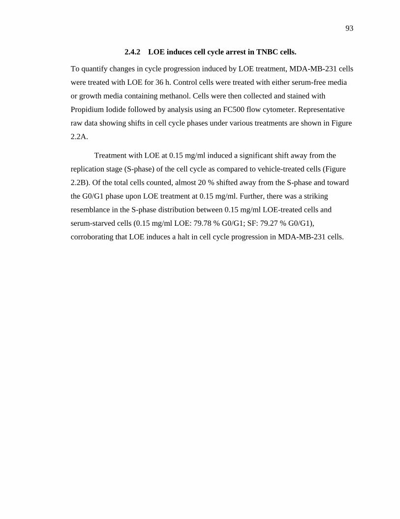
93
2.4.2 LOE induces cell cycle arrest in TNBC cells.
To quantify changes in cycle progression induced by LOE treatment, MDA-MB-231 cells
were treated with LOE for 36 h. Control cells were treated with either serum-free media
or growth media containing methanol. Cells were then collected and stained with
Propidium Iodide followed by analysis using an FC500 flow cytometer. Representative
raw data showing shifts in cell cycle phases under various treatments are shown in Figure
2.2A.
Treatment with LOE at 0.15 mg/ml induced a significant shift away from the
replication stage (S-phase) of the cell cycle as compared to vehicle-treated cells (Figure
2.2B). Of the total cells counted, almost 20 % shifted away from the S-phase and toward
the G0/G1 phase upon LOE treatment at 0.15 mg/ml. Further, there was a striking
resemblance in the S-phase distribution between 0.15 mg/ml LOE-treated cells and
serum-starved cells (0.15 mg/ml LOE: 79.78 % G0/G1; SF: 79.27 % G0/G1),
corroborating that LOE induces a halt in cell cycle progression in MDA-MB-231 cells.

94
A.
B.
Figure 2.2 LOE induces G0/G1 phase arrest in MDA-MB-231 cells. MDA-MB-231 cells
treated with indicated concentrations of LOE for 36h were stained with Propidium Iodide (PI)
and differential staining was measured using an FC500 flow cytometer (Bindley Flow
Cytometry Facility). (A) Representative plots of raw data indicating cell cycle stages from
treatment groups. (B) Average distribution across different cell cycle stages from various
treatment groups. SF: serum-free treatment. n=3; * significantly different from vehicle
treatment p<0.05 ★ significantly different from serum free treatment p<0.05

95
2.4.3 LOE induces apoptosis in TNBC cells.
Cell death may take the form of apoptosis, a programmed and controlled process
involving the degradation and clearance of cellular constituents; or necrosis, a traumatic
and inflammatory process which leads to the expulsion of cellular material into the
extracellular environment [40]. In order to analyze the impact of LOE on cell death in
triple-negative breast cancer cells, MDA-MB-231 cells were treated with LOE for 24h.
Cells were stained with Annexin V/7-Aminoactinomycin D and evaluated in a Muse Cell
Analyzer (Merck Millipore, MA). Representative raw data from various treatments
indicating shifts from live to early (EA) and late (LA) apoptosis as well as necrosis
(Dead) is shown in Figure 2.3.
Treatment with LOE induced a significant decrease in live cells as well a
significant increase in apoptosis but not necrosis in MDA-MB-231 cells. Furthermore,
40% of cells were apoptotic after 24h of 0.15 mg/ml LOE treatment while ~80% of cells
underwent apoptosis after 0.3 mg/ml LOE treatment over the same time period.

96
A.
B.
Figure 2.3 LOE induces apoptosis but not necrosis in MDA-MB-231 triple-negative
breast cancer cells. MDA-MB-231 cells were treated with indicated concentrations of LOE
for 24h and stained with Annexin-V/7-Aminoactinomycin D (7-AAD). This was followed by
measurement of differential Annexin-V/7-AAD staining using a Muse™ Cell Analyzer. (A)
Representative plots of raw data showing distribution of cells as Live, Early Apoptotic (EA),
Late Apoptotic (LA) or Necrotic. (B) Quantified graph showing effect of LOE on apoptosis in
MDA-MB-231 cells. N=3; Significant difference between LOE-treated cells and control
(Vehicle-treated) cells is indicated as *p<0.05 **p<0.005.

97
2.4.4 LOE impacts critical regulators of cell cycle progression.
As seen from Figure 2.2B, LOE treatment induced a shift toward the G0/G1 phase and
away from the S phase of the cell cycle in MDA-MB-231 cells. The progression from
G0/G1 is mediated by Cyclin D1, with cellular inhibitor of apoptosis (cIAP) proteins
playing a supportive role by suppressing apoptosis [41, 42]. As observed in Figure 2.4B,
there was a significant (5-fold) decrease in Cyclin D1 protein at 24 h post-treatment.
cIAP2 levels were significantly reduced by 40% after 12 h and by ~65% after 24 h.
Taken together, these results suggest that the halt in cell cycle progression
observed upon treatment of MDA-MB-231 cells with LOE is brought about by its impact
on Cyclin D1 and cIAP2 expression.
2.4.5 LOE induces apoptosis via caspase-8/-3 activation.
Based on the observation that LOE induces apoptosis and not necrosis in MDA-MB-231
cells (Figure 2.3B), our goal was to determine if LOE induces the extrinsic pathway of
apoptosis via caspase-8 activation. As shown in Figure 2.5B, LOE induced the rapid
cleavage of caspase-8 within 3 h post-treatment with PARP cleavage peaking at 6 h post-
treatment.
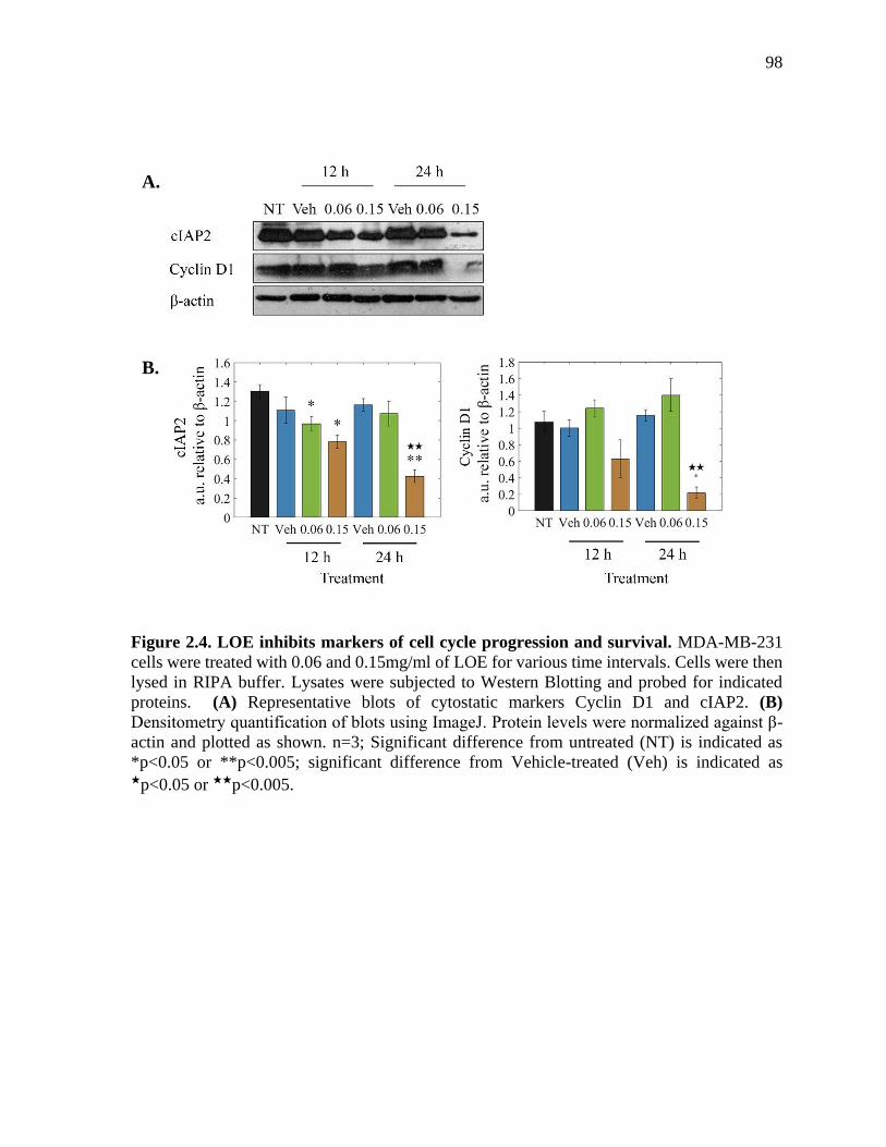
98
A.
B.
Figure 2.4. LOE inhibits markers of cell cycle progression and survival. MDA-MB-231
cells were treated with 0.06 and 0.15mg/ml of LOE for various time intervals. Cells were then
lysed in RIPA buffer. Lysates were subjected to Western Blotting and probed for indicated
proteins. (A) Representative blots of cytostatic markers Cyclin D1 and cIAP2. (B)
Densitometry quantification of blots using ImageJ. Protein levels were normalized against β-
actin and plotted as shown. n=3; Significant difference from untreated (NT) is indicated as
*p<0.05 or **p<0.005; significant difference from Vehicle-treated (Veh) is indicated as ★p<0.05 or ★★p<0.005.

99
A.
B.
Figure 2.5 LOE induces apoptosis accompanied by caspase-8 activation and PARP
cleavage. MDA-MB-231 cells treated with LOE for various time intervals were lysed and
subjected to Western Blotting and probed for indicated proteins (A) Representative blots of
apoptotic markers cleaved caspase-8, cleaved caspase-3 and cleaved PARP (B) Densitometry
quantification of blots using ImageJ. Protein levels were normalized against β-actin and
plotted as shown. n=3. Significant difference from untreated (NT) is indicated as *p<0.05 or
**p<0.005; significant difference from Vehicle-treated (Veh) is indicated as ★p<0.05 or ★★p<0.005.

100
To further understand apoptotic signaling, MDA-MB-231 cells were analyzed for
executioner caspase-3/-7 activity via live cell imaging using the IncuCyte ZOOM system.
These studies showed a marked visible increase in caspase-3/-7 activation upon LOE
treatment. As quantified in Figure 2.6, there was a significant increase in caspase-3/-7
activity 3 h post-treatment with 0.15 mg/ml and 0.3 mg/ml LOE, with peak activity at
approximately 10 h post-treatment. This was supported by Western Blotting time-course
data, which indicated a peak in caspase-3 activity at 12 h post-treatment with 0.15 mg/ml
LOE (Figure 2.6B).
2.4.6 LOE reduces RIP1 protein expression levels.
TNBC is often characterized by constitutive activation of NF-κB signaling via
recruitment of key effector proteins by RIP1 [43, 44]. Immunoblotting revealed a
significant decrease of ~ 40% in RIP1 protein levels in lysates from MDA-MB-231 cells
treated with LOE for 9 h as compared to controls (Figure 2.7B).

101
A.
B.
Figure 2.6 LOE induces executioner caspase-3/-7 activity in TNBC cells. (A) MDA-MB-
231 cells incubated with full growth medium containing indicated treatments and IncuCyte™
Caspase-3/7 apoptosis assay reagent were imaged periodically over 24h in an IncuCyte®
ZOOM live-cell analyzer to look at changes in caspase-3/-7 activity. (B) Activity was
quantified as the mean Green Fluorescent Object Count/Image for each treatment group and
plotted as shown. n=5; * significantly different from control (Vehicle-treated) cells; p<0.005.

102
Figure 2.7 RIP1 protein levels are reduced upon treatment of TNBC cells with LOE.
MDA-MB-231 cells were treated with 0.15 mg/ml LOE for 9 h and cell lysates were
immunoblotted for RIP1. (A) Representative blots of RIP1 (B) Densitometry quantification
of blots using ImageJ. Protein levels were normalized against β-tubulin and plotted as shown.
N = average of 3 replicates run twice. Significant difference from Vehicle-treated cells is
indicated as *p<0.05.

103
2.5 Discussion
TNBC continues to be a monumental concern in women’s health issues. As our
understanding of the mechanisms that govern the initiation and progression of breast
cancer grows, it has become apparent that conventional chemotherapeutics are limited by
their inability to target subtypes that are inherently more aggressive while being drug
resistant. One such subtype is TNBC, which lacks expression of ER, PR and HER2/neu,
thereby rendering TNBC resistant to conventional endocrine-targeted therapy. In this
study we provide support that an extract from the tropical South American plant L.
origanoides (LOE) may be a possible source for therapeutic agents against TNBCs.
2.5.1 LOE is a source of TNBC-specific cytotoxic compounds
In brief, we utilize the MTT metabolic activity assay to show LOE induces a dose-
dependent decrease in the viability of MDA-MB-231 mammary adenocarcinoma cells, a
universal standard in vitro model of highly invasive and drug resistant TNBC. This effect
was much greater compared to normal-like MCF10A mammary epithelial cells (Figure
2.1). Our observation that LOE also significantly reduced cell viability in triple-negative
Hras-transformed MCF10A invasive cells as well as the CRL-2321 cell-line, recently
shown to be the best in vitro phenotypic representative of grade 3 TNBC tumors [45],
further confirms its anti-TNBC effects. This observation suggests the LOE induces
molecular actions that target pathways specific to cancer cells. We also note that while
CRL-2321 cells have a greater sensitivity to low doses of LOE than MDA-MB-231 cells,
higher dosages of LOE do not induce decreases in CRL-2321 viability beyond 80%.
Previous studies have shown that cancer cell line subpopulations and sub-lines can
exhibit a high degree of transcriptome and phenotypic variability leading to observable
differences in metastatic activity [46] and drug sensitivity [47]. This suggests the
possibility of heterogeneity within subpopulations of the CRL-2321 cell line allowing for
compensatory survival pathways providing modest resistance to LOE treatment. The
identification of novel mechanisms targeted by new small molecular inhibitors is of
major interest in our continued studies of the actions of LOE.

104
2.5.2 LOE is a source of cell cycle inhibitors
Our study also quantified the molecular influences of the extract within cancer cells.
Cyclin D1 is a protein which associates with cyclin-dependent kinase-4 and -6 (CDK-4
and -6) to form heterodimers that progress the cell cycle from G0/G1 to S phase [41]. In
addition, inhibitor of apoptosis proteins (IAPs) prevent apoptosis and permit cell cycle
progression via binding and possibly inactivating executioner caspases, i.e. caspase-3 and
caspase-7 [42]. cIAP2 is known to be constitutively expressed in MDA-MB-231 cells,
and has been shown to protect against death-inducing ligands such as TNF-α by possibly
enhancing NF-κB signaling through RIP1 activation as well as by binding and inhibiting
caspase-3 [43]. Here, we observed that LOE induced G0/G1 phase halt in MDA-MB-231
cells (Figure 2.2). Western blot analysis revealed the effects of LOE on cell cycle
progression could be explained by a reduction in Cyclin D1 and cIAP2 levels upon
treatment of the cells with the extract (Figure 2.3). These experiments begin to build an
important framework for the actions of LOE on cell cycle regulation.
2.5.3 LOE is a source of apoptotic compounds
We next studied LOE’s impact on MDA-MB-231 cell death. Upon triggering of the
extrinsic pathway of apoptosis, procaspase-8 is activated via dimerization-induced auto-
cleavage and subsequently cleaves and activates the master “executioner” caspase,
caspase-3 [48, 49]. We revealed a potential mechanism behind the actions of the extract
by showing LOE-induced apoptosis involved activation of caspase-8/-3 and cleavage of
PARP (Figure 2.5 and 2.6), hallmarks of extrinsic apoptotic pathway activation [40].
Additionally, LOE induced apoptosis in these cells unaccompanied by necrotic cell death
(Figure 2.3). As cancer therapy is focused on preventing damage to non-targeted tissue
areas, this result provides additional support for LOE as a source for potential
therapeutics with desirable characteristics.
2.5.4 LOE is a source of compounds targeting NF-B signaling
In previous work, Stashenko et al. have characterized the major components of LOE [32].
However, it remains to be defined which component, or combination of components, is
responsible for the extract's apoptotic effects. Furthermore, it is imperative that major

105
cellular pathways involved in LOE signal transduction be delineated. Based on previous
studies involving components present in the LOE, one possible candidate pathway central
to LOE-induced apoptosis is NF-κB signaling. Constitutive activation of this pathway has
been previously linked to triple-negative breast cancer and blocking NF-κB signaling can
spontaneously induce cell death via a caspase-8/-3-dependent mechanism or render cells
susceptible to pro-apoptotic signals [44, 50, 51]. In normal cells, ligand binding to
TNFR1 is followed by rapid activation of the extrinsic pathway of apoptosis. However, in
contrast to normal cells, ligand binding to TNFR1 in TNBC cells instead activates NF-κB
signaling via membrane-recruitment of the scaffold protein RIP1, a Ser/Thr kinase [43].
In brief, cIAP2 exerts E3 ubiquitin ligase activity to polyubiquitinate and activate RIP1,
which goes on to phosphorylate the IKKα/β-NEMO complex. Active IKKs then
phosphorylate IκB proteins which sequester p50/RelA (NF-κB) dimers to the cytoplasm,
targeting the IkBs for polyubiquitination and subsequent degradation. This frees
p50/RelA to translocate to the nucleus and act as a transcription factor for pro-survival
genes such as Cyclin D1 and cIAPs [52, 53].
We hypothesized that components of LOE could interfere with NF-κB signaling,
leading to a halt in the cell cycle and induction of apoptosis. Indeed, we showed LOE
treatment resulted in a 40% decrease in protein levels of RIP1 in MDA-MB-231 cells
(Figure 2.7B). This may, at least in part, be explained by our previous observation that
LOE treatment induced activation of caspase-8/-3 (Figure 2.5 and 2.6). Active caspase-8
has previously been shown to cleave RIP-1, thereby inhibiting pro-survival NF-κB
signaling and inducing apoptosis [54]. This result, coupled with our findings that LOE
inhibits Cyclin D1 and cIAP2, proteins activated by NF-κB signaling, while
simultaneously stimulating caspase-8 dependent cell death, a characteristic of NF-κB
inhibition, sheds critical light on the mechanistic foreground of LOE action. All together,
these data highlight the potential of L. origanoides extract and its major constituents as
cancer therapeutics.

106
2.6 Summary
In summary, we have shown that LOE promotes apoptosis in TNBC cells, while having a
much reduced impact on cell death in normal mammary cells. We also found that LOE
reduces cell viability, alters the cell cycle and primarily leads to tumor cell apoptosis and
not necrosis. Further, we established some of the mechanisms of LOE actions, showing
that it suppresses markers of cell cycle progression and cell survival while inducing the
extrinsic pathway of apoptosis via caspase-8/-3 activation. Finally, we reveal that levels
of RIP1, an upstream effector of pro-survival NF-κB signaling, are reduced upon LOE
treatment. These data collectively support that LOE can be a valuable source for
development of novel anticancer agents. Moving forward, beyond fully describing the
cellular mechanisms of LOE action, a top priority will be identifying the major
component or combination of components responsible for its bioactivity. Plant-derived
compounds offer a vast source of small molecule inhibitors that could be used both in
their native state as well as chemically modified to optimize their pharmacological
activity. The bio-reactivity of the LOE-treatment in TNBC cells in vitro supports the idea
that future treatments for individual cancer subtypes lie in naturally-derived chemicals.
2.7 References
1. Jordan, V.C. (1992). The role of tamoxifen in the treatment and prevention of breast
cancer. Current Problems in Cancer, 16, 134-176.
2. Buzdar, A., & Howell, A. (2001). Advances in aromatase inhibition, clinical efficacy
and tolerability in the treatment of breast cancer. Clinical Cancer Research, 7, 2620-
2635.
3. Romond, E.H., Perez, E.A., Bryant, J., Suman, V.J., Geyer, C.E. Jr., Davidson, N.E.,
et al. (2005). Trastuzumab plus adjuvant chemotherapy for operable HER2-positive
breast cancer. New England Journal of Medicine, 353, 1673-1684.
4. Cakir, A., Gonul, I., & Uluoglu, O. (2012). A comprehensive morphological study for
basal-like breast carcinomas with comparison to nonbasal-like carcinomas. Diagnostic
Pathology, 7(1), 145.

107
5. Bertucci, F., Finetti, P., Cervera, N., Esterni, B., Hermitte, F., Viens, P., & Birnbaum,
D. (2008). How basal are triple-negative breast cancers? International Journal of
Cancer, 123(1), 236–240.
6. Rakha, E. A., El-Sayed, M. E., Green, A. R., Paish, E. C., Lee, A. H. S., & Ellis, I. O.
(2007). Breast carcinoma with basal differentiation, a proposal for pathology
definition based on basal cytokeratin expression. Histopathology, 50(4), 434–438.
7. Alluri, P., & Newman, L. A. (2014). Basal-like and triple-negative breast cancers,
searching for positives among many negatives. Surgical oncology clinics of North
America, 23(3), 567-77.
8. The Cancer Genome Atlas Network. (2012). Comprehensive molecular portraits of
human breast tumors. Nature, 490, 61-70.
9. Qi, X., Yin, N., Ma, S., Lepp, A., Tang, J., Jing, W., et al. (2015). p38gamma MAPK
is a therapeutic target for triple-negative breast cancer by stimulation of cancer stem-
like cell expansion. Stem Cells, 33, 2738-2747.
10. Darvishi, B., Farahmand, L., Eslami-S, Z., & Majidzadeh-A, K. (2017). NF-κB as the
main node of resistance to receptor tyrosine kinase inhibitors in triple-negative breast
cancer. Tumor Biology, 39(6), 101042831770691.
11. Sero, J.E., Sailem, H.Z., Ardy, R.C., Almuttaqi, H., Zhang, T. & Bakal, C. (2015).
Cell shape and the microenvironment regulate nuclear translocation of NF-kappaB in
breast epithelial and tumor cells. Molecular Systems Biology, 11, 790.
12. Khoshnan, A., Tindell, C., Laux, I., Bae, D., Bennett, B. & Nel, A.E. (2000) The NF-
kappaB cascade is important in Bcl-xL expression and for the anti-apoptotic effects of
the Cd28 receptor in primary human CD4+ lymphocytes. Journal of Immunology,
165, 1743-1754.
13. Wang, C.Y., Mayo, M.W., Korneluk, R.G., Goeddel, D.V. & Baldwin, A.S. Jr. (1998).
NF-kappaB antiapoptosis, induction of TRAF1 and TRAF2 and c-IAP1 and c-IAP2
to suppress caspase-8 activation. Science, 281, 1680-1683.
14. Huber, M.A., Azoitei, N., Baumann, B., Grünert, S., Sommer, A., Pehamberger, H.
(2004). NF-kappaB is essential for epithelial-mesenchymal transition and metastasis
in a model of breast cancer progression. Journal of Clinical Investigations, 114, 569-
581, 2004.

108
15. Hartman, Z.C., Poage, G.M., den Hollander, P., Tsimelzon, A., Hill, J., Panupinthu,
N., et al. (2013). Growth of triple-negative breast cancer cells relies upon coordinate
autocrine expression of the pro-inflammatory cytokines IL-6 and IL-8. Cancer
Research, 73, 3470-3480.
16. Hinz, M., Krappmann, D., Eichten, A., Heder, A., Scheidereit, C., & Strauss M, NF-
kappaB function in growth control, regulation of Cyclin D1 Expression and G0/G1-
to-S-phase transition. Molecular and Cellular Biology, 19, 2690-2698.
17. Li, C., Xia, W., Huo, L., Lim, S., Wu, Y., Hsu, J. L., et al. (2012). Epithelial-
Mesenchymal Transition Induced by TNF- Requires NF- B-Mediated
Transcriptional Upregulation of Twist1. Cancer Research, 72(5), 1290–1300.
18. Nakshatri, H., Bhat-Nakshatri, P., Martin, D.A., Goulet, R.J. Jr., & Sledge, G.W. Jr.
(1997). Constitutive activation of NF-kappaB during progression of breast cancer to
hormone-independent growth. Molecular and Cellular Biology, 17, 3629-3639.
19. Smith, S. M., Lyu, Y. L., & Cai, L. (2014). NF-κB Affects Proliferation and
Invasiveness of Breast Cancer Cells by Regulating CD44 Expression. PLoS ONE,
9(9), e106966.
20. Matsumoto, G., Namekawa, J., Muta, M., Nakamura, T., Bando, H., Tohyama, K., et
al. (2005). Targeting of nuclear factor kappaB pathways by dehydroxy-methyl-
epoxyquinomicin, a novel inhibitor of breast carcinomas, antitumor and anti-
angiogenic potential in vivo. Clinical Cancer Research, 11(3),1287-93.
21. Connelly, L., Robinson-Benion, C., Chont, M., Saint-Jean, L., Li, H., Polosukhin,
V.V., et al. (2007). A transgenic model reveals important roles for the NF-kappaB
alternative pathway (p100/p52) in mammary development and links to tumorigenesis.
Journal of Biological Chemistry, 282,10028–10035.
22. Thomas, G.W., Muss, H.B., Jackson, D.V., McCulloch, J., Ramseur, W., McFarland,
J., et al. (1994). Vincristine with high-dose etoposide in advanced breast cancer, a
phase II trial of the Piedmont Oncology Association. Cancer Chemotherapy and
Pharmacology, 35(2),165-8.
23. Stemmler, H.J., Gutschow, K., Sommer, H., Malekmohammadi, M., Kentenich, C.H.,
Forstpointner, R., et al. (2001). Weekly docetaxel (Taxotere) in patients with
metastatic breast cancer. Annals of Oncology, 12(10),1393-8.

109
24. Ahmad, A., Banerjee, S., Wang, Z., Kong, D., & Sarkar, F.H. (2008). Plumbagin-
induced apoptosis of human breast cancer cells is mediated by inactivation of NF-
kappaB and Bcl-2. Journal of Cellular Biochemistry, 105, 1461-1471.
25. Jin, M.L., Park, S.Y., Kim, Y.H., Park, G., & Lee, S. (2014) Halofuginone induces the
apoptosis of breast cancer cells and inhibits migration via downregulation of matrix
metalloproteinase-9. International Journal of Oncology, 44, 309-318.
26. Pozo-Guisado, E., Merino, J.M., Mulero-Navarro, S., Lorenzo-Benayas, M.J.,
Centeno, F., Alvarez-Barrientos, A., & Fernandez-Salguero, P.M. (2005) Resveratrol-
induced apoptosis in MCF-7 human breast cancer cells involves a caspase-
independent mechanism with downregulation of Bcl-2 and NF-kappaB. International
Journal of Cancer, 115, 74-84.
27. Singh., S, & Aggarwal, B.B. (1995). Activation of transcription factor NF-kappaB is
suppressed by curcumin (diferuloylmethane). The Journal of Biological Chemistry,
270, 24995-25000.
28. Tse, A.K., Wan, C.K., Shen, X.L., Yang, M., & Fong, W.F. (2005). Honokiol inhibits
TNF-alpha-stimulated NF-kappaB activation and NF-kappaB-regulated gene
expression through suppression of IKK activation. Biochemical Pharmacology, 70,
1443-1457.
29. Chen, K.S., Shi, M.D., Chien, C.S., & Shih, Y.W. (2014). Pinocembrin suppresses
TGF-betta1-induced epithelial-mesenchymal transition and metastasis of human Y-79
retinoblastoma cells through inactivating alphavbetta3 integrin/FAK/p38alpha
signaling pathway. Cell Biosciences, 4, 41.
30. Pascual, M.E., Slowing, K., Carretero, E., Sanchez Mata, D., & Villar, A. (2001)
Lippia, traditional uses, chemistry and pharmacology, a review. Journal of
Ethnopharmacology, 76, 201-214.
31. Stashenko, E.E., Martínez, J.R., Ruíz, C.A., Arias, G., Durán, C., Salgar, W., & Cala,
M. (2010). Lippia origanoides chemotype differentiation based on essential oil GC-
MS and principal component analysis. Journal of Separation Science, 33, 93-103.

110
32. Stashenko, E.E., Martínez, J.R., Cala, M.P., Durán, D.C., & Caballero, D. (2013).
Chromatographic and mass spectrometric characterization of essential oils and
extracts from Lippia (Verbenaceae) aromatic plants. Journal of Separation Science,
36, 192-202.
33. Legault, J., & Pichette, A. (2007). Potentiating effect of beta-caryophyllene on
anticancer activity of alpha-humulene, isocaryophyllene, and paclitaxel. Journal of
Pharmacy and Pharmacology, 59, 1643-1647.
34. Aristatile, B., Al-Assaf, A.H., & Pugalendi, K.V. (2013). Carvacrol suppresses the
expression of inflammatory marker genes in D-galactosamine-hepatotoxic rats. Asian
Pacific Journal of Tropical Medicine. 6(3),205-11.
35. Greiner, J.F., Müller, J., Zeuner, M.T., Hauser, S., Seidel, T., Klenke, C., et al. (2013).
1,8-Cineol inhibits nuclear translocation of NF-κB p65 and NF-κB-dependent
transcriptional activity. Biochimica et Biophysica Acta, 1833(12),2866-78.
36. Lee, J.C., Kundu, J.K., Hwang, D.M., Na, H.K., & Surh, Y.J. (2007). Humulene
inhibits phorbol ester-induced COX-2 expression in mouse skin by blocking activation
of NF-kappaB and AP-1, IkappaB kinase and c-Jun-N-terminal kinase as respective
potential upstream targets. Carcinogenesis, 28(7),1491-8.
37. Liang, D., Li, F., Fu, Y., Cao, Y., Song, X., Wang, T., et al. (2014). Thymol inhibits
LPS-stimulated inflammatory response via down-regulation of NF-κB and MAPK
signaling pathways in mouse mammary epithelial cells. Inflammation, 37(1),214-22.
38. Kim, C., Cho, S.K., Kim, K.D., Nam, D., Chung, W.S., Jang, H.J., et al. (2014). β-
Caryophyllene oxide potentiates TNFα-induced apoptosis and inhibits invasion
through down-modulation of NF-κB-regulated gene products. Apoptosis 19(4),708-
18.
39. Schneider, C.A., Rasband, W.S. & Eliceiri, K.W. (2012). NIH Image to ImageJ, 25
years of image analysis. Nature Methods, 9, 671-675.
40. Portt, L., Norman, G., Clapp, C., Greenwood, M., & Greenwood, M.T. (2011). Anti-
apoptosis and cell survival, a review. Biochimica Biophysica Acta, 1813,238-59.
41. Baldin,V., Lukas, J., Marcote, M.J., Pagano, M., & Draetta, G. (1993). Cyclin D1 is a
nuclear protein required for cell cycle progression in G1. Genes and Development, 7,
812-821.
111
42. Salvesen, G.S., & Duckett, C.S. (2002). IAP proteins, blocking the road to death's
door. Nature Reviews Molecular Cell Biology, 3, 401-410.
43. Bertrand, M.J., Milutinovic, S., Dickson, K.M., Ho, W.C., Boudreault, A., Durkin, J.,
et al. (2008). cIAP1 and cIAP2 facilitate cancer cell survival by functioning as E3
ligases that promote RIP1 ubiquitination. Molecular Cell, 30, 689-700.
44. Yamaguchi, N., Ito, T., Azuma, S., Ito, E., Honma, R., Yanagisawa, Y., et al. (2009).
Constitutive activation of nuclear factor-kappaB is preferentially involved in the
proliferation of basal-like subtype breast cancer cell lines. Cancer Science, 100, 1668-
1674.
45. Grigoriadis, A., Mackay, A., Noel, E., Wu, P.J., Natrajan, R., Frankum, J., Reis-Filho,
J.S., Tutt, A. (2012). Molecular characterisation of cell line models for triple-negative
breast cancers. BMC Genomics, 13,619.
46. Nguyen, A., Yoshida, M., Goodarzi, H., & Tavazoie, S.F. (2016). Highly variable
cancer subpopulations that exhibit enhanced transcriptome variability and metastatic
fitness. Nature Communications 7,11246.
47. Leung, E., Kannan, N., Krissansen, G.W., Findlay, M.P., & Baguley, B.C. (2010).
MCF-7 breast cancer cells selected for tamoxifen resistance acquire new phenotypes
differing in DNA content, phospho-HER2 and PAX2 expression, and rapamycin
sensitivity. Cancer Biology and Therapy, 1,9(9),717-24.
48. Peter, M.E., & Krammer, P.H. (2003). The CD95(APO-1/Fas) DISC and beyond. Cell
Death and Differentiation, 10, 26-35.
49. Beaudouin, J., Liesche, C., Aschenbrenner, S., Horner, M., & Eils, R. (2013). Caspase-
8 cleaves its substrates from the plasma membrane upon CD95-induced apoptosis.
Cell Death and Differentiation, 20, 599-610.
50. Yamamoto, M., Taguchi, Y., Ito-Kureha, T., Semba, K., Yamaguchi, N., & Inoue, J.
(2013). NF-kappaB non-cell-autonomously regulates cancer stem cell populations in
the basal-like breast cancer subtype. Nature Communications, 4, 2299.
51. Micheau, O., & Tschopp, J. (2003). Induction of TNF receptor I-mediated apoptosis
via two sequential signaling complexes. Cell, 114, 181-190.

112
52. Guttridge, D.C., Albanese, C., Reuther, J.Y., Pestell, R.G., & Baldwin, A.S. Jr. (1999).
NF-kappaB controls cell growth and differentiation through transcriptional regulation
of Cyclin D1. Molecular and Cellular Biology, 19, 5785-5799.
53. You, M., Ku, P.T., Hrdličková, R., & Bose, H.R. Jr. (1997). ch-IAP1, a member of the
inhibitor-of-apoptosis protein family, is a mediator of the antiapoptotic activity of the
v-Rel oncoprotein. Molecular and Cellular Biology, 17, 7328-7341.
54. Lin, Y., Devin, A., Rodriguez, Y., & Liu, Z. (1999). Cleavage of the death domain
kinase RIP by Caspase-8 prompts TNF-induced apoptosis. Genes and Development,
13(19),2514-2526.

113
PROTEOMIC ANALYSIS REVEALS LIPPIA
ORIGANOIDES EXTRACT (L42) TARGETS MITOCHONDRIAL
METABOLISM IN TRIPLE-NEGATIVE BREAST CANCER
CELLS
A version of this chapter has been previously published in the Journal of Proteome
Research, 2018, 17(10):3370-3383. DOI: 10.1021/acs.jproteome.8b00255
3.1 Abstract
Recent studies indicate triple-negative breast cancers possesses an altered metabolic state
with higher rates of glycolysis, mitochondrial oxidative phosphorylation, and increased
generation and utilization of tricarboxylic acid cycle intermediates. Here we utilized
label-free quantitative proteomics to gain insight into the anti-cancer mechanisms of a
methanolic extract from the Central American plant Lippia origanoides on MDA-MB-
231 triple-negative breast cancer cells. The L. origanoides extract dysregulated
mitochondrial oxidative phosphorylation by targeting Complex I of the electron transport
chain and suppressed cellular metabolism by targeting key tricarboxylic acid cycle
enzymes and mitochondrial lipid and amino acid metabolic pathways. Our study also
revealed treatment with the extract activated the stress response, and pathways related to
cell cycle progression and DNA repair. Overall, our results reveal new compelling
evidence that the extract from Lippia origanoides triggers rapid irreversible apoptosis in
MDA-MB-231 cells by effectively ‘starving’ the cells of metabolites and ATP. We
continue to study the specific bioactive components of the extract in the search for novel,
highly effective mitochondrial inhibitors to selectively target triple-negative breast
cancer.
3.2 Introduction
As mentioned in section 1.4., the initial discovery of anticancer drugs from natural
sources prompted the NIH to begin screening natural extracts using in vitro cancer
models to isolate and identify their bioactive compounds [1, 2]. Such extracts have often
shown inhibitory effects on the viability and proliferation of cancer cells while remaining

114
relatively harmless to normal cells, and non-toxic in vivo [3-5]. Bioassay-driven
fractionation and target-identification schemes have led to the discovery of several drugs,
notably the Vinca alkaloids and taxanes; however, this is both time-consuming and very
expensive [6, 7]. In addition, the use of Western-blots, PCR, and ELISA to identify the
effects of treatments on specific signaling pathways does not provide information on off-
target effects, which is a major drawback when dealing with complex mixtures of
compounds. The advent of cDNA microarray-driven biomarker identification techniques
provided a more holistic picture of the cell- and tissue-wide effects of chemotherapy [8].
Unfortunately, microarrays have the limitation of only providing information on
fluctuations in mRNA levels, which are often disproportional to corresponding changes
in protein expression [9-11]. Hence, it is critical that new techniques are developed to
more efficiently screen natural extracts for their effects on the entire proteome on a cell-
and tissue-wide basis. Realizing this goal will greatly expedite the identification of viable
new cancer targets.
Recently, there has been tremendous interest in targeting cancer cell metabolism
as a new therapeutic strategy, given that the most recognized hallmark of cancer cells is
increased aerobic glycolysis, a phenomenon termed the Warburg effect [12, 13]. In
particular, TNBCs are known to have an altered metabolic profile with elevated uptake
and utilization of glucose [14, 15], glutamine [16-18], and TCA cycle intermediates [19],
as well as increased fatty acid β-oxidation [20]. We speculate that this allows for the
heightened growth, proliferation and invasiveness observed in TNBCs by providing
alternative sources of ATP generation. For these reasons, identifying specific targets and
novel small molecule inhibitors against TNBC metabolism is of considerable clinical
importance.
As shown in Chapter 2, the Lippia origanoides extract (LOE), had substantial
anti-proliferative and pro-apoptotic effects on several TNBC cells lines, while remaining
relatively non-toxic to normal mammary epithelial cells [21]. In testing multiple extracts
from Lippia and other species, we discovered a new extract from a different chemotype
of the same species possessed an even higher potency against TNBC cells while retaining
reduced toxicity against normal cells. Hence, this extract, termed ‘L42’, was used in the
115
present study to determine its proteome-wide effects on MDA-MB-231 TNBC cells. We
applied precursor ion (MS1) intensity based label-free quantitation (LFQ) via LC-
MS/MS, which is becoming the method of choice to study cell functional responses at the
proteome level [22-26], to determine relative changes in protein abundances due to L42
treatment.
In brief, we quantified 2,407 proteins, of which 519 were significantly
dysregulated upon L42 treatment. The majority of down-regulated proteins were
mitochondrial, and known to play critical roles in cellular metabolism pathways,
including the TCA cycle, β-oxidation of fatty acids, and glutamine utilization. Core
subunits of Complex I and the mitochondrial ribosome were down-regulated upon
treatment, providing evidences of the disruptive effects of L42 on mitochondrial activity
and function.
Considering these actions, Chapter 3 provides critical and holistic mechanistic
insight into the TNBC-specific inhibitory effects of L42, and proposes that L42 is a novel
source of potent mitochondrial-specific inhibitors for tumor metabolism-based cancer
therapy.
3.3 Materials and Methods
3.3.1 Cell culture
MDA-MB-231 triple-negative breast cancer and MCF10A normal mammary epithelial
cell lines were obtained from the American Type Culture Collection. MDA-MB-231 cells
were cultured in Dulbecco’s Modified Essential Medium (DMEM; Life Technologies,
NY, USA) supplemented with 10 % fetal bovine serum (FBS; Atlanta Biologicals, GA,
USA), 100 IU/ml penicillin and 100 µg/ml streptomycin (Life Technologies, NY, USA).
MCF10A cells were cultured in a 1:1 ratio of DMEM:Ham’s F12 supplemented with 5 %
horse serum (HS; Atlanta Biologicals, GA, USA), 20 ng/ml human epidermal growth
factor (Sigma-Aldrich, MO, USA), 0.5 mg/mL hydrocortisone (Sigma-Aldrich, MO), 100
ng/ml cholera toxin (Sigma-Aldrich, MO, USA), 10 µg/mL bovine insulin (Sigma-
Aldrich, MO, USA), 100 IU/ml penicillin and 100 µg/ml streptomycin.
116
3.3.2 L42 extract preparation
The extract from Lippia origanoides (COL 560267) was prepared as described previously
[27]. Briefly, 200 g of finely ground L. origanoides plant material (leaves and stems)
was placed in the extraction chamber of a Thar SFE-2000-2-FMC50 (Thar Instruments,
PA, USA) and subjected to supercritical CO2 extraction at 50 MPa and 333 K at the
Universidad Industrial de Santander, Bucaramanga, Colombia. The material deposited on
the walls of the chamber was collected, and 250 mg of this extracted material was
dissolved in 5 ml of methanol, sonicated for 15 min, and centrifuged at 10000 rpm for a
further 15 min. The 50 mg/ml stock solution of the supernatant collected (L42) was used
in the present study.
3.3.3 Assessment of cell viability using MTT assay
MCF10A and MDA-MB-231 cells were seeded at 104 cells/well in 96-well plates and
allowed to attach. Spent media was then replaced with treatment media containing either
L42 or methanol (vehicle control, VEH). Following 24, 48 and 72 h treatments, 20 µl of
12 mM MTT (Life Technologies, NY, USA), was added to the cells followed by
incubation overnight at 37 °C. Formazan crystals formed at the end of incubation period
were dissolved using dimethyl sulfoxide (DMSO) and cell viability was determined as a
measure of absorbance at 570 nm.
3.3.4 Western blotting
MDA-MB-231 cells were seeded at 106 cells/well in 6-well plates and allowed to attach
overnight, then treated with fresh media containing L42 (0.15 mg/ml) or vehicle
(methanol) for 12 h. Treated cells were collected in RIPA buffer and sonicated, following
which samples were centrifuged to obtain lysates. Protein concentrations of lysates were
estimated via the bicinchoninic acid (BCA) assay (Thermo Fisher, IL, USA). 20 μg of
protein from each sample was mixed with 4X sample buffer and boiled, and denatured
proteins were loaded and run via SDS-PAGE, then transferred onto a polyvinyl difluoride
(PVDF) membrane and immunoblotted. Primary antibodies for Cyclin D1 and cleaved
caspase-8 were purchased from Cell Signaling Technologies, MA, USA. GLS and β-actin
antibodies were purchased from Abcam, MA, USA and Sigma-Aldrich, MO, USA

117
respectively. β-tubulin antibody was purchased from the Developmental Studies
Hybridoma Bank, University of Iowa, IA, USA. Densitometric quantification was carried
out using ImageJ software.
3.3.5 LC-MS/MS: sample preparation and analysis
Protein extraction: 3 x 10⁶ MDA-MB-231 cells, treated with L42 or vehicle control (N =
3 replicates/ treatment), were collected via scraping, centrifuged and then washed twice
with ice-cold 1X PBS. Samples were centrifuged at 3,000 rpm for 5 min after each wash,
and pelleted cells were re-suspended in 100 µl of 100 mM ammonium bicarbonate
(ABC). Samples were then homogenized at 35,000 psi using a barocycler (Pressure
Biosciences, MA, USA) with high-pressure time of 20 s followed by low-pressure time
of 10 s, for each cycle for a total of 90 cycles.
Homogenized samples were centrifuged at 14,000 rpm for 20 min at 4 °C and the
soluble fraction (SF, supernatant) and membrane-fraction (MF, pellet) were collected
separately. SF was subjected to bicinchoninic acid (BCA) assay to measure protein
concentration. A total of 100 µg of SF was made up to 100 µl using Milli Q water and
then precipitated using 5 volumes of cold (-20 °C) acetone overnight to allow for protein
precipitation. For pellet (membrane fraction, MF), 10 µl of urea/DTT was added and MF
samples were placed in 37 °C rotary shaker for 1 h to reduce and denature proteins.
Denatured MF samples were then mixed with 10 µl alkylation mixture (195 µl
acetonitrile (ACN), 1 µl triethylphosphine, and 4 µl 2-iodoethanol) and placed for a
further 1 h in 37 °C shaker. MF samples were then dried in vacuum centrifuge
(Labconco, MO, USA) for 1 h after which the dry protein pellets were stored at -20 °C
until further use. Precipitated SF samples were centrifuged at 14,000 rpm for 20 min and
pellets were dried in vacuum centrifuge briefly, after which SF samples were also
subjected to reduction, denaturation and alkylation as described previously.
Protein digestion and solid phase extraction (SPE): Lys-C/trypsin (Promega, WI, USA)
vial containing 20 µg of the mixture was dissolved by adding 400 µl of 25 mM ABC to
80 µl of each sample (both SF and MF) to achieve enzyme:substrate ratio of 1:25.
Samples were then transferred to a barocycler for digestion. Proteins were digested for 1
h at 50 °C at 20 kpsi using a program set at high pressure for 50 s followed by a low

118
(atmospheric) pressure setting for 10 s per cycle, for a total of 60 cycles. Digested
peptides were desalted using C18 SPE columns using a protocol provided by the
manufacturer. Clean peptides were eluted using 50 µl 80 % ACN/ 0.1 % FA/ 20 % Milli
Q, and elution step was repeated twice. Eluates were dried in a vacuum centrifuge
following which the clean, dry peptides were stored at -20 °C until further use.
LC-MS/MS analysis: Samples were analyzed by reverse-phase HPLC-ESI-MS/MS using
the Dionex UltiMate 3000 RSLC nano System (Thermo Fisher Scientific) coupled to the
Q-Exactive High Field (HF) Hybrid Quadrupole Orbitrap MS (Thermo Fisher Scientific)
and a Nano- electrospray Flex ion source (Thermo Fisher Scientific). Peptides were re-
suspended in 3 % ACN/ 0.1 % FA/ 97 % milliQ formic acid, and loaded onto a trap
column (300 µm ID x 5 mm) packed with 5 µm 100 Å PepMap C18 medium and washed
using a flow rate of 5 µl/ min with 98 % purified water/ 2 % acetonitrile (ACN)/ 0.01 %
formic acid (FA). The trap column was then switched in-line with the analytical column
after 5 minutes. Peptides were separated using a reverse phase Acclaim PepMap RSLC
C18 (75 µm x 15 cm) analytical column using a 120-min method at a flow rate of 300
nl/min. The analytical column was packed with 2 µm 100 Å PepMap C18 medium
(Thermo Fisher Scientific). Mobile phase A consisted of 0.01 % FA in water and a
mobile phase B consisted of 0.01 % FA in 80 % ACN. The linear gradient started at 5 %
B and reached 30 % B in 80 min, 45 % B in 91 min, and 100 % B in 93 min. The column
was held at 100% B for the next 5 min before being brought back to 5 % B and held for
20 min to equilibrate the column.
Sample was injected into the QE HF through the Nanospray Flex™ Ion Source
fitted with an emission tip from Thermo Scientific. Column temperature was maintained
at 35 ˚C. MS data were acquired with a Top 20 data-dependent MS/MS scan method. The
full scan MS spectra were collected over 300-1,650 m/z range with a maximum injection
time of 100 ms, a resolution of 120,000 at 200 m/z, spray voltage of 2 and AGC target of
1 ×10⁶. Fragmentation of precursor ions was performed by high-energy C-trap
dissociation (HCD) with the normalized collision energy of 27 eV. MS/MS scans were
acquired at a resolution of 15,000 at m/z 200. The dynamic exclusion was set at 20 s to
avoid repeated scanning of identical peptides.

119
Three biological sample replicates from each treatment were utilized for LC-
MS/MS, each run as a single technical replicate, which was sufficient for good statistical
power. Instrument optimization and recalibration was carried out at the start of each batch
run using the Pierce calibration solution. The sensitivity of the instrument was also
monitored using an E. coli digest at the start of the sample run.
Bioinformatics: MS/MS data was processed using MaxQuant (v1.6.1.0, [28]) with
the spectra matched against the Uniprot Homo sapiens fasta (http://www.uniprot.org/;
26,232 entries, state 10/25/17) concatenated with a common contaminants database and a
reverse decoy database. MS/MS spectra from Soluble (SF) and Membrane (MF) fractions
from each sample were combined during MaxQuant analysis. The cleavage enzyme used
was set as Trypsin/P; LysC while allowing for up to 2 missed cleavages. Mass error was
set to 10 ppm for precursor ions and 20 ppm for fragment ions. Variable modifications
included were oxidation of methionine and acetylation of N-terminal amino acids, with
alkylation of cysteine residues set as a fixed modification and oxidation of methionine
was defined as a variable modification. Estimated False Discovery Rate (FDR) threshold
was set to 1 % both at the peptide and protein level. The ‘unique plus razor peptides’
were used for peptide quantitation. Razor peptides are non-unique peptides assigned to
the protein group with most other peptides.
After MaxQuant searches, proteins without any LFQ intensity and without any
MS/MS counts were filtered out. MaxQuant results were exported to Data Analysis and
Extension Tool (DAnTE) [29], and analyzed for Pearson correlations coefficients, and
visualized using heatmaps generated by Heatmapper [30]. The complete RAW
proteomics data files, peptide sequence files, protein identification files, parameters used,
LC-MS/MS methodology, and Supplementary Tables have been deposited in the
MassIVE public proteomics data repository and can be accessed at
ftp://massive.ucsd.edu/MSV000082159.
3.3.6 Proteomics data analysis:
We identified 3,061 proteins across all 3 replicates from both treatment groups. Proteins
identified and quantified in ≥ 2 replicates in both treatment groups were retained, and
zero-fill proteins (i.e. proteins identified in all replicates of one treatment group, and not

120
found in any replicates of the other) were also retained, leaving 2,407 proteins for further
analyses. Bioinformatics analysis was performed using the R statistical software. LFQ
intensities of identified proteins were converted to log2 values and averaged across
replicates. The difference in average log2 values [Δlog2 (LFQ intensity)] between proteins
from L42-treated and control-treated groups was used as a measure of fold change.
Proteins with fold-change values Δlog2 < -0.5 or Δlog2 > 0.5, and p < 0.05 (Student’s
unpaired, two-tailed, t-test) were considered to be significantly regulated by L42
treatment.
GO enrichment analysis: Upon L42 treatment, 519 proteins were significantly
changed, of which 293 were found to be down-regulated and 226 were up-regulated (See
Supplementary Table 3.1C). Gene ontology enrichment analysis was performed by
comparing the total pool of 2,407 quantified proteins to down-regulated proteins (Blue
font, Supplementary Table 3.1C) and up-regulated (Red font, Supplementary Table 3.1C)
using the GOrilla software tool (for biological process, localization and function
enrichment) [31, 32] and DAVID 6.8 (for pathway enrichment using the KEGG database)
[33, 34]. GO terms with an FDR q-value (Bejamini-Hochberg corrected p-value) < 0.05
were considered significant.
STRING Interaction Analysis: Proteins were further divided into lists (Fatty Acid
Metabolism, Amino Acid Metabolism, Oxidative Phosphorylation etc.) based on pathway
enrichment, and lists were mapped for possible interactions using the STRING
interaction database tool [35]. Interactions were considered significant only at confidence
scores of ≥ 0.9 (highest confidence). Mitochondrial localization was also displayed using
the STRING enrichment analysis feature for Cellular Component.
Pathway visualization: Pathways were downloaded using the WikiPathways app
(v3.3.1) [36] onto Cytoscape software (v3.6.0) [37]. Pathway proteins found in
Supplementary Table 3.1C were highlighted, with the extent of up- or down-regulation
portrayed using a blue-red gradient indicating Δlog2 values.

121
3.3.7 Validation experiment: mitochondrial membrane potential assay
MDA-MB-231 cells were seeded at 104 cells/well in 96-well plates and allowed to grow
to 70 % confluency, following which cells were treated with L42 (0.15 mg/ml) or vehicle
for 8 h. At the end of the treatment period, media was aspirated gently and 200 nM
TMRE (Abcam, MA) prepared in fresh media was added to the cells. Following a 30 min
incubation period at 37 °C, TMRE media was gently aspirated and cells were washed
with 200 µl 1X PBS/ 0.2 % BSA. 100 µl of the same was then added to the cells prior to
imaging with a Zeiss Axiovert 200m fluorescence microscope.
3.3.8 Statistical analysis
Cell viability data from the MTT assay was analyzed using JMP 12 (SAS) software. Box
Cox transformation (λ = 0.403) was performed on data to satisfy the normality and equal
variance assumptions for Analysis of Variance (ANOVA), which was used to test for
overall statistical significance, p < 0.0001. An F-test was then performed to measure
significance of different effects (individual and crossed), p < 0.0001. Significant
difference between different levels for third level interaction (Treatment*Time*Cell
Line) was then assessed using Tukey’s Honest Significant Difference test (Tukey’s
HSD). The complete report is summarized in Supplementary Table 3.2.
Western Blotting data acquired using denistometric analysis via ImageJ software
was subjected to Student’s t-test [38]. Statistical analysis for Immunofluorescence
Microscopy was performed using an unpaired, two-tailed Mann-Whitney Test.
3.3.9 Data availability
RAW proteomics data files, peptide sequence files, protein identification files,
parameters used, LC-MS/MS methodology, and Supplementary Tables have been
deposited in the MassIVE public proteomics data repository and can be accessed using
the link ftp://massive.ucsd.edu/MSV000082159.

122
3.4 Results
3.4.1 Effect of L42 on TNBC cell viability.
To investigate the effects of L42 on cell viability of triple-negative and normal mammary
epithelial cells, MDA-MB-231 (TNBC) and MCF10A (normal) cells were treated with
increasing dosages of L42 for 24, 48 and 72 h, following which the cells were analyzed
for viability using the MTT assay. As summarized in the Tukey HSD Report from
Supplementary Table 3.2, we observed that L42 induced a significant decrease in
viability of MDA-MB-231 cells at 0.09 mg/ml while a dose of 0.13 mg/ml was required
to induce a significant reduction in MCF10A viability at 24 h (Figure 3.1A). Importantly,
at this time-point, 0.12 mg/ml L42 reduced cell viability in MDA-MB-231 cells to 51%,
with no loss of viability in MCF10A cells. At doses ≥ 0.09 mg/ml, at both 48 h and 72 h
time-points, MDA-MB-231 cell viability was significantly reduced upon L42 treatment
compared to MCF10A viability, even at the highest dosage of 0.15 mg/ml. At the end of
72 h, a single dose of 0.15 mg/ml L42 was seen to reduce MDA-MB-231 viability to <
2 %. Replacing treatment media after 24 h with fresh media did not rescue MDA-MB-
231 cell viability (Figure 3.1B)
As a reduction in cell viability may be explained by either cell death or slowing of
cell proliferation, we studied the effects of L42 on known effectors of these pathways.
Similar to LOE (See Figure 2.4B and Figure 2.5B), L42 treatment of MDA-MB-231 cells
induced a significant loss of Cyclin D1 while inducing Caspase-8 activation (Figure
3.1C). These results indicate that L42 dysregulates both proliferative as well as survival
pathways to induce apoptosis in TNBC cells.
3.4.2 Overview of proteome analysis and LC-MS reproducibility.
The multi-component nature of L42 could conceivably induce the dysregulation of
multiple cellular pathways to bring about its apoptotic effects, and so we sought to study
its actions on the proteome landscape of TNBC cells (see Figure 3.2A for workflow).
Reproducibility of peptide intensity is critical for accurate MS1 based label free
quantitation [26]. We obtained quantifiable data for 2,407 proteins. To assess the
accuracy, we ran 3 technical replicates of a complex E. coli digest. The average CV of

123
peptide intensity was 16.7 % (Figure 3.2B), and the median CV was 15.4%
(Supplementary Table 3.3). In addition, accuracy was confirmed by analyzing log2
intensity values of Supplementary Table 3.1C proteins which showed high correlation
(R2 ≥ 0.99) between MDA-MB-231 biological replicates from each treatment group
(Figure 3.2C). Correlation between replicates from all 2,407 proteins was also similarly
high (Figure 3.2D).

124
A.
B.
C.
Figure 3.1 L42 inhibits survival and induces apoptosis in MDA-MB-231 cells. A. MDA-
MB-231 and MCF10A cells were seeded in 96-well plates and treated with indicated
concentrations of L42 or vehicle control (VEH) for 24, 48 and 72 h and subjected to MTT
assay. This was followed by absorbance reading at 570 nm. N = 5 replicates; Significance was
determined using Tukey’s HSD test (see Supplementary Table 3.2), p < 0.0001. B. MDA-MB-
231 cells were treated with 0.15 mg/ml of L42 for various time intervals and lysates were
probed for indicated proteins by Western Blot. (Upper panel) Representative blots of Cyclin
D1 and Caspase-8. (Lower panel) Densitometry quantification of blots using ImageJ. Protein
levels were normalized against β-actin and plotted as shown. N = 3; * significant difference
from VEH, p < 0.05.
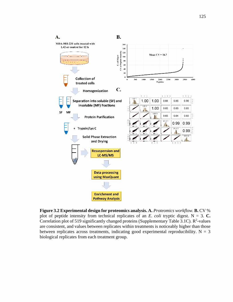
125
Figure 3.2 Experimental design for proteomics analysis. A. Proteomics workflow. B. CV %
plot of peptide intensity from technical replicates of an E. coli tryptic digest. N = 3. C.
Correlation plot of 519 significantly changed proteins (Supplementary Table 3.1C). R2-values
are consistent, and values between replicates within treatments is noticeably higher than those
between replicates across treatments, indicating good experimental reproducibility. N = 3
biological replicates from each treatment group.

126
3.4.3 Functional classification of identified proteins.
To obtain an overall assessment of the effects of L42 on TNBC cells, proteins from Table
3.1C were divided into separate lists of significantly up-regulated (Red font,
Supplementary Table 3.1C) and down-regulated (Blue font, Supplementary Table 3.1C)
proteins. GO enrichment for cellular localization, function and process was performed
using the GOrilla tool and expression levels of replicates of proteins from significantly
enriched terms were averaged and visualized with heat-maps. L42 was found to
significantly down-regulate components of oxidoreductase (part of the electron transport
chain) and TCA cycle complexes, both localized to the mitochondria, as well as both
large and small mitochondrial ribosomal subunits (Figure 3.3A). Further, the functional
terms enriched consisted of mitochondrial enzymes significantly down-regulated by L42
treatment (Figure 3.3B). Finally, Figure 3.3C shows with striking clarity that L42
suppressed multiple mitochondrial functions including the TCA cycle, oxidative
phosphorylation and protein translation. Interestingly, L42 treatment was also seen to up-
regulate proteins involved in stress response, cell cycle regulation and DNA repair
(Figure 3.3D).
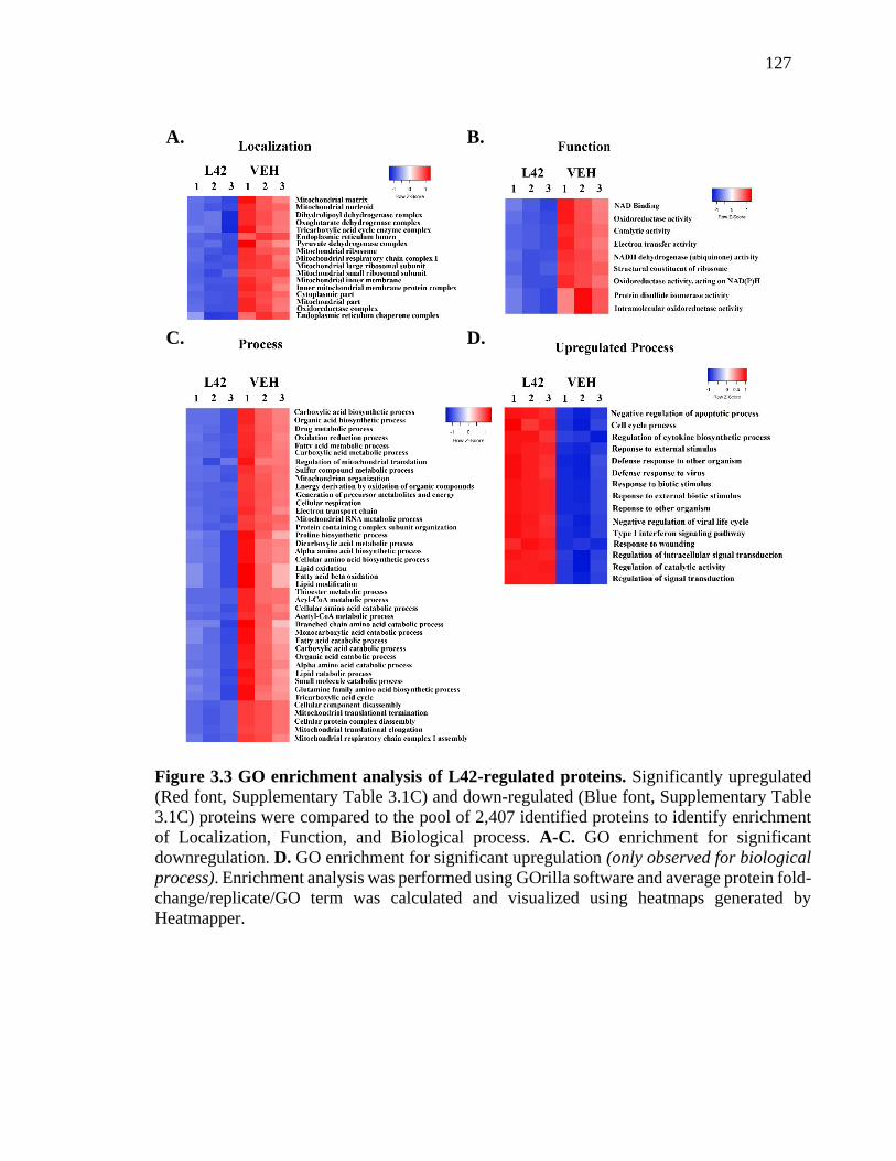
127
A. B.
D. C.
Figure 3.3 GO enrichment analysis of L42-regulated proteins. Significantly upregulated
(Red font, Supplementary Table 3.1C) and down-regulated (Blue font, Supplementary Table
3.1C) proteins were compared to the pool of 2,407 identified proteins to identify enrichment
of Localization, Function, and Biological process. A-C. GO enrichment for significant
downregulation. D. GO enrichment for significant upregulation (only observed for biological
process). Enrichment analysis was performed using GOrilla software and average protein fold-
change/replicate/GO term was calculated and visualized using heatmaps generated by
Heatmapper.

128
3.4.4 L42 disrupts metabolism in MDA-MB-231 TNBC cells.
We next examined whether L42 modulated the altered metabolic pathways of TNBC
cells. GO pathway enrichment analysis using the KEGG pathway database revealed that
L42 strongly suppressed proteins involved in lipid and amino acid metabolism (Figure
3.4A) and STRING analysis provided evidence for interaction between these proteins
(Figure 3.4B) and confirmed their mitochondrial localization. Of particular note are the
losses of enzymes such as acetyl-CoA acetyltransferease 1 (ACAT1) and AMPK-
activated protein kinase 1 (AMPK1) which are key to pyruvate and fatty acid β-oxidation
respectively [39, 40]. This was also accompanied by a significant reduction of
glutaminase (GLS) and the glutamine transporter solute carrier family 1 member 5
(SLC1A5). Given that targeting glutamine metabolism has emerged as an avenue for
TNBC therapy [16], we validated the loss of GLS expression by Western Blot (Figure
3.4C).

129
Figure 3.4 Hierarchical clustering of differentially-expressed proteins. Significantly
changed (Supplementary Table 3.1C) proteins were compared to the pool of 2,407
identified proteins to identify pathway enrichment using DAVID 6.8 software, with the
pathways matched to the KEGG database. A. Heatmaps of Lipid Metabolism (Top, GO
terms included: “Fatty Acid Metabolism”, “Fatty Acid Elongation”, and “Fatty Acid
Degradation”) and Amino Acid Metabolism (Bottom, GO terms included: “Amino Acid
Biosynthesis”, “Valine, Leucine and Isoleucine Degradation”, “Tryptophan Metabolism”,
and “Lysine Metabolism”). B. STRING interaction analysis of proteins from Lipid
Metabolism (Top) and Amino Acid Metabolism (Bottom), with coloring indicating
localization to mitochondria (Red: Mitochondrial matrix, Blue: Mitochondrial inner
membrane, Green: Mitochondrial unspecified). Shown are interactions at highest
confidence i.e. confidence score ≥ 0.9. C. Validation of GLS levels. (Left) Densitometry
quantification of blots using ImageJ. GLS levels were normalized against β-actin and
plotted as shown. N = 3. Significant difference from VEH is indicated as *p<0.05. (Right)
Representative western blot showing expression of glutaminase (GLS) in MDA-MB-231
cells treated with L42 (0.15 mg/ml) or vehicle control (VEH) for 12 h.

130
Figure 3.5 Hierarchical clustering of differentially-expressed proteins.
C.
A. B.

131
3.4.5 L42 impacts expression of TCA cycle enzymes.
Targeting the TCA cycle has recently generated interest as a novel route to cancer
therapy, in light of the observation that several TCA enzymes are mutated or
dysregulated in cancer cells [41]. Based on our GO analysis reflecting enrichment of
TCA cycle enzymes among the significantly down-regulated proteins, we compared the
expression levels of individual TCA cycle enzymes between L42- and control-treated
MDA-MB-231 cells. The full extent of TCA cycle suppression by L42 is made apparent
in Figure 3.5A, with substantial reductions in expression of multiple enzymes. As shown
in Figure 3.5B, L42 severely cripples the TCA cycle by inducing significant reductions in
nearly all TCA enzymes. In particular, the all subunits of the α-ketoglutarate
dehydrogenase complex (α-KGHDC) complex were down-regulated, with the key
component oxoglutarate decarboxylase (OGDH) exhibiting a nearly 2-fold reduction in
protein levels.
3.4.6 L42 dysregulates Complex I of the Electron Transport Chain.
Based on our GO analysis revealing down-regulation of proteins involved in oxidative
phosphorylation, we evaluated the impact of L42 on the specific complexes of the ETC.
We found L42 diminished expression of all the core subunits of Complex I, while also
suppressing subunits of Complex IV (Figure 3.6A). Comparison of expression levels
between L42- and control-treated groups shows the marked down-regulation of multiple
ETC complexes, with Complex I being particularly affected (Figure 3.6B).

132
Figure 3.6. Identification of TCA cycle proteins inhibited by L42 in MDA-MB-231
cells. A. Bar graph showing relative protein fold-change between treatment groups for the
GO term “Citrate cycle” from KEGG pathway analysis. B. Significantly changed
(Supplementary Table 3.1C) proteins were uploaded onto Cytoscape software (v3.6.0) and
matched to the TCA cycle using the WikiPathways app (v3.3.1), with degree of shading of
matching proteins set as a measure of fold-change (see key).

133
A.
B.
Figure 3.7 Identification of ETC proteins inhibited by L42 in MDA-MB-231 cells. A.
Bar graph showing relative protein fold-change between treatment groups for the GO term
“Oxidative Phosphorylation” from KEGG pathway analysis. B. Significantly changed
(Supplementary Table 3.1C) proteins were uploaded onto Cytoscape software (v3.6.0) and
matched to the Electron Transport Chain pathway using the WikiPathways app (v3.3.1),
with degree of shading of matching proteins set as a measure of fold-change (see key).

134
3.4.7 L42 disrupts mitochondrial membrane potential in TNBC cells
A major role of the ETC is maintenance of the mitochondrial membrane potential [42],
the loss of which disrupts the proton motive force required for ATP production, and is a
well-known trigger for apoptosis [43]. Our proteomics data strongly indicated that L42
induces apoptosis in TNBC cells by restricting oxidative phosphorylation through ETC
inhibition. To confirm this, we utilized a trimethylrhodamine, ethyl ester (TMRE)-based
staining assay to assess the status of the mitochondrial membrane potential in L42-treated
TNBC cells. As shown in Figure 3.7A, vehicle-treated MDA-MB-231 cells showed
robust staining with TMRE indicating intact and viable mitochondria, while L42-treated
cells displayed a significant 50 % reduction in TMRE staining (Figure 3.7B).

135
Figure 3.8 Assessment of mitochondrial membrane potential upon L42 treatment. A.
MDA-MB-231 cells were treated with L42 (0.15 mg/ml) or vehicle for 8 h and stained using
200 nM TMRE. Samples were imaged using a Zeiss Axiovert 200m fluorescence microscope.
B. Mitochondrial membrane potential was measured as the relative TMRE fluorescent
staining intensity between L42 and Vehicle-treated cells, quantified using ImageJ. Statistical
analysis was performed using an unpaired, two-tailed Mann-Whitney Test. A minimum of
250 cells were analyzed from a total of 4 replicates. *p < 0.00001.

136
3.5 Discussion
3.5.1 L42 inhibits survival and induces apoptosis in TNBC cells
Our study focused on elucidating the effects of an extract from the plant Lippia
origanoides, on the proteome of MDA-MB-231 TNBC cells. TNBCs are a particularly
aggressive, drug-resistant subtype of breast cancer with no known therapeutic targets.
Our previous research revealed that a Lippia extract had significant cytostatic and
apoptotic effects on TNBC cells with much reduced cytotoxicity against normal
mammary cells [21].
We reasoned that, since TNBC cells are known to have an altered metabolic
profile which includes heightened glycolysis and glutamine uptake, as well as generation
of TCA cycle intermediates, it was possible that the extract suppressed such metabolism
to selectively induce cell death in these cells. Through testing of extracts from other L.
origanoides chemotypes, we identified the L42 extract, which demonstrated an even
more potent dose-dependent cytotoxicity against TNBC cells while retaining a reduced
effect on normal cells (Figure 3.1A). L42 induced rapid, sustained and irreversible loss of
viability in MDA-MB-231 cells. L42 also inhibited Cyclin D1 (regulator of G0/G1 cell
cycle phase transition [44]) protein levels and induced Caspase-8 (extrinsic apoptosis
pathway marker [45]) activation (Figure 3.1B). These results shed light on the
mechanism by which L42 reduced TNBC cell viability but were insufficient to explain its
specificity. It was our intention to achieve a molecular understanding of the cancer cell
selectivity of L42 through quantitative proteomics.
3.5.2 L42 inhibits mitochondrial function and metabolism in TNBC cells.
We utilized LFQ to study the effects of a complex mixture, L42, on the TNBC proteome
landscape (Figure 3.2A). Data analysis revealed the LFQ data was remarkably
reproducible, with highly consistent quantitation achieved across replicates from both
treatment groups (Figure 3.2B). 2,407 proteins were quantified, with 519 proteins
significantly changed quantitatively between L42- and control-treated groups. L42 was
found to down-regulate proteins predominantly localized to various mitochondrial
complexes and compartments (Figure 3.3). Also apparent was that L42 inhibited

137
expression of proteins involved in oxidoreductase, iron sulfur-binding, NADH
dehydrogenase activity, and electron transport chain activity; all further indicative of its
mitochondrial-specificity.
This comprehensive and rapid loss of mitochondrial proteins required for
sustained cellular growth and proliferation could be expected to induce compensatory
pathways to prolong survival. The observation that L42 treatment induced stress
response, cell cycle, and DNA repair pathways is therefore not surprising (Figure 3.3B).
However, these cellular responses were insufficient to significantly bolster viability,
which continued to decrease up to 72 h post-treatment (Figure 3.1A). Together, these
results led us to further examine the nature of L42’s effects on mitochondrial metabolism.
3.5.3 L42 extensively inhibits the TCA Cycle in TNBC cells.
The TCA cycle (coupled to oxidative phosphorylation) provides a major alternative route
of ATP production for cancer cells compared to the inefficient process of anaerobic
glycolysis. TCA cycle intermediates are made readily available in TNBC cells by
upregulation of fatty acid β-oxidation and amino acid uptake and degradation [16-20].
We found that L42 downregulated several enzymes involved in mitochondrial amino acid
catabolism and fatty acid oxidation, effectively cutting off sources of acetyl CoA for the
TCA cycle (Figure 3.4). It has recently been revealed that Glutaminase (GLS), an enzyme
involved in the conversion of glutamine to glutamate (the rate-limiting step in glutamine
utilization), is necessary for TNBC growth in vitro and in vivo [16]. In support of its
actions on regulating TCA cycle substrate availability, we observed L42 reduced GLS
levels, a result validated by Western Blotting (Figure 3.4C).
It is recognized that the rate-limiting enzyme of the TCA cycle is α-KGHDC, a
complex consisting of oxoglutarate dehydrogenase (OGDH), dihydrolipoamide S-
succinyltransferase (DLST), and dihydrolipoamide dehydrogenase (DLD) [46]. Both
OGDH and DLST are implicated in cancer cell proliferation and survival, and targeting
α-KGHDC has become an exciting prospect for metabolism-based cancer therapy [41].
L42 severely disrupted the TCA cycle by down-regulating almost every enzyme involved
in the process, and reduced the expression of OGDC, DLST and DLD, (Figure 3.5).
Together, these results provide conclusive evidence that L42 treatment impacts the TCA

138
cycle in TNBC cells, and strongly suggests the resulting depletion of ATP could
contribute to the observed rapid apoptotic process.
3.5.4 L42 induces loss of NADH dehydrogenase (Complex I) expression
The electron transport chain (ETC) is critical for the generation of ATP via oxidative
phosphorylation, and also maintains the mitochondrial membrane potential (ΔΨm) via
the transfer of protons across the membrane by Complex I and Complex IV (cytochrome
c oxidase) [42]. The maintenance of ΔΨm is crucial for cellular integrity, and
depolarization of the membrane potential is a known catalyst for apoptosis [43]. For these
reasons, suppressing oxidative phosphorylation via electron transport chain (ETC)
inhibition has shown promise in cancer therapy. Metformin and Phenformin, both small
molecule Complex I inhibitors, were shown to reduce cell proliferation in HCT116
human colon cancer cells [47]. In addition, AG311, also a Complex I inhibitor, induced
rapid ATP depletion, mitochondrial depolarization, and necrosis in breast cancer cell
lines, and also retarded tumor growth and metastases in mouse models of breast cancer
without exhibiting any obvious toxicity [48].
We studied the effects of L42 on the electron transport chain, the major source of
ATP in the cell. L42 diminished expression of Complex I as well as Complex IV, down-
regulating several of their core subunits (Figure 3.6). Together, these two complexes
produce the proton gradient necessary for ATP production through ATP synthase, and
their severe down-regulation by L42 leads to rapid energy depletion in the cell and
subsequent apoptosis. In order to validate our proteomics data, we stained control- and
L42-treated MDA-MB-231 cells with trimethylrhodamine, ethyl ester (TMRE). TMRE is
a positively-charged fluorescent dye which readily stains active mitochondria via their
negative charge that is maintained by the functioning ETC. Disruption of ETC activity
leads to depolarization of the mitochondrial membrane and loss of TMRE staining.
Indeed, L42-treated MDA-MB-231 cells showed significantly reduced TMRE staining,
confirming impaired ETC function in these cells (Figure 3.7).

139
3.6 Summary
Our observation that L42 extensively downregulated mitochondrial proteins in TNBC
cells is significant for a number of reasons. It is well known that mitochondria are the
metabolic centers in cells, responsible for ATP production via oxidative phosphorylation
(OXPHOS) through the electron transport chain (ETC), while also housing the TCA
cycle as well as key enzymes involved in amino acid metabolism and fatty acid oxidation
[49-52]. Recent studies have implicated dysregulated mitochondrial function as key to the
acquisition of drug resistance in several forms of cancer; with targeting of mitochondrial
metabolism emerging as a novel route to cancer therapy. For example, inhibiting
mitochondrial protein synthesis and OXPHOS using tigecycline and imatinib was shown
to eradicate tyrosine kinase inhibitor-resistant CML leukemic stem cells (CML LSCs)
[53]. In addition, AraC (cytarabine, a nucleoside analogue)-resistant AML cells from
patient-derived xenografts (PDX) were shown to have significantly higher OXPHOS
signatures than AraC-sensitive cells, and targeting OXPHOS along with AraC treatment
was shown to be an effective combinatorial treatment [54]. In a recent study on TNBC,
Lee et al. discovered that targeting of MYC and myeloid-cell leukemia 1 (MCL1)
protein, along with hypoxia-inducible factor 1α (HIF-1α), inhibited mitochondrial
OXPHOS and drug resistance of cancer stem cells (CSCs) [55]. These studies strongly
suggest that targeting the mitochondria may be an effective method of treating aggressive
cancer and overcoming drug resistance [56].
Our study reveals suppression of TNBC metabolism to be a potentially powerful,
and as-yet unexploited, therapeutic approach. We hypothesize that the combined
inhibitory effects of L42 on TCA cycle enzymes, Acetyl CoA production, and OXPHOS
via ETC Complex I inhibition could trigger a cellular famine, which also provides an
explanation as to why L42 selectively affects MDA-MB-231 TNBC cells, which are
known to be highly metabolically active, as compared to normal mammary epithelial
MCF10A cells.
The difficulty of isolating bioactive components from complex mixtures and
identifying their cellular targets has limited the utility of studies on naturally-derived
extracts. Here, we show that the comprehensive landscape of information gleaned from

140
proteomics experiments both validates and advocates for the use of LFQ via LC-MS/MS
in the discovery of new cancer drugs from complex mixtures, as well as sheds light on
their mechanisms of action. The dose-dependent loss of cancer cell viability in response
to L42-treatment is comparable to responses commonly seen with the use of single
compounds. Our subsequent studies will focus on identifying the composition of L42,
and describing its effects in vivo.
3.7 References
1. Mann, J. (2002). Natural Products in Cancer Chemotherapy, Past, Present and Future.
Nature Reviews Cancer, 2, 143-148.
2. Chabner, B.A., & Roberts, T.G. (2005). Timeline - Chemotherapy and the War on
Cancer. Nature Reviews Cancer, 5, 65-72.
3. Milajerdia, A., Djafarian, K., & Hosseinia, B. (2016). The toxicity of saffron (Crocus
sativus L.) and its constituents against normal and cancer cells. Journal of Nutrition
and Intermediary Metabolism, 3, 23-32.
4. Graidist, P., Martla, M., & Sukpondma, Y. (2015). Cytotoxic Activity of Piper cubeba
Extract in Breast Cancer Cell Lines. Nutrients, 7, 2707-2718.
5. Konarikova, K., Jezovicova, M., Kerestes, J., Gbelcova, H., Durackova, Z., &
Zitnanova, I. (2015). Anticancer Effect of Black Tea Extract in Human Cancer Cell
Lines. Springerplus, 4, 4-127
6. Kingston, D.G.I. (2011). Modern Natural Products Drug Discovery and Its Relevance
to Biodiversity Conservation. Journal of Natural Products, 74, 496-511.
7. Beutler, J.A. (2009). Natural Products as a Foundation for Drug Discovery. Current
protocols in pharmacology. Current Protocol in Pharmacology, 46, 9.11.1–9.11.21
8. Macgregor, P.F., & Squire, J.A. (2002). Application of Microarrays to the Analysis of
Gene Expression in Cancer. Clinical Chemistry, 48, 1170-1177.
9. Ghazalpour, A., Bennett, B., Petyuk, V.A., Orozco, L., Hagopian, R., Mungrue, I.N.
et al. (2011). Comparative Analysis of Proteome and Transcriptome Variation in
Mouse. PLoS Genetics, 7, e1001393.
10. Ghaemmaghami, S., Huh, W., Bower, K., Howson, R.W., Belle, A., Dephoure, N. et
al. (2003). Global Analysis of Protein Expression in Yeast. Nature, 425, 737-741.

141
11. Tian, Q., Stepaniants, S.B., Mao, M., Weng, L., Feetham, M.C., Doyle, M.J. et al.
(2004). Integrated Genomic and Proteomic Analyses of Gene Expression in
Mammalian Cells. Molecular and Cellular Proteomics, 3, 960-969.
12. Warburg, O. (1956). Origin of Cancer Cells. Science, 123, 309-314.
13. Teicher, B.A., Linehan, W.M., & Helman, L.J. (2012). Targeting Cancer Metabolism.
Clinical Cancer Research, 18, 5537-5545.
14. Shen, L.L., O'shea, J.M., Kaadige, M.R., Cunha, S., Wilde, B.R., Cohen, A.L. et al.
(2015). Metabolic Reprogramming in Triple-Negative Breast Cancer through Myc
Suppression of TXNIP. Proceedings of the National Academy of Sciences U.S.A., 112,
5425-5430.
15. Lim, S.O., Li, C.W., Xia, W.Y., Lee, H.H., Chang, S.S., Shen, J. et al. (2016). EGFR
Signaling Enhances Aerobic Glycolysis in Triple-Negative Breast Cancer Cells to
Promote Tumor Growth and Immune Escape. Cancer Research, 76, 1284-1296.
16. Lampa, M., Arlt, H., He, T., Ospina, B., Reeves, J., Zhang, B.L. et al. Glutaminase Is
Essential for the Growth of Triple-Negative Breast Cancer Cells with a Deregulated
Glutamine Metabolism Pathway and Its Suppression Synergizes with Mtor Inhibition.
PLoS One. 2017, 12, e0185092
17. Goode, G., Gunda, V., Chaika, N.V., Purohit, V., Yu, F., Singh, P.K. Muc1 Facilitates
Metabolomic Reprogramming in Triple-Negative Breast Cancer. PLoS One 2017, 12,
e0176820
18. Van Geldermalsen, M., Wang, Q., Nagarajah, R., Marshall, A.D., Thoeng, A., Gao,
D. et al. Asct2/Slc1a5 Controls Glutamine Uptake and Tumour Growth in Triple-
Negative Basal-Like Breast Cancer. Oncogene. 2016, 35, 3201-3208.
19. Kanaan, Y.M., Sampey, B.P., Beyene, D., Esnakula, A.K., Naab, T.J., Ricks-Santi,
L.J. et al. Metabolic Profile of Triple-Negative Breast Cancer in African-American
Women Reveals Potential Biomarkers of Aggressive Disease. Cancer Genomics
Proteomics. 2014, 11, 279-294.
20. Wright, H.J., Hou, J., Xu, B., Cortez, M., Potma, E.O., Tromberg, B.J., Razorenova,
O.V. CDCP1 regulates lipid metabolism in TNBC. Proc. Natl. Acad. Sci. U.S.A. 2017,
8, 114 (32)

142
21. Raman, V., Fuentes-Lorenzo, J.L., Stashenko, E.E., Levy, M., Levy, M.M., Camarillo,
I.G. Lippia origanoides Extract Induces Cell Cycle Arrest and Apoptosis and
Suppresses Nf-Kappa B Signaling in Triple-Negative Breast Cancer Cells. Int. J.
Oncol. 2017, 51, 1801-1808.
22. Aryal, U.K., McBride, Z., Chen, D., Xie, J., Szymanski, D.B. Analysis of protein
complexes in Arabidopsis leaves using size exclusion chromatography and label-free
protein correlation profiling. J. Proteomics. 2017, 166, 8-18.
23. Bantscheff, M., Schirle, M., Sweetman, G., Rick, J., Kuster, B. Quantitative Mass
Spectrometry in Proteomics, A Critical Review. Anal. Bioanal. Chem. 2007, 389,
1017-1031.
24. Patel, V.J., Thalassinos, K., Slade, S.E., Connolly, J.B., Crombie, A., Murrell, J.C. et
al. A Comparison of Labeling and Label-Free Mass Spectrometry-Based Proteomics
Approaches. J. Proteome Res. 2009, 8, 3752-3759.
25. Michalski, A., Damoc, E., Hauschild, J.P., Lange, O., Wieghaus, A., Makarov, A. et
al. Mass Spectrometry-Based Proteomics Using Q Exactive, a High-Performance
Benchtop Quadrupole Orbitrap Mass Spectrometer. Mol. Cell. Proteomics. 2011, 10,
M111.
26. Cox, J., Hein, M.Y., Luber, C.A., Paron, I., Nagaraj, N., Mann, M. MaxLFQ allows
accurate proteome-wide label-free quantification by delayed normalization and
maximal peptide ratio extraction. Mol. Cell. Proteomics. 2014, 13, 2513-26.
27. Stashenko, E.E., Martinez, J.R., Cala, M.P., Duran, D.C., Caballero, D.
Chromatographic and Mass Spectrometric Characterization of Essential Oils and
Extracts from Lippia (Verbenaceae) Aromatic Plants. J. Sep Sci. 2013, 36, 192-202.
28. Cox, J., Mann, M. Maxquant Enables High Peptide Identification Rates,
Individualized P.P.B.-Range Mass Accuracies and Proteome-Wide Protein
Quantification. Nat. Biotechnol. 2008, 26, 1367-1372.
29. Polpitiya, A.D., Qian, W.J., Jaitly, N., Petyuk, V.A., Adkins, J.N., Camp, D.G. 2nd,
Anderson G.A., Smith, R.D. DAnTE, a statistical tool for quantitative analysis of -
omics data. Bioinformatics. 2008, 24, 1556-8.

143
30. Babicki, S., Arndt, D., Marcu, A., Liang, Y., Grant, J.R., Maciejewski, A., Wishart,
D.S. Heatmapper, web-enabled heat mapping for all. Nucleic Acids Res. 2016, 44,
W147-W153.
31. Eden, E., Navon, R., Steinfeld, I., Lipson, D., Yakhini, Z. Gorilla, A Tool for
Discovery and Visualization of Enriched Go Terms in Ranked Gene Lists. BMC
Bioinformatics. 2009, 10, 48.
32. Eden, E., Lipson, D., Yogev, S., Yakhini, Z. Discovering Motifs in Ranked Lists of
DNA Sequences. PLoS Comput. Biol. 2007, 3, 508-522.
33. Huang, D.W., Sherman, B.T., Lempicki, R.A. Systematic and Integrative Analysis of
Large Gene Lists Using David Bioinformatics Resources. Nat. Protoc. 2009, 4, 44-57.
34. Huang, D.W., Sherman, B.T., Lempicki, R.A. Bioinformatics Enrichment Tools,
Paths toward the Comprehensive Functional Analysis of Large Gene Lists. Nucleic
Acids Res. 2009, 37, 1-13.
35. Jensen, L.J., Kuhn, M., Stark, M., Chaffron, S., Creevey, C., Muller, J. et al. String 8-
a Global View on Proteins and Their Functional Interactions in 630 Organisms.
Nucleic Acids Res. 2009, 37, D412-D416.
36. Slenter, D.N., Kutmon, M., Hanspers, K., Riutta, A., Windsor, J., Nunes, N. et al.
Wikipathways, A Multifaceted Pathway Database Bridging Metabolomics to Other
Omics Research. Nucleic Acids Res. 2018, 46, D661-D667.
37. Shannon, P., Markiel, A., Ozier, O., Baliga, N.S., Wang, J.T., Ramage, D. et al.
Cytoscape, A Software Environment for Integrated Models of Biomolecular
Interaction Networks. Genome Res. 2003, 13, 2498-2504.
38. Schneider, C.A., Rasband, W.S. & Eliceiri, K.W. (2012). NIH Image to ImageJ, 25
years of image analysis. Nature Methods, 9, 671-675.
39. Fan, J., Lin, R.T., Xia, S.Y., Chen, D., Elf, S.E., Liu, S.P. et al. Tetrameric Acetyl-
Coa Acetyltransferase 1 Is Important for Tumor Growth. Mol. Cell. 2016, 64, 859-
874.
40. Hardie, D.G., Pan, D.A. Regulation of Fatty Acid Synthesis and Oxidation by the
Amp-Activated Protein Kinase. Biochem. Soc. Trans. 2002, 30, 1064-1070.
41. Anderson, N.M., Mucka, P., Kern, J.G., Feng, H. The emerging role and targetability
of the TCA cycle in cancer metabolism. Protein Cell. 2017, 9, 216-237.
144
42. Mitchell, P. Coupling of Phosphorylation to Electron and Hydrogen Transfer by a
Chemi-Osmotic type of Mechanism. Nature. 1961, 191,144–148
43. Ly, J.D., Grubb, D.R., Lawen, A. The Mitochondrial Membrane Potential (Delta Psi
M) in Apoptosis, an Update. Apoptosis. 2003, 8, 115-128.
44. Baldin, V., Lukas, J., Marcote, M.J., Pagano, M., Draetta, G. Cyclin D1 Is a Nuclear-
Protein Required for Cell-Cycle Progression in G(1). Genes Dev. 1993, 7, 812-821.
45. Muzio, M., Chinnaiyan, A.M., Kischkel, F.C., Orourke, K., Shevchenko, A., Ni, J. et
al. Flice, a Novel Fadd-Homologous Ice/Ced-3-Like Protease, Is Recruited to the
Cd95 (Fas/Apo-1) Death-Inducing Signaling Complex. Cell. 1996, 85, 817-827.
46. Lai, J.C.K., Cooper, A.J.L. Brain Alpha-Ketoglutarate Dehydrogenase Complex -
Kinetic-Properties, Regional Distribution, and Effects of Inhibitors. J. Neurochem.
1986, 47, 1376-1386.
47. Wheaton, W.W., Weinberg, S.E., Hamanaka, R.B., Soberanes, S., Sullivan, L.B.,
Anso, E. et al. Metformin Inhibits Mitochondrial Complex I of Cancer Cells to Reduce
Tumorigenesis. Elife. 2014, 3, e02242.
48. Bastian, A., Thorpe, J.E., Disch, B.C., Bailey-Downs, L.C., Gangjee, A., Devambatla,
R.K.V. et al. A Small Molecule with Anticancer and Antimetastatic Activities Induces
Rapid Mitochondrial-Associated Necrosis in Breast Cancer. J. Pharmacol. Exp. Ther.
2015, 353, 392-404.
49. Bertram, R., Pedersen, M.G., Luciani, D.S., Sherman, A. A Simplified Model for
Mitochondrial ATP Production. J. Theor. Biol. 2006, 243, 575-586.
50. Nunes-Nesi, A., Araujo, W.L., Obata, T., Fernie, A.R. Regulation of the
Mitochondrial Tricarboxylic Acid Cycle. Curr. Opinion Plant Biol. 2013, 16, 335-343.
51. Shah, Y.M., Lyssiotis, C.A. Mitochondrial Amino Acid Metabolism Provides
Vulnerabilities in Mutant Kras-Driven Cancers. Gastroenterology. 2016, 151, 798-801.
52. Currie, E., Schulze, A., Zechner, R., Walther, T.C., Farese, R.V. Cellular Fatty Acid
Metabolism and Cancer. Cell Metab. 2013, 18, 153-161.
53. Kuntz, E.M., Baquero, P., Michie, A.M., Dunn, K., Tardito, S., Holyoake, T.L. et al.
Targeting Mitochondrial Oxidative Phosphorylation Eradicates Therapy-Resistant
Chronic Myeloid Leukemia Stem Cells. Nat. Med. 2017, 23, 1234.
145
54. Farge, T., Saland, E., De Toni, F., Aroua, N., Hosseini, M., Perry, R. et al.
Chemotherapy-Resistant Human Acute Myeloid Leukemia Cells Are Not Enriched
for Leukemic Stem Cells but Require Oxidative Metabolism. Cancer Discov. 2017, 7,
716-735.
55. Lee, K.M., Giltnane, J.M., Balko, J.M., Schwarz, L.J., Guerrero-Zotano, A.L.,
Hutchinson, K.E. et al. Myc and Mcl1 Cooperatively Promote Chemotherapy-
Resistant Breast Cancer Stem Cells Via Regulation of Mitochondrial Oxidative
Phosphorylation. Cell Metab. 2017, 26, 633.
56. Bosc, C., Selak, M.A., Sarry, J.E. Resistance Is Futile, Targeting Mitochondrial
Energetics and Metabolism to Overcome Drug Resistance in Cancer Treatment. Cell
Metab. 2017, 26, 705-707.

146
ANALYSIS OF COMPOSITION AND IN VIVO
TOXICITY OF LIPPIA EXTRACTS
4.1 Abstract
Chapter 2 and Chapter 3 described our investigations into the in vitro effects and
mechanisms of action of the Lippia origanoides chemotype-variant extracts LOE and
L42. Here, we analyzed the composition of L42 via GC/MS to identify likely bioactive
components that mediate its apoptotic effects on TNBC cells. L42 was found to contain
30 different components, with the major peaks identified as the oxygenated monoterpenes
carvacrol and thymol. This is in contrast to LOE, which was previously shown by the
Stashenko lab to contain the flavonoid pinocembrin as the major component. However,
MDA-MB-231 cells treated with pinocembrin or carvacrol showed no significant
difference in viability, indicating that minor components and/or synergistic effects
potentiate the apoptotic effects of these extracts. In addition, in vivo toxicity assays
showed no significant adverse effects in NSG mice treated with intra-peritoneal
injections of L42. Collectively, these studies provide strong support for i) further
exploring the individual and synergistic apoptotic effects of L42 components on TNBC
cells, and ii) studying the ability of L42 to prevent tumor growth and progression in vivo.
4.2 Introduction
We have previously shown that Lippia origanoides extract LOE induced apoptosis in
multiple TNBC cell lines accompanied by NF-B inhibition and caspase-8 activation (see
Chapter 2) [1]. Compositional analysis of the extract by the Fuentes lab revealed it to
contain 16 components (Table 4.2), many of which have also been shown to inhibit NF-
B signaling and induce apoptosis in in vitro and in vivo models of cancer [2-13, 21-23].
Subsequently, we observed that an L. origanoides extract from a different chemotype,
L42, showed even greater potency against TNBC cells in vitro (see Chapter 3) [14]. L42
treatment of TNBC cells induced significant loss of expression of mitochondrial enzymes
necessary for cellular metabolism [14]. Similar to LOE, L42 also induced loss of RIP1,
Cyclin D1 and cIAP2, and activated caspase-8, leading to rapid, irreversible cell death

147
(Figure 4.1). We hypothesized that the similar downstream molecular effects of the
extracts on TNBC cells would be consistent with a conservation of composition across
the two chemotypes. Accordingly, we performed GC/MS analysis of L42 to compare its
components with those from LOE (Table 4.1), and were able to identify 30 compounds
present in the extract. While several of these compounds were shared with LOE,
including thymol, carvacrol, caryophyllene, and humulene, L42 also contained multiple
unique compounds, many of which have not been studied in the context of cancer.
Major components identified in LOE and L42 have been previously shown to
possess anti-cancer effects. The flavonoid pinocembrin makes up ~ 55 % of LOE, and as
mentioned in Chapter 2, was observed to inhibit epithelial-mesenchymal and transition
and reduce cell viability in Y-79 retinoblastoma cells [7]. Pinocembrin also induced
apoptosis in HCT-116 colon cancer cells by triggering the loss of mitochondrial
membrane potential accompanied by caspase-3/-9 activation and cytochrome c release
[11]. The oxygenated monoterpene carvacrol, found in significant quantities in both L42
and LOE, has also shown inhibitory effects on NF-B signaling and induced apoptosis in
multiple cancer cell lines [2-6]. We therefore investigated the effects of these components
on MDA-MB-231 cell viability.
Investigations into plant-derived extracts as a source of cancer therapeutics are
supported by a number of studies observing them to be non-toxic to normal cells as well
as in vivo [15-17]. To serve as a foundation for future studies investigating the benefits of
L42 for prevention of cancer initiation and progression, we performed toxicity tests of
L42 in the C57BL/6 mouse model, with no adverse reactions recorded over the period of
observation.
Taken together, our results provide a detailed composition of L42, and suggest
that its apoptotic effects are modulated by minor components or require multiple
components working synergistically. We also validate the safety of L42 for use as a
treatment in future studies of TNBC in vivo.

148
4.3 Materials and Methods
4.3.1 Plant material and extract
Lippia origanoides plants were collected from the Chicamocha River Canyon (Los
Santos, Santander, Colombia). Taxonomic identification of L. origanoides was performed
by Dr. José Luis Fernández Alonso (National University, Bogotá, Colombia). Fresh
leaves and stems from L. origanoides specimens (LOE: COL560259; L42: COL560267)
were used for extraction as previously described [18]. Briefly, 200 g of finely ground L.
origanoides plant material was placed in the extraction chamber of a Thar SFE-2000-2-
FMC50 (Thar Instruments, PA, USA) and subjected to supercritical CO2 extraction at 50
MPa and 333 K at the Universidad Industrial de Santander, Bucaramanga, Colombia. The
material deposited on the walls of the chamber was collected, and 250 mg of this
extracted material was dissolved in 5 ml of methanol, sonicated for 15 min, and
centrifuged at 10,000 rpm for a further 15 min. The 50 mg/ml stock solutions of the
supernatants collected from each specimen (LOE: COL560259; L42: COL560267) were
used in the present study.
4.3.2 GC/MS analysis of L. origanoides extracts
20 μl of L42 stock solution was dried in a speedvac (Labconco, MO, USA) for 30 min.
The dried pellet was resuspended in 40 uL methoxyamination reagent (10 mg/ml
methoxyamine HCl in anhydrous pyridine, prepared daily), then incubated at 37C for 90
min. A second sample prepared as above was derivatized by trimethylsilylation by
incubating with 70 μl N-Methyl-N-(trimethylsilyl) trifluoroacetamide (MSTFA) for 30
min at 37C.
GC/MS/MS analyses of both derivatized and non-derivatized samples were
performed at the Metabolite Profiling Facility (MPF), Purdue University, using a Thermo
TSQ 8000 triple quadrupole mass spectrometer coupled to a Thermo Trace 1310 GC.
Data acquisition and analysis was performed using the Chromeleon software tool, and
peaks were identified via spectral matching to the NIST compound library.

149
4.3.3 MTT cell viability assay
MDA-MB-231 cells were seeded at 105 cells/well in 96-well plates and allowed to attach
overnight. Spent media was then replaced with fresh media containing carvacrol (Sigma-
Aldrich, MO, USA) dosages between 60 - 85 μM. Media in control wells was replaced
with fresh media without additives (No-treatment, NT) or media containing the vehicle,
methanol (Veh). Following 24 h treatment, 12 mM of MTT, 3-(4,5-dimethylthiazol-2-yl)-
2,5-diphenyltetrazolium bromide (Life Technologies, NY), was added to the cells
followed by incubation for 4 h at 37°C. Formazan crystals formed at the end of
incubation period were dissolved using dimethyl sulfoxide (DMSO) and plates were read
at 570 nm with a reference wavelength of 630 nm.
4.3.4 In vivo toxicity study
Female virgin C57BL/6 mice were acquired from the Purdue Biological Evaluation
Facility and housed at the Purdue Animal Housing Facility. Animals were provided rat
chow and water ad libitum. At Day 0, mice (n = 3 per treatment group) were
intraperitoneally injected with a single 200 μl dose of control (20% v/v methanol/1X
PBS), or 100 mg/kg BW L42 (high dose), or 50 mg/kg BW L42 (medium dose), or 25
mg/kg BW L42 (low dose). In order to keep dosage volumes consistent within treatment
groups, mice with similar weights were grouped together. Animals were periodically
monitored for changes in body weight and adverse reactions over the course of 2 weeks,
at the end of which all animals were sacrificed via isoflurane anesthesia followed by
cervical dislocation. All experimental procedures were conducted under Purdue Animal
Care and Use Committee (PACUC) ethical regulations.
4.3.5 Histological analysis of mouse mammary gland
Left inguinal mammary glands were removed from mice post-sacrifice, and fixed in 10 %
formalin for 48 h prior to being transferred to the Purdue Histology Research Laboratory.
Tissues were placed in a Sakura Tissue-VIP6 tissue processor for dehydration through
graded ethanol solutions. Next, dehydrated tissues were cleared in xylene and paraffin-
fixed using Leica Paraffin Plus. 4 μm microsections were taken using a Themo Shandon
Finesse Me microtome, and the sections were then mounted on charged slides and dried

150
for 1 h at 60C in an oven. Slides were then placed on a Leica ST5010-CV5030
autostainer, deparaffinized in 3 changes of xylene and rehydrated using graded ethanols
to water. Slides were then stained using Gill’s II hematoxylin, blued, and eosin/phloxine
B mixture. Finally, slides were dehydrated, cleared in xylene and coverslipped in a
toluene-based mounting media. Slides were imaged at 10X and 40X magnification using
a DSC-F717 camera (Carl-Zeiss) attached to an inverted brightfield microscope (VWR
International). The author acknowledges the assistance of the Purdue University
Histology Research Laboratory, a core facility of the NIH-funded Indiana Clinical and
Translational Science Institute
4.4 Results
4.4.1 GC/MS reveals compositional differences in L42 and LOE
In order to understand if the increased potency exhibited by L42 over LOE on MDA-MB-
231 TNBC cell viability was due to compositional differences between the two extracts,
we performed GC/MS analysis of L42. Figure 4.1. shows the GC/MS spectra of L42,
with Table 4.1 describing the composition of L42 identified by matching the spectra to
the NIST compound library. Compared to the composition of LOE, which has 15
identified components (Table 4.2, taken with permission from Castellanos et al. [17]),
L42 is a more complex extract, with 32 components. However, 7 components (-cymene,
D-limonene, -terpinene, thymol, carvacrol, trans--caryophyllene, and -humulene) are
shared between the two extracts (components highlighted in red, Table 4.1 and Table
4.2).
4.4.2 Major components of LOE and L42 do not induce loss of viability in TNBC
cells.
GC/MS analysis by Castellanos et al. revealed that the flavanone pinocembrin makes up
54.9 % of LOE [19]. In addition, analysis of L42 at the MPF revealed carvacrol to be the
major component of this extract, followed by -cymene and thymol. We tested the major
component of each of these extracts for their effects on MDA-MB-231 cell viability.
Interestingly, while no significant effect on viability was observed for carvacrol (Figure

151
4.2), pinocembrin appeared to significantly increase the viability of these cells (not
shown).

152
Figure 4.1 GC/MS spectra of L42. (upper panel) Spectra of L42 run without TMS
derivatization using N-Methyl-N-(trimethylsilyl) trifluoroacetamide (MSTFA). (lower
panel) Spectra of L42 run after derivatization using MSTFA. Numbers on major peaks
indicate component identified in Table 4.1

153
Table 4.1 Composition of L42. Peaks from GC/MS spectra of non-derivatized and
derivatized L42 (Figure 4.1) were searched against the NIST compound library based on
retention times, and the respective components were identified. Components in red are shared
between L42 and LOE (See Table 4.2).

154
Table 4.2 Composition of LOE. Peaks from GC/MS spectra of non-derivatized LOE.
Components in red are shared between L42 and LOE (See Table 4.1). (Taken with
permission from Castellanos et. al [17])

155
Figure 4.2. Effect of carvacrol on TNBC cell viability. MDA-MB-231 cells were treated
with 60 – 85 μM carvacrol for 24 h, then tested for viability using the MTT assay. Plot
shows viability relative to vehicle (methanol)-treated cells.

156
4.4.3 L42 is well-tolerated in vivo upon IP administration
As a precursor to future experiments testing the effects of L42 on preventing
tumorigenesis and inhibiting tumor growth in vivo, we performed a Maximum Tolerated
Dose (MTD) study using high (100 mg/kg BW), medium (50 mg/kg BW), and low (25
mg/kg BW) of L42 intraperitoneally injected into female C57BL/6 mice. This strain of
mice was chosen due to possession of an intact immune system, which allowed for
observation of any immune reactions to components, toxins, or pathogens that may have
been present in the extract.
Immediately after injection, mice from ‘high’ and ‘medium’ dosage groups
exhibited slightly unsteady gait and dragging of the hind limb. However, these mice
quickly recovered 30 min post-injection, and mice from all groups remained bright, alert,
and responsive (BAR) throughout the duration of the study period (Table 4.3).
Mice were also monitored for changes in body weight during the 2-week
observational period following IP injection. As can be seen from Figure 4.3, mice from
all treatment groups exhibited no significant change in body weight during this time,
confirming that L42 is well-tolerated among mice with intact immune systems.

157
Table 4.3 L42 does not induce adverse effects in C57BL/6 mice. Female virgin C57BL/6
mice were intraperitoneally injected with L42 at 100 mg/kg BW (High dose), or 50 mg/kg BW
(Medium dose), or 25 mg/kg BW (Low dose) or Control, and monitored for adverse effects
over the course of 2 weeks. BAR: Bright, alert, and responsive. Mouse number reflects ID
number assigned to mouse at the start of experiment; numbers are not sequential because mice
were grouped based on body weight, not assigned number.

158
Figure 4.3 C57BL/6 mice do not exhibit weight loss upon L42 administration. Female
virgin C57BL/6 mice were intraperitoneally injected with L42 at 100 mg/kg BW (High dose),
or 50 mg/kg BW (Medium dose), or 25 mg/kg BW (Low dose) or Control, and weighed at
indicated days post-treatment over the course of 2 weeks.

159
4.4.4 IP injection of L42 is non-toxic to mouse mammary glands
In order to identify any potential adverse effects that L42 may have on the mammary
gland, the left inguinal mammary glands were isolated from C57BL/6 mice 2 weeks post-
IP injection of L42. H&E staining of mammary gland sections from control-treated and
L42-treated mice showed no significant pathological differences (Figure 4.4, left).
Specifically, the lactiferous ducts were intact, and surrounded by adipocytes of uniform
size with peripheral nuclei, and there were no visible signs of an immune response such
as the infiltration of polymorphonuclear neutrophils (PMNs), a common characteristic of
inflammation [20]. A closer examination of the lactiferous ducts also revealed no
differences in ductal morphology (Figure 4.4, right). Ducts from both groups showed a
single layer of polarized luminal epithelial cells lining the ductal lumen, with a thin
stromal layer forming the basement membrane. Mammary glands from both groups were
also free of necrotic cells and damage to lymph nodes (not shown).

160
Figure 4.4 Mammary glands from C57BL/6 mice do not show pathological differences
upon L42 administration. Mammary glands from control- and L42-treated mice were fixed
and H&E stained. (Left) Representative mammary glands from control (CTRL)- and L42 (100
mg/kg BW, HIGH dose)-treated mice showing intact lactiferous glands (boxed), adipocytes
with peripheral nuclei, and no sign of inflammation. (Right) Magnified lactiferous glands
showing normal phenotype from both groups, i.e. a single layer of luminal epithelial cells
attached to a stroma-rich basement membrane, surrounding the ductal lumen.
161
4.5 Summary
In Chapter 2 and 3, the Lippia origanoides extracts LOE and L42 were shown to exert
potent apoptotic effects against TNBC cells, with reduced effects on normal mammary
epithelial cells in vitro. Our investigations shed light on their mechanisms of action. Both
LOE and L42 significantly reduced RIP1 levels and induced caspase-8 activation, which
support the inhibition of NFB signaling as a common apoptotic mechanism of the
extracts. However, L42 also showed significant inhibitory effects on mitochondrial
metabolism, and showed greater potency against MDA-MB-231 TNBC cells than LOE.
In order to understand the reasons behind the similarities and differences in the effects of
LOE and L42, the composition of L42 was analyzed by GC/MS, and compared to that of
LOE (previously revealed by Castellanos et al. [19]).
As can be seen from Table 4.1 and 4.2, LOE and L42 share multiple major
components including carvacrol, thymol, -humulene, trans--caryophyllene, and -
cymene; all of which have been shown to block NFB signaling in previous studies [2,
13, 21-23]. However, L42 was shown to contain twice the number of components as
LOE, with 32 identified in total. It cannot be ruled out that the added potency of L42
against TNBC cells is due to synergism between components not found in LOE.
The major components of LOE and L42 were the flavanone pinocembrin, and the
monoterpene carvacrol, respectively. MDA-MB-231 TNBC cells were treated with these
components individually, to determine if they were sufficient for the apoptotic effects
exhibited by the extracts. As can be seen from Figure 4.2, carvacrol had no effect on
MDA-MB-231 viability, while surprisingly, pinocembrin increased cell viability (not
shown), possibly by stimulating proliferation or cellular oxidoreductase activity (as the
MTT assay measures oxidoreductase activity to assess viability). These results point to
synergism between Lippia components as a requirement for apoptosis induction in TNBC
cells. Further experiments using individual L. origanoides components alone or in
combinations, as well as bioassay-driven fractionation methods, may be required to
isolate the bioactive components (BACs) from these extracts. Also, in collaboration with
the Purdue Proteomics Facility and Metabolomics Profiling Facility, we are also
developing LC/MS and GC/MS-based tools to identify BACs and their cellular protein

162
binding partners, thereby improving on existing techniques while reducing the time
requirements.
Our studies have revealed that L. origanoides extracts are capable of inhibiting
NFB signaling, an inflammatory pathway often constitutively activated in TNBC [1, 24,
25], while also targeting mitochondrial metabolism, a major source of ATP for
proliferating cancer cells [26-28]. These extracts as a whole may have multiple health
benefits including anti-inflammation, anti-tumorigenic, and anti-tumor progression
effects in vivo. To serve as a foundation to such future studies, we performed a Maximum
Tolerated Dose study of L42 to assess its toxicity in the C57BL/6 mouse model. These
mice showed no significant adverse effects upon IP administration of up to 100 mg/kg
BW L42 (Figure 4.3), and mammary glands from L42-treated and control group mice
showed no histopathological differences (Figure 4.4), indicating that L42 is non-toxic in
vivo and can be safely used at high dosages in future studies.
Taken together, these results provide a compositional basis for the similarities and
differences seen in the effects of LOE and L42, while also indicating that the apoptotic
effects of these extracts on TNBC cells may be due to multi-component action. Finally,
our MTD study supports the safe use of L42 in future in vivo research investigating its
ability to prevent TNBC initiation and progression.
4.6 References
1. Raman, V., Lorenzo, J. L. F., Stashenko, E. E., Levy, M., Levy, M. M., & Camarillo,
I. G. (2017). Lippia origanoides extract induces cell cycle arrest and apoptosis and
suppresses NF-κB signaling in triple-negative breast cancer cells. International
Journal of Oncology, 51(6), 1801–1808.
2. Aristatile, B., Al-Assaf, A. H., & Pugalendi, K. V. (2013). Carvacrol suppresses the
expression of inflammatory marker genes in D-galactosamine-hepatotoxic rats. Asian
Pacific Journal of Tropical Medicine, 6(3), 205–211.
3. Arunasree, K. M. (2010). Anti-proliferative effects of carvacrol on a human metastatic
breast cancer cell line, MDA-MB 231. Phytomedicine, 17(8), 581–588.

163
4. Dai, W., Sun, C., Huang, S., & Zhou, Q. (2016). Carvacrol suppresses proliferation
and invasion in human oral squamous cell carcinoma. OncoTargets and Therapy, 9,
2297–2304.
5. Fan, K., Li, X., Cao, Y., Qi, H., Li, L., Zhang, Q., & Sun, H. (2015). Carvacrol inhibits
proliferation and induces apoptosis in human colon cancer cells. Anti-Cancer Drugs,
26(8), 813–823.
6. Kang, S.-H., Kim, Y.-S., Kim, E.-K., Hwang, J.-W., Jeong, J.-H., Dong, X., et al.
(2016). Anticancer Effect of Thymol on AGS Human Gastric Carcinoma Cells.
Journal of Microbiology and Biotechnology, 26(1), 28–37.
7. Chen, K.S., Shi, M.D., Chien, C.S., & Shih, Y.W. (2014). Pinocembrin suppresses
TGF-betta1-induced epithelial-mesenchymal transition and metastasis of human Y-79
retinoblastoma cells through inactivating alphavbetta3 integrin/FAK/p38alpha
signaling pathway. Cell Biosciences, 4, 41.
8. Legault, J., & Pichette, A. (2007). Potentiating effect of beta-caryophyllene on
anticancer activity of alpha-humulene, isocaryophyllene and paclitaxel. The Journal
of Pharmacy and Pharmacology, 59(12), 1643–1647.
9. Li, Y., Wen, J., Du, C., Hu, S., Chen, J., Zhang, S., et al. (2017). Thymol inhibits
bladder cancer cell proliferation via inducing cell cycle arrest and apoptosis.
Biochemical and Biophysical Research Communications, 491(2), 530–536.
10. Ahmad, A., Banerjee, S., Wang, Z., Kong, D., & Sarkar, F.H. (2008). Plumbagin-
induced apoptosis of human breast cancer cells is mediated by inactivation of NF-
kappaB and Bcl-2. Journal of Cellular Biochemistry, 105, 1461-1471.
11. Kumar, M. A. S., Nair, M., Hema, P. S., Mohan, J., & Santhoshkumar, T. R. (2007).
Pinocembrin triggers Bax-dependent mitochondrial apoptosis in colon cancer cells.
Molecular Carcinogenesis, 46(3), 231–241.
12. Jin, M.L., Park, S.Y., Kim, Y.H., Park, G., & Lee, S. (2014) Halofuginone induces the
apoptosis of breast cancer cells and inhibits migration via downregulation of matrix
metalloproteinase-9. International Journal of Oncology, 44, 309-318.
13. Liang, D., Li, F., Fu, Y., Cao, Y., Song, X., Wang, T., et al. (2014). Thymol inhibits
LPS-stimulated inflammatory response via down-regulation of NF-κB and MAPK
signaling pathways in mouse mammary epithelial cells. Inflammation, 37(1), 214–222.

164
14. Raman, V., Aryal, U. K., Hedrick, V., Ferreira, R. M., Fuentes Lorenzo, J. L.,
Stashenko, E. E., et al. (2018). Proteomic Analysis Reveals That an Extract of the
Plant Lippia origanoides Suppresses Mitochondrial Metabolism in Triple-Negative
Breast Cancer Cells. Journal of Proteome Research, 17(10), 3370–3383.
15. Milajerdia, A., Djafarian, K., & Hosseinia, B. (2016). The toxicity of saffron (Crocus
sativus L.) and its constituents against normal and cancer cells. Journal of Nutrition
and Intermediary Metabolism, 3, 23-32.
16. Graidist, P., Martla, M., & Sukpondma, Y. (2015). Cytotoxic Activity of Piper cubeba
Extract in Breast Cancer Cell Lines. Nutrients, 7, 2707-2718.
17. Konarikova, K., Jezovicova, M., Kerestes, J., Gbelcova, H., Durackova, Z., &
Zitnanova, I. (2015). Anticancer Effect of Black Tea Extract in Human Cancer Cell
Lines. Springerplus, 4, 4-127
18. Stashenko, E.E., Martínez, J.R., Cala, M.P., Durán, D.C., & Caballero, D. (2013).
Chromatographic and mass spectrometric characterization of essential oils and
extracts from Lippia (Verbenaceae) aromatic plants. Journal of Separation Science,
36, 192-202.
19. Castellanos, N.R., Castro, C.E., Stashenko, E.E., & Fuentes, J. Lippia origanoides
supercritical fluid extract and its major constituent pinocembrin diminish ultraviolet
radiation-induced DNA damage and modulate cell division in Escherichia coli. (In
review)
20. Camperio, C., Armas, F., Biasibetti, E., Frassanito, P., Giovannelli, C., Spuria, L., et
al. (2017). A mouse mastitis model to study the effects of the intramammary infusion
of a food-grade Lactococcus lactis strain. PLOS ONE, 12(9), e0184218.
21. Xie, G., Chen, N., Soromou, L. W., Liu, F., Xiong, Y., Wu, Q., et al. (2012). p-Cymene
protects mice against lipopolysaccharide-induced acute lung injury by inhibiting
inflammatory cell activation. Molecules (Basel, Switzerland), 17(7), 8159–8173.
22. Lee, J.C., Kundu, J.K., Hwang, D.M., Na, H.K., & Surh, Y.J. (2007). Humulene
inhibits phorbol ester-induced COX-2 expression in mouse skin by blocking activation
of NF-kappaB and AP-1, IkappaB kinase and c-Jun-N-terminal kinase as respective
potential upstream targets. Carcinogenesis, 28(7),1491-8.

165
23. Kim, C., Cho, S.K., Kim, K.D., Nam, D., Chung, W.S., Jang, H.J., et al. (2014). β-
Caryophyllene oxide potentiates TNFα-induced apoptosis and inhibits invasion
through down-modulation of NF-κB-regulated gene products. Apoptosis 19(4),708-
18.
24. Nakshatri, H., Bhat-Nakshatri, P., Martin, D. A., Goulet, R. J., & Sledge, G. W. (1997).
Constitutive activation of NF-kappaB during progression of breast cancer to hormone-
independent growth. Molecular and Cellular Biology, 17(7), 3629–3639.
25. Sero, J. E., Sailem, H. Z., Ardy, R. C., Almuttaqi, H., Zhang, T., & Bakal, C. (2015).
Cell shape and the microenvironment regulate nuclear translocation of NF-κB in
breast epithelial and tumor cells. Molecular Systems Biology, 11(3), 790.
26. Vyas, S., Zaganjor, E., & Haigis, M. C. (2016). Mitochondria and Cancer. Cell, 166(3),
555-566.
27. Ashton, T. M., McKenna, W. G., Kunz-Schughart, L. A., & Higgins, G. S. (2018).
Oxidative Phosphorylation as an Emerging Target in Cancer Therapy. Clinical Cancer
Research: An Official Journal of the American Association for Cancer Research,
24(11), 2482–2490.
28. Rodríguez-Enríquez, S., Carreño-Fuentes, L., Gallardo-Pérez, J. C., Saavedra, E.,
Quezada, H., Vega, A., et al. (2010). Oxidative phosphorylation is impaired by
prolonged hypoxia in breast and possibly in cervix carcinoma. The International
Journal of Biochemistry & Cell Biology, 42(10), 1744–1751.

166
CONCLUSIONS AND FUTURE DIRECTIONS
For thousands of years, human beings have used natural products in traditional medical
remedies; for healing wounds, alleviating gastrointestinal diseases, fighting infections,
and also as nociceptives, sedatives, and laxatives. Multiple modern cancer therapeutics
have their bases in natural products; with taxanes, Vinca alkaloids, camptothecins, and
epipodophyllotoxins the most well-known among them [1-4]. While most of these
therapies are cytotoxic, often causing serious side-effects when used in aggressive
combination regimens, by increasing our understanding of tumor-specific vulnerabilities,
future naturally-sourced chemo-drugs could be screened for enhanced tumor-directed
toxicity, thereby significantly reducing side-effects. Further, the tremendous diversity of
natural compounds offers significant opportunities for the identification of entirely new
classes of drugs, which can be further modified to specifically and more potently target
cancer cells.
Modern technology and software tools have helped to profile the genome and
transcriptome of tumors, and helped identify novel, exploitable characteristics.
Treatments for triple-negative breast cancer (TNBC), an aggressive subtype with higher
relapse and mortality rates, currently rely on combination chemotherapy regimens.
However, an improved understanding of TNBC tumor characteristics through
transcriptomics and metabolomics has identified several, previously unknown
vulnerabilities, two of which will be discussed here [5, 6].
Firstly, TNBCs are often characterized by constitutive activation of NF-B
signaling, an inflammatory pathway normally utilized normal immune cells [7].
Activation of the pathway leads to the nuclear translocation of a transcription factor,
p65/RelA, which induces transcription of genes involved in survival, EMT, and
proliferation such as cIAP2, Cyclin D1, and MMP-9 [8-14]. Inhibiting NF-B signaling
can induce tumor cell death both in vitro and in vivo, and may thus be a potentially target
for future chemotherapeutics.

167
Secondly, TNBCs are characterized by extensive metabolic reprogramming to
fuel the massive ATP demands required for proliferation, growth, and migration.
Unsurprisingly, TNBCs have been shown to have elevated levels of multiple amino acids,
including aspartate, asparagine, glutamate, phenylalanine, methionine, and the branched-
chain amino acids (BCAAs) leucine, isoleucine and valine [15]. These amino acids are
subsequently converted into TCA cycle intermediates such as succinate, fumarate, and
malate, with the by-products of the TCA cycle going on to fuel oxidative phosphorylation
as well [16]. TNBCs also show increases in glycolysis, and a dependency on glutamine
metabolism, a phenomenon known as ‘glutamine addiction’ [17-21]. As both the TCA
cycle and oxidative phosphorylation are mitochondria-localized, the inhibition of
mitochondrial metabolism may be another avenue to novel TNBC-specific therapies.
Our search for new natural sources of chemotherapeutics against TNBC lead to
our current investigations of the South American plant Lippia origanoides. Extracts from
this species have been used by the native people as topical analgesics and to alleviate
gastrointestinal disorders, and have also been shown as anti-mutagenic and anti-microbial
by Dr. Fuentes at the Universidad Industrial de Santander [22]. In the present studies, we
used two extracts from L. origanoides, LOE and L42, prepared from different
chemotypes of the plant, and studied their cytotoxic effects on TNBC cells in vitro, while
also shedding light on their mechanisms of action, using conventional molecular biology
tools as well as label-free quantitative (LFQ) proteomics. We also characterized the
composition of L42 and finally, we studied its in vivo toxicity.
5.1 LOE inhibits NF-B signaling and induces apoptosis in TNBC cells
Our studies initially identified LOE as capable of inducing a dose-dependent loss of
viability in MDA-MB-231, CRL-2321, and MCF-10AH triple-negative breast cancer
cells in the 0.15 – 0.2 mg/ml range (Chapter 2), with MDA-MB-231 cells showing a
50 % loss of viability upon 24 h treatment with 0.15 mg/ml LOE, and 95 % loss with 0.2
mg/ml LOE. In contrast, MCF-10A normal mammary epithelial cells showed only a
20 % loss of viability with 0.15 mg/ml, and 40 % loss with 0.2 mg/ml. This important

168
result indicated that LOE was targeting mechanisms specific to cancer cells, and our
subsequent studies focused on defining a mechanism of action for the extract.
Differential staining analysis by flow cytometry revealed that LOE induced a loss
in MDA-MB-231 cell viability by halting the cell cycle in the G0/G1 phase and promoted
apoptosis without inducing necrotic cell death. We showed that these results were
accompanied by a loss in protein levels of the proliferative and survival markers, Cyclin
D1 and cIAP2, and the activation of caspase-8 and caspase-3. As mentioned previously,
Cyclin D1 and cIAP2 are downstream products of NF-B signaling, and inhibition of this
pathway can activate caspase-8-induced cell death. In line with our observations, we
hypothesized that upstream effectors of NF-B signaling were inhibited by the extract
and found that indeed, levels of RIP1, a scaffold protein required for downstream
signaling, were significantly lost upon treatment with LOE (Chapter 2).
In summary, these results provided the first evidence that L. origanoides was a
source of cytotoxic compounds that could be used to induce apoptosis in triple-negative
breast cancer cells, with minimal effects on normal cells. The L. origanoides extract
(LOE) was shown to inhibit NF-B effector proteins, and based on previous studies
identifying this pathway as important to TNBC proliferation and survival, we proposed
that NF-B inhibition could be involved in mediating the TNBC-specific loss of cell
viability induced by the extract.
5.2 L42 induces extensive loss of mitochondrial proteins in TNBC cells
To ascertain that the effects of the L. origanoides extract (LOE) on TNBC vs. normal cell
viability would be retained in other extracts from the plant, we tested L42, an extract
prepared using an identical protocol but from a different chemotype (genetic variant) of
L. origanoides. Treatment of MDA-MB-231 and MCF-10A cells with L42 confirmed
that the extract retained the significant TNBC-specific cytotoxicity observed with LOE,
and was also significantly more potent than LOE at the same concentration (See Figure
2.1, Chapter 2 and Figure 3.1, Chapter 3). For example, at 0.15 mg/ml for 24 h, LOE
reduced viability by 50 %, compared to 80 % loss of viability with L42. In addition,

169
when treated for 24 h with up to 0.12 mg/ml L42, MCF-10A cells did not undergo any
loss of viability, compared to a 50 % loss of viability in MDA-MB-231 cells.
Having established similar TNBC-specific cytotoxic effects of the extracts, we
next studied the mechanism of action of L42. We confirmed that L42 induced caspase-8
activation and loss of Cyclin D1 (Figure 3.1, Chapter 3), and also induced a loss of RIP1
and cIAP2, while inducing the cleavage of PARP (not shown). These results highlighted
that both extracts could inhibit NF-B signaling while activating the extrinsic pathway of
apoptosis.
We acknowledged that multiple components could be responsible for the extracts’
apoptotic effects, and multiple cellular pathways may be affected by treatment with
Lippia extracts. We therefore utilized label-free quantitative proteomics to study the
proteome-wide changes induced in MDA-MB-231 TNBC cells by treatment with L42.
As described in Chapter 3, treatment with L42 lead to the significant loss of proteins
involved in mitochondrial amino acid and lipid metabolism, the TCA cycle, and oxidative
phosphorylation.
Using the freely-available STRING software and database, we observed that the
down-regulated proteins were strongly-interacting, pointing to a loss of distinct protein
complexes as occurring during treatment with L42 (Figure 3.4, Chapter 3). Further
studies will be required to identify if these proteins do indeed function as part of large
protein complex, and if disruption by L42 components leads to degradation of the entire
complex.
In addition, L42 treatment lead to a significant loss of the rate-limiting enzyme of
the TCA cycle, -ketoglutarate dehydrogenase complex (KGDHC), a complex consisting
of oxoglutarate dehydrogenase (OGDH), dihydrolipoamide S-succinyltransferase
(DLST), and dihydrolipoamide dehydrogenase (DLD), which, in combination with its
inhibitory effects on other TCA enzymes, would effectively halt the TCA cycle and
starve the electron transport chain of the NADH and succinate required for Complex I
and Complex II functioning (See Figure 3.5, Chapter 3, and Figure 5.1).

170
Treatment with L42 also lead to the loss of multiple matrix-localized Complex I
subunits (Figure 3.6, Chapter 3). All the down-regulated Complex I subunits were
nuclear-genome encoded; L42 components could therefore be disrupting the translocation
of these proteins into the mitochondria or interfering with Complex I assembly, leading to
subunit degradation. Further studies will be required to verify the mechanism of Complex
I dysregulation by L42.
We also confirmed that the loss in TCA cycle and OXPHOS proteins was
accompanied by reduction in mitochondrial membrane potential, which is a known
trigger for apoptosis (Figure 3.7, Chapter 3) [23]. In combination with the inhibition of
NF-B signaling and loss of key enzymes involved in ATP and metabolite production,
the depolarization of the mitochondrial membrane observed with L42 treatment could be
an additional mechanism by which L42 induces rapid, irreversible apoptosis in TNBC
cells (See Figure 5.1). Normal mammary epithelial cells (such as the MCF-10A cell line)
may show resistance to one or more of these mechanisms, and proteomics analysis of the
effects of L42 treatment on these cells is the subject of continuing studies.
Chapters 2 and 3 provided the mechanisms by which Lippia extracts exerted their
cytotoxic effects on cancer cells. Future investigations will focus on identifying the
bioactive components (BACs) of L42 and LOE, as well as their cellular protein binding
partners, to validate Lippia origanoides as a source of the next generation of
chemotherapeutics targeting triple-negative breast cancer.

171
Figure 5.1 Mechanism of apoptosis induced in TNBC cells by L42. Treatment with
Lippia leads to a significant reduction in the levels of several mitochondrial proteins
involved in metabolism, including the rate-limiting enzyme of the TCA cycle, -
ketoglutarate dehydrogenase complex (KGDHC) (1). Halting the TCA cycle would lead to
a loss in NADH and succinate levels (2, 3). This, in combination with the decrease in levels
of multiple subunits of Complex I of the electron transport chain (4), would result in
reduced H+ ions pumped into the intermembrane space (5), preventing the synthesis of ATP
by ATP Synthase (Complex V) (6), and also depolarizing the mitochondrial membrane,
activating cellular apoptosis.
Source: Modified from Osellame, L. D., Blacker, T. S., & Duchen, M. R. (2012). Clinical
Endocrinology & Metabolism, 26(6), 711–723.

172
5.3 L42 treatment does not lead to in vivo toxicity
A major end goal of our lab’s work on Lippia extracts are to investigate their ability to
prevent tumor initiation and inhibit tumor growth in vivo. As a basis for this future work,
we assessed the in vivo toxicity of L42 with IP injections of up to 100 mg/kg BW in
C57BL/6 mice. The mice did not exhibit adverse effects during the two-week observation
period, and had no significant changes in body weight. In addition, mammary glands
excised from the L42-treated mice showed no pathological signs of inflammation, de-
differentiation, or necrosis compared to control mice. We concluded that L42 could be
used safely for future in vivo cancer studies. In preparation for these studies, we have also
established an in vivo model of TNBC tumor establishment and growth, in female nude
athymic mice (Figure 5.2), with the eventual goal of investigating if IP injections of L42
can prevent or delay tumor establishment, while diminishing tumor growth and overall
size.
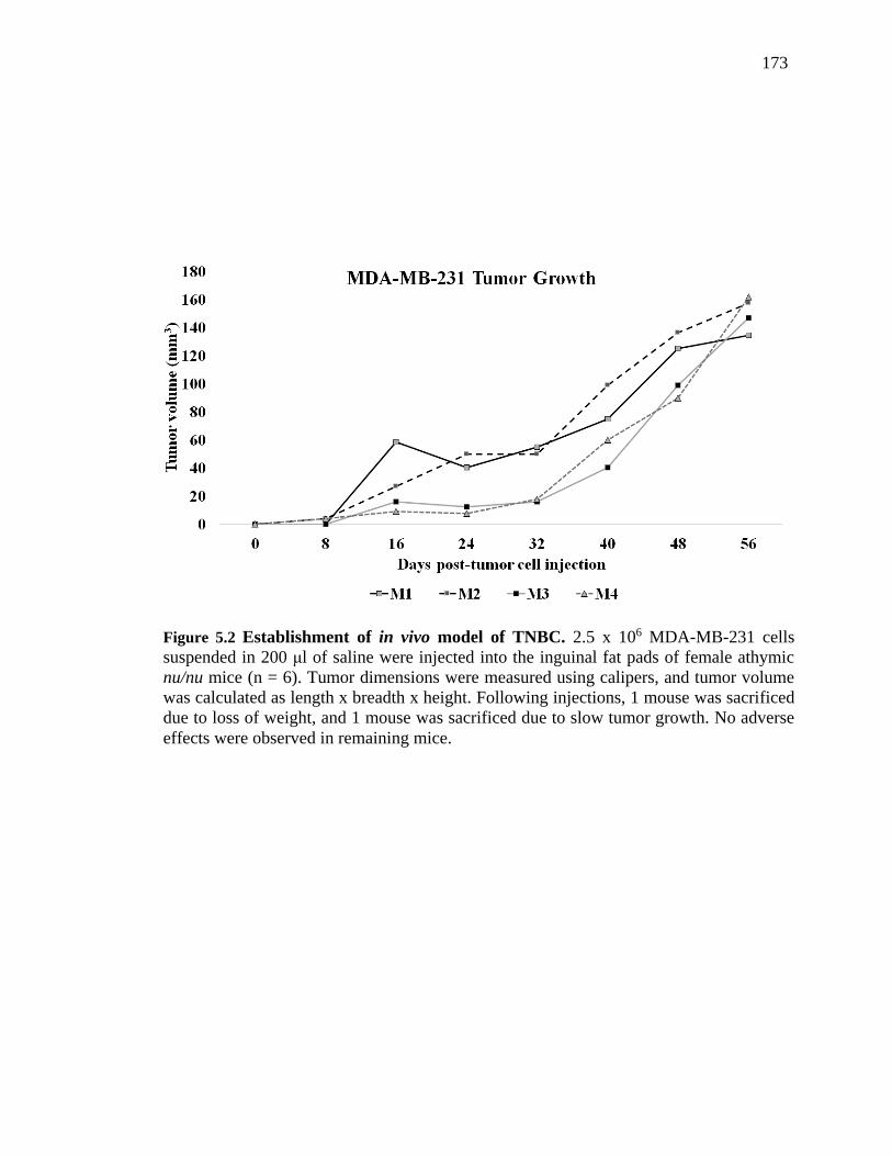
173
Figure 5.2 Establishment of in vivo model of TNBC. 2.5 x 10⁶ MDA-MB-231 cells
suspended in 200 μl of saline were injected into the inguinal fat pads of female athymic
nu/nu mice (n = 6). Tumor dimensions were measured using calipers, and tumor volume
was calculated as length x breadth x height. Following injections, 1 mouse was sacrificed
due to loss of weight, and 1 mouse was sacrificed due to slow tumor growth. No adverse
effects were observed in remaining mice.

174
5.4 Summary and Conclusions
Taken together, we have discovered that Lippia origanoides plant extracts can exert
potent and specific cytotoxicity against triple-negative breast cancer cells. We have
provided significant mechanistic insights into the extracts’ effects, including the
identification of inhibition of NF-B signaling and mitochondrial metabolism as key to
the induction of apoptosis upon treatment. Our results also confirm that the Lippia extract
L42 is non-toxic in vivo and can be further investigated for its anti-cancer effects in
future, using mouse models of TNBC. In line with this, our lab has established an MDA-
MB-231 tumor growth model in nu/nu mice, which will be used to determine the
inhibitory effects of L42 on tumor initiation and progression.
During the course of these studies, we showed for the first time that LC/MS-based
quantitative proteomics could be used to identify the complex cellular changes induced in
cancer cells by treatment with a combination of compounds. These results re-iterate that
LC/MS-based technologies are powerful tools that can be used to rapidly screen extracts
for novel compounds, investigate compounds individually or in combination for their
proteome-wide effects, and perhaps even identify bioactive compounds (BACs) and their
in vivo protein binding partners. In collaboration with the Purdue Proteomics and
Metabolomics Facilities (PPF and MPF), our lab is in the process of developing an
ambitious co-elution profiling-based scheme to simultaneously identify and quantify both
the BACs and their protein binding partners from MDA-MB-231 cell lysates incubated
with L42, with the ultimate aim of establishing this novel protocol as the gold standard
for BAC identification; thereby replacing the expensive and time-consuming bioassay-
driven fractionation methods in current use.
Finally, in addition to laying the foundation for future efforts attempting to identify
the BACs of Lippia extracts, this report provides support for studies into the anti-cancer
effects of other medicinal plants in order to better exploit the diversity of bioactive
compounds found naturally.
175
5.5 References
1. Hartwell, J.L., & Shear, M.J. (1947). Chemotherapy of cancer: Classes of compounds
under investigation, and active components of podophyllin. Cancer Research, 7, 716-
717.
2. Noble, R. L., Beer, C. T., & Cutts, J. H. (1958). Role of chance observations in
chemotherapy: Vinca rosea. Annals of the New York Academy of Sciences, 76(3),
882–894.
3. McAndrew, N., & DeMichele, A. (2018). Neoadjuvant Chemotherapy Considerations
in Triple-Negative Breast Cancer. The Journal of Targeted Therapies in Cancer, 7(1),
52-69.
4. Wall, M. E., Wani, M. C., Cook, C. E., Palmer, K. H., McPhail, A. T., & Sim, G. A.
(1966). Plant Antitumor Agents. I. The Isolation and Structure of Camptothecin, a
Novel Alkaloidal Leukemia and Tumor Inhibitor from Camptotheca acuminata.
Journal of the American Chemical Society, 88(16), 3888–3890
5. Liu, Y.-R., Jiang, Y.-Z., Xu, X.-E., Hu, X., Yu, K.-D., & Shao, Z.-M. (2016).
Comprehensive Transcriptome Profiling Reveals Multigene Signatures in Triple-
Negative Breast Cancer. Clinical Cancer Research: An Official Journal of the
American Association for Cancer Research, 22(7), 1653–1662.
6. Lehmann, B. D., Bauer, J. A., Chen, X., Sanders, M. E., Chakravarthy, A. B., Shyr,
Y., & Pietenpol, J. A. (2011). Identification of human triple-negative breast cancer
subtypes and preclinical models for selection of targeted therapies. Journal of Clinical
Investigation, 121(7), 2750–2767.
7. Nakshatri, H., Bhat-Nakshatri, P., Martin, D. A., Goulet, R. J., & Sledge, G. W. (1997).
Constitutive activation of NF-kappaB during progression of breast cancer to hormone-
independent growth. Molecular and Cellular Biology, 17(7), 3629–3639.
8. Khoshnan, A., Tindell, C., Laux, I., Bae, D., Bennett, B., & Nel, A. E. (2000). The
NF- B Cascade Is Important in Bcl-xL Expression and for the Anti-Apoptotic Effects
of the CD28 Receptor in Primary Human CD4+ Lymphocytes. The Journal of
Immunology, 165(4), 1743–1754.
176
9. Hartman, Z. C., Poage, G. M., den Hollander, P., Tsimelzon, A., Hill, J., Panupinthu,
N., et al. (2013). Growth of triple-negative breast cancer cells relies upon coordinate
autocrine expression of the proinflammatory cytokines IL-6 and IL-8. Cancer
Research, 73(11), 3470–3480.
10. Hinz, M., Krappmann, D., Eichten, A., Heder, A., Scheidereit, C., & Strauss, M.
(1999). NF-kappaB function in growth control: regulation of cyclin D1 expression and
G0/G1-to-S-phase transition. Molecular and Cellular Biology, 19(4), 2690–2698.
11. Huber, M. A., Azoitei, N., Baumann, B., Grünert, S., Sommer, A., Pehamberger, H.,
et al. (2004). NF-kappaB is essential for epithelial-mesenchymal transition and
metastasis in a model of breast cancer progression. The Journal of Clinical
Investigation, 114(4), 569–581.
12. Kagoya, Y., Yoshimi, A., Kataoka, K., Nakagawa, M., Kumano, K., Arai, S., …
Kurokawa, M. (2014). Positive feedback between NF-κB and TNF-α promotes
leukemia-initiating cell capacity. The Journal of Clinical Investigation, 124(2), 528–
542.
13. Wang, C. Y., Mayo, M. W., Korneluk, R. G., Goeddel, D. V., & Baldwin, A. S. (1998).
NF-kappaB antiapoptosis: induction of TRAF1 and TRAF2 and c-IAP1 and c-IAP2
to suppress caspase-8 activation. Science (New York, N.Y.), 281(5383), 1680–1683.
14. Yousef, E. M., Tahir, M. R., St-Pierre, Y., & Gaboury, L. A. (2014). MMP-9
expression varies according to molecular subtypes of breast cancer. BMC Cancer,
14(1), 609.
15. Kanaan, Y.M., Sampey, B.P., Beyene, D., Esnakula, A.K., Naab, T.J., Ricks-Santi,
L.J. et al. (2014). Metabolic Profile of Triple-Negative Breast Cancer in African-
American Women Reveals Potential Biomarkers of Aggressive Disease. Cancer
Genomics and Proteomics, 11, 279-294.
16. Berg, J. M., Tymoczko, J. L., & Stryer, L. (2002). Carbon Atoms of Degraded Amino
Acids Emerge as Major Metabolic Intermediates. Biochemistry. 5th Edition.
17. Kung, H.-N., Marks, J. R., & Chi, J.-T. (2011). Glutamine Synthetase Is a Genetic
Determinant of Cell Type–Specific Glutamine Independence in Breast Epithelia.
PLOS Genetics, 7(8), e1002229.

177
18. Lim, S.O., Li, C.W., Xia, W.Y., Lee, H.H., Chang, S.S., Shen, J. et al. (2016). EGFR
Signaling Enhances Aerobic Glycolysis in Triple-Negative Breast Cancer Cells to
Promote Tumor Growth and Immune Escape, Cancer Research, 76, 1284-1296.
19. Lampa, M., Arlt, H., He, T., Ospina, B., Reeves, J., Zhang, B.L. et al. (2017).
Glutaminase Is Essential for the Growth of Triple-Negative Breast Cancer Cells with
a Deregulated Glutamine Metabolism Pathway and Its Suppression Synergizes with
Mtor Inhibition. PLoS One, 12, e0185092.
20. Goode, G., Gunda, V., Chaika, N.V., Purohit, V., Yu, F., Singh, P.K. (2017). Muc1
Facilitates Metabolomic Reprogramming in Triple-Negative Breast Cancer. PLoS
One, 12, e0176820.
21. Van Geldermalsen, M., Wang, Q., Nagarajah, R., Marshall, A.D., Thoeng, A., Gao,
D. et al. (2016). ASCT2/SLC1A5 Controls Glutamine Uptake and Tumor Growth in
Triple-Negative Basal-Like Breast Cancer. Oncogene, 35, 3201-3208.
22. Castellanos, N.R., Castro, C.E., Stashenko, E.E., & Fuentes, J. Lippia origanoides
supercritical fluid extract and its major constituent pinocembrin diminish ultraviolet
radiation-induced DNA damage and modulate cell division in Escherichia coli. (In
review)
23. Ly, J.D., Grubb, D.R., Lawen, A. The Mitochondrial Membrane Potential (Delta Psi
M) in Apoptosis, an Update. Apoptosis. 2003, 8, 115-

178
VITA
VISHAK RAMAN (812) 391-4038 [email protected]
SUMMARY
Highly-motivated Cell and Molecular Biologist possessing strong presentation and communication
skills, with a core interest in disease marker identification, immunotherapy, and drug discovery.
RESEARCH EXPERIENCE
Purdue University, Department of Biological Sciences Indiana, USA
Ph. D Student, Camarillo Lab, Purdue Center for Cancer Research (PCCR) 2013 – Present
Identified the targeted apoptotic effects of a Lippia origanoides extract (L42) on triple-negative
breast cancer (TNBC) using plate-based viability assays
Demonstrated L42 induced cell cycle arrest in TNBC cells using flow cytometry and
fluorescence microscopy
Utilized label-free quantitative proteomics to show L42 induced metabolic arrest in TNBC cells
by inhibiting the TCA cycle and OXPHOS
Mastered several data analysis and visualization tools (InfernoRDN, Cytoscape, DAVID 6.8,
GOrilla) to process large datasets from proteomics experiments
SRM University, Department of Biotechnology Chennai, India
Undergraduate Research Intern, Integrative Medicine Lab 2011 – 2012
Received extensive F344 rat-handling experience including basic maintenance, oral gavage
treatment, anesthetic injection, sacrifice, and lymphatic tissue isolation
Studied the immuno-modulatory anti-ageing effects of Morinda citrifolia extracts on the
immune system of old F344 male rats
Performed plate-based bioassays (ELISA and enzyme activity assays) to measure cytokine
levels and antioxidant enzyme activity in draining lymph nodes during ageing
PRESENTATIONS AND PUBLICATIONS
Talks:
Vishak Raman. “A Lippia origanoides extract induces apoptosis and suppresses NF-κB
signalling in MDA-MB-231 triple-negative breast cancer cells.” Graduate Student Retreat,
Burton D. Morgan Center, West Lafayette, IN. (2016) [1st Place Winner]
Posters:
Vishak Raman. “Proteomic analysis reveals that an extract of the plant Lippia origanoides
suppresses mitochondrial metabolism in triple-negative breast cancer cells.” Eight
International Breast Cancer Prevention Symposium, Dauch Alumni Center, Purdue
University, West Lafayette, IN, 2018.
Vishak Raman. “Proteomic analysis reveals L. origanoides extract targets mitochondrial
metabolism and function to induce rapid apoptosis in triple-negative breast cancer cells.”
Amelia Project, Alumni Hall, Indiana University, Kokomo, IN. (July 14, 2018)
Vishak Raman. “A novel mass spectrometry-based approach to discover anti-cancer
bioactive compounds and their protein binding partners.” Dept. of Biol. Sci. Retreat,
Fourwinds Lakeside Inn, Bloomington, IN. (2017)

179
Vishak Raman. “A Lippia origanoides extract induces cell cycle arrest and apoptosis and
suppresses NF-κBsignaling in MDA-MB-231 triple-negative breast cancer cells.” Purdue
Sigma Xi Chapter Poster Competition, Stewart Center, Purdue University, IN. (2017)
Publications:
Raman V, Aryal UK, Hedrick V, Fuentes Lorenzo JL, Stashenko EE, Levy M, Levy MM,
Mohallem R, and Camarillo IG. “Proteomic analysis reveals an extract of the plant Lippia
origanoides suppresses mitochondrial metabolism in triple-negative breast cancer cells.” J
Proteome Res. 2018 [Epub ahead of print]
Raman V, Fuentes Lorenzo JL, Stashenko EE, Levy M, Levy MM, Camarillo IG. “Lippia
origanoides extract induces cell cycle arrest and apoptosis and suppresses NF-κB signaling in
triple-negative breast cancer cells.” Int J Oncol. 51(6): 1801-1808, 2017.
Pratap UP, Priyanka HP, Ramanathan KR, Raman V, Hima L, Thyagarajan S. “Noni
(Morinda citrifolia L.) fruit juice delays immunosenescence in lymph nodes of old F344 male
rats. J Integr Med. 16(3):199-207, 2018.
Mittal L, Raman V, Camarillo IG, and Sundararajan R. “Ultra-microsecond pulsed curcumin
for effective treatment of triple negative breast cancers.” Biochem Biophys Res Commun.
2017 Sep 30;491(4):1015-1020 2.
EDUCATION
Purdue University Indiana, USA
Ph. D, Biological Sciences 2013 – Present
Characterizing the apoptotic effects of L. origanoides extract on triple-negative breast cancer using
quantitative proteomics and metabolomics
SRM University Chennai, India
B. Tech, Biotechnology 2009 – 2013
Modulation of cytokine production and antioxidant enzyme activities in draining lymph nodes of old
F344 male rats treated with M. citrifolia extract.
SCIENTIFIC LEADERSHIP EXPERIENCE
Purdue Center for Cancer Research Indiana, USA
Sr. Research Mentor, Camarillo Lab 2016 – 2019
Established standard laboratory protocols for several cell and molecular biology techniques
including:
Quantitative Real-time PCR (qPCR)
Western Blotting
Immunofluorescence staining
Sample preparation for LC-MS/MS
Spear-headed new collaborations with Purdue Proteomics Facility and Flow Cytometry and
Cell Sorting Facility
Lead directly to 2 first-author manuscripts
Mentored 5 non-thesis,1 thesis-undergraduate student, and 3 Ph. D students
Designed and directed individual and group research projects for all students mentioned above
Dept. of Biological Sciences, Purdue University Indiana, USA
Teaching Assistant Sep. 2013 – Present
Lectured for over 400 students in the Anatomy and Physiology Lab (BIOL 203/204) over 4
years
Oversaw lab experiments, graded quizzes and worksheets, and held office hours for students
every week
Frequently substituted for senior instructors for recitation lectures
Dept. of Biotechnology, SRM University Chennai, India

180
Conference Host, NCFB 2012
Selected to host the Animal Biotechnology Conference as part of the National Conference on
Frontiers in Biotechnology (NCFB) 2012
AWARDS
Purdue University, Department of Biological Sciences Indiana, USA
Graduate School Summer Research Grant 2016
Best Presentation at Annual Graduate Student Retreat, Burton D.Morgan Center, 2016
SRM University, Department of Biotechnology Chennai, India
50% tuition-waiver scholarship for academic year 2010-2011
50% tuition-waiver scholarship for academic year 2009-2010
RELEVANT TECHNICAL SKILLS
Cell and Molecular Biology Techniques:
Mammalian Cell Culture
Western Blotting
qRT-PCR
ELISA Differential-staining analysis via Flow Cytometry
Sample preparation for quantitative proteomics via LC-MS/MS
Software and Omics Tools:
ImageJ (Densitometry for Western Blotting/IF Imaging)
InfernoRDN (Proteomic data analysis and visualization)
STRING (Protein interaction analysis)
Cytoscape (Pathway visualization)
DAVID 6.8 (Gene enrichment analysis)
GOrilla (Gene Ontology Enrichment Analysis and Visualization tool)