inhibitionoferk1/2andactivationofliverxreceptor ... ·...
TRANSCRIPT
Inhibition of ERK1/2 and Activation of Liver X ReceptorSynergistically Induce Macrophage ABCA1 Expression andCholesterol Efflux*
Received for publication, October 6, 2009, and in revised form, December 21, 2009 Published, JBC Papers in Press, December 25, 2009, DOI 10.1074/jbc.M109.073601
Xiaoye Zhou‡§¶, Zhinan Yin‡, Xianzhi Guo�, David P. Hajjar¶1, and Jihong Han‡¶**1,2
From the Colleges of ‡Life Sciences and �Chemistry and the **Key Lab of Bioactive Materials of the Ministry of Education of China,Nankai University, Tianjin 300071, China, the §Department of Neurobiology, Xuanwu Hospital, Capital Medical University,Beijing 100053, China, and the ¶Center of Vascular Biology and Department of Pathology, Weill Medical College of CornellUniversity, New York, New York 10065
ATP-binding cassette transporter A1 (ABCA1), a moleculemediating free cholesterol efflux from peripheral tissues toapoAI and high density lipoprotein (HDL), inhibits the forma-tion of lipid-ladenmacrophage/foam cells and the developmentof atherosclerosis. ERK1/2 are important signaling moleculesregulating cellular growth and differentiation. The ERK1/2signaling pathway is implicated in cardiac development andhypertrophy. However, the role of ERK1/2 in the developmentof atherosclerosis, particularly in macrophage cholesterolhomeostasis, is unknown. In this study, we investigated theeffects of ERK1/2 activity on macrophage ABCA1 expressionand cholesterol efflux. Compared with a minor effect by inhibi-tion of other kinases, inhibition of ERK1/2 significantlyincreased macrophage cholesterol efflux to apoAI and HDL. Incontrast, activation of ERK1/2 reducedmacrophage cholesterolefflux and ABCA1 expression. The increased cholesterol effluxby ERK1/2 inhibitors was associated with the increased ABCA1levels and the binding of apoAI to cells. The increased ABCA1by ERK1/2 inhibitors was due to increased ABCA1 mRNA andprotein stability. The induction of ABCA1 expression and cho-lesterol efflux by ERK1/2 inhibitors was concentration-depen-dent. The mechanism study indicated that activation of liver Xreceptor (LXR) had little effect on ERK1/2 expression and acti-vation. ERK1/2 inhibitors had no effect onmacrophage LXR�/�expression, whereas they did not influence the activation or theinhibition of the ABCA1 promoter by LXR or sterol regulatoryelement-binding protein (SREBP). However, inhibition ofERK1/2 and activation of LXR synergistically induced macro-phage cholesterol efflux and ABCA1 expression. Our data sug-gest that ERK1/2 activity can play an important role in macro-phage cholesterol trafficking.
Development of atherosclerotic lesions in coronary arteriesis an underlying cause of coronary heart disease. Lipid-ladenmacrophage/foam cells are a prominent part of atheroscleroticlesions (1). Cellular cholesterol content in macrophages isdetermined by uptake and efflux of cholesterol (2). The type Ascavenger receptor and type B scavenger receptor (CD36)mediate the binding and internalization ofmodified lowdensitylipoprotein (LDL),3 thus, demonstrating pro-atherogenic prop-erties (3, 4). In contrast, type BI scavenger receptor and ATP-binding cassette transporter A1 (ABCA1) can mediate cellularfree cholesterol efflux to extracellular high density lipoprotein(HDL) or lipid-free apolipoprotein AI (apoAI) thereby inhibit-ing the development of atherosclerosis (5, 6). Compared withbi-directional cholesterol transport across cellular membranesthat are mediated by type BI scavenger receptor (7, 8), ABCA1stimulates free cholesterol efflux from macrophages and otherperipheral cell types to apoAI and/or HDL by using the energyfrom ATP hydrolysis (9). The binding of free cholesterol andphospholipids to apoAI also leads to the generation of nascentHDL (10). ABCA1 can also mediate cholesterol efflux to exog-enous apolipoprotein E (apoE) (11). Although the removal ofmacrophage cholesterol by endogenous apoE is an ABCA1-in-dependent process (12), ABCA1 activity can contribute to basalconstitutive secretion of apoE from macrophages (13).Anti-atherogenic properties of ABCA1 have been well inves-
tigated in both humans and animal models. In humans, muta-tions in ABCA1 expression cause Tangier disease, which ischaracterized by very low levels of serum HDL cholesterol,rapid catabolism of apoAI, severe cholesteryl ester accumula-tion in peripheral tissues, and a high risk of development ofcoronary heart disease (14–16). In animal models, overexpres-sion of human ABCA1 reduces total cholesterol levels and ath-erosclerosis, whereas selective suppression of macrophageABCA1 increases atherosclerosis without affecting total cho-lesterol levels (1, 6, 17, 18).
* This work was supported, in whole or in part, by National Institutes of HealthGrant P01HL046403 (to D. P. H. and J. H.). This work was also supported bythe Julia and Gross Foundation (to D. P. H.), a 973 Basic Research Programof China Grant 2010CB945003 (to J. H.), Natural Science Foundation ofChina (NSFC) Grant 30971271 (to J. H.), the Tianjin Municipal Science andTechnology Commission of China (Grant 08ZCKFSH04400 to J. H.), the Min-istry of Science and Technology of China (Grant 2006AA02A408 to X. Z.),and the National Key Scientific Program of China (Grant 2007CB914801 toZ. Y.).
1 Both authors contributed equally to this work.2 To whom correspondence should be addressed: Center of Vascular Biol-
ogy, Weill Medical College of Cornell University, 1300 York Ave., NewYork, NY 10065. Tel.: 212-746-6900; Fax: 212-746-8789; E-mail:[email protected].
3 The abbreviations used are: LDL, low density lipoprotein; ABCA1, ATP-bind-ing cassette transporter A1; HDL, high density lipoprotein; apoAI, apoli-poprotein AI; apoE, apolipoprotein E; LXR, liver X receptor; SREBP, sterolregulatory element-binding protein; LXRE, LXR response element; ERK1/2,extracellular signal-regulated kinases 1 and 2; MAPK, mitogen-activatedprotein kinase; MEK, MAPK/ERK kinase; EGF, epidermal growth factor; PBS,phosphate-buffered saline; FACS, fluorescence-activated cell sorting;siRNA, small interference RNA; PKA, protein kinase A; JNK, c-Jun N-terminalkinase.
THE JOURNAL OF BIOLOGICAL CHEMISTRY VOL. 285, NO. 9, pp. 6316 –6326, February 26, 2010© 2010 by The American Society for Biochemistry and Molecular Biology, Inc. Printed in the U.S.A.
6316 JOURNAL OF BIOLOGICAL CHEMISTRY VOLUME 285 • NUMBER 9 • FEBRUARY 26, 2010
by guest on June 22, 2018http://w
ww
.jbc.org/D
ownloaded from
Expression ofABCA1 can be up-regulated by liverX receptor(LXR) and down-regulated by sterol regulatory element-bind-ing proteins (SREBPs) 1,2 (19, 20). After ligand binding, LXRforms a heterodimer with another nuclear protein, retinoid Xreceptor. The heterodimer of LXR/retinoid X receptor binds tothe LXR response element (LXRE) in the proximal region ofABCA1 gene promoter and induces ABCA1 transcription.Thus, synthetic LXR ligands can inhibit the development ofatherosclerosis in animal models (21–23). In contrast, SREBP2binds to the E-box in ABCA1 promoter to reduce ABCA1expression (24). In macrophages, SREBP1 suppresses ABCA1expression in an E-box-independent manner (19). Most of theoxysterols can suppress SREBPs. Therefore, they increaseABCA1 expression. Interestingly, some oxysterols, such as22(R)-hydroxycholesterol, function as LXR ligands and SREBPsuppressors simultaneously. The simultaneous activation ofLXR and inactivation of SREBP by this type of oxysterols syn-ergistically induce ABCA1 expression. In addition to transcrip-tion factors, the cellular ABCA1 levels are also regulated bypost-translational mechanisms. ABCA1 is a molecule with ahalf-life of�1–2 h. Thus, the decreasedABCA1 degradation byapoAI results in increasedABCA1 levels, whereas the enhancedABCA1 degradation by unsaturated fatty acids decreasesABCA1 levels (25, 26).Extracellular signal regulated kinases 1 and 2 (ERK1/2) or
p44/42 mitogen-activated protein kinases (p44/42 MAPK)belong to a highly conserved family of Ser-Thr protein kinasesand have been were characterized to function through the Ras-Raf-MEK-ERK1/2 cascade (27). ERK1/2 are implicated in widecellular processes, such as in embryogenesis, differentiation,proliferation, and cell death (28). ERK1/2 are ubiquitouslyexpressed in all tissues/cell types and are strongly activated bymultiple stimuli, including growth factors, such as epithelialgrowth factor (EGF) (28). In fact, overexpression or constitutiveactivation of ERK1/2 pathway can lead to progression of severalcancers. Inhibitors of ERK1/2 have been investigated as poten-tial therapeutic targets for cancer treatment (27). ERK1/2 havealso been demonstrated to play a role in several aspects of car-diac system, such as cardiac development, hypertrophy, andprotection (29). However, it is unclear if ERK1/2 play an impor-tant role inmacrophage cholesterol metabolism and traffickingas well as the development of atherosclerosis. In this study, weinvestigated the effects of ERK1/2 activity on macrophage freecholesterol efflux and ABCA1 expression. We found thatinhibition of ERK1/2 greatly increasedmacrophage free choles-terol efflux to apoAI andHDL. This increased cholesterol effluxwas associated with ABCA1 expression. Further, the increasedmacrophage ABCA1 expression occurred by enhancingABCA1 stability at both mRNA and protein levels. AlthoughERK1/2 inhibitor-induced ABCA1 expression was indepen-dent of LXR activation, ERK1/2 inhibitor, and LXR ligand syner-gistically induced macrophage cholesterol efflux and ABCA1expression. Our studies reported herein describe a new func-tion for ERK1/2 in cholesterol trafficking processes.
EXPERIMENTAL PROCEDURES
Reagents—Inhibitors for different kinases were purchasedfrom CalBiochemistry (San Diego, CA). All other chemicals
were purchased from Sigma-Aldrich except as indicated. Rab-bit anti-ABCA1 polyclonal antibody was obtained from NovusBiologicals (Littleton, CO). Rabbit anti-total ERK1/2 and phos-pho-ERK1/2 polyclonal antibodies were purchased from CellSignaling Technology (Beverly, MA). Rabbit anti-LXR� andLXR� polyclonal antibodies were purchased from Santa CruzBiotechnology, Inc. (Santa Cruz, CA). Acetylated low densitylipoprotein, HDL, and apoAI were prepared as described (24,30). The SDS-PAGE analysis indicated that the prepared HDLdid not contain apoE.Cells—RAW cells, a murine macrophage cell line, were pur-
chased from ATCC (Rockville, MD) and cultured in completeRPMI medium containing 10% fetal calf serum, 50 �g/ml ofpenicillin/streptomycin, and 2mM glutamine. At�90% conflu-ence, cells were switched to serum-free medium and receivedtreatment.To collect peritoneal macrophages, C57 wild-type mice
(Jackson Laboratory, Bar Harbor, ME) were injected with 3 mlof 4% thioglycolate and maintained with access to water andnormal chow for 5 days. Peritonealmacrophageswere collectedfrom the mouse abdomen by lavage with PBS. Cells were cul-tured in complete RPMI medium for 3 h, and all floating cellswere removed. Adhesive cells were continued to be culturedwith complete RPMI medium for additional 2 days and thentreated as indicated.Determination of Free Cholesterol Efflux from Macrophages—
Macrophages in 12-well plates were labeled in macrophageserum-free medium (Invitrogen, 1.5 ml/well) containing 50�g/ml acetylated lowdensity lipoprotein (used as the carrier forfree cholesterol labeling) and 150 nCi/ml [3H]cholesterol for24 h. After treatment, cells were washed twice with PBS andincubated for 1 h in serum-free medium, then switched toserum-free medium or medium containing purified apoAI (10�g/ml) or HDL (15 �g/well). After 5-h incubation at 37 °C,medium from each well was collected for determination ofradioactivity in supernatants. The remaining cells were lysed byaddition of 0.2 N NaOH, and the lysate was determined forprotein content which was used to normalize cholesterol efflux(dpm/�g of protein).Northern Blot and Real-time Reverse Transcription-PCR
Analysis of ABCA1 mRNA—Total cellular RNA was extractedfromcells in 60-mmdishes and used to determine expression ofABCA1 mRNA by Northern blot and quantitative real-timereverse transcription-PCR. The Northern blot was performedas described previously (31), and the probe for mouse ABCA1mRNA was generated by reverse transcription-PCR with thefollowing primers: forward, 5�-TGGACATCCTGAAGCCAG-3�, and backward, 5�-TTCTTCCCACATGCCCT-3�.To quantitative analyze ABCA1 transcript, 1�g of total RNA
was used to synthesize the first strand DNA with oligo(dT)18.The real-timePCRwas performedbyusing SYBRGreenMasterMix (Applied Biosystems, Foster City, CA) and the followingprimers. Abca1: forward, 5�-CTCAGTTAAGGCTGCTG-CTG-3�; backward, 5�-TCAGGCGTACAGAGATCAGG-3�;Gapdh: forward, 5�-ACAACTTTGGCATTGTGGAA-3�;backward, 5�-GATGCAGGGATGATGTTCTG-3�. The quan-titative results forABCA1were normalized by the levels of glyc-eraldehyde-3-phosphate dehydrogenase mRNA.
Regulation of ABCA1 Expression by ERK1/2
FEBRUARY 26, 2010 • VOLUME 285 • NUMBER 9 JOURNAL OF BIOLOGICAL CHEMISTRY 6317
by guest on June 22, 2018http://w
ww
.jbc.org/D
ownloaded from
Western Blot Analysis of ABCA1, LXR�, LXR�, andTotal andPhospho-ERK1/2—Whole cellular proteins were extracted asfollows: after treatment cells were washed twice with cold PBS,then scraped and lysed in ice-cold lysis buffer (50 mM Tris, pH7.5, 150mMNaCl, 1%TritonX-100, 1% sodiumdeoxycholate, 1mM phenylmethylsulfonyl fluoride, 50 mM sodium fluoride, 1mM sodium orthovanadate, 50 �g/ml aprotinin/leupeptin).Lysatewas sonicated for 20 cycles, thenmicrocentrifuged for 15min at 4 °C. The supernatant was transferred to a new test tubeand stored at �20 °C.Nuclear proteinswere extracted as described (32)withminor
modifications. Briefly, cells were firstly suspended in coldbuffer A (20mMHepes, pH 7.9, 10mMNaCl, 3mMMgCl2, 0.1%Nonidet P-40, 10% glycerol, 0.2mMEDTA, 50�g/ml aprotinin/leupeptin) and incubated on ice for 20 min followed by centri-fugation for 5 min with a Microfuge (Beckman) at 3,000 rpmand 4 °C. The resultant pellet of nuclei was washed once withbuffer A and then buffer B (20 mMHepes, pH 7.9, 20% glycerol,0.2 mM EDTA, 50 �g/ml aprotinin/leupeptin). The nucleic pel-let was re-suspended in buffer C (20 mM Hepes, pH 7.9, 0.4 M
NaCl, 20% glycerol, 0.2 mM EDTA, 50 �g/ml aprotinin/leupep-tin) and incubated on ice for 1 h with vortex several times. Thesuspension was centrifuged for 30 min at 14,000 rpm and 4 °C.The supernatant was collected and stored at �20 °C.After the content was determined by Lowry method,
whole cellular or nuclear proteins were loaded and separatedon a 7% (for determination of ABCA1) or 12% (for determi-nation of the rest proteins) SDS-PAGE and then transferredonto nylon enhanced nitrocellulose membrane. The mem-brane was blocked with a solution of 0.1% Tween 20/PBS(PBS-T) containing 5% fat-free milk for 1 h, then incubatedwith primary antibody for 2 h at room temperature or over-night at 4 °C followed by washing for 3 � 10 min with PBS-Tbuffer. The blot was re-blocked with PBS-T containing 5%milk followed by incubation with horseradish peroxidase-conjugated goat anti-rabbit IgG for 1 h at room temperature.After washing 3 � 10 min with PBS-T, the membrane wasincubated for 1 min in a mixture of equal volumes of West-ern blot chemiluminescence reagents 1 and 2 and thenexposed to film before development.FACS Assay of Macrophage Surface ABCA1 Protein and the
Binding of ApoAI to Cells—After treatment, macrophages werescraped and washed twice with PBS containing 1% fetal calfserum. Approximately 1 � 106 cells from each sample wereblocked for 30 min at room temperature with PBS containing5% goat serum. After washing with PBS, cells were incubatedwith rabbit anti-ABCA1 antibody (1:100) for 1 h at room tem-perature. Cells were then incubated with goat anti-rabbit fluo-rescein isothiocyanate-conjugated IgG (1:50) for 45 min atroom temperature. After washing with PBS, cells were sub-jected to flow cytometric evaluation.To determine the binding of apoAI tomacrophages, purified
apoAI was fluorescein-conjugated with a reactive succinimidyl
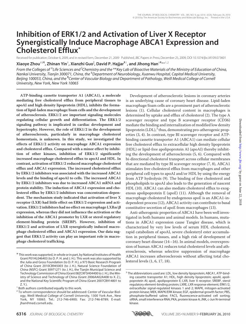
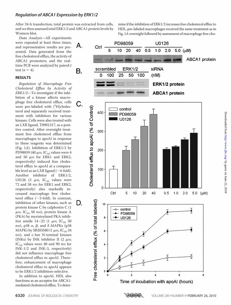
FIGURE 1. Regulation of macrophage free cholesterol efflux by ERK1/2activity. A and B, inhibition of ERK1/2 increases macrophage free cholesterolefflux to apoAI and HDL, respectively. RAW macrophages in 12-well plateswere pre-labeled as described under “Experimental Procedures” and thenreceived treatment overnight. Free cholesterol efflux was performed by incu-bating the treated cells in serum-free medium containing apoAI (10 �g/ml)(A) or HDL (15 �g/ml) (B) for 5 h at 37 °C. Radioactivity in medium was deter-mined and normalized by cellular protein content. Treatments were as fol-lows: ERK1/2 inhibitors, PD98059 (PD, 40 �M) and U0126 (2 �M); protein kinaseC inhibitor, calphostin C (Cal, 1 �M); PKA inhibitor, myristoylated PKA inhibitoramide 14 –22 (PKAI, 1 �M); p38 MAPK inhibitor, SB203580 (SB, 2 �M), and JNKsinhibitor, JNK inhibitor II (JNKI-2, 2 �M). LXR ligand, T0901317 (T0, 200 nM),was used as a positive control. # and *, significantly different from the corre-sponding controls at p � 0.05 by Student’s t test (n � 4). C, EGF decreases
macrophage free cholesterol efflux to apoAI. Pre-labeled RAW cells weretreated with EGF at the indicated concentrations overnight followed bydetermination of free cholesterol efflux to apoAI (10 �g/ml). *, significantlydifferent from control at p � 0.05 by Student’s t test (n � 4).
Regulation of ABCA1 Expression by ERK1/2
6318 JOURNAL OF BIOLOGICAL CHEMISTRY VOLUME 285 • NUMBER 9 • FEBRUARY 26, 2010
by guest on June 22, 2018http://w
ww
.jbc.org/D
ownloaded from
ester of carboxyl fluorescein by using a labeling kit from Prin-ceton Separations (Adelphia, NJ). After lift, cells were washedtwice with PBS and then incubated with 10 �g/ml of labeledapoAI in serum-free medium for 2 h at 37 °C. After washedtwice with PBS, cells were used to determine the binding ofapoAI by FACS.
Preparation of Plasmid DNA andDetermination of ABCA1 PromoterActivity—cDNA encoding nuclearform of mouse SREBP1a (N-termi-nal 1–460 amino acids) was gener-ated by PCR using the clone pur-chased from Invitrogen (cDNAclone MGC:66503 IMAGE:6824948)as template DNA and followingprimers: forward, 5�-ACTCAGAT-CTCGATGGACGAGCTGGCCT-TCG-3�; backward, 5�-CCGCGGT-ACCCTAGACCTGGCTATCCT-CAAAGG-3�. After sequence wasconfirmed, the PCR product wasdigested with BglII and KpnI fol-lowed by subcloning into anexpression vector, pEGFP-C2(pEGFP-nSREBP1a).Mouse ABCA1 promoters were
constructed by PCR with mousegenomic DNA and following prim-ers. For ABCA1 promoter A (from�179 to �227): forward, 5�-TAG-CCTCGAGGTCGCCGGTTTAA-GGGGCG-3�; backward, 5�-TGC-CAAGCTTCCTCTTACCTGTT-TTCCACTTTG-3�; for ABCA1promoter B (from �113 to �227):forward, 5�-TAGCCTCGAGCA-GAGGCCGGGAACGGGGCG-3�,and backward was same as in pro-moter A. After sequence was con-firmed, the PCR product wasdigested by XhoI and HindIII fol-lowed by ligation with pGL4 luci-ferase reporter vector (Promega,Madison, WI), transformed, andamplified. To analyze ABCA1 pro-moter activity, �95% confluent 293cells in 24-well plates were trans-fected with DNA for ABCA1 pro-moter, nSREBP1a and Renilla (forinternal normalization) by usingLipofectamineTM 2000 (Invitro-gen). After 24 h of transfection plustreatment, cells were lysed, and cel-lular lysate was used to determinethe activity of firefly and Renillaluciferases by using the Dual-Lucif-erase� Reporter Assay System fromPromega.
Transfection of siRNA—The siRNA against mouse ERK1and ERK2, and the scrambled siRNA were purchased fromSanta Cruz Biotechnology. RAW cells (�80% confluence) in a6-well platewere transfectedwith siRNAof ERK1 andERK2 (anequal amount of each was mixed), and scrambled siRNA usingTransIT-TKO transfection kit from Mirusbio (Madison, WI).
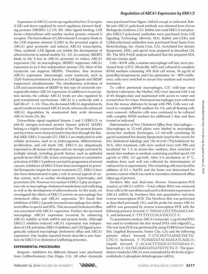
FIGURE 2. Inhibition of ERK1/2 induces macrophage ABCA1 expression and the binding of apoAI to cells.A, RAW macrophages in serum-free medium were treated with inhibitors for various kinases as indicatedovernight. LXR ligand, T0901317, was used as a positive control. Total cellular RNA was extracted and used todetermine expression of ABCA1 mRNA by quantitative real-time PCR as described under “Experimental Proce-dures.” *, significantly different from control at p � 0.05 by Student t test (n � 4). B, RAW cells in serum-freemedium were treated with inhibitors for various kinases (40 �M PD98059 (PD), 1 �M U0126 (U0), 1 �M calphostinC (Cal), 1 �M PKA inhibitor (PKAI), 2 �M SB203580 (SB), and 2 �M JNK inhibitor II (JNKI-2)) overnight. After washingwith PBS, surface ABCA1 protein levels were determined for cells by FACS assay as described under “Experi-mental Procedures.” Blue line, control cells. C, macrophages received same treatments as in Fig. 2B. The bindingof apoAI to treated cells was completed as described under “Experimental Procedures” and assessed by FACS assay.Blue line, control cells. D, RAW macrophages were treated with EGF at the indicated concentrations overnight.Expression of ABCA1 protein was determined by Western blot as described under “Experimental Procedures.”
Regulation of ABCA1 Expression by ERK1/2
FEBRUARY 26, 2010 • VOLUME 285 • NUMBER 9 JOURNAL OF BIOLOGICAL CHEMISTRY 6319
by guest on June 22, 2018http://w
ww
.jbc.org/D
ownloaded from
After 24-h transfection, total protein was extracted from cells,andwe then assessed total ERK1/2 andABCA1protein levels byWestern blot.Data Analysis—All experiments
were repeated at least three times,and representative results are pre-sented. Data generated from thefree cholesterol efflux, the activity ofABCA1 promoters, and the real-time PCR were analyzed by paired ttest (n � 4).
RESULTS
Regulation of Macrophage FreeCholesterol Efflux by Activity ofERK1/2—To investigate if the inhi-bition of a kinase affects macro-phage free cholesterol efflux, cellswere pre-labeled with [3H]choles-terol and separately received treat-ment with inhibitors for variouskinases. Cells were also treated withan LXR ligand, T0901317, as a posi-tive control. After overnight treat-ment free cholesterol efflux frommacrophages to apoAI in responseto these reagents was determined(Fig. 1A). Inhibition of ERK1/2 byPD98059 (40 �M, IC50 values were 4and 50 �M for ERK1 and ERK2,respectively) induced free choles-terol efflux to apoAI at a compara-ble level as an LXR ligand (�4-fold).Another inhibitor of ERK1/2,U0126 (2 �M, IC50 values were72 and 58 nM for ERK1 and ERK2,respectively) also markedly in-creased macrophage free choles-terol efflux (�3-fold). In contrast,inhibition of other kinases, such asprotein kinase C by calphostin C (1�M, IC50 50 nM), protein kinase A(PKA) by myristoylated PKA inhib-itor amide 14–22 (1 �M, IC50 50nM), p38 �, �, and � MAPKs (p38MAPK) by SB203580 (1 �M, IC50 34nM), and c-Jun N-teminal kinases(JNKs) by JNK inhibitor II (2 �M;IC50 values were 40 and 90 nM forJNK-1/2 and JNK-3, respectively)did not influence macrophage freecholesterol efflux to apoAI. There-fore, enhancement of macrophagecholesterol efflux to apoAI appearsto be ERK1/2 inhibition-selective.In addition to apoAI, HDL also
functions as an acceptor for ABCA1-mediated cholesterol efflux.Todeter-
mine if the inhibitionofERK1/2 increases free cholesterol efflux toHDL, pre-labeledmacrophages received the same treatment as inFig. 1Aovernight followedby assessment ofmacrophage free cho-
Regulation of ABCA1 Expression by ERK1/2
6320 JOURNAL OF BIOLOGICAL CHEMISTRY VOLUME 285 • NUMBER 9 • FEBRUARY 26, 2010
by guest on June 22, 2018http://w
ww
.jbc.org/D
ownloaded from
lesterol efflux to HDL. The similar observations obtained forapoAI showed that inhibition of ERK1/2, but not of other kinases,increasedmacrophage cholesterol efflux to HDL (Fig. 1B).To further determine the effects of ERK1/2 activity on mac-
rophage cholesterol efflux, pre-labeled cells were treated withEGF to increase ERK1/2 activity followed by assessment of cho-lesterol efflux to apoAI (Fig. 1C). In contrast to the inhibition,the activation of ERK1/2 by EGF significantly suppressed themacrophage free cholesterol efflux. Taken together, results inthese experiments (Fig. 1, A–C) suggest that ERK1/2 activitycan affect macrophage cholesterol efflux processes.Inhibition of ERK1/2 Increased Expression of Macrophage
ABCA1and the Binding of ApoAI toCells—ABCA1 is an impor-tant molecule regulating cellular free cholesterol efflux toapoAI and HDL. To determine if ERK1/2 inhibitor-inducedmacrophage free cholesterol efflux is due to increased ABCA1levels, cells were treated with different kinase inhibitors.Changes in ABCA1 mRNA in response to these reagents wereassessed by quantitative real-time reverse transcription-PCR.Consistent with the effects on cholesterol efflux, ERK1/2 inhib-itors (PD98059 and U0126) increased ABCA1mRNA levels. Incontrast, inhibition of PKA, protein kinase C, p38 MAPK, andJNKs had little effect on ABCA1 mRNA levels (Fig. 2A).To study if the increased macrophage ABCA1 mRNA levels
by ERK1/2 inhibitors can lead to an increase in cell surfaceABCA1 protein, after treatment cells were determined for sur-faceABCA1protein levels by FACS (Fig. 2B). Clearly, inhibitionof ERK1/2 by PD98059 andU0126 elevated cell surface ABCA1protein levels. However, inhibition of other kinases had noeffect on cell surface ABCA1 protein levels. Associatedwith thechanges in cell surface ABCA1 protein, the binding of apoAI tocells, which is the initial step in ABCA1-mediated free choles-terol efflux, was also selectively increased by ERK1/2 inhibitors,but not by inhibitors for other kinases (Fig. 2C). In contrast toinhibition of ERK1/2, activation of ERK1/2 by EGF reducedmacrophage ABCA1 protein levels in a concentration-depen-dent manner suggesting the reduced macrophage cholesterolefflux by EGF is, at least partially, attributed to the EGF-inhib-ited ABCA1 expression (Fig. 2D).To study the physiological relevance of ERK1/2 inhibitors on
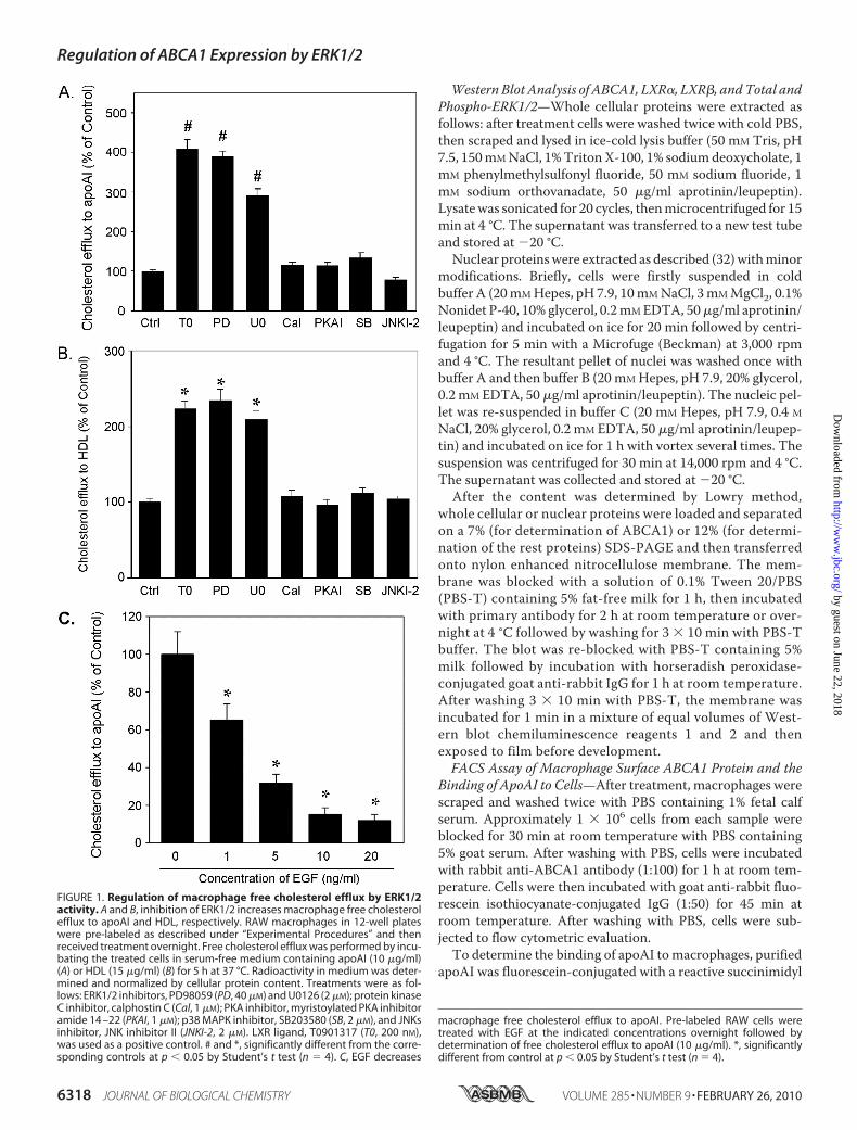
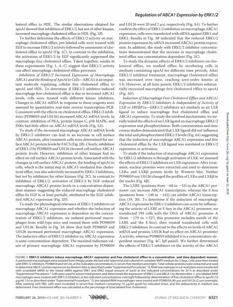
macrophage ABCA1 expression and whether the induction ofmacrophage ABCA1 expression is dependent on the concen-tration of ERK1/2 inhibitors, we isolated peritoneal macro-phages from wild-type mice and treated them with PD98059and U0126. Results in Fig. 3A show that both PD98059 andU0126 increased peritoneal macrophage ABCA1 expression.The inductive effect of ERK1/2 inhibitor on ABCA1 expressionis semi-concentration-dependent. The maximal induction val-ues of primary macrophage ABCA1 expression by PD98059
and U0126 were 20 and 2 �M, respectively (Fig. 3A). To furtherconfirm the effect of ERK1/2 inhibition onmacrophageABCA1expression, cellswere transfectedwith siRNAagainst ERK1 andERK2. Results in Fig. 3B indicated that the reduced ERK1/2protein expression by siRNA increased ABCA1 protein expres-sion. In addition, the study with ERK1/2 inhibitor concentra-tions demonstrated that the increase in macrophage choles-terol efflux was concentration-dependent (Fig. 3C).To study the dynamic effects of ERK1/2 inhibitors on cho-
lesterol efflux, we studied efflux by incubating cells inmedium containing apoAI for different time periods. AfterERK1/2 inhibitor treatment, macrophage cholesterol effluxwas increased over time, reaching zero-order kinetic at5 h. However, at all time points, ERK1/2 inhibitors substan-tially increased macrophage free cholesterol efflux to apoAI(Fig. 3D).Induction of Macrophage Free Cholesterol Efflux and ABCA1
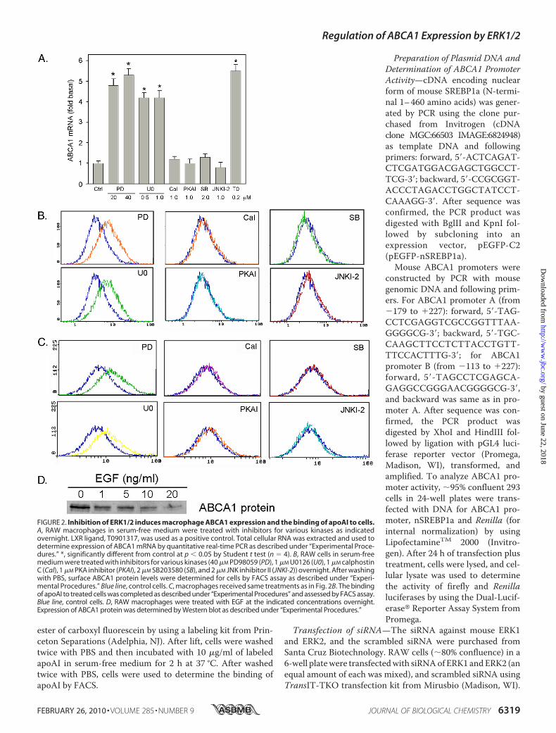
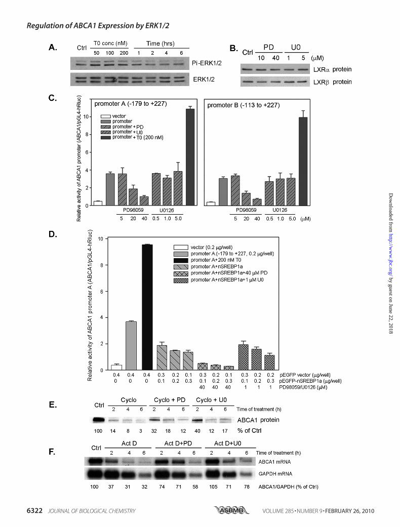
Expression by ERK1/2 Inhibitors Is Independent of Activity ofLXR or SREBP1a—ERK1/2 inhibitors act similarly as an LXRligand to induce macrophage free cholesterol efflux andABCA1 expression. To study the involved mechanisms, we ini-tially tested the effects of an LXR ligand onmacrophage ERK1/2expression and phosphorylation. Both concentration and timecourse studies demonstrated that LXR ligand did not influencethe total and phosphorylated ERK1/2 levels (Fig. 4A) suggestingthat the induction of macrophage ABCA1 expression and freecholesterol efflux by the LXR ligand was unrelated to ERK1/2expression or activation.To study if the induction of macrophage ABCA1 expression
by ERK1/2 inhibitors is through activation of LXR, we assessedthe effects of ERK1/2 inhibitors on LXR expression. After treat-ment, nuclear proteins were extracted and used to determineLXR� and LXR� protein levels by Western blot. NeitherPD98059 norU0126 changed the profiles of LXR� and LXR� inthe nuclei (Fig. 4B).The LXRE (positions from �68 to �53) in the ABCA1 pro-
moter can increase ABCA1 transcription, whereas the E-box(positions from �148 to �142) can inhibit ABCA1 transcrip-tion (19, 20). To determine if the induction of macrophageABCA1 expression by ERK1/2 inhibitors can occur by influenc-ing the activity of LXRE or E-box in the ABCA1 promoter, wetransfected 293 cells with the DNA of ABCA1 promoter A(from �179 to �227; this promoter includes motifs of theLXRE and the E-box), then treated transfected cells withERK1/2 inhibitors. In contrast to the effects on levels of ABCA1mRNA and protein, U0126 had no effect on ABCA1 promoterA activity, whereas PD98059 inhibited it in a concentration-de-pendent manner (Fig. 4C, left panel). We further determinedthe effects of ERK1/2 inhibitors on the activity of the ABCA1
FIGURE 3. ERK1/2 inhibitors induce macrophage ABCA1 expression and free cholesterol efflux in a concentration- and time-dependent manner.A, peritoneal macrophages were isolated from thioglycolate-elicited wild-type mice and cultured in complete RMPI medium for 2 days. Cells were then treatedwith ERK1/2 inhibitors, PD98059 and U0126, at the indicated concentrations in serum-free medium overnight. Total cellular proteins were extracted and usedto determine ABCA1 protein expression by Western blot as described under “Experimental Procedures.” B, RAW macrophages in a 6-well plate were transfectedwith scrambled siRNA or the mixed siRNA against ERK1 and ERK2 (equal amount of each) at the indicated concentrations for 24 h as described under“Experimental Procedures.” Cells were used to extract total protein and determined the expression of ERK1/2 and ABCA1 by Western blot. C, pre-labeled RAWmacrophages were treated with ERK1/2 inhibitors at the indicated concentrations overnight followed by determination of free cholesterol efflux to apoAI (10�g/ml, 5 h) as described under “Experimental Procedures.” D, pre-labeled RAW macrophages were treated with PD98059 (40 �M) and U0126 (2 �M) overnight.After washing with PBS, cells were incubated in serum-free medium containing 10 �g/ml apoAI for indicated times, and the radioactivity in medium wasdetermined. Free cholesterol efflux was calculated as the percentage of total labeled free cholesterol.
Regulation of ABCA1 Expression by ERK1/2
FEBRUARY 26, 2010 • VOLUME 285 • NUMBER 9 JOURNAL OF BIOLOGICAL CHEMISTRY 6321
by guest on June 22, 2018http://w
ww
.jbc.org/D
ownloaded from
Regulation of ABCA1 Expression by ERK1/2
6322 JOURNAL OF BIOLOGICAL CHEMISTRY VOLUME 285 • NUMBER 9 • FEBRUARY 26, 2010
by guest on June 22, 2018http://w
ww
.jbc.org/D
ownloaded from
promoter B (from�113 to�227), which includes the LXREbutnot the E-box. We observed that the effects of ERK1/2 inhibi-tors on the ABCA1 promoter B (Fig. 4C, right panel) were sim-ilar to that on the ABCA1 promoter A.Previously, we reported that SREBP1 inhibited ABCA1
expression by decreasing ABCA1 promoter activity (24). Totest if ERK1/2 inhibitor-induced ABCA1 expression occurs byblocking SREBP1 action, 293 cells were co-transfected withDNA of active SREBP1a (nuclear form) and ABCA1 promoterA followed by treatment with ERK1/2 inhibitors. Results in Fig.4D demonstrate that nSREBP1a inhibited ABCA1 promoteractivity, and this inhibition was not reversed by U0126 butenhanced by PD98059. Thus, the induction of macrophageABCA1 expression by ERK1/2 inhibitors was also independentof SREBP1 activity.Increased ABCA1 can occur by post-transcriptional modifi-
cations. To test if ERK1/2 inhibitors increase macrophageABCA1 levels by increasing its stability, we treated cells withcycloheximide to arrest cellular protein synthesis in theabsence or presence of ERK1/2 inhibitors. ABCA1 is a quicklydegraded protein, thus, in the presence of cycloheximide,ABCA1 protein declined dramatically and was almost unde-tectable after 6-h treatment. In contrast, ERK1/2 inhibitors(PD98059 and U0126) reduced the decline at all time points oftreatment suggesting ERK1/2 inhibitors are able to reduce thedegradation of ABCA1 protein (Fig. 4E). Moreover, weobserved that ABCA1 mRNA also degrades quickly (t1⁄2 � 2 h)and that this degradation was also reduced by ERK1/2 inhibi-tors (Fig. 4F).ERK1/2 Inhibitor and LXR Ligand Have Synergistic Effects on
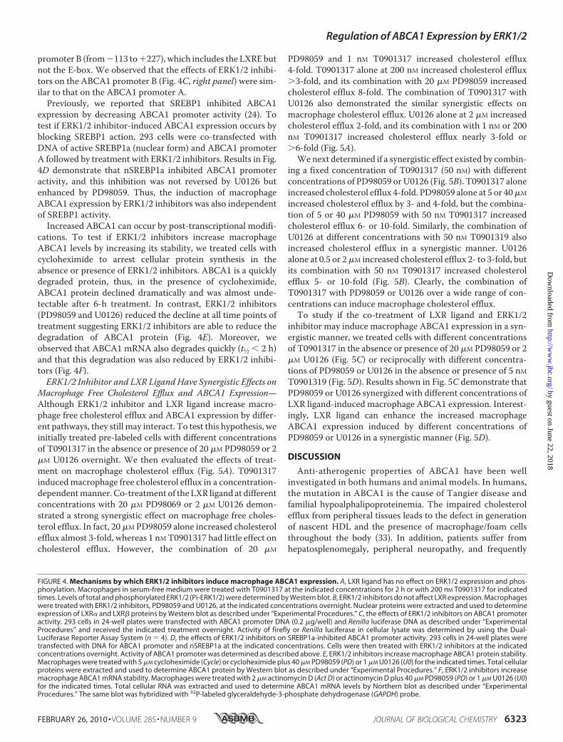
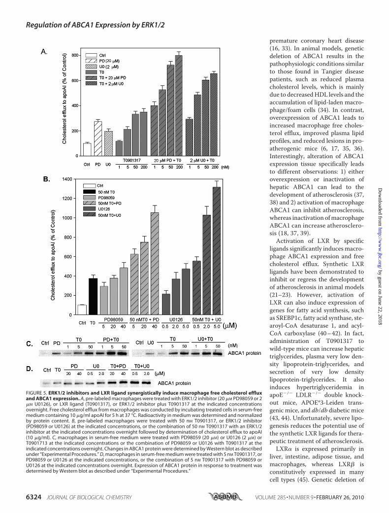
Macrophage Free Cholesterol Efflux and ABCA1 Expression—Although ERK1/2 inhibitor and LXR ligand increase macro-phage free cholesterol efflux and ABCA1 expression by differ-ent pathways, they still may interact. To test this hypothesis, weinitially treated pre-labeled cells with different concentrationsof T0901317 in the absence or presence of 20 �M PD98059 or 2�M U0126 overnight. We then evaluated the effects of treat-ment on macrophage cholesterol efflux (Fig. 5A). T0901317inducedmacrophage free cholesterol efflux in a concentration-dependentmanner. Co-treatment of the LXR ligand at differentconcentrations with 20 �M PD98069 or 2 �M U0126 demon-strated a strong synergistic effect on macrophage free choles-terol efflux. In fact, 20 �M PD98059 alone increased cholesterolefflux almost 3-fold, whereas 1 nMT0901317 had little effect oncholesterol efflux. However, the combination of 20 �M
PD98059 and 1 nM T0901317 increased cholesterol efflux4-fold. T0901317 alone at 200 nM increased cholesterol efflux�3-fold, and its combination with 20 �M PD98059 increasedcholesterol efflux 8-fold. The combination of T0901317 withU0126 also demonstrated the similar synergistic effects onmacrophage cholesterol efflux. U0126 alone at 2 �M increasedcholesterol efflux 2-fold, and its combination with 1 nM or 200nM T0901317 increased cholesterol efflux nearly 3-fold or�6-fold (Fig. 5A).We next determined if a synergistic effect existed by combin-
ing a fixed concentration of T0901317 (50 nM) with differentconcentrations of PD98059 orU0126 (Fig. 5B). T0901317 aloneincreased cholesterol efflux 4-fold. PD98059 alone at 5 or 40�M
increased cholesterol efflux by 3- and 4-fold, but the combina-tion of 5 or 40 �M PD98059 with 50 nM T0901317 increasedcholesterol efflux 6- or 10-fold. Similarly, the combination ofU0126 at different concentrations with 50 nM T0901319 alsoincreased cholesterol efflux in a synergistic manner. U0126alone at 0.5 or 2�M increased cholesterol efflux 2- to 3-fold, butits combination with 50 nM T0901317 increased cholesterolefflux 5- or 10-fold (Fig. 5B). Clearly, the combination ofT0901317 with PD98059 or U0126 over a wide range of con-centrations can induce macrophage cholesterol efflux.To study if the co-treatment of LXR ligand and ERK1/2
inhibitor may induce macrophage ABCA1 expression in a syn-ergistic manner, we treated cells with different concentrationsof T0901317 in the absence or presence of 20 �M PD98059 or 2�M U0126 (Fig. 5C) or reciprocally with different concentra-tions of PD98059 or U0126 in the absence or presence of 5 nMT0901319 (Fig. 5D). Results shown in Fig. 5C demonstrate thatPD98059 or U0126 synergized with different concentrations ofLXR ligand-induced macrophage ABCA1 expression. Interest-ingly, LXR ligand can enhance the increased macrophageABCA1 expression induced by different concentrations ofPD98059 or U0126 in a synergistic manner (Fig. 5D).
DISCUSSION
Anti-atherogenic properties of ABCA1 have been wellinvestigated in both humans and animal models. In humans,the mutation in ABCA1 is the cause of Tangier disease andfamilial hypoalphalipoproteinemia. The impaired cholesterolefflux from peripheral tissues leads to the defect in generationof nascent HDL and the presence of macrophage/foam cellsthroughout the body (33). In addition, patients suffer fromhepatosplenomegaly, peripheral neuropathy, and frequently
FIGURE 4. Mechanisms by which ERK1/2 inhibitors induce macrophage ABCA1 expression. A, LXR ligand has no effect on ERK1/2 expression and phos-phorylation. Macrophages in serum-free medium were treated with T0901317 at the indicated concentrations for 2 h or with 200 nM T0901317 for indicatedtimes. Levels of total and phosphorylated ERK1/2 (Pi-ERK1/2) were determined by Western blot. B, ERK1/2 inhibitors do not affect LXR expression. Macrophageswere treated with ERK1/2 inhibitors, PD98059 and U0126, at the indicated concentrations overnight. Nuclear proteins were extracted and used to determineexpression of LXR� and LXR� proteins by Western blot as described under “Experimental Procedures.” C, the effects of ERK1/2 inhibitors on ABCA1 promoteractivity. 293 cells in 24-well plates were transfected with ABCA1 promoter DNA (0.2 �g/well) and Renilla luciferase DNA as described under “ExperimentalProcedures” and received the indicated treatment overnight. Activity of firefly or Renilla luciferase in cellular lysate was determined by using the Dual-Luciferase Reporter Assay System (n � 4). D, the effects of ERK1/2 inhibitors on SREBP1a-inhibited ABCA1 promoter activity. 293 cells in 24-well plates weretransfected with DNA for ABCA1 promoter and nSREBP1a at the indicated concentrations. Cells were then treated with ERK1/2 inhibitors at the indicatedconcentrations overnight. Activity of ABCA1 promoter was determined as described above. E, ERK1/2 inhibitors increase macrophage ABCA1 protein stability.Macrophages were treated with 5 �M cycloheximide (Cycle) or cycloheximide plus 40 �M PD98059 (PD) or 1 �M U0126 (U0) for the indicated times. Total cellularproteins were extracted and used to determine ABCA1 protein by Western blot as described under “Experimental Procedures.” F, ERK1/2 inhibitors increasemacrophage ABCA1 mRNA stability. Macrophages were treated with 2 �M actinomycin D (Act D) or actinomycin D plus 40 �M PD98059 (PD) or 1 �M U0126 (U0)for the indicated times. Total cellular RNA was extracted and used to determine ABCA1 mRNA levels by Northern blot as described under “ExperimentalProcedures.” The same blot was hybridized with 32P-labeled glyceraldehyde-3-phosphate dehydrogenase (GAPDH) probe.
Regulation of ABCA1 Expression by ERK1/2
FEBRUARY 26, 2010 • VOLUME 285 • NUMBER 9 JOURNAL OF BIOLOGICAL CHEMISTRY 6323
by guest on June 22, 2018http://w
ww
.jbc.org/D
ownloaded from
premature coronary heart disease(16, 33). In animal models, geneticdeletion of ABCA1 results in thepathophysiologic conditions similarto those found in Tangier diseasepatients, such as reduced plasmacholesterol levels, which is mainlydue to decreasedHDL levels and theaccumulation of lipid-laden macro-phage/foam cells (34). In contrast,overexpression of ABCA1 leads toincreased macrophage free choles-terol efflux, improved plasma lipidprofiles, and reduced lesions in pro-atherogenic mice (6, 17, 35, 36).Interestingly, alteration of ABCA1expression tissue specifically leadsto different observations: 1) eitheroverexpression or inactivation ofhepatic ABCA1 can lead to thedevelopment of atherosclerosis (37,38) and 2) activation of macrophageABCA1 can inhibit atherosclerosis,whereas inactivation ofmacrophageABCA1 can increase atherosclero-sis (18, 37, 39).Activation of LXR by specific
ligands significantly inducesmacro-phage ABCA1 expression and freecholesterol efflux. Synthetic LXRligands have been demonstrated toinhibit or regress the developmentof atherosclerosis in animal models(21–23). However, activation ofLXR can also induce expression ofgenes for fatty acid synthesis, suchas SREBP1c, fatty acid synthase, ste-aroyl-CoA desaturase 1, and acyl-CoA carboxylase (40–42). In fact,administration of T0901317 towild-type mice can increase hepatictriglycerides, plasma very low den-sity lipoprotein-triglycerides, andsecretion of very low densitylipoprotein-triglycerides. It alsoinduces hypertriglyceridemia inapoE�/� LDLR�/� double knock-out mice, APOE*3-Leiden trans-genic mice, and db/db diabetic mice(43, 44). Unfortunately, severe lipo-genesis reduces the potential use ofthe synthetic LXR ligands for thera-peutic treatment of atherosclerosis.LXR� is expressed primarily in
liver, intestine, adipose tissue, andmacrophages, whereas LXR� isconstitutively expressed in manycell types (45). Genetic deletion of
FIGURE 5. ERK1/2 inhibitors and LXR ligand synergistically induce macrophage free cholesterol effluxand ABCA1 expression. A, pre-labeled macrophages were treated with ERK1/2 inhibitor (20 �M PD98059 or 2�M U0126), or LXR ligand (T0901317), or ERK1/2 inhibitor plus T0901317 at the indicated concentrationsovernight. Free cholesterol efflux from macrophages was conducted by incubating treated cells in serum-freemedium containing 10 �g/ml apoAI for 5 h at 37 °C. Radioactivity in medium was determined and normalizedby protein content. B, pre-labeled macrophages were treated with 50 nM T0901317, or ERK1/2 inhibitor(PD98059 or U0126) at the indicated concentrations, or the combination of 50 nM T0901317 with an ERK1/2inhibitor at the indicated concentrations overnight followed by determination of cholesterol efflux to apoAI(10 �g/ml). C, macrophages in serum-free medium were treated with PD98059 (20 �M) or U0126 (2 �M) orT0901713 at the indicated concentrations or the combination of PD98059 or U0126 with T0901317 at theindicated concentrations overnight. Changes in ABCA1 protein were determined by Western blot as describedunder “Experimental Procedures.” D, macrophages in serum-free medium were treated with 5 nM T0901317, orPD98059 or U0126 at the indicated concentrations, or the combination of 5 nM T0901317 with PD98059 orU0126 at the indicated concentrations overnight. Expression of ABCA1 protein in response to treatment wasdetermined by Western blot as described under “Experimental Procedures.”
Regulation of ABCA1 Expression by ERK1/2
6324 JOURNAL OF BIOLOGICAL CHEMISTRY VOLUME 285 • NUMBER 9 • FEBRUARY 26, 2010
by guest on June 22, 2018http://w
ww
.jbc.org/D
ownloaded from
LXR�profoundly impacts on expression of those genes for fattyacid biosynthesis while the absence of LXR� has little effect(41). Thus, the selective modulators of LXR� may have littleadverse effect on lipogenesis while reducing atherosclerosis.However, due to the high identity of LXR� and LXR� indomains for the DNA and ligand binding, the identification ofthe selective LXR� modulators has not been advanced (46).GW3965, another synthetic LXR ligand, can inhibit the devel-opment of atherosclerosis in both LDLR�/� and apoE�/�mice.It has no effect on lipogenesis in both wild-type and LDLR�/�
mice, but not in apoE�/�mice (21, 47). The reduced side effectsmay be due to the fact that GW3965 is a weak activator of LXRrather than it has a higher selectivity for LXR� than LXR� (47).Phosphorylation of ABCA1 can influence ABCA1 protein
stability and activity in a different manner. The constitutivephosphorylation of Ser-1042 and Ser-2054 by PKA in humanABCA1 increases ABCA1-mediated phospholipid efflux. How-ever, PKA activity has no effect on ABCA1 expression (48, 49).ApoAI and synthetic amphiphilic helical peptides increaseABCA1 protein stability by activating PKA (50, 51). ApoAI alsophosphorylates ABCA1 through protein kinase C� to enhanceABCA1 protein stability (52). In contrast, phosphorylation of aPEST sequence (rich in proline, glutamic acid, serine, and thre-onine) in ABCA1 by calpain promotes a quick degradation ofABCA1. Thus, inhibition of calpain by calpeptin or N-Ac-Leu-Leu-norleucinal increases ABCA1 protein stability and cellularlevels (25, 53, 54). Each of the above reports suggests the mod-ification of ABCA1 can occur at the post-translational levels. Incontrast, we observed that inhibition of cellular ERK1/2 activitycan increase ABCA1 stability at both the mRNA and proteinlevel (Fig. 4, E and F) suggesting a different mechanism bywhich regulates ABCA1 expression.More importantly, an LXRligand can increase ABCA1 transcription, and the increasedABCA1 transcript is protected by an ERK1/2 inhibitor from thedegradation. Thus, ERK1/2 inhibitor synergizes with LXRligand-induced ABCA1 expression and cholesterol efflux evenwhen partial ERK1/2 activity is inhibited. Indeed, we observedthat co-treatment of cells with an ERK1/2 inhibitor and an LXRligand greatly increasesmacrophage cholesterol efflux to apoAI(Fig. 5). Others have found that reduced doses of T0901317 caninhibit atherosclerosis in LDLR�/� mice and is associated withlesser adverse effects on lipogenesis (23). Our findings implythat it may be feasible to use in vivo the combined ERK1/2inhibitor and LXR ligand at very low doses to inhibit/regressatherosclerosis without unfavorable lipogenic effects.In summary, our results demonstrate that blockage of
ERK1/2 increases macrophage ABCA1 expression and freecholesterol efflux to apoAI and HDL. ERK1/2 inhibitorsincrease ABCA1 expression at both mRNA and protein levels.In addition, ERK1/2 inhibitors synergize with LXR ligand-in-duced ABCA1 expression and cholesterol efflux. Takentogether, our studies suggest a new function of ERK1/2 activityin cholesterol trafficking in the macrophage.REFERENCES1. Arora, S., and Nicholls, S. J. (2008) Drugs Today 44, 711–7182. de Villiers, W. J., and Smart, E. J. (1999) J. Leukocyte Biol. 66, 740–7463. Febbraio, M., Podrez, E. A., Smith, J. D., Hajjar, D. P., Hazen, S. L., Hoff,
H. F., Sharma, K., and Silverstein, R. L. (2000) J. Clin. Invest. 105,
1049–10564. Suzuki, H., Kurihara, Y., Takeya, M., Kamada, N., Kataoka, M., Jishage, K.,
Ueda, O., Sakaguchi, H., Higashi, T., Suzuki, T., Takashima, Y., Kawabe,Y., Cynshi, O., Wada, Y., Honda, M., Kurihara, H., Aburatani, H., Doi, T.,Matsumoto, A., Azuma, S., Noda, T., Toyoda, Y., Itakura, H., Yazaki, Y.,and Kodama, T. (1997) Nature 386, 292–296
5. Braun, A., Trigatti, B. L., Post, M. J., Sato, K., Simons, M., Edelberg, J. M.,Rosenberg, R. D., Schrenzel, M., and Krieger, M. (2002) Circ. Res. 90,270–276
6. Singaraja, R. R., Fievet, C., Castro, G., James, E. R., Hennuyer, N., Clee,S. M., Bissada, N., Choy, J. C., Fruchart, J. C., McManus, B. M., Staels, B.,and Hayden, M. R. (2002) J. Clin. Invest. 110, 35–42
7. Ji, Y., Jian, B., Wang, N., Sun, Y., Moya, M. L., Phillips, M. C., Rothblat,G. H., Swaney, J. B., and Tall, A. R. (1997) J. Biol. Chem. 272, 20982–20985
8. Swarnakar, S., Temel, R. E., Connelly, M. A., Azhar, S., andWilliams, D. L.(1999) J. Biol. Chem. 274, 29733–29739
9. Wang, N., Silver, D. L., Thiele, C., and Tall, A. R. (2001) J. Biol. Chem. 276,23742–23747
10. Lee, J. Y., and Parks, J. S. (2005) Curr. Opin. Lipidol. 16, 19–2511. Krimbou, L., Denis, M., Haidar, B., Carrier, M., Marcil, M., and Genest, J.,
Jr. (2004) J. Lipid Res. 45, 839–84812. Huang, Z. H., Fitzgerald, M. L., and Mazzone, T. (2006) Arterioscler.
Thromb. Vasc. Biol. 26, 157–16213. Von Eckardstein, A., Langer, C., Engel, T., Schaukal, I., Cignarella, A.,
Reinhardt, J., Lorkowski, S., Li, Z., Zhou, X., Cullen, P., and Assmann, G.(2001) FASEB J. 15, 1555–1561
14. Brooks-Wilson, A., Marcil, M., Clee, S. M., Zhang, L. H., Roomp, K., vanDam, M., Yu, L., Brewer, C., Collins, J. A., Molhuizen, H. O., Loubser, O.,Ouelette, B. F., Fichter, K., Ashbourne-Excoffon, K. J., Sensen, C. W.,Scherer, S., Mott, S., Denis, M., Martindale, D., Frohlich, J., Morgan, K.,Koop, B., Pimstone, S., Kastelein, J. J., Genest, J., Jr., and Hayden, M. R.(1999) Nat. Genet. 22, 336–345
15. Bodzioch,M., Orso, E., Klucken, J., Langmann, T., Bottcher, A., Diederich,W., Drobnik,W., Barlage, S., Buchler, C., Porsch-Ozcurumez,M., Kamin-ski, W. E., Hahmann, H. W., Oette, K., Rothe, G., Aslanidis, C., Lackner,K. J., and Schmitz, G. (1999) Nat. Genet. 22, 347–351
16. Rust, S., Rosier, M., Funke, H., Real, J., Amoura, Z., Piette, J. C., Deleuze,J. F., Brewer, H. B., Duverger, N., Denefle, P., and Assmann, G. (1999)Nat.Genet. 22, 352–355
17. Joyce, C. W., Amar, M. J., Lambert, G., Vaisman, B. L., Paigen, B., Najib-Fruchart, J., Hoyt, R. F., Jr., Neufeld, E. D., Remaley, A. T., Fredrickson,D. S., Brewer, H. B., Jr., and Santamarina-Fojo, S. (2002) Proc. Natl. Acad.Sci. U.S.A. 99, 407–412
18. Van Eck, M., Singaraja, R. R., Ye, D., Hildebrand, R. B., James, E. R., Hay-den, M. R., and Van Berkel, T. J. (2006) Arterioscler. Thromb. Vasc. Biol.26, 929–934
19. Zeng, L., Liao, H., Liu, Y., Lee, T. S., Zhu,M.,Wang, X., Stemerman,M. B.,Zhu, Y., and Shyy, J. Y. (2004) J. Biol. Chem. 279, 48801–48807
20. Schmitz, G., and Langmann, T. (2005) Biochim. Biophys. Acta 1735, 1–1921. Joseph, S. B., McKilligin, E., Pei, L., Watson, M. A., Collins, A. R., Laffitte,
B. A., Chen, M., Noh, G., Goodman, J., Hagger, G. N., Tran, J., Tippin,T. K.,Wang, X., Lusis, A. J., Hsueh,W.A., Law, R. E., Collins, J. L.,Willson,T.M., andTontonoz, P. (2002)Proc. Natl. Acad. Sci. U.S.A. 99, 7604–7609
22. Levin, N., Bischoff, E. D., Daige, C. L., Thomas, D., Vu, C. T., Heyman,R. A., Tangirala, R. K., and Schulman, I. G. (2005) Arterioscler. Thromb.Vasc. Biol. 25, 135–142
23. Terasaka, N., Hiroshima, A., Koieyama, T., Ubukata, N., Morikawa, Y.,Nakai, D., and Inaba, T. (2003) FEBS Lett. 536, 6–11
24. Zhou, X., He, W., Huang, Z., Gotto, A. M., Jr., Hajjar, D. P., and Han, J.(2008) J. Biol. Chem. 283, 2129–2138
25. Arakawa, R., and Yokoyama, S. (2002) J. Biol. Chem. 277, 22426–2242926. Wang, Y., and Oram, J. F. (2002) J. Biol. Chem. 277, 5692–569727. Sebolt-Leopold, J. S., and Herrera, R. (2004) Nat. Rev. Cancer 4, 937–94728. Pearson, G., Robinson, F., Beers, Gibson, T., Xu, B. E., Karandikar, M.,
Berman, K., and Cobb, M. H. (2001) Endocr. Rev. 22, 153–18329. Wang, Y. (2007) Circulation 116, 1413–142330. Brinton, E. A., Eisenberg, S., and Breslow, J. L. (1989) J. Clin. Invest. 84,
262–269
Regulation of ABCA1 Expression by ERK1/2
FEBRUARY 26, 2010 • VOLUME 285 • NUMBER 9 JOURNAL OF BIOLOGICAL CHEMISTRY 6325
by guest on June 22, 2018http://w
ww
.jbc.org/D
ownloaded from
31. Han, J., Hajjar, D. P., Zhou, X., Gotto, A. M., Jr., and Nicholson, A. C.(2002) J. Biol. Chem. 277, 23582–23586
32. Zerivitz, K., and Akusjarvi, G. (1989) Gene Anal. Tech. 6, 101–10933. Clee, S. M., Kastelein, J. J., van, Dam, M., Marcil, M., Roomp, K., Zwarts,
K. Y., Collins, J. A., Roelants, R., Tamasawa, N., Stulc, T., Suda, T., Ceska,R., Boucher, B., Rondeau, C., DeSouich, C., Brooks-Wilson, A., Molhui-zen, H. O., Frohlich, J., Genest, J., Jr., and Hayden, M. R. (2000) J. Clin.Invest. 106, 1263–1270
34. McNeish, J., Aiello, R. J., Guyot, D., Turi, T., Gabel, C., Aldinger, C.,Hoppe, K. L., Roach, M. L., Royer, L. J., deWet, J., Broccardo, C., Chimini,G., and Francone, O. L. (2000) Proc. Natl. Acad. Sci. U.S.A. 97, 4245–4250
35. Cavelier, L. B., Qiu, Y., Bielicki, J. K., Afzal, V., Cheng, J. F., andRubin, E.M.(2001) J. Biol. Chem. 276, 18046–18051
36. Singaraja, R. R., Bocher, V., James, E. R., Clee, S. M., Zhang, L. H., Leavitt,B. R., Tan, B., Brooks-Wilson,A., Kwok,A., Bissada,N., Yang, Y. Z., Liu,G.,Tafuri, S. R., Fievet, C., Wellington, C. L., Staels, B., and Hayden, M. R.(2001) J. Biol. Chem. 276, 33969–33979
37. Brunham, L. R., Singaraja, R. R., Duong, M., Timmins, J. M., Fievet, C.,Bissada,N., Kang,M.H., Samra, A., Fruchart, J. C.,McManus, B., Staels, B.,Parks, J. S., and Hayden, M. R. (2009) Arterioscler. Thromb. Vasc. Biol. 29,548–554
38. Joyce, C.W., Wagner, E. M., Basso, F., Amar, M. J., Freeman, L. A., Sham-burek, R.D., Knapper, C. L., Syed, J.,Wu, J., Vaisman, B. L., Fruchart-Najib,J., Billings, E. M., Paigen, B., Remaley, A. T., Santamarina-Fojo, S., andBrewer, H. B., Jr. (2006) J. Biol. Chem. 281, 33053–33065
39. Aiello, R. J., Brees, D., Bourassa, P. A., Royer, L., Lindsey, S., Coskran, T.,Haghpassand, M., and Francone, O. L. (2002) Arterioscler. Thromb. Vasc.Biol. 22, 630–637
40. Joseph, S. B., Laffitte, B. A., Patel, P. H., Watson, M. A., Matsukuma, K. E.,Walczak, R., Collins, J. L., Osborne, T. F., and Tontonoz, P. (2002) J. Biol.Chem. 277, 11019–11025
41. Repa, J. J., Liang, G., Ou, J., Bashmakov, Y., Lobaccaro, J. M., Shimomura,I., Shan, B., Brown, M. S., Goldstein, J. L., and Mangelsdorf, D. J. (2000)
Genes Dev. 14, 2819–283042. Schultz, J. R., Tu, H., Luk, A., Repa, J. J., Medina, J. C., Li, L., Schwendner,
S., Wang, S., Thoolen, M., Mangelsdorf, D. J., Lustig, K. D., and Shan, B.(2000) Genes Dev. 14, 2831–2838
43. Chisholm, J. W., Hong, J., Mills, S. A., and Lawn, R. M. (2003) J. Lipid Res.44, 2039–2048
44. Grefhorst, A., Elzinga, B.M., Voshol, P. J., Plosch, T., Kok, T., Bloks, V.W.,van der Sluijs, F. H., Havekes, L. M., Romijn, J. A., Verkade, H. J., andKuipers, F. (2002) J. Biol. Chem. 277, 34182–34190
45. Repa, J. J., and Mangelsdorf, D. J. (2000) Annu. Rev. Cell Dev. Biol. 16,459–481
46. Teboul, M., Enmark, E., Li, Q., Wikstrom, A. C., Pelto-Huikko, M., andGustafsson, J. A. (1995) Proc. Natl. Acad. Sci. U.S.A. 92, 2096–2100
47. Miao, B., Zondlo, S., Gibbs, S., Cromley, D., Hosagrahara, V. P., Kirchgess-ner, T. G., Billheimer, J., and Mukherjee, R. (2004) J. Lipid Res. 45,1410–1417
48. Haidar, B., Denis, M., Krimbou, L., Marcil, M., and Genest, J., Jr. (2002) J.Lipid Res. 43, 2087–2094
49. See, R. H., Caday-Malcolm, R. A., Singaraja, R. R., Zhou, S., Silverston, A.,Huber, M. T., Moran, J., James, E. R., Janoo, R., Savill, J. M., Rigot, V.,Zhang, L. H., Wang, M., Chimini, G., Wellington, C. L., Tafuri, S. R., andHayden, M. R. (2002) J. Biol. Chem. 277, 41835–41842
50. Haidar, B., Denis, M., Marcil, M., Krimbou, L., and Genest, J., Jr. (2004)J. Biol. Chem. 279, 9963–9969
51. Arakawa, R., Hayashi,M., Remaley, A. T., Brewer, B. H., Yamauchi, Y., andYokoyama, S. (2004) J. Biol. Chem. 279, 6217–6220
52. Yamauchi, Y., Hayashi, M., Abe-Dohmae, S., and Yokoyama, S. (2003)J. Biol. Chem. 278, 47890–47897
53. Martinez, L. O., Agerholm-Larsen, B.,Wang, N., Chen,W., and Tall, A. R.(2003) J. Biol. Chem. 278, 37368–37374
54. Wang, N., Chen, W., Linsel-Nitschke, P., Martinez, L. O., Agerholm-Larsen, B., Silver, D. L., and Tall, A. R. (2003) J. Clin. Invest. 111, 99–107
Regulation of ABCA1 Expression by ERK1/2
6326 JOURNAL OF BIOLOGICAL CHEMISTRY VOLUME 285 • NUMBER 9 • FEBRUARY 26, 2010
by guest on June 22, 2018http://w
ww
.jbc.org/D
ownloaded from
Xiaoye Zhou, Zhinan Yin, Xianzhi Guo, David P. Hajjar and Jihong HanMacrophage ABCA1 Expression and Cholesterol Efflux
Inhibition of ERK1/2 and Activation of Liver X Receptor Synergistically Induce
doi: 10.1074/jbc.M109.073601 originally published online December 25, 20092010, 285:6316-6326.J. Biol. Chem.
10.1074/jbc.M109.073601Access the most updated version of this article at doi:
Alerts:
When a correction for this article is posted•
When this article is cited•
to choose from all of JBC's e-mail alertsClick here
http://www.jbc.org/content/285/9/6316.full.html#ref-list-1
This article cites 54 references, 37 of which can be accessed free at
by guest on June 22, 2018http://w
ww
.jbc.org/D
ownloaded from