inhibitory effect of verapamil upon calcium and potassium ... · inhibitory effect of verapamil...
TRANSCRIPT
Gen. Physiol. Biophys. (1983), 2, 181—192 181
Inhibitory Effect of Verapamil upon Calcium and Potassium Currents in Crayfish Muscle Membrane
I. ZÁHRADNÍK and J. ZACHAR
Department of General Physiology, Centre of Physiological Sciences, Slovak Academy of Sciences, Vlárska 5, 833 06 Bratislava, Czechoslovakia
Abstract. The effect of verapamil upon ionic currents in the muscle membrane of the crayfish (Astacus fluviatilis) was investigated under voltage clamp conditions both in intact fibres and cut muscle fibre segments. In intact isolated fibres millimolar concentrations of verapamil inhibit both the early and the late potassium outward currents, however to a different extent. The effect of verapamil on pure calcium inward current (ica) was studied in more detail applying the voltage clamp technique to muscle fibre fragments equilibrated in K+-free medium. At the resting membrane potential ( -80 mV) Jca was only partially inhibited after 22 min exposure to verapamil (2 mmol/1) in the external solution ([Ca2+]0 = 20 mmol/1; pH = 7.5; t = 16 — 18°C). No changes in threshold, maximum or reversal potentials were observed in Jo — V curves. If the fibre was depolarized during the action of verapamil, Ica was decreased to zero. The restitution was very slow; Tea was absent even after 4 min repolarization to —80 mV. In a control experiment, reversibility of Jo. after prolonged depolarization in the absence of verapamil was very rapid and almost complete. Inhibition of ica by verapamil (0.5 mmol/1) was substantially enhanced when the frequency and pulse duration of repeated stimulation were increased. These results are in agreement with the hypothesis of the use-dependent mechanism in the verapamil-Ca channel interaction.
Key words: Muscle membrane — Verapamil — Calcium current — Internal perfusion — Use-dependent action
Introduction
Verapamil belongs to drugs with antiarrhytmic action characterized as calcium antagonists (Fleckenstein 1977; Bayer and Ehara 1978). However, it is not a specific blocker of calcium channels. At low concentrations, its ( —)-optical isomer blocks calcium channels, while the ( + )-isomer was reported to block sodium channels in mammalian heart muscle cells (Bayer et al. 1975b, c). In higher concentrations, it can inhibit potassium currents as well (Nawrath et al. 1977; Kass and Tsien 1975). Local anaesthetic activity of verapamil, comparable with that of procaine was also observed at higher concentrations (Singh and Vaughan Williams 1972; Kraynack et al. 1982). The analysis of Jca and contractility inhibition by

182 Záhradník and Zachar
verapamil or D 600 has shown (Bayer et al. 1975a, b, c; McDonald et al. 1980; Ehara and Kaufmann 1978) that, contrary to Ni 2 + and La3*, organic calcium antagonists block the cell calcium influx in a frequency and potential dependent manner.
Heart and smooth muscle preparations are less advantageous for a detailed study of the effects of calcium antagonists, since it is difficult to separate the respective ionic conductances from each other and because of the multicellular nature of the preparations. The effects of calcium antagonists were therefore studied on isolated excitable cells characterized by the calcium-type electrogenesis. Rojas and Luxoro (1974) reported inhibition of Ic by D 600 in barnacle muscle cells. Suarez-Kurtz and Sorenson (1977) observed a higher efficacy of the verapamil (-)-isomer in inhibiting both Go, and GK in crab muscle fibres as compared with the ( + )-isomer. Lower amplitudes and shorter action potentials were observed by van der Kloot and Kita (1975) in frog and crayfish muscle fibres, apparently due to the inhibition of both the outward and inward currents by verapamil. Kostyuk and Krishtal (1977) reported inhibitory action of verapamil and D 600 on Jo, and, to a lesser extent, on JN«, in isolated snail neurons. Akaike et al. (1981) performed detailed measurements of the effects of verapanul and diltiazem in the same experimental object. They found that organic Ca antagonists inhibit Zca by a competitive mechanism similarly as do metal cations, namely by an interaction with the calcium receptor in the calcium channel. In snail neurons Akaike et al. (1979) reported a non-specific inhibitory effect of organic Ca-an-tagonists on potassium currents.
It may be generally stated that the results obtained in single-cell preparations with Ca-electrogenesis coincide well with those obtained by less direct methods in multicellular preparations, although the potency of calcium antagonists appears to be lower in the former preparations.
Our experiments were aimed at determining the effect of verapamil on total ionic currents in the crayfish muscle membrane, and at studying the mechanism of verapamil-calcium channel interaction using the method of isolated pure calcium currents. Some of the results were reported previously (Záhradník and Zachar 1980, 1981).
Methods
The voltage-clamp method used in the present work was described in detail elsewhere (Záhradník and Zachar 1982b). A bundle containing several muscle fibres was cut from the m. extensor carpopoditi of the crayfish Astacus fluviatilis and transferred into the "internal" relaxing solution. The fibres were subsequently separated and cut into 3 mm long fragments. The fragments were allowed to equilibrate over 30—60 min, and were then fixed in the experimental chamber with two isolating sucrose and one grounding rings (Henček and Zachar 1977). In this arrangement, only the membrane over the central channel, 50 um in diameter, is exposed to the external solution.

Verapamil on Crayfish Muscle Membrane 183
10 ms j I
Fig. 1. Effect of verapamil on total ionic currents. The voltage clamp depolarization pulses were increased in 20 mV steps. Arrows point to currents corresponding to 100 mV depolarizations. The scales in pannel a are common for all pannels. a: currents recorded in the normal external solution, b and c: currents recorded 5 and 9 min respectively, following verapamil application (2.3 mmol/1). d, e and /: currents recorded 7, 16 and 24 min.respectively, following the return into the normal external solution.
After installing the voltage-clamp regime, the membrane potential was measured and adjusted to -80 mV (inside of the fibre negative) using an external source. Total membrane currents to rectangular depolarizing pulses were compensated for the capacitance and leak components using a compensatory amplifier with its parameters being fixed at 10 mV hyperpolarizing pulses. Pure ionic currents were recorded on the memory oscilloscope screen.
Under these conditions, pure currents flowing through calcium channels were recorded, since KMree solutions were in contact with both sides of the membrane. For the composition of the solutions, see the Results section. The working temperature was maintained between 16— 18°C, pH was 7.5. Verapamil (racemic mixture) was supplied by Knoll AG (FRG).
Results
In crayfish muscle fibres the blocking action of verapamil on calcium channels occured at concentrations about 1 mmol/1; at these concentrations potassium currents were also partially blocked. A typical experiment is illustrated in Fig. 1 (a, b, c) showing records from intact fibres with their membrane testing area exposed

184 Záhradník and Zachar
120 160 mV
120 160 mV
Fíg. 2. Effect of verapamil on potassium currents. Left: current-voltage relations of peak outward current values. Right: current-voltage relations of steady outward current (at the end of the 70 ms pulse). Symbols: <3 and • are values obtained 5 and 9 min following the addition of verapamil (2.3 mmol/l), respectively, X, A and • are values obtained 7, 16 and 24 min following verapamil wash-out, O are control values before addition of verapamil.
to a solution containing (in mmol/1): 27 SrCl2; 200 NaCl; 5.4 KC1; 5 Tris; pH 7.4. The addition of verapamil (2.3 mmol/1) resulted in a gradual decrease of the Sr2+
inward currents and K+ outward currents (upwards). The Sr2+ currents apparently disappeared after 9 min; their presence was, however, indicated by the irregularity of the potassium current increments with the increase of depolarization in 20 mV steps. Reversibility of the verapamil action is shown in Fig. 1 (d, e, f). The figure shows that both the decay and recovery of K+ currents are more rapid than those of Sr2+ currents, which may suggest different mechanisms of action. Current-voltage relationships constructed from peak and steady (at the end of the pulse) outward current values were used to evaluate verapamil induced changes in JK (Fig. 2). No significant shift of the JK — V dependence along the voltage axis could be observed; the slope of the linear part of the relation was, however, decreased. Prior to verapamil application as well as 7 min after its removal from the external solution, the slopes were 0.85 nA/mV and 0.61 nA/mV for JK peak and steady values, respectively. The slopes decreased to 53% for peak potassium currents and to 39% for steady values of JK, 5 min following the addition of verapamil. Only slightly smaller slopes were observed after 9 min of verapamil action: 44% and 26%, respectively. Verapamil thus affects potassium currents by decreasing the membrane conductance for K+ ions. However, the early and delayed potassium conductances (Zachar et al. 1978) are affected to different extent.
The mechanism of verapamil interactions with calcium channels was studied in

Verapamil on Crayfish Muscle Membrane 185
I depolarization on pure calcium currents. Currents are s-clamp pulses. A: records of pure la, obtained 47 min internal solution. B: calcium currents 22 min following irds obtained 4 min following membrane depolarization obtained 14 min following verapamil wash-out.
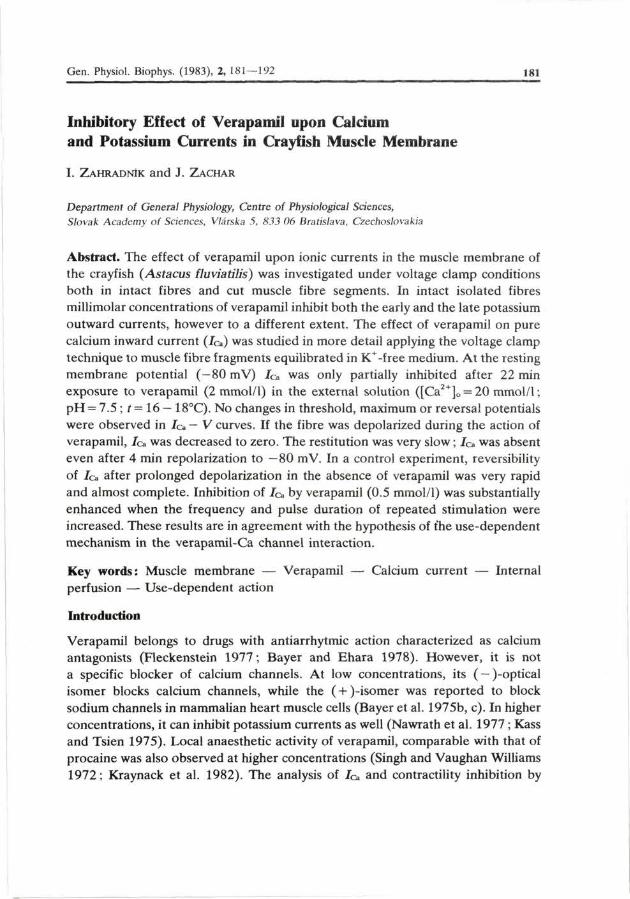
further experiments on muscle fibre fragments. The records shown in Fig. 3A represent the typical appearance of pure calcium currents recorded following the removal of K+ ions from the environment of the membrane. The fragment was equilibrated over 47 min in the internal solution, containing (in mmol/1): 4 ATP; 0.5 EGTA; 10 NaCl; 220 Tris-propionate. The external solution contained (in mmol/1): 27 CaCl2; 210 TrisCl. The addition of verapamil (2.1 mmol/1) resulted in a decrease in Jo, (Fig. 3B) to 4 5 % of the initial value after 22 min (steady-state). The decay in Jc» was identical for both the peak and steady (at the end of the pulse) current values. This is evident from Fig. 4 showing current-voltage relations of the peak inward currents (fop) and steady values (bottom). No shifts of the threshold, maximum or extrapolated reversal potential values along voltage axis could be observed. This result suggests that, in the crayfish muscle fibre membrane, verapamil inhibits inward Ca2+ currents by suppressing the calcium conductance Gca, most likely through decreasing the number of channels in conductive state.
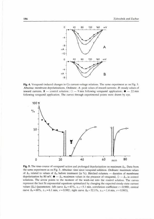
Fig. 5 (left) shows the kinetics of the verapamil-induced Jo, inhibition in the above experiment. At the 100 mV depolarization the decrease in peak current values with the duration of verapamil action was exponential, with a half-time of 5.1 min. The rate as well as the extent of verapamil-induced Jo inhibition became
186 Záhradník and Zachar
O 40 80 120 160 mV
O 40 80 120 160 mV
-4 -
nA -
- 8 -
Fig. 4. Verapamil-induced changes in Ca current-voltage relations. The same experiment as on Fig. 3. Abscissa: membrane depolarizations. Ordinate: A: peak values of inward currents: B: steady values of inward currents. X — control solution; O — 9 min following verapamil application • — 22 min following verapamil application. The curves through experimental points were drawn by eye.
100
%
50
0 20 40 60 m i n 80
Fig. 5. The time-course of verapamil action and prolonged depolarization on maximum Ic. Data from the same experiment as on Fig. 3. Abscissa: time since verapamil addition. Ordinate: maximum values of / c related to values of Ic before treatment (in %). Hatched columns — duration of membrane depolarization by 80 mV. • — Ic maximum values in the presence of verapamil, O — Ic in control solutions. The arrow points to the moment of the wash-out into the control solution. The curves represent the best fit exponential equations optimalized by changing the expected steady-state current values (Jss) (parameters: left curve iss = 43%, ii 2 = 5.1 min, correlation coefficient r = 0.980; central curve /ss = 60%, /i,2 = 6.1 min, r = 0.992: right curve Jss = 52.1%, i1/2=1.4min, r = 0.993).

Verapamil on Crayfish Muscle Membrane 187
A O
a O
aA d
ä
** e
t-
+ + + + + f 1 1 1 1 1 1 1 1 1 —
0 20 40 s 0 20 40 s
Fig. 6. Effect of verapamil at repeated membrane stimulations. Maximum inward currents related to the first depolarizing pulse (in the train of pulses) are plotted against the duration of repeated stimulation. Left: Ic decrease in the control solution. Right: decrease in the presence of verapamil (5 x 1 0 " mol/I). The duration and frequency of 80 mV depolarization pulses were: (a) 10ms; 0.5 Hz; (b) 10ms; 1.0 Hz; (c)50 ms; 1.0 Hz; (d) 300 ms; 0.5 Hz; (e) 300 ms; 1.0 Hz; (f) 1000 ms; 0.5 Hz. Corresponding stimulation patterns refer to the same fragment for both solutions.
increased following a prolonged membrane depolarization. This is demonstrated in Fig. 3 C, which shows original records obtained 4 min after membrane depolarization by 80 mV lasting 2 min. In fact, membrane currents disappeared; the "inward" current residue was mostly due to the overcompensation of the capacita-tive current.
After the removal of verapamil (Fig. 3D) the currents were only partially restituted, reaching 55% of the pre-application values. The half-time of the current restitution was 6.1 min (Fig 5 middle). As shown in Fig. 5 (right), calcium currents became restituted very quickly following a 2 min lasting membrane depolarization in the absence of verapamil, with a half-time of 1.4 min, and they almost reached the initial values.
From these experiments it follows that the verapamil block of G o is only partial and develops very slowly; however, both the rate and the extent of inhibition became rapidly increased following membrane depolarization.
Further experiments were therefore designed to study the dependence of verapamil action on the duration and frequency of depolarizing pulses. In crayfish muscle fibres, this type of experiments is complicated by the fact that the amplitude of inward currents decreases already at relatively low stimulation frequencies (Fig. 6, left). The reversibility of Jo after longer and more frequent depolarizations is slow and often only partial. Manifold overloading of the fibre membrane even
100i
so
A O * t A O
5 \ o
A
* b o C
100-,
%
50^

188 Záhradník and Z achar
results in disappearance of the inward current and in a permanent drop in the membrane resistance. With respect to the fact that survival of the fragment membrane under present experimental conditions usually ranges between 1 and 2 h, larger series of experiments cannot be performed in the same preparations.
Fig. 6 illustrates the dependence of the /c., decrease on frequency and duration of 80 mV depolarizations both in the control solution (in mmol/1: 20 CaCl2; 220 TrisCl) and in the presence of verapamil (control solution 4- 0.5 mmol/1 of verapamil). In these experiments, the fragments were equilibrated in the internal solution containing (in mmol/1): 4 ATP; 1 EGTA; 2 MgCl2; 240 Tris propionate. The blocking effect of 0.5 mmol/1 of verapamil on Jo was about 50% at the resting membrane potential. The decrease in Jo during repeated stimulation in the absence of verapamil was due to Ca-channel inactivation. Based on the results obtained by Záhradník and Zachar (1983) it may be suggested that both the voltage-dependent and Jo-dependent inactivation mechanisms are operative. In the presence of verapamil the decrease in Jo during repeated stimulation is the more pronounced the longer the pulse and the higher the frequency of stimulation are. It may therefore be stated that the blocking action of verapamil depends on Ca-channel activation. However, the present experiments do not allow to establish whether the number of activated channels or the duration of their open state is substantial or both. Qualitatively the mechanism resembles sodium channel inhibition by local anaesthetics, termed the use-dependent block.
Discussion
From the qualitative aspect, the results obtained under voltage clamp conditions are in agreement with the results obtained by other authors studying changes in the action potential. Van der Kloot and Kita (1975) observed in somatic muscle fibres of the crayfish a decrease in Jo and JK to 30% and 6% of control values, respectively, following exposure to 0.5 mmol/1 of verapamil for 8 min at resting membrane potential ([Ca2+]Q= 14 mmol/1). Suarez-Kurtz and Sorenson (1977) observed an almost by one order of magnitude stronger effect of verapamil on G o and GK in crab muscle fibres; this may have been due to lower Ca2+ concentration (12.5 mmol/1) and particularly to the higher temperature (22—25°C) used by these authors. In the above-mentioned studies, the intervals within which the steady-state effect was reached, were comparable to values obtained in our experiments, i.e. about 10 min.
Zachar et al. (1978) could distinguish three types of potassium conductances in crayfish muscle fibres. Our experiments have shown that verapamil acts differently on the first two K+ conductances. Time differences in the onset of the verapamil effect on inward and outward currents suggest different mechanisms of action. The inhibition of potassium conductance was also observed under vol-

Verapamil on Crayfish Muscle Membrane 189
tage-clamp conditions in number of other preparations (Baker et al. 1973; Kass and Tsien 1975; Akaike et al. 1979; Nawrath et al. 1977).
The verapamil-Ca channel interaction was studied by a method which enables measuring pure calcium currents over the whole range of membrane depolarizations (Záhradník and Zachar 1982). These currents are characterized by a two-phase inactivation (Fig. 3A). Measurements at the resting membrane potential showed that verapamil decreased proportionally both the peak and steady Jo values with their current-voltage parameters remaining unchanged. Verapamil thus blocks both the fast and slow components of the calcium current.
Effects of organic calcium antagonists on pure Ca2+ currents have only rarely been studied. Akaike et al. (1981) studied dose-response dependence of verapamil and diltiazem at resting membrane potentials (—60 mV to —50 mV) in isolated snail neurons. They blocked the sodium and potassium currents by substitution of Tris+ for Na+ and of Cs+ for K+, respectively. At pH = 7.4 and r = 20-25°C, a 50% inhibitory effect of verapamil on Jo at 4 x 10~5 mol/1 was found after 15 min of exposure. Kostyuk and Krishtal (1977) using a slightly modified technique under similar conditions reported verapamil (5 x 10"" mmol/1) and D 600 (1 x 10~4 mmol/1) to inhibit Jo significantly though not completely. These latter results practically agree with those obtained in our experiments. Also, experiments in barnacle muscle fibres showed that the inhibitory effect of calcium antagonists on pure Jo is relatively low at resting potentials (Roj as and Luxoro 1974). Pelzer et al. (1982) and McDonald et al. (1980) did not observe a significant decrease in Jo following the application of 2 X 10~6 mol/1 D 600 in a resting mammalian myocardial preparation. For the sake of comparison, it would be interesting to test whether higher concentrations of calcium antagonists would also show inhibitory effect in resting heart muscle.
The inhibition of Jo at repeated and long-lasting membrane depolarizations is qualitatively similar to the results obtained on heart muscle preparations (Ehara and Kaufmann 1978 ; McDonald et al. 1980; Pelzer et al. 1982). Inhibition of Jo by calcium antagonists is of use-dependent nature, similar to that of JN» by local anaesthetics (Hille 1977; Khodorov et al. 1976). Thus the inhibition of Jo is the more pronounced, the higher the frequency of and the longer the membrane depolarizations are. The model hypothesis is that during membrane depolarization the calcium channel gets opened, allowing the blocker to interact with the receptor. This reaction results in the blockage of Ca2+ ions flow through the channel. The rate of dissociation of the blocker-receptor complex is a function of the membrane potential; the more negative it is, the faster the dissociation and thus the Jo restitution are (Pelzer et al. 1982).
This mechanism implies a blocker-channel interaction from the inner side of the membrane, as was shown by Hescheler et al. (1982). It may explain the observation of the slow development of the verapamil effect, as the drug has to

190 Záhradník and Zachar
overcome a membrane barrier when diffusing to the site of its action. This idea is also supported by slower and incomplete restitution of inward currents following verapamil wash-out from the external environment, as well as by different onset of its effect on JSr as compared with JK. At the resting membrane potential with the channel being closed the slow blocker-receptor association may also be the rate-limiting step.
Another mechanism was disclosed by Akaike et al. (1981) in snail neurones. They could show that, at the resting membrane potential, verapamil and diltiazem compete with divalent cations for the binding site near the outer mouth of the channel. However, a significantly slower time constant to reach the steady-state effect by organic blockers as compared with metal cations, and a slower J o restitution following the removal of organic blockers was observed. Contrary to results of Hescheler et al. (1982) a weaker effect of Ca-antagonists following intracellular administration was reported by these authors. The decrease in Jo during repeated stimulation and in the presence of verapamil, can be compared with that observed on heart preparations only indirectly. Both the rate and extent of the decrease, as reported by McDonald et al. (1980) and Pelzer et al. (1982) for the frequency of 0.33 Hz and pulse duration of 300 ms, was in qualitative agreement with the decrease observed in our experiments at 0.5 Hz and 300 ms pulse duration.
It is very difficult to compare directly the effects of organic Ca-antagonists obtained on different kinds of tissues. It is generally accepted (see e.g. Hagiwara and Byerly 1981) that only effects of concentrations below 10 umol/1 may be considered as specific for calcium channels. It is, however, possible to define a set of common conditions which influence measurements of blocker — Ca-channel interactions. These include: external concentration of Ca2+ ions; presence of K+
outward currents, pH and temperature, resting (or holding) membrane potentials and stimulation pattern. Then, on the basis of the most recent reports and our present data, it seems quite probable that mechanisms of organic antagonists — Ca-channels interactions are the same for any objects studied. This conclusion supports the idea on a similarity of Ca-channels in different tissues.
References
Akaike N., Lee K. S., Brown A. M. (1979): Direct action of diltiazem on Ic and IK of single snail neuron. Pharmacologist 21, 150
Akaike N., Brown A. M., Nishi K., Tsuda K. (1981): Actions of verapamil, diltiazem and other divalent cations on the calcium-current of Helix neurones. Brit. J. Pharmacol. 74, 87—95
Baker P. F., Meves H., Ridgway E. R. (1973): Effects of manganese and other agents on the calcium uptake that follows depolarization of squid axons. J. Physiol. (London) 231, 511—526
Bayer R., Hennekes R., Kaufmann R., Mannhold R. (1975a): Inotropic and electrophysiological actions of verapamil and D 600 in mammalian myocardium. I. Pattern of inotropic effects of the racemic compounds. Naunyn-Schmied. Arch. Pharmacol. 290, 49—68

Verapamil on Crayfish Muscle Membrane 191
Bayer R., Kaufmann R., Mannhold R. (1975b): Inotropic and electrophysiological actions of verapamil and D-600 in mammalian myocardium. II. Pattern of inotropic effects of the optical isomers. Naunyn-Schmied. Arch. Pharmacol. 290, 69—80
Bayer R., Kalusche D., Kaufmann R., Mannhold R. (1975c): Inotropic and electrophysiological actions of verapamil and D-600 in mammalian myocardium. III. Effects of the optical isomers on transmembrane action potentials. Naunyn-Schmied. Arch. Pharmacol. 290, 81—97
Bayer R., Ehara T. (1978): Comparative studies on calcium antagonists. Progr. Pharmacol. 2,31—37 Ehara T., Kaufmann R. (1978): The voltage- and time- dependent effects of ( - )-verapamil on the slow
inward current in isolated cat ventricular myocardium. J. Pharmacol. Exp. Ther. 207, 49—55 Fleckenstein A. (1977): Specific pharmacology of calcium in myocardium, cardiac pacemakers, and
vascular smooth muscle. Annu. Rev. Pharmacol. Toxicol. 17, 149—166 Hagiwara S., Byerly L. (1981): Calcium channel. Annu. Rev. Neurosci. 4, 69—125 Henček M., Zachar J., (1977): Calcium currents and conductances in the muscle membrane of the
crayfish. J. Physiol. (London) 268, 51—71 Hescheler J., Pelzer D., Trúbe G, Trautwein W. (1982): Does the organic calcium channel blocker
D-600 act from inside or outside on the cardiac cell membrane? Pfliigers Arch. 393,287—291 Hille B. (1977): Local anaesthetics: hydrophilic and hydrophobic pathways for the drug-receptor
reaction. J. Gen. Physiol. 69, 475—496 Kass R. S., Tsien R. W. (1975): Multiple effects of calcium antagonists on plateau currents in cardiac
Purkinje fibers. J. Gen. Physiol. 66, 169—192 Khodorov B. I., Shishkova L. D., Peganov E. M., Revnenko S. V. (1976): Inhibition of sodium currents
in frog Ranvier node treated with local anaesthetics. Role of slow sodium inactivation. Biochim. Biophys. Acta 433, 4 0 9 ^ 3 5
Kostyuk P. G, Krishtal O. A. (1977): Separation of sodium and calcium currents in the somatic membrane of mollusc neurons. J. Physiol. (London) 270, 545—568
Kraynack B. J., Gintautas J., Lawson N. W. (1982): Effects of verapamil on excitable membranes. Proc. W. Pharmacol. Soc. 25, 61—64
McDonald T. F., Pelzer D., Trautwein W. (1980): On the mechanism of slow calcium channel block in heart. Pfliigers Arch. 385, 175—179
Nawrath H., Ten Eick R. E., McDonald T. F., Trautwein W. (1977): On the mechanism underlying the action of D-600 on slow inward current and tension in mammalian myocardium. Circ. Res. 40, 408-^114
Pelzer D., Trautwein W., McDonald T. F. (1982): Calcium channel block and recovery from block in mammalian ventricular muscle treated with organic channel inhibitors. Pfliigers Arch. 394, 97—105
Rojas E., Luxoro M. (1974): Coupling between ionic conductance changes and contraction in barnacle muscle fibres under membrane potential control. Actualites Neurophysiol. 10, 159—169
Singh B. N., Vaughan Williams E. M. (1972): A fourth class of antidysrhythmic action? Effect of verapamil on ouabain toxicity, on atrial and ventricular potentials, and on other features of cardiac function. Cardiovasc. Res. 6, 109—119
Suarez-Kurtz G, Sorenson A. L. (1977): Effects of verapamil on excitation-contraction coupling in single crab muscle fibres. Pfliigers Arch. 368, 231—239
Van der Kloot W., Kita H. (1975): The effects of the "calcium-antagonist" verapamil on muscle action potentials in the frog and crayfish and on neuromuscular transmission in the crayfish. Comp. Biochem. Physiol. 500, 121—125
Zachar J., Henček M., Záhradník I. (1978): Three potassium current components in the muscle membrane of the crayfish. Abstracts of the VI. Int. Biophys. Congress, Kyoto, Japan, p. 149
Záhradník I., Zachar J. (1980): Calcium channel behaviour in the muscle membrane. Proc. IUPS (XXVUI. Int. Congress), 14, 797

192 Záhradník and Zachar
Záhradník L, Zachar J. (1981): The effect of verapamil and of prolonged stimulation on isolated calcium currents. Physiol. Bohemoslov. 30, 467—468
Záhradník I., Zachar J. (1982): Calcium currents in the muscle membrane of the crayfish in the K+-free internal environment. Gen. Physiol. Biophys. 1, 457—461
Záhradník I., Zachar J. (1983): Are there two inactivation mechanisms of Ca current in crayfish muscle fibres? J. Physiol. (Paris) (in press)
Received November 12, 1982 / Accepted December 17, 1982