initiation and progression of mÜllerian duct derived ... paul henryk van der - bewerkt.pdf ·...
TRANSCRIPT

INITIATION AND PROGRESSION OF MÜLLERIAN
DUCT DERIVED MALIGNANCIES
Paul van der Horst

INITIATION AND PROGRESSION OF MÜLLERIAN DUCT DERIVED MALIGNANCIES
Paul Henryk van der Horst

Initiation and progression of Müllerian duct derived malignancies
Thesis, Erasmus University Rotterdam, The Netherlands
The research described in this thesis has been performed at the Department of Obstetrics and
Gynaecology, Erasmus MC, Rotterdam, The Netherlands and was financially supported by the
Dutch Cancer Society (KWF Kankerbestrijding).
The printing of this thesis has been supported by the Department of Obstetrics and Gynaecology,
Erasmus MC, Rotterdam; the Erasmus University Rotterdam; the Dutch Cancer Society (KWF
Kankerbestrijding) and the Nederlandse Vereniging voor Obstetrie en Gynaecologie (NVOG).
Further support for this dissertation was kindly provided by:
Stichting Olijf (www.olijf.nl)
Cover: SEM image of the fimbriae of a fallopian tube, science photo library (www.anp.nl)
Lay-out: Simone Vinke, Ridderprint B.V., Ridderkerk, the Netherlands
Printing: Ridderprint B.V., Ridderkerk, the Netherlands
Copyright © 2013 by P.H. van der Horst, Rotterdam, The Netherlands.
All rights reserved. No part of this thesis may be reproduced, stored in a retrieval system of any
nature, or transmitted in any form or by any means, without prior written permission of the author,
or, when appropriate, of the holder of the copyright.

INITIATION AND PROGRESSION OF MÜLLERIAN DUCT DERIVED MALIGNANCIES
Ontstaan en progressie van maligniteiten van de Müllerse gang.
Proefschrift
ter verkrijging van de graad van doctor aan de
Erasmus Universiteit Rotterdam
op gezag van de rector magnificus
Prof.dr. H.G. Schmidt
en volgens besluit van het College voor Promoties.
De openbare verdediging zal plaatsvinden op
vrijdag 27 september 2013 om 11.30 uur
door
Paul Henryk van der Horst
geboren te Rotterdam

Promotiecommissie:
Promotor: Prof.dr. C.W. Burger
Overige leden: Dr. P.M.J.J. Berns
Prof.dr. L.H.J. Looijenga
Prof.dr. R.F.P.M. Kruitwagen
Copromotor: Dr.ir. L.J. Blok
Paranimfen: H.H. Rensink, LLB
Drs. K.A. Vakalopoulos

Contents
Chapter 1: General introduction 7
Chapter 2: Interaction between sexhormones and WNT/β-catenin signal 29
transduction in endometrial physiology and disease
Chapter 3: Progesterone inhibits epithelial-to-mesenchymal transition in 47
endometrial cancer
Chapter 4: Müllerian origin of ovarian cancer 67
Chapter 5: Endometrioid ovarian cancer arising from the distal oviduct 87
Chapter 6: Malignant transformation of tubal precursors into serous 105
ovarian cancer
Chapter 7: General discussion 123
Chapter 8: Summary 133
Samenvatting 137
Appendices 139
List of abbreviations 141
PhD Portfolio 145
Publications and awards 149
About the author 151
Dankwoord 153


Chapter 1
General introduction

General introduction 9
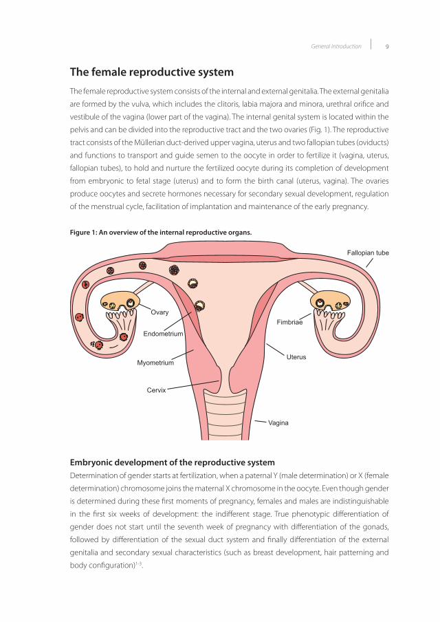
The female reproductive systemThe female reproductive system consists of the internal and external genitalia. The external genitalia
are formed by the vulva, which includes the clitoris, labia majora and minora, urethral orifice and
vestibule of the vagina (lower part of the vagina). The internal genital system is located within the
pelvis and can be divided into the reproductive tract and the two ovaries (Fig. 1). The reproductive
tract consists of the Müllerian duct-derived upper vagina, uterus and two fallopian tubes (oviducts)
and functions to transport and guide semen to the oocyte in order to fertilize it (vagina, uterus,
fallopian tubes), to hold and nurture the fertilized oocyte during its completion of development
from embryonic to fetal stage (uterus) and to form the birth canal (uterus, vagina). The ovaries
produce oocytes and secrete hormones necessary for secondary sexual development, regulation
of the menstrual cycle, facilitation of implantation and maintenance of the early pregnancy.
Figure 1: An overview of the internal reproductive organs.
Fimbriae
Uterus
Fallopian tube
Ovary
Cervix
Endometrium
Myometrium
Vagina
Figure 1: An overview of the internal reproductive organs.Embryonic development of the reproductive systemDetermination of gender starts at fertilization, when a paternal Y (male determination) or X (female
determination) chromosome joins the maternal X chromosome in the oocyte. Even though gender
is determined during these first moments of pregnancy, females and males are indistinguishable
in the first six weeks of development: the indifferent stage. True phenotypic differentiation of
gender does not start until the seventh week of pregnancy with differentiation of the gonads,
followed by differentiation of the sexual duct system and finally differentiation of the external
genitalia and secondary sexual characteristics (such as breast development, hair patterning and
body configuration)1-3.

Chapter 110
Development of the ovaries
Gonadal development starts in the caudal part of the ventromedial border of the mesonephros
when gonadal rigdes become prominent in the coelomic cavity during the fifth week of pregnancy.
These early gonads develop from migrating somatic cells, derived from the mesonephros, the
surrounding mesenchymal and coelomic epithelium, and primordial germ cells migrating from the
endodermal layer on the posterior wall of the yolk sac along the mesentery of the hindgut into the
gonad1, 2. As described earlier, until the seventh week of pregnancy the gonads are indifferent. The
initial development of the gonads into either a male or female phenotype, however, is depended
on the presence of the SRY gene, located on the male Y-chromosome3. Under the influence of SRY,
SOX9 is expressed and DAX1 is inhibited, which leads to the formation and final differentiation of
Sertoli cells and eventually gonadal development into testis. In absence of SRY, DAX1 is continuously
expressed, causing suppression of testis formation and development of the gonads into ovaries4.
The presence of viable primordial germ cells is crucial for ovarian differentiation and if primordial
germ cells fail to reach the primitive gonads or if they are abnormal, the gonads regress resulting
in streak (vestigal) ovaries2. Upon entry into the ovary, primordial germ cells nest in the secondary
sex chord, concentrated in the cortical region of the ovary, and are now called oogonia. While
most oogonia continue to proliferate by mitosis, some oogonia in the inner medulla enter the
prophase of the first meitotic division upon which they are called oocytes. These oocytes become
surrounded with granulosa cells and form primordial follicles. Meiosis of these oocytes proceeds
until the diplotene stage of the prophase of the first meitotic division and at that point is arrested
until the blockade is removed during reproductive life1, 2.
Development of the reproductive tract
The reproductive tract, consisting of the upper vagina, uterus and fallopian tubes, stems from the
embryonic paramesonephic or Müllerian duct. During the sixth week of pregnancy, the Müllerian
duct develops from a specific subset of cells in the anterior region of the coelomic epithelium
adjacent to the mesonephros. Müllerian duct initiation is dependent on WNT signaling and
under the influence of WNT4 secreted by the coelomic epithelium, LIM1 and PAX2 expressing
mesoepithelial cells invaginate, thereby creating a coelomic opening5-7. Upon invagination, the
primitive Müllerian duct extends and under the influence of WNT9B secreted by epithelial cells of
the Wolffian duct, posterior elongation is initiated and the Müllerian duct extends further towards
the cloaca8. Final outgrowth of the Müllerian duct is completed by widespread proliferation along
the developing duct and at the growing tip and as a last step, both Müllerian ducts fuse to form
the uterovaginal tube, which is completed at 16 weeks5, 9.
During the indifferent stage, both the Wolffian and the Müllerian ducts are present. If the gonads
develop into testes, testosterone secreted by the testicular Leydig cells and anti-Müllerian hormone
(AMH) secreted by testicular Sertoli cells, cause the Wolffian ducts to further differentiate in the
male reproductive tract and causes the Müllerian ducts to regress. However, if the gonads develop
into ovaries or if gonads are absent, testosterone and AMH are not secreted, and therefore the
Wolffian ducts regress and the Müllerian ducts further differentiate2.

General introduction 11
Differentiation of the primitive Müllerian duct into the components of the reproductive tract,
the upper two third of the vagina, uterus and fallopian tubes, is dependent on WNT7A expressed
by oviductal and uterine epithelial cells and WNT5A expressed by uterine, cervical and vaginal
mesenchymal cells10, 11. Next to WNT signaling, differentiation of the Müllerian duct is further
mediated by spatially restricted members of the HOX family of homeobox genes. HOXA9 is
expressed in the developing tubal epithelium, HOXA10 in the developing uterus, HOXA11 in the
lower uterine segment and cervix and HOXA13 in the upper two third of the vagina12. The lower
one third of the vagina is formed by epithelial cells from the urogenital sinus under the influence
of the Wolffian duct1. This process, however, is still poorly understood.
Development of the external genitalia
Similar to the gonads and reproductive tract, the external genitalia are indifferent during their
first development. The indifferent external genitalia are derived from mesodermal tissue near
the cloaca and in the fourth week of pregnancy the genital tubercle develops ventral from the
cloaca, flanked by a pair of genital folds and genital swellings. In the center of the genital folds,
the urogenital sinus opens into the abdomen. Under the influence of dihydrotestosterone, the
genital tubercle elongates and forms the penis, the urogenital folds fuse and enclose the urethra
and the genital swellings enlarge and fuse to form the scrotum. However, if testes are absent,
dihydrotestosterone is not synthesized and the indifferent external genitalia differentiate into a
female phenotype. Here, the genital tubercle inverts and becomes the clitoris, the genital folds
develop into the labia minora, the genital swellings become the labia majora and the urogenital
sinus forms the upper vagina and the vestibule in which the urethra and vagina open1, 2, 13.
The menstrual cycle:The menstrual cycle involves cyclic changes in the ovary and the uterus. The ovarian cycle includes
the follicular phase, ovulation and the luteal phase. The endometrial cycle includes the menses,
proliferative phase and the secretory phase. The reproductive phase of life starts at the menarche,
which marks the menses of the first menstrual cycle usually around 13 years of age, and continues
until approximately 50 years of age. The menstrual cycle is the effect of the ovary secreting
hormones during production of oocytes for fertilization. Under the control of estrogen and
progesterone the reproductive system undergoes functional and structural changes to optimize
uterine conditions for embryo implantation and subsequent placentation (Fig. 2a-b).
The uterus can be divided in two functional layers: the outer myometrium and the inner
endometrium. The endometrium facilitates implantation, development and outgrowth of
the embryo and can be divided into two layers: the functionalis and basalis. Every month, the
functionalis is shedded during menstruation, which marks the start of a new menstrual cycle (Fig.
2a). During the first two weeks of the menstrual cycle, the proliferative phase, estrogens produced
in the ovary induce proliferation of the endometrium and thereby generate a new functionalis.
In the ovary, this first phase of the menstrual cycle is called the follicular phase, during which the

Chapter 112
follicle matures and prepares to release its oocyt for fertilization. As stated, during this phase the
ovary produces estrogens crucial for endometrial proliferation (Fig. 2a). However, the cells present
in the ovary are not capable to synthesize estrogens in one step and therefore collaboration
between theca and granulosa cells is vital for estrogen production. Under the influence of the
pituitary secreted luteinizing hormone (LH), thecal cells convert cholesterol into androstenedione,
using 17α-hydroxylase, which serves as a precursor for estrogen. Upon diffusion through the basal
membrane into surrounding granulosa cells, androstenedione is then converted into estrogen
(estradiol) by aromatase and 17β-HSD under the influence of the pituitary secreted follicle-
stimulating hormone (FSH). Pituitary secretion of LH and FHS, in its turn, is under control of GnRH
secreted by the hypothalamus and inhibin, activin and estrogen secreted by the ovary. In addition
to the estrogenic effect on the endometrium, estrogens also influence the cervix by stimulation of
cervical mucus production, which allows the spermatozoa easy excess to the uterine cavity.
Figure 2: The endometrial cycle.
End
omet
rial a
ctiv
ity
Proliferative phase Secretory phaseOvulation
Normal menstrual cycle
Basalis Functionalis
End
omet
rial a
ctiv
ity
Window of implantation
Basalis Functionalis
Proliferative phase Secretory phaseOvulation
B.
A.
EstrogenProgesterone
EstrogenProgesterone
Figure 2: The endometrial cycle. (A + B) Functional and structural changes of the endometrium under the control of estrogen and progesterone during the normal menstrual cycle (A) and the window of implantation (B). Figure adapt-ed from Vd Horst et al. (2012) Mol Cell Endocrinol. 358(2):176-184.
(A + B) Functional and structural changes of the endometrium under the control of estrogen and progesterone during the normal menstrual cycle (A) and the window of implantation (B). Figure adapted from Vd Horst et al. (2012) Mol Cell Endocrinol. 358(2):176-184.

General introduction 13
After ovulation, during which the oocyt is released from the ovarian follicle into the fallopian tube,
the second half of the menstrual cycle or secretory phase starts (Fig. 2a). During this phase the
endometrium prepares for implantation of the fertilized ovum. Here, progesterone, counterbalances
the proliferative effects of estrogen and is responsible for the induction of differentiation of the
endometrium necessary for optimal implantation. The corresponding ovarian phase is called the
luteal phase, during which, progesterone is synthesized by LH stimulated ovarian conversion
of cholesterol in the corpus luteum. In contrast to estrogen production, progesterone is not
synthesized by both thecal and granulosa cells, but by luteinized granulosa cells of the follicle
alone.
Progesterone-induced endometrial differentiation is characterized by induction of secretory
activity of the glands, attraction of natural killer cells and transformation of endometrial stromal cells
into decidual cells, a process called decidualization. Furthermore, progesterone inhibits passage
of spermatozoa through the cervix by induction of very thick and acidic mucus production. If
fertilization is absent, progesterone production declines, the functional layer of the endometrium
degenerates and the menstrual cycle restarts at menses. In case a zygote is formed (Fig. 2b),
embryonic surface cells, called trophoblastic cells, will produce human chorionic gonadotropin
(HCG), stimulating the corpus luteum to continue the secretion of progesterone which inhibits
shedding of the functionalis layer of the endometrium (Fig. 2a)2, 13-17.
The role of WNT/β-catenin signaling during the menstrual cycleThe WNT signalling pathway has been shown to be a key regulator in development and disease
since the discovery of Wnt1 in 198218, 19. In humans, 18 WNT proteins have been identified and
upon binding of these WNT proteins to their Frizzled receptor the WNT/β-catenin signalling
pathway can be activated19, 20. Central to canonical WNT/β-catenin signalling is the degradation
complex, which consists of the scaffold proteins AXIN1 and AXIN2 (conductin), β-catenin, APC
(adenomatosis polyposis coli), CK1 (casein kinase I) and GSK3β (glycogen synthase kinase 3 beta).
In absence of WNT, β-catenin is phosphorylated by GSK3β and CK1, leading to its degradation.
However, upon binding of WNT, the Frizzled receptor cooperates with a member of the LRP family
and as a result, the degradation complex is dissociated and β-catenin becomes stably available in
the cytoplasm21, 22. Stabilized β-catenin can now translocate to the nucleus where it displaces the
transcription repressor Groucho (TLE), which leads to TCF/LEF transcription factor family regulated
WNT target gene transcription23.
WNT/β-catenin signaling is thought to be implicated in regulation of the regular menstrual cycle,
a process extensively described in chapter 2 of this thesis. During the proliferative phase of the
menstrual cycle increased estrogen levels stimulate WNT/β-catenin signaling in order to enhance
proliferation, while in the secretory phase, progesterone levels inhibit WNT/β-catenin signaling
thereby counterbalancing estradiol-induced proliferation and enhancing differentiation. This was
confirmed by the fact that nuclear β-catenin staining is observed during the proliferative phase
of the menstrual cycle, while nuclear β-catenin is absent during the second half of the menstrual

Chapter 114
cycle24. Furthermore, exogenous administration of estrogen resulted in accumulation of nuclear
β-catenin in endometrial cells and upon viral-induction of the WNT/β-catenin inhibitor SFRP2,
estrogen induced proliferation was inhibited25. The relationship between the menstrual cycle was
further confirmed using gene expression profiling, where WNT/β-catenin signaling activating
factors, such as WNT4, WNT5A, WNT6 and WNT7A were found to be upregulated in the proliferative
phase, in contrast to WNT/β-catenin signaling inhibitors, such as DKK1 and FOXO1, which were
upregulated during the secretory phase26-28. In addition, using data obtained from hormone
treated postmenopausal women it was shown that many targets and components of the WNT
signaling pathway were regulated by estrogen and progesterone28-30.
Endometrial cancerWorldwide, more than 288.000 women are diagnosed with endometrial cancer each year, making
it the most common gynecological malignancy and the fourth most common female malignancy
in developed countries31. In the Netherlands, in 2008, more than 1800 women were diagnosed
with endometrial cancer, accounting for an incidence of 22,4 per 100.000 women and a cumulative
risk of endometrial cancer up to 75 years of age of 1,55%31. Unfortunately, due to the increase in life
expectancy and a rising presence of endometrial cancer risk factors within the world population, a
substantial increase in endometrial cancer incidence is expected in the near future32.
Risk factorsAge is the most important risk factor for endometrial cancer as approximately seventy-five percent
of all cases occur in postmenopausal women33. Furthermore, obesity was found to be a major risk
factor due to its associated high estrogen level caused by conversion of androgen into estrogens
within the fat tissue34, 35. Next to age and obesity, other important risk factors for endometrial cancer
related to prolonged exposure to high levels of estrogens include long-term exposure to estrogen
therapy, polycystic ovary syndrome (PCOS), early menarche, late menopause and null parity33, 36-38.
Additional risk factors are long-term use of Tamoxifen, endometrial cancer family history in the first
degree, BRCA1 mutation and HNPCC family (Lynch) syndrome39-43. In contrast, factors decreasing
long term unopposed estrogen levels such as smoking, oral-contraceptive use, grand multi parity
and a diet with phytoestrogens, decrease the risk of endometrial cancer44-47.
Symptoms and diagnosisThe most prominent and early symptom of endometrial cancer is abnormal uterine bleeding or
spotting. Even though uterine bleeding is associated with many other diseases, all postmenopausal
women with uterine bleeding should be assessed for endometrial cancer. Additional symptoms
include nonspecific symptoms such as lower abdominal pain or pelvic cramps. Transvaginal
ultrasonography (TVU) is the first step in diagnosis and is used to assess the endometrial thickness
and irregularity of the endometrial-myometrial border. Final diagnosis of endometrial cancer is
done histologically using endometrial tissue obtained by Pipelle biopsy or hysteroscopy33, 48.

General introduction 15
PathologyIn case of endometrial cancer, using histological assessment of the endometrial biopsy,
endometrioid adenocarcinoma is identified in 80% of cases33. Other subtypes of endometrial
cancer are mucinous, serous, clear-cell, mixed Müllerian, squamous-cell, transitional cell, small-cell
and undifferentiated carcinoma49. Like many other types of cancer, endometrial carcinoma can be
further divided into two subgroups based on their differentiation. Most endometrial cancers are
well to moderately differentiated and are known as type I endometrial cancer. Type I endometrial
carcinomas are mainly found in postmenopausal women, generally have a good prognosis and
arise from atypical endometrial hyperplasia, which is thought to be caused by long term unopposed
estrogenic stimulation50. Type I carcinomas are frequently associated with mutations in the PTEN
tumor suppressor gene, the KRAS oncogene and the WNT/β-catenin signaling pathway51-53.
Next to type I, about 10% of all endometrial cancers are type II carcinomas. By definition, these
tumors are either poorly differentiated endometrioid or non-endometrioid carcinomas, of which
serous endometrial carcinoma is the most aggressive. Type II tumors are more common in
premenopausal women and are not caused by unopposed estrogen exposure, but are associated
with endometrial atrophy and, in case of serous carcinoma, associated with endometrial intra-
epithelial carcinoma (EIC)50, 54. Furthermore, in type II endometrial cancers, myometrial and vascular
invasion are more commonly found and patients are at high risk of recurrence and metastatic
disease33. Mutations associated with type II endometrial carcinoma are found in ERBB-2 (HER2/
NEU) and TP5355, 56. Interestingly, as in serous ovarian cancer, serous endometrial carcinomas show
nuclear accumulation of mutant P5357.
Treatment and prognosisFollowing initial diagnosis, surgery is the cornerstone of treatment and hysterectomy (either
alone or in combination with bilateral salpingo-oophorectomy and/or lymphadenectomy) by
laparoscopy or laparotomy is an adequate treatment in most cases with a 7-year survival rate of
80%33. Where there is recurrent or high stage metastatic disease, however, the situation is very
different and 5-year survival drops to 17%. Here, (neo)adjuvant radiation and/or systemic therapy
in combination with surgery is indicated and in general, progressive disease has a poor prognosis
accounting for 74.000 deaths worldwide each year (2,2 percent of all cancer related death in
women)31, 33. Important prognostic factors for recurrent and metastatic disease include FIGO
stage, tumor grade, age at diagnosis, depth of myometrial invasion, lymphovascular invasion,
immunological T-cell distribution and estrogen and progesterone receptor status58-68. In addition,
even though type II endometrial cancer only accounts for 10% of all endometrial cancer patients,
more than 50% of all endometrial cancer recurrences and deaths are related to type II disease69.
Because progesterone induced differentiation is thought to antagonize estrogen induced
endometrial proliferation, progesterone (as medroxyprogesterone acetate, MPA) is used in
palliative treatment of advanced and recurrent endometrial cancer with modest response-rates
(15-25%)70. Furthermore, MPA is used as a primary treatment for atypical endometrial hyperplasia

Chapter 116
and well differentiated endometrial carcinoma in premenopausal women determined to preserve
fertility. Here, response-rates can be up to 60%, indicating that progesterone signaling is a potent
inhibitor of carcinogenesis71, 72.
Tumor infiltrating T-lymphocytes and endometrial cancerInfiltrating solid tumor growth is thought to cause an inflammatory response similar to an acute
injury, which eventually results in infiltration of T-lymphocytes73. In several types of cancer, such
as melanoma, colorectal cancer, ovarian cancer and cervical cancer, the presence of these tumor-
infiltrating T-lymphocytes (TILs) has been extensively investigated and is associated with improved
prognosis and reduced cancer recurrence74-80. In endometrial cancer, infiltration of cytotoxic
(CD8+) T-lymphocytes within the tumor was positively correlated with improved disease free and
overall survival59, 64. Furthermore, as in ovarian cancer, a high cytotoxic/regulatory (CD8+/FOXP3+)
T-lymphocyte ratio was found to be associated with improved survival in type 1 endometrial
cancer59. In addition, low numbers of FOXP3+ T-lymphocytes were correlated with low vascular
density and estrogen receptor negativity, which are associated with improved endometrial cancer
prognosis81. However, the underling mechanisms by which TILs influence endometrial cancer
survival and recurrence is not understood.
WNT/β-catenin signaling and endometrial cancerAs described earlier, the WNT/β-catenin signaling pathway plays a rate-limiting role in maintenance
and control of the endometrium where it regulates the fine balance between proliferation (WNT-on)
and differentiation (WNT-off ) under influence of estrogen and progesterone. Therefore, a causal role
for WNT/β-catenin signaling in endometrial carcinogenesis was proposed. This role was confirmed
by the frequent finding of gene mutations in endometrial cancer, that can lead to constitutive
activation of canonical WNT/β-catenin signaling28, 82-86. In agreement to this, as measured by nuclear
β-catenin accumulation, approximately 40% of well differentiated endometrioid adenocarcinomas
actually show high levels of WNT/β-catenin signaling24, 87, 88. As indicated earlier, progesterone
induced inhibition of the WNT/β-catenin signaling pathway, for example by upregulation of DKK1
and FOXO1, was found to reduce endometrial cancer progression28, 89. Next to these more clinical
findings a number of mice models, which are extensively described in chapter 2 of this thesis,
also indicate a causal relationship between activated WNT/β-catenin signaling and endometrial
carcinogenesis90-92.
Ovarian cancerEvery year, worldwide, approximately 225.000 women are diagnosed with ovarian cancer,
accounting for 3,7% of all cancers found in women. Although this incidence is relatively low,
ovarian cancer accounts for 140.000 deaths each year, making it the most lethal gynecological
malignancy31. In the Netherlands, each year, approximately 1200 patients are diagnosed with
ovarian cancer, accounting for an incidence of 14,3 per 100.000 women and a cumulative risk of
endometrial cancer up to 75 years of age of 0,95%31.

General introduction 17
Risk factorsBecause of the high mortality of ovarian cancer, the identification of risk factors is of vital
importance. The most important risk factors are ovarian cancer specific genetic syndromes such
as the hereditary breast-ovarian cancer syndrome (BRCA1 and BRCA2 gene mutations) and Lynch
syndrome (MLH1, MSH2 and MSH6 gene mutations). The estimated lifetime risk for ovarian cancer is
35-46 percent for BRCA1 mutation carriers and 13-23 percent for BRCA2 mutation carriers. Because
of this high risk and since BRCA mutations are mainly associated with high grade serous ovarian
cancer, risk-reducing or prophylactic bilateral salpingo-oophorectomy is offered as preventive
treatment93, 94. Other risk factors include endometriosis and factors involved with a high number of
ovulations, such as: null parity, delayed childbearing, estrogen replacement therapy for more than
five years, late menopause, early menarche and a high fat diet95-99. In contrast, factors that reduce
the number of ovulations, such as oral contraceptive use, pregnancy and lactation, decrease the
risk of ovarian cancer99.
Symptoms and diagnosisThe high mortality is mainly caused by the fact that approximately 64% of women with ovarian
cancer are diagnosed at a late stage of disease (stage III or IV), where the disease has already spread
throughout the abdomen100. This delayed diagnosis is mainly caused by two factors: firstly, the
precursor lesion causing epithelial ovarian cancer is still debated amongst scientists and clinicians,
making development of tools for early detection and targeted therapy difficult. Secondly, ovarian
cancer shows late and unspecific symptoms such as fatigue, nausea, abdominal (pelvic) pain,
bloating and feeling full, symptoms commonly present in many women and in many types of
disease101.
Diagnosis of ovarian cancer commonly includes measurement of the serum CA125 level and
transvaginal ultrasonography, while internal gynecological examination is relatively sensitive for
detecting ovarian masses102. CA125, encoded by MUC16, was discovered in the eighties and is the
most frequently used biomarker for ovarian cancer. Elevated levels of serum CA125 are found in
approximately 80% of patients with advanced ovarian cancer103. However, although a combination
of CA125 level measurement and transvaginal ultrasonography is able to detect ovarian cancer
at a relatively early stage, this does not improve clinical outcome and therefore routine ovarian
cancer screening is not recommended104, 105. Furthermore, several other abdominal conditions,
such as pelvic inflammatory disease, endometriosis, functional ovarian cysts, menstruation and
pregnancy, can also result in increased CA125 levels106. Other biomarkers for ovarian carcinoma
are serum measurement of HE4, either alone or in combination with CA125 (ROMA algorithm),
and the biomarkerpanel OVA1 that includes serum measurement of CA125, β2-microglobulin,
apolipoprotein, prealbumin and transferrin107-109. Even though ultrasound and biomarker tests are
relatively good diagnostic tools, the final diagnosis of ovarian cancer is made during surgery.

Chapter 118
PathologyUpon histological diagnosis, three major types of ovarian cancer can be distinguished: epithelial
(85-95%), stromal (5-8%) and germ cell (3-5%)110. Epithelial ovarian cancer is most common in
postmenopausal women and can be divided in four distinct subtypes: serous, endometrioid,
mucinous and clear-cell ovarian cancer110. As in endometrial cancer, epithelial ovarian cancer
can be further divided in two subgroups: type I and type II111. Type I tumors include 25% of all
ovarian cancer cases, are slow growing, generally confined to the ovary, low grade and seem to
develop from endometriosis or well-established borderline lesions. Mutations associated with type
I tumors are found in PTEN, KRAS, BRAF and CTNNB1. Type II tumors account for 75% of all ovarian
cancer cases, are characterized by fast growing, highly aggressive and rapidly spreading tumors
and include high-grade serous carcinoma, carcinosarcomas and undifferentiated tumors. Genetic
mutations associated with type II disease are generally found in TP53111.
The origin of ovarian cancerFor many decades the ovarian surface epithelium (OSE) was appointed as the only origin of
epithelial ovarian cancer. Here, ovarian surface epithelial cells are thought to accumulated DNA
mutations due to repeated ovulation-induced mechanical and chemotoxic damage, followed by
entrapment of the OSE in a repaired ovulation site causing so called cortical inclusion cysts (CICs).
Under the influence of the ovarian micro-environment and additional genetic disturbances, these
CICs become metaplastic, obtain a Müllerian phenotype and eventually become malignant101.
Over the last decade, however, many researchers questioned this hypothesis for the following
reasons. Firstly the three most important epithelial ovarian subtypes strongly represent Müllerian
duct derived structures, while the OSE does not display these characteristics: serous ovarian cancer
resembles the epithelium of the fallopian tube; endometrioid ovarian cancer shows similarity to
endometrial glands; and mucinous ovarian cancer resembles the endocervical epithelium112.
Secondly, pathways and genes involved in Müllerian duct development such as WNT/β-catenin
signaling, HOX-genes and PAX-genes, are highly expressed in ovarian cancer but not in the OSE113-
121. Thirdly, upon review of fallopian tubes, early benign (P53 signatures), intermediate (serous
tubal intra-epithelial lesions, STILs) and malignant (serous tubal intra-epithelial carcinomas, STICs)
lesions were identified in patients at risk for or with a concurrent serous ovarian carcinoma122-130.
Interestingly, these malignant STICs showed similar histological and genetical characteristics as
concurrent serous ovarian cancer, which indicates a causal relationship125, 126. Fourthly, frequently
used ovarian cancer biomarkers such as CA125, PAX2 and WT1 are expressed by Müllerian duct
derived structures, but not in the OSE116, 119, 131, 132. Finally our group was able to show that a
population of stem-like cells is located in the distal and fimbriae part of the fallopian tube (near
the ovary) in mice, but not the OSE133. Upon isolation, these cells formed spheroids capable of self-
renewal and fetal calf serum (FCS) stimulation initiated differentiation of these cells into gland-like
structures with a clear Müllerian phenotype. Hence, due to their Müllerian characteristics and close
proximity to the ovary, it was hypothesized that these stem-like cells may seize ovulation induced

General introduction 19
DNA damage causing them to transform into malignant STICs, and initiate ovarian cancer133.
Based on these and other findings more extensively discussed in chapter 4 of this thesis, a
different origin of epithelial ovarian cancer was proposed: tissues derived from the Müllerian duct.
Unfortunately, good animal models aiming to confirm this hypothesis are still lacking.
Treatment and prognosisThe treatment of ovarian cancer consists of two pillars: tumor debulking surgery and (neo)adjuvant
chemotherapy. Surgical treatment involves total hysterectomy, bilateral salpingo-oophorectomy,
pelvic and paraaortic lymfadenectomy and removal of the omentum. As described before, during
surgery the final diagnosis is made and the tumor is staged. However, outcome of treatment is
highly dependent on the type, stage at diagnosis and the histological grade, with high stage
and poor cell differentiation (high grade) corresponding with poor prognosis100. Because in
most patients microscopic disease is still present after surgery, chemotherapy is an important
part of the treatment. Unfortunately, even though initially most tumors respond well, eventually
chemoresistant disease will develop and as a result, in the Netherlands overall survival of ovarian
cancer patients is only approximately 41% and in total almost 69% of patients die from the
disease100. Even more devastating, five year survival of the most frequently diagnosed stage III and
IV disease is only 28,6 and 14,1%, respectively100.
Cancer progression: epithelial to mesenchymal transition Epithelial cells are virtually incapable of migration, due to their strong cell-cell bindings, mediated
for example by E-cadherin, and the presence of the basement membranes. Migration of epithelial
cells, however, is vital during the most crucial steps of embryogenesis and to circumvent this
problem, epithelial cells are capable of transition into a more mesenchymal phenotype134.
Unfortunately, this transition of an epithelial phenotype towards a more mesenchymal phenotype
also acts as a subsequent step in progression from a confined tumor to invasive and metastatic
disease.
Central to epithelial to mesenchymal transition (EMT) is the activation of important signaling
pathways such as WNT/β-catenin, FGF, EGF and TGF-β134. Activation of these pathways results
in induction of EMT transcription factors such as SNAIL1, SLUG, ZEB1/2, TWIST1/2, GOOSEGOID
and KLF8. Upon expression, SNAIL1, SLUG, KLF8 and ZEB1/2 directly repress the activity of the
E-cadherin promotor, while TWIST1/2 and GOOSEGOID repress E-cadherin indirectly134-136. In
addition to the repression of epithelial E-cadherin, EMT transcription factors cause gain of
mesenchymal markers such as vimentin and N-cadherin134. Next to downregulation of E-cadherin
and upregulation of vimentin and N-cadherin, expression of SNAIL1 and ZEB1/2 also induces
matrix metalloproteinases (MMP), causing degradation of the basement membrane, thereby
facilitating invasion137-139. Furthermore, SNAIL1 and ZEB1 inhibit epithelial polarity by repression of
PAR, CRUMBS3 and SCRIBBLE140, 141.

Chapter 120
Eventually, EMT enables migration, invasion, intravasation, dissemination and extravasation of
tumor cells resulting in widespread metastasis (Fig. 3)142. In addition to metastasis, EMT is also an
important factor in resistance to cell death and senescence, chemo and immunotherapy and anti-
tumor immune response, and in induction of stem-like cell properties134.
Figure 3: Epithelial to mesenchymal transtion (EMT) in cancer progression.
Dysplasia Carcinoma in situ Invasive carcinoma Extravasation
Intravasation MetastasisEpithelial-mesenchymal transition
EMTEMTBasement membrane
WNTTGF-β SLUG
SNAIL
ZEB
E-cadherin Vimentin
Figure 3: Epithelial to mesenchymal transtion (EMT) in cancer progression. Upon activation, transition from an epithelial towards a more mesenchymal phenotype (EMT) enables migration, invasion, intravasation and extrava-sation of tumor cells, which can result in widespread metastasis. Figure adapted from Thiery (2002) Nat Rev Can-cer;2:442-54.
Upon activation, transition from an epithelial towards a more mesenchymal phenotype (EMT) enables migration, invasion, intravasation and extravasation of tumor cells, which can result in widespread metastasis. Figure adapted from Thiery (2002) Nat Rev Cancer;2:442-54.
Aims of the thesisThe main goal of the work presented in this thesis was to unravel the mechanisms involved in
initiation and progression of Müllerian duct derived malignancies. For this purpose, three research
questions were posed:
1. What is the effect of progesterone receptor signaling on the tumor specific immune response,
epithelial-to-mesenchymal transition and recurrence in endometrial cancer?
2. What is the effect of activation of WNT/β-catenin signaling on Müllerian duct derived tissues?
3. Are Müllerian duct derived tissues the origin of epithelial ovarian cancer; can we initiate ovarian
cancer from these tissues; and can we identify and characterize tubal precursor lesions of
serous ovarian carcinoma in controls, patients susceptible for and patients with serous ovarian
cancer?

General introduction 21
Outline of the thesisThe WNT/β-catenin signaling pathway plays a rate-limiting role in the development of many organs
and is of great importance in tissue development and homeostasis during adult live. Chapter 2
reviews the role of WNT/β-catenin signaling on the Müllerian-derived female reproductive tract,
especially focusing on its interaction with sex hormones during uterine development, pregnancy,
endometriosis and cancer. Since sex hormones were shown to interact with important pathways
involved in cancer initiation and development, the role of progesterone receptor signaling on
endometrial carcinoma was assessed in Chapter 3. In this study, using endometrial cancer cell
lines and patient tissue specimens, the role of progesterone receptor signaling on endometrial
cancer triggered immune response, cell migration, recurrence, and metastasis was investigated.
Early detection of ovarian cancer is hampered by the fact that the origin of ovarian cancer is still
debated. Over the last decades, researchers have proposed the hypothesis that epithelial ovarian
cancer originates from Müllerian derived structures and current perspectives on this Müllerian
origin of epithelial ovarian cancer are introduced and discussed in Chapter 4. Knowing that in
a high percentage of endometrioid ovarian cancers WNT/β-catenin signaling is activated, and in
view of the hypothesis that ovarian cancer may originate from the distal oviduct, in Chapter 5
we have documented an endometrioid ovarian cancer mouse model using conditional activation
of WNT/β-catenin signaling in Müllerian duct derived tissues. The role of Müllerian duct derived
tissues in epithelial ovarian cancer initiation and progression is further assessed for the human
situation in Chapter 6. Here we have investigated the prevalence of tubal precursor lesions of
serous ovarian cancer in different patient populations, studied the molecular and migratory
characteristics of the observed lesions and compared them to concurrent serous ovarian tumor.
Chapter 7 and 8 provide a summary of the results of the studies in this thesis and a general
discussion. Furthermore, directions for future research and possible clinical implications are
assessed.

Chapter 122
References1. Acien P. Embryological observations on the female genital tract. Hum Reprod 1992;7:437-45.
2. Carlson BM. Human Embryology and Developmental Biology. 3 ed: Mosby; 2004.
3. Wilhelm D, Palmer S, Koopman P. Sex determination and gonadal development in mammals. Physiol Rev 2007;87:1-28.
4. Ikeda Y, Takeda Y, Shikayama T, Mukai T, Hisano S, et al. Comparative localization of Dax-1 and Ad4BP/SF-1 during development of the hypothalamic-pituitary-gonadal axis suggests their closely related and distinct functions. Dev Dyn 2001;220:363-76.
5. Guioli S, Sekido R, Lovell-Badge R. The origin of the Mullerian duct in chick and mouse. Dev Biol 2007;302:389-98.
6. Kobayashi A, Shawlot W, Kania A, Behringer RR. Requirement of Lim1 for female reproductive tract development. Development 2004;131:539-49.
7. Masse J, Watrin T, Laurent A, Deschamps S, Guerrier D, et al. The developing female genital tract: from genetics to epigenetics. Int J Dev Biol 2009;53:411-24.
8. Carroll TJ, Park JS, Hayashi S, Majumdar A, McMahon AP. Wnt9b plays a central role in the regulation of mesenchymal to epithelial transitions underlying organogenesis of the mammalian urogenital system. Dev Cell 2005;9:283-92.
9. Orvis GD, Behringer RR. Cellular mechanisms of Mullerian duct formation in the mouse. Dev Biol 2007;306:493-504.
10. Mericskay M, Kitajewski J, Sassoon D. Wnt5a is required for proper epithelial-mesenchymal interactions in the uterus. Development 2004;131:2061-72.
11. Miller C, Sassoon DA. Wnt-7a maintains appropriate uterine patterning during the development of the mouse female reproductive tract. Development 1998;125:3201-11.
12. Podlasek C, Houston J, McKenna KE, McVary KT. Posterior Hox gene expression in developing genitalia. Evol Dev 2002;4:142-63.
13. Heineman MJ, Evers JLH, Massuger LFAG, Steegers EAP. Obstetrie en Gynaecologie. 6 ed. Maarssen: Elsevier Gezondheidszorg; 2007.
14. Gellersen B, Brosens IA, Brosens JJ. Decidualization of the human endometrium: mechanisms, functions, and clinical perspectives. Semin Reprod Med 2007;25:445-53.
15. Jungueira LC, Carneiro J. Basic Histology. 11 ed: McGraw-Hill; 2005.
16. Ryan KJ. Biochemistry of aromatase: significance to female reproductive physiology. Cancer Res 1982;42:3342s-4s.
17. Boron WF, Boulpaep EL. Medical Physiology. Updated ed. Philadelphia: Elsevier Saunders; 2005.
18. Nusse R, Varmus HE. Many tumors induced by the mouse mammary tumor virus contain a provirus integrated in the same region of the host genome. Cell 1982;31:99-109.
19. Clevers H. Wnt/[beta]-Catenin Signaling in Development and Disease. Cell 2006;127:469-80.
20. Nusse R. The Wnt Homepage. 2013 [cited; Available from: www.stanford.edu/group/nusselab/cgi-bin/wnt/
21. Pinson KI, Brennan J, Monkley S, Avery BJ, Skarnes WC. An LDL-receptor-related protein mediates Wnt signalling in mice. Nature 2000;407:535-8.
22. Behrens J, Jerchow BA, Wurtele M, Grimm J, Asbrand C, et al. Functional interaction of an axin homolog, conductin, with beta-catenin, APC, and GSK3beta. Science 1998;280:596-9.
23. Daniels DL, Weis WI. Beta-catenin directly displaces Groucho/TLE repressors from Tcf/Lef in Wnt-mediated transcription activation. Nat Struct Mol Biol 2005;12:364-71.
24. Nei H, Saito T, Yamasaki H, Mizumoto H, Ito E, et al. Nuclear localization of beta-catenin in normal and carcinogenic endometrium. Mol Carcinog 1999;25:207-18.
25. Hou X, Tan Y, Li M, Dey SK, Das SK. Canonical Wnt signaling is critical to estrogen-mediated uterine growth. Mol Endocrinol 2004;18:3035-49.
26. Cloke B, Huhtinen K, Fusi L, Kajihara T, Yliheikkila M, et al. The androgen and progesterone receptors regulate distinct gene networks and cellular functions in decidualizing endometrium. Endocrinology 2008;149:4462-74.

General introduction 23
27. Talbi S, Hamilton AE, Vo KC, Tulac S, Overgaard MT, et al. Molecular phenotyping of human endometrium distinguishes menstrual cycle phases and underlying biological processes in normo-ovulatory women. Endocrinology 2006;147:1097-121.
28. Wang Y, Hanifi-Moghaddam P, Hanekamp EE, Kloosterboer HJ, Franken P, et al. Progesterone inhibition of Wnt/beta-catenin signaling in normal endometrium and endometrial cancer. Clin Cancer Res 2009;15:5784-93.
29. Hanifi-Moghaddam P, Boers-Sijmons B, Klaassens AH, van Wijk FH, den Bakker MA, et al. Molecular analysis of human endometrium: short-term tibolone signaling differs significantly from estrogen and estrogen + progestagen signaling. J Mol Med (Berl) 2007;85:471-80.
30. Klaassens AH, van Wijk FH, Hanifi-Moghaddam P, Sijmons B, Ewing PC, et al. Histological and immunohistochemical evaluation of postmenopausal endometrium after 3 weeks of treatment with tibolone, estrogen only, or estrogen plus progestagen. Fertil Steril 2006;86:352-61.
31. Ferlay J SH, Bray F, Forman D, Mathers C and Parkin DM. GLOBOCAN 2008 v2.0, Cancer Incidence and Mortality Worldwide: IARC CancerBase No. 10 [Internet]. [cited; Available from: http://globocan.iarc.fr, accessed on day/month/year.
32. Howlader N, Noone AM, Krapcho M, Garshell J, N. N, et al. SEER Cancer Statistics Review 1975-2010. November 2012 [cited; Available from: http://seer.cancer.gov/csr/1975_2010/
33. Amant F, Moerman P, Neven P, Timmerman D, Van Limbergen E, et al. Endometrial cancer. Lancet 2005;366:491-505.
34. Bianchini F, Kaaks R, Vainio H. Overweight, obesity, and cancer risk. The Lancet Oncology 2002;3:565-74.
35. Weiderpass E, Persson I, Adami HO, Magnusson C, Lindgren A, et al. Body size in different periods of life, diabetes mellitus, hypertension, and risk of postmenopausal endometrial cancer (Sweden). Cancer Causes Control 2000;11:185-92.
36. Klip H, Burger CW, Kenemans P, van Leeuwen FE. Cancer risk associated with subfertility and ovulation induction: a review. Cancer Causes Control 2000;11:319-44.
37. Schouten LJ, Goldbohm RA, van den Brandt PA. Anthropometry, physical activity, and endometrial cancer risk: results from the Netherlands cohort study. Int J Gynecol Cancer 2006;16 Suppl 2:492.
38. Van Gorp T, Neven P. Endometrial safety of hormone replacement therapy: review of literature. Maturitas 2002;42:93-104.
39. Bergman L, Beelen ML, Gallee MP, Hollema H, Benraadt J, et al. Risk and prognosis of endometrial cancer after tamoxifen for breast cancer. Comprehensive Cancer Centres’ ALERT Group. Assessment of Liver and Endometrial cancer Risk following Tamoxifen. Lancet 2000;356:881-7.
40. Gruber SB, Thompson WD. A population-based study of endometrial cancer and familial risk in younger women. Cancer and Steroid Hormone Study Group. Cancer Epidemiol Biomarkers Prev 1996;5:411-7.
41. Hemminki K, Granstrom C. Familial clustering of ovarian and endometrial cancers. Eur J Cancer 2004;40:90-5.
42. Lynch HT, Shaw MW, Magnuson CW, Larsen AL, Krush AJ. Hereditary factors in cancer. Study of two large midwestern kindreds. Arch Intern Med 1966;117:206-12.
43. Pennington KP, Walsh T, Lee M, Pennil C, Novetsky AP, et al. BRCA1, TP53, and CHEK2 germline mutations in uterine serous carcinoma. Cancer 2013;119:332-8.
44. Hinkula M, Pukkala E, Kyyronen P, Kauppila A. Grand multiparity and incidence of endometrial cancer: a population-based study in Finland. Int J Cancer 2002;98:912-5.
45. Horn-Ross PL, John EM, Canchola AJ, Stewart SL, Lee MM. Phytoestrogen intake and endometrial cancer risk. J Natl Cancer Inst 2003;95:1158-64.
46. Lesko SM, Rosenberg L, Kaufman DW, Helmrich SP, Miller DR, et al. Cigarette smoking and the risk of endometrial cancer. N Engl J Med 1985;313:593-6.
47. Deligeoroglou E, Michailidis E, Creatsas G. Oral contraceptives and reproductive system cancer. Ann N Y Acad Sci 2003;997:199-208.
48. Clark TJ. Outpatient hysteroscopy and ultrasonography in the management of endometrial disease. Curr Opin Obstet Gynecol 2004;16:305-11.
49. Clement PB, Young RH. Endometrioid carcinoma of the uterine corpus: a review of its pathology with emphasis on recent advances and problematic aspects. Adv Anat Pathol 2002;9:145-84.
50. Bokhman JV. Two pathogenetic types of endometrial carcinoma. Gynecol Oncol 1983;15:10-7.

Chapter 124
51. Enomoto T, Inoue M, Perantoni AO, Buzard GS, Miki H, et al. K-ras activation in premalignant and malignant epithelial lesions of the human uterus. Cancer Res 1991;51:5308-14.
52. Kong D, Suzuki A, Zou TT, Sakurada A, Kemp LW, et al. PTEN1 is frequently mutated in primary endometrial carcinomas. Nat Genet 1997;17:143-4.
53. van der Horst PH, Wang Y, van der Zee M, Burger CW, Blok LJ. Interaction between sex hormones and WNT/beta-catenin signal transduction in endometrial physiology and disease. Mol Cell Endocrinol 2012;358:176-84.
54. Ambros RA, Sherman ME, Zahn CM, Bitterman P, Kurman RJ. Endometrial intraepithelial carcinoma: a distinctive lesion specifically associated with tumors displaying serous differentiation. Hum Pathol 1995;26:1260-7.
55. Okamoto A, Sameshima Y, Yamada Y, Teshima S, Terashima Y, et al. Allelic loss on chromosome 17p and p53 mutations in human endometrial carcinoma of the uterus. Cancer Res 1991;51:5632-5.
56. Santin AD. HER2/neu overexpression: has the Achilles’ heel of uterine serous papillary carcinoma been exposed? Gynecol Oncol 2003;88:263-5.
57. Wheeler DT, Bell KA, Kurman RJ, Sherman ME. Minimal uterine serous carcinoma: diagnosis and clinicopathologic correlation. Am J Surg Pathol 2000;24:797-806.
58. Cohn DE, Horowitz NS, Mutch DG, Kim SM, Manolitsas T, et al. Should the presence of lymphvascular space involvement be used to assign patients to adjuvant therapy following hysterectomy for unstaged endometrial cancer? Gynecol Oncol 2002;87:243-6.
59. de Jong RA, Leffers N, Boezen HM, ten Hoor KA, van der Zee AG, et al. Presence of tumor-infiltrating lymphocytes is an independent prognostic factor in type I and II endometrial cancer. Gynecol Oncol 2009;114:105-10.
60. Ehrlich CE, Young PC, Stehman FB, Sutton GP, Alford WM. Steroid receptors and clinical outcome in patients with adenocarcinoma of the endometrium. Am J Obstet Gynecol 1988;158:796-807.
61. Hanekamp EE, Gielen SC, Smid-Koopman E, Kuhne LC, de Ruiter PE, et al. Consequences of loss of progesterone receptor expression in development of invasive endometrial cancer. Clin Cancer Res 2003;9:4190-9.
62. Jeon YT, Park IA, Kim YB, Kim JW, Park NH, et al. Steroid receptor expressions in endometrial cancer: clinical significance and epidemiological implication. Cancer Lett 2006;239:198-204.
63. Jolly S, Vargas CE, Kumar T, Weiner SA, Brabbins DS, et al. The impact of age on long-term outcome in patients with endometrial cancer treated with postoperative radiation. Gynecol Oncol 2006;103:87-93.
64. Kondratiev S, Sabo E, Yakirevich E, Lavie O, Resnick MB. Intratumoral CD8+ T lymphocytes as a prognostic factor of survival in endometrial carcinoma. Clin Cancer Res 2004;10:4450-6.
65. Kosary CL. FIGO stage, histology, histologic grade, age and race as prognostic factors in determining survival for cancers of the female gynecological system: an analysis of 1973-87 SEER cases of cancers of the endometrium, cervix, ovary, vulva, and vagina. Semin Surg Oncol 1994;10:31-46.
66. Lindauer J, Fowler JM, Manolitsas TP, Copeland LJ, Eaton LA, et al. Is there a prognostic difference between depth of myometrial invasion and the tumor-free distance from the uterine serosa in endometrial cancer? Gynecol Oncol 2003;91:547-51.
67. van der Horst PH, Wang YY, Vandenput I, Kuhne LC, Ewing PC, et al. Progesterone Inhibits Epithelial-to-Mesenchymal Transition in Endometrial Cancer. PLoS One 2012;7.
68. Pecorelli S. Revised FIGO staging for carcinoma of the vulva, cervix, and endometrium. Int J Gynaecol Obstet 2009;105:103-4.
69. Creasman WT, Kohler MF, Odicino F, Maisonneuve P, Boyle P. Prognosis of papillary serous, clear cell, and grade 3 stage I carcinoma of the endometrium. Gynecol Oncol 2004;95:593-6.
70. Thigpen JT, Brady MF, Alvarez RD, Adelson MD, Homesley HD, et al. Oral medroxyprogesterone acetate in the treatment of advanced or recurrent endometrial carcinoma: a dose-response study by the Gynecologic Oncology Group. J Clin Oncol 1999;17:1736-44.
71. Kim YB, Holschneider CH, Ghosh K, Nieberg RK, Montz FJ. Progestin alone as primary treatment of endometrial carcinoma in premenopausal women. Report of seven cases and review of the literature. Cancer 1997;79:320-7.
72. Yahata T, Fujita K, Aoki Y, Tanaka K. Long-term conservative therapy for endometrial adenocarcinoma in young women. Hum Reprod 2006;21:1070-5.

General introduction 25
73. Clark CE, Hingorani SR, Mick R, Combs C, Tuveson DA, et al. Dynamics of the immune reaction to pancreatic cancer from inception to invasion. Cancer Res 2007;67:9518-27.
74. Clemente CG, Mihm MC, Jr., Bufalino R, Zurrida S, Collini P, et al. Prognostic value of tumor infiltrating lymphocytes in the vertical growth phase of primary cutaneous melanoma. Cancer 1996;77:1303-10.
75. de Vos van Steenwijk PJ, Heusinkveld M, Ramwadhdoebe TH, Lowik MJ, van der Hulst JM, et al. An unexpectedly large polyclonal repertoire of HPV-specific T cells is poised for action in patients with cervical cancer. Cancer Res 2010;70:2707-17.
76. Galon J, Costes A, Sanchez-Cabo F, Kirilovsky A, Mlecnik B, et al. Type, density, and location of immune cells within human colorectal tumors predict clinical outcome. Science 2006;313:1960-4.
77. Kilic A, Landreneau RJ, Luketich JD, Pennathur A, Schuchert MJ. Density of tumor-infiltrating lymphocytes correlates with disease recurrence and survival in patients with large non-small-cell lung cancer tumors. J Surg Res 2011;167:207-10.
78. Le DT, Ladle BH, Lee T, Weiss V, Yao X, et al. CD8(+) Foxp3(+) tumor infiltrating lymphocytes accumulate in the context of an effective anti-tumor response. Int J Cancer 2011;129:636-47.
79. Leffers N, Fehrmann RS, Gooden MJ, Schulze UR, Ten Hoor KA, et al. Identification of genes and pathways associated with cytotoxic T lymphocyte infiltration of serous ovarian cancer. Br J Cancer 2010;103:685-92.
80. Zhang L, Conejo-Garcia JR, Katsaros D, Gimotty PA, Massobrio M, et al. Intratumoral T cells, recurrence, and survival in epithelial ovarian cancer. N Engl J Med 2003;348:203-13.
81. Giatromanolaki A, Bates GJ, Koukourakis MI, Sivridis E, Gatter KC, et al. The presence of tumor-infiltrating FOXP3+ lymphocytes correlates with intratumoral angiogenesis in endometrial cancer. Gynecol Oncol 2008;110:216-21.
82. Hecht JL, Mutter GL. Molecular and pathologic aspects of endometrial carcinogenesis. J Clin Oncol 2006;24:4783-91.
83. Konopka B, Janiec-Jankowska A, Czapczak D, Paszko Z, Bidzinski M, et al. Molecular genetic defects in endometrial carcinomas: microsatellite instability, PTEN and beta-catenin (CTNNB1) genes mutations. J Cancer Res Clin Oncol 2007;133:361-71.
84. Moreno-Bueno G, Hardisson D, Sanchez C, Sarrio D, Cassia R, et al. Abnormalities of the APC/beta-catenin pathway in endometrial cancer. Oncogene 2002;21:7981-90.
85. Pijnenborg JM, Kisters N, van Engeland M, Dunselman GA, de Haan J, et al. APC, beta-catenin, and E-cadherin and the development of recurrent endometrial carcinoma. Int J Gynecol Cancer 2004;14:947-56.
86. Fukuchi T, Sakamoto M, Tsuda H, Maruyama K, Nozawa S, et al. Beta-catenin mutation in carcinoma of the uterine endometrium. Cancer Res 1998;58:3526-8.
87. Saegusa M, Okayasu I. Frequent nuclear beta-catenin accumulation and associated mutations in endometrioid-type endometrial and ovarian carcinomas with squamous differentiation. J Pathol 2001;194:59-67.
88. Scholten AN, Creutzberg CL, van den Broek LJ, Noordijk EM, Smit VT. Nuclear beta-catenin is a molecular feature of type I endometrial carcinoma. J Pathol 2003;201:460-5.
89. Ward EC, Hoekstra AV, Blok LJ, Hanifi-Moghaddam P, Lurain JR, et al. The regulation and function of the forkhead transcription factor, Forkhead box O1, is dependent on the progesterone receptor in endometrial carcinoma. Endocrinology 2008;149:1942-50.
90. Tanwar PS, Lee HJ, Zhang L, Zukerberg LR, Taketo MM, et al. Constitutive activation of Beta-catenin in uterine stroma and smooth muscle leads to the development of mesenchymal tumors in mice. Biol Reprod 2009;81:545-52.
91. Tanwar PS, Zhang L, Roberts DJ, Teixeira JM. Stromal deletion of the APC tumor suppressor in mice triggers development of endometrial cancer. Cancer Res 2011;71:1584-96.
92. van der Zee M, Jia Y, Wang Y, Heijmans-Antonissen C, Ewing PC, et al. Alterations in Wnt/beta-catenin and Pten signaling play distinct roles in endometrial cancer initiation and progression. J Pathol 2013.
93. Chen S, Parmigiani G. Meta-analysis of BRCA1 and BRCA2 penetrance. J Clin Oncol 2007;25:1329-33.
94. Lakhani SR, Manek S, Penault-Llorca F, Flanagan A, Arnout L, et al. Pathology of ovarian cancers in BRCA1 and BRCA2 carriers. Clin Cancer Res 2004;10:2473-81.
95. Folsom AR, Anderson JP, Ross JA. Estrogen replacement therapy and ovarian cancer. Epidemiology 2004;15:100-4.

Chapter 126
96. Prentice RL, Thomson CA, Caan B, Hubbell FA, Anderson GL, et al. Low-fat dietary pattern and cancer incidence in the Women’s Health Initiative Dietary Modification Randomized Controlled Trial. J Natl Cancer Inst 2007;99:1534-43.
97. Vo C, Carney ME. Ovarian cancer hormonal and environmental risk effect. Obstet Gynecol Clin North Am 2007;34:687-700, viii.
98. Whiteman DC, Siskind V, Purdie DM, Green AC. Timing of pregnancy and the risk of epithelial ovarian cancer. Cancer Epidemiol Biomarkers Prev 2003;12:42-6.
99. Whittemore AS. Characteristics relating to ovarian cancer risk: implications for prevention and detection. Gynecol Oncol 1994;55:S15-9.
100. van Altena AM, Karim-Kos HE, de Vries E, Kruitwagen RF, Massuger LF, et al. Trends in therapy and survival of advanced stage epithelial ovarian cancer patients in the Netherlands. Gynecol Oncol 2012;125:649-54.
101. Cannistra SA. Cancer of the Ovary. New England Journal of Medicine 2004;351:2519-29.
102. Goff BA, Mandel LS, Drescher CW, Urban N, Gough S, et al. Development of an ovarian cancer symptom index: possibilities for earlier detection. Cancer 2007;109:221-7.
103. Bast RC, Jr., Klug TL, St John E, Jenison E, Niloff JM, et al. A radioimmunoassay using a monoclonal antibody to monitor the course of epithelial ovarian cancer. N Engl J Med 1983;309:883-7.
104. Force USPST. Screening for ovarian cancer: recommendation statement. U.S. Preventive Services Task Force. Am Fam Physician 2005;71:759-62.
105. Stirling D, Evans DG, Pichert G, Shenton A, Kirk EN, et al. Screening for familial ovarian cancer: failure of current protocols to detect ovarian cancer at an early stage according to the international Federation of gynecology and obstetrics system. J Clin Oncol 2005;23:5588-96.
106. Barney SP, Muller CY, Bradshaw KD. Pelvic masses. Med Clin North Am 2008;92:1143-61, xi.
107. Anastasi E, Marchei GG, Viggiani V, Gennarini G, Frati L, et al. HE4: a new potential early biomarker for the recurrence of ovarian cancer. Tumour Biol 2010;31:113-9.
108. Ueland FR, Desimone CP, Seamon LG, Miller RA, Goodrich S, et al. Effectiveness of a multivariate index assay in the preoperative assessment of ovarian tumors. Obstet Gynecol 2011;117:1289-97.
109. Van Gorp T, Cadron I, Despierre E, Daemen A, Leunen K, et al. HE4 and CA125 as a diagnostic test in ovarian cancer: prospective validation of the Risk of Ovarian Malignancy Algorithm. Br J Cancer 2011;104:863-70.
110. Roett MA, Evans P. Ovarian cancer: an overview. Am Fam Physician 2009;80:609-16.
111. Kurman RJ, Shih Ie M. Pathogenesis of ovarian cancer: lessons from morphology and molecular biology and their clinical implications. Int J Gynecol Pathol 2008;27:151-60.
112. Karst AM, Drapkin R. Ovarian cancer pathogenesis: a model in evolution. J Oncol 2010;2010:932371.
113. Bitler BG, Nicodemus JP, Li H, Cai Q, Wu H, et al. Wnt5a suppresses epithelial ovarian cancer by promoting cellular senescence. Cancer Res 2011;71:6184-94.
114. Gatcliffe TA, Monk BJ, Planutis K, Holcombe RF. Wnt signaling in ovarian tumorigenesis. Int J Gynecol Cancer 2008;18:954-62.
115. Merritt MA, Parsons PG, Newton TR, Martyn AC, Webb PM, et al. Expression profiling identifies genes involved in neoplastic transformation of serous ovarian cancer. BMC Cancer 2009;9:378.
116. Ozcan A, Liles N, Coffey D, Shen SS, Truong LD. PAX2 and PAX8 expression in primary and metastatic mullerian epithelial tumors: a comprehensive comparison. Am J Surg Pathol 2011;35:1837-47.
117. Peng C, Zhang X, Yu H, Wu D, Zheng J. Wnt5a as a predictor in poor clinical outcome of patients and a mediator in chemoresistance of ovarian cancer. Int J Gynecol Cancer 2011;21:280-8.
118. Steg A, Wang W, Blanquicett C, Grunda JM, Eltoum IA, et al. Multiple gene expression analyses in paraffin-embedded tissues by TaqMan low-density array: Application to hedgehog and Wnt pathway analysis in ovarian endometrioid adenocarcinoma. J Mol Diagn 2006;8:76-83.
119. Tong GX, Chiriboga L, Hamele-Bena D, Borczuk AC. Expression of PAX2 in papillary serous carcinoma of the ovary: immunohistochemical evidence of fallopian tube or secondary Mullerian system origin? Mod Pathol 2007;20:856-63.
120. Varma RR, Hector SM, Clark K, Greco WR, Hawthorn L, et al. Gene expression profiling of a clonal isolate of oxaliplatin-resistant ovarian carcinoma cell line A2780/C10. Oncol Rep 2005;14:925-32.
121. Yoshioka S, King ML, Ran S, Okuda H, MacLean JA, 2nd, et al. WNT7A regulates tumor growth and progression in ovarian cancer through the WNT/beta-catenin pathway. Mol Cancer Res 2012;10:469-82.

General introduction 27
122. Callahan MJ, Crum CP, Medeiros F, Kindelberger DW, Elvin JA, et al. Primary fallopian tube malignancies in BRCA-positive women undergoing surgery for ovarian cancer risk reduction. J Clin Oncol 2007;25:3985-90.
123. Carcangiu ML, Peissel B, Pasini B, Spatti G, Radice P, et al. Incidental carcinomas in prophylactic specimens in BRCA1 and BRCA2 germ-line mutation carriers, with emphasis on fallopian tube lesions: report of 6 cases and review of the literature. Am J Surg Pathol 2006;30:1222-30.
124. Hirst JE, Gard GB, McIllroy K, Nevell D, Field M. High rates of occult fallopian tube cancer diagnosed at prophylactic bilateral salpingo-oophorectomy. Int J Gynecol Cancer 2009;19:826-9.
125. Kindelberger DW, Lee Y, Miron A, Hirsch MS, Feltmate C, et al. Intraepithelial carcinoma of the fimbria and pelvic serous carcinoma: Evidence for a causal relationship. Am J Surg Pathol 2007;31:161-9.
126. Lee Y, Miron A, Drapkin R, Nucci MR, Medeiros F, et al. A candidate precursor to serous carcinoma that originates in the distal fallopian tube. J Pathol 2007;211:26-35.
127. Manchanda R, Abdelraheim A, Johnson M, Rosenthal AN, Benjamin E, et al. Outcome of risk-reducing salpingo-oophorectomy in BRCA carriers and women of unknown mutation status. BJOG 2011;118:814-24.
128. Medeiros F, Muto MG, Lee Y, Elvin JA, Callahan MJ, et al. The tubal fimbria is a preferred site for early adenocarcinoma in women with familial ovarian cancer syndrome. Am J Surg Pathol 2006;30:230-6.
129. Mingels MJ, Roelofsen T, van der Laak JA, de Hullu JA, van Ham MA, et al. Tubal epithelial lesions in salpingo-oophorectomy specimens of BRCA-mutation carriers and controls. Gynecol Oncol 2012;127:88-93.
130. Piek JM, van Diest PJ, Zweemer RP, Jansen JW, Poort-Keesom RJ, et al. Dysplastic changes in prophylactically removed Fallopian tubes of women predisposed to developing ovarian cancer. J Pathol 2001;195:451-6.
131. Coppes MJ, Ye Y, Rackley R, Zhao XL, Liefers GJ, et al. Analysis of WT1 in granulosa cell and other sex cord-stromal tumors. Cancer Res 1993;53:2712-4.
132. Neunteufel W, Breitenecker G. Tissue expression of CA 125 in benign and malignant lesions of ovary and fallopian tube: a comparison with CA 19-9 and CEA. Gynecol Oncol 1989;32:297-302.
133. Wang Y, Sacchetti A, van Dijk MR, van der Zee M, van der Horst PH, et al. Identification of quiescent, stem-like cells in the distal female reproductive tract. PLoS One 2012;7:e40691.
134. Thiery JP, Acloque H, Huang RYJ, Nieto MA. Epithelial-Mesenchymal Transitions in Development and Disease. Cell 2009;139:871-90.
135. Peinado H, Olmeda D, Cano A. Snail, Zeb and bHLH factors in tumour progression: an alliance against the epithelial phenotype? Nat Rev Cancer 2007;7:415-28.
136. Olmeda D, Jorda M, Peinado H, Fabra A, Cano A. Snail silencing effectively suppresses tumour growth and invasiveness. Oncogene 2007;26:1862-74.
137. Jorda M, Olmeda D, Vinyals A, Valero E, Cubillo E, et al. Upregulation of MMP-9 in MDCK epithelial cell line in response to expression of the Snail transcription factor. J Cell Sci 2005;118:3371-85.
138. Miyoshi A, Kitajima Y, Sumi K, Sato K, Hagiwara A, et al. Snail and SIP1 increase cancer invasion by upregulating MMP family in hepatocellular carcinoma cells. Br J Cancer 2004;90:1265-73.
139. Yokoyama K, Kamata N, Fujimoto R, Tsutsumi S, Tomonari M, et al. Increased invasion and matrix metalloproteinase-2 expression by Snail-induced mesenchymal transition in squamous cell carcinomas. Int J Oncol 2003;22:891-8.
140. Spaderna S, Schmalhofer O, Wahlbuhl M, Dimmler A, Bauer K, et al. The transcriptional repressor ZEB1 promotes metastasis and loss of cell polarity in cancer. Cancer Res 2008;68:537-44.
141. Whiteman EL, Liu CJ, Fearon ER, Margolis B. The transcription factor snail represses Crumbs3 expression and disrupts apico-basal polarity complexes. Oncogene 2008;27:3875-9.
142. Thiery JP. Epithelial-mesenchymal transitions in tumour progression. Nat Rev Cancer 2002;2:442-54.


Chapter 2
Interaction between sexhormones and WNT/β-catenin signal transduction in endometrial
physiology and disease
Paul H. van der Horst, Yongyi Wang, Marten van der Zee, Curt W. Burger, Leen J. Blok
Department of Obstetrics and Gynaecology, Erasmus University Medical Centre Rotterdam, PO box 2040, 3000 CA Rotterdam, The Netherlands
Mol Cell Endocrinol. 2012; 358(2):176-184.

Chapter 230
Abstract:Wnt/β-catenin signalling plays a rate-limiting role in early development of many different organs
in a broad spectrum of organisms. In the developing Müllerian duct, Wnt/β-catenin signalling is
important for initiation, outgrowth, patterning and differentiation into vagina, cervix, uterus and
oviducts. In adult life, sex hormones modulate Wnt/β-catenin signalling in the endometrium to
maintain the monthly balance between estrogen-induced proliferation and progesterone-induced
differentiation, and enhanced Wnt/β-catenin signalling seems to be involved in endometrial
carcinogenesis. However, early in pregnancy enhanced Wnt/β-catenin signalling is prerequisite
for proper implantation and invasion of trophoblast cells into endometrium and myometrium
thus helping to form a placenta. Overall, it seems that tight control of Wnt/β-catenin signalling
in time and space is important for initiation, development and normal function of the female
reproductive tract. However, if Wnt/β-catenin signalling is not kept in check, it easily seems to
initiate or contribute to development of a number of uterine disorders.
Interaction between sexhormones and WNT/β-catenin signal transduction in endometrial physiology and disease 31
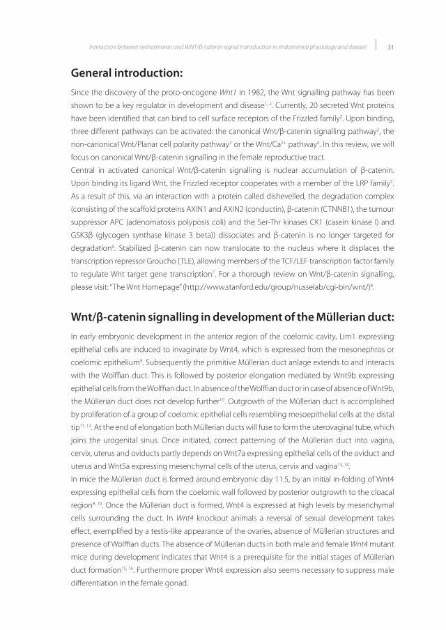
General introduction:Since the discovery of the proto-oncogene Wnt1 in 1982, the Wnt signalling pathway has been
shown to be a key regulator in development and disease1, 2. Currently, 20 secreted Wnt proteins
have been identified that can bind to cell surface receptors of the Frizzled family2. Upon binding,
three different pathways can be activated: the canonical Wnt/β-catenin signalling pathway2, the
non-canonical Wnt/Planar cell polarity pathway3 or the Wnt/Ca2+ pathway4. In this review, we will
focus on canonical Wnt/β-catenin signalling in the female reproductive tract.
Central in activated canonical Wnt/β-catenin signalling is nuclear accumulation of β-catenin.
Upon binding its ligand Wnt, the Frizzled receptor cooperates with a member of the LRP family5.
As a result of this, via an interaction with a protein called dishevelled, the degradation complex
(consisting of the scaffold proteins AXIN1 and AXIN2 (conductin), β-catenin (CTNNB1), the tumour
suppressor APC (adenomatosis polyposis coli) and the Ser-Thr kinases CK1 (casein kinase I) and
GSK3β (glycogen synthase kinase 3 beta)) dissociates and β-catenin is no longer targeted for
degradation6. Stabilized β-catenin can now translocate to the nucleus where it displaces the
transcription repressor Groucho (TLE), allowing members of the TCF/LEF transcription factor family
to regulate Wnt target gene transcription7. For a thorough review on Wnt/β-catenin signalling,
please visit: “The Wnt Homepage” (http://www.stanford.edu/group/nusselab/cgi-bin/wnt/)8.
Wnt/β-catenin signalling in development of the Müllerian duct:In early embryonic development in the anterior region of the coelomic cavity, Lim1 expressing
epithelial cells are induced to invaginate by Wnt4, which is expressed from the mesonephros or
coelomic epithelium9. Subsequently the primitive Müllerian duct anlage extends to and interacts
with the Wolffian duct. This is followed by posterior elongation mediated by Wnt9b expressing
epithelial cells from the Wolffian duct. In absence of the Wolffian duct or in case of absence of Wnt9b,
the Müllerian duct does not develop further10. Outgrowth of the Müllerian duct is accomplished
by proliferation of a group of coelomic epithelial cells resembling mesoepithelial cells at the distal
tip11, 12. At the end of elongation both Müllerian ducts will fuse to form the uterovaginal tube, which
joins the urogenital sinus. Once initiated, correct patterning of the Müllerian duct into vagina,
cervix, uterus and oviducts partly depends on Wnt7a expressing epithelial cells of the oviduct and
uterus and Wnt5a expressing mesenchymal cells of the uterus, cervix and vagina13, 14.
In mice the Müllerian duct is formed around embryonic day 11.5, by an initial in-folding of Wnt4
expressing epithelial cells from the coelomic wall followed by posterior outgrowth to the cloacal
region9, 10. Once the Müllerian duct is formed, Wnt4 is expressed at high levels by mesenchymal
cells surrounding the duct. In Wnt4 knockout animals a reversal of sexual development takes
effect, exemplified by a testis-like appearance of the ovaries, absence of Müllerian structures and
presence of Wolffian ducts. The absence of Müllerian ducts in both male and female Wnt4 mutant
mice during development indicates that Wnt4 is a prerequisite for the initial stages of Müllerian
duct formation15, 16. Furthermore proper Wnt4 expression also seems necessary to suppress male
differentiation in the female gonad.

Chapter 232
Wnt9b is expressed in the Wolffian ducts during early embryonic stages when both Wolffian and
Müllerian ducts are present (E9.5 – 14.5)10. In Wnt9b-/- embryos the Wolffian duct and the initial
Müllerian anlage are present, but there is no extension of the Müllerian duct. This indicates that
Wnt9b is necessary for posterior outgrowth during Müllerian duct formation10.
Throughout the Müllerian duct epithelium Wnt7a is expressed before birth and in oviduct and
uterine luminal epithelium after birth14. Targeted disruption of Wnt7a showed that oviducts were
absent in most mice and, when present, remained uncoiled resembling uterus morphology.
Furthermore, the uterus showed marked resemblance to the vagina with thickening of the
surrounding musculature, a relatively thin stroma, pronounced loss of glands and a luminal
epithelium with a clear squamous aspect. These data indicate that loss of Wnt7a seems to result in
posteriorization of the female reproductive tract, indicating an important role for Wnt7a in correct
patterning of the developing Müllerian duct14, 17.
In normal mice, Wnt5a is expressed in mesenchymal cells surrounding the Müllerian duct and later
in mesenchymal cells of uterus, cervix and vagina18. Wnt5a knockout female mice display normal
oviducts and anterior uterine horns, but lack the more posterior cervical and vaginal structures. The
uterine horns are severely coiled and either fused at midline or remain separated as blind ending
pouches. Because Wnt5a mutant mice die at birth due to severe kidney problems, uterine tissues
were grafted under the kidney capsule of immunodeficient mice. It was observed that in mutant
grafts, gland formation was markedly impaired. Further investigations revealed that in wild type
animals Wnt5a was highly expressed in the stromal region of the endometrium, and that Wnt5a
and Wnt7a seem to act side by side to control gland formation13.
In summary, the Wnt/β-catenin signalling pathway is important for initiation, outgrowth, patterning
and differentiation of the Müllerian duct into vagina, cervix, uterus and oviduct (Table 1).
Wnt/β-catenin signalling in uterine physiology:The human uterus can be divided in 2 functional layers: the outer myometrial layer (myometrium)
and the inner endometrial layer (endometrium). The endometrium is a dynamic tissue, which
facilitates implantation, development and outgrowth of the embryo. The endometrium can also
be divided in two layers: a functional and a basal layer. The functional layer, which is divested
every month during menses, is replenished by the basal layer during the proliferative phase of the
menstrual cycle. After menses during the first two weeks of the menstrual cycle estrogens, being
produced by ovarian thecal cells, induce proliferation of the endometrium thus generating a new
functional layer. During the second half of the menstrual cycle, the secretory phase, this functional
layer will differentiate to prepare for implantation of the fertilized ovum. During this phase
progesterone, which is produced by the corpus luteum, counterbalances estrogens proliferative
effects and is responsible for the induction of differentiation19 (Fig. 1).
In analogy to the situation in the gastrointestinal tract, where proliferating epithelial cells display
activated Wnt/β-catenin signalling and differentiated cells show diminished Wnt/β-catenin
signalling2, a central role for Wnt/β-catenin signalling was hypothesized for the endometrium.

Interaction between sexhormones and WNT/β-catenin signal transduction in endometrial physiology and disease 33
In short, during the proliferative phase of the menstrual cycle estrogens induce Wnt/β-
catenin signalling. During the secretory phase of the menstrual cycle, however, progestagens
counterbalance estrogen-induced proliferation by inhibition of Wnt/β-catenin signalling, thus
inducing differentiation. Over time there have been multiple reports in literature which corroborate
this hypothesis.
Nei et al. in 1999 observed clear nuclear localization of β-catenin during the proliferative phase
of the menstrual cycle when estrogen levels are high and unopposed by progestagens20.
Furthermore, during the secretory phase of the menstrual cycle, when progesterone levels
increase and estrogen levels decrease, nuclear β-catenin accumulation was found to decrease. In
line with these observations Hou et al., 2004, showed that exogenous estrogen treatment of mice
indeed results in nuclear localization of β-catenin in epithelial cells of the endometrium21. They also
observed that the proliferative effect of estrogens could be inhibited by adenovirus mediated in
vivo uterine delivery of Sfrp2 (a known Wnt antagonist)21. In agreement with these observations,
Gunin et al., 2004 could mimic estrogens proliferative effects on the endometrium by feeding their
mice LiCl, which is known to activate Wnt/β-catenin signalling by inhibiting Gsk3b activity22.
More data suggesting involvement of Wnt/β-catenin signalling in regulation of the menstrual
cycle came from gene expression profiling studies23-27. Wang et al., 200928 combined two large sets
of endometrial gene expression data: gene expression profiles from normal human endometrial
tissue acquired during different phases of the menstrual cycle27, and endometrial gene expression
data from postmenopausal women that were either untreated or were treated with estrogen or
estrogen+progestagen25. Combining these two data sets, large numbers of differentially expressed
genes were recognized as either downstream targets or integral parts of the Wnt/β-catenin
signalling pathway (n=228,28). For example, WNT4, WNT5a, WNT6 and WNT7a were up regulated
by estrogen during the proliferative phase of the menstrual cycle, while a number of inhibitors of
Wnt/β-catenin signalling were found up regulated by progesterone during the secretory phase of
the menstrual cycle (the complete list of regulated genes can be accessed from Supplementary
Table 1).
DKK1 and FOXO1 are two progesterone regulated Wnt/β-catenin signalling inhibitors which have
been investigated further28. Progesterone regulation of DKK1 was first observed by Kao et al., 2002,29
and Tulac et al., 2003,30, 31 in stromal cells of the human endometrium and progesterone regulation
of FOXO1 has been extensively studied by Takano et al., 2007, and Ward et al., 200832, 33. Using the
human endometrial cancer cell line Ishikawa Wang et al., 2009,28 has investigated progesterone
inhibition of Wnt/β-catenin signalling and the involvement of DKK1 and FOXO1 further. Here it was
shown that progesterone was very effective in inhibiting the TOP-Flash Wnt/β-catenin signalling
reporter in Ishikawa cells28. Furthermore, when progesterone was added to the medium and DKK1
or FOXO1 expression was inhibited by use of specific siRNAs, progesterone inhibition of Wnt/β-
catenin signalling was partly circumvented indicating that the Wnt/β-catenin signalling inhibitors
DKK1 and FOXO1 acted downstream from progesterone.
In summary, sexhormones regulate Wnt/β-catenin signalling in the endometrium to maintain
the monthly balance between estrogen-induced proliferation and progesterone-induced
differentiation (Table 1 and Fig. 1).

Chapter 234
Figure 1: Activation of Wnt/β-catenin signalling during the normal menstrual cycle (A), the window of implantation (B) and endometrial carcinogenesis (C).
End
omet
rial a
ctiv
ity
Proliferation Differentiation
Normal menstrual cycle
Basalis Functionalis
End
omet
rial a
ctiv
ity
Proliferation Decidualisation
Window of implantation
Basalis Functionalis
InvasionProliferation
End
omet
rial a
ctiv
ity
Endometrial cancer
Basalis Functionalis
B.
C.
A.
+
+ ++
++
++ ++
+ ++
+ +
+
++
+++
++
++ +
+ ++
++
++
++ +
++
+ +
Wnt/β-catenin signalling
Wnt/β-catenin signalling
Wnt/β-catenin signalling
A: During the proliferative phase of the menstrual cycle, estrogens induce Wnt/β-catenin signalling, while during the secretory phase of the menstrual cycle progestagens counterbalance estrogen-induced proliferation by inhibition of Wnt/β-catenin signalling, thus inducing differentiation. B: During implantation, blastocyst signalling to the endometrium activates the Wnt/β-catenin pathway at the site of implantation. Furthermore activation of Wnt/β-catenin signalling is a prerequisite for proper decidualization and correct invasion of trophoblast into the maternal endometrium. C: Constitutive activation of Wnt/β-catenin signalling in the endometrium induces endometrial hyperplasia, which can develop further into invasive disease. Furthermore, once a tumour has been initiated, Wnt/β-catenin signalling seems to facilitate transition from an epithelial phenotype towards a mesenchymal phenotype thus aiding endometrial cancer progression. +: represents locations where Wnt/β-catenin signalling is activated. Figure modified from Wang et al., Oncotarget 2010.

Interaction between sexhormones and WNT/β-catenin signal transduction in endometrial physiology and disease 35
Wnt/β-catenin signalling during decidualization, implantation and placenta formation:In humans, fertilization occurs within 24 to 48 hours after ovulation when the oocyte travels
through the fallopian tube towards the uterine cavity. When the embryo reaches the uterus it has
developed into a fluid filled mass of cells (blastocyst) displaying the first signs of differentiation.
Within 72 hours of reaching the uterus, the blastocyst is released from the surrounding zona
pellucida (hatching) thus exposing its surface cells (trophoblast) to the endometrial epithelium.
The first step towards implantation involves adhesion of these trophoblasts to the uterine wall
(apposition), which is followed by stabilization of binding (stable attachment). Subsequently,
invasion begins by penetration of the syncytiotrophoblasts into uterine epithelium34, 35. Ten days
after conception, the embryo has completely invaded into the endometrium and mononuclear
cytotrophoblasts start to invade the endometrium and inner third of the myometrium.
Receptivity of the endometrium for implantation depends highly on correct hormonal signalling
towards the moment of implantation (between days 20 and 24 of the menstrual cycle). Estrogens,
produced in increasing amounts during the first two weeks of the menstrual cycle, induce outgrowth
of the functional layer of the endometrium. Progesterone, being produced from the moment of
ovulation onwards, is very effective in inhibiting estrogenic effects and induces differentiation.
Differentiation is characterized by induction of secretory activity of the glands, attraction of natural
killer cells and initiation of transformation of endometrial stroma cells into decidual cells (start
of decidualization)36. This endometrial priming in humans is, in contrast to the situation in mice
where decidualization starts after implantation, a crucial step towards implantation, invasion of
trophoblasts and full decidualization of the uterine stroma37.
Based on the fact that Wnt/β-catenin signalling plays an important role in proliferation and
differentiation during normal uterine physiology and that Wnt/β-catenin signalling has an essential
function in embryonic development, a role for Wnt/β-catenin signalling in blastocyst implantation,
endometrial decidualization and placenta formation was hypothesized (recently reviewed by
Sonderegger et al., 201038).
In wild-type mice, Hayashi et al., 2009, studied the expression of different Wnt receptors (Fzd2,
Fzd3 and Fzd4) and ligands (Wnt4, Wnt5a, Wnt7a, Wnt7b, Wnt11, Wnt16) during peri-implantation
of pregnancy and it was observed that, except for Fzd6, all receptors and ligands were specifically
expressed at the site of implantation and around the moment of implantation39. Furthermore,
expression of Wnt4, Wnt7a, Wnt7b, Wnt11, Wnt16, Fzd2, Fzd4 and Fzd6 was found to be regulated
in ovariectomized mice treated with estradiol and/or progesterone39. Using Tcf/Lef-LacZ reporter
mice, Mohamed et al., 2005, actually measured activation of the Wnt/β-catenin signalling pathway
during the window of implantation40. It was observed that 4 days after fertilization, 5 – 7 bands
of transient Wnt/β-catenin activity were present in the inner circular smooth muscle layer of
the myometrium, probably marking future sites of implantation. Subsequently, at day 5 after
fertilization, the Wnt/β-catenin signalling pathway was activated in specific endometrial regions
in the vicinity of a blastocyst, indicating cross-talk between the blastocyst and the endometrium40.

Chapter 236
Furthermore, instead of assessing pregnant mice, pseudopregnant mice were injected with Wnt7a
and profound activation of Wnt/β-catenin signalling was observed throughout the exposed
region. Next, the authors showed that when mice blastocysts were treated with the Wnt/β-catenin
signalling inhibitor Sfrp2 or when high amounts of Sfrp2 were present during implantation, the
implantation rate dropped by approximately 50%40. In addition, Xie et al., 2008, inhibited Wnt/β-
catenin signalling in mice blastocysts using adenoviral delivered Dkk1 and also observed profound
inhibition of implantation in normal pseudopregnant recipients41. These investigations indicate
that in mice, embryo-induced Wnt/β-catenin signalling at the site of blastocyst attachment is
prerequisite for successful implantation40-42.
During implantation stromal cells of the endometrium undergo further decidualization. Interestingly
in humans and in pregnant mice, during the secretory phase of the menstrual cycle, progesterone
induced Wnt4 expression was shown to be responsible for Bmp2 mediated decidualization43, 44.
Wnt4 acts downstream from Bmp2 and Wnt4 conditional knockdown in mice was shown to affect
stromal cell survival, differentiation and responsiveness to progestagens45. Furthermore, Cloke et
al., 2008, indicated that next to progesterone signalling also androgen signalling was involved in
decidualization although androgen action does not seem to be mediated by the Wnt/β-catenin
signalling pathway24.
During implantation, placental formation is initiated as trophoblast cells start to invade into the
underlying decidualized maternal tissue. Subsequently, maternal blood vessels are broken down by
these invading trophoblasts, thus forming blood sinuses. In mice, these blood sinuses are invaded
by foetal vessels and capillaries (produced from the allantois) establishing the so called labyrinthine
zone46. A number of genetic mouse models support the hypothesis that Wnt/β-catenin signalling
activation is an important factor allowing trophoblast migration, placental vascularisation, chorion
allantois fusion and labyrinth function thus initiating a functional placenta. In mice, Wnt2 has been
shown to be expressed on the foetal side of the developing placenta and targeted disruption of
Wnt2, interestingly, resulted in placental defects caused by improper and defective vascularisation
of the placenta47. In addition, Wnt7b is expressed in the chorion and disruption of this gene in
mice results in embryonic death at midgestation. More in detail, chorion development was found
to be impaired as a consequence of absence of fusion (decreased cell adhesion through down
regulation of Wnt/β-catenin signalling target gene α4-integrin) between the allantois and chorion,
possibly causing a severe lack of nutrient supply from the mother48. Targeted disruption of Tcf1 or
Lef1, interestingly, also resulted in defects in the formation of the placenta due to loss of allantois-
chorion fusion49. Furthermore Fzd5 was found to be important for placenta development, as Fzd5
knock-out mice died in utero displaying poor placental vascularisation50.
In humans, many Wnt ligands and FZD receptors are detectable in placental tissues51 and recent
studies have indicated increased expression of TCF3/4 and nuclear β-catenin staining in invasive
trophoblasts during the early phases of placentation52. Furthermore, recombinant Wnt-3A
treatment of human trophoblasts induced the activity of the Wnt/β-catenin reporter TCF-luciferase,
and was shown to induce secretion of MMP2, which could help promote trophoblast migration
and invasion53. In agreement with this, treatment of primary human trophoblasts with the Wnt/β-

Interaction between sexhormones and WNT/β-catenin signal transduction in endometrial physiology and disease 37
catenin inhibitor DKK1 resulted in reduced migration and invasion52. Recently, gene expression
was studied in human embryonic stem cells that were differentiated down the trophoblast lineage
by culture with BMP4, and profound regulation of the Wnt/β-catenin pathway was observed54.
The involvement of Wnt/β-catenin signalling in migration and invasion is not a new finding. A
role for β-catenin-independent Wnt signalling in migration and invasion has also been described
for gliomas55 and breast cancer metastasis in the brain56. For β-catenin-dependent Wnt signalling
Schmalhofer et al., 2009, showed clear nuclear β-catenin staining at the invasive front of
progressive colorectal cancer, further indicating a role for Wnt/β-catenin signalling in epithelial to
mesenchymal transformation57. In addition, in endometrial cancer the Wnt/β-catenin signalling
pathway target and adhesion molecule L1CAM was also shown to be present specifically at the
leading edge of the tumour58.
In summary, blastocyst signalling to the endometrium activates the Wnt/β-catenin signalling
pathway at the site of implantation and is prerequisite for proper implantation. Activation of the
Wnt/β-catenin signalling pathway, furthermore, is a requirement for proper decidualization and
correct invasion of trophoblasts into the maternal endometrium and myometrium thus forming
the placenta (Table 1 and Fig. 1).
Wnt/β-catenin signalling in endometriosis:Endometriosis, a common and benign gynaecological disorder, is characterised by the presence
of endometrial glandular and stromal tissue outside the uterine cavity (pelvic peritoneum, on the
ovaries and in the rectovaginal septum) and is associated with pelvic pain and infertility. Because
endometriosis is an estrogen-dependent disease displaying reduced progesterone receptor levels
and resistance to progesterone therapy59-62, a role for Wnt/β-catenin signalling in development and
maintenance of the disease has been proposed.
Using gene expression profiling, indications were found that Wnt/β-catenin signalling was indeed
differentially regulated between eutopic and ectopic endometrium60, 63, 64. Furthermore, Gaetje et
al. in 2007, showed significantly higher expression of WNT7a in endometriotic tissues, most likely
caused by reduced progesterone signalling65. This is an interesting finding because WNT7A has
been described to induce HOXA10 expression which is strongly implicated in the development
of endometriosis66.
Besides endometrial tissues homing towards the abdominal cavity, there is a special form of
endometriosis which invades into the myometrium called adenomyosis. Interestingly, using a
mouse model where Wnt/β-catenin signalling was activated in the myometrium, endometrial
glands and stroma were observed to be present in the myometrium67, 68. Whether these
observations point towards an active process of endometrial tissue invading into the myometrium
or perhaps endometrial tissue is simply filling the gap generated by myometrial dystrophy, is not
entirely clear at this point.
In summary, enhanced estrogen signalling relative to inhibited progesterone signalling in ectopic
endometrium activates the Wnt/β-catenin signalling pathway, and may be a mechanism stimulating
survival, proliferation and invasion of endometrial tissue outside its normal environment (Table 1).

Chapter 238
Wnt/β-catenin signalling during endometrial carcinogenesis:Major risk factors for endometrial cancer are prolonged high levels of estrogens69. During the
normal menstrual cycle, high estrogen levels are counterbalanced each month by progesterone
during the secretory phase of the menstrual cycle. When these progesterone levels are too low,
or when estrogen levels are too high, the proliferative effect of estrogen becomes dominant and
will induce endometrial hyperplasia70. Endometrial hyperplasia can, over time, develop further into
type I endometrial cancer which makes up 90% of endometrial cancer cases70.
As indicated earlier, estrogens seem to induce Wnt/β-catenin signalling during the proliferative
phase of the menstrual cycle20 and artificial induction of Wnt/β-catenin signalling results in
endometrial hyperplasia22, 71. Based on these investigations, it was hypothesized that enhanced
Wnt/β-catenin signalling could be a causative factor in endometrial hyperplasia and in endometrial
carcinogenesis. In agreement with this Wnt/β-catenin signalling, as measured by nuclear β-catenin
staining, was found to be enhanced in about 40% of well differentiated endometrial cancers
(31%:72; 85%:73). Upon investigating the mechanism behind enhanced nuclear β-catenin staining,
activating β-catenin mutations were found in 15-40 % of endometrial tumours74, 75, truncating
APC mutations in 10% of all endometrial cancers76 and APC A1 promoter hypermethylation in
approximately 20% of endometrial cancers77. These findings seem to indicate that Wnt/β-catenin
signalling plays a significant role during endometrial carcinogenesis.
Using genetically modified mice the role of Wnt/β-catenin signalling during endometrial
carcinogenesis was investigated further. Because homozygous β-catenin deletion results in embryo
lethality, conditional knockdown was established using β-catenin gene targeting with the help of
C-recombinase, Cre78. Using this technique the β-catenin gene (Ctnnb1) is knocked out in a specific
tissue at a specific time. In Amhr2-Cre mice, Cre is expressed from E-12.5 onwards in mesenchymal
cells surrounding the Müllerian duct79, 80. In adult animals Amhr2 driven Cre-expression was clearly
observed in the myometrium but expression was much lower in endometrial stroma cells and Cre
was not expressed in epithelial cells79, 81. At birth, in β-catenin conditional knockdown animals a
smaller uterus was observed (due to decreased mesenchymal and epithelial cell proliferation) and
coiling of the oviduct was sometimes impaired (resembling the Wnt7a mutant 14)81, 82. Interestingly
in adult animals, over time myometrial cells were lost (dystrophy, resembling the Wnt7a mutant14)
and vast areas of adiposites appeared. This phenotype seems, to some extent, to resemble a
human condition called lipoleiomyoma82.
Tanwar et al., 2009, used Amhr2-Cre to induce an activating mutation of β-catenin and
macroscopically found large tumourous growths and multiple hemorrhagic sites on the uterine
surface67. Microscopically the authors observed an increase in the myometrial area and TGFβ3
positive dysplastic lesions of the myometrium (resembling human uterine leiomyomas). In addition,
endometrial stromal sarcomas and epithelial hyperplasia were observed. Finally, endometrial
glands were sometimes observed inside the myometrium, resembling a human situation called
adenomyosis (as discussed before). Recently, Tanwar et al., 2011, used Amhr2-Cre to force Apc deletion
to induce Wnt/β-catenin signalling. It was observed that besides myometrial defects these animals
displayed endometrial hyperplasia and cancer combined with defective estrogen signalling83.

Interaction between sexhormones and WNT/β-catenin signal transduction in endometrial physiology and disease 39
Table 1: Summary of WNT/β -catenin signalling in endometrial physiology and disease.
Wnt/β-catenin signalling in Müllerian duct development:
Wnt4 - Wnt4 is expressed by epithelial cells from the mesonephros or coelomic wall [9-10]
- Wnt4 is a prerequisite for Müllerian duct initiation [9-10, 15-16]
Wnt5a - Wnt5a is expressed in mesenchymal cells of the Müllerian duct [18]
- Wnt5a knockout mice lack cervical and vaginal structures [13]
Wnt7a - Wnt7a is expressed throughout the Müllerian duct epithelium [14]
- Wnt7a loss results in posteriorization of the female reproductive tract [14,17]
Wnt9a - Wnt9b is expressed in epithelial cells from the Wolffian duct, when the Müllerian duct is present
[10]
- Wnt9b is a prerequisite for posterior outgrowth of the early Müllerian duct [10]
Wnt/β-catenin signalling in uterine physiology:
DKK1 - DKK1 is progesterone induced and can inhibit Wnt/β-catenin signalling [28-31]
FOXO1 - FOXO1 is progesterone induced and can inhibit Wnt/β-catenin signalling [28, 32-33]
Gsk3b - Gsk3b inhibition leads to Wnt signaling activation and mimics estrogens induced proliferation
[22]
Sfrp2 - Sfrp2, a known Wnt antagonist, opposes the proliferative effect of estrogen [20]
Wnt/β-catenin signalling during decidualization, implantation and placenta formation
Dkk1 - Dkk1 treatment inhibits implantation in normal pseudopregnant recipients [41]
DKK1 - DKK1 treatment results in reduced trophoblast migration and invasion [52]
Fzd5 - Fzd5 knockout results in embryonic death through poor placental vascularisation
[50]
Lef1 - Lef1 targeted disruption results in defects in placental formation [49]
Sfrp2 - Sfrp2 treatment inhibits implantation in mice [40]
Tcf1 - Tcf1 targeted disruption results in defects in placental formation [49]
Wnt2 - Wnt2 targeted disruption results in defective placental vascularisation [47]
Wnt3a - Wnt3a treatment promotes trophoblast migration and invasion [53]
Wnt4 - Wnt4 is responsible for Bmp2 mediated decidualisation [43-44]
- Wnt4 knockout affects stromal cell survival, differentiation and progesterone responsiveness
[45]
Wnt7a - Wnt7a activates Wnt/β-catenin signalling in pseudopregnant mice [40]
Wnt7b - Wnt7b disruption results in embryonic death due to placental failure [48]
Wnt/β-catenin signalling in endometriosis and endometrial carcinogenesis:
Apc - Apc conditional knockdown results in endometrial hyperplasia and cancer [82-83]
- Apc conditional knockdown results in myometrial loss and reduced gland numbers
[68]
APC - APC is mutated in 10% and its promoter hypermethylated in 20% of endometrial cancers
[75-76]
β-catenin - Activating β-catenin mutations were found in 15-40 % of endometrial cancers [73-74]
- Conditional activation of β-catenin in mice results in tumour-like growths and multiple hemorrhagic sites at the uterine surface; increased myometrial area and TGFβ3 positive dysplastic lesions of the myometrium; endometrial stromal sarcomas; enlarged glands causing epithelial hyperplasia and sometimes endometrial glands were observed inside the myometrium.
[67]
- Conditional knockdown of β-catenin results in myometrial loss and areas of adipogenesis; less epithelial glands and squamous cell metaplasia.
[81]
WNT7A - WNT7A is enhanced in endometriosis and induces HOXA10 [65-66]
Not included in this summary are studies using gene expression analysis (micro-array and RT-PCR) that show WNT/β-catenin signalling involvement [23-28, 39, 54, 60, 63-64].

Chapter 240
Our own data, using Amhr2-Cre to drive Apc deletion, also indicate severe myometrial defects
and reduced endometrial gland formation as a result of induction of Wnt/β-catenin signalling in
mesenchymal cells surrounding the Müllerian duct68. However, in these animals we never observed
endometrial hyperplasia nor endometrial carcinogenesis.
Jeong et al., 2009, used the progesterone receptor to drive Cre expression in order to induce an
activating or inactivating mutation of β-catenin in all uterine cells (myometrium, stroma, glandular
epithelium and luminal epithelium)84. Both β-catenin mutations led to severe subfertility or even
infertility due to failure to undergo decidualization during embryo implantation. Furthermore,
Pgr-Cre induced constitutive β-catenin activation resulted in enlarged glands causing endometrial
hyperplasia. Conditional inactivation of β-catenin, however, resulted in less epithelial glands and
squamous cell metaplasia (resembling the Wnt7a mutant17). Recently our group has also used Pgr-
Cre to drive deletion of Apc and we observed clear endometrial hyperplasia which was sporadically
followed in time by endometrial carcinogenesis.
Recently we have been investigating progressive endometrial cancer and observed that loss of
progesterone signalling seems to release inhibition of epithelial to mesenchymal cell transition thus
facilitating tumour progression and malignant transformation. Interestingly, loss of progesterone
signalling also led to enhanced Wnt/β-catenin signalling in these progressive endometrial cancer
specimens (Van der Horst et al., submitted).
In summary, enhanced Wnt/β-catenin signalling in mesenchymal cells surrounding the Müllerian
duct results in severe myometrial problems, while continuous Wnt/β-catenin signalling in the
endometrium seems to be an important early step in endometrial carcinogenesis (Table 1 and Fig. 1).
Summary:The role of Wnt signalling in initiation, development and function of the female reproductive tract
is significant. During development, Wnt4 is essential for initiation of the Müllerian duct, Wnt9b
is essential for posterior outgrowth of the Müllerian duct and Wnt5a and Wnt7a are involved in
proper differentiation of the Müllerian duct, into vagina, cervix, uterus and oviduct10, 13, 14, 16. During
reproductive life, hormonal regulation of the menstrual cycle is mediated by estrogen induced
activation and progesterone induced inhibition of Wnt/β-catenin signalling20, 21, 23, 25, 28, 30. During
pregnancy, when the embryo is nearing its site of implantation, Wnt/β-catenin signalling is
profoundly induced and during early decidualization, Bmp2 induced Wnt4 signalling allows for
stromal survival and differentiation. Next to this, genetic models showed that Wnt2 and Wnt7b are
essential for invasion of trophoblasts that can form the interphase where exchange can take place
between mother and foetus (the placenta)40, 44, 48.
These normal functions of Wnt/β-catenin signalling, however, have a down site. Constitutively
activated Wnt/β-catenin signalling in the myometrial layer of the uterus can cause muscular
dystrophy, probably facilitating placental invasion during pregnancy. At the same time Wnt/β-
catenin signalling in the myometrium seems to induce a disorder called adenomyosis67, 68, 81, 82.

Interaction between sexhormones and WNT/β-catenin signal transduction in endometrial physiology and disease 41
Furthermore, activated Wnt/β-catenin signalling is involved in estrogen induced proliferation
of the endometrium during the first two weeks of the menstrual cycle. However, constitutive
Wnt signalling in the endometrium induces endometrial hyperplasia which may proceed to
endometrial cancer. Also, similar to Wnt/β-catenin activation during trophoblast invasion, once
a tumour has been initiated constitutive Wnt/β-catenin signalling seems to facilitate transition
from an epithelial phenotype towards a mesenchymal phenotype thus aiding endometrial cancer
progression22, 71, 74-77.

Chapter 242
References1. Nusse R, Varmus HE. Many tumors induced by the mouse mammary tumor virus contain a provirus
integrated in the same region of the host genome. Cell 1982;31:99-109.
2. Clevers H. Wnt/[beta]-Catenin Signaling in Development and Disease. Cell 2006;127:469-80.
3. Katoh M. WNT/PCP signaling pathway and human cancer (review). Oncol Rep 2005;14:1583-8.
4. Kohn AD, Moon RT. Wnt and calcium signaling: beta-catenin-independent pathways. Cell Calcium 2005;38:439-46.
5. Pinson KI, Brennan J, Monkley S, Avery BJ, Skarnes WC. An LDL-receptor-related protein mediates Wnt signalling in mice. Nature 2000;407:535-8.
6. Behrens J, Jerchow BA, Wurtele M, Grimm J, Asbrand C, et al. Functional interaction of an axin homolog, conductin, with beta-catenin, APC, and GSK3beta. Science 1998;280:596-9.
7. Daniels DL, Weis WI. Beta-catenin directly displaces Groucho/TLE repressors from Tcf/Lef in Wnt-mediated transcription activation. Nat Struct Mol Biol 2005;12:364-71.
8. Nusse R. The Wnt Homepage http://www.stanford.edu/group/nusselab/cgi-bin/wnt/.
9. Kobayashi A, Shawlot W, Kania A, Behringer RR. Requirement of Lim1 for female reproductive tract development. Development 2004;131:539-49.
10. Carroll TJ, Park JS, Hayashi S, Majumdar A, McMahon AP. Wnt9b plays a central role in the regulation of mesenchymal to epithelial transitions underlying organogenesis of the mammalian urogenital system. Dev Cell 2005;9:283-92.
11. Guioli S, Sekido R, Lovell-Badge R. The origin of the Mullerian duct in chick and mouse. Dev Biol 2007;302:389-98.
12. Orvis GD, Behringer RR. Cellular mechanisms of Mullerian duct formation in the mouse. Dev Biol 2007;306:493-504.
13. Mericskay M, Kitajewski J, Sassoon D. Wnt5a is required for proper epithelial-mesenchymal interactions in the uterus. Development 2004;131:2061-72.
14. Miller C, Sassoon DA. Wnt-7a maintains appropriate uterine patterning during the development of the mouse female reproductive tract. Development 1998;125:3201-11.
15. Stark K, Vainio S, Vassileva G, McMahon AP. Epithelial transformation of metanephric mesenchyme in the developing kidney regulated by Wnt-4. Nature 1994;372:679-83.
16. Vainio S, Heikkila M, Kispert A, Chin N, McMahon AP. Female development in mammals is regulated by Wnt-4 signalling. Nature 1999;397:405-9.
17. Parr BA, McMahon AP. Sexually dimorphic development of the mammalian reproductive tract requires Wnt-7a. Nature 1998;395:707-10.
18. Miller C, Pavlova A, Sassoon DA. Differential expression patterns of Wnt genes in the murine female reproductive tract during development and the estrous cycle. Mech Dev 1998;76:91-9.
19. Maruyama T, Yoshimura Y. Molecular and cellular mechanisms for differentiation and regeneration of the uterine endometrium. Endocr J 2008;55:795-810.
20. Nei H, Saito T, Yamasaki H, Mizumoto H, Ito E, et al. Nuclear localization of β-catenin in normal and carcinogenic endometrium. Molecular Carcinogenesis 1999;25:207-18.
21. Hou X, Tan Y, Li M, Dey SK, Das SK. Canonical Wnt signaling is critical to estrogen-mediated uterine growth. Mol Endocrinol 2004;18:3035-49.
22. Gunin AG, Emelianov VU, Mironkin IU, Morozov MP, Tolmachev AS. Lithium treatment enhances estradiol-induced proliferation and hyperplasia formation in the uterus of mice. Eur J Obstet Gynecol Reprod Biol 2004;114:83-91.
23. Catalano RD, Critchley HO, Heikinheimo O, Baird DT, Hapangama D, et al. Mifepristone induced progesterone withdrawal reveals novel regulatory pathways in human endometrium. Molecular Human Reproduction 2007;13:641-54.
24. Cloke B, Huhtinen K, Fusi L, Kajihara T, Yliheikkila M, et al. The Androgen and Progesterone Receptors Regulate Distinct Gene Networks and Cellular Functions in Decidualizing Endometrium. Endocrinology 2008;149:4462-74.
25. Hanifi-Moghaddam P, Boers-Sijmons B, Klaassens AH, van Wijk FH, den Bakker MA, et al. Molecular analysis of human endometrium: short-term tibolone signaling differs significantly from estrogen and estrogen + progestagen signaling. J Mol Med 2007;85:471-80.

Interaction between sexhormones and WNT/β-catenin signal transduction in endometrial physiology and disease 43
26. Kuokkanen S, Chen B, Ojalvo L, Benard L, Santoro N, et al. Genomic profiling of microRNAs and messenger RNAs reveals hormonal regulation in microRNA expression in human endometrium. Biol Reprod 2010;82:791-801.
27. Talbi S, Hamilton AE, Vo KC, Tulac S, Overgaard MT, et al. Molecular phenotyping of human endometrium distinguishes menstrual cycle phases and underlying biological processes in normo-ovulatory women. Endocrinology 2006;147:1097-121.
28. Wang Y, Hanifi-Moghaddam P, Hanekamp EE, Kloosterboer HJ, Franken P, et al. Progesterone inhibition of Wnt/beta-catenin signaling in normal endometrium and endometrial cancer. Clin Cancer Res 2009;15:5784-93.
29. Kao LC, Tulac S, Lobo S, Imani B, Yang JP, et al. Global Gene Profiling in Human Endometrium during the Window of Implantation. Endocrinology 2002;143:2119-38.
30. Tulac S, Nayak NR, Kao LC, Van Waes M, Huang J, et al. Identification, characterization, and regulation of the canonical Wnt signaling pathway in human endometrium. J Clin Endocrinol Metab 2003;88:3860-6.
31. Tulac S, Overgaard MT, Hamilton AE, Jumbe NL, Suchanek E, et al. Dickkopf-1, an Inhibitor of Wnt Signaling, Is Regulated by Progesterone in Human Endometrial Stromal Cells. J Clin Endocrinol Metab 2006;91:1453-61.
32. Takano M, Lu Z, Goto T, Fusi L, Higham J, et al. Transcriptional Cross Talk between the Forkhead Transcription Factor Forkhead Box O1A and the Progesterone Receptor Coordinates Cell Cycle Regulation and Differentiation in Human Endometrial Stromal Cells. Mol Endocrinol 2007;21:2334-49.
33. Ward EC, Hoekstra AV, Blok LJ, Hanifi-Moghaddam P, Lurain JR, et al. The regulation and function of the forkhead transcription factor, Forkhead box O1, is dependent on the progesterone receptor in endometrial carcinoma. Endocrinology 2008;149:1942-50.
34. Carson DD, Bagchi I, Dey SK, Enders AC, Fazleabas AT, et al. Embryo implantation. Dev Biol 2000;223:217-37.
35. Norwitz ER, Schust DJ, Fisher SJ. Implantation and the survival of early pregnancy. N Engl J Med 2001;345:1400-8.
36. Gellersen B, Brosens IA, Brosens JJ. Decidualization of the human endometrium: mechanisms, functions, and clinical perspectives. Semin Reprod Med 2007;25:445-53.
37. Dey SK, Lim H, Das SK, Reese J, Paria BC, et al. Molecular cues to implantation. Endocr Rev 2004;25:341-73.
38. Sonderegger S, Pollheimer J, Knöfler M. Wnt Signalling in Implantation, Decidualisation and Placental Differentiation - Review. Placenta 2010;31:839-47.
39. Hayashi K, Erikson DW, Tilford SA, Bany BM, Maclean JA, et al. Wnt Genes in the Mouse Uterus: Potential Regulation of Implantation. Biology of Reproduction 2009;80:989-1000.
40. Mohamed OA, Jonnaert M, Labelle-Dumais C, Kuroda K, Clarke HJ, et al. Uterine Wnt/beta-catenin signaling is required for implantation. Proc Natl Acad Sci U S A 2005;102:8579-84.
41. Xie H, Tranguch S, Jia X, Zhang H, Das SK, et al. Inactivation of nuclear Wnt-beta-catenin signaling limits blastocyst competency for implantation. Development 2008;135:717-27.
42. Mohamed OA, Dufort D, Clarke HJ. Expression and Estradiol Regulation of Wnt Genes in the Mouse Blastocyst Identify a Candidate Pathway for Embryo-Maternal Signaling at Implantation. Biology of Reproduction 2004;71:417-24.
43. Lee KY, Jeong JW, Wang J, Ma L, Martin JF, et al. Bmp2 is critical for the murine uterine decidual response. Mol Cell Biol 2007;27:5468-78.
44. Li Q, Kannan A, Wang W, DeMayo FJ, Taylor RN, et al. Bone Morphogenetic Protein 2 Functions via a Conserved Signaling Pathway Involving Wnt4 to Regulate Uterine Decidualization in the Mouse and the Human. Journal of Biological Chemistry 2007;282:31725-32.
45. Franco HL, Dai D, Lee KY, Rubel CA, Roop D, et al. WNT4 is a key regulator of normal postnatal uterine development and progesterone signaling during embryo implantation and decidualization in the mouse. FASEB J 2010.
46. Torry DS, Leavenworth J, Chang M, Maheshwari V, Groesch K, et al. Angiogenesis in implantation. J Assist Reprod Genet 2007;24:303-15.
47. Monkley SJ, Delaney SJ, Pennisi DJ, Christiansen JH, Wainwright BJ. Targeted disruption of the Wnt2 gene results in placentation defects. Development 1996;122:3343-53.
48. Parr BA, Cornish VA, Cybulsky MI, McMahon AP. Wnt7b regulates placental development in mice. Dev Biol 2001;237:324-32.

Chapter 244
49. Galceran J, Farinas I, Depew MJ, Clevers H, Grosschedl R. Wnt3a-/--like phenotype and limb deficiency in Lef1(-/-)Tcf1(-/-) mice. Genes Dev 1999;13:709-17.
50. Ishikawa T, Tamai Y, Zorn AM, Yoshida H, Seldin MF, et al. Mouse Wnt receptor gene Fzd5 is essential for yolk sac and placental angiogenesis. Development 2001;128:25-33.
51. Sonderegger S, Husslein H, Leisser C, Knöfler M. Complex Expression Pattern of Wnt Ligands and Frizzled Receptors in Human Placenta and its Trophoblast Subtypes. Placenta 2007;28:S97-S102.
52. Pollheimer J, Loregger T, Sonderegger S, Saleh L, Bauer S, et al. Activation of the canonical wingless/T-cell factor signaling pathway promotes invasive differentiation of human trophoblast. Am J Pathol 2006;168:1134-47.
53. Sonderegger S, Haslinger P, Sabri A, Leisser C, Otten JV, et al. Wingless (Wnt)-3A induces trophoblast migration and matrix metalloproteinase-2 secretion through canonical Wnt signaling and protein kinase B/AKT activation. Endocrinology 2010;151:211-20.
54. Marchand M, Horcajadas JA, Esteban FJ, McElroy SL, Fisher SJ, et al. Transcriptomic Signature of Trophoblast Differentiation in a Human Embryonic Stem Cell Model. Biol Reprod 2011.
55. Kamino M, Kishida M, Kibe T, Ikoma K, Iijima M, et al. Wnt-5a signaling is correlated with infiltrative activity in human glioma by inducing cellular migration and MMP-2. Cancer Sci 2011;102:540-8.
56. Klemm F, Bleckmann A, Siam L, Chuang HN, Rietkotter E, et al. {beta}-catenin-independent WNT signaling in basal-like breast cancer and brain metastasis. Carcinogenesis 2011;32:434-42.
57. Schmalhofer O, Brabletz S, Brabletz T. E-cadherin, beta-catenin, and ZEB1 in malignant progression of cancer. Cancer Metastasis Rev 2009;28:151-66.
58. Huszar M, Pfeifer M, Schirmer U, Kiefel H, Konecny GE, et al. Up-regulation of L1CAM is linked to loss of hormone receptors and E-cadherin in aggressive subtypes of endometrial carcinomas. J Pathol 2010;220:551-61.
59. Giudice LC, Kao LC. Endometriosis. Lancet 2004;364:1789-99.
60. Burney RO, Talbi S, Hamilton AE, Vo KC, Nyegaard M, et al. Gene expression analysis of endometrium reveals progesterone resistance and candidate susceptibility genes in women with endometriosis. Endocrinology 2007;148:3814-26.
61. Bulun SE, Cheng YH, Pavone ME, Xue Q, Attar E, et al. Estrogen receptor-beta, estrogen receptor-alpha, and progesterone resistance in endometriosis. Semin Reprod Med 2010;28:36-43.
62. Bulun SE, Cheng YH, Pavone ME, Yin P, Imir G, et al. 17Beta-hydroxysteroid dehydrogenase-2 deficiency and progesterone resistance in endometriosis. Semin Reprod Med 2010;28:44-50.
63. Kao LC, Germeyer A, Tulac S, Lobo S, Yang JP, et al. Expression profiling of endometrium from women with endometriosis reveals candidate genes for disease-based implantation failure and infertility. Endocrinology 2003;144:2870-81.
64. Wu Y, Kajdacsy-Balla A, Strawn E, Basir Z, Halverson G, et al. Transcriptional Characterizations of Differences between Eutopic and Ectopic Endometrium. Endocrinology 2006;147:232-46.
65. Gaetje R, Holtrich U, Karn T, Cikrit E, Engels K, et al. Characterization of WNT7A expression in human endometrium and endometriotic lesions. Fertility and Sterility 2007;88:1534-40.
66. Zanatta A, Rocha AM, Carvalho FM, Pereira RM, Taylor HS, et al. The role of the Hoxa10/HOXA10 gene in the etiology of endometriosis and its related infertility: a review. J Assist Reprod Genet 2010;27:701-10.
67. Tanwar PS, Lee H-J, Zhang L, Zukerberg LR, Taketo MM, et al. Constitutive Activation of Beta-Catenin in Uterine Stroma and Smooth Muscle Leads to the Development of Mesenchymal Tumors in Mice. Biology of Reproduction 2009;81:545-52.
68. Wang Y, Jia Y, Franken PF, Smits MJM, Ewing PC, et al. Loss of APC function in mesenchymal cells surrounding the Müllerian duct leads to myometrial defects in adult mice. Mol Cell Endocrinol Accepted 2011.
69. Schindler AE. Progestogen deficiency and endometrial cancer risk. Maturitas 2009;62:334-7.
70. Amant F, Moerman P, Neven P, Timmerman D, Van Limbergen E, et al. Endometrial cancer. Lancet 2005;366:491-505.
71. Polotsky AJ, Zhu L, Santoro N, Pollard JW. Lithium chloride treatment induces epithelial cell proliferation in xenografted human endometrium. Hum Reprod 2009;24:1960-7.
72. Scholten AN, Creutzberg CL, van den Broek LJ, Noordijk EM, Smit VT. Nuclear beta-catenin is a molecular feature of type I endometrial carcinoma. J Pathol 2003;201:460-5.

Interaction between sexhormones and WNT/β-catenin signal transduction in endometrial physiology and disease 45
73. Saegusa M, Okayasu I. Frequent nuclear beta-catenin accumulation and associated mutations in endometrioid-type endometrial and ovarian carcinomas with squamous differentiation. J Pathol 2001;194:59-67.
74. Hecht JL, Mutter GL. Molecular and pathologic aspects of endometrial carcinogenesis. J Clin Oncol 2006;24:4783-91.
75. Konopka B, Janiec-Jankowska A, Czapczak D, Paszko Z, Bidzinski M, et al. Molecular genetic defects in endometrial carcinomas: microsatellite instability, PTEN and beta-catenin (CTNNB1) genes mutations. J Cancer Res Clin Oncol 2007;133:361-71.
76. Pijnenborg JM, Kisters N, van Engeland M, Dunselman GA, de Haan J, et al. APC, beta-catenin, and E-cadherin and the development of recurrent endometrial carcinoma. Int J Gynecol Cancer 2004;14:947-56.
77. Moreno-Bueno G, Hardisson D, Sanchez C, Sarrio D, Cassia R, et al. Abnormalities of the APC/beta-catenin pathway in endometrial cancer. Oncogene 2002;21:7981-90.
78. Sauer B, Henderson N. Site-specific DNA recombination in mammalian cells by the Cre recombinase of bacteriophage P1. Proc Natl Acad Sci U S A 1988;85:5166-70.
79. Arango NA, Kobayashi A, Wang Y, Jamin SP, Lee HH, et al. A mesenchymal perspective of Mullerian duct differentiation and regression in Amhr2-lacZ mice. Mol Reprod Dev 2008;75:1154-62.
80. Jamin SP, Arango NA, Mishina Y, Hanks MC, Behringer RR. Requirement of Bmpr1a for Mullerian duct regression during male sexual development. Nat Genet 2002;32:408-10.
81. Deutscher E, Hung-Chang Yao H. Essential roles of mesenchyme-derived beta-catenin in mouse Mullerian duct morphogenesis. Dev Biol 2007;307:227-36.
82. Arango NA, Szotek PP, Manganaro TF, Oliva E, Donahoe PK, et al. Conditional deletion of beta-catenin in the mesenchyme of the developing mouse uterus results in a switch to adipogenesis in the myometrium. Dev Biol 2005;288:276-83.
83. Tanwar PS, Zhang L, Roberts DJ, Teixeira JM. Stromal deletion of the APC tumor suppressor in mice triggers development of endometrial cancer. Cancer Res 2011;71:1584-96.
84. Jeong JW, Lee HS, Franco HL, Broaddus RR, Taketo MM, et al. beta-catenin mediates glandular formation and dysregulation of beta-catenin induces hyperplasia formation in the murine uterus. Oncogene 2009;28:31-40.
Supplementary data associated with this article can be found, in the online version, at
doi:10.1016/j.mce.2011.06.010


Chapter 3
Progesterone inhibits epithelial-to-mesenchymal transition in Endometrial Cancer
Paul H. van der Horst1, Yongyi Wang1, Ingrid Vandenput2, Liesbeth C. Kühne1, Patricia C. Ewing3,
Wilfred F.J. van IJcken4, Marten van der Zee1, Frederic Amant2, Curt W. Burger1, Leen J. Blok1
1: Department of Obstetrics and Gynaecology, Erasmus University Medical Center Rotterdam, PO box 2040, 3000 CA Rotterdam, The Netherlands.
2: Division Gynecologic Oncology, University Hospital Gasthuisberg, Catholic University Leuven, Herestraat 49, 3000 Leuven, Belgium.
3: Department of Pathology, Erasmus University Medical Center Rotterdam, PO box 2040, 3000 CA Rotterdam, The Netherlands.
4: Department of Biomics, Erasmus University Medical Center Rotterdam, PO box 2040, 3000 CA Rotterdam, The Netherlands.
PLoS One. 2012;7(1):e30840.

Chapter 348
Abstract:Background:Every year approximately 74,000 women die of endometrial cancer, mainly due to recurrent or
metastatic disease. The presence of tumor infiltrating lymphocytes (TILs) as well as progesterone
receptor (PR) positivity has been correlated with improved prognosis. This study describes two
mechanisms by which progesterone inhibits metastatic spread of endometrial cancer: by
stimulating T-cell infiltration and by inhibiting epithelial-to-mesenchymal cell transition (EMT).
Methodology and principle findings: Paraffin sections from patients with (n=9) or without (n=9) progressive endometrial cancer
(recurrent or metastatic disease) were assessed for the presence of CD4+ (helper), CD8+ (cytotoxic)
and Foxp3+ (regulatory) T-lymphocytes and PR expression. Progressive disease was observed to
be associated with significant loss of TILs and loss of PR expression. Frozen tumor samples, used
for genome-wide expression analysis, showed significant regulation of pathways involved in
immunesurveillance, EMT and metastasis. For a number of genes, such as CXCL14, DKK1, DKK4, PEG10
and WIF1, quantitive RT-PCR was performed to verify up- or downregulation in progressive disease.
To corroborate the role of progesterone in regulating invasion, Ishikawa(IK) endometrial cancer
cell lines stably transfected with PRA (IKPRA), PRB(IKPRB) and PRA+PRB (IKPRAB) were cultured in
presence/absence of progesterone (MPA) and used for genome-wide expression analysis, Boyden-
and wound healing migration assays, and IHC for known EMT markers. IKPRB and IKPRAB cell lines
showed MPA induced inhibition of migration and loss of the mesenchymal marker vimentin at
the invasive front of the wound healing assay. Furthermore, pathway analysis of significantly MPA
regulated genes showed significant down regulation of important pathways involved in EMT,
immunesuppression and metastasis: such as IL6-, TGF-β and Wnt/β-catenin signaling.
Conclusion:Intact progesterone signaling in non-progressive endometrial cancer seems to be an important
factor stimulating immunosurveilance and inhibiting transition from an epithelial to a more
mesenchymal, more invasive phenotype.
Progesterone inhibits epithelial-to-mesenchymal transition in Endometrial Cancer 49
Introduction:Each year, worldwide, more than 287,000 women develop endometrial cancer making it the most
common gynecological cancer in the world and the fourth most common female malignancy in
developed countries1. Usually endometrial cancer is detected in an early stage and surgery is the
cornerstone of treatment. Where there is recurrent or metastatic disease, however, the situation
is different. (Neo-)Adjuvant radiation and/or systemic therapy in combination with surgery is
usually indicated and in general, progressive disease has a poor prognosis accounting for 74,000
deaths worldwide each year2, 3. Prognostic factors for recurrent and metastatic endometrial cancer
include surgical FIGO stage, grade of differentiation, histopathological subtype and myometrial
and lymphovascular invasion2, 4-7.
In several types of cancer, the presence of tumor infiltrating lymphocytes (TILs) has been
correlated with improved prognosis, and much research has been performed on this topic8-15. The
rationale is that well differentiated cancer evokes an inflammatory response similar to an acute
injury which, after sequential infiltration of different dendritic cell populations, eventually results
in T-lymphocyte infiltration16. Infiltration of TILs as a positive prognostic factor was first described
in cutaneous melanoma, where the presence of TILs was predictive for improved survival8. Galon
et al. in 2006, showed that infiltration of lymphocytes of the adaptive immune system into the
center and invasive margin of colorectal cancer was positively correlated with reduced recurrence
and improved survival10. In 2009 Kilic et al., showed that high levels of TILs within non-small-cell
lung cancer correlated with reduced recurrence and enhanced survival12. In ovarian cancer, the
presence of intratumoral T-lymphocytes was also positively correlated with improved survival and
delayed recurrence of the disease15. Furthermore, TILs in ovarian cancer were also associated with
increased levels of INF-γ, IL2 and chemokines which indicates T-cell activation and attraction15.
The presence of TILs has not been extensively investigated in endometrial cancer. In endometrial
cancer, infiltration of cytotoxic (CD8+) T-lymphocytes in the area of the lesion has been described
as an independent prognostic factor and is positively correlated to disease free- and overall
survival17, 18. In addition, a high cytotoxic T-lymphocyte/regulatory T-lymphocyte (CD8/FOXP3) ratio
has been described to be correlated to improved survival in type I endometrial cancer17.
Next to the influx of T-lymphocytes into the tumor area, the presence of progesterone receptors
(PR) is also described as an important asset in prognosis and treatment of endometrial cancer19-21.
In well differentiated endometrial cancer PR expression is usually maintained and treatment with
medroxyprogesterone acetate (MPA), of those patients with well differentiated disease who chose
to preserve fertility, is usually successful22, 23. Loss of PR, however, is a negative prognostic factor and
is associated with progressive disease in which MPA treatment is usually only temporally successful
in 15-20% of cases24.
Recently, our group has studied the mechanism through which progesterone can induce
differentiation during the normal menstrual cycle and can inhibit well differentiated endometrial
cancer growth. It was observed that progesterone treatment results in induction of expression of
two important inhibitors of Wnt/β-catenin signaling: DKK1 and FOXO125, 26. In endometrial cancer,

Chapter 350
activation of Wnt/β-catenin signaling is observed in 30-40% of well differentiated endometrioid
carcinomas27 and progesterone induced inhibition of the Wnt signaling pathway is hypothesized
to be an important mechanism to reduce cancer progression25.
In this study we aimed to investigate the role of progesterone as a direct inhibitor of the migratory
capacities of endometrial cancer cells and its role in T-lymphocyte associated inhibition of
progressive disease.
Materials and methods:Patient materials:Primary endometrial carcinoma tissue from women with (n=9) and without (n=9) a known
episode of recurrence or metastasis, was obtained from patients treated between 1997 and 2006
in the University Hospital Gasthuisberg, Catholic University Leuven, Belgium. From this point on,
non-recurrent disease is referred as non-progressive disease and recurrent/metastatic disease as
progressive disease. Histopathological grading, staging and typing were determined according to
the guidelines of the WHO and FIGO28, 29 and all tumors were revised by a pathologist experienced
in gynaecopathology (PCE). Patients with an endometrioid type and a FIGO stage I endometrial
carcinoma were included. Patients treated with radio- or chemotherapy prior to surgery, using
hormonal steroids or with a second malignancy were excluded. Complete clinical history was
obtained from all patients and follow-up was revised to date. Specimens were snap-frozen in liquid
nitrogen for RNA-isolation or fixed in formalin and embedded in paraffin for immunohistochemistry
(IHC). For microarray analysis, from 4 non-progressive and 4 progressive patients, snap frozen
tumor specimens were used. These were chosen because they contained > 80% tumor tissue and
good quality RNA could be isolated from them. For RT-PCR, 6 non-progressive and 6 progressive
snap frozen patient tissue samples were used. For IHC 9 non-progressive and 9 progressive
paraffin embedded patient tissue samples were available. Tissue and clinical data collection for the
current research study was approved by the Medical Ethical Committee of the University Hospital
Gasthuisberg and patients gave written informed consent for tissue collection and clinical data
collection for all research purposes.
Cell culture: For all cell line experiments, Ishikawa endometrial cancer cell lines stably transfected with PRA
(IKPRA-1), PRB (IKPRB-1) or PRA and PRB (IKPRAB-36) (previously described by Smit-Koopman et
al.30) were cultured and maintained in regular culture medium (DMEM/F12 Glutamax, Invitrogen,
Carlsbad, CA, USA) in the presence of 5% Fetal Calf Serum (Invitrogen) supplemented with penicillin
and streptomycin (Invitrogen). Neomycin (ICN Biomedicals, Costa Mesa, CA, USA) and hygromycin
(Invitrogen) 1:200 were used to maintain selection. For all assays, cells were cultured in DMEM/F12
Glutamax culture medium supplemented with penicillin and streptomycin (Invitrogen), containing
5% charcoal stripped FCS (Invitrogen) with addition of hygromycin and neomycin.

Progesterone inhibits epithelial-to-mesenchymal transition in Endometrial Cancer 51
Immunohistochemistry:IHC studies for CD4 (Sanbio BV, Uden, The Netherlands), CD8 (Dako, Glostrup, Denmark),
FOXP3 (Natutech, Frankfurt am Main, Germany) and PRA+PRB (Progesterone Receptor Ab-8,
Neomarkers, Fremont, CA, USA) were performed on 4 μm paraffin sections on Starfrost-slides
(Knittel, Braunschweig, Germany). Prior to incubation with the primary antibody, the slides were
deparaffinized in xylene and rehydrated to 70% ethanol. For CD4+ and CD8+ T-lymphocyte staining,
slides were microwaved at 850 Watt in Tris/EDTA pH 9.0 for 15 min. Endogenous peroxidase activity
was blocked with 30% H2O
2 in PBS for 5 min. Primary antibodies were applied at respectively
1:160 (CD4) and 1:200 (CD8) in Tris/HCl pH8.0 and incubated at room temperature for 30 min.
After washing with Tris/HCl pH8.0, sections were incubated for 30 min. at room temperature
with biotinylated secondary antibody (Dako, 1:400). After washing with Tris/HCL, the substrate
Diaminobenzidine (Dako) was used for visualization of antigen–antibody reactivity.
For FOXP3, slides were blocked (peroxidase deactivation) for 20 min at room temperature (RT) in
30% H2O
2 in methanol and boiled (antigen retrieval) in a citrate-buffer pH6.0 for 15 min. Primary
antibody was applied at 1:25 and incubated at 4°C overnight. After washing with PBS, slides were
incubated for 30 min. with a secondary rabbit-anti-rat antibody (DAKO, 1:150) and incubated for 30
min. with AB-complex (Dako). The substrate Diaminobenzidine (Dako) was used for visualization
of antigen–antibody reactivity.
For PRA+PRB staining, endogenous peroxidase activity was blocked for 5 min at RT in a 10% H2O
2 in
methanol solution and the slides were microwaved (antigen retrieval) in a microwave-oven at 850
Watt in 10nM citric acid buffer pH6.0 (DAKO) for 15 min. After cooling to room temperature slides
were washed with PBS and blocked for 30 min with 0.3% BSA/PBS. Primary antibody was applied at
1:50 and incubated at 4°C overnight. After washing with PBS, slides were incubated for 30 minutes
with a biotinylated secondary goat-anti-mouse antibody (Dako, 1:400). After the second wash the
slides were incubated for 30 min with AB-complex (Dako, 1:1:50). The substrate Diaminobenzidine
(Dako) was used for visualization of reactivity. All slides were counterstained with hematoxylin for
30s, then dehydrated and mounted.
For Vimentin staining, a wound-healing assay was performed in 2-well chamber slides (Lab-Tek,
Thermo Fisher Scientific, Waltham, MA, USA), in the presence and absence of 1 nM medroxy-
progesterone acetate (MPA), and terminated after 48 hr. The cells were washed three times with
PBS, fixed with 4% formaldehyde/PBS for 15 minutes and permeabilized with 0,3% Triton100/
PBS for 5 minutes. After washing, endogenous peroxidase activity was blocked with 10% H2O
2
in methanol for 5 minutes. Slides were washed and then blocked for 30 minutes with 0.3% BSA/
PBS. The anti-vimentin antibody (Invitrogen) was applied at 1:50 and the slides were incubated
for 30 minutes at room temperature. After washing with PBS, slides were incubated with a GFP-
fluorescent goat-anti-mouse secondary antibody (Invitrogen) at 1:500. After washing, the slides
were incubated for 5 minutes with DAPI Nucleic Acid Staining Solution (Invitrogen) for nuclear
staining. After termination of the reaction with dH2O, the slides were mounted and fluorescent
images were taken with the Axioplan 2 Imaging Fluorescent Microscope (Carl Zeiss AG, Jena,
Germany).

Chapter 352
Counting TILs:After staining, the slides were scanned with the NDP slide scanner (Hamamatsu, Hamamatsu City,
Japan) and CD4, CD8 and FOXP3 positive tumor infiltrating lymphocytes (TILs) were counted
using Image J software (National Institutes of Health, Bethesda, MD, USA). The number of TILs
was determined inside the tumor (Intratumoral), at the tumor edge (Tumor Edge) and at the
endometrial/myometrial border (EM). The complete tumor edge and endometrial/myometrial
border were evaluated for the presence of TILs. The intratumoral count was performed by counting
the TILs in 10 different randomly picked areas (1170μm x 932μm) chosen by an independent
investigator, thereby eradicating observer bias.
WST1 assay:For the WST1 proliferation assay, IKPRA-1, IKPRB-1 and IKPRAB-36 cell lines were cultured in the
absence or presence of MPA in a 96 well plate (Corning Costar, Cambridge, MA, USA). At time 0, the
cells were incubated with cell proliferation reagent WST1 (Roche, Basel, Switzerland) for 3 hours at
37 °C and absorbance was measured with the Microplate Reader (BIORAD, model 550, Hercules,
CA, USA). After baseline measurement the cell lines were cultured in the presence and absence of
1 nM MPA for 96 hours and at 96 hours, the WST1 assay was repeated.
Migration assays:For the wound-healing assay, IKPRA-1, IKPRB-1 and IKPRAB-36 cell lines were cultured in a 6-well
plate (Corning Costar). After inducing the wound, cells were incubated with 1 nM MPA for 96 hours.
Wound healing was verified every 24 hr by photography, and analyzed by measuring closure of
the wound.
For the modified Boydon assay, cells were seeded in the upper well of a modified Boydon chamber
(Transwell, 8 μm pores, 24 mm inserts, 6 well plate, Corning Costar) at 2.5 x 10⁵ cells per well in
the presence or absence of 1nM MPA. Furthermore as a control, cells were cultured in a Boyden
chamber in the presence or absence of 1nM MPA in combination with 100 nM of the anti-
progestagin Org31489 (Organon, Oss, The Netherlands). After 96 hours, cells that had migrated
through the filter into the lower well or to the bottom of the insert were trypsinized and counted
under the microscope.
Western blotting:IKPRA-1, IKPRB-1, IKPRAB-36 and IKLV-8 cell lines were cultured in the absence or presence of 1 nM
MPA for 96 hrs and subsequently lysed at 0°C in Cell Lysis Buffer (Cell Signaling Technology, Danvers,
MA, USA) for 5 minutes. Then the cells were scraped, centrifuged at 14.000 rpm for 10 minutes and
the supernatant was removed. The protein concentration was calculated using the Protein Assay Kit
(Pierce, Thermo Scientific, Rockford, IL, USA) and of each sample 4.5 μg protein in 30 μL lysisbuffer
+ BSA was loaded on a 10% SDS-PAGE gel. Western blotting was performed according to standard
procedures. The blotting paper was blocked for 30 minutes at RT with Blocking Buffer (LI-COR

Progesterone inhibits epithelial-to-mesenchymal transition in Endometrial Cancer 53
Biotechnology, Lincoln, NE, USA) and then incubated overnight at 4°C using rabbit polyclonal anti-
hFOXO1 antibody (1:5000, Bethyl Laboratories, Montgomery, TX, USA) in Blocking Buffer (LI-COR
Biotechnology). Next, the blotting membrane was incubated with the secondary goat-anti-rabbit
IgG (IRDye 800CW, 1:5000, LI-COR Biotechnology) for 30 minutes at RT and washed. As a loading
control, the membrane was incubated for 30 minutes with the monoclonal anti-β-actin (1:1000,
Sigma-Aldrich, Saint Louis, MO, USA), washed with PBS and incubated for 30 minutes with the
secondary goat-anti-mouse IgG (IRDye 680CW, 1:5000, LI-COR Biotechnology). The specific protein
bands were detected using the Odyssey Scanning System (LI-COR Biotechnology).
RNA-isolation, gene expression analyses and quantitative real-time RT-PCR:Patient tissue samples were sectioned (5 μm, cryostat) and every 10th section was HE stained and
revised by the pathologist (PCE) to assess tumor load. Only sections containing >80% tumor were
lysed in Trizol (Invitrogen) and sonified for 1 min. The PRA and PRB expressing Ishikawa cell line
(IKPRAB-36) was cultured for 48h in the absence or presence of 1nM MPA (n = 3), placed on ice and
lysed in Trizol (Invitrogen).
Phase separation was accomplished with 0.2 ml chloroform and centrifugation for 15 min. The
supernatant was transferred and isopropanol was added for RNA precipitation. The precipitated
RNA was washed with 75% ethanol. All RNA was cleaned with the Rneasy Minelute cleanup kit
(Qiagen, Venlo, The Netherlands). Amount and quality of the RNA was assessed by using the
Nanodrop (Nanodrop, Wilmington, DE, USA) and Bio-analyzer (Aligent, Santa Clara, CA, USA).
RNA isolated from patient and cell line material was labeled according to Affymetrix labeling
protocols and labeled RNA was applied to genome-wide expression arrays (Affymetrix U133plus2
GeneChips containing 54,614 probe sets, representing approximately 47.000 transcripts (Affymetrix,
Santa Clara, CA, USA)). Using RMA (Robust Multi-array Analysis31), normalization of raw data was
performed to be able to produce gene lists and eventually calculate significantly regulated genes
using SAM (Stanford University, Stanford, CA, USA32). Lists of SAM regulated genes (1.25 fold or
more; delta-values resembling p<0.05) were loaded in the Ingenuity pathway assist software to
assess the involvement of different biological pathways (Ingenuity, Redwood City, CA, USA). For the
patient materials raw lists of regulated genes (1.25 fold or more) were loaded in Ingenuity.
All micro-array data is MIAME compliant and raw data has been deposited in the MIAME compliant
GEO database under series: GSE29437 (consisting of GSE29435: cell line data; and GSE29436:
patient data).
Genes for quantitative real-time RT-PCR were identified by micro-array analysis and pathway
analysis. RNA was transcribed into cDNA with the use of the Affymetrix one-cycle cDNA synthesis
kit (Affymetrix). For identified genes, primers were ordered and tested (a list of primers is included
in Table S1). The housekeeping gene β-actin was used as a reference gene. RT-PCR was performed
and analyzed using the CFX RT-PCR system (Bio-Rad, Veenendaal, The Netherlands).

Chapter 354
Statistics:For the statistical analyses of the CD4+, CD8+ and FOXP3+ cell counts, modified Boyden chamber
assay data, WST1 assay data and RT-PCR data, SPSS 15.0 was used (IBM, Armonk, NY, USA). For
normal distributed data a t-test and for skewed data a Mann-Whitney U-test was performed to
assess P-values. A P-value < 0.05 was considered statistically significant. To calculate the p-value of
regulated pathways, Ingenuity pathway assist software uses a Fisher’s exact test.
Results:Table 1: Clinical characteristics of the included patients.
Non-progressive (n=9) Progressive (n=9) P-value
Patients 1-9 Patients 10-18
Age - year Mean 68,5 68,6 p = 0,606
Range 54-85 59-73
BMI Mean 28,3 32 p = 0,284
Sd 6,1 4,7
Histological type
no. (%) EndometrioidMixed
9 (100)0 ( -- )
8 (88,9)1 (11,1)
FIGO stage no. (%) Ia 4 (44,4) 7 (77,8)
Ib 5 (55,6) 2 (22,2)
Tumor grade no. (%) 1 2 (22,2) 5 (55,6)
2 3 (33,3) 1 (11,1)
3 4 (44,5) 3 (33,3)
Current status no. (%) NED 8 (88,9) 3 (33,3)
DOD 1 (11,1) 6 (66,7)
Recurrence no. (% No 9 (100) 0 ( -- )
Yes 0 ( -- ) 9 (100)
Metastasis no. (%) No 9 (100) 5 (55,6)
Yes 0 ( -- ) 4 (44,4)
Chemotherapy no. 0 0
Radiotherapy no. 0 1
Table 1 shows the characteristics of the patients included in the study. A p-value of < 0.05 was considered as statistically significant. BMI= body mass index; NED= no evidence of disease; DOD=death of disease.

Progesterone inhibits epithelial-to-mesenchymal transition in Endometrial Cancer 55
Patient characteristics (Table 1): Patients with (n=9) and without (n=9) progressive endometrial cancer were included. All included
patients underwent primary total abdominal- or laparoscopically assisted vaginal hysterectomy
and a bilateral salpingo-oophorectomy combined with lymph node removal. None of the
women received chemotherapy and only one woman in the progressive disease group was
given radiotherapy after surgery. Histopathological subtypes were endometrioid (n=17) and
mixed endometrioid/mucinoid (n=1). Tumor grades were 1 (n=7), 2 (n=4) and 3 (n=7) and FIGO
stages were Ia (n=11) and Ib (n=7). In the progressive disease group all 9 patients had one or
more episodes of local recurrence and 4 patients developed one or multiple distant metastases.
Recurrences were vaginal, pelvic or (retro)peritoneal, and metastatic sites were the lungs (n=3),
liver (n=1), spleen (n=1) and brain (n=1). Clinical follow-up to date was available for all patients.
In the non-progressive group 8 patients are currently free of disease and 1 patient died in follow-
up. In the progressive disease group 3 patients are free of disease and 6 patients died from their
endometrial cancer related disease. Patient characteristics are detailed in Table 1.
Progesterone receptor status and detection of CD4+ T-helper, CD8+ cytotoxic T-cells and FOXP3+ regulatory T-cells in non- progressive and progressive diseaseThe presence of tumor infiltrating lymphocytes has been correlated to prolonged survival in
endometrial cancer17, 18. Furthermore, loss of progesterone receptor (PR) expression in endometrial
cancer has been found to be a risk factor for progressive disease33. In order to substantiate
the relationship between intact PR signaling and the presence of infiltrating lymphocytes in
non-progressive disease, immunohistochemical staining and, when appropriate, quantitative
measurements were performed.
As exemplified in Fig. 1A, in progressive disease immunohistochemical staining for CD4+, CD8+
and FOXP3+ T-lymphocytes seems reduced as compared to staining in non-progressive disease.
Quantification of the number of CD4+, CD8+ and FOXP3+ T-lymphocytes in progressive disease
indeed confirmed a lower number of positive cells located on the endometrial-myometrial
border (Fig 1B, EM), at the edge of the tumor (Fig 1B, Tumor Edge) and within the tumor (Fig.
1B, Intratumoral). Whether the reduced cell counts were significantly different between the non-
progressive and progressive endometrial cancer tissues is indicated in the Figure (Fig. 1B).
Furthermore, reviewing consecutive sections in non-progressive disease for expression of
progesterone receptors (PR) revealed that the presence of CD4+ and CD8+ T-lymphocytes was
positively correlated with the presence of PR staining (Fig. 1C and 1D).
Genome-wide expression analyses of primary endometrial carcinoma tissueTo investigate whether the correlation between PR signaling and the presence of tumor infiltrating
lymphocytes could indicate a causative relationship, a genome-wide mRNA expression analysis
on snap-frozen primary endometrial carcinoma specimens from 4 patients without and 4 patients
with progressive disease was performed.

Chapter 356
Figure 1: Expression and histological distribution of PRA+PRB and CD4+, CD8+ and Foxp3+ T-lymphocytes in primary endometrial carcinoma specimens.
Tumor Edge Intratumoral EM border
Tumor Edge Intratumoral EM border
Non-progressive diseaseProgressive disease
Non-progressive diseaseProgressive disease
Tumor Edge Intratumoral EM border
Non-progressive diseaseProgressive disease
# C
ell
# C
ell
# C
ell
Non-progressive diseaseA BProgressive disease CD4+ T-lymphocytes
CD8+ T-lymphocytes
FOXP3+ T-lymphocytes
CD4
CD8
CD4
CD8
FOXP3 FOXP3
*
*
*
*
*
*
CD4 Patient#2 Patient#6 Patient#11 Patient#12
CD8
PRAB
C Non-progressive disease D Progressive disease
A: Overview of immunohistochemical staining for CD4, CD8 and FOXP3 in primary endometrial cancer specimens in non-progressive disease (n=9) compared to progressive disease (n=9) (magnification 0,4x, inlay 10x). Non-progressive disease shows pronounced staining, whereas progressive disease shows reduced staining. The scale-bar represents 10 mm. B: Quantification of CD4, CD8 and FOXP3 cell counts on the tumor edge (Tumor Edge), in the tumor (Intratumoral) and on the endometrial-myometrial border (EM border). *indicates a p-value <0.05 (Mann-Whitney U-test). C and D: Representative non-progressive (C) and progressive (D) patient tissues were stained for CD4, CD8 and PRA+PRB and show a positive correlation between the presence of TILs and the expression of PR. Magnification is 5x and the scale-bar represents 1 mm. Patients 6 and 11 were both included in the micro-array analyses. Furthermore patient 11 had only recurrent disease, while patient 12 had recurrent and metastatic disease.

Progesterone inhibits epithelial-to-mesenchymal transition in Endometrial Cancer 57
At the individual gene level it was observed that a marked number of chemokines and cytokines
were differentially regulated between non-progressive and progressive disease (Table S2). For
example, the chemokines CCL21 (-1.5x), CXCL9 (-2.9x), CXCL10 (-2.1x) and CXCL14 (three data sets
present: -33.0x; -20.5x; -6.4x, respectively) were all down regulated in progressive disease while the
cytokines IL8 (2.0x; 5.7x; 9.5x) and IL32 (1.9x) were up-regulated in progressive disease (Table S2).
Furthermore, earlier work from our group has indicated activation of Wnt/β catenin signaling in
progressive disease25 and in agreement with this a number of Wnt/β-catenin inhibitory- and target
genes were lost from progressive disease (DKK1, DKK4 and WIF1) (Table S2).
Interestingly, a number of the above mentioned genes which were down-regulated in progressive
disease, have been described in literature to be up-regulated by progesterone (CXCL1434 , DKK125,
MMP735 and SFRP436). This is in agreement with the finding that PR expression (at protein and
mRNA expression level (Fig. 1C and 1D and Table S2) is down regulated in progressive disease.
Upon reviewing pathways regulated between non-progressive and progressive disease,
regulation of a number of pathways involved in carcinogenesis and invasive disease and involved
in immunosurveillance was found to be significantly regulated: Integrin Signaling, Molecular
Mechanisms of Cancer, Antigen Presentation Pathway, Non-Small Cell Lung Cancer Signaling, IGF-
1 Signaling, Role of Tissue Factor in Cancer, Leukocyte Extravasation Signaling, ERK/MAPK Signaling,
Colorectal Cancer Metastasis Signaling (which includes Wnt/β catenin signaling), FGF Signaling,
FAK Signaling, etc (the complete list of regulated pathways and their consecutive p-values can be
accessed from Table S3).
For a number of genes (CXCL14, DKK1, DKK4, PEG10 and WIF1) a quantitative real-time RT-PCR was
performed in order to verify regulation (Fig. 2).
Figure 2: RT-PCR results of genes of interest in the patient samples.
p= 0.05 p= 0.065 p= 0.065
p= 0.004 p= 0.05
ProgressiveNon-progressive ProgressiveNon-progressive ProgressiveNon-progressive
ProgressiveNon-progressive ProgressiveNon-progressive
CXCL14 DKK1 DKK4
WIF1 PEG10
CXCL14, DKK1, DKK4, WIF1 and PEG10 were selected from the micro-array results and verified with real time RT-PCR. Significance was calculated using a Mann-Whitney U-test. A p-value of 0.05 was considered to be statistically significant.

Chapter 358
Figure 3: Progesterone induced inhibition of migration in a wound-healing assay.
IKPRA-1
0 24 48 72 960
20
40
60
80
100
Time (hrs)
Perc
enta
ge
IKPRB-1
0 24 48 72 960
20
40
60
80
100
Time (hrs)
IKPRAB-36
0 24 48 72 960
20
40
60
80
100
Time (hrs)
A
D
E
B C
IKPRB-1Control
IKPRB-1MPA
IKPRB-36Control
IKPRB-36MPA
IKPRA-1
IKPRB-1
IKPRAB-36
Control MPA Control MPA Control MPANuclear DAPI Vimentin Overlay DAPI/Vimentin
IKPRA-1 (A), IKPRB-1 (B) and IKPRAB-36 (C) cells were cultured in the absence (white bullets) or presence (black bullets) of 1 nM MPA and used for a wound-healing assay (n = 3) and closure of the wound was measured as a percentage of total closure (100% means the wound is open, 0% means the wound has closed). D shows representative images of the process of wound-healing with in red the wound. E shows IF for nuclei (DAPI) and vimentin expression on the invasive front of the manually inflicted wound. In this figure, the wound was always situated on the right side.

Progesterone inhibits epithelial-to-mesenchymal transition in Endometrial Cancer 59
Effect of progesterone on migration of the Ishikawa endometrial cancer cell linesIn order to further corroborate the possible role for progesterone in regulating invasion, Ishikawa
endometrial carcinoma cell lines stably transfected with PRA, PRB, or PRA and PRB30 were cultured in
the presence or absence of MPA for varying periods of time and used in two different experiments
measuring cell migration. To verify cell proliferation during the different experiments a WST1
proliferation test was performed which showed that within the indicated timeframe no significant
differences in proliferation could be detected between cells incubated with or without MPA.
In Figure 3, different Ishikawa cell lines were subjected to a wound-healing assay in the presence or
absence of MPA (1nM) for up to 96h. It was observed that, in the stably PRB expressing (IKPRB-1) and
PRA+PRB expressing (IKPRAB-36) Ishikawa cell lines, MPA inhibited closure of the manually inflicted
wound (Fig. 3A-D). Furthermore, when we stained the edge of the wound for the mesenchymal
marker vimentin, it was observed that in the presence of MPA vimentin expression was clearly
reduced (Fig. 3E). Next to this detail on expression of vimentin, the overall vimentin levels were
decreased in IKPRB-1 and IKPRAB-36 cell lines incubated with 1 nM MPA. It was also observed
that in the stably PRA expressing (IKPRA-1) Ishikawa cell line, neither wound healing nor vimentin
expression was affected by MPA (Fig. 3A and 3E).
In Figure 4, another approach was used to study the migratory capacity of different Ishikawa
cell lines in the presence or absence of progesterone. It was observed that for IKPRB-1 as well
as IKPRAB-36 cells, migration in a modified Boyden chamber was inhibited in the presence of
progesterone. Furthermore, for the IKPRA-1 cell line such a differential regulation of migration
under the influence of MPA was not observed.
Figure 4: Invasion of PR positive Ishikawa EC cell lines.
Modified Boyden Assay
*
*
Mig
rate
d ce
lls (x
10 )
IKPRA-1
Control MPA
IKPRB-1 IKPRAB-36
Control MPA Control MPA
4
IKPRA-1, IKPRB-1 and IKPRAB-36 cells were cultured in the absence (black dots) or presence (white dots) of 1 nM MPA in a modified Boyden chamber. After 96 hours, cells that had migrated through the pores of the upper well were counted. The figure represents three independent experiments performed in triplicate. *indicates a p-value of <0.05 (Mann-Whitney U-test).

Chapter 360
Genome-wide expression analysis of Ishikawa endometrial cancer cell lineTo further document progesterone-induced inhibition of cellular migration and to investigate the
involvement of progesterone signaling in T-lymphocyte infiltration, IKPRAB-36 cells were cultured
for 48h in the presence or absence of 1nM MPA and used for genome-wide expression analysis. It
was observed that 1616 genes were significantly regulated by progesterone in the IKPRAB-36 cell
line (1029 up-regulated, 587 down-regulated, Table S4).
Using Ingenuity pathway analysis of significantly regulated genes, the following pathways were
observed to be regulated by progesterone (the complete list of regulated pathways and their
consecutive p-values can be accessed from Table S5): IGF-1 signaling, Neuregulin signaling, TNFR1
signaling, P13K signaling in B-lymphocytes, VDR/RXR signaling, Acute Phase Response signaling,
Hepatic Fibrosis / Hepatic Stellate Cell activation, Molecular Mechanisms of Cancer (which includes
Wnt/β-catenin and TGF-β signaling), TGF-β signaling, Axonal Guidance Signaling etc. Interestingly,
it was noted that 41/67 pathways observed to be significantly regulated by progesterone in the
cell line were also found to be significantly regulated between non-progressive and progressive
disease (see Table S6). Furthermore, it was also noted that a number of pathways specifically
involved in transition from a epithelial state to a mesenchymal state (EMT) was significantly
regulated by progesterone and in the endometrial cancer samples: EGF signaling (p=0.029), IGF-1
signaling (p=0.0000006), IL-6 signaling (0.013), ILK signaling (p=0.018), PDGF signaling (p=0.03),
TGF-β (p=0.003), VEGF signaling (p=0.022) and Wnt/β-catenin signaling (p=0.036). In Figure 5A
and B, MPA-induced gene regulation in Wnt/β-catenin and TGF-β signaling is shown. Next to this,
a heat map confirmed a major overlap between gene regulation by MPA and differential gene
expression between non-progressive and progressive disease (Table S7).
Regulation of the Wnt signaling pathway was further confirmed by showing progesterone
induction of the Wnt inhibitor FOXO1 at the protein level (Fig. 5C).
Discussion:In general, patients with endometrial cancer have a good prognosis since early diagnosis is
frequent and the disease has usually not spread beyond the uterus. However, the prognosis for
recurrent or metastatic endometrial cancer remains poor and in order to improve therapy it is vital
to understand the processes which inhibit and stimulate cancer progression.
Infiltration of T-lymphocytes into the region of the lesion, for example, is an anticancer signal
which helps to confine a tumor until cancer-induced T-cell death establishes tumor immune
tolerance opening the road to progression. The transition of an epithelial phenotype towards a
more mesenchymal phenotype is a subsequent step which leads to further progression to invasive
disease. Central to this epithelial to mesenchymal transition (EMT) is the activation of important
signaling pathways such as Wnt/β-catenin and TGF-β37. Activation of these pathways results in
induction of Snail1/2 induced transcription, eventually causing degradation of the basement
membrane by induction of matrix metalloproteinases, loss of epithelial markers such as E-cadherin
and gain of mesenchymal markers such as vimentin37.

Progesterone inhibits epithelial-to-mesenchymal transition in Endometrial Cancer 61
Figure 5: MPA induced regulation of TGF-β and Wnt/β-catenin signaling in the IKPRAB-36 cell line.
Extracellular space
Cytoplasm
Nucleus
Extracellular space
Cytoplasm
Nucleus
TGF-β signalling Wnt/β-catenin signallingB:
TGF-β Nodal
RTK
GRB2SKI
SARASmurf2Smurf1
c-Raf
M EK 1/2
ERK 1/2
ERK 1/2
Smad4
Smad4 Smad4
Smad4
SOS Ras
TRAF6
TAK1
Smad7
Smad7
MKK 3/6
p38MAPK
TAB1
HPK1
TAK1
MKK4
JNK 1/2
Smad2/3
Smad2/3
Smad2/3
Smad2/3
Smad1/5/8
Smad1/5/8
P P
P
P P
P
Smad1/5/8
Smad1/5/8
Type IIReceptor
Type IReceptor
ActivinsInhibins
Apoptosis
PIASγ HDAC1
Nodal GSC PAI-1 Smad7 Bcl-2
TCFc-JUN TGIF
Smad4
CBP TFE3 VDR
AP-1
Runx3FoxH1
TLX2
OAZ Smad6
CBP
Hoxc8Runx2
Smad7Smad6 Nkx2.5 PITX2
BMP2/4/7
MIS
Smurf1
Smad6
TypeIIBMPR
TypeIBMPR
Wnt
WIF1
SFRP
TGF-β
TFGβR
HPK1
TAB1
TAK1
NLK
NLK
Axin2 CD44CX43MMP7TCF4TCF1Cyclin D1
SFRP1DKK1p14ARFOCT4
LRH1 c-Jun c-Myc
APPL
Reptin
HDAC1Grouche
LEF/TCF β-catenin
β-cateninβ-catenin
β-catenin
β-catenin
RA
RARSOX
Bd-9
LEF/TCF
PPARδ
Regulation of pluripotency
DKK
LRP1/5/6
Kremen Frizzled
CK1/2
FAR1DshGBP
GBP Dsh
MDM2
p53
p14ARF
Proteasomaldegradation
Ub
Ub
P
P
CBP
β-TrCPAxin
APC
PIN1
CK1ILK
AKT
CSK3
PSrc
Cadherin
PP2A
Gαq/o
A:
C: FOXO1
IKPRA-1 IKPRB-1 IKPRAB-36 IKLV-8Control MPA Control MPA Control MPA Control MPA
*
A and B: In these pathways a green color represents down regulation by MPA and a red color represents up regulation by MPA. Signaling pathways were provided by Ingenuity Pathway Assist Software© and individual gene expression levels are available in Table S4. C: Western blot showing FOXO1 expression in the IKPRA-1, IKPRB-1, IKPRAB-36 and IKLV-8 cell lines cultured in the absence (control) or presence (MPA) of 1 nM MPA. *indicates significant regulation in the micro-array analysis (Table S4).
In the current investigations non-progressive and progressive primary endometrial cancer tissues
were compared and it was observed that progression of disease was characterized by 1. Loss of
progesterone signaling, 2. Loss of CD4, CD8 and FOXP3 T-lymphocytes driven immunosuppression
and 3. Modulation of genes and pathways reminiscent of EMT. The aim of the present investigations
was to assess the role of decreased progesterone signaling in progressive disease, and more
particularly in relation to loss of immunosuppression and transition from an epithelial phenotype
to a more invasive mesenchymal phenotype.
Loss of PR expression correlates with loss of immunosupression and increased EMT in progressive diseaseMeasuring tumor infiltrating lymphocytes (TILs) in primary endometrial cancer tissues from non-
progressive and progressive disease indicated that in patients with non-progressive endometrial
cancer, TILs were abundantly present. This is in agreement with studies by Kondratiev et al. in
2004 18 and De Jong et al. in 200917, which showed that high levels of CD8+ T-lymphocytes were
associated with improved disease free survival. Furthermore, the presence of several chemokines
(CCL21, CXCL9, CXCL10, CXCL14, IL8 and IL32) indicated that there is an active process which
directs TILs to the site of the lesion38. Interestingly, a number of these chemokines are up-regulated
during the secretory phase of the menstrual cycle when progesterone levels are increased (CCL21:
1.5-fold up, CXCL10: 1.3-fold up and CXCL14: 90-fold up;39). Furthermore, CXCL14 has also been
described by other groups to be a progesterone induced gene in the endometrium involved in

Chapter 362
chemo-attraction of uterine natural killer cells to the epithelial glands34. In summary, this indicates
a putative role for progesterone signaling in attracting TILs in non-progressive endometrial cancer.
In the patient tissues which were used in the current investigations, progesterone receptor
expression was lost from progressive disease. The fact that hormonal control of a tissue is lost
upon progressive malignant transformation is not a new finding and besides loss of PR expression
in endometrial cancer20 this has also been described for other cancer types like breast cancer (loss
of estrogen signaling40) and prostate cancer (loss of androgen signaling41) as well.
According to previous work from our group, besides stimulating TILs, progesterone can inhibit
Wnt/β-catenin signaling and loss of progesterone signaling may be involved in tumor onset
and progression towards a more invasive disease21, 25, 42, 43. Interestingly, upon reviewing gene
expression profiles obtained from progressive and non-progressive endometrial cancer, a number
of inhibitors of Wnt/β-catenin signaling were indeed found to be down-regulated in progressive
disease (DKK1, DKK4 and WIF1). These findings are in accordance with the hypothesis that
Wnt/β-catenin signaling becomes activated through loss of PR signaling, thus accommodating
progressive disease25. Down-regulation of the Wnt/β-catenin signaling inhibitor WIF1, in this
respect, is of interest because down regulation of WIF-1 in prostate cancer cells was observed to
be associated with an increased capacity for cell migration and invasion44. In keeping with this, in
colorectal cancer, overexpression of activated nuclear β-catenin (the hallmark of activated Wnt/β-
catenin signaling) is located at the invasive front of the tumor45 and in colorectal cancer cell lines,
activation of β-catenin directly induces EMT46.
PEG10 was found to be significantly up regulated in progressive disease. Interestingly, PEG10 is
a biomarker for progressive development and invasion of hepatocellular carcinoma, gallbladder
adenocarcinoma and acute lymphoid leukemia and is found to be regulated by androgens47-50.
Next to this, PEG10 and IL10 expression is activated by ligation of CCL10-CCR7 and CXCL13-CXCR5
in B-cell acute lymphatic leukemia, and PEG10 contributes to the up-regulation of IL10, which
can lead to impairment of the cytotoxicity of CD8+ T-lymphocytes51. It was observed that CXCL13
(3,17x) and PEG10 (9,38x and 4,38x, p=0,05) were both up-regulated in progressive disease and
possibly this up-regulation can contribute to impairment of the T-lymphocyte mediated anti-
tumor response in progressive disease.
Upon reviewing other pathways which were differentially expressed between non-progressive
and progressive endometrial cancer, significant up-regulation of a number of pathways involved
in progression towards a more mesenchymal phenotype was noted (Table S3). IL8 signaling is one
of those regulated pathways and IL8 itself was found to be up regulated 9.5-fold in progressive
disease. These data are in line with literature showing that IL8 is a progesterone down-regulated
gene52 and that high levels of IL8 correlate with endometrial metastatic disease53.
MPA inhibits EMT in the Ishikawa endometrial cancer cell line.In order to further substantiate the above finding that loss of progesterone signaling in progressive
disease may play a role in diminished T-cell infiltration and induction of EMT, progesterone signaling
in the well differentiated Ishikawa endometrial cancer cell line was investigated.
Progesterone inhibits epithelial-to-mesenchymal transition in Endometrial Cancer 63
Although both PRA and PRB can activate transcription of target genes in response to progesterone,
PRA and PRB have different transcriptional activities54. It has been documented that PRB is a
stronger activator of transcription than PRA and PRA is thought to be a dominant repressor of
PRB55. Next to this, the difference in transcriptional activity is further explained by the recruitment
of different cofactors by PRA and PRB56, 57.
In the present study, it was observed that culture of the IKPRB-1 and IKPRAB-36 endometrial cancer
cell line, but not IKPRA-1, in the presence of MPA resulted in inhibition of migration and down
regulation of the mesenchymal marker vimentin at the edge of a manually inflicted wound.
These findings suggest that progesterone, in vitro, can inhibit cancer cell migration due to
inhibition of EMT. Assessment of pathways involved in EMT showed progesterone modulated
down regulation of EGF, IGF-1, IL-6, Integrin/ILK, PDGF, TGF-β, VEGF and Wnt/β-catenin signaling.
Interestingly, all of these pathways were also observed to be modulated in progressive disease
(Table S6). As shown, many of the observed altered signaling pathways in the patient samples
(Table S3) were also significantly altered in the Ishikawa cell line, when incubated with or without
progesterone (Table S5). In the Ishikawa culture obviously no tumor infiltrating lymphocytes
are present and it is only progesterone signaling that contributes to these changes in signaling.
Therefore we conclude that regulation of signaling pathways in patient samples can not only be
attributed to the presence or absence of tumor infiltrating lymphocytes, but also to changes in
progesterone receptor signaling.
Progesterone inhibition of TGF-β signaling and induction of TGF-β signaling in progesterone
insensitive progressive disease is an interesting finding because enhanced TGF-β signaling has been
shown to be a very potent immunosuppressant signal used in transplantation medicine. Several
agents inhibiting TGF-β signaling (anti-TGF-beta antibodies, small molecule inhibitors of TGF-beta,
Smad inhibitors) are in the early stages of development aiming to alleviate immunosuppression
during carcinogenesis58. Furthermore, neutralizing TGF-β resulted in a CD8+ T-lymphocyte anti-
tumor immune response in mouse models59.
Enhanced TGF-β signaling is also of interest because it has been described as an important major
driving force of EMT. Reviewing the pathway in more detail revealed for example up regulation of
cell adhesion molecule L1CAM. For L1CAM, regulation of transcription by TGF-β signaling has been
described60, but, interestingly, in colorectal cancer L1CAM has also been shown to be a target gene
of Wnt/β-catenin signaling and expression of L1CAM was found to co-localize with β-catenin in
the invasive front of the tumor61. Recently, for endometrial cancer similar observations have been
described confirming promoter-binding sites for the Wnt/β-catenin inducing transcription factor
LEF-1 and, interestingly, also for the EMT inducing transcription factors SNAI1 and SNAI260.
In summary, intact progesterone signaling in non-progressive endometrial cancer seems to be an
important factor stimulating immunosuppression and inhibiting transition from an epithelial to a
more mesenchymal, more invasive phenotype.

Chapter 364
References1. Ferlay J, Shin H-R, Bray F, Forman D, Mathers C, et al. Estimates of worldwide burden of cancer in 2008:
GLOBOCAN 2008. International Journal of Cancer 2010;127:2893-917.
2. Amant F, Moerman P, Neven P, Timmerman D, Van Limbergen E, et al. Endometrial cancer. Lancet 2005;366:491-505.
3. Jemal A, Siegel R, Xu J, Ward E. Cancer Statistics, 2010. CA Cancer J Clin 2010;60:277-300.
4. Cohn DE, Horowitz NS, Mutch DG, Kim S-M, Manolitsas T, et al. Should the Presence of Lymphvascular Space Involvement Be Used to Assign Patients to Adjuvant Therapy Following Hysterectomy for Unstaged Endometrial Cancer? Gynecologic Oncology 2002;87:243-6.
5. Jolly S, Vargas CE, Kumar T, Weiner SA, Brabbins DS, et al. The impact of age on long-term outcome in patients with endometrial cancer treated with postoperative radiation. Gynecologic Oncology 2006;103:87-93.
6. Kosary CL. FIGO stage, histology, histologic grade, age and race as prognostic factors in determining survival for cancers of the female gynecological system: an analysis of 1973-87 SEER cases of cancers of the endometrium, cervix, ovary, vulva, and vagina. Semin Surg Oncol 1994;10:31-46.
7. Lindauer J, Fowler JM, Manolitsas TP, Copeland LJ, Eaton LA, et al. Is there a prognostic difference between depth of myometrial invasion and the tumor-free distance from the uterine serosa in endometrial cancer? Gynecologic Oncology 2003;91:547-51.
8. Clemente CG, Mihm MC, Jr., Bufalino R, Zurrida S, Collini P, et al. Prognostic value of tumor infiltrating lymphocytes in the vertical growth phase of primary cutaneous melanoma. Cancer 1996;77:1303-10.
9. de Vos van Steenwijk PJ, Heusinkveld M, Ramwadhdoebe TH, Löwik MJ, van der Hulst JM, et al. An Unexpectedly Large Polyclonal Repertoire of HPV-Specific T Cells Is Poised for Action in Patients with Cervical Cancer. Cancer Research 2010;70:2707-17.
10. Galon J, Costes A, Sanchez-Cabo F, Kirilovsky A, Mlecnik B, et al. Type, density, and location of immune cells within human colorectal tumors predict clinical outcome. Science 2006;313:1960-4.
11. Hiraoka N. Tumor-infiltrating lymphocytes and hepatocellular carcinoma: molecular biology. International Journal of Clinical Oncology 2010:1-8.
12. Kilic A, Landreneau RJ, Luketich JD, Pennathur A, Schuchert MJ. Density of Tumor-Infiltrating Lymphocytes Correlates with Disease Recurrence and Survival in Patients with Large Non-Small-Cell Lung Cancer Tumors. J Surg Res 2009.
13. Le DT, Ladle BH, Lee T, Weiss V, Yao X, et al. CD8+Foxp3+ tumor infiltrating lymphocytes accumulate in the context of an effective anti-tumor response. International Journal of Cancer 2010:n/a-n/a.
14. Leffers N, Fehrmann RSN, Gooden MJM, Schulze URJ, ten Hoor KA, et al. Identification of genes and pathways associated with cytotoxic T lymphocyte infiltration of serous ovarian cancer. Br J Cancer 2010;103:685-92.
15. Zhang L, Conejo-Garcia JR, Katsaros D, Gimotty PA, Massobrio M, et al. Intratumoral T Cells, Recurrence, and Survival in Epithelial Ovarian Cancer. New England Journal of Medicine 2003;348:203-13.
16. Clark CE, Hingorani SR, Mick R, Combs C, Tuveson DA, et al. Dynamics of the immune reaction to pancreatic cancer from inception to invasion. Cancer Res 2007;67:9518-27.
17. de Jong RA, Leffers N, Boezen HM, ten Hoor KA, van der Zee AGJ, et al. Presence of tumor-infiltrating lymphocytes is an independent prognostic factor in type I and II endometrial cancer. Gynecologic Oncology 2009;114:105-10.
18. Kondratiev S, Sabo E, Yakirevich E, Lavie O, Resnick MB. Intratumoral CD8+ T lymphocytes as a prognostic factor of survival in endometrial carcinoma. Clin Cancer Res 2004;10:4450-6.
19. Ehrlich CE, Young PC, Stehman FB, Sutton GP, Alford WM. Steroid receptors and clinical outcome in patients with adenocarcinoma of the endometrium. Am J Obstet Gynecol 1988;158:796-807.
20. Jeon Y-T, Park I-A, Kim Y-B, Kim JW, Park N-H, et al. Steroid receptor expressions in endometrial cancer: Clinical significance and epidemiological implication. Cancer Letters 2006;239:198-204.
21. Hanekamp EE, Gielen SC, Smid-Koopman E, Kuhne LC, de Ruiter PE, et al. Consequences of loss of progesterone receptor expression in development of invasive endometrial cancer. Clin Cancer Res 2003;9:4190-9.
22. Kim YB, Holschneider CH, Ghosh K, Nieberg RK, Montz FJ. Progestin alone as primary treatment of endometrial carcinoma in premenopausal women. Cancer 1997;79:320-7.

Progesterone inhibits epithelial-to-mesenchymal transition in Endometrial Cancer 65
23. Yahata T, Fujita K, Aoki Y, Tanaka K. Long-term conservative therapy for endometrial adenocarcinoma in young women. Hum Reprod 2006;21:1070-5.
24. Thigpen JT, Brady MF, Alvarez RD, Adelson MD, Homesley HD, et al. Oral medroxyprogesterone acetate in the treatment of advanced or recurrent endometrial carcinoma: a dose-response study by the Gynecologic Oncology Group. J Clin Oncol 1999;17:1736-44.
25. Wang Y, Hanifi-Moghaddam P, Hanekamp EE, Kloosterboer HJ, Franken P, et al. Progesterone inhibition of Wnt/beta-catenin signaling in normal endometrium and endometrial cancer. Clin Cancer Res 2009;15:5784-93.
26. Ward EC, Hoekstra AV, Blok LJ, Hanifi-Moghaddam P, Lurain JR, et al. The Regulation and Function of the Forkhead Transcription Factor, Forkhead Box O1, Is Dependent on the Progesterone Receptor in Endometrial Carcinoma. Endocrinology 2008;149:1942-50.
27. Scholten AN, Creutzberg CL, van den Broek L, Noordijk EM, Smit V. Nuclear β-catenin is a molecular feature of type I endometrial carcinoma. The Journal of Pathology 2003;201:460-5.
28. Tavassoli FA, Devilee P. World Health Organisation: Tumours of the Breast and Female Genital Organs (Classification of Tumours). 1 ed: IARCPress-WHO; 2003.
29. Pecorelli S. Revised FIGO staging for carcinoma of the vulva, cervix, and endometrium. Int J Gynaecol Obstet 2009;105:103-4.
30. Smid-Koopman E, Kuhne LC, Hanekamp EE, Gielen SC, De Ruiter PE, et al. Progesterone-induced inhibition of growth and differential regulation of gene expression in PRA- and/or PRB-expressing endometrial cancer cell lines. J Soc Gynecol Investig 2005;12:285-92.
31. Bolstad BM, Irizarry RA, Astrand M, Speed TP. A comparison of normalization methods for high density oligonucleotide array data based on variance and bias. Bioinformatics 2003;19:185-93.
32. Tusher VG, Tibshirani R, Chu G. Significance analysis of microarrays applied to the ionizing radiation response. Proceedings of the National Academy of Sciences of the United States of America 2001;98:5116-21.
33. Kim JJ, Chapman-Davis E. Role of progesterone in endometrial cancer. Semin Reprod Med 2010;28:81-90.
34. Mokhtar NM, Cheng CW, Cook E, Bielby H, Smith SK, et al. Progestin regulates chemokine (C-X-C motif ) ligand 14 transcript level in human endometrium. Mol Hum Reprod 2010;16:170-7.
35. Bruner-Tran KL, Eisenberg E, Yeaman GR, Anderson TA, McBean J, et al. Steroid and Cytokine Regulation of Matrix Metalloproteinase Expression in Endometriosis and the Establishment of Experimental Endometriosis in Nude Mice. J Clin Endocrinol Metab 2002;87:4782-91.
36. Ace CI, Okulicz WC. Microarray profiling of progesterone-regulated endometrial genes during the rhesus monkey secretory phase. Reprod Biol Endocrinol 2004;2:54.
37. Thiery JP, Acloque H, Huang RYJ, Nieto MA. Epithelial-Mesenchymal Transitions in Development and Disease. Cell 2009;139:871-90.
38. Allavena P, Germano G, Marchesi F, Mantovani A. Chemokines in cancer related inflammation. Experimental Cell Research;In Press, Accepted Manuscript.
39. Talbi S, Hamilton AE, Vo KC, Tulac S, Overgaard MT, et al. Molecular phenotyping of human endometrium distinguishes menstrual cycle phases and underlying biological processes in normo-ovulatory women. Endocrinology 2006;147:1097-121.
40. Nicholson RI, Gee JM, Knowlden J, McClelland R, Madden TA, et al. The biology of antihormone failure in breast cancer. Breast Cancer Res Treat 2003;80 Suppl 1:S29-34; discussion S5.
41. Avila DM, Zoppi S, McPhaul MJ. The androgen receptor (AR) in syndromes of androgen insensitivity and in prostate cancer. J Steroid Biochem Mol Biol 2001;76:135-42.
42. Hanekamp EE, Gielen SC, De Ruiter PE, Chadha-Ajwani S, Huikeshoven FJ, et al. Differences in invasive capacity of endometrial cancer cell lines expressing different progesterone receptor isotypes: possible involvement of cadherins. J Soc Gynecol Investig 2005;12:278-84.
43. Hanekamp EE, Kuhne EC, Smid-Koopman E, de Ruiter PE, Chadha-Ajwani S, et al. Loss of progesterone receptor may lead to an invasive phenotype in human endometrial cancer. Eur J Cancer 2002;38 Suppl 6:S71-2.
44. Yee DS, Tang Y, Li X, Liu Z, Guo Y, et al. The Wnt inhibitory factor 1 restoration in prostate cancer cells was associated with reduced tumor growth, decreased capacity of cell migration and invasion and a reversal of epithelial to mesenchymal transition. Mol Cancer 2010;9:162.

Chapter 366
45. Brabletz T, Jung A, Hermann K, Gunther K, Hohenberger W, et al. Nuclear overexpression of the oncoprotein beta-catenin in colorectal cancer is localized predominantly at the invasion front. Pathol Res Pract 1998;194:701-4.
46. Naishiro Y, Yamada T, Takaoka AS, Hayashi R, Hasegawa F, et al. Restoration of epithelial cell polarity in a colorectal cancer cell line by suppression of beta-catenin/T-cell factor 4-mediated gene transactivation. Cancer Res 2001;61:2751-8.
47. Hu C, Xiong J, Zhang L, Huang B, Zhang Q, et al. PEG10 activation by co-stimulation of CXCR5 and CCR7 essentially contributes to resistance to apoptosis in CD19+CD34+ B cells from patients with B cell lineage acute and chronic lymphocytic leukemia. Cell Mol Immunol 2004;1:280-94.
48. Ip WK, Lai PB, Wong NL, Sy SM, Beheshti B, et al. Identification of PEG10 as a progression related biomarker for hepatocellular carcinoma. Cancer Lett 2007;250:284-91.
49. Jie X, Lang C, Jian Q, Chaoqun L, Dehua Y, et al. Androgen activates PEG10 to promote carcinogenesis in hepatic cancer cells. Oncogene 2007;26:5741-51.
50. Liu D-c, Yang Z-l, Jiang S. Identification of PEG10 and TSG101 as Carcinogenesis, Progression, and Poor-Prognosis Related Biomarkers for Gallbladder Adenocarcinoma. Pathology & Oncology Research 2011:1-8.
51. Wang X, Yuling H, Yanping J, Xinti T, Yaofang Y, et al. CCL19 and CXCL13 synergistically regulate interaction between B cell acute lymphocytic leukemia CD23+CD5+ B Cells and CD8+ T cells. J Immunol 2007;179:2880-8.
52. Kelly RW, Illingworth P, Baldie G, Leask R, Brouwer S, et al. Progesterone control of interleukin-8 production in endometrium and chorio-decidual cells underlines the role of the neutrophil in menstruation and parturition. Hum Reprod 1994;9:253-8.
53. Berry KK, Varney ML, Dave BJ, Bucana CD, Fidler IJ, et al. Expression of interleukin-8 in human metastatic endometrial carcinoma cells and its regulation by inflammatory cytokines. Int J Gynecol Cancer 2001;11:54-60.
54. Giangrande PH, McDonnell DP. The A and B isoforms of the human progesterone receptor: two functionally different transcription factors encoded by a single gene. Recent Prog Horm Res 1999;54:291-313; discussion -4.
55. Giangrande PH, Pollio G, McDonnell DP. Mapping and characterization of the functional domains responsible for the differential activity of the A and B isoforms of the human progesterone receptor. J Biol Chem 1997;272:32889-900.
56. Tetel MJ, Giangrande PH, Leonhardt SA, McDonnell DP, Edwards DP. Hormone-dependent interaction between the amino- and carboxyl-terminal domains of progesterone receptor in vitro and in vivo. Mol Endocrinol 1999;13:910-24.
57. Giangrande PH, Kimbrel EA, Edwards DP, McDonnell DP. The opposing transcriptional activities of the two isoforms of the human progesterone receptor are due to differential cofactor binding. Mol Cell Biol 2000;20:3102-15.
58. Yang L. TGFbeta, a potent regulator of tumor microenvironment and host immune response, implication for therapy. Curr Mol Med 2010;10:374-80.
59. Yang L, Pang Y, Moses HL. TGF-[beta] and immune cells: an important regulatory axis in the tumor microenvironment and progression. Trends in Immunology 2010;31:220-7.
60. Pfeifer M, Schirmer U, Geismann C, Schafer H, Sebens S, et al. L1CAM expression in endometrial carcinomas is regulated by usage of two different promoter regions. BMC Mol Biol 2010;11:64.
61. Gavert N, Conacci-Sorrell M, Gast D, Schneider A, Altevogt P, et al. L1, a novel target of beta-catenin signaling, transforms cells and is expressed at the invasive front of colon cancers. J Cell Biol 2005;168:633-42.
Supporting information is available at:
www.plosone.org/article/info%3Adoi%2F10.1371%2Fjournal.pone.0030840

Chapter 4
Müllerian origin of ovarian cancer
Paul H. van der Horst, Curt W. Burger and Leen J. Blok
Department of Obstetrics and Gynaecology, Erasmus University Medical Center Rotterdam, PO box 2040, 3000 CA Rotterdam, The Netherlands
In preparation


Müllerian origin of ovarian cancer 69
Each year, approximately 225,000 women worldwide are diagnosed with epithelial ovarian cancer,
accounting for 3,7% of all women-related cancers. Although this incidence may seem relatively
low, with more than 140.000 deaths each year, it is considered the most lethal gynecological
malignancy1. High mortality is caused by the fact that by the time a patient experiences symptoms,
the disease is usually spread-out in the abdomen. Furthermore, since in most patients microscopic
disease is present after debulking surgery, chemotherapy is a crucial part of the treatment and
even though initially most patients respond well, eventually chemoresistant disease will develop2.
As a result, in the Netherlands overall 5-year-survival of ovarian cancer patients is approximately
41%, and in total almost 69% of patients die from the disease. Even more devastating, five-year
survival from the most frequently diagnosed stage III or IV disease, is only a disappointing 28,6%
and 14,1% respectively3.
Delayed diagnosis is mainly caused by two important factors. Firstly, ovarian cancer shows late and
unspecific symptoms such as fatigue, nausea, abdominal (pelvic) pain, bloating and feeling full,
symptoms commonly present in many women and in many types of disease. Secondly, the origin
of epithelial ovarian cancer is still debated amongst scientists and clinicians, making development
of tools for early detection very difficult.
For many decades the ovarian surface epithelium (OSE) was appointed as the origin of epithelial
ovarian cancer2. In the OSE model, ovarian carcinogenesis is thought to be triggered by reactive
oxygen species and cytokine induced genotoxic damage of the OSE with each ovulation. Damaged
OSE cells would invaginate into the ovarian stroma, thus forming so called cortical inclusion cysts
(CICs). Through a process called metaplasia these cysts eventually obtain a Müllerian phenotype
and under influence of locally produced high hormone levels, these cells eventually become
malignant. However, this model needs revision because inclusion cysts were found to be similarly
represented in both high risk patients and controls and equally important, precursor lesions of
ovarian cancer in the OSE were never found2, 4. This hypothesis also suggests that ovarian cancer
is better differentiated then its tissue of origin which goes against our current opinion on the
development of cancer. Furthermore, although sometimes found as a cystic mass within the
ovarian cortex, an important subset of serous ovarian carcinomas is found at the ovarian surface,
frequently associated with serous tubal and peritoneal carcinoma. Knowing this, in 1999, Dubeau
suggested that, because of the resemblance of ovarian cancer to Müllerian duct derived tissues,
the role of components of the Müllerian system should be considered in ovarian carcinogenesis5.
This hypothesis initiated a shift in paradigm about ovarian cancer and triggered many researchers
and clinicians to search for an alternative origin for ovarian cancer. In this review we will describe
clinical and more basic research that has been performed to reveal the origin of ovarian cancer and
unravel the process of early carcinogenesis.

Chapter 470
1. Paradigm shift: Identification of precursor lesions for serous ovarian cancer in the distal fallopian tube.Triggered by reports on occult serous tumors in the fallopian tubes (oviducts) of women at risk
for hereditary ovarian cancer (BRCA1 and 2 mutation carriers)6-8, Piek et al., 2001, investigated the
fallopian tubes of woman undergoing prophylactic bilateral salpingo-oophorectomy (pBSO) for
a BRCA gene mutation9. Here, it was found that 50% of patients harbored regions of epithelial
dysplasia in the distal fallopian tube epithelium, characterized by a shift towards the secretory
phenotype with complete loss of ciliated cells and an increase of proliferative capacity, while
no aberrations were found within the OSE9. Based on this and on the limitations of the existing
hypothesis on serous ovarian carcinogenesis, a new hypothesis appointed the distal fallopian tube
epithelium as the origin of serous ovarian cancer.
As more researchers investigated the fallopian tube epithelium as the site of origin for serous ovarian
carcinoma, next to dysplasia, serous tubal intraepithelial carcinomas (STICs) were identified. Using
a well thought-out protocol for examination of the fallopian tube, the SEE-FIM protocol, Meideros
et al found STICs in 30,8% of women undergoing pBSO because of a BRCA gene mutation10. The
presence of STICs was confirmed by many other research groups, although the high prevalence
found by Medeiros et al. appears to be an exception and prevalence of STICs in pBSO patients
usually varies between 1,0% and 12,0%11-17. STICs are characterized by intra-epithelial carcinoma in
continuity with the normal mucosal epithelium, epithelial stratification, high nuclear to cytoplasmic
ratio, nuclear atypia, loss of ciliated cells, high numbers of proliferating cells and mutations in the
P53 tumor suppressor gene, characteristics also present in serous ovarian carcinoma10. STICs were
also found to be present in the fallopian tube epithelium in as much as 45-60% of serous ovarian
carcinoma patients17-19. These data suggest STICs to be a potential precursor lesion for serous
ovarian cancer.
Next to malignant STICs, the presence of P53 signatures was described10. P53 signatures are regions
that show strong p53-immunostaining but are non-proliferative and appear histopathologically
benign10. The regions are composed of secretory cells that exhibit a serous phenotype and stain for
γ-H2AX, a DNA-damage marker. P53 signatures occur both in BRCA gene mutation carriers and in
controls, suggesting that the presence of these signatures is a normal phenomenon10, 17. However,
P53 signatures were observed to be more frequently present in fallopian tubes containing STICs
and were found in continuity with STICs17, 20 (Fig. 1). Furthermore, the presence of yH2AX staining
and abnormal P53 expression indicates that the tubal epithelium has experienced genotoxic
damage, which can potentially trigger malignant transformation. Therefore, P53 signatures can be
assessed as “benign” precursors of STICs, and subsequent serous ovarian cancer.
In confirmation of the relation between P53 signatures and STICs, morphological intermediates
between p53 signatures and STICs were shown and identified as ‘serous tubal intraepithelial
lesions’ (STILs)20, 21.
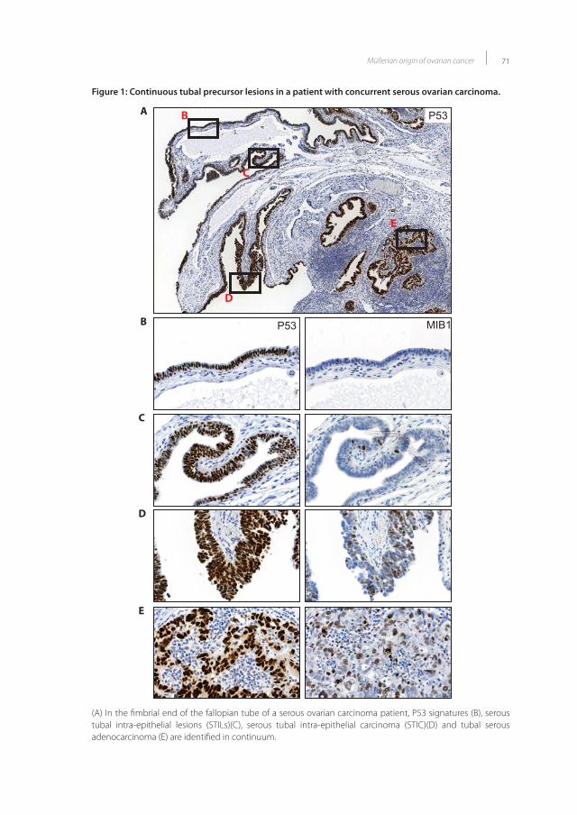
Müllerian origin of ovarian cancer 71
Figure 1: Continuous tubal precursor lesions in a patient with concurrent serous ovarian carcinoma.
A
B
C
D
E
P53
P53 MIB1
B
C
D
E
Figure 1
Figure 1: Continuous tubal precursor lesions in a patient with concurrent serous ovarian carcinoma. (A) In the �mbrial end of the fallopian tube of a serous ovarian carcinoma patient, P53 signatures (B), serous tubal intra-epithelial lesions (STILs)(C), serous tubal intra-epithelial carcinoma (STIC)(D) and tubal serous adenocarcinoma (E) are identi�ed in continuum.
(A) In the fimbrial end of the fallopian tube of a serous ovarian carcinoma patient, P53 signatures (B), serous tubal intra-epithelial lesions (STILs)(C), serous tubal intra-epithelial carcinoma (STIC)(D) and tubal serous adenocarcinoma (E) are identified in continuum.

Chapter 472
Additionally, identical P53 mutations were found in P53 signatures, STICs and concurrent
serous carcinomas, making the hypothesis that STICs develop from ‘precursor’ p53 signatures
and eventually spread to the ovaries a feasible one20. The suggestion that STICs may eventually
disseminate to the ovaries was further strengthened by the finding of signs of possible epithelial-
to-mesenchymal transition and the observation of P53 positive cells in abdominal washings
of women with only a STIC at the fimbrial end of the distal oviduct11, 17. Furthermore, next to a
shared P53 gene mutation, important molecular characteristics of serous ovarian cancer, such as
expression of CA125, WT1, ER, PR, Vimentin, PAX2, PAX8 and HMGA2 were also found to be similar
between STIC and concurrent ovarian cancer17, 22-24..
In summary, malignant tubal precursor lesions for serous ovarian cancer (STICs) were identified in
patients with a hierarchy in prevalence from patients susceptible for serous ovarian cancer, to patients
with a concurrent serous ovarian cancer.
2. Similarities between embryonic development of the Müllerian duct and the different ovarian cancer subtypes.In the first paragraph we have indicated the fallopian tube as a possible site of origin of ovarian
cancer, however, cells initiating the early malignant precursors of ovarian cancer in the fallopian
tube are still unknown. In order to shed light on this issue, embryonic development will be
discussed next.
The female reproductive tract stems from the intermediate mesoderm, and phenotypic
development of the reproductive tract starts in the seventh week of development. Gonadal
development is initiated a few weeks earlier, in the fifth week of pregnancy, in the caudal part
of the ventromedial surface of the mesonephros and becomes prominent as the gonadal ridge
protruding into the coelomic cavity. The gonads develop from migrating somatic cells, derived
from the mesonephros, the surrounding mesenchymal and coelomic epithelium, and primordial
germ cells migrating from the endodermal layer on the posterior wall of the yolk sac. During early
development, the gonads are indifferent and development into male or female phenotype is
depended on the presence of the SRY-gene on the Y-chromosome25. Under the influence of this
gene, testes are formed, but in the absence of SRY, a gene called DAX1 is continuously expressed,
causing suppression of testis formation and allowing the gonads to develop into ovaries26.
Development of the gonads into either the testes or ovaries, influences the development of the
reproductive tract. The indifferent phase (bipotential stage) consists of the mesonephic (Wolffian)
and the paramesonephic (Müllerian) ducts. If testes are present, Sertoli cells secrete testosterone
and anti-Müllerian hormone (AMH), which causes the Wolffian duct to further develop and the
Müllerian ducts to regress, respectively. If ovaries are present or if gonads are absent, testosterone
and AMH are not secreted and the system differentiates into a female phenotype27.

Müllerian origin of ovarian cancer 73
Even though the Müllerian duct and ovarian surface epithelium are both derived from the
embryonic coelomic epithelium, the Müllerian duct stems from a specific subset of cells in
the anterior region of the coelomic epithelium adjacent to the mesonephros. Müllerian duct
development is initiated under the influence of WNT4 secreted by the coelomic epithelium, by
invagination of LIM1 and PAX2 expressing mesoepithelial cells creating a coelomic opening28-31.
After invagination, the primitive Müllerian duct extends to and interacts with the still preexisting
Wolffian duct. Under the influence of WNT9B expressing epithelial cells of the Wolffian duct,
posterior elongation of the LIM1 expressing epithelial cells is initiated and the Müllerian duct
extends towards the cloaca32. Final outgrowth of the Müllerian duct epithelium is completed
by widespread proliferation along the developing duct and at its growing tip and both of the
Müllerian ducts fuse to form the uterovaginal tube31, 33.
After initiation and posterior elongation of the Müllerian duct, posterior differentiation of the
primitive Müllerian duct into vagina, cervix, uterus and oviducts depends on WNT7A expressed
by oviductal and uterine epithelial cells and WNT5A expressed by uterine, cervical and vaginal
mesenchymal cells34, 35. In addition to Wnt signaling, posterior differentiation of the Müllerian
duct is also mediated by the actions of members of the Hox family of homeobox genes: HOXA9 is
expressed in the developing tubal epithelium, HOXA10 in the developing uterus, HOXA11 in the
lower uterine segment and cervix and HOXA13 in the upper part of the vagina36. Interestingly,
maintenance of HOXA10 and HOXA11 expression is under the influence of WNT5A and WNT7A34, 35.
Due to their involvement in Müllerian duct initiation and development, the role of WNT signaling,
HOX genes and PAX2 in ovarian carcinogenesis was studied. Although mainly investigated in
endometrioid ovarian cancer, WNT signaling is an important factor in progression, survival and
chemoresistance of serous ovarian cancer. High levels of WNT5A expression in serous ovarian
cancer predict poor overall and progression-free survival37. Furthermore, WNT5A overexpression,
induced in the human ovarian cancer cell line SKOV3, causes decreased chemosensitivity, which
is in agreement with the earlier observed increased WNT5A expression in ovarian cancer cells
with acquired oxaliplatin resistance37, 38. In contrast, WNT5A was also found to suppress growth of
ovarian cancer cell lines by triggering cellular senescence39. Overexpression of WNT7A was found
in invasive serous ovarian carcinoma and overexpression of WNT7A in OVCAR-3 and SKOV3 ovarian
cancer cells promotes proliferation, migration and invasion40. Interestingly, WNT7A expression in
adult life becomes restricted to epithelial cells of the oviduct and uterine luminal epithelium, but
not in the ovary and the OSE35, 40, 41. Furthermore, WNT9B is highly expressed in ovarian cancer, but
not the OSE42.
Next to their function in Müllerian duct differentiation, the special restricted expression of HOX
genes continues to be present throughout adult life and is thought to be crucial for maintaining
the epithelial plasticity necessary for functional changes which occur during menstruation and
ovulation43. This finding is of interest, because the major subtypes of epithelial ovarian cancer are

Chapter 474
distinguished by their morphological resemblance to the specialized epithelia of the reproductive
tract that have been derived from the Müllerian duct. Serous ovarian cancer is typically papillary or
cystic and resembles the epithelium of the fallopian tube. In contrast, endometrioid and mucinous
ovarian cancer resemble the endometrial-like glands and endocervical epithelium, respectively2.
Because of this resemblance and because the expression of HOX genes is confined to specific
parts of the Müllerian derived epithelium, the expression of HOX genes in epithelial ovarian cancer
was investigated. Interestingly, overexpression of specifically HOXA9, HOXA10, HOXA11 was shown
in serous, endometrioid and mucinous ovarian carcinoma, respectively44. These findings are of
interest because this expression pattern coincides with the physiological expression pattern of
these HOX genes: HOXA9 is expressed in the fallopian tube, HOXA10 in the endometrium and
HOXA11 in the endocervix. Importantly, HOXA9, HOXA10 and HOXA11 are not expressed in the
ovarian surface epithelium44.
Finally, PAX2 is coexpressed with LIM1 by cells in the earliest anlage of the Müllerian duct (Kobayashi
2003). Interestingly, PAX2 was found to be expressed in ovarian papillary serous carcinomas, the
epithelium of the fallopian tube, endometrium and endocervix, but not in the OSE, ovarian surface
epithelium derived inclusion cysts and the ovary itself23. In contrast, Ozcan et al., 2001, did show
focal PAX2 expression in the OSE, next to high expression within the fallopian tube and epithelial
ovarian cancer45. However, since PAX2 expressing cells initiate Müllerian duct invagination from the
coelomic epithelium and number of rudimentary Müllerian cells in proximity of this area might
cause focal OSE expression.
In summary, many similarities and shared characteristics have been identified between early
development of the various Müllerian duct derived organs and the different epithelial ovarian cancer
subtypes.
3. The identification of stem cells that could be involved in initiation of ovarian cancer.There is tentative evidence to postulate that at least in a number of cases a genetically changed
stem cell is the initiating event in malignant transformation46, 47. Therefore, investigations into the
identification of stem cells that could be involved in ovarian carcinogenesis are important.
In 2008, using doxycycline inducible histone2B-GFP and BrdU pulse-chase experiments, Szotek
et al., identified a population of long term label-retaining cells (3 months of chase) in the ovarian
surface epithelium of adult mice as potential stem or progenitor cells48. Label-retaining cells
were slow cycling and showed asymmetric division. Furthermore, label-retaining cells showed a
functional proliferative response to estrogen exposure in vivo and enhanced colony formation in
vitro. However, no evidence of self-renewal, a main characteristic of stem cells, was found 46. Next
to this, the capacity of identified label retaining cells upon mutation to induce ovarian cancer was

Müllerian origin of ovarian cancer 75
not addressed and other regions surrounding the ovaries, such as the fallopian tube, as a putative
source of stem or progenitor cells were not assessed.
In a subsequent effort to investigate the origin of ovarian cancer in mice, a localized pool of stem-
like cells was found to be clustered in the ovarian hilum region, the transitional area which forms the
junction between the OSE, mesothelial peritoneum and tubal epithelium49. Cells were identified
using BrdU pulse chase experiments and immunohistochemical analysis for Aldh1. Microdisected
ovarian hilum cells were slow cycling, formed larger colonies, developed more spheroids and
could be propagated longer as compared to normal OSE cells. Furthermore, using FACsorting,
Aldh1 expressing OSE cells were isolated and were shown to express stem cell markers Aldh1,
CD133, Ck6b, Lgr5 and Lef1. In order assess the malignant potential of ovarian hilum cells, adenoviral
delivery of C-recombinase in the ovarian bursa of Trp53loxp/loxp; Rb1loxp/loxp animals was accomplished,
resulting in early neoplastic lesions in the hilum. Additionally, Trp53 and Rb1-deficient primary
cultured hilum and OSE cells were transplanted intraperitoneally. Upon transplantation, 7/8 mice
injected with hilum cells developed high grade serous adenocarcinomas with metastasis to the
lung, while only 1/12 mice injected with OSE cells developed a non-metastatic carcinoma. The
results of this study led to the postulation that the transitional zone between OSE, mesothelial
peritoneum and tubal epithelium, harbors a stem cell niche, which, when it becomes mutated, has
the potential to give rise to serous ovarian cancer.
Also using the doxycycline inducible histone2B-GFP model, Wang et al (2012) identified a
population of long term label-retaining cells (12 weeks of chase) in the distal and fimbrial part
of the fallopian tube50. These cells could, after FACsorting, form spheroids capable of self-renewal
and upon serum stimulation (differentiation) these spheroids formed glandular structures, which
expressed markers of mature Müllerian epithelial cells (ERα, PRab, Paep and Cd44). In addition, in
this study, no label-retaining cells were found to be present within the OSE, while label-retaining
cells were present in the distal oviduct up to 47 weeks of chase. The presence of these stem-like cells
in the distal and fimbrial part of the fallopian tube is of interest, because their location coincides
with the earliest anlage of the Müllerian duct during embryonic development. Interestingly, the
distal fallopian tube contains a segment that is in continuity with the ovarian hilum and pelvic
mesothelium, forming a Müllerian-mesothelial (mesoepihtelial) junction. Therefore the stem-like
cells identified in the ovarian hilum might be interrelated with stem-like cells identified in the distal
oviduct (Flesken-Nikitin, Wang). In addition to this, 80-93% of tubal precursors of ovarian cancer are
identified within the distal oviduct17, 19.
Because endometrial intra-epithelial carcinoma (EIC) is also hypothesized to be a precursor lesion
of serous ovarian cancer, a potential role for endometrial stem cells in ovarian carcinogenesis was
proposed51. The first evidence of an endometrial stem cell was obtained by plating out purified
single cell suspensions of endometrial epithelial and stromal cells, which showed 0,22% of
epithelial and 1,25% of stromal cells to be able to form large colonies, which could be replated
several times52. This clonal capacity was confirmed by a number of research groups53-55 and when

Chapter 476
grown in Matrigel, Gargett et al. (2009) demonstrated that a single colony forming epithelial
cell was able to form large cytokeratin expressing gland-like structures56. Furthermore, putative
endometrial stromal stem cells were shown to be able to differentiate in multiple mesenchymal
lineages and even into functional epithelium56-59. However, in all studies, a single and specific
stem cell was not identified nor isolated. Using BrdU labeling, Chan et al., (2006) showed label
retaining cells (LRCs) to be present in the luminal epithelium at 8 weeks of chase60 and in the
stromal endometrial-myometrial junction at 12 weeks of chase. The presence of BrdU-LRCs in both
the endometrial epithelium and stroma, was confirmed by Cervelló et al. (2007) and here, LRCs
were found to co-localize with stem cell markers c-KIT and POU5F1/OCT-461. Unfortunately, in
both studies, stem cell characteristics of the LRCs, such as self-renewal, spheroid forming capacity
and growth in recipient animals were not addressed. Wang et al. (2012) confirmed the presence
of LRCs in the endometrium, using doxycycline H2B-GPF pulse-chase labeling, and found LRCs
to be present up until 4 and 12 weeks in epithelial and stromal endometrial cells respectively50.
Interestingly, as described earlier, LRCs were identified in the distal fallopian tube up to 1 year after
pulse and showed stem-like characteristics. Other investigations on the presence of endometrial
stem cells showed that, donor-derived bone marrow cells were identified in the endometrium
of patients receiving bone marrow transplantation62. Lethally irradiated female mice, in which
LacZ-expressing bone marrow cells of a male donor were identified in the epithelium of the
endometrium and peritoneal endometriosis, further confirmed the potential of bone marrow cells
as stem cells of the endometrium63.
Summarizing, progenitor or stem-like cells were identified in the OSE, ovarian hilum, fallopian tube and
endometrium. However, their true potential in ovarian cancer initiation is still to be determined.
4. Ovarian cancer initiation in mouse models.Ovarian cancer cell lines and xenografts have been used extensively over the last decades and
proved effective to investigate chemoresistance, molecular mechanisms of action and biological
behavior of epithelial ovarian cancer64. However, cell lines and xenograft models have their
limitation and animal models mimicking initiation, early development and metastatic spread of
epithelial ovarian cancer are rare. Therefore, models in which genes are conditionally knocked in or
out have been developed for epithelial ovarian cancer. Below we will discuss the most important
models and review what data are presented that add to the discussion on the origin of ovarian
cancer.
Adenoviral delivery of C-recombinase (Ad-Cre) has been extensively used as a tool to induce
recombination in tissues inside the bursal pouch surrounding the ovary and distal oviduct in
mice. In order to assess genes frequently involved in ovarian carcinogenesis, bursal injection of
adenoviral-Cre (AdCMV-Cre) in P53lox/lox;Rb1lox/lox animals was used65. Recombination of P53 and Rb1
resulted in ovarian epithelial cancer in 97% of animals, with ascites (24%) and metastasis spread
to the contralateral ovary (15%), lung (18%) and liver (6%). Control experiments indicated that Ad-

Müllerian origin of ovarian cancer 77
Cre administration resulted in recombinase activity in the OSE cells. Furthermore, OSE cells with
conditional deletions of P53 and Rb1 displayed an increased proliferative activity65. Importantly,
injection of Ad-Cre into the bursal cavity also delivers Ad-Cre to the fimbrial and distal part of the
oviduct. However, possible recombination and involvement of Müllerian duct derived tissues as an
origin of epithelial ovarian cancer in this study was not discussed.
Simultaneously, Dinulescu et al. (2005) used bursal delivered Ad-Cre to induce recombination
in Ptenlox/lox;lsl-KrasG12D/+ animals and found rapidly developing, widely metastatic, endometrioid
ovarian adenocarcinomas in 100% of animals, only 7 weeks after delivery66. Interestingly, animals
which were recombined for KrasG12D alone, only showed ovarian endometriosis, which is associated
with endometrioid ovarian carcinogenesis66-68. Importantly, Cre-activity in these animals was
confirmed in OSE cells, but was also documented in the bursa and the distal oviduct.
Wu et al (2007) reviewed 72 ovarian endometrioid adenocarcinoma tissues and observed defects
in the PI3K/Pten and Wnt/β-catenin signaling pathways in a subset of these tumors69. Based
on this, Ad-Cre injection into the bursa was used to recombine Apclox/lox and Ptenlox/lox. Here,
adenocarcinomas developed which were morphologically similar to human ovarian endometrioid
adenocarcinoma in 100% of animals. Furthermore, 76% of mice developed hemorrhagic ascites
and 21% developed overt peritoneal dissemination69. Even though whole organ staining for
Adenoviral-Cre revealed recombinase activity in OSE cells, the authors were inconclusive for Cre
activity in the distal oviduct.
Using Adenoviral-GFP and Adenoviral-LacZ as controls, Clark-Knowles et al. (2007) showed infection
to be seemingly confined to the OSE cells (no expression in ovarian fatpad, oviduct and uterus)70.
Ad-Cre delivery to Brca1lox/lox animals resulted in increased accumulation of premalignant changes
(hyperplasia, a 4-fold increase in epithelial invaginations and inclusion cysts), while Ad-Cre delivery
to P53lox/lox animals resulted in tumors in 100% of animals and tumor progression was accelerated
in P53lox/lox;Brca1lox/lox mice70. Interestingly, the induced tumors were classified as leiomyosarcomas,
which the authors themselves suggested to have arisen from the ovarian bursa and not from OSE
cells or distal oviduct. Kim at al., 2010 performed similar experiments using Adenoviral-Cre, and
was able to show increased proliferation of OSE from Brca1lox/lox and Brca2lox/lox;P53lox/lox recombined
mice71. However and surprisingly, no evidence of involvement of recombined Brca1lox/lox, Brca2lox/lox
or P53lox/lox in ovarian carcinogenesis was shown.
Finally, Laviolett et al. (2010) induced recombination of tgCAG-LS-Tag (resulting in a functional
SV40 Tag) by bursal injection of Ad-Cre and these mice developed poorly differentiated ovarian
tumors, with metastasis in the pancreas and spleen72. However, the distal oviduct and fimbriae
were not assessed in these investigations.
Even though in many models epithelial ovarian cancer growth was established, adenoviral-Cre
injections into the ovarian bursa will not only recombine the affected (loxed) genes in the OSE
cells, but will also cause Cre-meditated recombination in cells of the fimbriae and distal oviduct.
Therefore, using this technique it is not possible to discriminate between OSE cells and cells
located in the fimbrial region of the distal oviduct as the origin of ovarian carcinogenesis.

Chapter 478
In order to use a more targeted approach, Connolly et al., 2003, used the AmhR2 (MISIIR) promoter
to drive SV40 TAg73. Here, poorly differentiated serous ovarian cancer was observed in 50% of
all animals. Next to these ovarian tumors, intraperitoneal ascites and peritoneal implants were
observed. Immunohistological staining to detect SV40-TAg revealed expression in OSE cells, but
also in patches of epithelial cells in the oviduct and uterus. Furthermore, using PCR, AmhR2 was
shown to be expressed in the ovary as well at low levels in the oviduct and uterus. In contrast,
transgenic mice in which the AmhR2 promoter was used to drive PIK3CA expression and activity (a
much weaker oncogenic signal), only showed hyperplasia of the OSE74.
In mice in which AmhR2-Cre was used to drive recombination of Ptenlox/lox;lsl-KrasG12D, low-grade
ovarian serous papillary adenocarcinomas were formed in 100% of mice75, 76. Interestingly, isolated
recombined OSE cells displayed a temporal change in expression of Müllerian epithelial markers,
grew in soft agar and developed ectopic invasive tumors in recipient mice76. The Müllerian duct as
a possible site of origin of ovarian cancer, however, was neither reviewed, nor discussed in relation
to these experiments75, 76.
Using AmhR2-Cre, Dicer, an essential gene for micro RNA synthesis, and Pten, a key tumor suppressor
inhibiting the PI3K pathway were conditionally deleted77. As a result, high-grade serous carcinomas
arising from the fallopian tube with spread to the ovary and metastasis throughout the abdominal
cavity were identified in 100% of mice and closely resembled human serous cancer. Interestingly,
removal of the oviducts at an early age prevented cancer formation. However, it is important to
note that so far there has not been a role for Dicer in ovarian carcinogenesis and, furthermore,
using this model, cancer initiation seems to start from stromal cells of the oviduct while in humans
tubal precursors of serous ovarian cancer are epithelial.
Tanwar et al., 2012 combined AmhR2-Cre with Apclox/lox and observed development of epithelial
inclusion cysts and, in much older animals, high grade ovarian endometrioid adenocarcinoma78.
The finding of endometrioid ovarian cancer is in agreement with observations that in this subtype,
Wnt/β-catenin signaling is often activated79.
In an effort to prove the Müllerian origin of endometrioid ovarian cancer, Pgr-Cre induced
conditional recombination of Apclox/lox in the oviduct was used80. Interestingly, in this model the
OSE cells are not affected. As described before, Wnt signaling is an important oncologic factor
in human endometrioid ovarian cancer and APC mutations are frequently found79. Interestingly,
in this model, tubal intra-epithelial carcinomas developed, starting from 10 weeks of age, which
show high resemblance to human tubal intra-epithelial carcinomas. With age, these TICs were
shown to evolve and developed into endometrioid tubal and ovarian tumors resembling human
endometrioid tubal and ovarian cancer growth. Next to these tubal and ovarian tumors, loco-
regional spread to the utero-ovarian ligament was shown80.
Additionally models, in which not the OSE cells or Müllerian duct but the granulosa cells were
targeted, also need to be discussed81-83. Chen et al., described early alterations in OSE cells in
FshR-knockout animals, eventually resulting in serous papillary adenoma of the ovaries81. Another

Müllerian origin of ovarian cancer 79
model used FshR-Cre to target Brca1lox/lox in granulosa cells of the ovary. In these animals, grossly
visible serous cystadenomas were attached to the ovary, within the wall of the uterus, or on the
external surface of the uterine horns. Interestingly, in these cystadenomas the Brca1 gene was
not recombined indicating that factors secreted by the granulosa cells must have influenced
tumorigenesis indirectly82. The finding that the uterine horns are also involved next to the ovaries
is in line with the finding of tubal intraepithelial lesions in asymptomatic carriers of BRCA1
mutations10, 15, 16 and seems to point to an extraovarian origin of ovarian cancer.
In summary, some mouse models point towards the OSE cells and others to Müllerian duct derived
tissues as the origin of epithelial ovarian cancer, but in essence none of these models are specific enough
to provide a definitive answer to the question whether it are mutated or modified OSE cells, or cells from
Müllerian origin that develop into the earliest malignant precursors of ovarian cancer.
5. The secondary Müllerian system as a source of ovarian carcinogenesis.In 1999, Dubeau suggested the secondary Müllerian system as a possible origin of epithelial ovarian
cancer. The secondary Müllerian system consists of microscopic structures lined with Müllerian
epithelium, commonly present in the paratubal and paraovarian areas, the ovarian medulla near
the hilum and the deeper portions of the ovarian cortex84. These structures might be rudimentary
remnants from the developing Müllerian duct but also include endosalphyngiosis (cysts lined with
tubal epthelium), endocervicosis (cysts lined with endocervical epithelium) and endometriosis
(functional endometrial-like tissue outside the uterus)5, 84. Interestingly, these structures can
develop into large extra- or intra-ovarian cysts which share morphological characteristics with
serous, mucinous or endometrioid ovarian cancer.
Endometriosis affects 5- 10% of woman of reproductive age and is therefore considered as a major
gynecological health problem85. As in ovarian cancer, the origin of endometriosis is not clear but
the most prevalent hypothesis is that endometrial stem cells appear in the abdominal cavity where
they attach and migrate into surrounding tissues and organs86. Interestingly, endometrioid ovarian
cancer also resembles the endometrium and recent investigations have indicated an association
between endometriosis and endometrioid ovarian cancer67, 68. A strong increased risk for ovarian
malignancies in women with endometriosis was identified in a large pooled case-control study
where a significant association was found between history of self-reported endometriosis and
clear-cell, low-grade serous and endometrioid ovarian cancer67. Furthermore, similar gene
mutations in ARID1a in endometrioid ovarian cancer and neighboring atypical endometriosis
were found, indicating a genetical association between the two diseases68. The epidemiological
relationship between endometriosis and ovarian cancer was further confirmed by Buis et al.,
2013, who found an increased ovarian cancer risk in subfertile women with surgically diagnosed
endometrioisis (REF). In addition to endometriosis, serous borderline tumors also were found in
foci of endosalpingiosis in pelvic and para-aortic lymph nodes87.

Chapter 480
Furthermore, it was suggested that the rete ovarii, which consist of coiled microscopic ducts near
the ovarian hilum, are part of the secondary Müllerian system. Interestingly, in some rodents,
although rarely diagnosed, epithelial ovarian cancer seems to naturally arise from a dilatation of
these rete ovarii88, 89.
These findings are of interest because stem-like cells were identified in the ovarian hilum
and therefore an association with lesions located in the ovarian hilum, such as rete ovarri,
endosalpingiosis and endometriosis may be hypothesized49.
Even though most research is focused on either the fallopian tube or OSE as the origin of ovarian cancer,
important findings appoint a role for other Müllerian duct derived structures such as the secondary
Müllerian system in epithelial ovarian carcinogenesis.
6. Not the OSE but tissues derived from the Müllerian duct are the origin of epithelial ovarian cancer: conclusions and future perspectivesCortical inclusion cysts (CICs), derived from either the Müllerian duct or OSE, have been appointed
as the origin of epithelial ovarian cancer5, 90-92. Even though some CICs appear mesothelial (OSE),
most CICs resemble a Müllerian morphology4, 93-95. The OSE hypothesis corrects for this Müllerian
appearance, by stating that stem or progenitor cells from the OSE acquire genetic modifications
and regain Müllerian characteristics through metaplasia90. Ovarian cancer is induced by additional
genetic disturbances and stimuli from the surrounding microenvironment, leading to dysplasia
of the metaplastic CIC and culminating as full-scale epithelial ovarian cancer. If we, however,
summarize all supporting and opposing arguments for either the OSE or the Müllerian duct as the
origin of ovarian cancer the balance tips towards a Müllerian origin of ovarian cancer.
Scientific evidence supporting an OSE origin of ovarian cancer:
- OSE lined CICs have been described4, 90, 93, 95,
- OSE cells and the Müllerian duct are both derived from a shared embryonic precursor,
indicating that metaplasia to Müllerian duct like cells may be possible,
- From all cells present in the ovary, OSE cells are the only cell type for which metaplasia is
feasible,
- Stem-like cell characteristics have been described for OSE cells48, 96, 97,
- Atypical OSE cells were found directly adjacent serous ovarian cancer98,
- Isolated mutated OSE cells, when transplanted in recipient mice, can show serous ovarian
cancer growth49.
Scientific evidence supporting a Müllerian duct origin of ovarian cancer:
- The three most important epithelial ovarian cancer subtypes represent Müllerian duct derived
tissues92,
- Genes important for Müllerian duct development and maintenance are highly expressed in
ovarian cancer, but are not expressed in the OSE23, 37-42, 44, 45, 79,

Müllerian origin of ovarian cancer 81
- Stem-like cells forming spheroids and capable of self-renewal were identified in the distal
oviduct50, 60, 61,
- Müllerian duct derived structures are found in the ovarian hilum and are possibly associated
with stem or progenitor cells found in the ovarian hilum49, 84,
- Mouse models in which the Müllerian duct is mutated, but not the OSE, show serous and
endometrioid ovarian cancer77, 80,
- Components of the secondary Müllerian system, such as endometriosis, endosalpingiosis,
endocervicosis and rete ovarii, are found in the ovary and are associated with epithelial ovarian
cancer5, 67, 68, 84, 87-89,
- Most CICs are lined with Müllerian epithelium, and P53 expressing, dysplastic cells are found
within CICs lined with Müllerian epithelium94,
- Early benign (P53 signatures), intermediate (STILs) and clearly malignant (STICs) precursors
of high grade serous ovarian cancer (all lesions of the distal oviduct) were identified with a
hierarchy in prevalence from control, to patients at risk, to patients with a concurrent serous
ovarian cancer9-21.
- STICs are only identified in patients at risk or with a concurrent serous ovarian cancer9-21,
- P53 signatures, STILs and STICs share identical P53 mutations with the concurrent serous
ovarian cancer20,
- In patients with pelvic serous carcinoma, which is indistinguishable from serous ovarian
carcinoma, STICs are found but no ovarian lesions19. Furthermore, STICs and concurrent pelvic
serous carcinoma display similar P53 mutations.
Upon reviewing these data, we appoint two possible mechanisms in which epithelial ovarian
cancer is initiated based on the histopathological model of Kurman and Shih(2008)99.
First, since type I ovarian tumors are typically ovarian confined and develop from borderline
precursors, we hypothesize that these are derived from the oviduct or components of the
secondary Müllerian system, such as ovarian endosalpingiosis or endometriosis, which over time
have acquired further genetical disturbances due to ovulation-induced distress or stimuli from the
ovarian stroma.
Second, type II high grade serous ovarian carcinomas are mainly confined to the ovary and are
characterized by mutation of TP53. Therefore we hypothesize that ovulation-induced damage to
the distal fallopian tube epithelium results in areas mutated for TP53 (P53 signatures). Upon further
genetic damage and increased proliferation, P53 signatures develop into STILs, which, progress
to become STICs. When transformed, malignant STIC cells can exfoliate and, in addition to other
peritoneal sites, implant on or in the ovary. As a result, type II high grade serous ovarian cancer
develops.
In conclusion, there is abundant evidence that not the OSE but the Müllerian duct should be
appointed as the origin of epithelial ovarian cancer and research aiming to unravel the earliest
carcinogenic changes in Müllerian derived tissues is key to facilitate early detection and targeted
therapy for ovarian cancer.

Chapter 482
References1. Ferlay J SH, Bray F, Forman D, Mathers C and Parkin DM. GLOBOCAN 2008 v2.0, Cancer Incidence and
Mortality Worldwide: IARC CancerBase No. 10 [Internet]. [cited; Available from: http://globocan.iarc.fr, accessed on day/month/year.
2. Cannistra SA. Cancer of the Ovary. New England Journal of Medicine 2004;351:2519-29.
3. van Altena AM, Karim-Kos HE, de Vries E, Kruitwagen RF, Massuger LF, et al. Trends in therapy and survival of advanced stage epithelial ovarian cancer patients in the Netherlands. Gynecol Oncol 2012;125:649-54.
4. Stratton JF, Buckley CH, Lowe D, Ponder BA. Comparison of prophylactic oophorectomy specimens from carriers and noncarriers of a BRCA1 or BRCA2 gene mutation. United Kingdom Coordinating Committee on Cancer Research (UKCCCR) Familial Ovarian Cancer Study Group. J Natl Cancer Inst 1999;91:626-8.
5. Dubeau L. The cell of origin of ovarian epithelial tumors and the ovarian surface epithelium dogma: does the emperor have no clothes? Gynecol Oncol 1999;72:437-42.
6. Rose PG, Shrigley R, Wiesner GL. Germline BRCA2 mutation in a patient with fallopian tube carcinoma: a case report. Gynecol Oncol 2000;77:319-20.
7. Zweemer RP, van Diest PJ, Verheijen RH, Ryan A, Gille JJ, et al. Molecular evidence linking primary cancer of the fallopian tube to BRCA1 germline mutations. Gynecol Oncol 2000;76:45-50.
8. Colgan TJ, Murphy J, Cole DE, Narod S, Rosen B. Occult carcinoma in prophylactic oophorectomy specimens: prevalence and association with BRCA germline mutation status. Am J Surg Pathol 2001;25:1283-9.
9. Piek JM, van Diest PJ, Zweemer RP, Jansen JW, Poort-Keesom RJ, et al. Dysplastic changes in prophylactically removed Fallopian tubes of women predisposed to developing ovarian cancer. J Pathol 2001;195:451-6.
10. Medeiros F, Muto MG, Lee Y, Elvin JA, Callahan MJ, et al. The tubal fimbria is a preferred site for early adenocarcinoma in women with familial ovarian cancer syndrome. Am J Surg Pathol 2006;30:230-6.
11. Callahan MJ, Crum CP, Medeiros F, Kindelberger DW, Elvin JA, et al. Primary fallopian tube malignancies in BRCA-positive women undergoing surgery for ovarian cancer risk reduction. J Clin Oncol 2007;25:3985-90.
12. Carcangiu ML, Peissel B, Pasini B, Spatti G, Radice P, et al. Incidental carcinomas in prophylactic specimens in BRCA1 and BRCA2 germ-line mutation carriers, with emphasis on fallopian tube lesions: report of 6 cases and review of the literature. Am J Surg Pathol 2006;30:1222-30.
13. Hirst JE, Gard GB, McIllroy K, Nevell D, Field M. High rates of occult fallopian tube cancer diagnosed at prophylactic bilateral salpingo-oophorectomy. Int J Gynecol Cancer 2009;19:826-9.
14. Manchanda R, Abdelraheim A, Johnson M, Rosenthal AN, Benjamin E, et al. Outcome of risk-reducing salpingo-oophorectomy in BRCA carriers and women of unknown mutation status. BJOG 2011;118:814-24.
15. Mingels MJ, Roelofsen T, van der Laak JA, de Hullu JA, van Ham MA, et al. Tubal epithelial lesions in salpingo-oophorectomy specimens of BRCA-mutation carriers and controls. Gynecol Oncol 2012;127:88-93.
16. Reitsma W, de Bock GH, Oosterwijk JC, Bart J, Hollema H, et al. Support of the ‘fallopian tube hypothesis’ in a prospective series of risk-reducing salpingo-oophorectomy specimens. Eur J Cancer 2013;49:132-41.
17. Van der Horst PH, Wijnhoven RKE, Daal S, Mouthaan MH, Heijmans-Antonissen C, et al. Malignant transition of tubal precursors into serous ovarian cancer. In submission 2013.
18. Roh MH, Kindelberger D, Crum CP. Serous tubal intraepithelial carcinoma and the dominant ovarian mass: clues to serous tumor origin? Am J Surg Pathol 2009;33:376-83.
19. Kindelberger DW, Lee Y, Miron A, Hirsch MS, Feltmate C, et al. Intraepithelial carcinoma of the fimbria and pelvic serous carcinoma: Evidence for a causal relationship. Am J Surg Pathol 2007;31:161-9.
20. Lee Y, Miron A, Drapkin R, Nucci MR, Medeiros F, et al. A candidate precursor to serous carcinoma that originates in the distal fallopian tube. J Pathol 2007;211:26-35.
21. Vang R, Visvanathan K, Gross A, Maambo E, Gupta M, et al. Validation of an algorithm for the diagnosis of serous tubal intraepithelial carcinoma. Int J Gynecol Pathol 2012;31:243-53.
22. Jarboe EA, Miron A, Carlson JW, Hirsch MS, Kindelberger D, et al. Coexisting intraepithelial serous carcinomas of the endometrium and fallopian tube: frequency and potential significance. Int J Gynecol Pathol 2009;28:308-15.
23. Tong GX, Chiriboga L, Hamele-Bena D, Borczuk AC. Expression of PAX2 in papillary serous carcinoma of the ovary: immunohistochemical evidence of fallopian tube or secondary Mullerian system origin? Mod Pathol 2007;20:856-63.
24. Wei JJ, Wu J, Luan C, Yeldandi A, Lee P, et al. HMGA2: a potential biomarker complement to P53 for detection of early-stage high-grade papillary serous carcinoma in fallopian tubes. Am J Surg Pathol 2010;34:18-26.

Müllerian origin of ovarian cancer 83
25. Wilhelm D, Palmer S, Koopman P. Sex determination and gonadal development in mammals. Physiol Rev 2007;87:1-28.
26. Ikeda Y, Takeda Y, Shikayama T, Mukai T, Hisano S, et al. Comparative localization of Dax-1 and Ad4BP/SF-1 during development of the hypothalamic-pituitary-gonadal axis suggests their closely related and distinct functions. Dev Dyn 2001;220:363-76.
27. Acien P. Embryological observations on the female genital tract. Hum Reprod 1992;7:437-45.
28. Masse J, Watrin T, Laurent A, Deschamps S, Guerrier D, et al. The developing female genital tract: from genetics to epigenetics. Int J Dev Biol 2009;53:411-24.
29. Kobayashi A, Shawlot W, Kania A, Behringer RR. Requirement of Lim1 for female reproductive tract development. Development 2004;131:539-49.
30. van der Horst PH, Wang Y, van der Zee M, Burger CW, Blok LJ. Interaction between sex hormones and WNT/beta-catenin signal transduction in endometrial physiology and disease. Mol Cell Endocrinol 2012;358:176-84.
31. Guioli S, Sekido R, Lovell-Badge R. The origin of the Mullerian duct in chick and mouse. Dev Biol 2007;302:389-98.
32. Carroll TJ, Park JS, Hayashi S, Majumdar A, McMahon AP. Wnt9b plays a central role in the regulation of mesenchymal to epithelial transitions underlying organogenesis of the mammalian urogenital system. Dev Cell 2005;9:283-92.
33. Orvis GD, Behringer RR. Cellular mechanisms of Mullerian duct formation in the mouse. Dev Biol 2007;306:493-504.
34. Mericskay M, Kitajewski J, Sassoon D. Wnt5a is required for proper epithelial-mesenchymal interactions in the uterus. Development 2004;131:2061-72.
35. Miller C, Sassoon DA. Wnt-7a maintains appropriate uterine patterning during the development of the mouse female reproductive tract. Development 1998;125:3201-11.
36. Podlasek C, Houston J, McKenna KE, McVary KT. Posterior Hox gene expression in developing genitalia. Evol Dev 2002;4:142-63.
37. Peng C, Zhang X, Yu H, Wu D, Zheng J. Wnt5a as a predictor in poor clinical outcome of patients and a mediator in chemoresistance of ovarian cancer. Int J Gynecol Cancer 2011;21:280-8.
38. Varma RR, Hector SM, Clark K, Greco WR, Hawthorn L, et al. Gene expression profiling of a clonal isolate of oxaliplatin-resistant ovarian carcinoma cell line A2780/C10. Oncol Rep 2005;14:925-32.
39. Bitler BG, Nicodemus JP, Li H, Cai Q, Wu H, et al. Wnt5a suppresses epithelial ovarian cancer by promoting cellular senescence. Cancer Res 2011;71:6184-94.
40. Merritt MA, Parsons PG, Newton TR, Martyn AC, Webb PM, et al. Expression profiling identifies genes involved in neoplastic transformation of serous ovarian cancer. BMC Cancer 2009;9:378.
41. Yoshioka S, King ML, Ran S, Okuda H, MacLean JA, 2nd, et al. WNT7A regulates tumor growth and progression in ovarian cancer through the WNT/beta-catenin pathway. Mol Cancer Res 2012;10:469-82.
42. Steg A, Wang W, Blanquicett C, Grunda JM, Eltoum IA, et al. Multiple gene expression analyses in paraffin-embedded tissues by TaqMan low-density array: Application to hedgehog and Wnt pathway analysis in ovarian endometrioid adenocarcinoma. J Mol Diagn 2006;8:76-83.
43. Taylor HS, Vanden Heuvel GB, Igarashi P. A conserved Hox axis in the mouse and human female reproductive system: late establishment and persistent adult expression of the Hoxa cluster genes. Biol Reprod 1997;57:1338-45.
44. Cheng W, Liu J, Yoshida H, Rosen D, Naora H. Lineage infidelity of epithelial ovarian cancers is controlled by HOX genes that specify regional identity in the reproductive tract. Nat Med 2005;11:531-7.
45. Ozcan A, Liles N, Coffey D, Shen SS, Truong LD. PAX2 and PAX8 expression in primary and metastatic mullerian epithelial tumors: a comprehensive comparison. Am J Surg Pathol 2011;35:1837-47.
46. Visvader JE. Cells of origin in cancer. Nature 2011;469:314-22.
47. Jordan CT, Guzman ML, Noble M. Cancer stem cells. N Engl J Med 2006;355:1253-61.
48. Szotek PP, Chang HL, Brennand K, Fujino A, Pieretti-Vanmarcke R, et al. Normal ovarian surface epithelial label-retaining cells exhibit stem/progenitor cell characteristics. Proc Natl Acad Sci U S A 2008;105:12469-73.
49. Flesken-Nikitin A, Hwang CI, Cheng CY, Michurina TV, Enikolopov G, et al. Ovarian surface epithelium at the junction area contains a cancer-prone stem cell niche. Nature 2013;495:241-5.

Chapter 484
50. Wang Y, Sacchetti A, van Dijk MR, van der Zee M, van der Horst PH, et al. Identification of quiescent, stem-like cells in the distal female reproductive tract. PLoS One 2012;7:e40691.
51. Roelofsen T, van Kempen LC, van der Laak JA, van Ham MA, Bulten J, et al. Concurrent endometrial intraepithelial carcinoma (EIC) and serous ovarian cancer: can EIC be seen as the precursor lesion? Int J Gynecol Cancer 2012;22:457-64.
52. Chan RW, Schwab KE, Gargett CE. Clonogenicity of human endometrial epithelial and stromal cells. Biol Reprod 2004;70:1738-50.
53. Dimitrov R, Timeva T, Kyurkchiev D, Stamenova M, Shterev A, et al. Characterization of clonogenic stromal cells isolated from human endometrium. Reproduction 2008;135:551-8.
54. Schwab KE, Chan RW, Gargett CE. Putative stem cell activity of human endometrial epithelial and stromal cells during the menstrual cycle. Fertil Steril 2005;84 Suppl 2:1124-30.
55. Schwab KE, Hutchinson P, Gargett CE. Identification of surface markers for prospective isolation of human endometrial stromal colony-forming cells. Hum Reprod 2008;23:934-43.
56. Gargett CE, Schwab KE, Zillwood RM, Nguyen HP, Wu D. Isolation and culture of epithelial progenitors and mesenchymal stem cells from human endometrium. Biol Reprod 2009;80:1136-45.
57. Cervello I, Gil-Sanchis C, Mas A, Delgado-Rosas F, Martinez-Conejero JA, et al. Human endometrial side population cells exhibit genotypic, phenotypic and functional features of somatic stem cells. PLoS One 2010;5:e10964.
58. Schuring AN, Schulte N, Kelsch R, Ropke A, Kiesel L, et al. Characterization of endometrial mesenchymal stem-like cells obtained by endometrial biopsy during routine diagnostics. Fertil Steril 2011;95:423-6.
59. Cervello I, Mas A, Gil-Sanchis C, Peris L, Faus A, et al. Reconstruction of endometrium from human endometrial side population cell lines. PLoS One 2011;6:e21221.
60. Chan RW, Gargett CE. Identification of label-retaining cells in mouse endometrium. Stem Cells 2006;24:1529-38.
61. Cervello I, Martinez-Conejero JA, Horcajadas JA, Pellicer A, Simon C. Identification, characterization and co-localization of label-retaining cell population in mouse endometrium with typical undifferentiated markers. Hum Reprod 2007;22:45-51.
62. Taylor HS. Endometrial cells derived from donor stem cells in bone marrow transplant recipients. JAMA 2004;292:81-5.
63. Du H, Taylor HS. Contribution of bone marrow-derived stem cells to endometrium and endometriosis. Stem Cells 2007;25:2082-6.
64. Vanderhyden BC, Shaw TJ, Ethier JF. Animal models of ovarian cancer. Reprod Biol Endocrinol 2003;1:67.
65. Flesken-Nikitin A, Choi KC, Eng JP, Shmidt EN, Nikitin AY. Induction of carcinogenesis by concurrent inactivation of p53 and Rb1 in the mouse ovarian surface epithelium. Cancer Res 2003;63:3459-63.
66. Dinulescu DM, Ince TA, Quade BJ, Shafer SA, Crowley D, et al. Role of K-ras and Pten in the development of mouse models of endometriosis and endometrioid ovarian cancer. Nat Med 2005;11:63-70.
67. Pearce CL, Templeman C, Rossing MA, Lee A, Near AM, et al. Association between endometriosis and risk of histological subtypes of ovarian cancer: a pooled analysis of case-control studies. Lancet Oncol 2012;13:385-94.
68. Wiegand KC, Shah SP, Al-Agha OM, Zhao Y, Tse K, et al. ARID1A mutations in endometriosis-associated ovarian carcinomas. N Engl J Med 2010;363:1532-43.
69. Wu R, Hendrix-Lucas N, Kuick R, Zhai Y, Schwartz DR, et al. Mouse model of human ovarian endometrioid adenocarcinoma based on somatic defects in the Wnt/beta-catenin and PI3K/Pten signaling pathways. Cancer Cell 2007;11:321-33.
70. Clark-Knowles KV, Garson K, Jonkers J, Vanderhyden BC. Conditional inactivation of Brca1 in the mouse ovarian surface epithelium results in an increase in preneoplastic changes. Exp Cell Res 2007;313:133-45.
71. Kim KY, Park DW, Jeung EB, Choi KC. Conditional knockout of brca1/2 and p53 in mouse ovarian surface epithelium: do they play a role in ovarian carcinogenesis? J Vet Sci 2010;11:291-7.
72. Laviolette LA, Garson K, Macdonald EA, Senterman MK, Courville K, et al. 17beta-estradiol accelerates tumor onset and decreases survival in a transgenic mouse model of ovarian cancer. Endocrinology 2010;151:929-38.
73. Connolly DC, Bao R, Nikitin AY, Stephens KC, Poole TW, et al. Female mice chimeric for expression of the simian virus 40 TAg under control of the MISIIR promoter develop epithelial ovarian cancer. Cancer Res 2003;63:1389-97.

Müllerian origin of ovarian cancer 85
74. Liang S, Yang N, Pan Y, Deng S, Lin X, et al. Expression of activated PIK3CA in ovarian surface epithelium results in hyperplasia but not tumor formation. PLoS One 2009;4:e4295.
75. Fan HY, Liu Z, Paquet M, Wang J, Lydon JP, et al. Cell type-specific targeted mutations of Kras and Pten document proliferation arrest in granulosa cells versus oncogenic insult to ovarian surface epithelial cells. Cancer Res 2009;69:6463-72.
76. Mullany LK, Fan HY, Liu Z, White LD, Marshall A, et al. Molecular and functional characteristics of ovarian surface epithelial cells transformed by KrasG12D and loss of Pten in a mouse model in vivo. Oncogene 2011;30:3522-36.
77. Kim J, Coffey DM, Creighton CJ, Yu Z, Hawkins SM, et al. High-grade serous ovarian cancer arises from fallopian tube in a mouse model. Proc Natl Acad Sci U S A 2012;109:3921-6.
78. Tanwar PS, Kaneko-Tarui T, Lee HJ, Zhang L, Teixeira JM. PTEN loss and HOXA10 expression are associated with ovarian endometrioid adenocarcinoma differentiation and progression. Carcinogenesis 2013;34:893-901.
79. Gatcliffe TA, Monk BJ, Planutis K, Holcombe RF. Wnt signaling in ovarian tumorigenesis. Int J Gynecol Cancer 2008;18:954-62.
80. Van der Horst PH, Van der Zee M, Heijmans-Antonissen C, Jia Y, deMayo FJ, et al. Endometrioid ovarian cancer arising from the distal oviduct. In submission 2013.
81. Chen X, Aravindakshan J, Yang Y, Sairam MR. Early alterations in ovarian surface epithelial cells and induction of ovarian epithelial tumors triggered by loss of FSH receptor. Neoplasia 2007;9:521-31.
82. Chodankar R, Kwang S, Sangiorgi F, Hong H, Yen HY, et al. Cell-nonautonomous induction of ovarian and uterine serous cystadenomas in mice lacking a functional Brca1 in ovarian granulosa cells. Curr Biol 2005;15:561-5.
83. Dierich A, Sairam MR, Monaco L, Fimia GM, Gansmuller A, et al. Impairing follicle-stimulating hormone (FSH) signaling in vivo: targeted disruption of the FSH receptor leads to aberrant gametogenesis and hormonal imbalance. Proc Natl Acad Sci U S A 1998;95:13612-7.
84. Lauchlan SC. The secondary mullerian system revisited. Int J Gynecol Pathol 1994;13:73-9.
85. Bulun SE. Endometriosis. New England Journal of Medicine 2009;360:268-79.
86. D’Hooghe TM, Debrock S. Endometriosis, retrograde menstruation and peritoneal inflammation in women and in baboons. Hum Reprod Update 2002;8:84-8.
87. Kadar N, Krumerman M. Possible metaplastic origin of lymph node “metastases” in serous ovarian tumor of low malignant potential (borderline serous tumor). Gynecol Oncol 1995;59:394-7.
88. Rutgers JL, Scully RE. Cysts (cystadenomas) and tumors of the rete ovarii. Int J Gynecol Pathol 1988;7:330-42.
89. Quattropani SL. Serous cystadenoma formation in guinea pig ovaries. J Submicrosc Cytol 1981;13:337-45.
90. Auersperg N. Ovarian surface epithelium as a source of ovarian cancers: Unwarranted speculation or evidence-based hypothesis? Gynecol Oncol 2013.
91. Crum CP, Drapkin R, Miron A, Ince TA, Muto M, et al. The distal fallopian tube: a new model for pelvic serous carcinogenesis. Curr Opin Obstet Gynecol 2007;19:3-9.
92. Karst AM, Drapkin R. Ovarian cancer pathogenesis: a model in evolution. J Oncol 2010;2010:932371.
93. Mittal KR, Zeleniuch-Jacquotte A, Cooper JL, Demopoulos RI. Contralateral ovary in unilateral ovarian carcinoma: a search for preneoplastic lesions. Int J Gynecol Pathol 1993;12:59-63.
94. Pothuri B, Leitao MM, Levine DA, Viale A, Olshen AB, et al. Genetic analysis of the early natural history of epithelial ovarian carcinoma. PLoS One 2010;5:e10358.
95. Resta L, Russo S, Colucci GA, Prat J. Morphologic precursors of ovarian epithelial tumors. Obstet Gynecol 1993;82:181-6.
96. Bowen NJ, Walker LD, Matyunina LV, Logani S, Totten KA, et al. Gene expression profiling supports the hypothesis that human ovarian surface epithelia are multipotent and capable of serving as ovarian cancer initiating cells. BMC Med Genomics 2009;2:71.
97. Gamwell LF, Collins O, Vanderhyden BC. The mouse ovarian surface epithelium contains a population of LY6A (SCA-1) expressing progenitor cells that are regulated by ovulation-associated factors. Biol Reprod 2012;87:80.
98. Plaxe SC, Deligdisch L, Dottino PR, Cohen CJ. Ovarian intraepithelial neoplasia demonstrated in patients with stage I ovarian carcinoma. Gynecol Oncol 1990;38:367-72.
99. Kurman RJ, Shih Ie M. Pathogenesis of ovarian cancer: lessons from morphology and molecular biology and their clinical implications. Int J Gynecol Pathol 2008;27:151-60.


Chapter 5
Endometrioid ovarian cancer arising from the distal oviduct
Paul H. van der Horsta, Marten van der Zeea,b, Claudia Heijmans-Antonissena,
Yundan Jiaa,b, Francesco J. DeMayoc, John P. Lydonc,
Carolien H.M. van Deurzenb, Patricia C. Ewingb,
Curt W. Burgera, Leen J. Bloka
a: Department of Obstetrics and Gynecology, Erasmus University Medical Center Rotterdam, PO box 2040, 3000 CA Rotterdam, The Netherlands
b: Department of Pathology, Erasmus University Medical Center Rotterdam PO box 2040, 3000 CA Rotterdam, The Netherlands
c: Department of Molecular and Cellular Biology, Baylor College of Medicine, 1 Baylor Plaza, 77030, Houston, TX, USA
Submitted for publication

Chapter 6
Malignant transformation of tubal precursors into serous ovarian cancer
Paul H. van der Horst1, Renske K.E Wijnhoven1*, Sadé N.S. Daal1*, Marthe H. Mouthaan1*,
Claudia Heijmans-Antonissen1, Ronald van der Knaap1, Ramon G.V. Smolders1,
Diederick de Jong1, Jurgen M. Piek3, Patricia C. Ewing2, Curt W. Burger1, Leen J. Blok1
1: Department of Obstetrics and Gynecology, Erasmus University Medical Center Rotterdam, PO box 2040, 3000 CA Rotterdam, The Netherlands
2: Department of Pathology, Erasmus University Medical Center Rotterdam PO box 2040, 3000 CA Rotterdam, The Netherlands
3: Comprehensive Cancer Center South, location TweeSteden Hospital, Dr. Deelenlaan 5, 5042 AD Tilburg, The Netherlands
*: Contributed equally to this study
Submitted for publication

Chapter 7
General discussion


General discussion 125
In this thesis, we describe our investigation on the mechanisms involved in the initiation and
progression of Müllerian duct derived malignancies. The research was focused on the role of
progesterone signaling in the progression of endometrial cancer (chapter 2, 3) and on the origin of
epithelial ovarian cancer (chapter 4, 5 and 6). For this, 3 aims were described:
1. What is the effect of progesterone receptor signaling on the tumor specific immune response,
Epithelial-Mesenchymal Transition (EMT) and recurrence in endometrial cancer?
2. What is the effect of activation of WNT/β-catenin signaling on Müllerian duct derived tissues?
3. Are Müllerian duct derived tissues the origin of epithelial ovarian cancer, can we initiate ovarian
cancer from these tissues and can we identify and characterize tubal precursor lesions of
serous ovarian carcinoma in controls, patients susceptible for and patients with serous ovarian
cancer?
Progesterone signaling stimulates infiltration of T-lymphocytes and inhibits epithelial-to-mesenchymal transition in endometrial cancer.In general, patients with endometrial cancer have a good prognosis due to the fact that the
disease is usually diagnosed at an early stage, in which it has not spread beyond the uterus1.
However, if there is recurrent or metastatic disease, the situation is very different and progressive
disease has a very poor prognosis accounting for 74.000 deaths worldwide each year2. Therefore,
in order to improve therapy it is vital to understand the processes that inhibit and stimulate
endometrial cancer progression. The research performed in chapter 3 aimed to investigate two
mechanisms involved in metastatic spread of endometrial cancer: tumor infiltrating lymphocytes
and progesterone induced inhibition of EMT. For this, primary endometrial cancer specimens from
progressive and non-progressive endometrial cancer patients were assessed for the presence of
CD4+ (helper), CD8+ (cytotoxic) and FOXP3+ (regulatory) T-lymphocytes and PR expression. As
expected3-5, patients with progressive (recurrent and/or metastatic) disease, showed a significant
decrease in tumor infiltrating lymphocytes coinciding with loss of PR expression. Conformingly,
gene expression analysis of frozen tumor samples of these patients, showed significant regulation
of pathways involved in immunesurveillance, in addition to pathways involved in EMT and
metastasis. Interestingly, inhibitors of WNT/β-catenin signaling, DKK1, DKK4 and WIF1, were down
regulated in progressive disease, which was confirmed by quantitative RT-PCR analysis. These
results were in line with our previous investigations, which showed that WNT/β-catenin signaling
becomes activated at the same time as the progesterone receptor is lost6.
In order to substantiate the finding that loss of progesterone signaling in progressive disease
plays a role in diminished T-cell infiltration and induction of EMT, well differentiated Ishikawa
endometrial cell lines stably transfected with PRA (IKPRA-1), PRB (IKPRB-1) and PRA+PRB
(IKPRAB-36) were cultured in the presence or absence of progesterone (MPA) and subsequently
used for immunohistochemistry, wound healing and modified Boyden chamber migration assay,
and genome wide gene expression analysis. Culture of IKPRB-1 and IKPRAB-36, but not IKPRA-1, in

Chapter 7126
the presence of MPA resulted in inhibition of migration and down regulation of the mesenchymal
marker vimentin at the invasive front of the wound. Furthermore, as in progressive disease,
progesterone stimulated immunosuppression, but inhibited pathways and genes involved in EMT
and metastasis: such as Integrin/ILK, EGF, PDGF, TGF-β and WNT/β-catenin-signaling. Interestingly,
many of the differentially expressed signaling pathways in the Ishikawa cell lines, were also
significantly altered in the patient samples.
In summary, we conclude that loss of progesterone signaling in progressive endometrial cancer
causes a decrease in tumor infiltrating lymphocytes numbers and induces a transition from an
epithelial to a more mesenchymal, more invasive phenotype in vivo, as well as in vitro.
Activation of WNT/β-catenin signaling in Müllerian duct derived tissues causes endometrioid ovarian cancer.As described in chapter 2, tight control of WNT/β-catenin signaling is crucial for the
embryonic initiation and development of the Müllerian duct, cycle-depended proliferation
and differentiation of the endometrium during reproductive life, and proper implantation
and placenta formation during pregnancy. However, unbalanced WNT/β-catenin signaling
is associated with endometriosis, endometrial hyperplasia and endometrial cancer.
Due to its contribution in Müllerian duct development7, 8, many investigators studied the role of WNT/
β-catenin signaling in ovarian carcinogenesis. As in endometrial cancer, WNT/β-catenin signaling
was found to be an important factor in progression, metastasis, survival and chemoresistance
of epithelial ovarian cancer9-14. Furthermore, several WNT-associated genes, WNT5A, WNT7A and
WNT9B, were highly expressed in epithelial ovarian cancer11-15 and endometrioid ovarian cancers
frequently show gene mutations in CTNNB1 and APC16-21.
Knowing that WNT/β-catenin signaling plays an important role in endometrioid ovarian cancer
and in view of the hypothesis that ovarian cancer may originate from Müllerian derived tissues, we
studied mice in which WNT/β-catenin signaling was activated in Müllerian derived tissues (chapter
5). Here, Pgr-Cre induced mutation of APC resulted in the activation of WNT/β-catenin signaling in
tissues derived from the Müllerian duct and granulosa cells, but not the OSE or ovarian stroma. In
the oviducts of these mice, but not the uterus or OSE, precursor lesions were found that resembled
human tubal intra-epithelial carcinoma (TIC). Over time and through a process of glandular
transition, these precursor lesions developed into endometrial tubal tumors, which resembled
human endometrioid tubal cancer. Interestingly, while no abnormalities were found in the OSE,
starting from 10 weeks of age, simple endometrioid ovarian cysts were present. Over time, these
cysts developed into large endometrioid ovarian tumors that resembled human endometrioid
ovarian cancer. In addition, in 9,4% of mice, loco-regional spread to the uterine-ovarian ligament
was observed.
These findings are in clear contrast with ovarian cancer models that appoint the OSE as a credible
source of ovarian carcinoma. Interestingly, mouse models aiming to induce ovarian cancer from
the OSE, either do not show epithelial ovarian cancer22-25, recombine cells in both the oviduct

General discussion 127
and OSE making discrimination between origins very difficult26-28, or do not addresses possible
Müllerian involvement29-32. Therefore, together with our recent finding of stem-like cells located in
the distal oviduct33, these findings strengthen the hypothesis that the Müllerian duct is the origin
of ovarian cancer and the current mouse model can be a valuable tool for further research on
ovarian cancer initiation, behavior and therapy.
Malignant transition of tubal precursors into serous ovarian cancer.To further substantiate the Müllerian origin of ovarian cancer, we studied the prevalence and
characteristics of tubal precursor lesions of serous ovarian cancer (chapter 6). In this study, early
benign (P53 signatures), intermediate (serous tubal intra-epithelial lesions, STILs) and clearly
malignant (serous tubal intra-epithelial carcinomas, STICs) precursors of high grade serous ovarian
cancer were identified with a hierarchy in prevalence from control, to patients at risk, to patients
with a concurrent serous ovarian cancer. In the control group, P53 signatures were present in 6,7%
of cases and in patients with a BRCA mutation this incidence increased to 26,7% for BRCA1 and
46,7% for BRCA2. However, in none of these patients, lesions of malignant potential, STILs and STICs,
were identified. Although P53 signature prevalence in BRCA gene mutation carriers is comparable
with other studies, the absence of malignant lesions in this group was inconsistent34-36. Medeiros et
al., 2006, identified STICs in 30% of tubal specimens collected during pBSO of BRCA gene mutation
carriers. However, this high prevalence appears to be an exception as the prevalence of STICs in
pBSO patients in many other studies usually varies between 1% and 6%36-41.
Finally, serous ovarian carcinoma patients with or without a BRCA gene mutation were screened
for tubal lesions. As expected, these patients showed a considerable increase in P53 signature
prevalence and only here STILs, STICs and tubal adenocarcinomas were detected. P53 signatures
were identified in 47% of cases and in addition to P53 signatures, STILs, STICs and tubal carcinomas
were detected with a prevalence of 15,8%, 52,6% and 31,6% respectively. Furthermore, as indicated
by several other studies36, 42, tubal precursors were most commonly located in the fimbrial end of
the fallopian tubes. Interestingly, in patients with a STIC, P53 signature prevalence was notably
higher than in patients without a STIC. Further affirming the relationship between P53 signatures
and STIC was the presence of P53 signatures and STILs aside STIC in a patient with concurrent
serous ovarian carcinoma.
Upon further characterization of the identified STICs, a high resemblance of STIC to serous ovarian
carcinoma was found on a morphological and molecular level. Using immunohistochemical
analyses, STICs as well as concurrent ovarian cancer, showed enhanced WT1 and CA125 expression,
decreased ERα and PRab expression and strong reduction of the mesenchymal marker vimentin.
Furthermore, in STILs and STICs, membranous E-cadherin and β-catenin function was somewhat
reduced, which indicates evidence of epithelial-to-mesenchymal transition.
In conclusion, our results support the hypothesis that serous ovarian cancer originates from
lesions in the fallopian tube. Using a well-defined protocol (SEE-FIM) for total embedding of the

Chapter 7128
oviduct, benign, intermediate and malignant precursor tubal lesions of serous ovarian cancer
were identified. Upon identification, immunohistochemical analysis confirmed the malignant and
metastatic potential of STICs and further indicated its contributory relation as the origin of serous
ovarian cancer.
The Müllerian duct as origin of epithelial ovarian cancer.Upon reviewing current literature and research described in this thesis, we appoint two possible
mechanisms in which epithelial ovarian cancer arises based on the two pathway model of Kurman
and Shih(2008)43.
First, since type I ovarian tumors are typically ovarian confined and develop from borderline
precursors, we hypothesize that these are derived from the oviduct or components of the
secondary Müllerian system, such as ovarian endosalpingiosis or endometriosis, which over time
acquire further genetical disturbances due to ovulation-induced distress or stimuli from the
ovarian stroma.
Second, type II high grade serous ovarian carcinomas are mainly confined to the ovary and are
characterized by mutation of TP53. Therefore we hypothesize that ovulation-induced mechanical,
inflammatory and biochemical damage to the nearby distal fallopian tube epithelium results in
areas mutated for TP53 (P53 signatures). Upon further genetic damage and increased proliferation,
P53 signatures develop into STILs, which, progress to become STICs. When transformed, malignant
STIC cells can exfoliate and, in addition to other peritoneal sites, implant on or in the ovary. As a
result, type II high grade serous ovarian cancer can develop.
In conclusion, not the OSE but the Müllerian duct should be appointed as the origin of epithelial
ovarian cancer and research aiming to unravel the earliest carcinogenic changes in Müllerian
derived tissues is key to facilitate early detection and targeted therapy for ovarian cancer.
Future research into the Müllerian origin of ovarian cancer:In order to further investigate the origin of ovarian cancer and to be able to detect and treat early
lesions of epithelial ovarian cancer, a number of important research questions have to be answered.
1. Are the stem-like cells observed in the distal oviduct in mice truly stem cells and if so, for which
tissues do they serve as stem cells?
2. Can these ductal stem cells, when mutated, serve as progenitor cells for epithelial ovarian
cancer?
3. How can we translate our mice findings to facilitate improved management of ovarian carci-
nogenesis?
In order to proof stemness of the oviductal stem-like cells, lineage tracing needs to be developed.
Lineage tracing however, is not as straightforward as one would hope. What is needed is a stem-like
cell specific gene from which the promoter can be used to drive C-recombinase (Cre) expression.
Stem-like cell specific Cre expression can then be used to drive reconstitution of a defective

General discussion 129
reporter gene (YFP for example) essentially marking the stem-like cells and all cells derived from
them44-46.
Once a mouse model is available which specifically targets stem cells in the distal oviduct this model
can now also be combined with conditionally mutated mice models. For example, combining
oviductal stem cells specific Cre with Apclox will most likely result in endometrioid ovarian cancer
while combining it with Brca1/2lox and/or P53lox may induce serous ovarian cancer.
A significant challenge lies in translating animal data into human applications. For this the
oviductal stem cells and early malignant precursors of ovarian cancer need to be analyzed
in order to identify specific maker genes using genome wide expression analysis. From these
ovarian cancer precursor specific genes, those which are upregulated and which encode proteins
expressed at the cell surface will be selected. For these cell surface expressed proteins antibodies
will be obtained and these antibodies will be labeled with a fluorophore. These labeled antibodies
can be used for three applications. First these antibodies can be used to identify ovarian cancer
precursors in vivo, second these antibodies can be used to isolate precursor cells (which can be
used in transplantation experiments to proof carcinogenic properties of these cells) and, thirdly,
these in vivo labeled cells can be removed using a sophisticated laser device. Furthermore, it is also
possible that among these marker genes there will be biomarkers, which can be used to detect the
presence of precursors using serum or other body fluids such as urine or menstrual blood.
Finally, upon review of the results from these future investigations, a large multicenter trial
could be undertaken to assess the safety of salpingectomy without oophorectomy in patients
predisposed for ovarian cancer (BRCA1 and BRCA2 carriers). If serous ovarian cancer only originates
from the fallopian tube, salpingectomy should be sufficient to reduce the life-time ovarian
cancer risk. Therefore, mastectomized patients should be randomly divided into two groups: 1:
complete salpingo-oophorectomy at 40 years of age (standard protocol in the Erasmus MC) and
with standard care; 2: salpingectomy alone at 30 years of age or after fulfilled child wish followed
by oophorectomy after natural menopause. As a protective measure, in between salpingectomy
and oophorectomy, patients should undergo intensive follow-up every 6 months by means of
transvaginal sonogram, measurement of serum CA125 and possibly measurement of markers
identified in the research described before. For patients predisposed for ovarian cancer, it is
anticipated that, after a careful review of the success of the here suggested research program,
prophylactic removal of the ovaries may no longer be necessary. This will immediately improve
quality of life, since prophylactic removal of the ovary induces surgical menopause at young age,
which is associated with increased cardiovascular risk, osteoporosis and declined psychological
and sexual wellbeing47-51.

Chapter 7130
References1. Amant F, Moerman P, Neven P, Timmerman D, Van Limbergen E, et al. Endometrial cancer. Lancet
2005;366:491-505.
2. Ferlay J SH, Bray F, Forman D, Mathers C and Parkin DM. GLOBOCAN 2008 v2.0, Cancer Incidence and Mortality Worldwide: IARC CancerBase No. 10 [Internet]. [cited; Available from: http://globocan.iarc.fr, accessed on day/month/year.
3. de Jong RA, Leffers N, Boezen HM, ten Hoor KA, van der Zee AG, et al. Presence of tumor-infiltrating lymphocytes is an independent prognostic factor in type I and II endometrial cancer. Gynecol Oncol 2009;114:105-10.
4. Hanekamp EE, Gielen SC, Smid-Koopman E, Kuhne LC, de Ruiter PE, et al. Consequences of loss of progesterone receptor expression in development of invasive endometrial cancer. Clin Cancer Res 2003;9:4190-9.
5. Kondratiev S, Sabo E, Yakirevich E, Lavie O, Resnick MB. Intratumoral CD8+ T lymphocytes as a prognostic factor of survival in endometrial carcinoma. Clin Cancer Res 2004;10:4450-6.
6. Wang Y, Hanifi-Moghaddam P, Hanekamp EE, Kloosterboer HJ, Franken P, et al. Progesterone inhibition of Wnt/beta-catenin signaling in normal endometrium and endometrial cancer. Clin Cancer Res 2009;15:5784-93.
7. van der Horst PH, Wang Y, van der Zee M, Burger CW, Blok LJ. Interaction between sex hormones and WNT/beta-catenin signal transduction in endometrial physiology and disease. Mol Cell Endocrinol 2012;358:176-84.
8. van der Horst PH, Burger CW, Blok LJ. Müllerian origin of ovarian cancer. To be submitted 2013.
9. Bitler BG, Nicodemus JP, Li H, Cai Q, Wu H, et al. Wnt5a suppresses epithelial ovarian cancer by promoting cellular senescence. Cancer Res 2011;71:6184-94.
10. Gatcliffe TA, Monk BJ, Planutis K, Holcombe RF. Wnt signaling in ovarian tumorigenesis. Int J Gynecol Cancer 2008;18:954-62.
11. Merritt MA, Parsons PG, Newton TR, Martyn AC, Webb PM, et al. Expression profiling identifies genes involved in neoplastic transformation of serous ovarian cancer. BMC Cancer 2009;9:378.
12. Peng C, Zhang X, Yu H, Wu D, Zheng J. Wnt5a as a predictor in poor clinical outcome of patients and a mediator in chemoresistance of ovarian cancer. Int J Gynecol Cancer 2011;21:280-8.
13. Varma RR, Hector SM, Clark K, Greco WR, Hawthorn L, et al. Gene expression profiling of a clonal isolate of oxaliplatin-resistant ovarian carcinoma cell line A2780/C10. Oncol Rep 2005;14:925-32.
14. Yoshioka S, King ML, Ran S, Okuda H, MacLean JA, 2nd, et al. WNT7A regulates tumor growth and progression in ovarian cancer through the WNT/beta-catenin pathway. Mol Cancer Res 2012;10:469-82.
15. Steg A, Wang W, Blanquicett C, Grunda JM, Eltoum IA, et al. Multiple gene expression analyses in paraffin-embedded tissues by TaqMan low-density array: Application to hedgehog and Wnt pathway analysis in ovarian endometrioid adenocarcinoma. J Mol Diagn 2006;8:76-83.
16. Gamallo C, Palacios J, Moreno G, Calvo de Mora J, Suarez A, et al. beta-catenin expression pattern in stage I and II ovarian carcinomas : relationship with beta-catenin gene mutations, clinicopathological features, and clinical outcome. Am J Pathol 1999;155:527-36.
17. Moreno-Bueno G, Gamallo C, Perez-Gallego L, de Mora JC, Suarez A, et al. beta-Catenin expression pattern, beta-catenin gene mutations, and microsatellite instability in endometrioid ovarian carcinomas and synchronous endometrial carcinomas. Diagn Mol Pathol 2001;10:116-22.
18. Palacios J, Gamallo C. Mutations in the beta-catenin gene (CTNNB1) in endometrioid ovarian carcinomas. Cancer Res 1998;58:1344-7.
19. Saegusa M, Okayasu I. Frequent nuclear beta-catenin accumulation and associated mutations in endometrioid-type endometrial and ovarian carcinomas with squamous differentiation. J Pathol 2001;194:59-67.
20. Wright K, Wilson P, Morland S, Campbell I, Walsh M, et al. beta-catenin mutation and expression analysis in ovarian cancer: exon 3 mutations and nuclear translocation in 16% of endometrioid tumours. Int J Cancer 1999;82:625-9.
21. Wu R, Zhai Y, Fearon ER, Cho KR. Diverse mechanisms of beta-catenin deregulation in ovarian endometrioid adenocarcinomas. Cancer Res 2001;61:8247-55.

General discussion 131
22. Clark-Knowles KV, Garson K, Jonkers J, Vanderhyden BC. Conditional inactivation of Brca1 in the mouse ovarian surface epithelium results in an increase in preneoplastic changes. Exp Cell Res 2007;313:133-45.
23. Flesken-Nikitin A, Choi KC, Eng JP, Shmidt EN, Nikitin AY. Induction of carcinogenesis by concurrent inactivation of p53 and Rb1 in the mouse ovarian surface epithelium. Cancer Res 2003;63:3459-63.
24. Kim KY, Park DW, Jeung EB, Choi KC. Conditional knockout of brca1/2 and p53 in mouse ovarian surface epithelium: do they play a role in ovarian carcinogenesis? J Vet Sci 2010;11:291-7.
25. Liang S, Yang N, Pan Y, Deng S, Lin X, et al. Expression of activated PIK3CA in ovarian surface epithelium results in hyperplasia but not tumor formation. PLoS One 2009;4:e4295.
26. Connolly DC, Bao R, Nikitin AY, Stephens KC, Poole TW, et al. Female mice chimeric for expression of the simian virus 40 TAg under control of the MISIIR promoter develop epithelial ovarian cancer. Cancer Res 2003;63:1389-97.
27. Dinulescu DM, Ince TA, Quade BJ, Shafer SA, Crowley D, et al. Role of K-ras and Pten in the development of mouse models of endometriosis and endometrioid ovarian cancer. Nat Med 2005;11:63-70.
28. Tanwar PS, Kaneko-Tarui T, Lee HJ, Zhang L, Teixeira JM. PTEN loss and HOXA10 expression are associated with ovarian endometrioid adenocarcinoma differentiation and progression. Carcinogenesis 2013;34:893-901.
29. Fan HY, Liu Z, Paquet M, Wang J, Lydon JP, et al. Cell type-specific targeted mutations of Kras and Pten document proliferation arrest in granulosa cells versus oncogenic insult to ovarian surface epithelial cells. Cancer Res 2009;69:6463-72.
30. Laviolette LA, Garson K, Macdonald EA, Senterman MK, Courville K, et al. 17beta-estradiol accelerates tumor onset and decreases survival in a transgenic mouse model of ovarian cancer. Endocrinology 2010;151:929-38.
31. Mullany LK, Fan HY, Liu Z, White LD, Marshall A, et al. Molecular and functional characteristics of ovarian surface epithelial cells transformed by KrasG12D and loss of Pten in a mouse model in vivo. Oncogene 2011;30:3522-36.
32. Wu R, Hendrix-Lucas N, Kuick R, Zhai Y, Schwartz DR, et al. Mouse model of human ovarian endometrioid adenocarcinoma based on somatic defects in the Wnt/beta-catenin and PI3K/Pten signaling pathways. Cancer Cell 2007;11:321-33.
33. Wang Y, Sacchetti A, van Dijk MR, van der Zee M, van der Horst PH, et al. Identification of quiescent, stem-like cells in the distal female reproductive tract. PLoS One 2012;7:e40691.
34. Jarboe E, Folkins A, Nucci MR, Kindelberger D, Drapkin R, et al. Serous carcinogenesis in the fallopian tube: a descriptive classification. Int J Gynecol Pathol 2008;27:1-9.
35. Lee Y, Miron A, Drapkin R, Nucci MR, Medeiros F, et al. A candidate precursor to serous carcinoma that originates in the distal fallopian tube. J Pathol 2007;211:26-35.
36. Medeiros F, Muto MG, Lee Y, Elvin JA, Callahan MJ, et al. The tubal fimbria is a preferred site for early adenocarcinoma in women with familial ovarian cancer syndrome. Am J Surg Pathol 2006;30:230-6.
37. Callahan MJ, Crum CP, Medeiros F, Kindelberger DW, Elvin JA, et al. Primary fallopian tube malignancies in BRCA-positive women undergoing surgery for ovarian cancer risk reduction. J Clin Oncol 2007;25:3985-90.
38. Leeper K, Garcia R, Swisher E, Goff B, Greer B, et al. Pathologic findings in prophylactic oophorectomy specimens in high-risk women. Gynecol Oncol 2002;87:52-6.
39. Mingels MJ, Roelofsen T, van der Laak JA, de Hullu JA, van Ham MA, et al. Tubal epithelial lesions in salpingo-oophorectomy specimens of BRCA-mutation carriers and controls. Gynecol Oncol 2012;127:88-93.
40. Powell CB, Chen LM, McLennan J, Crawford B, Zaloudek C, et al. Risk-reducing salpingo-oophorectomy (RRSO) in BRCA mutation carriers: experience with a consecutive series of 111 patients using a standardized surgical-pathological protocol. Int J Gynecol Cancer 2011;21:846-51.
41. Reitsma W, de Bock GH, Oosterwijk JC, Bart J, Hollema H, et al. Support of the ‘fallopian tube hypothesis’ in a prospective series of risk-reducing salpingo-oophorectomy specimens. Eur J Cancer 2013;49:132-41.
42. Kindelberger DW, Lee Y, Miron A, Hirsch MS, Feltmate C, et al. Intraepithelial carcinoma of the fimbria and pelvic serous carcinoma: Evidence for a causal relationship. Am J Surg Pathol 2007;31:161-9.
43. Kurman RJ, Shih Ie M. Pathogenesis of ovarian cancer: lessons from morphology and molecular biology and their clinical implications. Int J Gynecol Pathol 2008;27:151-60.
44. Schepers AG, Snippert HJ, Stange DE, van den Born M, van Es JH, et al. Lineage tracing reveals Lgr5+ stem cell activity in mouse intestinal adenomas. Science 2012;337:730-5.

Chapter 7132
45. Alcolea MP, Jones PH. Tracking cells in their native habitat: lineage tracing in epithelial neoplasia. Nat Rev Cancer 2013;13:161-71.
46. Buczacki SJ, Zecchini HI, Nicholson AM, Russell R, Vermeulen L, et al. Intestinal label-retaining cells are secretory precursors expressing Lgr5. Nature 2013;495:65-9.
47. Castelo-Branco C, Palacios S, Combalia J, Ferrer M, Traveria G. Risk of hypoactive sexual desire disorder and associated factors in a cohort of oophorectomized women. Climacteric 2009;12:525-32.
48. Colditz GA, Willett WC, Stampfer MJ, Rosner B, Speizer FE, et al. Menopause and the risk of coronary heart disease in women. N Engl J Med 1987;316:1105-10.
49. Hreshchyshyn MM, Hopkins A, Zylstra S, Anbar M. Effects of natural menopause, hysterectomy, and oophorectomy on lumbar spine and femoral neck bone densities. Obstet Gynecol 1988;72:631-8.
50. Kelsey JL, Prill MM, Keegan TH, Quesenberry CP, Jr., Sidney S. Risk factors for pelvis fracture in older persons. Am J Epidemiol 2005;162:879-86.
51. Shifren JL. Androgen deficiency in the oophorectomized woman. Fertil Steril 2002;77 Suppl 4:S60-2.

Chapter 8
SummarySamenvatting


Summary 135
SummaryThe main goal of the work presented in this thesis is to unravel the mechanisms involved in the
initiation and progression of Müllerian duct derived malignancies.
Chapter 1 provides a general introduction of the female reproductive tract, endometrial and
ovarian carcinoma and the aims of the study.
Chapter 2 reviews the role of WNT/β-catenin signaling in the female reproductive tract, especially
focusing on its interaction with sex hormones during embryonic development, pregnancy,
endometriosis and endometrial cancer. It was concluded that tight control of WNT/β-catenin
signaling is crucial for the embryonic initiation and development of the Müllerian duct, cycle-
depended proliferation and differentiation of the endometrium during reproductive life, and
proper implantation and placenta formation during pregnancy. However, if WNT/β-catenin
signaling is not maintained in control, it may initiate endometriosis, endometrial hyperplasia and
endometrial carcinoma.
The role of progesterone receptor signaling involved in important pathways in endometrial cancer
progression was assessed in Chapter 3. In this study, it was observed that progression (recurrence
and/or metastasis) of disease in endometrial cancer patients is characterized by loss of progesterone
signaling, loss of tumor infiltrating T-lymphocytes and significant inhibition of pathways
involved in immune surveillance and stimulation of pathways and genes involved in epithelial-
to-mesenchymal transition and metastasis. In order to substantiate the role of progesterone
signaling, Ishikawa endometrial cancer cell lines stably transfected with PRA(IKPRA-1), PRB(IKPRB-1)
or PRA and PRB(IKPRAB-36) were subsequently cultured in presence/absence of progesterone
(medroxyprogesterone acetate, MPA). Here, culture of IKPRB and IKPRAB in the presence of MPA
resulted in inhibition of migration and downregulation of the mesenchymal marker vimentin.
Furthermore, progesterone stimulated immunosuppression, but inhibited pathways and genes
involved in EMT and metastasis. Based on these results it was concluded that loss of progesterone
signaling in progressive endometrial cancer causes a decrease in tumor infiltrating lymphocyte
numbers and induces a transition from an epithelial to a more mesenchymal, more invasive
phenotype.
Epithelial ovarian cancer is the deadliest gynecological malignancy in Western countries, which
is mainly caused by the fact that the origin of ovarian cancer and consequently its therapeutic
approach, is still under debate. Therefore, Chapter 4 extensively reviews the clinical and more
basic research that has been performed to reveal the origin of ovarian cancer and unravel the
process of early carcinogenesis. Here it was concluded, that not the ovarian surface epithelium
(OSE), but the Müllerian duct should be appointed as the origin of epithelial ovarian cancer.
Knowing that in a high percentage of endometrioid ovarian cancers WNT/β-catenin signaling is
activated, and in view of the hypothesis that ovarian cancer originates from the Müllerian duct,
in Chapter 5 we studied mice in which WNT/β-catenin signaling was conditionally activated
in Müllerian duct derived tissues. These Pgrcre/+;Apcex15lox/lox mice developed tubal intraepithelial
carcinomas (TICs), which, through a process of glandular transition, developed into endometrioid

Chapter 8136
tubal tumors. In the ovaries, mainly at young age, simple epithelial cysts were noted that
developed further into endometrioid ovarian tumors, resembling human endometrioid ovarian
cancer. Furthermore, loco-regional spread to the utero-ovarian ligament was shown. Since the OSE
was not affected in these mice, it was concluded that endometrioid ovarian cancer develops from
precursor lesions in the oviduct.
In order to further investigate the Müllerian origin of epithelial ovarian cancer, in chapter 6 we
determined the prevalence and characteristics of tubal precursor lesions in patients with serous
ovarian cancer, with susceptibility for serous ovarian cancer as well as healthy controls. In this study
a hierarchy in prevalence of lesions from controls, to patients with an increased risk, to patients
with serous ovarian cancer was identified. However, while “benign” P53 signatures were found in all
groups, precursors considered of malignant potential, STILs and STICs, were only found in patients
with serous ovarian cancer. Furthermore, STICs showed similar characteristics as concurrent ovarian
carcinoma and some evidence of epithelial-to-mesenchymal transition in STICs was found, making
metastatic spread of malignant tubal cells to the ovary plausible. Therefore, it was concluded that
serous ovarian cancer originates from precursor lesions in the oviduct.
Chapter 7 and 8 provide a summary of the results of the studies in this thesis and a general
discussion. Furthermore, directions for future research and possible clinical implications are
assessed.

Samenvatting 137
SamenvattingHet doel van het onderzoek beschreven in dit proefschrift is het ontrafelen van mechanismen die
betrokken zijn bij het ontstaan en bij de progressie van maligniteiten van Müllerse gang afgeleide
weefsels.
In hoofdstuk 1 wordt een algemene inleiding over het vrouwelijke voortplantingssysteem,
endometrium- en ovariumcarcinoom gegeven. Daarnaast beschrijft dit hoofdstuk de doelstellingen
behorende bij dit proefschrift.
Hoofdstuk 2 beschrijft de rol van WNT/β-catenine signalering in het vrouwelijke
voortplantingssysteem en richt zich in het bijzonder op de interactie tussen WNT/β-catenine
signalering en de werking van de vrouwelijke geslachtshormonen oestradiol en progesteron
tijdens embryonale ontwikkeling, normale fysiologie, zwangerschap, endometriose en
endometriumkanker. Geconcludeerd werd dat nauwkeurige regulatie van WNT/β-catenine
signalering cruciaal is voor de initiatie en ontwikkeling van de Müllerse gang tijdens de
embryogenese, de menstruele cyclus, de innesteling van het embryo en de vorming van de
placenta tijdens de zwangerschap. Wanneer WNT/β-catenine signalering niet goed wordt
gereguleerd kunnen endometriose, endometriumhyperplasie en endometriumkanker ontstaan.
In hoofdstuk 3 wordt de rol van progesteron en de progesteronreceptoren (PR) in relatie tot
de progressie van endometriumcarcinoom onderzocht. In deze studie werd waargenomen dat
progressie (recidivering en/of metastasering) van endometriumcarcinoom wordt gekenmerkt
door het verlies van progesteron werking, verlies van tumor-infiltrerende T-lymfocyten,
een significante remming van signaleringssystemen betrokken bij de immuunrespons en
stimulering van signaleringssystemen en genen betrokken bij epitheliale naar mesenchymale
transitie (EMT) en metastase. Om de rol van progesteron verder te onderzoeken werden
Ishikawa endometriumcarcinoom cellijnen stabiel getransfecteerd met de A-vorm van de
progesteronreceptor (IKPRA-1), de B-vorm van de progesteronreceptor (IKPRB-1) of de A- en
B-vorm van de progesteronreceptor (IKPRAB-36), en vervolgens gekweekt in aan- of afwezigheid
van progesteron (medroxyprogesteronacetaat, MPA). Het kweken van IKPRB en IKPRAB in
aanwezigheid van MPA resulteerde in remming van celmigratie en verminderde expressie van
de mesenchymale marker vimentine. Bovendien stimuleerde progesteron de immuunrespons
en remde signaleringssystemen en genen betrokken bij EMT en metastase. Aan de hand
van deze resultaten werd geconcludeerd dat verlies van progesteron werking in progressief
endometriumcarcinoom een verlaging van de lokale immuunrespons en een overgang van een
epitheliaal naar een mesenchymaal, meer invasief fenotype, initieert.
Epitheliaal ovariumcarcinoom is de dodelijkste gynaecologische maligniteit in westerse landen.
Deze hoge mortaliteit wordt voornamelijk veroorzaakt door het feit dat de oorsprong van
ovariumcarcinoom nog ter discussie staat, waardoor vroege diagnose en gerichte therapeutische
benadering zeer moeilijk zijn. In hoofdstuk 4 beschrijven we uitvoerig het klinisch en fundamenteel
onderzoek dat is uitgevoerd naar het ontstaan van ovariumcarcinoom. Geconcludeerd werd dat

Chapter 8138
niet het ovariële oppervlakte-epitheel maar weefsels afkomstig vanuit de Müllerse gang moeten
worden aangewezen als de oorsprong van epitheliaal ovariumcarcinoom.
Omdat in een hoog percentage van de endometrioide ovariumcarcinomen het WNT/β-catenine
signaleringssysteem is geactiveerd en gezien de hypothese dat ovariumcarcinoom afkomstig zou
kunnen zijn vanuit weefsels van de Müllerse gang, hebben we in hoofdstuk 5 muizen bestudeerd
waarin WNT/β-catenine signalering is geactiveerd in weefsels afkomstig van de Müllerse gang.
Deze PgrCre/+;Apcex15lox/lox muizen ontwikkelden tubaire intra-epitheliale carcinomen (TIC) welke,
door middel van een proces van glandulaire transitie, zich ontwikkelden tot endometrioïde tubaire
tumoren. Daarnaast vonden wij in de ovaria van deze muizen, eenvoudige endometrioïde cysten
die zich verder ontwikkelden tot endometrioïde ovariële tumoren die grote gelijkenis vertonen
met humaan endometrioïd ovariumcarcinoom. Bovendien, werd locoregionale verspreiding van
de endometrioïde tumoren in het utero-ovariële ligament aangetoond. Aangezien het ovariële
oppervlakte-epitheel in deze muizen niet gemuteerd wordt, concluderen wij aan de hand van
deze resultaten dat endometrioïd ovarium carcinoom ontwikkelt vanuit precursor laesies in de
tuba.
Om verder te onderzoeken of weefsels van de Müllerse gang de oorsprong zijn van epitheliaal
ovarium carcinoom, hebben we in hoofdstuk 6 de prevalentie en kenmerken van tubaire
precursor laesies onderzocht in patiënten met sereus ovariumcarcinoom, patiënten met een
verhoogd erfelijk risico op sereus ovariumcarcinoom en gezonde controles. In deze studie werd
een oplopende prevalentie van laesies gevonden van controles, naar patiënten met een verhoogd
erfelijk risico, naar patiënten met sereus ovariumcarcinoom. Verder bleek dat (pre)maligne STILs
en STICs alleen werden gevonden bij patiënten met een sereus ovariumcarcinoom, terwijl de
“goedaardige” P53 signatures aanwezig waren in alle groepen. Bovendien vertoonde de gevonden
TICs dezelfde moleculaire kenmerken als het bijbehorende ovariumcarcinoom. Daarnaast vonden
we aanwijzingen van epitheliale-naar-mesenchymale transitie in de TICS, wat de metastatische
verspreiding van kwaadaardige cellen van de tuba naar het ovarium plausibel maakt. Derhalve
werd geconcludeerd dat sereus ovariumcarcinoom afkomstig is van precursor laesies in de tuba.
Hoofdstuk 7 en 8 vormen de samenvatting van de resultaten van de studies beschreven in dit
proefschrift en een algemene discussie. Verder worden aanwijzingen voor toekomstig onderzoek
en mogelijke klinische implicaties gegeven.

Appendices
List of abbreviations
PhD Portfolio
Publications and awards
About the author
Dankwoord


List of abbreviations 141
List of abbreviationsAd-Cre Adenoviral C-recombinase
ALDH1 Aldehyde dehydrogenase
AMH Anti-Müllerian hormone
APC Adenomatosis polyposis coli
ARID1a AT rich interactive domain 1A
BMP2 (4,…) Bone morphogenetic protein 2
BRCA1 Breast cancer 1, early onset
BRCA2 Breast cancer 2, early onset
BSA Bovine serum albumin
CA125 Cancer antigen 125
CCL21 Chemokine (C-C motif ) ligand 21
CCR C-C motif receptor
CD4 (8,…) Cluster of differentiation 4
CICs Cortical inclusion cysts
CK1 Casein kinase 1
Ck6b Cytokeratin-6B (mouse)
CRE C-recombinase
CRUMBS3 Crumbs protein homolog 3
CTNNB1 Catenin (cadherin-associated protein), beta 1
CXCL9 (10,…) Chemokine (C-X-C motif ) ligand 9
CXCR C-X-C motif receptor
DAX1 Dosage-sensitive sex reversal, adrenal hypoplasia critical region,
on chromosome X, gene
DKK1 Dickkopf WNT signaling pathway inhibitor 1
E12.5 Embyonic day 12,5
EGF Epidermal growth factor
EIC Endometrial intra-epithelial carcinoma
EMT Epithelial-to-mesenchymal transition
ER Estrogen receptor
ERBB-2 V-erb-b2 avian erythroblastic leukemia viral oncogene homolog 2
FACsorting Fluorescence-activated cell sorting
FCS Fetal calf serum
FGF Fibroblast growth factor
FOXL2 Forkhead box L2
FOXO1 Forkhead box O1
FOXP3 Forkhead box P3
FSH Follicle-stimulating hormone
FSHR follicle-stimulating hormone receptor

Appendices142
FZD Frizzled receptor
GFP Green fluorescent protein
GnRH Gonadotropin-releasing hormone
GSK3β Glycogen synthase kinase 3 beta
HCG Human chorionic gonadotropin
HE4 Human epididymis protein 4
HMGA2 High mobility group AT-hook 2
HNPCC Hereditary nonpolyposis colorectal cancer
HOXA9 (10,…) Homeobox A9
H-Y H-Y antigen
IGF Insulin-like growth factor
IHC Immunohistochemistry
IL2 (8,…) Interleukin 2
ILK Integrin-linked kinase
INF-γ Interferon-gamma
KLF8 Kruppel-like factor 8
KRAS Kirsten rat sarcoma viral oncogene homolog
L1CAM L1 cell adhesion molecule
LEF Lymphoid enhancer-binding factor
LGR5 Leucine-rich repeat containing G protein-coupled receptor 5
LH Luteinizing hormone
LIM1 LIM homeobox 1
LRCs Label-retaining cells
MAPK Mitogen-activated protein kinase
MLH1 MutL homolog 1
MMP2 (7,..) Matrix metallopeptidase 2
MPA Medroxyprogesterone acetate
MSH2 (6,…) MutS homolog 2
MUC16 Mucin 16
OCT4 Octamer-binding transcription factor 4
OSE Ovarian surface epithelium
(T)P53 (63,..) Tumor protein p53
PAEP Progestagen-associated endometrial protein
PAX2 (8,…) Paired box gene 2
pBSO Prophylactic bilateral salpingo-oophorectomy
PCOS Polycystic ovary syndrome
PDGF Platelet-derived growth factor
PEG10 Paternally expressed 10
PIK3CA phosphatidylinositol-4,5-bisphosphate 3-kinase, catalytic subunit alpha

List of abbreviations 143
POU5F1 POU domain, class 5, transcription factor 1
PR Progesterone receptor
PTEN Phosphatase and tensin homolog
Rb1 Retinoblastoma 1 (mouse)
RT Room temperature
RT-PCR Real-time polymerase chain reaction
SAM Statistical analysis of microarray
SFRP4 secreted frizzled-related protein 4
SLUG SLUG zinc-finger protein
SNAIL1 Snail family zinc finger 1
SRY Sex-determining region Y
STICs Serous tubal intra-epithelial carcinomas
STILs Serous tubal intra-epithelial lesions
SV40 Simian virus 40
TCF T-cell factor
TDF Testis determining factor
TGF-β transforming growth factor beta
TICs Tubal intra-epithelial carcinoma
TILs Tumor-infiltrating T-lymphocytes
TVU Transvaginal ultrasonography
TWIST1 (2,…) Twist basic helix-loop-helix transcription factor 1
VEGF Vascular endothelial growth factor
WIF1 Wnt inhibitory factor 1
WNT1 (1A,…) Wingless-type MMTV integration site family, member 1
WT1 Wilms tumor 1
γH2AX gamma-H2A histone family, member X
ZEB1 (2,…) Zinc finger E-box-binding homeobox 1
ZFY Zinc finger Y-chromosomal protein


PhD Portfolio 145
PhD Portfolio
SummarySummary of PhD training and teaching activities
Name PhD student: P.H. van der Horst
Erasmus MC Department: Obstetrics and Gynaecology
Research School: Molecular medicine
PhD period: 2009-2013
Promotor: Prof. dr. C.W. Burger
Copromotor: Dr. ir. L.J. Blok
1. PhD training
Year Workload
(ECTS)General academic skills
- Laboratory animal science (Art. 9 course) (6-9-2010 – 24-9-
2010) Rotterdam
- Course on presentation skills (5-4, 26-4, 10-5-12) Rotterdam
2010 (second)
2012 (third)
4.50
1.00Research skills
- Statistics (Basic Introduction Course on SPSS 10 & 11-06-2010
- Molecular Diagnostics IV (28-5-2009 - 29-5-2009) Rotterdam
- Biomedical Research Techniques (12-10-09 - 16-10-09)
Rotterdam
- Basic Data Analysis on Gene Expression Arrays (26-10-09 - 27-
10-09) Rotterdam
- Radiation safety course 5A and 5B (2012) Rotterdam
- Basic course on using ‘R’ for data manipulation and statistical
analyses
2010 (first)
2009 (first)
2009 (first)
2009 (first)
2012 (third)
2012 (fourth)
0.60
1.00
1.60
1.20
3.00
1.40
In-depth courses (e.g. Research school, Medical Training)
- Basic and Translational Oncology (09-11-2009 - 13-11-2009)
Rotterdam
- Research Management (15-06-2010 & 29-06-2010) Rotterdam
- Photoshop and Illustrator CS5 course (29-3-11 - 30-3-11)
Rotterdam
- InDesign CS5 course (13-04-11) Rotterdam
- Finance for non-financials (01-08-2011 – 05-08-2011),
Nyenrode Business University Breukelen
2009 (first)
2010 (first)
2011 (second)
2011 (second)
2011 (second)
1.80
1.00
0.30
0.20
2.00

Appendices146
Presentations
- Presentation at the Leuven University Hospital (08-07-2009)
- Presentation at the SGGO meeting Rotterdam (29-07-2009)
- Presentation at the SGGO meeting Rotterdam (25-01-2010)
- Presentation at the Juriy Wladimiroff Symposium (12-03-2010)
- Presentation at the JNI scientific meeting (14-06-2010)
- Presentation at the Wetenschapslunch Cluster 12 (28-10-2010)
- Presentation at the Leuven University Hospital (29-10-2010)
- Presentation at the Gynaecongres (11-11-2010)
- Presentation at the JNI scientific meeting (06-06-2011)
- Presentation at the SEOHS Amsterdam (18-11-2011)
- Presentation at the JNI scientific meeting (12-12-2011)
- Presentation at the JNI scientific meeting (24-09-2012)
- Presentation at the Gynaecongres (15-11-2012)
- Presentation at the Science meeting cluster 15 (06-02-2013)
- Presentation at the MolMed Day (13-02-2013)
- Presentation at the Juriy Wladimiroff Symposium (15-03-2013)
- Presidents Elect Young Investigator Session, SGI, USA, 20-03-
2013
2009 (first)
2009 (first)
2010 (first)
2010 (first)
2010 (first)
2010 (second)
2010 (second)
2010 (second)
2011 (second)
2011 (third)
2011 (third)
2012 (fourth)
2012 (fourth)
2013 (fourth)
2013 (fourth)
2013 (fourth)
2013 (fourth)
0.50
0.50
0.50
0.50
0.50
0.50
0.50
0.50
0.50
0.50
0.50
0.50
0.50
0.50
0.50
0.50
0.50
International conferences
- Ovarian Cancer Screening, London (UK) (29-11 – 30-11-2011)
- 2nd ESGO/ENTRIGO Translational Research Workshop, London
(UK), (16-11-2012)
- SGI Annual Scientific Meeting, Orlando, Florida (USA) (20 – 23-
03-2013)
2011 (third)
2012 (fourth)
2013 (fourth)
0.50
0.25
0.75
Seminars and workshops
- 14th Molecular Medicine Day Rotterdam (04-03-2010)
- PhD day 2010 (20-05-2010)
- PhD day 2011 (27-05-2011)
- 16th Molecular Medicine Day Rotterdam (29-02-2012)
2010 (first)
2010 (first)
2011 (second)
2012 (third)
0.25
0.25
0.25
0.25Other
- Organisation of the SEOHS symposium 2010 (19-11-2010) 2010 (1st &
2nd)
2.00

PhD Portfolio 147
2. Teaching activities
Year Workload
(Hours/
ECTS)Supervising practicals and excursions
- Designing and supervising the Junior Science Program for
Gynaecological Oncology, 2 high school students (16-11-09 -
20-11-09) Rotterdam
- Designing and supervising the Junior Science Program for
Gynaecological Oncology, 2 high school students (21-06-10 -
25-06-10) Rotterdam
- Designing and supervising the Junior Science Program for
Gynaecological Oncology / Pathology, 2 high school students
(10-10-11 - 14-10-10) Rotterdam
2009 (first)
2010 (first)
2011 (third)
1.00
1.00
1.00
Supervising Master’s theses
- Substitute supervisor for a fourth year medical student elective
research program (4 weeks, Matthijs van Dijk)
- Designing and supervising a master’s thesis medical student
elective research program (21 weeks, Nov - Jun, Ms. Sadé Daal)
- Designing and supervising a master’s thesis medical student
elective research program (21 weeks, Jul - Dec, Ms. Renske
Wijnhoven)
- Designing and supervising a master’s thesis medical student
elective research program (21 weeks, Oct - Mar, Ms. Marthe
Mouthaan)
- Designing and supervising a master’s thesis medical student
elective research program (17 weeks, Mar - Jul, Ms. Margot
Cloostermans)
2009 (first)
2011 (third)
2012 (fourth)
2012 (fourth)
2013 (fourth)
0.50
3.00
3.00
3.00
3.00


Publications and awards 149
Publications and awards:Publications:Interaction between sexhormones and Wnt/β-catenin signal transduction in endometrial physiology
and disease.
Paul H. van der Horst, Yongyi Wang, Marten van der Zee, Curt W. Burger and Leen J. Blok
Mol Cell Endocrinol 2012;358:176-84.
Progesterone inhibits epithelial-to-mesenchymal transition in endometrial cancer.
Paul H. van der Horst, Yongyi Wang, Ingrid Vandenput, Liesbeth C. Kuhne, Patricia C. Ewing,
Wilfred F.J. Van IJcken, Marten van der Zee, Frederic Amant, Curt W. Burger and Leen J. Blok
PLoS One 2012;7(1): e30840
Identification of quiescent stem- like cells in the distal female reproductive tract.
Yongyi Wang, Andrea Sacchetti, Matthijs R. van Dijk, Marten van der Zee, Paul H. van der
Horst, Rosalie Joosten, Curt W. Burger, J. Anton Grootegoed, Leen J. Blok and Ricardo Fodde
PLoS One 2012;7:e40691.
Endometrioid ovarian cancer arising from the distal oviduct.
Paul H. van der Horst, Marten van der Zee, Claudia Heijmans-Antonissen, Yundan Jia, Francesco J.
DeMayo, John P. Lydon, Carlolien H.M. van Deurzen, Patricia C. Ewing, Curt W. Burger and Leen J. Blok.
Submitted for publication
Malignant transition of tubal precursors into serous ovarian cancer.
Paul H. van der Horst, Renske K.E. Wijnhoven, Sadé Daal, Marthe H. Mouthaan,
Claudia Heijmans-Antonissen, Ronald van der Knaap, Ramon G.V. Smolders,
Diederick de Jong, Jurgen M. Piek, Patricia C. Ewing, Curt W. Burger and Leen J. Blok.
Submitted for publication
Müllerian origin of ovarian cancer.
Paul H. van der Horst, Curt W. Burger and Leen J. Blok.
In preparation
A rat model of anastomotic leakage created by insufficient sutures after colectomy.
Zhouqiao Wu, G. Simone A. Boersma, King Lam, Paul H. van der Horst, Gert-Jan J. Kleinrensink,
Johannes Jeekel, Johan F. Lange.
Submitted for publication

Appendices150
Reinforcement of anastomosis by tissue adhesive in a contaminated environment.
Zhouqiao Wu, Konstantinos A. Vakalopoulos, G. Simone A. Boersma, F. Daams, King Lam,
Leen J. Blok, Paul H. van der Horst, Gert-Jan J. Kleinrensink, Johannes Jeekel, Johan F. Lange.
Submitted for publication
An in vivo overview of the adhesive strength and healing effects of commercially available tissue
adhesives.
K.A. Vakalopoulos, Z. Wu, L. Kroese, P.H. van der Horst, L.J Blok, J. Jeekel, J.F. Lange.
In preparation
Quality and quantity of memories in patients undergoing awake brain tumour resection.
M. Klimek, P.H van der Horst, C. Müller, R.J. Stolker.
In preparation
Awards:Beste Jonge Onderzoeker tijdens het Gynaecongres van de Nederlandse Vereniging voor Obstetrie
en Gynaecologie (NVOG) op 15 november 2012, Congrescentrum Papendal, Arnhem.
Giorgio Pardi Foundation Plenary Award for outstanding research by a junior investigator 2013,
Giorgio Pardi Foundation, Milaan, Italië (uitgereikt tijdens de 2013 Annual World Meeting, Society
of Gynecological Investigation (SGI), Orlando, Florida, USA).
Prof.dr. Juriy Wladimiroff Onderzoeksprijs 2013, Rotterdamse Gynaecologen Opleidingscluster
(RGOC), Erasmus Universitair Medisch Centrum Rotterdam.

About the author 151
About the authorPaul Henryk van der Horst was born in Rotterdam on April 21st, 1987. During secondary school
(“atheneum”), he studied jazz drums from 2001 until 2004 at the Young Talent School of the
Rotterdam Conservatory. After his graduation from secondary school in 2005, he enrolled in
medical school at the Erasmus University Medical Center in Rotterdam and from that moment
on he participated in scientific research. From 2005 until 2006 he performed an assay on patient
experiences and awareness during the Awake Craniotomy at the department of Anesthesiology
(supervision by Dr. M. Klimek). During this period he also worked on a trail for Near-Infrared
Cerebral Oximetry during aortal aneurysm repair intervention procedures at the departments of
Anesthesiology and Surgery (supervision by Dr. F. Grüne). From 2006 until 2008 he participated in
a study on the prognostic value of homocysteine testing before and after methionine loading in
predicting long-term mortality and major adverse cardiac events (Dept. of Surgery, supervision
by Dr. M. Dunkelgrun). Since 2008, he is involved in the research conducted at the department of
Obstetrics and Gynaecology. In 2009, he performed his 21-weeks elective research period on the
immunological mechanisms of endometrial cancer and progesterone receptor gene expression
profiling (supervision by Dr.ir. L.J. Blok and Dr. Y. Wang). After his doctoral graduation in August
2009, Paul started working as a fulltime PhD-student in this department (supervision by Prof.
dr. C.W. Burger and Dr.ir. L.J. Blok). During this period he investigated the mechanisms involved
in the initiation and progression of Müllerian derived malignancies with a special focus on the
progression and recurrence of endometrial cancer and the origin, development and progression
of epithelial ovarian cancer. During his PhD research, Paul was rewarded with a number of (inter)
national awards and successfully published his results in international scientific journals.


Dankwoord 153
DankwoordNu het proefschrift af is, rest mij als laatste het dankwoord. Misschien is dit nog wel het moeilijkste
onderdeel van het boekje, want hoe bedank je iedereen die de afgelopen jaren heeft bijgedragen
aan het tot stand komen van dit proefschrift en alles er omheen? Ik ga het toch proberen en als ik
iemand vergeten ben, ook jij bedankt!
Allereerst wil ik mijn promotor bedanken. Geachte Prof. Burger, beste Curt, tijdens een
casusbespreking in mijn derde jaar van geneeskunde kwam ik u voor het eerst tegen. Het klikte
(ondanks uw sterke affectie voor die club in 020) en ik heb direct een afspraak gemaakt om te
bespreken of ik in het onderzoek binnen uw afdeling kon participeren. Mijn eerste stappen waren
bij Peggy Vencken en Carolien Seynaeve, maar al snel kwam ik via u in contact met Leen Blok wat
uiteindelijk heeft geleidt tot mijn aanstelling en dit prachtige proefschrift. Ik wil u graag bedanken
voor uw frisse en soms kritische blik tijdens al onze voortgangsgesprekken en de grote steun de
afgelopen jaren. Deze steun was niet alleen op onderzoeksgebied, maar vooral ook in mijn eigen
ontwikkeling en carrière. Ik waardeer uw commentaar en mening zeer sterk en nogmaals mijn
excuses dat ik ondanks uw herhaaldelijke stimulering geen gynaecoloog-oncoloog wordt.
Natuurlijk komt hierna direct mijn copromotor. Geachte Dr.ir. Blok, allerbeste Leen. Het is af! Dit
betekend dat je eindelijk wat rust krijgt en geen last meer hebt van je bellende (ja inderdaad,
zelfs in je vakantie), e-mailende, smsende en in je kamer stormende promovendus. Ik maakte je
het niet altijd makkelijk, maar het is echt gelukt en ik ben er trots op! Je bent niet alleen een super
onderzoeksbegeleider geweest, maar je hebt me ook een hoop over mijzelf geleerd. Ik bewonder
de manier waarop jij mensen motiveert en stimuleert. Ik heb het gevoel dat ik door jou tijdens mijn
promotie niet alleen een goede academicus ben geworden, maar vooral ook een completer mens.
Ondanks het feit dat de zaken niet gaan zoals we dat hadden verwacht wil ik je ontzettend veel
succes wensen in het vervolg. Ps. onze Oviscope® die komt er echt nog een keer!
Daarna wil ik graag de leden van de leescommissie, dr. P.M.J.J. Berns, prof.dr. L.H.J Looijenga en prof.
dr. R.F.P.M. Kruitwagen bedanken. Beste Prof. Looijenga en Kruitwagen, ik wil u hartelijk danken
voor uw waardevolle commentaar op het proefschrift, het is er absoluut beter van geworden. Een
speciaal woord heb ik nog voor Dr. Berns. Beste Els, we hebben elkaar leren kennen tijdens mijn
tweede jaar van mij studie. Ik was mentor en jij de tutor van een groepje eerstejaars studenten
en wat is het ontzettend leuk dat we elkaar later weer tegenkwamen. Ik wil je bedanken voor je
interesse, steun en commentaar de afgelopen jaren en het is dan ook niet meer dan terecht dat
jij binnenkort als hoogleraar je carrière nog glansrijker gaat maken. Daarnaast wil ik natuurlijk de
overige leden van de grote commissie bedanken voor hun aanwezigheid en discussie, prof.dr. A.
Grootegoed, prof.dr. L. Massuger en dr. C. van Deurzen.

Appendices154
Dan kom ik bij het lab. Als eerste mag Claudia natuurlijk niet ontbreken. Claudia, jij was mijn
absolute steun en toeverlaat. Ik wil je bedanken voor al je harde werk. Het is bijna niet te bevatten
hoeveel jij voor mijn onderzoek in de laatste 2 jaar hebt gedaan. Ik wens je ontzettend veel geluk
toe met Pieter, Dennis en Merel en ik weet zeker dat je binnenkort je carrière weer een nieuwe
boost gaat geven! Daarna komt Liesbeth. Waar Claudia mijn steun en toeverlaat was in de laatste
2 jaar, was jij dat in de eerste twee. Ik heb alles van je geleerd. Ik vond het ontzettend leuk met je
samen te werken en bij de faillissementsveilingen meubels uit te zoeken voor jullie zeer succesvolle
restaurant!
Natuurlijk wil ik ook mijn studenten, Sadé, Renske, Marthe en Margot, bedanken. Jullie hebben
enorm veel werk verricht en ik ben er trots op dat ik jullie heb mogen begeleiden tijdens jullie
master scriptie. Jullie waren samen de ideale student en ik weet zeker dat jullie er allemaal gaan
komen.
Dear Yongyi, as a PhD student you were the supervisor of my medical thesis. I enjoyed working
with you and I was proud to be a paranifm during your PhD defence. I wish you great happiness
with Yanan and Amelie and all the best in your further career. Beste Marten, ondanks het feit dat
we soms lijnrecht tegen over elkaar stonden heb ik toch veel van je geleerd. Heel veel succes
bij Sanquin en natuurlijk veel geluk met Jolanda en de toekomstige kleine. Beste Liza, heel veel
succes, je komt er zeker!
Daarnaast wil ik de clinici in onze groep bedanken. Beste Lindy en Annelinde, mijn voorgangsters.
Dank voor alle discussies en gezelligheid in de groep. Succes met jullie specialisaties! Beste Ramon,
dank voor je interesse, inclusies en commentaar. Beste Diederick, dank voor het initiëren van de
STIC studie en ik wens je heel veel succes in Azerbeidzjan. Beste Jurgen, jij bent de peetvader van
het STIC onderzoek. Het was een eer samen met je te mogen werken en ik wil je danken voor je
commentaar, frisse blik en de vele patiënten die je voor ons hebt geïncludeerd. Daarnaast wil ik
alle stafleden van de afdeling verloskunde en gynaecologie, in het bijzonder de stafleden van de
sectie gynaecologie en gynaecologische oncologie, bedanken voor de inclusie van alle patiënten,
de belletjes vanaf de OK als er weefsel beschikbaar was, het commentaar en de interesse in de
afgelopen jaren. Een speciaal woord is voor Bea. Beste Bea, volgens mij ben je de beste assistent
die Prof. Burger zich maar kan wensen. Ik heb je zo vaak gestoord, maar altijd was je vriendelijk.
Een andere afdeling die ik zeer veel dank verschuldigd ben is de Pathologie. Allereerst natuurlijk
Patricia Ewing en Carolien van Deurzen. Ik wil jullie beiden bedanken voor alle urenlange sessies
waarin ik duizenden coupes door jullie heb laten beoordelen. Daarnaast wil ik jullie bedanken
voor jullie oprechte belangstelling, kritiek, discussie en steun. Carolien, het is een absolute eer
dat jij aan mijn grote commissie wil deelnemen. Daarnaast kan ik natuurlijk niet Lisette de Vogel
vergeten. Ontzettend bedankt voor alle weefsels die we via jou hebben kunnen gebruiken. Jij
maakte ons leven zoveel gemakkelijker. Verder wil ik de dames van het immunolab bedanken

Dankwoord 155
voor alle antwoorden over antilichamen, het kleuren en het gebruik van de antilichamen (meestal
buiten de standaardtijden om). Daarnaast wil ik natuurlijk Prof.dr. Riccardo Fodde bedanken voor
de goede samenwerking en de bruikbare commentaren. Ook wil ik alle medewerkers van het
Fodde lab bedanken voor de gezelligheid en hulp op het lab: Patrick, Joel, Rosalie, Andrea (of
course for all the FACS experiments), Medine, Matthias, Yaser en Marieke.
Een andere speciale dank gaat uit naar de collega’s in Leuven. Geachte prof. Amant, beste Frederic,
het was een eer om met u samen te mogen werken. Ik wil u danken voor alle materialen en ideeën
waarvan u ons samen met Ingrid Vandenput heeft voorzien.
Daarnaast wil ik de leden van de REPAIR groep, Prof. Jeekel, Prof. Lange, Prof. Kleinrensink,
Zhouqiao, Simone, Ruth, Leonard, Diman en Konstantinos, danken voor de stimulerende en goede
samenwerking. We hebben een aantal prachtige studies samen kunnen doen.
Geachte dr. Klimek, allerbeste Markus. Jij hebt mij de eerste stappen laten zetten op het
onderzoekspad. In mijn 2e week van geneeskunde kwam ik je tegen en klikte het goed. Na
een dagje meelopen op OK ben ik bij jou gestart met mijn allereerste eerste onderzoek, de
ervaringen van patiënten tijdens de Awake Craniotomie. Dit heeft geleid tot een presentatie op
de anesthesiologendagen en binnenkort een prachtige publicatie. Ik kijk enorm tegen je op. Voor
mij ben jij het ultieme voorbeeld van een topclinicus, topwetenschapper en ook nog eens een
topmanager. Daarnaast heb ik er een vriendschap voor het leven bij gekregen. Ik wil je heel veel
geluk wensen samen met Thomas, en Ilse en ik hopen dat we nog vaak samen kunnen afspreken.
Dan de mannen van Stichting Steun de Wetenschap. Al gaan de dingen niet zo snel als we hadden
gehoopt, wij gaan de wereld van de wetenschapsfinanciering opschudden! Kasper, als neef van
Ilse heb ik je leren kennen en jij bent het voorbeeld dat briljant zijn toch samen kan gaan met
ontzettend goede sociale omgang. Ik vind het super bijzonder dat we nu samen in dit bestuur
zitten en ik weet zeker dat die Ferrari van je er komt. Nanne, samen in het bestuur en samen in
dezelfde week promoveren. Het was een eer alle zorgen en frustraties met je te kunnen delen.
Lieve vrienden, naast werk is ontspanning een essentieel onderdeel van succes. Daarom hebben
jullie allemaal deel uitgemaakt van dit proefschrift. Heeren van Fermentum, dank voor alle steun en
vriendschap die ik van jullie krijg, ondanks het feit dat ik mij de afgelopen jaren veel te weinig heb
laten zien. Ik hoop dat ik dat nu weer een beetje goed kan gaan maken. “Op de Ferm poes!” Kevin
& Anouschka, Kim & Niels, Mark en alle anderen, dank voor alles. Kevin, wanneer ga jij Anouschka
nu eindelijk eens vragen?
Dan mijn paranimfen, mijn beste vrienden. Hidde, vriend van het eerste uur. We hebben alles
samen meegemaakt, van absolute hoogtepunten tot de allergrootste dieptepunten. Ik weet zeker
dat jij er komt en ik hoop dat onze vriendschap nog zeer lang mag duren. Konstantinos, we leerden
elkaar kennen tijdens de Art. 9 cursus en het klikte meteen. Dank voor al je hulp, je vriendschap

Appendices156
en je wijze raad. We hebben binnenkort zelfs een aantal publicaties samen en die promotie van
je moet er ook even snel komen hoor! Ik weet zeker dat je een ontzettend goede chirurg gaat
worden en we moeten snel die poker/sigaren/whisky avonden gaan organiseren!
Beste Sjef en Trees, dank voor jullie steun en wijze raad. Beste Sjef, als oudste vriend van mijn vader
heb jij je over mij ontfermt. Ondanks dat we soms te weinig contact hebben wil ik je danken voor
al je steun en het feit dat jij mij al sinds dat ik klein ben kennis laat maken met alle goede dingen in
het leven: eten, de wijnkelder en de humidor.
Lieve schoonfamilie. Dank voor jullie warme en lieve ontvangst. Door jullie voel ik mij een compleet
en volwaardig onderdeel van de familie! Beste Patrick, onwijs veel succes op de universiteit, je
wordt een top ingenieur! Beste Laurens, jij bent mijn favoriete neefje!
Lieve familie. Ondanks het feit dat het voor jullie niet altijd even duidelijk is waar ik nu precies mee
bezig ben, wil ik jullie bedanken voor alle steun en gezelligheid. Onze grote gemeenschappelijke
tegenslagen, maar ook alle hoogtepunten en nieuwe leden van de familie, hebben ons closer
gemaakt dan ooit te voren!
Lieve schoonouders, jullie hebben mij echt opgenomen als jullie zoon. Ik voel mij onwijs prettig
bij jullie en ik kan echt mezelf zijn. Dank voor al jullie steun en wijze raad. Beste schoonpap, jij bent
toch een beetje de vader die ik niet meer heb.
Lieve mam, het zijn een aantal hele rare jaren geweest. Alles is veranderd. Ik ben trots om te
zien hoe je er doorheen hebt geslagen en je eigen leven weer hebt opgebouwd. Dank voor je
toegewijde steun, je wijze raad, het vertrouwen en de fijne omgeving waarin ik ben opgegroeid.
Lieve pap, helaas kun jij er niet meer bij zijn, maar wat zou je trots op me zijn geweest.
Dan als laatste mijn allerliefste Ilse, mijn aanstaande vrouw. Wat is het een jaar: allebei een nieuwe
baan, mijn promotie, misschien wel verhuizen en ons huwelijk. Jij geeft het leven kleur en ik ben
ontzettend trots op je! Jij staat altijd voor me klaar, je steunt me in alles wat ik doe en je sleept me
er doorheen als ik het niet meer zie zitten. Wij zijn een echt team en ik ben nog steeds iedere dag
dankbaar dat jij bij mij wilt zijn. Ik hou van je!
