inner ear anatomy
TRANSCRIPT
Anatomy of Inner ear
DR YATISH KUMAR B L MS ENT PG/JR 1
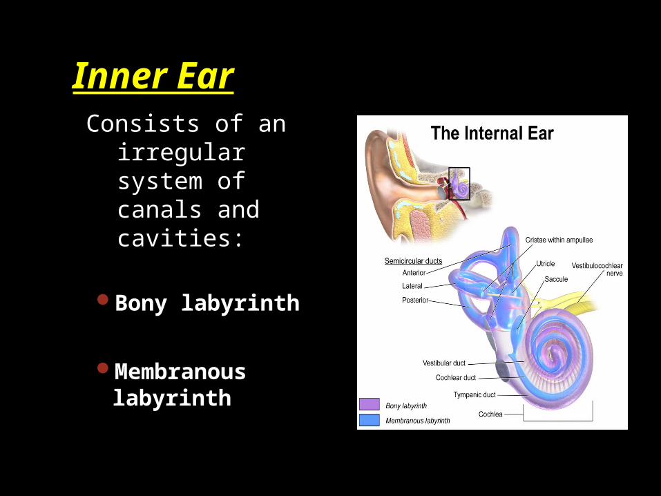
Inner Ear Consists of an
irregular system of canals and cavities:
Bony labyrinth
Membranous labyrinth
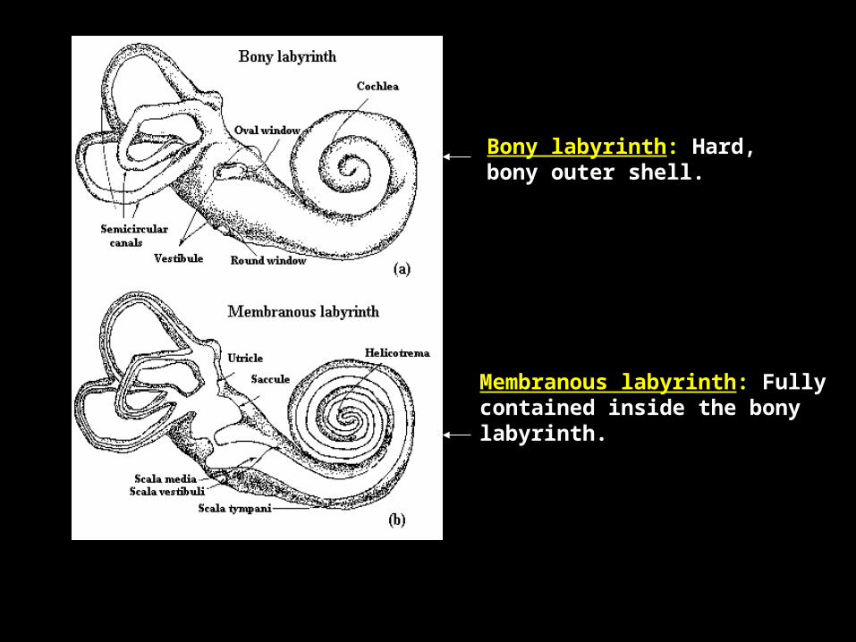
Bony labyrinth: Hard, bony outer shell.
Membranous labyrinth: Fully contained inside the bony labyrinth.
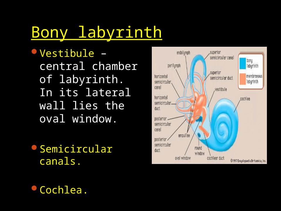
Bony labyrinthVestibule – central
chamber of labyrinth. In its lateral wall lies the oval window.
Semicircular canals.
Cochlea.
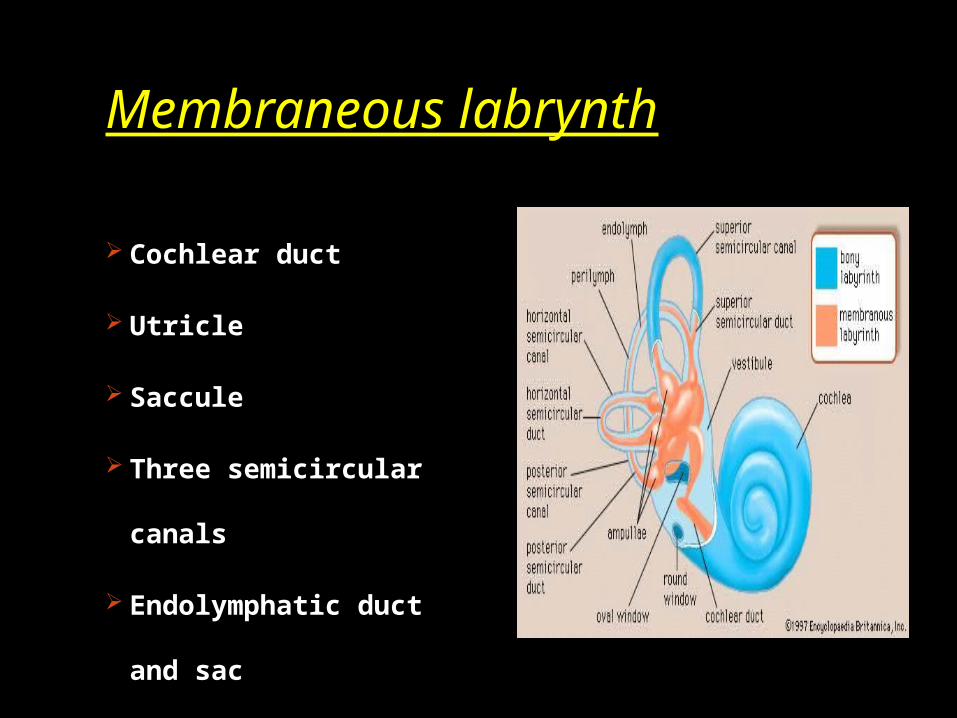
Membraneous labrynth
Cochlear duct
Utricle
Saccule
Three semicircular
canals
Endolymphatic duct
and sac
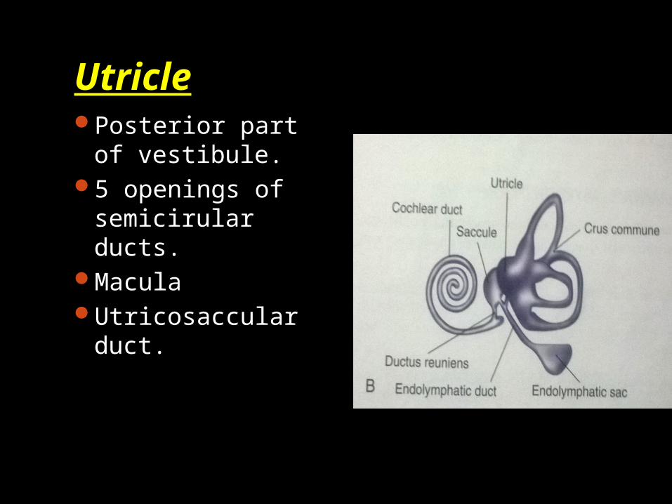
UtriclePosterior part of
vestibule.5 openings of
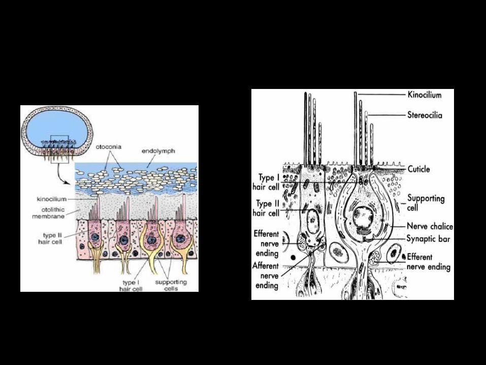
semicirular ducts.MaculaUtricosaccular
duct.
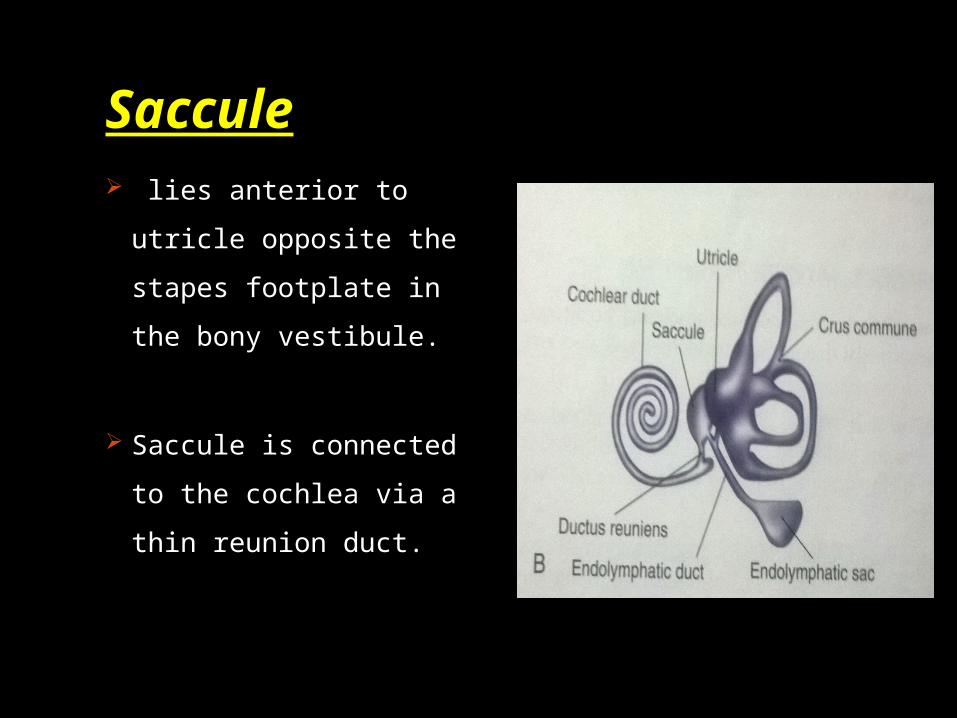
Saccule lies anterior to
utricle opposite the
stapes footplate in
the bony vestibule.
Saccule is connected
to the cochlea via a
thin reunion duct.
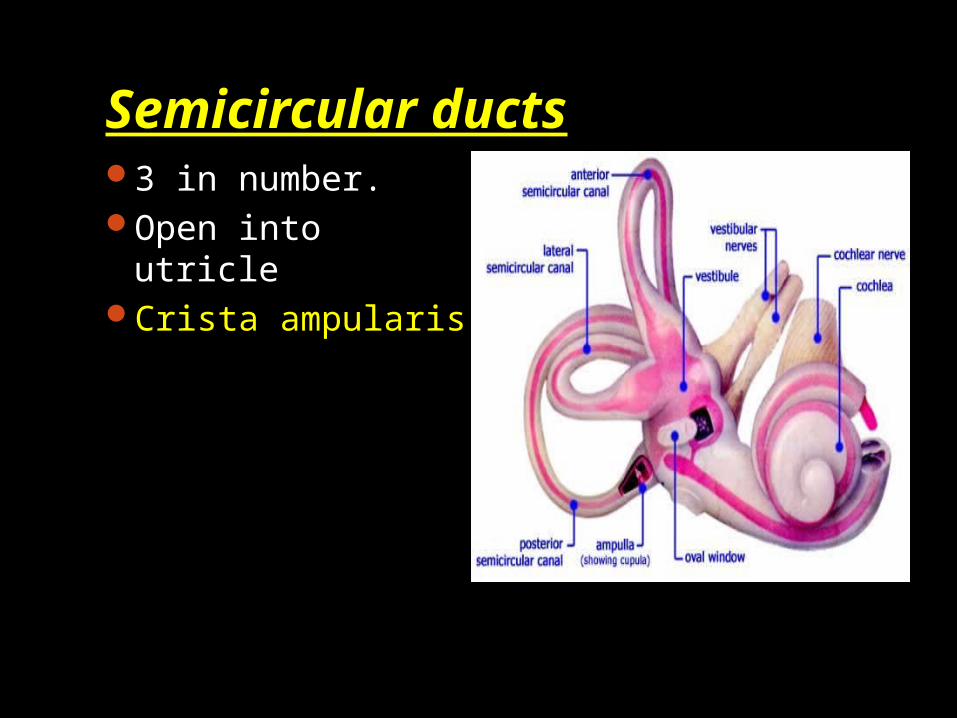
Semicircular ducts3 in number.Open into utricleCrista ampularis
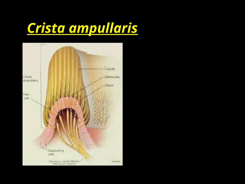
Crista ampullaris
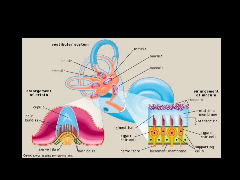
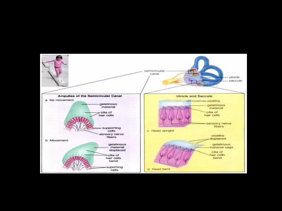

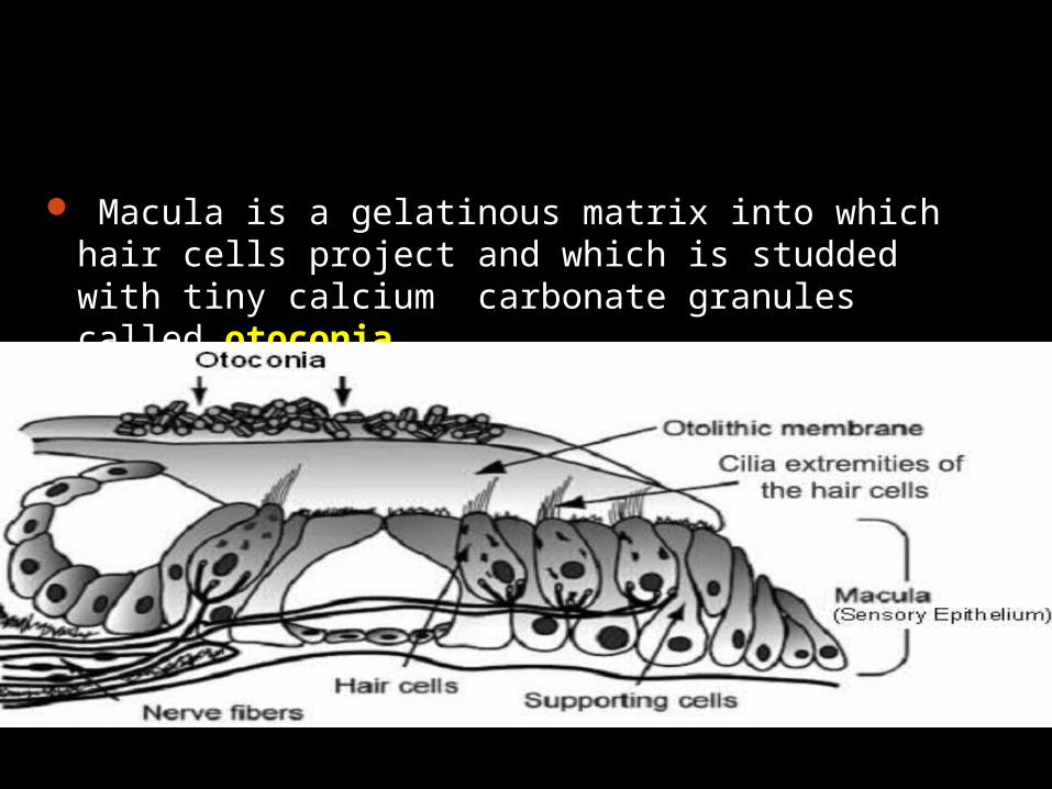
Macula is a gelatinous matrix into which hair cells project and which is studded with tiny calcium carbonate granules called otoconia.
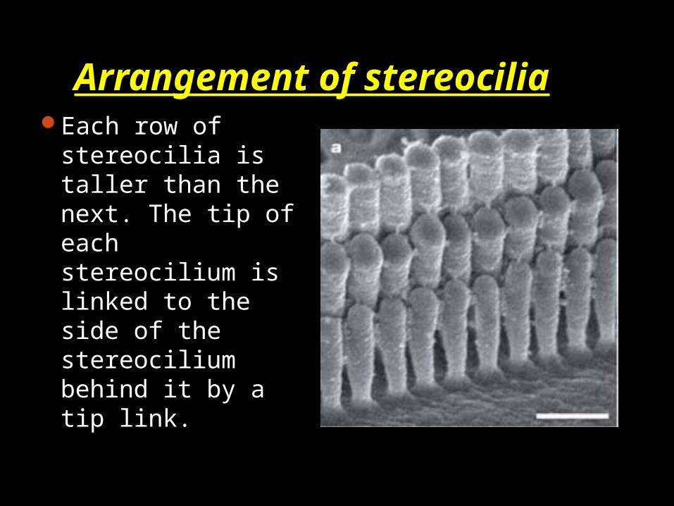
Arrangement of stereocilia
Each row of stereocilia is taller than the next. The tip of each stereocilium is linked to the side of the stereocilium behind it by a tip link.
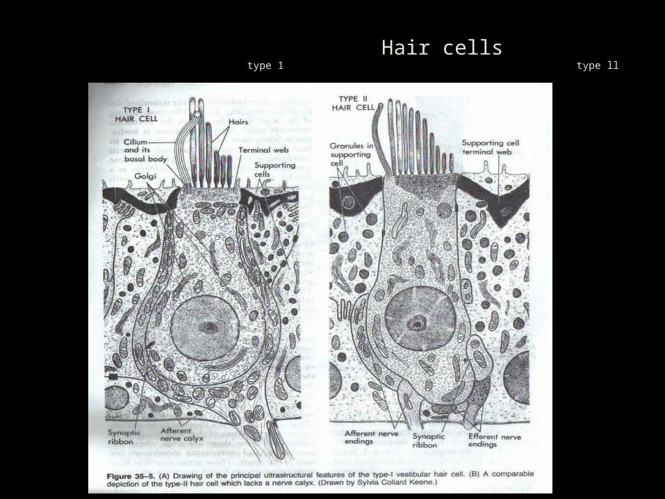
Hair cells type 1 type ll
Types of Hair cells
Type 1 Hair cells = flask shaped cells with a rounded base, narrow neck.
Nucleus is basal, surrounded by mitochondria
With supranuclear Golgi complex, occasional cisternae of RER & small vesicles.
With 50-100 sterocilia on free surface. Tallest hair is 10um near kinocilium & shortest is 1um on the opposite side.
Type ll Hair cells = more columnar, kinocilium, sterocilia, cytoplasmic roganelles are similar to type 1.
Golgi complex is larger, small vesicles found in great numbers in cytoplasm.
Synaptic ribbons are found in the peripheral cytoplasm opposite the plasmalemma of terminal boutons.
Some endings contain clear synaptic vesicles (non granulated vesicles) carry afferent nerves info to brain.
Semicircular CanalsThree semicircular
canals- Lateral
(Horizontal)Superior
( Anterior vertical)
Posterior ( Posterior vertical)
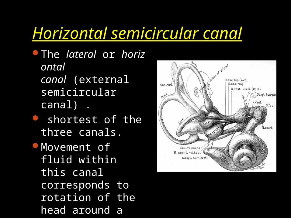
Horizontal semicircular canalThe lateral or horiz
ontal canal (external semicircular canal) .
shortest of the three canals.
Movement of fluid within this canal corresponds to rotation of the head around a vertical axis (i.e. the neck).
Projects as rounded bulge in middle ear, aditus & antrum
Makes an angle of 30 with horizontal plane.It measures from 12 to 15 mm., and its
arch is directed horizontally backward and laterally; thus each semicircular canal stands at right angles to the other two.
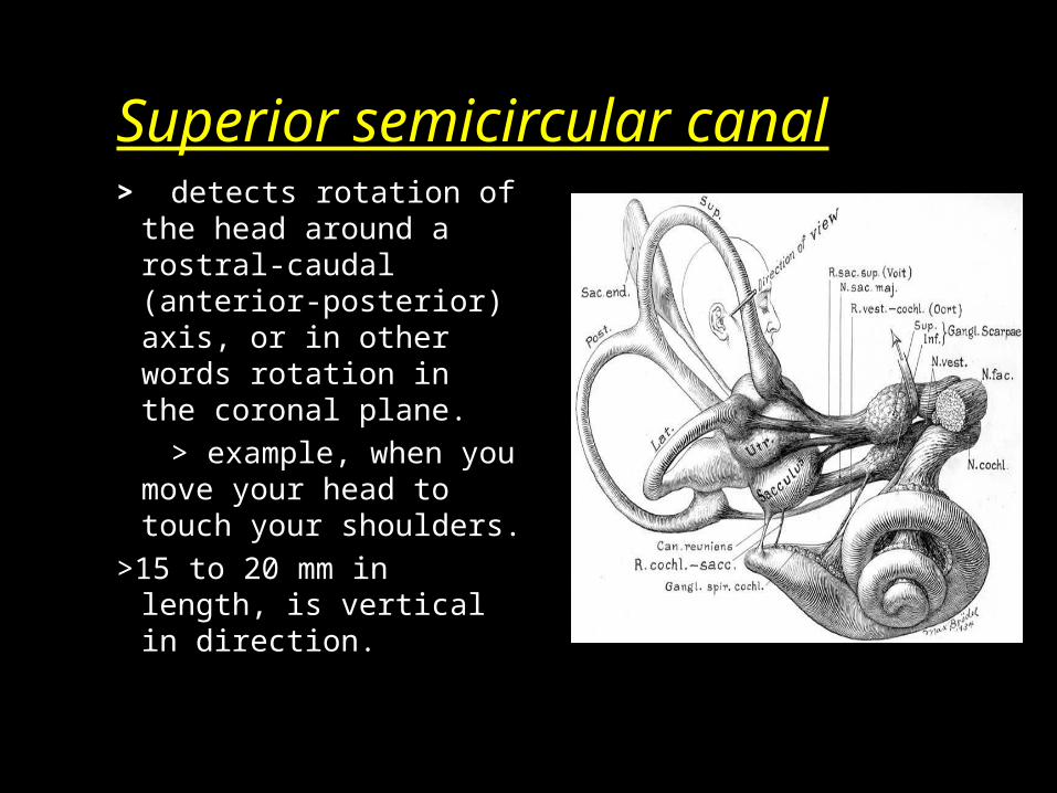
Superior semicircular canal> detects rotation of
the head around a rostral-caudal (anterior-posterior) axis, or in other words rotation in the coronal plane.
> example, when you move your head to touch your shoulders.
>15 to 20 mm in length, is vertical in direction.
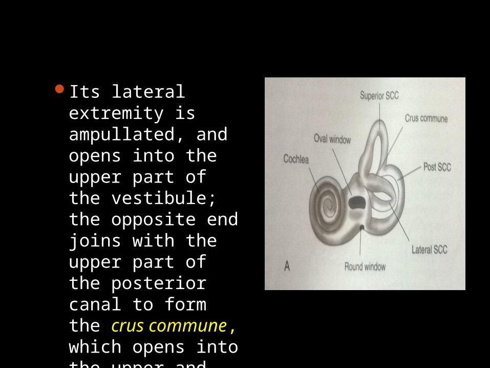
Its lateral extremity is ampullated, and opens into the upper part of the vestibule; the opposite end joins with the upper part of the posterior canal to form the crus commune, which opens into the upper and medial part of the vestibule.

Posterior semicircular canalThe posterior
semicircular canal detects rotations of the head in around the lateral axis. This occurs, for example, when nodding your head.
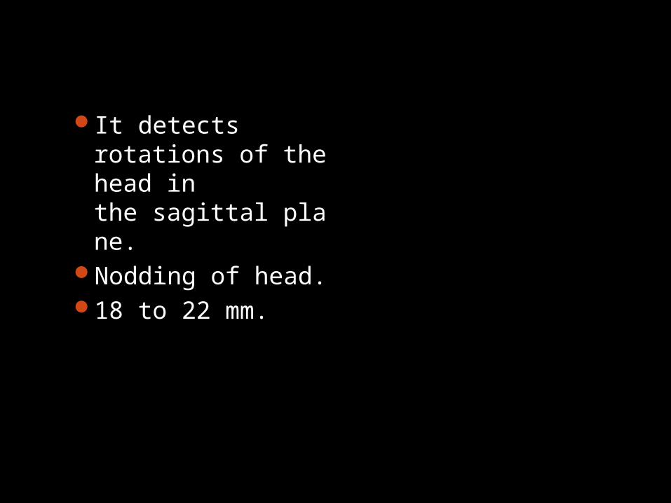
It detects rotations of the head in the sagittal plane.
Nodding of head.18 to 22 mm.
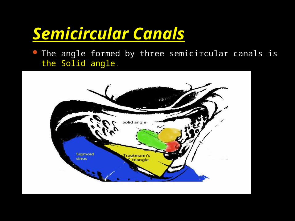
Semicircular Canals The angle formed by three semicircular canals is the
Solid angle.

Trautmann’s triangle.The triangle bounded by the bony labyrinth,
sigmoid sinus, and superior petrosal sinus is known as Trautmann’s triangle.

Cochlea
Snail shaped coiled tube.
2.5 to 2.75 turns round a central pyramid of bone called modiolus.
30 mm long
5 mm from base to apex & 9 mm around its base Anterio medial to vestibule.
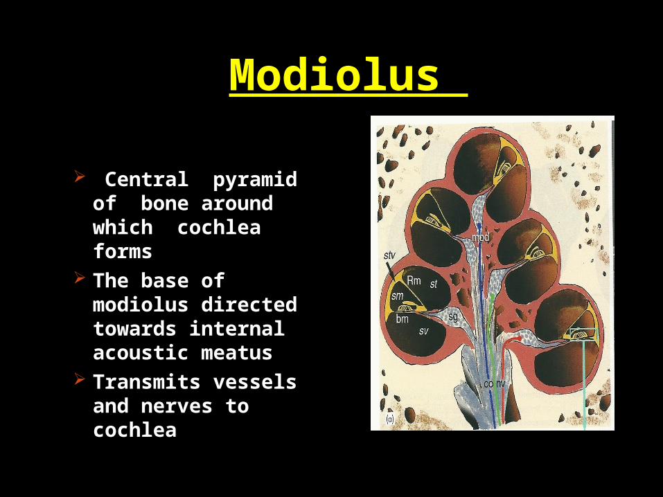
Modiolus
Central pyramid of bone around which cochlea forms
The base of modiolus directed towards internal acoustic meatus
Transmits vessels and nerves to cochlea
Promontary
A bony bulge in the medial wall of middle ear , represents the basal coil of cochlea.
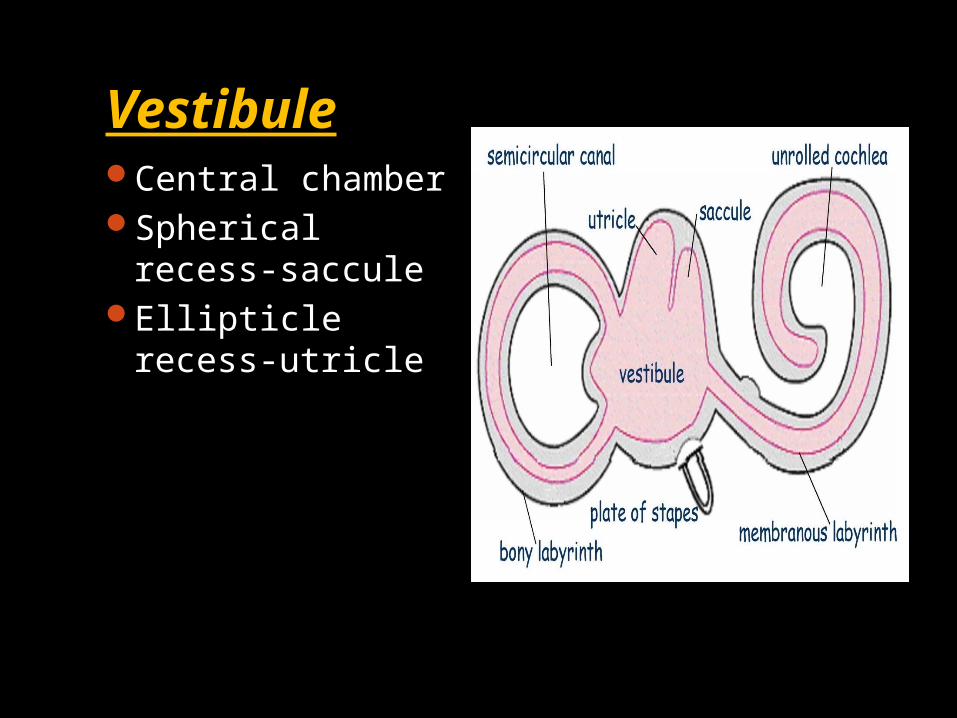
VestibuleCentral chamberSpherical recess-
sacculeEllipticle recess-
utricle
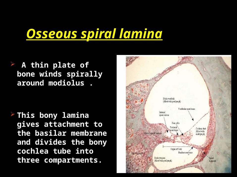
Osseous spiral lamina
A thin plate of bone winds spirally around modiolus .
This bony lamina gives attachment to the basilar membrane and divides the bony cochlea tube into three compartments.
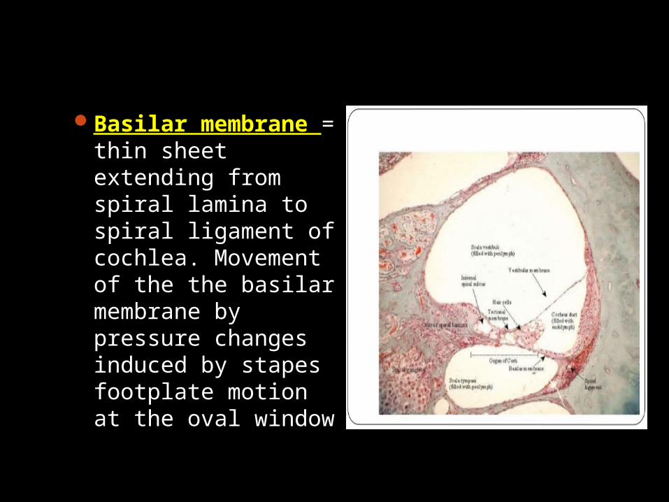
Basilar membrane = thin sheet extending from spiral lamina to spiral ligament of cochlea. Movement of the the basilar membrane by pressure changes induced by stapes footplate motion at the oval window
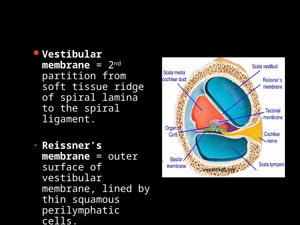
Vestibular membrane = 2nd partition from soft tissue ridge of spiral lamina to the spiral ligament.
• Reissner’s membrane = outer surface of vestibular membrane, lined by thin squamous perilymphatic cells.
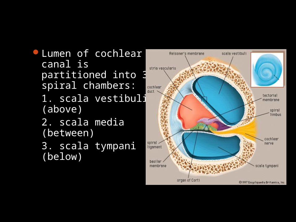
Lumen of cochlear canal is partitioned into 3 spiral chambers:1. scala vestibuli (above)2. scala media (between)3. scala tympani (below)
Rosenthal’s canal Spiral ganglions are
situated in this canal which runs along the osseous spiral lamina.
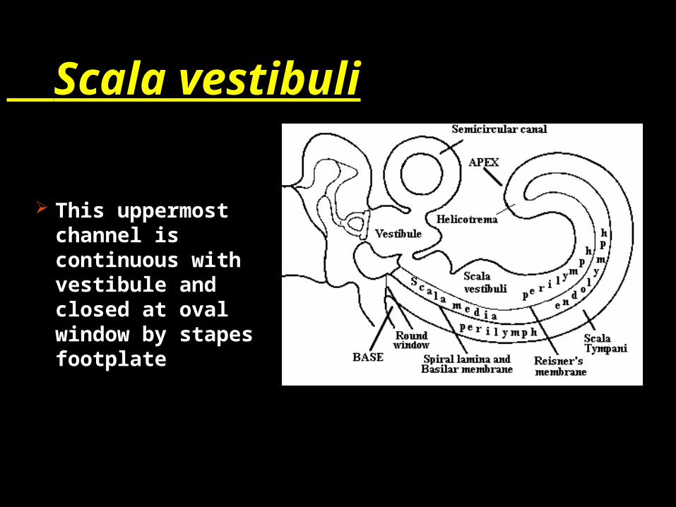
Scala vestibuli
This uppermost channel is continuous with vestibule and closed at oval window by stapes footplate
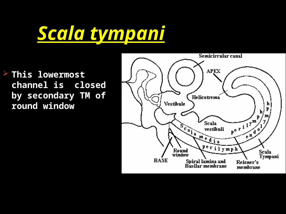
Scala tympani
This lowermost channel is closed by secondary TM of round window
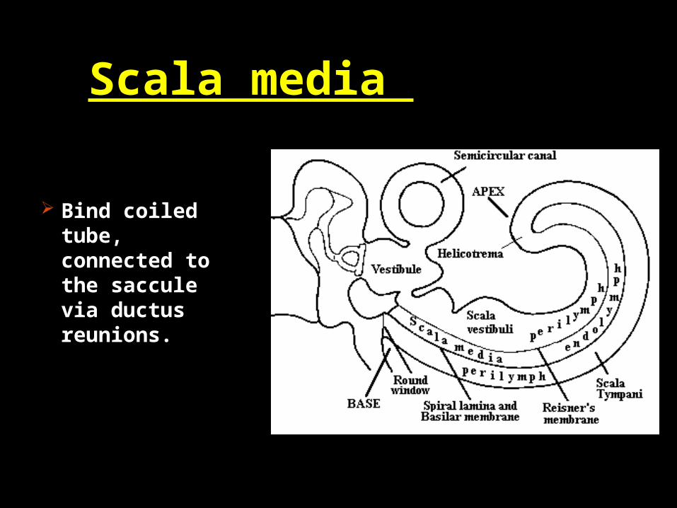
Scala media
Bind coiled tube, connected to the saccule via ductus reunions.
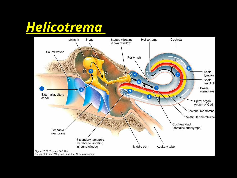
Helicotrema

Aqueduct of cochlea Scala tympani
is connected with subarachnoid space via aqueduct of cochlea.
It is thought to regulate perilymph & pressure in bony labrynth
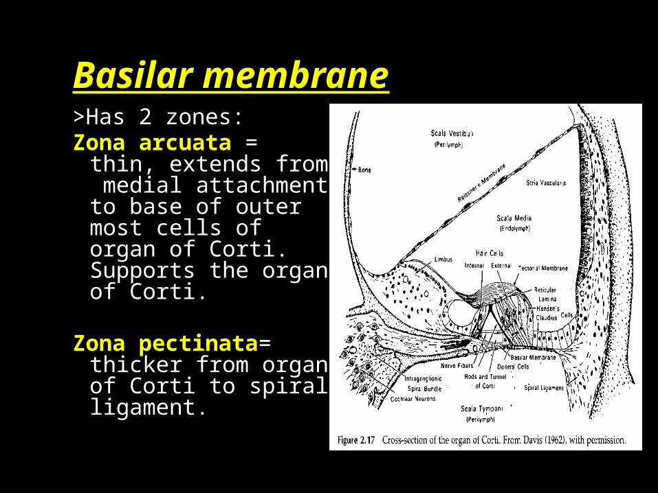
Basilar membrane>Has 2 zones:Zona arcuata =
thin, extends from medial attachment to base of outer most cells of organ of Corti. Supports the organ of Corti.
Zona pectinata= thicker from organ of Corti to spiral ligament.
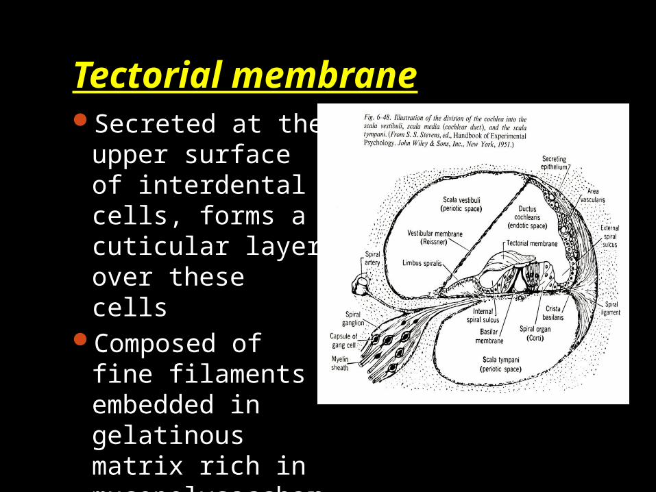
Tectorial membraneSecreted at the
upper surface of interdental cells, forms a cuticular layer over these cells
Composed of fine filaments embedded in gelatinous matrix rich in mucopolysaccharides.

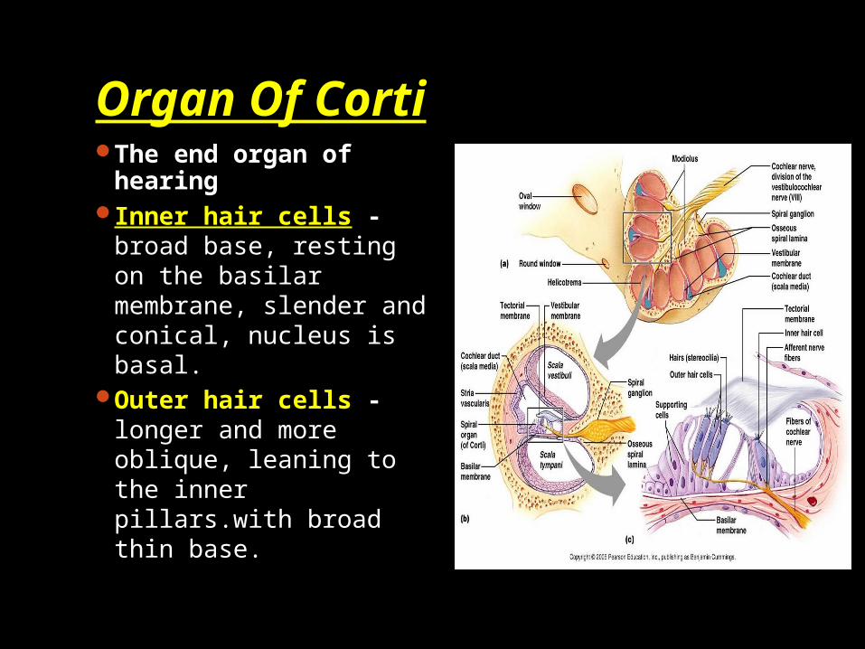
Organ Of CortiThe end organ of
hearingInner hair cells -
broad base, resting on the basilar membrane, slender and conical, nucleus is basal.
Outer hair cells -longer and more oblique, leaning to the inner pillars.with broad thin base.

Tunnel of corti- wide triangular intercellular space continuous through cochlear length. Bounded above converging inner and outer pillar cells.

Deiters cells -the supporting cells for the 3 -4 rows of outer hair cells. Base is columnar with cup shaped upper end. The apex does not reach the free surface of the organ of Corti.
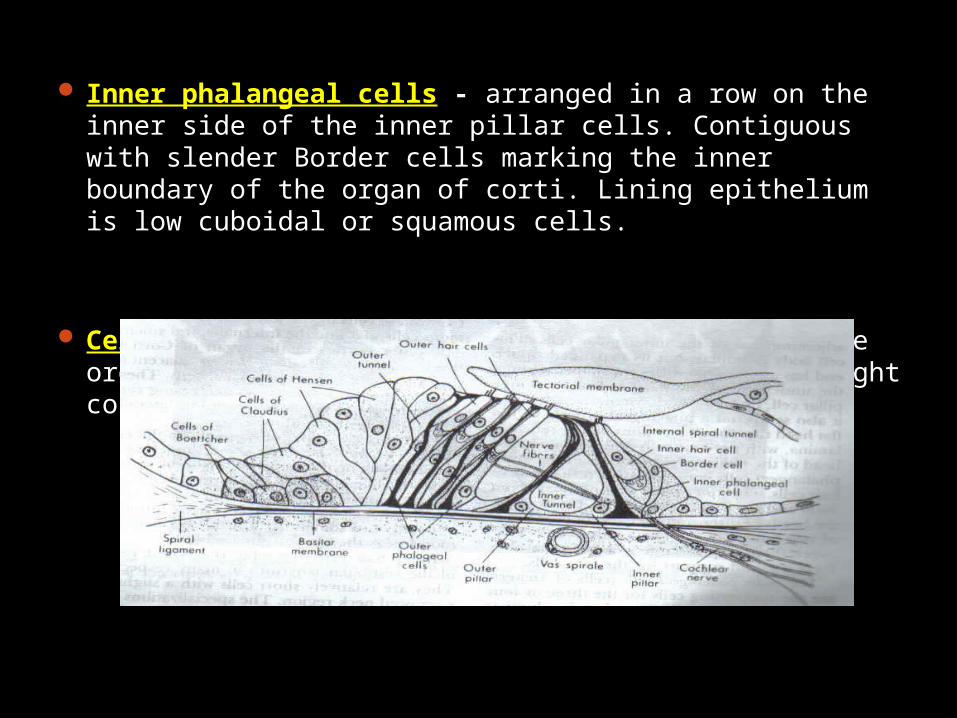
Inner phalangeal cells - arranged in a row on the inner side of the inner pillar cells. Contiguous with slender Border cells marking the inner boundary of the organ of corti. Lining epithelium is low cuboidal or squamous cells.
Cells of Hensen - delimit the outer border of the organ of Corti arrange in rows decreasing in height continuous with the cells of Claudius.
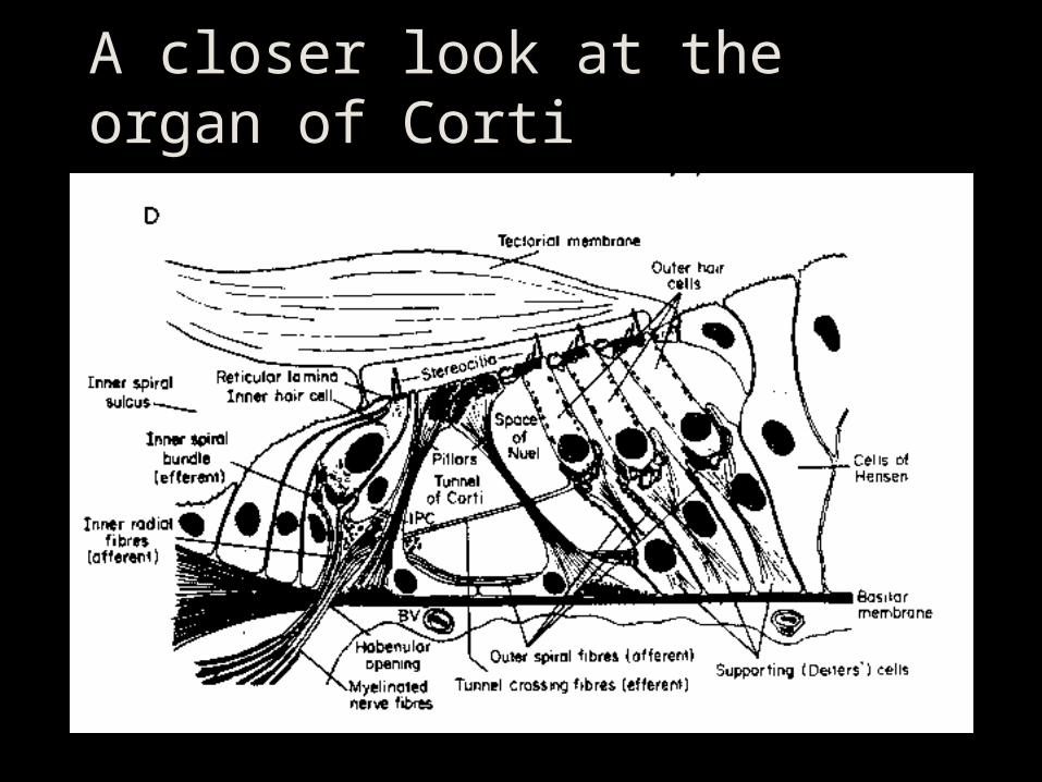
A closer look at the organ of Corti

Reticular laminaThe reticular
lamina is a solid surface at the tops of the hair cells, so the tops of the hair cells are in endolymph and the bottom of the hair cells are in perilymph.
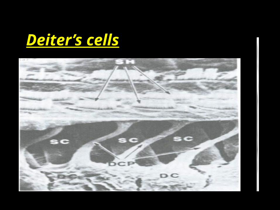
Deiter’s cells
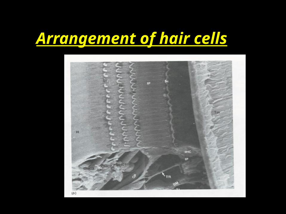
Arrangement of hair cells

The spiral ganglionThe cell bodies of the neurons that form the auditory nerve are located within the cochlear modiolus. The collection of cell bodies is called the spiral ganglion.
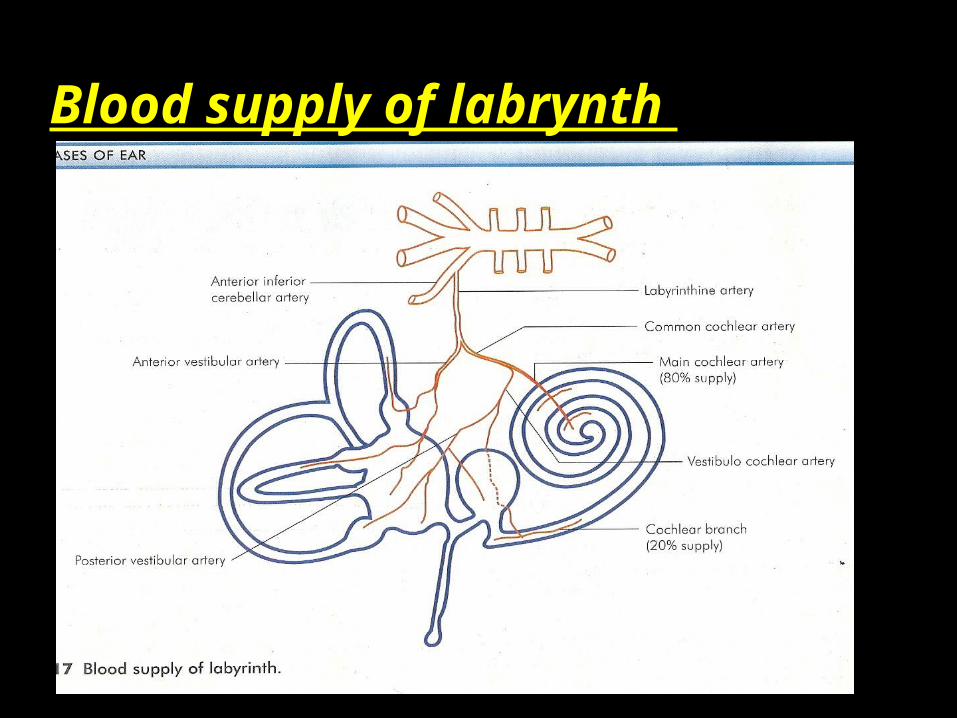
Blood supply of labrynth
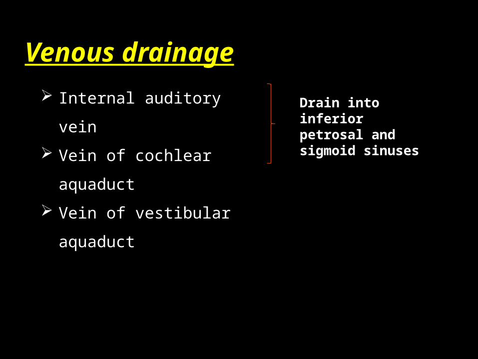
Venous drainage
Internal auditory vein
Vein of cochlear
aquaduct
Vein of vestibular
aquaduct
Drain into inferior petrosal and sigmoid sinuses

Internal auditory canal
About 1 cm long
Passes into
petrous part of
temporal bone in
a lateral direction
Lined by dura


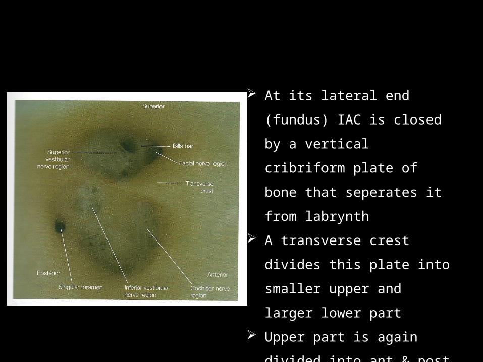
At its lateral end (fundus)
IAC is closed by a vertical
cribriform plate of bone
that seperates it from
labrynth
A transverse crest divides
this plate into smaller
upper and larger lower part
Upper part is again divided
into ant & post part by a
vertical crest called BILL’S
BAR.
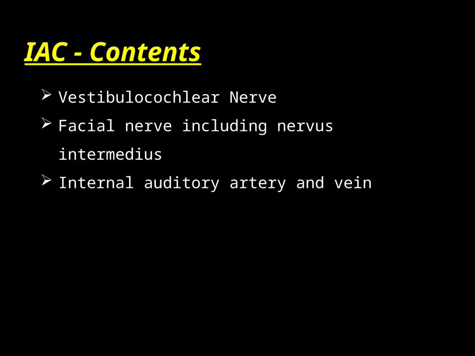
IAC - Contents
Vestibulocochlear Nerve
Facial nerve including nervus intermedius
Internal auditory artery and vein
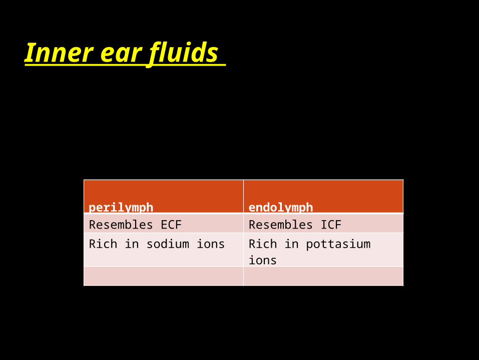
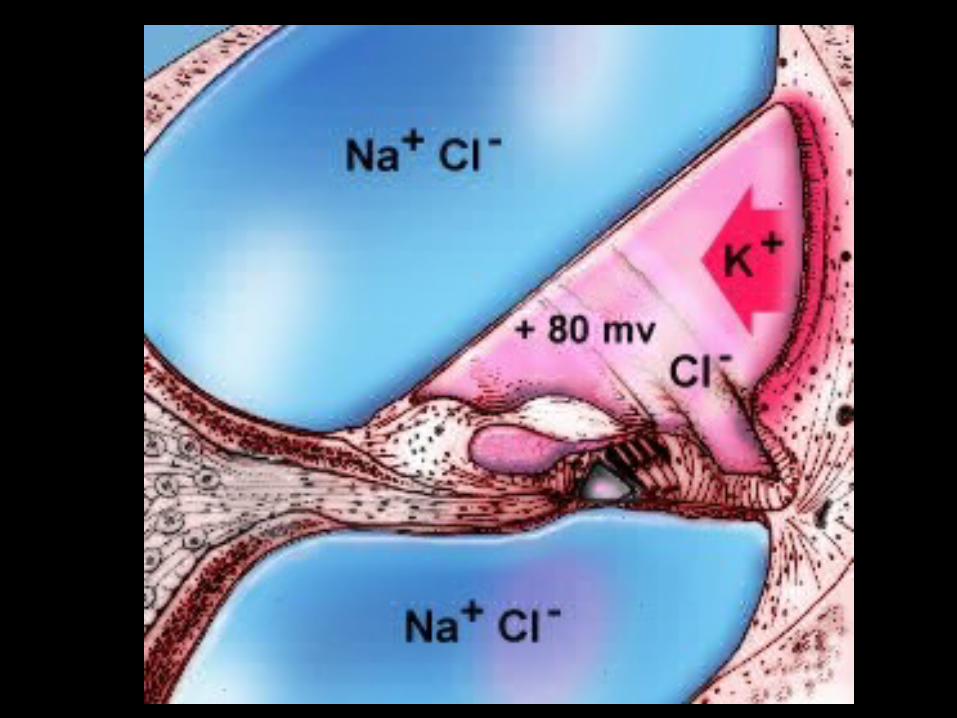
Inner ear fluids
perilymph endolymph
Resembles ECF Resembles ICF
Rich in sodium ions Rich in pottasium ions
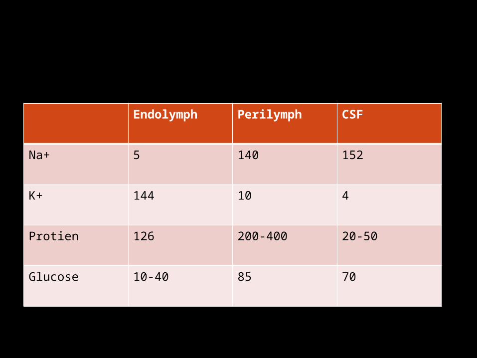
Endolymph Perilymph CSF
Na+ 5 140 152
K+ 144 10 4
Protien 126 200-400 20-50
Glucose 10-40 85 70

THANK YOU