inner ear development - university of minnesota duluthpschoff/documents/innereardevelopment.pdf ·...
TRANSCRIPT
Inner Ear Development
• Review of adult anatomy and physiology
• Generation of specification of the sensory organs
• Birth and differentiation of hair cells
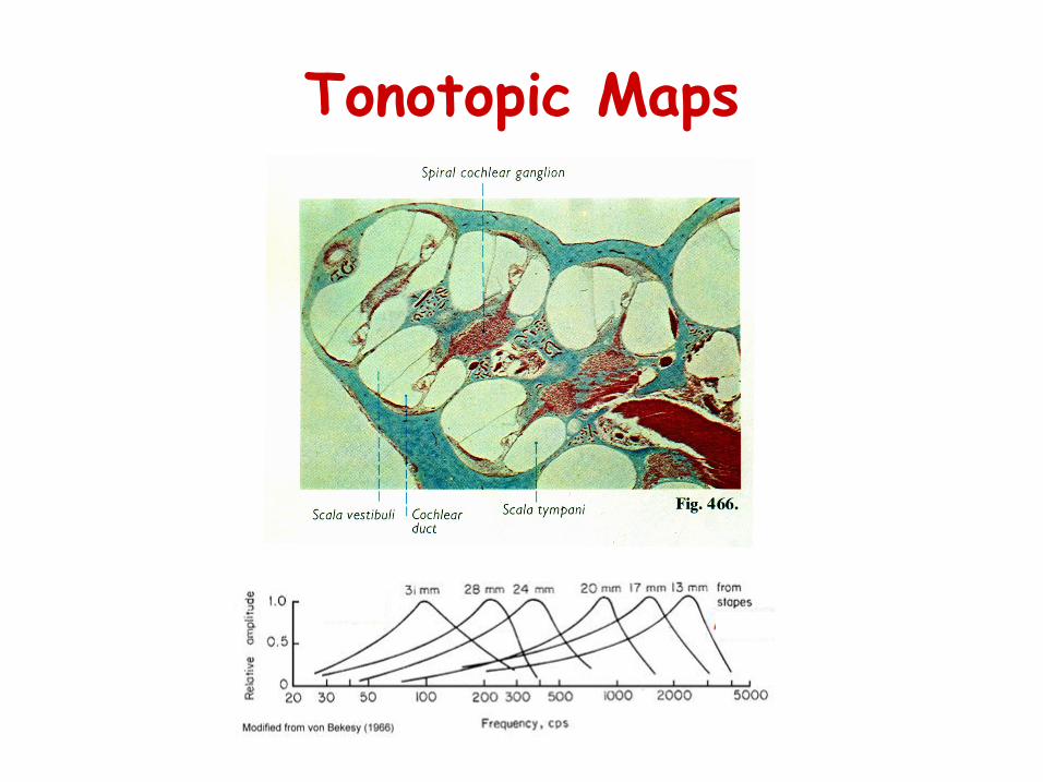
Tonotopic Maps
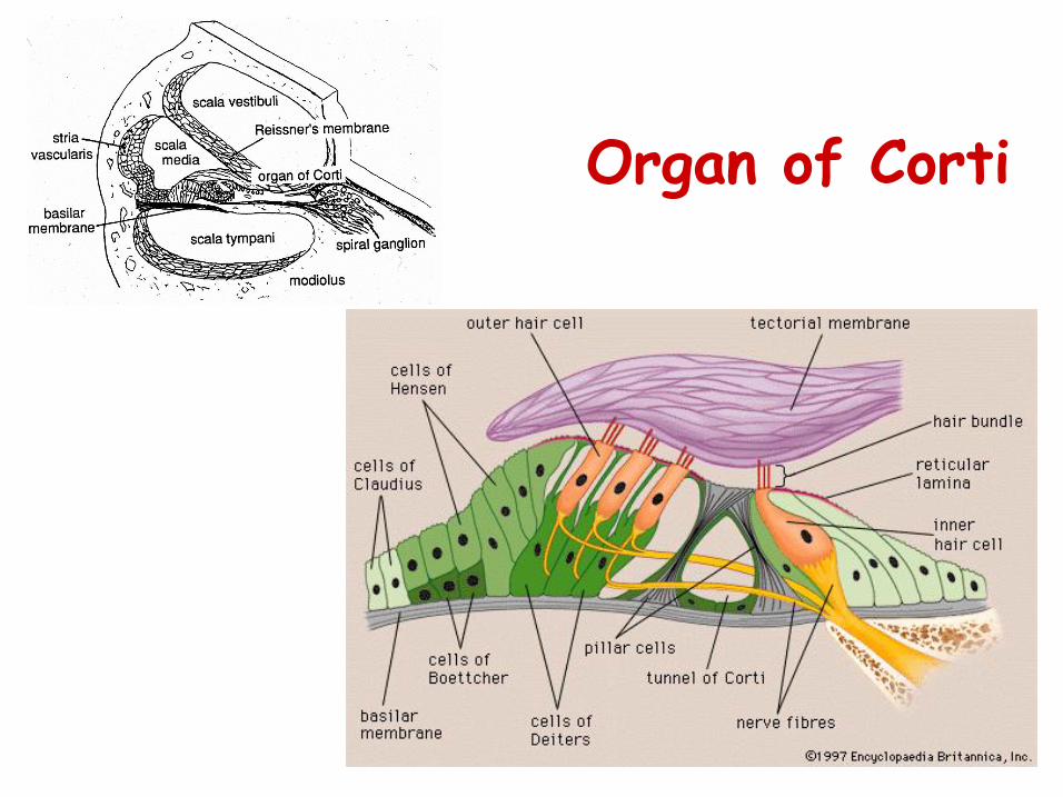
Organ of Corti
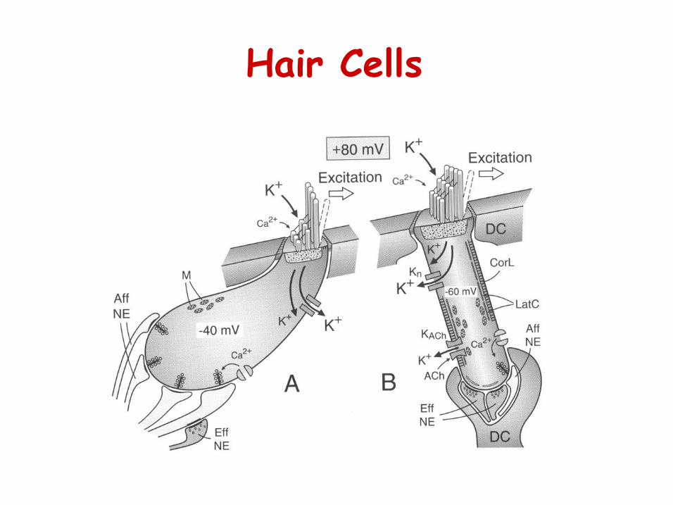
Hair Cells
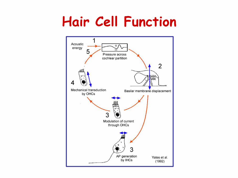
Hair Cell Function
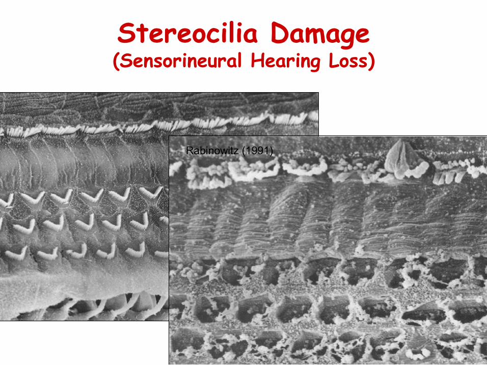
Stereocilia Damage (Sensorineural Hearing Loss)
Rabinowitz (1991)
Inner Ear Development
• Review of adult anatomy and physiology
• Generation of specification of the sensory organ
• Birth and differentiation of hair cells
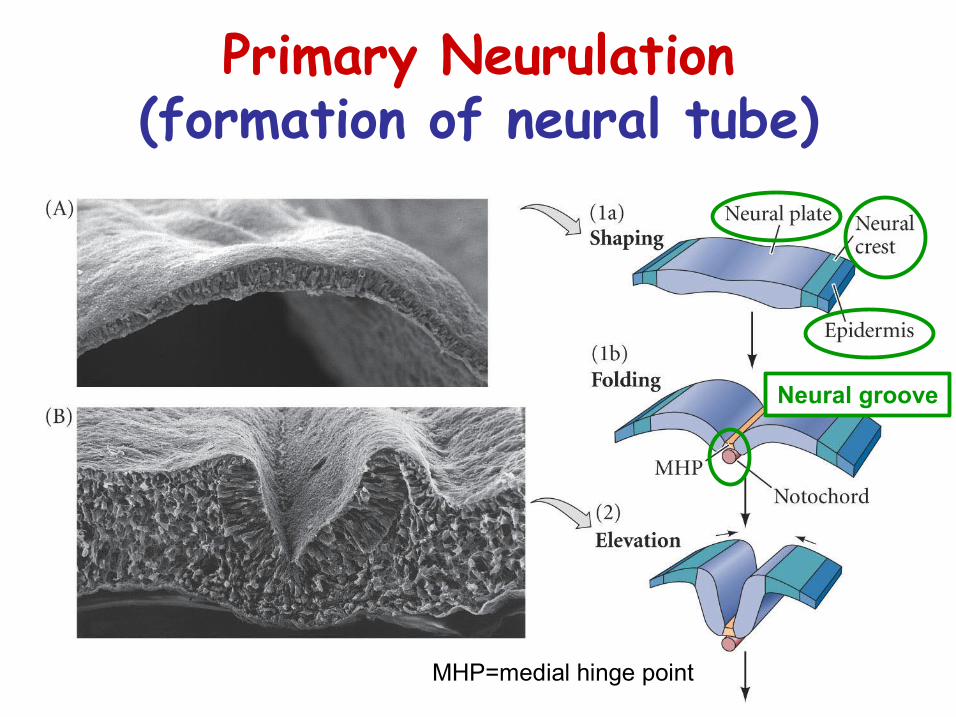
Primary Neurulation (formation of neural tube)
MHP=medial hinge point
Neural groove
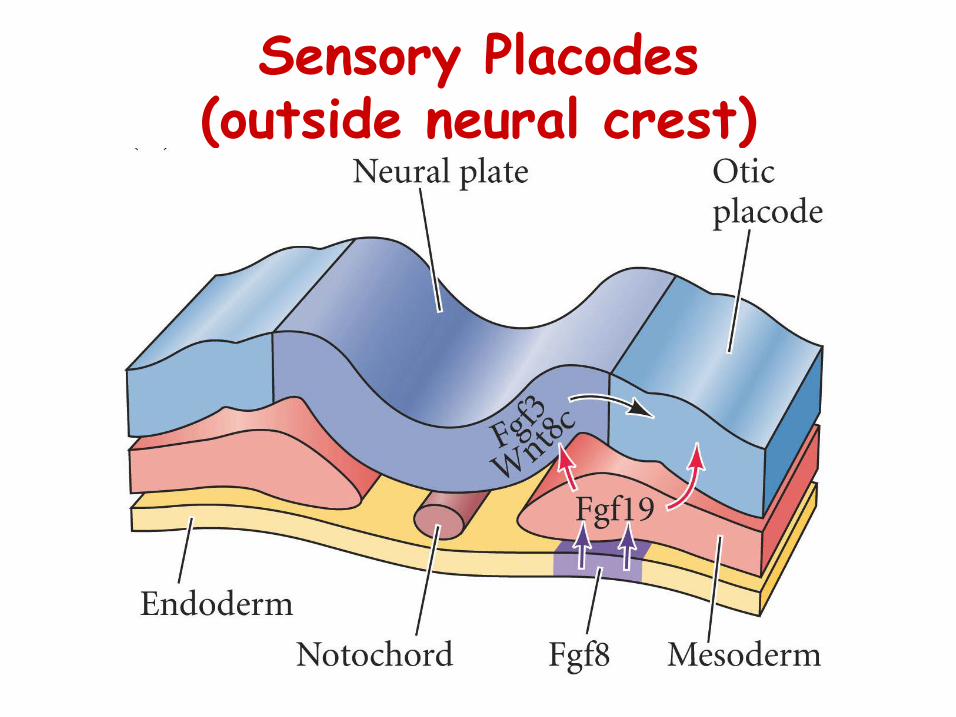
Sensory Placodes (outside neural crest)
Formation of Sensory Ganglia
Streit (2007)
• Before the placodes have emerged, head ectoderm cells have become distinct re. final placement in a specific placode o This region is called the preplacodal region
Differentiation of Preplacodal
Region
• Inhibitory signals from the lateral (BMP) and posterior (Wnt) ectoderm, as well as mesoderm and the neural folds define preplacodal region
Streit (2007)
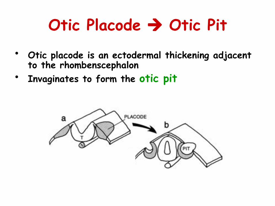
Otic Placode à Otic Pit
• Otic placode is an ectodermal thickening adjacent to the rhombenscephalon
• Invaginates to form the otic pit
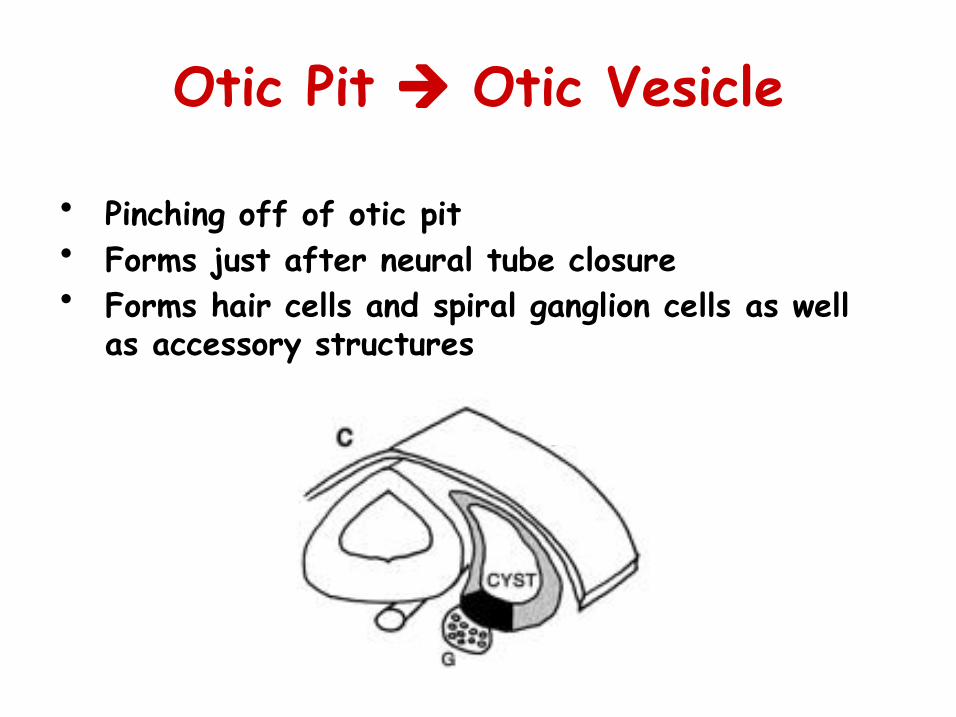
Otic Pit à Otic Vesicle
• Pinching off of otic pit • Forms just after neural tube closure • Forms hair cells and spiral ganglion cells as well
as accessory structures
Formation of the Pro-sensory Patch
• forms in the ventromedial wall • defined by expression patterns of Serrate1,
Lunatic fringe and BEN à involved in Notch signaling
• asymmetric expression patterns of these and other molecules appear to define ventral vs. dorsal halves, and anterior/posterior compartments
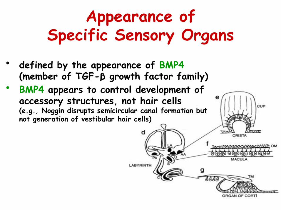
Appearance of Specific Sensory Organs
• defined by the appearance of BMP4 (member of TGF-β growth factor family)
• BMP4 appears to control development of accessory structures, not hair cells (e.g., Noggin disrupts semicircular canal formation but not generation of vestibular hair cells)
Inner Ear Development
• Review of adult anatomy and physiology
• Generation of specification of the sensory organs
• Birth and differentiation of hair cells o Cell fate o Stereocilia polarity o Outer vs. inner
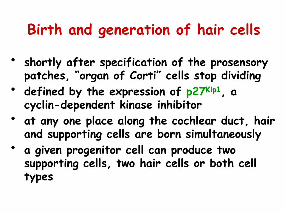
Birth and generation of hair cells
• shortly after specification of the prosensory patches, “organ of Corti” cells stop dividing
• defined by the expression of p27 Kip1 , a cyclin-dependent kinase inhibitor
• at any one place along the cochlear duct, hair and supporting cells are born simultaneously
• a given progenitor cell can produce two supporting cells, two hair cells or both cell types

Kelley (2007)
Cell Fate (Early)
• 4 potential fates • Decision by prosensory
cells to become a hair cell or a supporting cell involves LATERAL INHIBITION mediated by Notch signalling
Kelley (2007)
Cell Fate (Late)
• nascent hair cells begin to express Brn3.1, a POU domain transcription factor that is specifically expressed by hair cells
• a day later, the hair cell markers myosin VI and VIIa are expressed
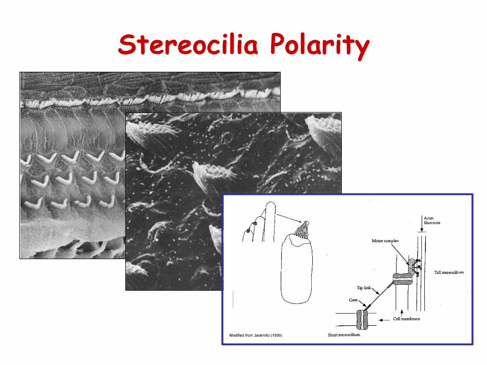
Stereocilia Polarity
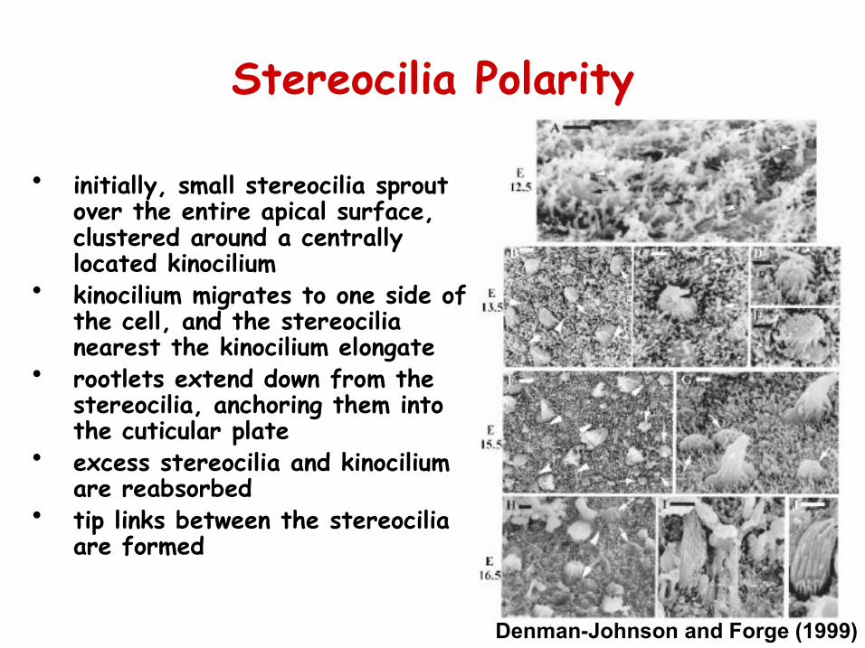
Stereocilia Polarity
• initially, small stereocilia sprout over the entire apical surface, clustered around a centrally located kinocilium
• kinocilium migrates to one side of the cell, and the stereocilia nearest the kinocilium elongate
• rootlets extend down from the stereocilia, anchoring them into the cuticular plate
• excess stereocilia and kinocilium are reabsorbed
• tip links between the stereocilia are formed
DenmanJohnson and Forge (1999)
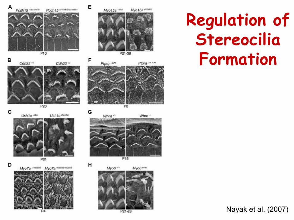
Regulation of Stereocilia Formation
Nayak et al. (2007)
Inner vs. Outer Hair Cells
• expression of PRESTIN