inria’sresearchcenters oma mia -mva 2017-2018 2,800 scientists in computer science 1300...
TRANSCRIPT
1
1
Introduction to medical image
acquisition and processing
X. Pennec
Asclepios team
2004, route des Lucioles B.P. 93
06902 Sophia Antipolis Cedex
http://www-sop.inria.fr/asclepios
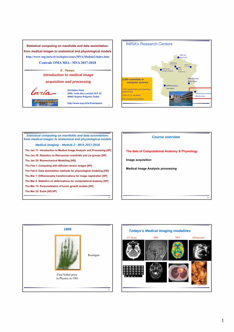
Statistical computing on manifolds and data assimilation:
from medical images to anatomical and physiological models
http://www-sop.inria.fr/asclepios/cours/MVA/Module2/index.htm
Centrale OMA MIA - MVA 2017-2018
2,800 scientists in computer science
1300 researchers and teaching-researchers
1000 Ph.D. students
500 postdoctoral fellows
•INRIA Nancy
•Grand Est
INRIA Grenoble
Rhône-Alpes
•INRIA Sophia Antipolis
•Méditerranée
•INRIA Rennes
•Bretagne Atlantique
INRIA Bordeaux
Sud-Ouest
INRIA Lille
Nord Europe
INRIA
Saclay
Île-de-France
INRIA ParisRocquencourt
INRIA’s Research Centers•4
3
Statistical computing on manifolds and data assimilation: from medical images to anatomical and physiological models
Medical Imaging – Module 2 - MVA 2017-2018
Thu Jan 11: Introduction to Medical Image Analysis and Processing [XP]
Thu Jan 18: Statistics on Riemannian manifolds and Lie groups [XP]
Thu Jan 25: Biomechanical Modelling [HD]
Thu Feb 1: Computing with diffusion tensor images [XP]
Thu Feb 8: Data assimilation methods for physiological modeling [HD]
Thu Mar 1: Diffeomorphic transformations for image registration [XP]
Thu Mar 8: Statistics on deformations for computational anatomy [XP]
Thu Mar 15: Personalization of tumor growth models [XP]
Thu Mar 22: Exam [HD,XP]
4
Course overview
The data of Computational Anatomy & Physiology
Image acquisition
Medical Image Analysis processing
5
1895
First Nobel prize in Physics in 1901
Roentgen
Todays’s Medical Imaging modalities
UltrasoundCT Scan MRI PET
•Source :T. Peters
2
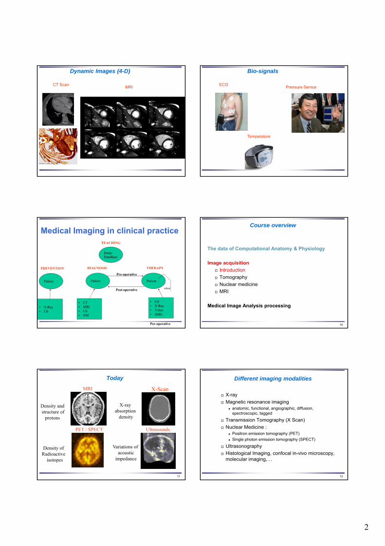
Dynamic Images (4-D)
MRICT Scan
Bio-signals
Pressure SensorECG
Temperature
Patient
DIAGNOSIS
• CT• MRI• US• NM
Patient
THERAPY
• US• X-Ray• Video• iMRI
Per-operative
Pre-operative
Post-operative robot
Patient
PREVENTION
• X-Ray• US
TEACHING
Image DataBase
Medical Imaging in clinical practice
10
Course overview
The data of Computational Anatomy & Physiology
Image acquisition
Introduction
Tomography
Nuclear medicine
MRI
Medical Image Analysis processing
11
Today
MRI
UltrasoundsPET / SPECT
X-Scan
X-ray absorption
density
Variations of acoustic
impedance
Density of Radioactive
isotopes
Density and structure of
protons
12
Different imaging modalities
X-ray
Magnetic resonance imaging anatomic, functional, angiographic, diffusion,
spectroscopic, tagged
Transmission Tomography (X Scan)
Nuclear Medicine : Positron emission tomography (PET)
Single photon emission tomography (SPECT)
Ultrasonography
Histological Imaging, confocal in-vivo microscopy, molecular imaging,…
3
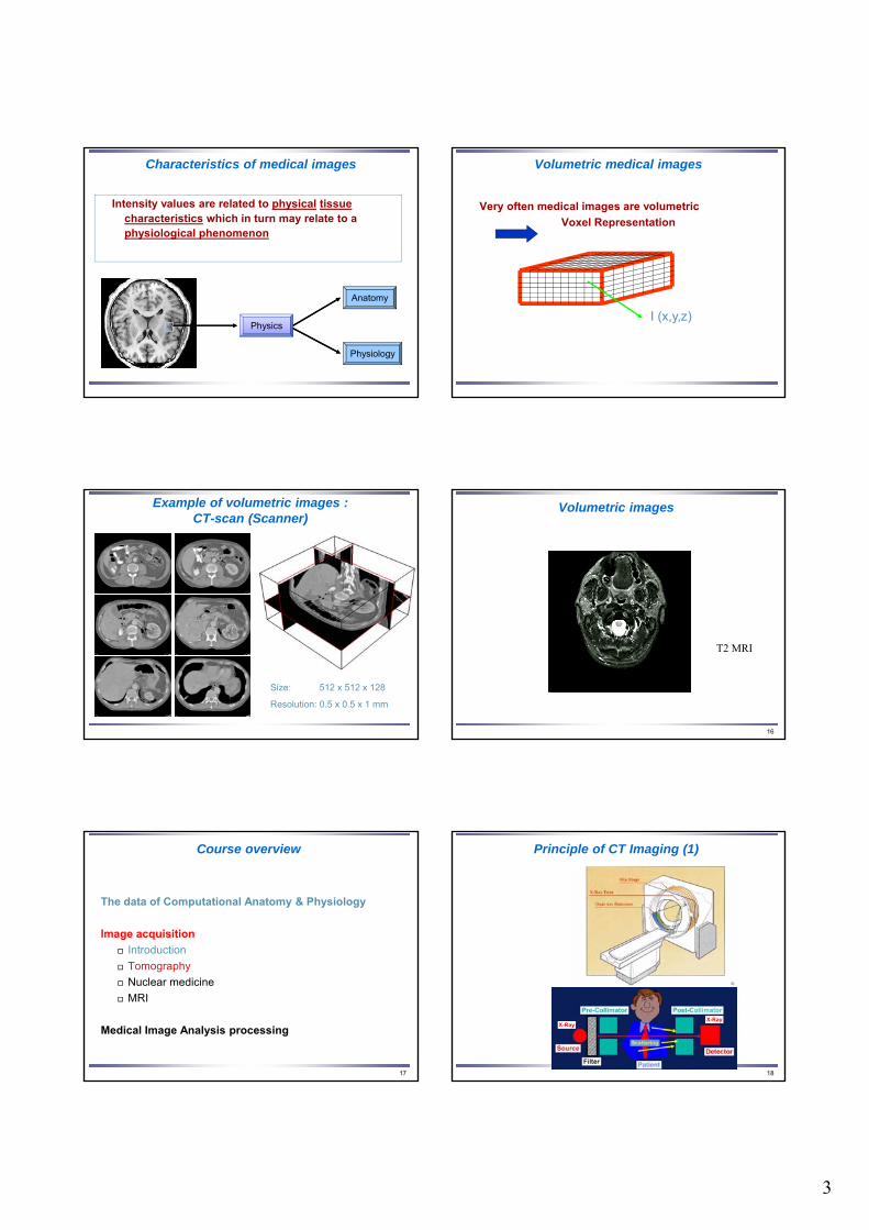
Characteristics of medical images
Intensity values are related to physical tissue characteristics which in turn may relate to a physiological phenomenon
Physics
Anatomy
Physiology
Volumetric medical images
Very often medical images are volumetric
Voxel Representation
I (x,y,z)
Example of volumetric images :CT-scan (Scanner)
Size: 512 x 512 x 128
Resolution: 0.5 x 0.5 x 1 mm
16
Volumetric images
T2 MRI
17
Course overview
The data of Computational Anatomy & Physiology
Image acquisition
Introduction
Tomography
Nuclear medicine
MRI
Medical Image Analysis processing
Principle of CT Imaging (1)
18
X-RayX-Ray
4
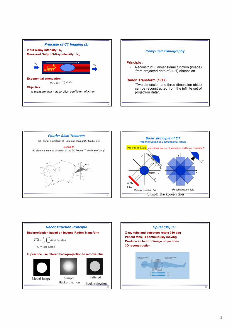
Principle of CT Imaging (2)
Input X-Ray intensity : Ni
Measured Output X-Ray intensity : No
Exponential attenuation :
Objective :
measure (x) = absorption coefficient of X-ray
19
Ni N0
Computed Tomography
Principle :• Reconstruct n dimensional function (image)
from projected data of (n–1) dimension
Radon Transform (1917)• “Two dimension and three dimension object
can be reconstructed from the infinite set of projection data”.
Fourier Slice Theorem
1D Fourier Transform of Projected slice of 2D field (x,y)
is equal to
1D slice in the same direction of the 2D Fourier Transform of (x,y)
21
Basic principle of CT-Reconstruction of 2 dimensional image-
Simple Backprojection
Projection Data
x
y
x
y
curvilinear integral of absorption coefficient regarding Y
object
X-ray
tubeData Acquisition field Reconstruction field
Reconstruction Principle
Backprojection based on inverse Radon Transform
In practice use filtered back-projection to remove blur
23
12
, .
cos , sin
Model Image SimpleBackprojection
Filtered
Backprojection
Spiral (3D) CT
X-ray tube and detectors rotate 360 deg
Patient table is continuously moving
Produce an helix of image projections
3D reconstruction
26
5
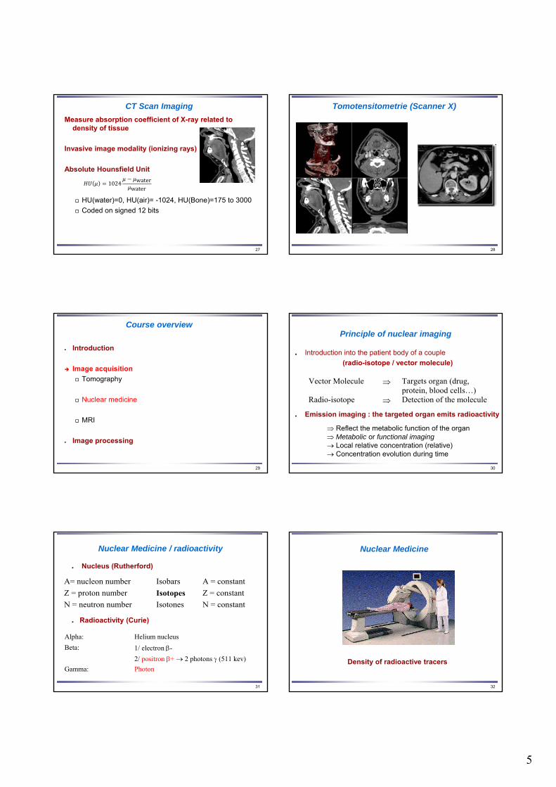
CT Scan Imaging
Measure absorption coefficient of X-ray related to density of tissue
Invasive image modality (ionizing rays)
Absolute Hounsfield Unit
HU(water)=0, HU(air)= -1024, HU(Bone)=175 to 3000
Coded on signed 12 bits
27
1024 waterwater
28
Tomotensitometrie (Scanner X)
29
Course overview
Introduction
Image acquisition
Tomography
Nuclear medicine
MRI
Image processing
30
Principle of nuclear imaging
Introduction into the patient body of a couple
(radio-isotope / vector molecule)
Emission imaging : the targeted organ emits radioactivity
Vector Molecule Targets organ (drug, protein, blood cells…)
Radio-isotope Detection of the molecule
Reflect the metabolic function of the organ Metabolic or functional imaging Local relative concentration (relative) Concentration evolution during time
31
Nuclear Medicine / radioactivity
Nucleus (Rutherford)
Radioactivity (Curie)
A= nucleon number Isobars A = constant
Z = proton number Isotopes Z = constant
N = neutron number Isotones N = constant
Alpha: Helium nucleus
Beta: 1/ electron -
2/ positron + 2 photons (511 kev)
Gamma: Photon
32
Nuclear Medicine
Density of radioactive tracers

6
33
Single photon gamma imaging
Radio-isotopes
Single photon emitters Technetium Tc 99m 6 h 140 kev Portative generator Iodine I 131 8 j 360 kev Reacteur (fission) Iodine I 123 13 h 159 kev Cyclotron (industry) Thallium Tl 201 73 h 80 kev Cyclotron (industry)
Krypton (Kr 81 m), Gallium (Ga 67), Indium (In 111), Xenon (Xe 133, gaz)
34
Single photon gamma imaging
Heart (myocardium perfusion) Stress/rest examperfusion / perfusion Healthy area perfusion / (hypo/non-)perfusion Zone at risk (ischemia) (hypo/non-)perfusion / (hypo/non-)perfusion Infarcted Area
Radio-isotopes
Physiological molecules
35
Positron emission tomography (PET)
Positron emitters Carbon 11C 20 mn cyclotron (medical)Nitrogen 13N 10 mn cyclotron (medical)Oxygen 15O 2 mn cyclotron (medical)Fluor 18F 112 mn cyclotron (medical)
Emission : positron (+) Annihilation
2 photons of 511 kev at 180º
water H2O15
glucose fluoro-deoxyglucose (F18DG)
36
Positron emission tomography (PET)
PET Scan
37
https://www.youtube.com/watch?v=GHLBcCv4rqk
38
Course overview
Introduction
Image acquisition
Tomography
Nuclear medicine
MRI
Image processing
7
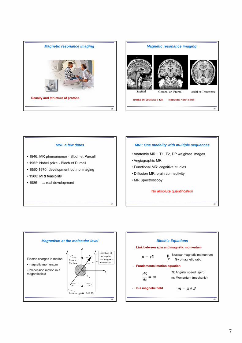
39
Magnetic resonance imaging
Density and structure of protons
40
Magnetic resonance imaging
dimension: 256 x 256 x 128 résolution: 1x1x1.5 mm
Sagittal Coronal or Frontal Axial or Transverse
41
MRI: a few dates
• 1946: MR phenomenon - Bloch et Purcell
• 1952: Nobel prize - Bloch et Purcell
• 1950-1970: development but no imaging
• 1980: MRI feasibility
• 1986 - …: real development
42
MRI: One modality with multiple sequences
• Anatomic MRI: T1, T2, DP weighted images
• Angiographic MR
• Functional MR: cognitive studies
• Diffusion MR: brain connectivity
• MR Spectroscopy
No absolute quantification
44
Magnetism at the molecular level
Electric charges in motion
• magnetic momentum
• Precession motion in a magnetic field
45
Bloch’s Equations
Fundamental motion equation
Link between spin and magnetic momentum
S: Angular speed (spin)
m: Momentum (mechanic)
Nuclear magnetic momentum
Gyromagnetic ratio
In a magnetic field
μ
∧
8
46
Bloch’s Equations
Thus
0
0 0
0
B
B
0
)(sin
)( cos
z
Lt
Lt
t
t
0 BL Larmor’s frequency
d∧
49
Magnetism at the macroscopic level
P+1/2=0.5000049
P-1/2=0.4999951B0=1.5 T
50
Magnetism at the macroscopic level
1023 spins
51
Resonance
52
Magnetic Resonance / excitation
• Electro-magnetic field at Larmor’s frequency
0 BL
• Hydrogen protons enter into resonance
Flip of the macroscopic momentum M
53
Magnetic Resonance / excitation
9
54
Magnetic Resonance / relaxation
1T
MBM
dt
dM zz
z
• Return to equilibrium / B0: time constant T1
• Spin dephasing: Time constant T2
2
,,
,
T
MBM
dt
dM yxyx
yx
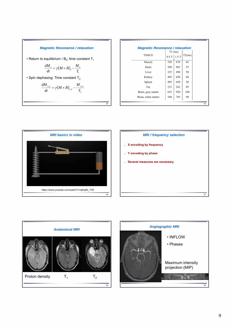
55
Magnetic Resonance / relaxationT1 (ms)
TISSUE 0.5 T 1.5 T
T2(ms)
Muscle 550 870 45
Heart 580 865 55
Liver 325 490 50
Kidney 495 650 60
Spleen 495 650 58
Fat 215 262 85
Brain, grey matter 655 920 100
Brain, white matter 540 785 90 540
MRI basics in video
56
https://www.youtube.com/watch?v=djAxjtN_7VE
57
MRI / frequency selection
X encoding by frequency
Y encoding by phase
Several measures are necessary
58
Anatomical MRI
Proton density T1 T2
59
Angiographic MRI
Maximum intensity projection (MIP)
• INFLOW
• Phases
10
60
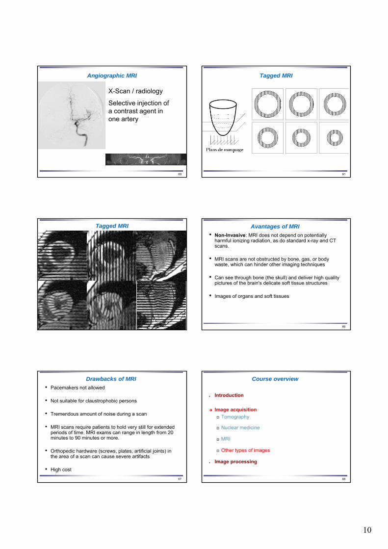
Angiographic MRI
X-Scan / radiology
Selective injection of a contrast agent in one artery
61
Tagged MRI
62
Tagged MRI Avantages of MRI
• Non-Invasive: MRI does not depend on potentially harmful ionizing radiation, as do standard x-ray and CT scans.
• MRI scans are not obstructed by bone, gas, or body waste, which can hinder other imaging techniques
• Can see through bone (the skull) and deliver high quality pictures of the brain's delicate soft tissue structures
• Images of organs and soft tissues
66
Drawbacks of MRI
• Pacemakers not allowed
• Not suitable for claustrophobic persons
• Tremendous amount of noise during a scan
• MRI scans require patients to hold very still for extended periods of time. MRI exams can range in length from 20 minutes to 90 minutes or more.
• Orthopedic hardware (screws, plates, artificial joints) in the area of a scan can cause severe artifacts
• High cost
67 68
Course overview
Introduction
Image acquisition
Tomography
Nuclear medicine
MRI
Other types of images
Image processing
11
Echography
Local variation of acoustic impedance
Echography
Gall Blader
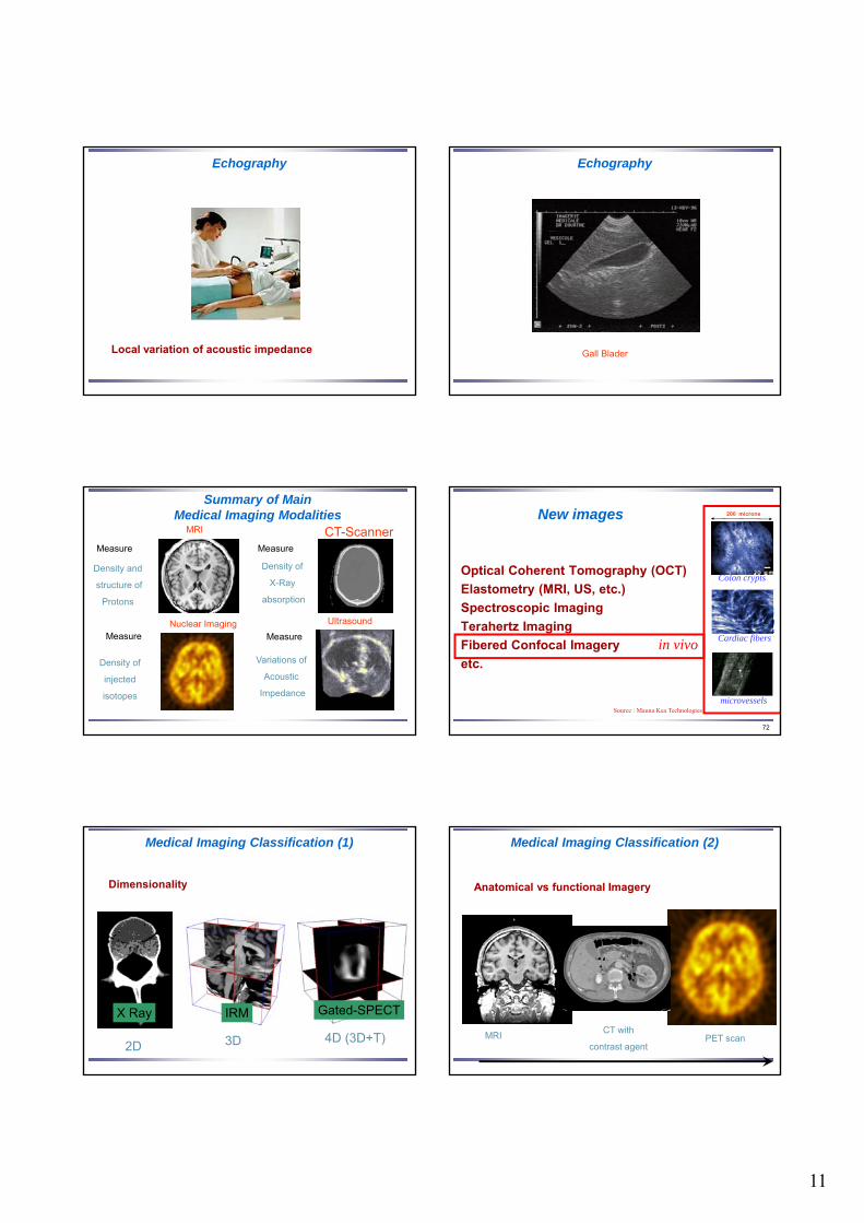
Summary of Main Medical Imaging Modalities
MRI
UltrasoundNuclear Imaging
CT-Scanner
Density of
X-Ray
absorption
Variations of
Acoustic
Impedance
Density of
injected
isotopes
Density and
structure of
Protons
Measure Measure
Measure Measure
72
New images
Optical Coherent Tomography (OCT)
Elastometry (MRI, US, etc.)
Spectroscopic Imaging
Terahertz Imaging
Fibered Confocal Imagery
etc.
200 microns
Cardiac fibers
microvessels
Colon crypts
in vivo
Source : Mauna Kea Technologies
Medical Imaging Classification (1)
Dimensionality
2D 3D 4D (3D+T)
X Ray IRM Gated-SPECT
Medical Imaging Classification (2)
Anatomical vs functional Imagery
MRICT with
contrast agentPET scan

12
75
Multiparametric Images
Angio MRI fMRIDTIMRI T1, T2
76
Course overview
The data of Computational Anatomy & Physiology
Image acquisition
Medical Image Analysis processing
Medical Imaging
Registration
Segmentation
Medical Image Processingvs
Computer Vision
Computer Vision Medical Image Processing
•Projective Geometry •Complex Image Formation
•Large Datasets•Occluding Objects
•Easy to acquire
•Low dimensionality
•Cartesian Geometry
•Statistics Information
•Intensity links to physics
•Intensity depends on lighting •Patient Images
•Patient Images
Why Medical Image Analysis?
78
Imprecise
Gets tired
Inconsistent
Expensive
Capable to learn
Lots Knowledge
...
Precise
Never gets tired
Consistent
Cheap
Stupid thing!
Few Knowledge
...
General Trends in Medical Imaging
More image modalities,
Improved image qualities
Global Aging : More patients, less doctors
Digital Patient Record Infrastructure
A new concept
Personnalisation
in vivo Medical
Imagesand
Bio‐signals
GeometryPhysics
PhysiologyCognition
ComputationalModels
& Tools
in silico
Statistics
The Digital Patient
13
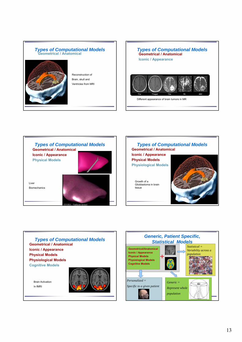
Types of Computational ModelsGeometrical / Anatomical
Reconstruction of
Brain, skull and
Ventricles from MRI
Types of Computational ModelsGeometrical / Anatomical
Iconic / Appearance
T1w T1wgad T2w FA ADC
Different appearance of brain tumors in MR
Types of Computational ModelsGeometrical / Anatomical
Iconic / Appearance
Physical Models
Liver
Biomechanics
Types of Computational ModelsGeometrical / Anatomical
Iconic / Appearance
Physical Models
Physiological Models
Growth of a Glioblastoma in brain tissue
Types of Computational ModelsGeometrical / Anatomical
Iconic / Appearance
Physical Models
Physiological Models
Cognitive Models
Brain Activation
In fMRI
Generic, Patient Specific, Statistical Models
Geometrical/Anatomical
Iconic / Appearance
Physical Models
Physiological Models
Cognitive Models
Personalized =
Specific to a given patient
Statistical = Variability across a population+
Generic =
Represent whole
population
14
Applications of Digital Patient
Therapy
Simulation
Computer-Aided
Diagnosis
Therapy
Planning
Therapy
Guidance
Image
Fusion
Patient
DIAGNOSIS
•CT•MRI•US•NM
Patient
THERAPY
•US•X-Ray•Video•iMRI
Per-operative
Pre-operative
Post-operative •robot
Patient
PREVENTION
•X-Ray•US
TEACHING
Image DataBase
Applications of Digital Patient
Computer-Aided
Diagnosis Image
FusionImage
Fusion
Image
Fusion
Therapy
PlanningTherapy
Guidance
Therapy
Simulation
Computer-Aided
Diagnosis
89
Classification of 3D image processing problems
Segmentation (organs, lesions, activations,…)
Registration (comparison, fusions)
Motion analysis (cardiac imaging)
Deformable models (Surgery simulation)
Medical Robotics (image guided surgery, telesurgery…)
90
Course overview
The data of Computational Anatomy & Physiology
Image acquisition
Medical Image Analysis processing
Medical Imaging
Registration
Segmentation
91
Medical Image Registration
A dual problem
Find the point y of image J which is corresponding (homologous) to each points x of image I.
Determine the best transformation T that superimposes homologous points
I
T
J
kxky
92
Principal Applications
Fusion of multimodal images
Temporal evolution of a pathology
Inter-subject comparisons
Superposition of an atlas
Augmented reality
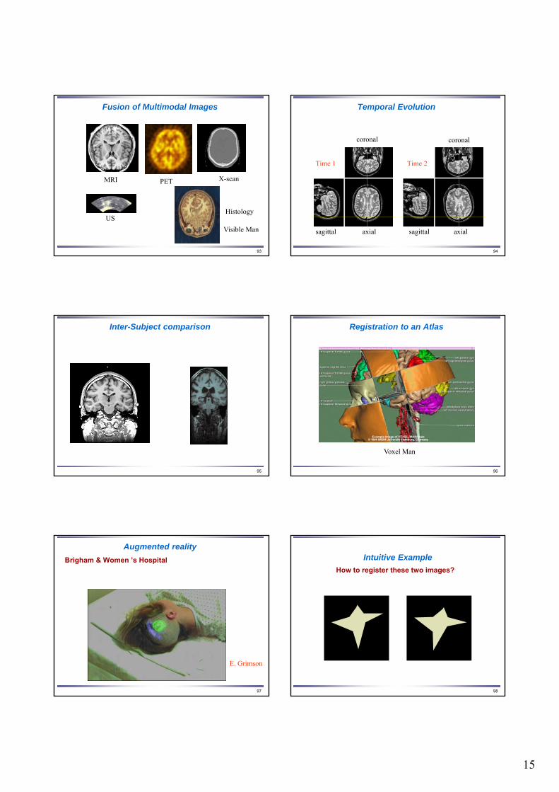
15
93
Fusion of Multimodal Images
MRI PET X-scan
US
Visible Man
Histology
94
Temporal Evolution
Time 1
axialsagittal
coronal coronal
axialsagittal
Time 2
95
Inter-Subject comparison
96
Registration to an Atlas
Voxel Man
97
Augmented reality
Brigham & Women ’s Hospital
E. Grimson
98
Intuitive Example
How to register these two images?
16
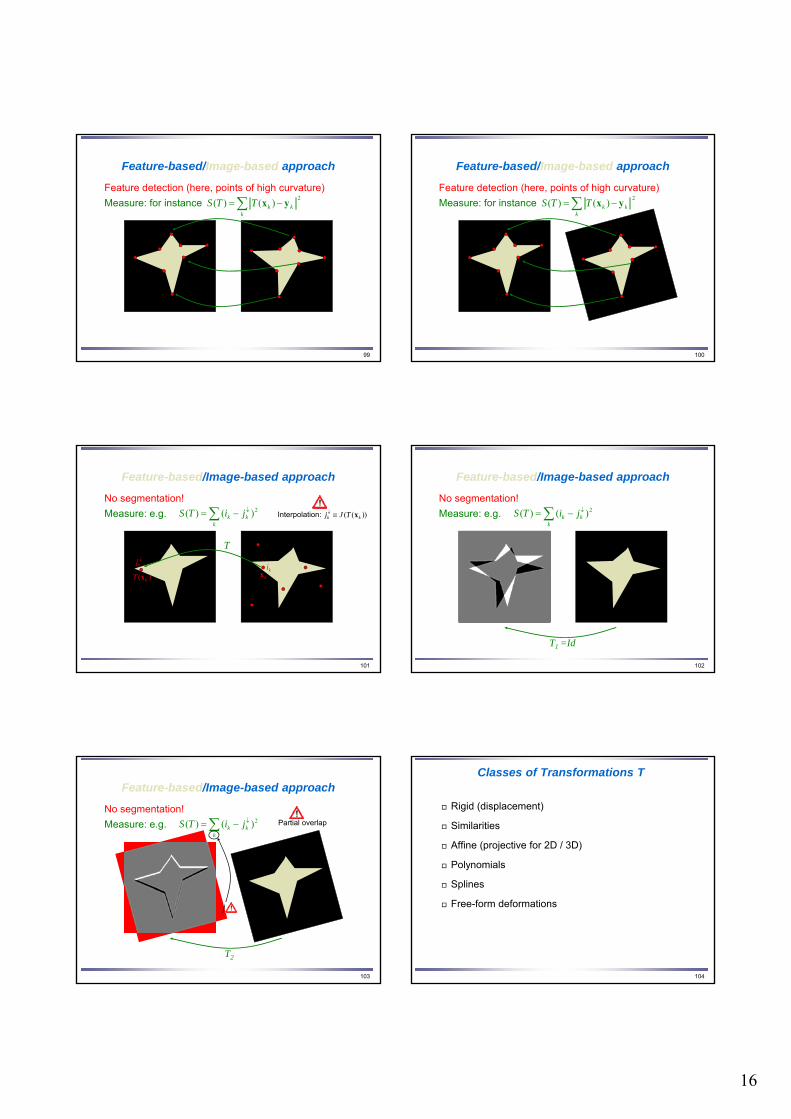
99
Feature-based/Image-based approach
Feature detection (here, points of high curvature)
Measure: for instance k
kkTTS2
)()( yx
100
Feature detection (here, points of high curvature)
Measure: for instance k
kkTTS2
)()( yx
Feature-based/Image-based approach
101
No segmentation!
Measure: e.g. k
kk jiTS 2)()(
kikj
)( kT x kx
T
))(( kk TJj xInterpolation:
Feature-based/Image-based approach
102
No segmentation!
T1 =Id
Measure: e.g. k
kk jiTS 2)()(
Feature-based/Image-based approach
103
No segmentation!
T2
Measure: e.g. k
kk jiTS 2)()( Partial overlap
Feature-based/Image-based approach
104
Classes of Transformations T
Rigid (displacement)
Similarities
Affine (projective for 2D / 3D)
Polynomials
Splines
Free-form deformations
17
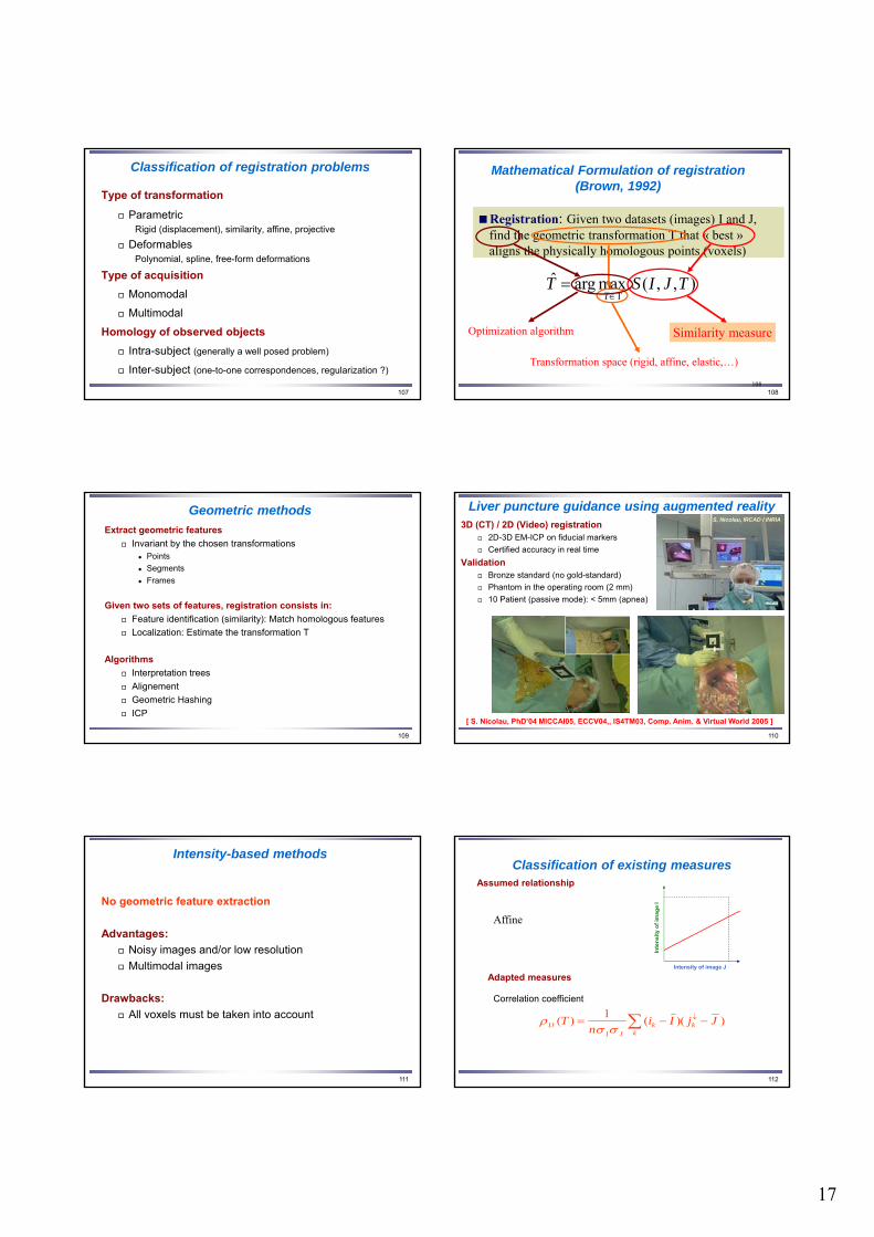
107
Classification of registration problems
Type of transformation
ParametricRigid (displacement), similarity, affine, projective
DeformablesPolynomial, spline, free-form deformations
Type of acquisition
Monomodal
Multimodal
Homology of observed objects
Intra-subject (generally a well posed problem)
Inter-subject (one-to-one correspondences, regularization ?)
108108
),,(maxargˆΤ
TJISTT
Registration: Given two datasets (images) I and J, find the geometric transformation T that « best » aligns the physically homologous points (voxels)
Registration: Given two datasets (images) I and J, find the geometric transformation T that « best » aligns the physically homologous points (voxels)
Mathematical Formulation of registration (Brown, 1992)
Transformation space (rigid, affine, elastic,…)
Similarity measureSimilarity measureOptimization algorithm
109
Geometric methods
Extract geometric features
Invariant by the chosen transformations Points
Segments
Frames
Given two sets of features, registration consists in:
Feature identification (similarity): Match homologous features
Localization: Estimate the transformation T
Algorithms
Interpretation trees
Alignement
Geometric Hashing
ICP
110
Liver puncture guidance using augmented reality3D (CT) / 2D (Video) registration
2D-3D EM-ICP on fiducial markers
Certified accuracy in real time
Validation Bronze standard (no gold-standard)
Phantom in the operating room (2 mm)
10 Patient (passive mode): < 5mm (apnea)
[ S. Nicolau, PhD’04 MICCAI05, ECCV04,, IS4TM03, Comp. Anim. & Virtual World 2005 ]
S. Nicolau, IRCAD / INRIA
111
Intensity-based methods
No geometric feature extraction
Advantages:
Noisy images and/or low resolution
Multimodal images
Drawbacks:
All voxels must be taken into account
112
Classification of existing measuresAssumed relationship
Intensity of image J
Inte
nsi
ty o
f im
age
I
Affine
Adapted measures
Correlation coefficient
kkk
JIIJ JjIi
nT ))((
1)(
18
113
Classification of existing measuresAssumed relationship
Woods’ criterion (1993)Woods’ variants (Ardekani, 95; Alpert, 96; Nikou, 97)Correlation ratio (Roche, 98)
Intensity of image JIn
ten
sity
of
imag
e I
Functional
Adapted measures
)(
))](|([2
IVar
TJIEVar
114
Classification of existing measuresAssumed relationship
Joint Entropy (Hill, 95; Collignon, 95)Mutual Information (Collignon, 95; Viola, 95)Normalized Mutual Information (Studholme, 98)
Intensity of image J
Inte
nsi
ty o
f im
age
I
Statistical
Adapted measures
i j jPiP
jiPjiPJIHJHIHJIMI
)()(
),(log),(),()()(),(
115
• Rigid matching tools for MR
• MR / US registration
• US tracking of brain deformations
Roboscope
Robot / patient coordinate system
MR 0 with surgical plan
Virtual MR n
MR coordinate system
MR 1
Virtual MR 2
TMR/US
Bra
ind
efo
rmat
ion
ove
r ti
me
US coordinate system
US 1
US 2
US n
TMR0 / MR1
118
MR-US Images
Pre - Operative MR Image
Acquisition of images : L. & D. Auer, M. Rudolf
axial
coronal sagittal
Per - Operative US Image
axial
coronal sagittal
119
Elementary principles of US imagery
Zu
x
)()(.)( xxux ZIrf
)()(.log)( xxux BZAI
Logarithmic
compression
US MRI Grad MRI
Ultrasound image / MRI registration
120
Ultrasound image / MRI registration
Assumption: acoustic impedance is a function of the MR signal (denote by J)
)())(()())(()( xxxxx JJgZJgZ
Relation between US and MR signals
)()(.),()( xxuxx JJfI
In practice, the influence of orientation is neglected
)()(),()( xxxx JJfI
19
121
Dependency Hypothesis
Bivariate Correlation Ratio
A. Roche, X. Pennec, G. Malandain, and N. A.Rigid Registration of 3D Ultrasound with MR Images:a New Approach Combining Intensity and Gradient Information.IEEE Transactions on Medical Imaging, 20(10):1038--1049, October 2001.
2)))((),(((ˆ)((minargˆ k
kkkT
xTJxTJfxIT
|)|,( JJfI
I function of 2 variables
2 iterated stages
Robust polynomial approx. of f
Estimation of T:
122
Per - Operative US Image
Typical Registration Resultwith Bivariate Correlation Ratio
Pre - Operative MR Image
Registered
Acquisition of images : L. & D. Auer, M. Rudolf
axial
coronal sagittal
axial
coronal sagittal
123
US Intensity MR Intensity and Gradient
124
Course overview
The data of Computational Anatomy & Physiology
Image acquisition
Medical Image Analysis processing
Medical Imaging
Registration
Segmentation
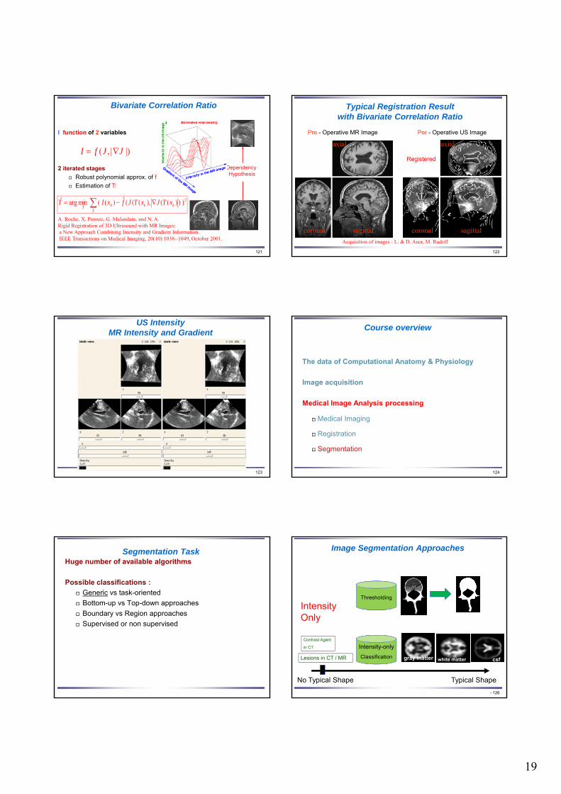
Segmentation TaskHuge number of available algorithms
Possible classifications :
Generic vs task-oriented
Bottom-up vs Top-down approaches
Boundary vs Region approaches
Supervised or non supervised
Image Segmentation Approaches
- 12610/01/2018 126
No Typical Shape Typical Shape
Lesions in CT / MR
Contrast Agent
in CT
Thresholding
Intensity-only
Classification
Intensity Only
gray matter white matter csf
20
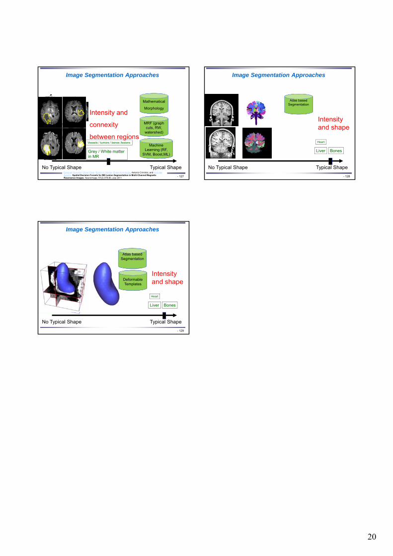
Image Segmentation Approaches
- 127
No Typical Shape Typical Shape
Grey / White matter in MR
Vessels / tumors / bones /lesions
Mathematical
Morphology
MRF (graph cuts, RW,
watershed)
Intensity and
connexity
between regionsMachine
Learning (RF, SVM, Boost,ML)
Camille Couprie, Leo Grady, Laurent Najman and Hugues Talbot , "Power watershed: A UnifyingGraph-Based Optimization Framework" , IEEE Transactions on Pattern Analysis and Machine Intelligence, vol. 33 (7), pp 1384-1399 (2011)
Ezequiel Geremia, Olivier Clatz, Bjoern H. Menze, Ender Konukoglu, Antonio Criminisi, and Nicholas Ayache. Spatial Decision Forests for MS Lesion Segmentation in Multi-Channel MagneticResonance Images. NeuroImage, 57(2):378-90, July 2011
Image Segmentation Approaches
- 128
No Typical Shape Typical Shape
Liver
Heart
Bones
Atlas based Segmentation
Intensity and shape
Image Segmentation Approaches
- 129
No Typical Shape Typical Shape
Liver
Heart
Atlas based Segmentation
Deformable Templates
Intensity and shape
Bones