insect physiology - integument systems department of entomology national chung hsing university
TRANSCRIPT

Insect PhysiologyInsect Physiology- - Integument SystemsIntegument Systems
Department of EntomologyDepartment of EntomologyNational Chung Hsing UniversityNational Chung Hsing University

CONTENTSCONTENTSAdvantages of an exoskeletonAdvantages of an exoskeletonInsect growth and developmentInsect growth and developmentStrategies for growthStrategies for growthOrigins of Origins of holometabolyholometabolyInstars, stadia, and hidden phasesInstars, stadia, and hidden phasesStructure of the integumentStructure of the integumentModified features of the integumentModified features of the integumentChemistry of the cuticleChemistry of the cuticleThe molting processThe molting processEndocrine control of moltingEndocrine control of moltingEndocrine control of metamorphosisEndocrine control of metamorphosisMetamorphosis and the radically changing cutMetamorphosis and the radically changing cuticleicle

Insect Integument Insect Integument Insect integument system – exoskeleton– like the skin of vertebrates - provide a barrier to
the environmentwater (*high surface-to-volume ratio)ionsparasitesenvironment chemicals, including pesticides
– as the skeleton system in insects - allow for the insertion of muscles to locomotion
– as food reservoir (???) / molting & stravation– mating recognition - responsible for releasing p
articular behavioral sequences– many other functions

Insect IntegumentInsect Integument
Advantages– significant mechanical strength over an
endoskeleton of the same weight (next slide)
Disadvantages– restrict insect growth - molt– molting is dangerous to insects– molting consumes time, energy, and
metabolic resources

Insect Growth and Development Insect Growth and Development
The growth and development of insects
are largely a function of the growth and
development of their integuments.
– Molting
– Metamorphosis

Strategies for Growth Strategies for Growth Metamorphosis: the change that occurs as an insect develops from an immature to an adult; separates and early feeding stage from a later reproductive stage.– Ametabolous development - continue to molt as sexua
lly mature adults and there is no real metamorphosis– Hemimetabolous (incomplete) development - immatur
es lack wings and genitalia (exoptergotes)– Holometabolous (complete) development - a sometim
es very radical change in form and ecological habits between immatures and adults (endopterygotes)

Three Major Types of Metamorphosis in InsectsThree Major Types of Metamorphosis in Insects

Origins of HolometabolyOrigins of Holometaboly
(Berlese, 1913)
(Hinton, 1963)
(Truman and Riddiford, 1999)

Instars Stadia and Hidden PhaseInstars Stadia and Hidden PhaseInstars: a term to describe an immature insect between ecdysesStadium: a term to describe the length of time spent between ecdysesPharate instar (adult): a term to describe an insect within the loosened, but not yet shed, cuticle

Structure of the IntegumentStructure of the IntegumentThe outer covering of insects is referred to both as an exoskeleton and an integument.The integument consists of– basement membrane– epidermal cell layer – epidermis– nonliving cuticle

Structure of the IntegumentStructure of the IntegumentBasement membrane 基底膜 : a continuous sheet of mucopolysaccharide, as much as 0.5 m in thickness; initially secreted by hemocytesEpidermis 上皮層 : the only living portion of the integument; modifications of these cells produce dermal glands, sensory receptors and their support cells, and oenocytes.Cuticle 表皮 : secreted by epidermis; divided into two main regions– epicuticle: consists of cement layer, wax layer, outer
epicuticle (cuticulin layer), and inner epicuticle– procuticle: consists of exocuticle, mesocuticle, and en
docuticle, contains largely of chitin and protein

The ProcuticleThe Procuticle 原表皮原表皮The procuticle is secreted by the epidermal cells and consists largely of chitin and protein. (next slide)– exocuticle: the proteins become heavily cross-linked
and insoluble; are not broken down during the molting cycle; pigments deposited within it
– endocuticle: synthesis continues after the old cuticle is shed, often in daily layers; cross-linking is reduced; completely broken down during molting process
– mesocuticle: as a transitional layer in which the proteins are untanned like the endocuticle but impregnated with lipid and proteins like the exocuticle.

The EpicuticleThe Epicuticle 上表皮 上表皮 The epicuticle is a complex consisting of several layers that are produced by both the epidermal cells and dermal glands. (next slide)– Cement layer 固結層 : consists mostly of lipoprotein secret
ed by dermal glands.– Wax layer 腊層 : are mixtures of hydrocarbons with 25-31 c
arbon atoms, alcohols of 24-34 carbon atoms, and esters of fatty acids; produced by the epidermal cells
– Outer epicuticle (i.e. cuticulin): synthesized by epidermal cells; present in all insects; the first layer of the new cuticle to be synthesized
– Inner epicuticle: contains both polyphenols 多酚 and the enzyme polyphenol oxidase 多酚氧化酵素 , which involved in tanning the cuticle. 體壁硬化作用
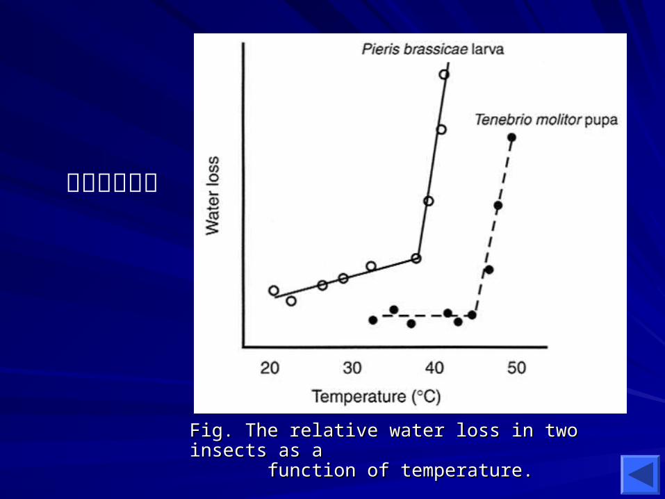
Fig. The relative water loss in two insects as a Fig. The relative water loss in two insects as a function of temperature. function of temperature.
水分散失測試

Modified Features of the IntegumentModified Features of the Integument
Arthrodial membrane 節間膜 : the flexible membranes between body segments where the exocuticle is absent; untanned endocuticle contains special acidic proteins and resilin (a flexible protein) to provide the flexibility in the region. (next slide)
Ecdysial line 脫皮線 : areas of reduced exocuticle that they are programmed areas of weakness that serve as emergence points during ecdyses. (next slide)
Pore canals 孔道 : cytoplasmic extensions of the epidermal cells extend from the epidermis through the cuticle to its surface. (next slide)

Cuticle between Two SegmentsCuticle between Two Segments

The Cuticle in Ecdysial LinesThe Cuticle in Ecdysial Lines

The Generalized Insect IntegumentThe Generalized Insect Integument

Chemistry of the CuticleChemistry of the Cuticle The insect cuticle is composed largely of– Proteins
comprise more than half the dry weight of the insect cuticleprimarily located within the procuticlesynthesized mainly by epidermal cells
– Chitinconsisting of 20-40% of the total dry weight of the cuticle (the other major component of procuticle)a polymer of N-acetyl-D-glucosamine (-galactosamine)synthesized by epidermal cells
– Lipidsmainly located in the wax layer of epicuticlesynthesized largely by the oenocytes and the fat body

Families of protein in insect
Class C proteins Class BD proteins Class H proteins Class T proteins
Kinds of cuticular proteins Varies
Heavily sclerotized: hydrophobic, positively charged proteinsFlexible cuticle: acidic proteins (bind water)
R. prolixus, lower the pH of portions of cuticle to below 6 more plastic to expand when blood meal

Fig. The mechanism of ootheca production in the cockroach.

A Portion of the Chitin ChainA Portion of the Chitin Chain
N-acetyl-D-glucosamine
glucosamine
1-4 -linkage

The Orientation of the Chitin The Orientation of the Chitin Chains in the CuticleChains in the Cuticle
(A) The orientation of chitin, the most common form in insects;
(B) The orientation of chain;
(C) Two possible orientation of chitin;
(D) The location of the chains of chitin in a single chitin microfibril.
• cross-linked by hydrogen bonds

Fig. The helicoidal arrangement of the chitin layers as they are rotated by a constant angle during their synthesis.

The steps inchitin biosynthesis.
Chitosan

Sclerotization Sclerotization 骨化作用骨化作用Cuticular Cuticular sclerotizationsclerotization, also known as , also known as tanningtanning, stabilizes the protein matrix of the , stabilizes the protein matrix of the cuticle to make it stiffer and harder, more cuticle to make it stiffer and harder, more insoluble, and more resistant to degradation.insoluble, and more resistant to degradation.
The process of sclerotization The process of sclerotization cross-links the cross-links the functional groups of cuticular proteins when functional groups of cuticular proteins when they react with they react with quinonesquinones..
The amino acid The amino acid tyrosinetyrosine provides one of the provides one of the precursors (DOPA or NADA) for sclerotization.precursors (DOPA or NADA) for sclerotization.
The precursors are oxidized by The precursors are oxidized by phenoloxidasesphenoloxidases to form reactive quinones. to form reactive quinones.

The Steps in the Synthesis of Cuticular The Steps in the Synthesis of Cuticular Tanning PrecursorsTanning Precursors
(NADA)
(NBAD)
(*less dark than NBAD)
More dark
Less dark

Fig. Differences between quinone sclerotization and -sclerotization in where the cross-linked proteins are attached.
Catecholamines phenolosidases
quinones

Hormonal Regulation of Hormonal Regulation of Sclerotization Sclerotization
At least two hormone are involved in the regulation of sclerotization– Ecdysteroids: induce the epidermal cel
ls to synthesize the dopa decarboxylase (to synthesize NADA)
– Bursicon: induced by declining ecdysteroid titers; increase the permeability of epidermal cells to tyrosine and to hemolymph catecholamines.

The Molting Process The Molting Process
The molting process involves an elaborate sequence of events that produces a new cuticle capable of significant expansion before the old one is discarded.
The molting process begins with apolysis and ends with ecdysis.– Apolysis 剝離作用 : the separation of the epidermal
cells from the old cuticle
– Ecdysis 脫皮作用 : the casting off of the old cuticle

The Steps of Molting ProcessThe Steps of Molting Process
Exuvial space: the area between the cuticle and epidermis; fills with a molting gel that contains inactive enzymes including a chitinase and proteases for digesting the old cuticle.

The Steps of Molting ProcessThe Steps of Molting Process
• The epidermal cells secrete a new outer epicuticle (lipoprotein: cuticulin);• The activation of the enzyme in the molting gel, now called the molting fluid;• The molting fluid begin the digestion of the old unsclerotized endocuticle;• The epidermal cells begin to secrete the new procuticle;•

The Steps of Molting ProcessThe Steps of Molting Process
• Formation of the new epicuticle;• Absorption of the molting fluid;• Ecdysis: induced by eclosion hormone.

Eclosion Behavior and Eclosion Behavior and Its Endocrine RegulationIts Endocrine Regulation
Behavior of ecdysis are divided into two phases: (control by central nervous system)– Pre-ecdysis behavior: loosen the old cuticle through
rotational movements of the abdomen– Ecdysis behavior: shed the old cuticle by means of
peristaltic contractions
A cascade of neurohormones is responsible for eliciting eclosion behavior– Ecdysis-triggering hormone: from epitracheal glands– Eclosion hormone: from CC– Crustacean cardioactive peptide (CCAP): from the
ventral ganglion

Endocrine Control of MoltingEndocrine Control of Molting
Control of PTTH release– nervous stimuli such as stretch
receptors and critical size (or body mass)
– environmental stimuli such as photoperiod, temperature
Mode of action– via a second messenger, cAMP

Correction of Cellular Events during A Correction of Cellular Events during A Molting Cycle with the Ecdysteroid TiterMolting Cycle with the Ecdysteroid Titer

Endocrine Control of Endocrine Control of MetamorphosisMetamorphosis
Insect metamorphosis is a function of gene expression by epidermal cells and the temporal pattern of their protein synthesis.Two major hormones are involved in the metamorphosis– juvenile hormone– ecdysteroids

Fig. Hormonal regulation of insect metamorphosis.

Fig. The correlation of hormone levels with developmental events of Drosophila melanogaster.

Fig. The relationship between the size of the Manduca larva and its tendency to pupate and undergo metamorphosis.

Supernumerary Larvae of Supernumerary Larvae of Spodoptera lituraSpodoptera litura

The Mechanism of JH ActionThe Mechanism of JH Action

Imaginal Discs Imaginal Discs Imaginal discsImaginal discs are derived from ectoderm and are small are derived from ectoderm and are small groups of embryonic cells that persist in larvae of the groups of embryonic cells that persist in larvae of the Holometabola.Holometabola.
When the insect pupates, the imaginal discs When the insect pupates, the imaginal discs provide the provide the cells to make adult structurescells to make adult structures..
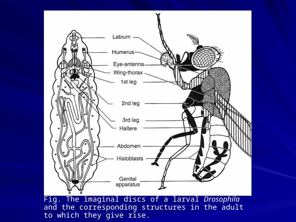
Fig. The imaginal discs of a larval Drosophila and the corresponding structures in the adult to which they give rise.

Fig. The evagination of a leg disc during Drosophila development.


Two Cross Section of Two Cross Section of Two AppendagesTwo Appendages

The OenocytesThe Oenocytes
The The oenocytesoenocytes are large polyploid cells are large polyploid cells associated with the basement membrane.associated with the basement membrane.– some oenocytes might be involved in the some oenocytes might be involved in the
production of production of cuticular lipidcuticular lipid that are deposited that are deposited in the epicuticle.in the epicuticle.
– other types of oenocytes may secrete other types of oenocytes may secrete ecdysteroid hormonesecdysteroid hormones..