insights into the membrane-interaction properties of the pseudomonas aeruginosa type iii secretion...

1
proteins, even alpha-helical ones, because the contribution of the backbone conformation is subtracted from our final measurements. We were also able to use OMPLA to measure how the energetics of side-chain insertion vary with depth in the bilayer. Both Arginine and Leucine have their most extreme insertion energies when they are closest to the middle of the hy- drophobic region of OMPLA. Further, we carried out a double mutant cycle with Arginines and discovered that the insertion of a second Arginine is aided by the insertion of the first. This result is particularly relevant for understanding the function of the voltage sensing domains of some ion channels, which may involve multiple Arginines penetrating the lipid bilayer. 1128-Pos Board B38 UV Excited Resonance Raman Analysis of Lipid Solvated Polypeptides Christopher Halsey, Jian Xiong, Carol Roach, Renee D. Jiji, Jason W. Cooley. The study of membrane protein structure is hampered by a well-documented litany of technical issues associated with sample preparation, peptide solubil- ity and sample complexity. Deep-UV excited resonance Raman (DUVRR) spectroscopy has proven to be a means by which to attain discrete structural information for some of the most dynamic or insoluble protein samples. We will present here our initial efforts to address the feasibility of using DUVRR to probe the lipid solvated polypetide backbone secondary structures. 1129-Pos Board B39 Investigation of Transmembrane Helix Hetero-Dimerization by Combina- torial Peptide Libraries and High-Throughput Measurement of FO ¨ RSTER Resonance Energy Transfer in Liposomes Jing He, William C. Wimley. Receptor tyrosine kinases (RTKs) are membrane proteins containing an N- terminal extracellular ligand-binding domain, a single helical transmembrane (TM) domain, and an intracellular catalytic kinase domain. Lateral dimeriza- tion driven in part by the TM domain is proposed to be a crucial intermediate step in signal transduction across the plasma membrane. Defects in this process such as over-dimerization are closely linked to unregulated signaling and disease. Therefore, dimerization inhibitors developed using the chemical- physical basis of RTK TM domain dimerization could be promising therapeu- tic agents. Here we present a first step towards this goal. We designed a rational combinatorial peptide library based on the TM domain of Neu, an RTK from the Erb-B/HER epidermal growth factor receptor family (EGFR) with a path- ogenic V to E mutation in the TM domain. Fo ¨ rster Resonance Energy Transfer (FRET) in lipid vesicles was used as a method to probe the dimerization between the library members and mutant Neu sequence. To enable high- throughput screening in liposomes, we developed a novel multi-well FRET assay. FRET donor-labeled library members and acceptor-labeled Neu peptides were incorporated into lipid vesicles in a rapid high-throughput 96-well plate format. We are using this high throughput screen to identify in- hibitors of Neu dimerization. Our findings will be discussed in a context of throughput and sensitivity along with a statistical analysis that takes into account the FRET that arises from random proximity of donors and acceptors in the bilayer. 1130-Pos Board B40 Spectroscopic Studies of the Dimerization of ATP-Binding Cassette Nucleotide-Binding Domains Maria E. Zoghbi, Guillermo A. Altenberg. Cancer cells are frequently resistant against chemically unrelated anticancer agents or develop resistance to those agents during treatment (multidrug resis- tance). ATP-binding cassette (ABC) proteins such as P-glycoprotein are key mediators of multidrug resistance. They catalyze the efflux of chemotherapeu- tic agents out of cancer cells, preventing their intracellular accumulation and cytotoxic effect. There are two competing models to explain the mechanism of ABC exporters: 1) The alternating-access model, where large conforma- tional changes take place during the transport cycle as a consequence of ATP-induced nucleotide-binding domain (NBD) dimerization. 2) An alterna- tive model, which suggests that the power stroke triggered by ATP binding con- sists of only moderate rearrangements, with the NBDs in contact at all times during the transport cycle. Here, we performed experiments on isolated bacte- rial NBDs using luminescence resonance energy transfer (LRET) and trypto- phan fluorescence quenching, to determine the ATP dependence on NBD dimerization. As a model, we used MJ0976, a NBD from the thermophile M. jannaschii. Under non-hydrolysis conditions (in the absence of Mg2þ), tryptophan fluorescence quenching and LRET experiments showed a similar apparent affinity for ATP, in the 20-40 micromolar range. A mutation that abol- ishes ATPase activity increases the apparent affinity for ATP approximately 10 folds. Under conditions of ATP hydrolysis, in the presence of Mg2þ-ATP, the decrease in LRET indicates that there was an increase in the proportion of NBDs in the monomeric form. The results show: 1) The feasibility of LRET studies to determine dimerization of ABC protein NBDs, and 2) That ATP- induced dimerization is a transient phenomenon. This work was supported by CPRIT grant RP101073. 1131-Pos Board B41 Structural Aspects of Transmembrane Domain Interactions of Receptor Tyrosine Kinases Eduard Bocharov, Konstantin Mineev, Dmitry Lesovoy, Marina Goncharuk, Sergey Goncharuk, Olga Bocharova, Pavel Volynsky, Roman Efremov, Alexander Arseniev. Specific helix-helix interactions of single-span transmembrane domains of re- ceptor tyrosine kinases are critical for their lateral dimerization and biological function. Establishing structure-function relationship as well as rational drug design requires precise structural information about this class of biologically significant bitopic membrane proteins. A combination of NMR, optical spec- troscopy, protein engineering and molecular modelling made it possible studies of the nature and the mechanisms of important transmembrane helix-helix in- teractions inside the supramolecular complexes mimicking membrane environ- ment. Using a robust strategy we investigated recombinant transmembrane fragments from different families of receptor tyrosine kinases: EphA, ErbB and FGFR, which play important roles in normal and in pathological conditions of human organism by providing cell signalling, maintaining cellular homeostasis and controlling cell fate. We established spatial structure and internal dynamics of the homo- and heterodimeric transmembrane domains, characterized diverse transmembrane helix-helix packing interfaces and obtained detailed picture of intra- and intermolecu- lar interactions in membrane. The already available information about structural-dynamic properties of the dimeric transmembrane domains of studied re- ceptor tyrosine kinases along with the available biophysical and biochemical data provides useful insights into their functioning in the human organ- ism at the atomic level. 1132-Pos Board B42 Insights into the Membrane-Interaction Properties of the PSEUDOMO- NAS Aeruginosa Type III Secretion Translocator Popd Fabian B. Romano Chernac, Kyle C. Rossi, Min Chen, Alejandro P. Heuck. In order to infect human cells, pathogenic Gram-negative bacteria depend on their ability to secrete toxins into the cytoplasm of an adjacent eukaryotic host cell. A conserved and efficient delivery mechanism, called the type III se- cretion (T3S) system, acts as a syringe to inject these toxic proteins called ‘‘ef- fectors’’ which need to translocate through the eukaryotic plasma membrane. In Pseudomonas aeruginosa PopB and PopD are essential for effector transloca- tion. Substantial genetic and biochemical data support a model with PopB and PopD inserting into the target membrane and forming an oligomeric translocon pore, however virtually nothing is known about this critical transmembrane complex. Our primary goal is to determine the arrangement of PopD in membrane assem- bled translocons. What segments of PopD interact with the membrane? Does PopB influence the PopD/membrane interaction? Using membrane binding as- says, conductivity measures in planar lipid bilayers, site-directed fluorescence labeling, and multiple fluorescence approaches we have analyzed the interac- tion of PopD with model membranes and the pore formation activity of the translocator in the presence and absence of PopB. Despite both proteins can individually form pores in membranes, we found that PopD oligomerizes with PopB and forms pores with an inner diameter of ap- proximately 30A ˚ . Our analysis also showed that the PopD predicted transmem- brane segment (residues L119-V137), does not adopt a transmembrane orientation. Our data support a model that consists in the binding of the un- folded translocators to the membrane, oligomerization, and formation of dis- crete transmembrane pores. Monday, March 7, 2011 207a

Upload: alejandro-p

Post on 30-Dec-2016

212 views

Category:

Documents


0 download

TRANSCRIPT

Monday, March 7, 2011 207a

proteins, even alpha-helical ones, because the contribution of the backboneconformation is subtracted from our final measurements.We were also able to use OMPLA to measure how the energetics of side-chaininsertion vary with depth in the bilayer. Both Arginine and Leucine have theirmost extreme insertion energies when they are closest to the middle of the hy-drophobic region of OMPLA. Further, we carried out a double mutant cyclewith Arginines and discovered that the insertion of a second Arginine is aidedby the insertion of the first. This result is particularly relevant for understandingthe function of the voltage sensing domains of some ion channels, which mayinvolve multiple Arginines penetrating the lipid bilayer.

1128-Pos Board B38UV Excited Resonance Raman Analysis of Lipid Solvated PolypeptidesChristopher Halsey, Jian Xiong, Carol Roach, Renee D. Jiji,Jason W. Cooley.The study of membrane protein structure is hampered by a well-documentedlitany of technical issues associated with sample preparation, peptide solubil-ity and sample complexity. Deep-UV excited resonance Raman (DUVRR)spectroscopy has proven to be a means by which to attain discrete structuralinformation for some of the most dynamic or insoluble protein samples.We will present here our initial efforts to address the feasibility of usingDUVRR to probe the lipid solvated polypetide backbone secondarystructures.

1129-Pos Board B39Investigation of Transmembrane Helix Hetero-Dimerization by Combina-torial Peptide Libraries and High-Throughput Measurement ofFORSTER Resonance Energy Transfer in LiposomesJing He, William C. Wimley.Receptor tyrosine kinases (RTKs) are membrane proteins containing an N-terminal extracellular ligand-binding domain, a single helical transmembrane(TM) domain, and an intracellular catalytic kinase domain. Lateral dimeriza-tion driven in part by the TM domain is proposed to be a crucial intermediatestep in signal transduction across the plasma membrane. Defects in thisprocess such as over-dimerization are closely linked to unregulated signalingand disease. Therefore, dimerization inhibitors developed using the chemical-physical basis of RTK TM domain dimerization could be promising therapeu-tic agents. Here we present a first step towards this goal. We designed a rationalcombinatorial peptide library based on the TM domain of Neu, an RTK fromthe Erb-B/HER epidermal growth factor receptor family (EGFR) with a path-ogenic V to E mutation in the TM domain. Forster Resonance Energy Transfer(FRET) in lipid vesicles was used as a method to probe the dimerizationbetween the library members and mutant Neu sequence. To enable high-throughput screening in liposomes, we developed a novel multi-well FRETassay. FRET donor-labeled library members and acceptor-labeled Neupeptides were incorporated into lipid vesicles in a rapid high-throughput96-well plate format. We are using this high throughput screen to identify in-hibitors of Neu dimerization. Our findings will be discussed in a context ofthroughput and sensitivity along with a statistical analysis that takes intoaccount the FRET that arises from random proximity of donors and acceptorsin the bilayer.

1130-Pos Board B40Spectroscopic Studies of the Dimerization of ATP-Binding CassetteNucleotide-Binding DomainsMaria E. Zoghbi, Guillermo A. Altenberg.Cancer cells are frequently resistant against chemically unrelated anticanceragents or develop resistance to those agents during treatment (multidrug resis-tance). ATP-binding cassette (ABC) proteins such as P-glycoprotein are keymediators of multidrug resistance. They catalyze the efflux of chemotherapeu-tic agents out of cancer cells, preventing their intracellular accumulation andcytotoxic effect. There are two competing models to explain the mechanismof ABC exporters: 1) The alternating-access model, where large conforma-tional changes take place during the transport cycle as a consequence ofATP-induced nucleotide-binding domain (NBD) dimerization. 2) An alterna-tive model, which suggests that the power stroke triggered by ATP binding con-sists of only moderate rearrangements, with the NBDs in contact at all timesduring the transport cycle. Here, we performed experiments on isolated bacte-rial NBDs using luminescence resonance energy transfer (LRET) and trypto-phan fluorescence quenching, to determine the ATP dependence on NBDdimerization. As a model, we used MJ0976, a NBD from the thermophile

M. jannaschii. Under non-hydrolysis conditions (in the absence of Mg2þ),tryptophan fluorescence quenching and LRET experiments showed a similarapparent affinity for ATP, in the 20-40 micromolar range. A mutation that abol-ishes ATPase activity increases the apparent affinity for ATP approximately 10folds. Under conditions of ATP hydrolysis, in the presence of Mg2þ-ATP, thedecrease in LRET indicates that there was an increase in the proportion ofNBDs in the monomeric form. The results show: 1) The feasibility of LRETstudies to determine dimerization of ABC protein NBDs, and 2) That ATP-induced dimerization is a transient phenomenon. This work was supportedby CPRIT grant RP101073.

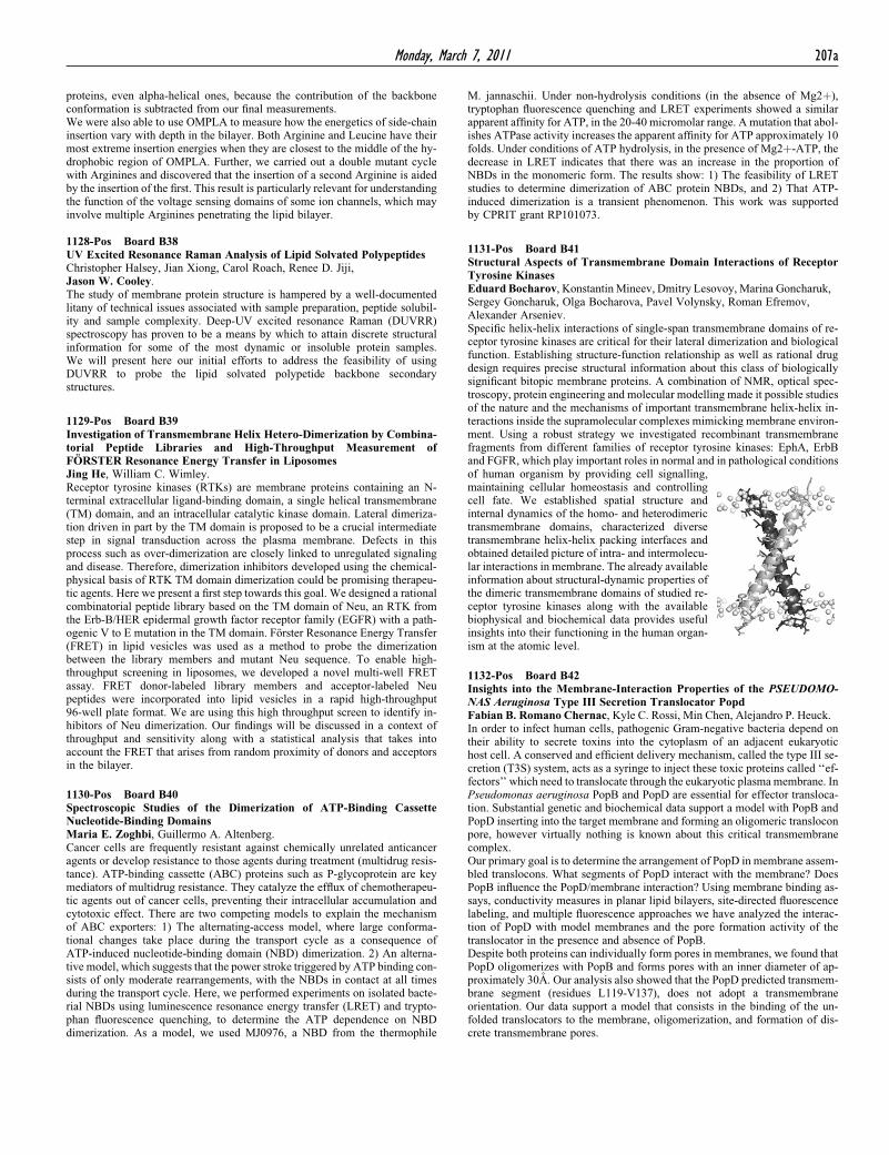
1131-Pos Board B41Structural Aspects of Transmembrane Domain Interactions of ReceptorTyrosine KinasesEduard Bocharov, Konstantin Mineev, Dmitry Lesovoy, Marina Goncharuk,Sergey Goncharuk, Olga Bocharova, Pavel Volynsky, Roman Efremov,Alexander Arseniev.Specific helix-helix interactions of single-span transmembrane domains of re-ceptor tyrosine kinases are critical for their lateral dimerization and biologicalfunction. Establishing structure-function relationship as well as rational drugdesign requires precise structural information about this class of biologicallysignificant bitopic membrane proteins. A combination of NMR, optical spec-troscopy, protein engineering and molecular modelling made it possible studiesof the nature and the mechanisms of important transmembrane helix-helix in-teractions inside the supramolecular complexes mimicking membrane environ-ment. Using a robust strategy we investigated recombinant transmembranefragments from different families of receptor tyrosine kinases: EphA, ErbBand FGFR, which play important roles in normal and in pathological conditions

of human organism by providing cell signalling,maintaining cellular homeostasis and controllingcell fate. We established spatial structure andinternal dynamics of the homo- and heterodimerictransmembrane domains, characterized diversetransmembrane helix-helix packing interfaces andobtained detailed picture of intra- and intermolecu-lar interactions in membrane. The already availableinformation about structural-dynamic properties ofthe dimeric transmembrane domains of studied re-ceptor tyrosine kinases along with the availablebiophysical and biochemical data provides usefulinsights into their functioning in the human organ-ism at the atomic level.

1132-Pos Board B42Insights into the Membrane-Interaction Properties of the PSEUDOMO-NAS Aeruginosa Type III Secretion Translocator PopdFabian B. Romano Chernac, Kyle C. Rossi, Min Chen, Alejandro P. Heuck.In order to infect human cells, pathogenic Gram-negative bacteria depend ontheir ability to secrete toxins into the cytoplasm of an adjacent eukaryotichost cell. A conserved and efficient delivery mechanism, called the type III se-cretion (T3S) system, acts as a syringe to inject these toxic proteins called ‘‘ef-fectors’’ which need to translocate through the eukaryotic plasma membrane. InPseudomonas aeruginosa PopB and PopD are essential for effector transloca-tion. Substantial genetic and biochemical data support a model with PopB andPopD inserting into the target membrane and forming an oligomeric transloconpore, however virtually nothing is known about this critical transmembranecomplex.Our primary goal is to determine the arrangement of PopD in membrane assem-bled translocons. What segments of PopD interact with the membrane? DoesPopB influence the PopD/membrane interaction? Using membrane binding as-says, conductivity measures in planar lipid bilayers, site-directed fluorescencelabeling, and multiple fluorescence approaches we have analyzed the interac-tion of PopD with model membranes and the pore formation activity of thetranslocator in the presence and absence of PopB.Despite both proteins can individually form pores in membranes, we found thatPopD oligomerizes with PopB and forms pores with an inner diameter of ap-proximately 30A. Our analysis also showed that the PopD predicted transmem-brane segment (residues L119-V137), does not adopt a transmembraneorientation. Our data support a model that consists in the binding of the un-folded translocators to the membrane, oligomerization, and formation of dis-crete transmembrane pores.