insights into the novel function of system xc– in
TRANSCRIPT

1650
Abstract. – System Xc–, also named cystine/glutamate antiporter, is an important intracellu-lar antioxidant element. It is composed of the light chain SLC7A11 (xCT) and the heavy chain SLC3A2 (4F2hc) and functions as raw materi-als for the synthesis of glutathione (GSH). Re-cent studies have demonstrated that system Xc– plays an important role in different types of regulated cell death, which is referred to cell death controlled by dedicated molecular machin-ery. It has been shown that system Xc– involves in ferroptosis, apoptosis, and autophagy-depen-dent cell death, contributing to different diseas-es and drug resistance, such as cancer, neu-rological disorders, and cisplatin resistance to cancers. To date, the intervention of system Xc– by its inhibitors or activators displays a benefi-cial effect on the treatment of certain diseases. In this review, we summarize recent findings on the role of system Xc– in regulated cell death,
including molecular mechanisms and potential therapeutic applications.
Key Words: System Xc–, Cystine/glutamate antiporter, Ferropto-
sis, Apoptosis, Autophagy-dependent cell death.
Introduction
Types of cell death are mainly divided into apoptosis, necrosis, and autophagy-dependent cell death with the following morphological or biochemical features: (1) apoptosis forms apop-totic bodies taken up by neighboring cells with phagocytic activity and degraded in lysosomes, showing cytoplasmic shrinkage, nuclear frag-
European Review for Medical and Pharmacological Sciences 2021; 25: 1650-1662
H. TU1, L.-J. TANG1, X.-J. LUO2, K.-L. AI1,3, J. PENG1,3
1Department of Pharmacology, Xiangya School of Pharmaceutical Sciences, Central South University, Changsha, China2Department of Laboratory Medicine, Xiangya Third Hospital, Central South University, Changsha, China3Hunan Provincial Key Laboratory of Cardiovascular Research, School of Pharmaceutical Sciences, Central South University, Changsha, China
Corresponding Author: Jun Peng, MD, Ph.D; e-mail: [email protected]
Insights into the novel function of system Xc– in regulated cell death
Graphical abstract.

Insights into the novel function of system Xc– in regulated cell death
1651
mentation and plasma membrane blebbing; (2) autophagy-dependent cell death, is triggered by autophagy-inducing peptides, starvation, isch-emia or hypoxia manifesting with extensive cyto-plasmic vacuolization and similarly culminating with phagocytic uptake and consequent lysosom-al degradation; and (3) necrosis deals with the cell corpses in phagocytic and lysosomal-independent manner, showing irreversible plasma membrane permeabilization or complete cellular fragmen-tation. Traditionally, classic necrosis is thought a passive and unregulated cell death. Some necro-sis also occurs in a programmed fashion, such as necroptosis, pyroptosis, and ferroptosis1,2. Based on morphological, biochemical and function-al perspectives, cell death relying on dedicated molecular machinery is defined as regulated cell death (RCD), including apoptosis, ferroptosis, necroptosis, MPTP-dependent necrosis, pyropto-sis, autophagy-dependent cell death, entotic cell death, and NETotic cell death. These RCD modes are initiated and propagated by molecular mecha-nisms that exhibit a considerable degree of inter-connectivity.
There is growing evidence that system Xc– modulates a number of RCDs under pathophysio-logical conditions. To date, ferroptosis, apoptosis, and autophagy-dependent cell death are the most well-studied RCD relevant to system Xc–. Among them, apoptosis is a classical form of caspase-de-pendent cell death, dividing into intrinsic or ex-trinsic pathways; ferroptosis is a form of RCD ini-tiated by oxidative perturbations of the intracel-lular microenvironment that is under constitutive control by glutathione peroxidase 4 (GPX4); while autophagy-dependent cell death is a form of RCD that mechanistically depends on the autophagic machinery (or components thereof). Numerous studies have demonstrated that system Xc– plays an important role in ferroptosis3-7, apoptosis8-11, as well as autophagy-dependent cell death12,13. This review will focus on recent progress in the role of system Xc– in ferroptosis and apoptosis.
System Xc–
Structure and Localization of System Xc–System Xc–, also referred to cystine/glutamate
antiporter, was first reported in human fetal lung fibroblasts in 198014, and then a similar transport system was identified in a rat hepatoma cell line15. System Xc– consists of the light chain SLC7A11 (xCT) and the 4F2 heavy chain (4F2hc), and they
are linked by a disulfide bond. The 4F2hc heavy subunit is common to several amino acid transport systems and can interact with other homologous light chains, for instance, with LAT1 to form a system l neutral amino acid transporter. However, the transport-specific light chain xCT is unique in that and it only interacts with 4F2hc to mediate the cysteine-glutamate exchange. In mouse, sys-tem Xc– is widely distributed in the thymus and spleen but it is lowly expressed in lung, heart, liv-er, and kidney; while in human, system Xc– is highly expressed in the brain and spinal cord, but it is not detected in peripheral leukocytes, spleen, thymus and lymph nodes.
The Physiological Functions of System Xc–
The primary function of system Xc– is to im-port cystine and export glutamate simultaneously with a ratio of 1:1. Usually, system Xc– transfers cystine into the cells, where it is reduced to cyste-ine immediately. In cells, cysteine and glutamate are utilized to synthetize γ-glutamyl cysteine (γGC) via glutamate cysteine ligase (GCL), and to produce glutathione (GSH) by adding glycine under glutathione synthase catalysis. Of note, cysteine is a rate-limiting substrate for generating GSH, an important antioxidant and a vital ele-ment of a redox couple maintaining oxidation-re-duction balance.
System Xc– possesses dual functions, in-cluding control of extracellular glutamate and defense against oxidative stress. On one hand, system Xc–plays a key role in preserving gluta-mate homeostasis in the nervous system. Gluta-mate exported by system Xc– is largely respon-sible for the extracellular glutamate concen-tration in the brain. Disruptions in glutamate homeostasis have been linked to numerous dis-eased states of the brain and can lead to wide-spread changes in synaptic activity, called cen-tral nervous (CNS) toxicity. On the other hand, the imported cystine via system Xc– is import-ant for the endogenous defense system against oxidative stress. Disturbances in the function of system Xc– have been shown to decrease the intracellular cysteine and subsequent glu-tathione, which results in antioxidant dysfunc-tion concomitant with reactive oxygen species (ROS) overload and lipid peroxidation, leading to cell death and disease ultimately. More in-formation regarding the role of system Xc– in the modulation of glutamate homeostasis can be found in several comprehensive reviews16-18.

H. Tu, L.-J. Tang, X.-J. Luo, K.-L. Ai, J. Peng
1652
Transcriptional Regulation of System Xc-Light Chain: SLC7A11
Transcriptional regulation of SLC7A11 is one of the most important determinants for system Xc– activity. Thereby, the physiological function of system Xc– mainly relies on the SLC7A11 sub-unit. The transcriptional level of SLC7A11 in can-cer cells is significantly higher than that in normal cells. It has been demonstrated that the NRF2-Keap1 pathway is critical for glioma cell growth, which involves in SLC7A11 transcription. Nucle-ar factor erythroid 2-related factor 2 (Nrf2) dis-sociates from Kelch-like ECH-associated protein 1 (Keap1), translocates into nucleus, and inter-acts with antioxidant response element (ARE) in the SLC7A11 promotor, leading to the upreg-ulation of SLC7A11 transcription against oxida-tive stress19. When amino acid starvation leads to phosphorylation of eIF2, activating transcription factor 4 (ATF4) interacts with ARE, leading to tumor proliferation via increase of the transcrip-tion of SLC7A11. The activating transcription factor 3 (ATF3) enhances erastin-induced fer-roptosis through forming ATF3 dimer retaining SLC7A11’s transcription. However, ferroptosis promoted by ATF3 is not related to response to oxidative stress3,20.
There are reports showing that p53 could sup-press SLC7A11 mRNA expression via modulat-ing certain protein deubiquitination. It has been shown that p53 promotes the nuclear transloca-tion of the deubiquitinase ubiquitin special pep-tidase 7 (USP7), leading to negatively regulating monoubiquitination of histone H2B on lysine 120 (H2Bub1). Deubiquitinated H2Bub1 occupies the SLC7A11 gene regulatory region and represses the expression of SLC7A11 in the presence of erastin21. Similarly, another tumor suppressor, BRCA1-associated protein 1 (BAP1), can also decrease histone 2A ubiquitination (H2Aub). The interaction between H2Aub and BAP1 can retain the transcription of SLC7A11, which represses tu-mor growth22. In addition, PRC1, a major H2Aub ubiquitin ligase, increases the binding of H2Aub to the SLC7A11 promoter. Both BAP1 and PRC1 suppress SLC7A11 expression, revealing that dynamic regulation of H2Aub is important for SLC7A11 repression.
Of note, BAP1 promotes ferroptosis induced by erastin but not by GPX4 inhibitor (RSL3), and BAP1-mediated SLC7A11 repression does not require NRF2 and ATF4 transcription factors23. Furthermore, it has been shown that p53 increas-es the sensibility of ALOX12-dependent ferro-
ptosis by indirectly repressing the transcription of SLC7A115. As a tumor suppressor, ARF inhib-its NRF2 or promotes p53 to suppress SLC7A11 transcriptional activation, causing cancer cell de-mise via ferroptosis24. The bromodomain 4 pro-tein (BRD4) level in multiple cancer is elevated, accompanied by upregulation of GPX4, SLC7A11, and SLC3A2 and downregulation of ferritin heavy light (FHT), leading to poor prognosis of cancer patients25. POU2F1, an octamer transcription fac-tor-1, is associated with anti-cytotoxicity, stem cell function, and cancer tumor deterioration. A recent study illustrated that POU2F1 binding to the SLC7A11 promotor inhibited its transcription in skin pigmentation4. The scheme for the tran-scriptional regulation of SLC7A11 is summarized in Figure 1.
Post-Translational Regulation of SLC7A11
The function of SLC7A11 in ferroptosis is tight-ly controlled by post-translational regulation. On one hand, AMP-activated protein kinase phos-phorylates BECN1 at Ser90/93/96, which acceler-ates BECN1 binding to SLC7A11 in erastin-treat-ed cells. The formation of BECN1-SLC7A11 com-plex leads to SLC7A11 dysfunction, accompanied by intracellular lipid peroxidation and ferroptosis in the end26. On the other hand, ferroptosis can be triggered due to the change in SLC7A11 sta-bility. CD44 or CD44 variant (CD44v) is stem cell marker in cancer cells. It has been shown that CD44-positive cells promote tumor growth and metastasis through forming CD44-SLC7A11 complex27, and its stability is affected by certain pathways. Firstly, OTUB1 (ovarian tumor fami-ly member deubiquitinase) directly interacts with SLC7A11 and increases the connection between CD44 and SLC7A11, which enhances the stabil-ity of CD44-SLC7A11 complex6,28. Secondly, the transmembrane protein mucin 1 (MUC1-C) al-so binds directly to CD44v, which increases the stability of SLC7A11 and maintains the redox balance in triple-negative breast cancer29. Final-ly, an increase of intracellular iron concentration elevates the expression of lipocalin 2 (LCN2), an iron transporter, and antioxidant element, which enhances the interaction between CD44 and SLC7A11 via upregulation of both of them, re-sulting in malignant transformation of tumors30. Therefore, increasing the phosphorylation of BECN1 or targeting the MUC1-C/SLC7A11 or SLC7A11/OTUB1 pathway or LCN2 can induce ferroptosis and decrease tumors’ survival effec-

Insights into the novel function of system Xc– in regulated cell death
1653
tively. The scheme for the post-translational reg-ulation of SLC7A11 is summarized in Figure 2.
The Pathological Roles of System Xc–
Relations Among System Xc–, Ferroptosis and Cancer
The occurrence of ferroptosis is controlled by multiple signaling pathways, including iron homeostasis pathway, system Xc– pathway, and voltage-dependent anion channel (VDAC) path-way. It is well recognized that the suppression of system Xc– leads to ferroptosis. Erastin, a com-monly-used ferroptosis inducer, not only inhibits system Xc– but also blocks VDAC. In the pres-ence of erastin, mitochondria become smaller,
and the membrane density is increased due to the inhibition of VDAC2 and VDAC3, concom-itant with loss of structural integrity, collapse of the mitochondrial transmembrane potential and overproduction of ROS, resulting in mitochon-drial dysfunction and ferroptosis ultimately31. Although erastin-induced ferroptosis involves in inhibition of system Xc– as well as VDACs, it is believed that targeting system Xc– plays a major role in this process32.
The system Xc– expression is upregulated in many tumors, such as glioma33-35, hand and neck cancers (HNC)36,37, triple-negative breast can-cers (TNBC)29,38, colorectal carcinoma cancer (CCC)39, ovarian clear cell carcinoma30, liver car-cinoma40, oral squamous cell carcinoma41, and non-small cell lung cancer42. There are reports
Figure 1. A schematic diagram for transcriptional regulation of SLC7A11. In tumors, NRF2-keap1 and POU2F pathway is activated via interacting with antioxidant response element (ARE) while the ATF4 pathway is activated by interacting with amino-acid response element (AARE), leading to the upregulation of SLC7A11 transcription. p53 can block these pathways by binding a cis-regulatory element proximal to ARE; p53 also can promote the nuclear translocation of USP7, leading to H2Bub1 deubiquitination and negatively regulating the transcription of SLC7A11; meanwhile, BAP1 can also remove H2Aub ubiquiti-nation to retain the transcription of SLC7A11; ARF inhibits NRF2 or promotes p53 to suppress SLC7A11 transcriptionally ac-tivation simultaneously; and ATF3 dimer enhances the retaining SLC7A11’s transcription in erastin-induced ferroptosis. Sys-tem Xc–, cystine/glutamate antiporter; SLC7A11, solute carrier family 7 member 11; NRF2, nuclear factor erythroid 2-related factor 2; keap1, Kelch-like ECH-associated protein 1; POUF2, octamer transcription factor-1; ATF4, activating transcription factor 4; USP7, ubiquitin special peptidase 7; H2Bub1, histone H2B on lysine 120; BAP1, BRCA1-associated protein 1; H2Aub, histone 2A ubiquitination; ARF, p14 alterative reading frame; ATF3, activating transcription factor 3.

H. Tu, L.-J. Tang, X.-J. Luo, K.-L. Ai, J. Peng
1654
that specific inhibition of the system Xc– against cystine uptake by erastin can decrease GSH pro-duction, which compromises the GPX4 ability to counteract iron-dependent ROS generation and in turn leads to ferroptosis. Thus, suppression of the system Xc– can promote cancer cell death via initialing ferroptosis. As a key component of the system Xc–, SLC7A11 might be a potential target for the treatment of cancer in clinic43.
SLC7A11’s expression is involved in promot-ing anti-cancer drug resistance (such as cisplatin resistance) and ferroptosis-inducer can overcome this phenomenon. It had been shown that a very short pre-treatment of erastin can synergize with cisplatin to induce cancer cell death44. Sulfasal-azine has been reported to decrease the genera-tion of GSH by suppressing SLC7A11. GSH in-teracts with cisplatin to form a conjugate, which is transferred outside of the cells via multidrug
resistance-associated protein (MRPs) in tumors. Therefore, repressing the function of SLC7A11 effectively enhanced the intracellular platinum level and cytotoxicity of cisplatin in colorectal cancer (CRC)45. In addition, the silence of the SLC7A11 gene in cisplatin-resistant head and neck cancer (HNC) cells led to a significant in-hibition of glutamate release and cancer cell via-bility36. Based on these reports, it is likely that a combination of SLC7A11 inhibitor with cisplatin can raise the therapeutic effect on cisplatin-resis-tant tumors.
Sorafenib is the only ferroptosis inducer ap-proved for the treatment of liver cancer, thyroid cancer, and kidney cancer. There was a report that sorafenib increased the anti-tumor activity of cis-platin in drug resistant HNC cells46. In this study, sorafenib or aspirin alone enhanced the toxicity of cisplatin in HNC cells via inhibition of SLC7A11
Figure 2. A schematic diagram for post-translational regulation of SLC7A11. Under the conditions of erastin, sulfasalazine, glutamate or ischemia/reperfusion treatment, the function of SLC7A11 is restricted, which decreases the production of GSH and compromise the GPX4 ability to counteract ROS production, leading to ferroptosis. When erastin induces the activation of AMPK, BECN1 is phosphorylated and forms a complex with SLC7A11, causing SLC7A11 dysfunction. However, it will be changed in CD44-positive tumor cells. CD44 (or CD44v)-SLC7A11 complex promotes tumor growth and metastasis, and its stability is affected by OTUB1 and MUC-1, respectively. GSH: glutathione; GPX4: glutathione peroxidase 4; ROS: reac-tive oxygen species; AMPK: AMP-activated protein kinase; BECN1: beclin 1; OTUB1: OTU deubiquitinase ubiquitin alde-hyde-binding 1; MUC-1: mucin 1.

Insights into the novel function of system Xc– in regulated cell death
1655
(mRNA and protein) expression, deletion of GSH, accumulation of ROS and damage of DNA in vi-tro and in a tumor xenograft mouse model, and these effects were further enhanced by a combi-nation of sorafenib and aspirin. The mechanisms for the inhibitory effect of sorafinib on SLC7A11 expression remain unclear. Previously, Drayton et al47 reported that miRNA-27a was able to target SLC7A11 and contributed to cisplatin resistance via modulation of GSH biosynthesis. It is not known, however, whether the inhibitory effect of sorafenib on SLC7A11 expression also involves miRNA-27a or not. It is worth mentioning that the application of sorafenib plus aspirin in cancer treatment is reported to overcome resistance to chemotherapy. However, this is based on a limit-ed number of studies, and the efficacy is poor to non-existent. Therefore, research for better strat-egies is warranted.
Contribution of System Xc– to Ferroptosis in Neurological Disorders
Besides its involvement in ferroptosis of tu-mors, system Xc– also contributes to ferroptosis in neurological disorders. It has been shown that BECN1-SLC7A11 complex not only plays a pivot-al role in tumors but also involves in subarachnoid hemorrhage (SAH) in rats. Inhibition of BECN1-SLC7A11 by siRNA enhanced its antioxidative capacity and ameliorated neurological deficits and brain edema, suggesting that BECN1 mod-ulates ferroptosis through interacting with the system Xc–7. Actually, ferroptosis also exists in other neurological disorders, such as Alzheimer’s disease, ischemic and/or hemorrhagic stroke48,49. A number of hydroxylated chalcones have been reported to inhibit amyloid-beta peptide aggrega-tion as well as ferroptosis simultaneously, indicat-ing that chalcones compounds were good inhibi-tors of erastin-induced ferroptosis and could pre-vent or slow down Alzheimer’s disease48. In the ischemic stroke of rodents and humans, SLC7A11 and its function are upregulated, and these ef-fects could last several days. Genetic deletion of SLC7A11 in cortical cells of mice inhibited either oxygen-glucose deprivation/reoxygenation (OG-DR) or cerebral ischemia/reperfusion-induced glutamate excitotoxicity-related cell death in vitro and in vivo49; using a mouse model of hemorrhag-ic stroke, Karuppagounder et al49 have found that N-acetylcysteine, a cysteine prodrug, prevented hemin-induced ferroptosis via neutralizing toxic lipids generated by arachidonate-dependent ara-chidonate 5-lipoxygenase (ALOX5) activity, and
the efficacy of N-acetylcysteine required the in-crease of glutathione and was correlated with sup-pression of reactive lipids by glutathione-depen-dent enzymes such as glutathione S-transferase. These reports confirmed that system Xc– exerts an important role in ferroptosis in cerebral isch-emia and/or hemorrhagic stroke.
Actually, system Xc– also plays an important role in the mediation of central nervous system (CNS) toxicity. Oxidative stress, inflammation, mitochondrial dysfunction, and excitotoxicity are key players in the pathogenesis of many neuro-logical diseases/disorders. Two important neuro-logical disorder players are GSH and glutamate. Increased extracellular glutamate levels, which induce excitotoxic damage, can also compromise the proper functioning of system Xc–, resulting in GSH depletion and cell death. Piani and Fon-tana’s study50 demonstrated that neuronal kill-ing by macrophage-mediated glutamate release was dependent on system Xc– activity in vitro, firstly showing the neurotoxic potential for this transporter. In addition, the export of glutamate via system Xc– from glioma cells produces an ex-citotoxic necrosis that aids in tumor growth, mi-gration, and invasion both in vitro and in vivo51. These reports support the key role of system Xc–in mediating the CNS excitotoxicity, which might be involved in ferroptosis.
System Xc– and Apoptosis Apoptosis is a caspase-dependent regulated
cell death with the intrinsic and extrinsic path-ways. Caspase-3 is the common executioner caspase for both intrinsic and extrinsic pathways of apoptosis52. Since system Xc– itself possess-es the anti-apoptotic function, the suppression of system Xc– has been repeatedly reported to accelerate the activation of apoptosis pathway. Here are the reports to support the involvement of system Xc– in apoptosis: (1) GSH levels were decreased rapidly while the caspase 3-dependent apoptosis was elevated in the SLC7A11-/- neutro-phils cells compared with the wild-type cells53; (2) in case of oxidative stress, SLC7A11-deficient cells triggered apoptosis via the c-Jun N-terminal kinase (JNK) pathway and the latter induced the caspase-dependent (caspase-9-caspase-3) apop-tosis as well as the endoplasmic reticulum (ER) apoptotic signaling pathway (eIF2-CHOP)54. Un-der such condition, TNF receptor-associated pro-tein 1(TRAP1), the member of heat shock protein 90 (HSP90) family located in mitochondria, fa-vors the phosphorylation of eIF2a and attenuates

H. Tu, L.-J. Tang, X.-J. Luo, K.-L. Ai, J. Peng
1656
caspase-dependent translation, which enhances the synthesis of stress-responsive proteins in-cluding ATF4 and SLC7A11, attenuating the ER stress, oxidative damage, and nutrient depriva-tion8; (3) in addition to endogenous factors, the change of amino acid content also affects the ac-tivity of SLC7A11. On one hand, lysine starvation causes the downregulation of SLC7A11, leading to cell-cycle arrest and apoptosis. On the other hand, excess glutamate regulates chondrogenic differentiation toward mineralization via apop-tosis mechanism-mediated the depletion of intra-cellular GSH after the retrogradation of system Xc–9,55; (4) SLC7A11 is highly expressed in Kapo-si’s sarcoma-associated herpesvirus (KSHV)-in-fected primary effusion lymphoma (PEL) cell lines, which induce KSHV-infected PEL apop-tosis10; and (5) the upregulation of miR-375 can inhibit the proliferation and invasion of oral squa-mous cell carcinoma via inhibiting the expres-sion of SLC7A11. The miR-375 directly targeted SLC7A11 3’ UTR and suppressed its expression, contributing to the cell cycle arrest in the G0/G1 phase and induced cell apoptosis compared with negative controls41.
Besides ferroptosis and apoptosis, there is evi-dence that system Xc– also involves in autopha-gy-dependent cell death. In hepatocellular carci-noma cells, knockdown of SLC7A11 results in the aggregation of PE-conjugated microtubule-associ-ated protein LC3-II signals with an increase of in-tracellular ROS levels, following by autophagy-de-pendent cell death12,56; while in sut melanocytes, SLC7A11 deficiency leads to autophagy-dependent cell death via activation of p38 MAPK and NF-κB pathways and subsequently inhibiting the func-tions of Akt/mTOR/p70S6K survival pathways. However, phenyl butyric acid, function as facilitat-ing protein folding and ameliorating endoplasmic reticulum stress, prevents the conversion of LC3 I to LC3 II in SLC7A11-deficient sut melanocytes, further confirming the involvement of system XcXc– in autophagy-dependent cell death13.
Inhibitors or Activators of System Xc–: the Potential Drugs for Treating Cancer or Neurological Disorders
Due to its involvement in ferroptosis, apop-tosis, and autophagy-dependent cell death, sys-tem Xc– might be a potential therapeutic target for multiple diseases, particularly for cancer and neurological disorders. To date, a number of po-tential inhibitors for system Xc– are identified as follows: (1) Erastin, the firstly identified system
XcXc– nhibitor, is frequently used to induce fer-roptosis20,21,26,28,44. Imidazole ketone erastin, an erastin analogue, also exerts anti-tumor effect via inhibition of system Xc–, leading to GSH de-pletion and lipid peroxidation in a diffuse large B cell lymphoma57; (2) Sorafenib, a new type of oral anti-cancer drug, dramatically reduces plas-modium liver stage parasite infection via down-regulation of SLC7A1158; (3) Sulfasalazine, an FDA approved anti-inflammatory drug, retains the activity of SLC7A11 and prevents KSHV plus PEL cells progression10. Meanwhile, sulfasala-zine selectively kill the CD44v-expressing cells, enhancing the cytotoxicity of cisplatin27; (4) (+)-JQ1, an inhibitor of BRD4, can downregulate the GPX4, SLC7A11 and SLC3A2, and increase the anti-cancer function of erastin- or RSL3-induced ferroptosis25; (5) (S)-4-carboxyphenylglycine is able to inhibit cysteine uptake by inhibiting sys-tem Xc– 59; and (6) Pseudolaric acid B, a natu-ral compound, can deplete intracellular GSH via p53-mediated SLC7A11 pathway, which further exacerbates accumulation of H2O2 and lipid per-oxides in glioma cells60.
Although the above-mentioned system Xc– in-hibitors all possess anti-tumor activity on certain cancer cells, their clinical potentials are varied. As the most commonly used system Xc– inhib-itor, erastin’s clinical prospect is dim due to the poor water solubility and renal toxicity61. Com-pare to erastin, imidazole ketone erastin, a deriva-tive of erastin, shows an increased potency (about 100-fold over erastin), solubility, and stability, making it an attractive molecule for in vivo pre-clinical study57. As a selective BET bromodomain inhibitor, (+)-JQ1 shows effects on tumor growth and survival, cell cycle arrest, and differentia-tion62,63. Some BET inhibitors structurally similar to (+)-JQ1 are being tested in clinical trials for a variety of cancers64. More information regarding the possible system Xc– inhibitors and therapeu-tic potentials are summarized in Table I.
In addition to the inhibitors of system Xc– in-hibitor, several potential activators of system Xc–
have also been reported. SRS 16-86 can recover spinal cord injury from ferroptosis through the upregulation of SLC7A11 and GPX4, accompa-nied by downregulation of 4-HNE production and lipid peroxidation65,66; lactacystin, a protea-some inhibitor, elevates the mRNA and protein levels of SLC7A11, resulting in the proliferation of colorectal cancer cells39; pituitary adenylate cyclase-activating polypeptide activates system Xc–, which can convey signals to astrocytes at

Insights into the novel function of system Xc– in regulated cell death
1657
Chemical structure Molecular formula
Pharmacological actions Ref
C30H31ClN4O4
Irreversible inhibitor of system Xc–, exert an-ti-tumor effect via inducing ferroptosis.
23,25,27,30,41
C35H35ClN6O5
Erastin analogue, slowing tumor growth in a diffuse large B cell lymphoma.
57
C21H16ClF3N4O3
Oral anti-cancer drug, reducing plasmodium liver stage parasite infection and cisplatin resis-tance in head and neck cancer cells.
44,58
C18H14N4O5S
Anti-inflammatory drug, retain KSHV plus PEL cells and CD44v- positive cancer cell progression as well as cisplatin resistance in colorectal cancer.
3, 6,7,11,58
C23H25ClN4O2S
Increasing the anti-cancer function of erastin- or RSL3-induced ferroptosis.
21
C9H9NO4
Decreasing cystine uptake in primary neurons cells and intestinal epithelial cells.
59,67
C23H28O8
Exacerbating accumulation of H2O2 and lipid peroxides in glioma cells.
60
Table I. System Xc– inhibitors and therapeutic potentials.
Pseudolaric acid B
Erastin
Imidazole Ketone Erastin
Sorafenib
Sulfasalazine
(+)-JQ1
(S)-4-carboxyphenylglycine
H. Tu, L.-J. Tang, X.-J. Luo, K.-L. Ai, J. Peng
1658
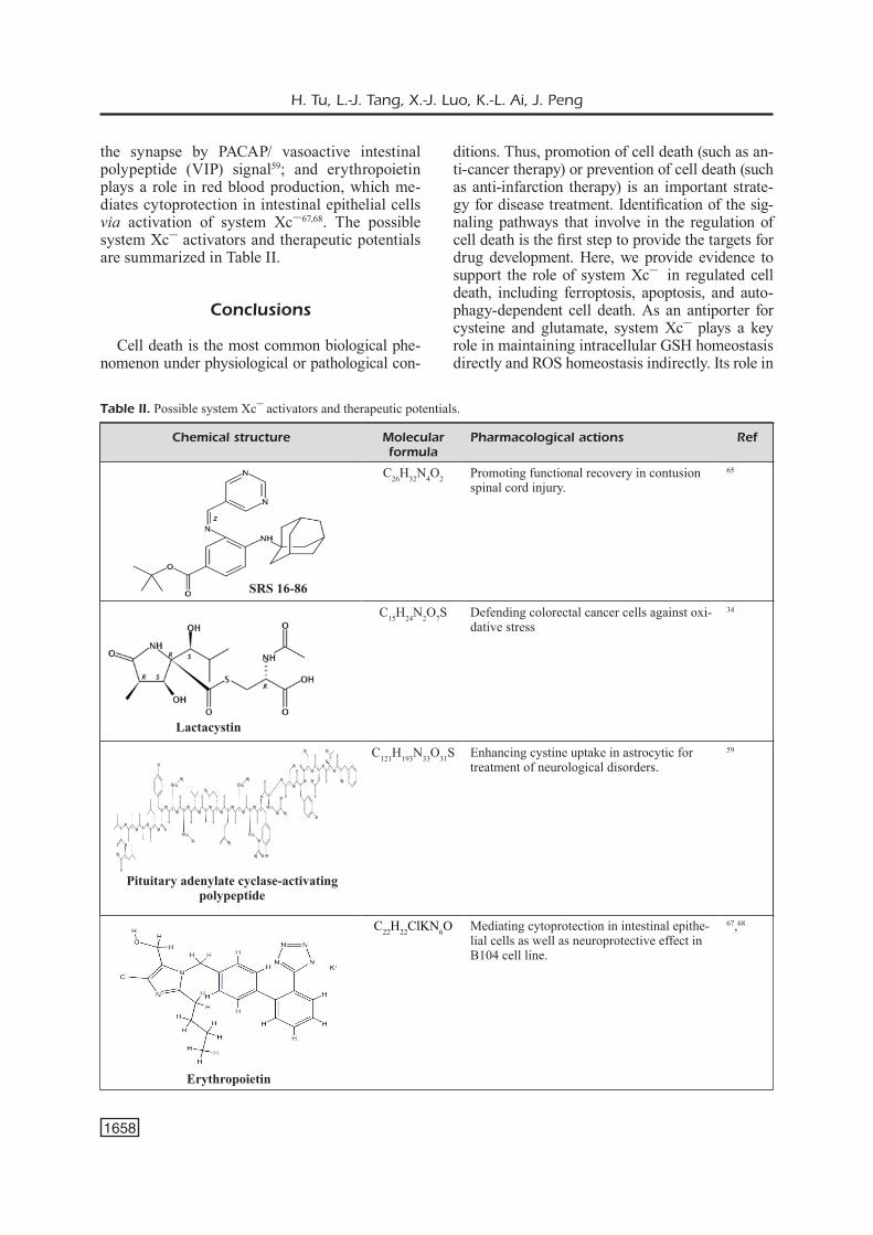
the synapse by PACAP/ vasoactive intestinal polypeptide (VIP) signal59; and erythropoietin plays a role in red blood production, which me-diates cytoprotection in intestinal epithelial cells via activation of system Xc–67,68. The possible system Xc– activators and therapeutic potentials are summarized in Table II.
Conclusions
Cell death is the most common biological phe-nomenon under physiological or pathological con-
ditions. Thus, promotion of cell death (such as an-ti-cancer therapy) or prevention of cell death (such as anti-infarction therapy) is an important strate-gy for disease treatment. Identification of the sig-naling pathways that involve in the regulation of cell death is the first step to provide the targets for drug development. Here, we provide evidence to support the role of system Xc– in regulated cell death, including ferroptosis, apoptosis, and auto-phagy-dependent cell death. As an antiporter for cysteine and glutamate, system Xc– plays a key role in maintaining intracellular GSH homeostasis directly and ROS homeostasis indirectly. Its role in
Chemical structure Molecular formula
Pharmacological actions Ref
C26H32N4O2 Promoting functional recovery in contusion spinal cord injury.
65
C15H24N2O7S Defending colorectal cancer cells against oxi-dative stress
34
C121H193N33O31S Enhancing cystine uptake in astrocytic for treatment of neurological disorders.
59
C22H22ClKN6O Mediating cytoprotection in intestinal epithe-lial cells as well as neuroprotective effect in B104 cell line.
67,68
Erythropoietin
Table II. Possible system Xc– activators and therapeutic potentials.
Lactacystin
Pituitary adenylate cyclase-activating polypeptide
SRS 16-86
Insights into the novel function of system Xc– in regulated cell death
1659
the regulation of ferroptosis was immerging with Dixon et al32 report in 2012 because system Xc– specific inhibitor (erastin)-induced cell death could be attenuated by iron chelator (deferoxamine). In addition to its key role in ferroptosis, system Xc– was also reported to be involved in apoptosis and autophagy-dependent cell death. To date, system Xc–-relevant regulated cell death has been found to play an important role in cancer, neurodegen-erative diseases, myocardial infarction, and stroke. While the intervention of system Xc– by inhibi-tor or activator has achieved a beneficial effect on anti-cancer or anti-infarction therapy, there is still a long way to go before translating these findings into clinical applications.
Conflict of InterestThe Authors declare that they have no conflict of interests.
AcknowledgementsThis work was supported by the National Natural Sci-ence Foundation of China (No. 81872873 to Jun Peng; No. 82073849 to Xiu-Ju Luo).
References
1) Tian J, Guo S, Chen H, Peng JJ, Jia MM, Li NS, Zhang XJ, Yang J, Luo XJ, Peng J. Combination of emricasan with ponatinib synergistically reduc-es ischemia/reperfusion injury in rat brain through simultaneous prevention of apoptosis and necro-ptosis. Transl Stroke Res 2018; 9: 382-392.
2) She L, Tu H, Zhang YZ, Tang LJ, Li NS, Ma QL, Liu B, Li Q, Luo XJ, Peng J. Inhibition of phos-phoglycerate mutase 5 reduces necroptosis in rat hearts following ischemia/reperfusion through suppression of dynamin-related protein 1. Cardio-vasc Drugs Ther 2019; 33: 13-23.
3) Chen D, Fan Z, Rauh M, Buchfelder M, Eyupog-lu IY, Savaskan N. ATF4 promotes angiogenesis and neuronal cell death and confers ferroptosis in a xCT-dependent manner. Oncogene 2017; 36: 5593-5608.
4) Chen Y, Hu S, Mu L, Zhao B, Wang M, Yang N, Bao G, Zhu C, Wu X. Slc7a11 modulated by POU2F1 is involved in pigmentation in rabbit. Int J Mol Sci 2019; 20.
5) Chu B, Kon N, Chen D, Li T, Liu T, Jiang L, Song S, Tavana O, Gu W. ALOX12 is required for p53-me-diated tumour suppression through a distinct fer-roptosis pathway. Nat Cell Biol 2019; 21: 579-591.
6) Gan B. DUBbing ferroptosis in cancer cells. Can-cer Res 2019; 79: 1749-1750.
7) Guo Y, Liu X, Liu D, Li K, Wang C, Liu Y, He B, Shi P. Inhibition of BECN1 suppresses lipid perox-
idation by increasing System Xc(-) activity in early brain injury after subarachnoid hemorrhage. J Mol Neurosci 2019; 67: 622-631.
8) Matassa DS, Amoroso MR, Agliarulo I, Maddale-na F, Sisinni L, Paladino S, Romano S, Romano MF, Sagar V, Loreni F, Landriscina M, Esposito F. Translational control in the stress adaptive re-sponse of cancer cells: a novel role for the heat shock protein TRAP1. Cell Death Dis 2013; 4: e851.
9) Yin J, Li Y, Han H, Zheng J, Wang L, Ren W, Chen S, Wu F, Fang R, Huang X, Li C, Tan B, Xiong X, Zhang Y, Liu G, Yao J, Li T, Yin Y. Effects of lysine deficiency and Lys-Lys dipeptide on cellular apop-tosis and amino acids metabolism. Mol Nutr Food Res 2017; 61(9).
10) Dai L, Cao Y, Chen Y, Parsons C, Qin Z. Target-ing xCT, a cystine-glutamate transporter induces apoptosis and tumor regression for KSHV/HIV-as-sociated lymphoma. J Hematol Oncol 2014; 7: 30.
11) Hu K, Li K, Lv J, Feng J, Chen J, Wu H, Cheng F, Jiang W, Wang J, Pei H, Chiao PJ, Cai Z, Chen Y, Liu M, Pang X. Suppression of the SLC7A11/glutathione axis causes synthetic lethality in KRAS-mutant lung adenocarcinoma. J Clin Invest 2020; 130: 1752-1766.
12) Guo W, Zhao Y, Zhang Z, Tan N, Zhao F, Ge C, Li-ang L, Jia D, Chen T, Yao M, Li J, He X. Disruption of xCT inhibits cell growth via the ROS/autophagy pathway in hepatocellular carcinoma. Cancer Lett 2011; 312: 55-61.
13) Zheng X, Li Y, Zhao R, Yan F, Ma Y, Zhao L, Qiao H. xCT deficiency induces autophagy via endoplasmic reticulum stress activated p38-mi-togen-activated protein kinase and mTOR in sut melanocytes. Eur J Cell Biol 2016; 95: 175-181.
14) Bannai S, Kitamura E. Transport interaction of L-cys-tine and L-glutamate in human diploid fibroblasts in culture. J Biol Chem 1980; 255: 2372-2376.
15) Makowske M, Christensen HN. Contrasts in transport systems for anionic amino acids in he-patocytes and a hepatoma cell line HTC. J Biol Chem 1982; 257: 5663-5670.
16) Reissner KJ, Kalivas PW. Using glutamate ho-meostasis as a target for treating addictive disor-ders. Behav Pharmacol 2010; 21: 514-522.
17) Massie A, Boillee S, Hewett S, Knackstedt L, Lewerenz J. Main path and byways: non-vesicular glutamate release by system xc(-) as an import-ant modifier of glutamatergic neurotransmission. J Neurochem 2015; 135: 1062-1079.
18) Bridges R, Lutgen V, Lobner D, Baker DA. Think-ing outside the cleft to understand synaptic ac-tivity: contribution of the cystine-glutamate anti-porter (System xc-) to normal and pathological glutamatergic signaling. Pharmacol Rev 2012; 64: 780-802.
19) Qaisiya M, Coda ZCD, Bellarosa C, Tiribelli C. Bilirubin mediated oxidative stress involves anti-oxidant response activation via Nrf2 pathway. Cell Signal 2014; 26: 512-520.
20) Wang L, Liu Y, Du T, Yang H, Lei L, Guo M, Ding HF, Zhang J, Wang H, Chen X, Yan C. ATF3 pro-

H. Tu, L.-J. Tang, X.-J. Luo, K.-L. Ai, J. Peng
1660
motes erastin-induced ferroptosis by suppress-ing system Xc. Cell Death Differ 2020; 27: 662-675.
21) Wang Y, Yang L, Zhang X, Cui W, Liu Y, Sun QR, He Q, Zhao S, Zhang GA, Wang Y, Chen S. Epigenetic regulation of ferroptosis by H2B monoubiquitination and p53. EMBO Rep 2019; 20: e47563.
22) Zhang Y, Zhuang L, Gan B. BAP1 suppresses tumor development by inducing ferroptosis up-on SLC7A11 repression. Mol Cell Oncol 2019; 6: 1536845.
23) Zhang Y, Koppula P, Gan B. Regulation of H2A ubiquitination and SLC7A11 expression by BAP1 and PRC1. Cell Cycle 2019; 18: 773-783.
24) Chen D, Tavana O, Chu B, Erber L, Chen Y, Baer R, Gu W. NRF2 is a major target of ARF in p53-independent tumor suppression. Mol Cell 2017; 68: 224-232.e224.
25) Sui S, Zhang J, Xu S, Wang Q, Wang P, Pang D. Ferritinophagy is required for the induction of ferroptosis by the bromodomain protein BRD4 inhibitor (+)-JQ1 in cancer cells. Cell Death Dis 2019; 10: 331.
26) Song X, Zhu S, Chen P, Hou W, Wen Q, Liu J, Xie Y, Liu J, Klionsky DJ, Kroemer G, Lotze MT, Zeh HJ, Kang R, Tang D. AMPK-mediated BECN1 phosphorylation promotes ferroptosis by directly blocking System Xc(-) activity. Curr Biol 2018; 28: 2388-2399.
27) Yoshikawa M, Tsuchihashi K, Ishimoto T, Yae T, Motohara T, Sugihara E, Onishi N, Masuko T, Yoshizawa K, Kawashiri S, Mukai M, Asoda S, Kawana H, Nakagawa T, Saya H, Nagano O. xCT inhibition depletes CD44v-expressing tu-mor cells that are resistant to EGFR-targeted therapy in head and neck squamous cell carci-noma. Cancer Res 2013; 73: 1855-1866.
28) Liu T, Jiang L, Tavana O, Gu W. The deubiquityl-ase OTUB1 mediates ferroptosis via stabilization of SLC7A11. Cancer Res 2019; 79: 1913-1924.
29) Hasegawa M, Takahashi H, Rajabi H, Alam M, Suzuki Y, Yin L, Tagde A, Maeda T, Hiraki M, Sukhatme VP, Kufe D. Functional interactions of the cystine/glutamate antiporter, CD44v and MUC1-C oncoprotein in triple-negative breast cancer cells. Oncotarget 2016; 7: 11756-11769.
30) Yamada Y, Miyamoto T, Kashima H, Kobara H, Asaka R, Ando H, Higuchi S, Ida K, Shiozawa T. Lipocalin 2 attenuates iron-related oxidative stress and prolongs the survival of ovarian clear cell carcinoma cells by up-regulating the CD44 variant. Free Radic Res 2016; 50: 414-425.
31) Yagoda N, von Rechenberg M, Zaganjor E, Bauer AJ, Yang WS, Fridman DJ, Wolpaw AJ, Smukste I, Peltier JM, Boniface JJ, Smith R, Lessnick SL, Sahasrabudhe S, Stockwell BR. RAS-RAF-MEK-dependent oxidative cell death involving voltage-dependent anion channels. Nature 2007; 447: 864-868.
32) Dixon SJ, Lemberg KM, Lamprecht MR, Skouta R, Zaitsev EM, Gleason CE, Patel DN, Bauer
AJ, Cantley AM, Yang WS, Morrison BR, Stock-well BR. Ferroptosis: an iron-dependent form of nonapoptotic cell death. Cell 2012; 149: 1060-1072.
33) Haryu S, Saito R, Jia W, Shoji T, Mano Y, Sa-to A, Kanamori M, Sonoda Y, Sampetrean O, Saya H, Tominaga T. Convection-enhanced de-livery of sulfasalazine prolongs survival in a gli-oma stem cell brain tumor model. J Neurooncol 2018; 136: 23-31.
34) Wang XY, Li YL, Wang HY, Zhu M, Guo D, Wang GL, Gao YT, Yang Z, Li T, Yang CY, Chen YM. Propofol inhibits invasion and proliferation of C6 glioma cells by regulating the Ca(2+) permeable AMPA receptor-system xc(-) pathway. Toxicol In Vitro 2017; 44: 57-65.
35) Arensman MD, Yang XS, Leahy DM, Toral-Bar-za L, Mileski M, Rosfjord EC, Wang F, Deng S, Myers JS, Abraham RT, Eng CH. Cystine-glu-tamate antiporter xCT deficiency suppresses tumor growth while preserving antitumor immu-nity. Proc Natl Acad Sci U S A 2019; 116: 9533-9542.
36) Roh JL, Kim EH, Jang HJ, Park JY, Shin D. In-duction of ferroptotic cell death for overcoming cisplatin resistance of head and neck cancer. Cancer Lett 2016; 381: 96-103.
37) Kim EH, Shin D, Lee J, Jung AR, Roh JL. CISD2 inhibition overcomes resistance to sulfasala-zine-induced ferroptotic cell death in head and neck cancer. Cancer Lett 2018; 432: 180-190.
38) Timmerman LA, Holton T, Yuneva M, Louie RJ, Padro M, Daemen A, Hu M, Chan DA, Ethier SP, van T VLJ, Polyak K, McCormick F, Gray JW. Glutamine sensitivity analysis identifies the xCT antiporter as a common triple-negative breast tumor therapeutic target. Cancer Cell 2013; 24: 450-465.
39) Huseby NE, Ravuri C, Moens U. The protea-some inhibitor lactacystin enhances GSH syn-thesis capacity by increased expression of an-tioxidant components in an Nrf2-independent, but p38 MAPK-dependent manner in rat col-orectal carcinoma cells. Free Radic Res 2016; 50: 1-13.
40) Zhang L, Huang Y, Ling J, Zhuo W, Yu Z, Luo Y, Zhu Y. Overexpression of SLC7A11: a novel on-cogene and an indicator of unfavorable progno-sis for liver carcinoma. Future Oncol 2018; 14: 927-936.
41) Wu Y, Sun X, Song B, Qiu X, Zhao J. MiR-375/SLC7A11 axis regulates oral squamous cell car-cinoma proliferation and invasion. Cancer Med 2017; 6: 1686-1697.
42) Ji X, Qian J, Rahman SMJ, Siska PJ, Zou Y, Harris BK, Hoeksema MD, Trenary IA, Heidi C, Eisenberg R, Rathmell JC, Young JD, Massion PP. xCT (SLC7A11)-mediated metabolic repro-gramming promotes non-small cell lung cancer progression. Oncogene 2018; 37: 5007-5019.
43) Lewerenz J, Hewett SJ, Huang Y, Lambros M, Gout PW, Kalivas PW, Massie A, Smolders I,

Insights into the novel function of system Xc– in regulated cell death
1661
Methner A, Pergande M, Smith SB, Ganapathy V, Maher P. The cystine/glutamate antiporter system x(c)(-) in health and disease: from mo-lecular mechanisms to novel therapeutic oppor-tunities. Antioxid Redox Signal 2013; 18: 522-555.
44) Sato M, Kusumi R, Hamashima S, Kobayashi S, Sasaki S, Komiyama Y, Izumikawa T, Conrad M, Bannai S, Sato H. The ferroptosis inducer erastin irreversibly inhibits system xc- and syn-ergizes with cisplatin to increase cisplatin’s cy-totoxicity in cancer cells. Sci Rep 2018; 8: 968.
45) Ma MZ, Chen G, Wang P, Lu WH, Zhu CF, Song M, Yang J, Wen S, Xu RH, Hu Y, Huang P. Xc- inhibitor sulfasalazine sensitizes colorectal cancer to cisplatin by a GSH-dependent mech-anism. Cancer Lett 2015; 368: 88-96.
46) Roh JL, Kim EH, Jang H, Shin D. Aspirin plus sorafenib potentiates cisplatin cytotoxicity in re-sistant head and neck cancer cells through xCT inhibition. Free Radic Biol Med 2017; 104: 1-9.
47) Drayton RM, Dudziec E, Peter S, Bertz S, Hart-mann A, Bryant HE, Catto JW. Reduced ex-pression of miRNA-27a modulates cisplatin re-sistance in bladder cancer by targeting the cys-tine/glutamate exchanger SLC7A11. Clin Cancer Res 2014; 20: 1990-2000.
48) Cong L, Dong X, Wang Y, Deng Y, Li B, Dai R. On the role of synthesized hydroxylated chal-cones as dual functional amyloid-beta aggrega-tion and ferroptosis inhibitors for potential treat-ment of Alzheimer’s disease. Eur J Med Chem 2019; 166: 11-21.
49) Karuppagounder SS, Alin L, Chen Y, Brand D, Bourassa MW, Dietrich K, Wilkinson CM, Nadeau CA, Kumar A, Perry S, Pinto JT, Dar-ley-Usmar V, Sanchez S, Milne GL, Pratico D, Holman TR, Carmichael ST, Coppola G, Col-bourne F, Ratan RR. N-acetylcysteine targets 5 lipoxygenase-derived, toxic lipids and can synergize with prostaglandin E2 to inhibit fer-roptosis and improve outcomes following hem-orrhagic stroke in mice. Ann Neurol 2018; 84: 854-872.
50) Piani D, Fontana A. Involvement of the cystine transport system xc- in the macrophage-in-duced glutamate-dependent cytotoxicity to neu-rons. J Immunol 1994; 152: 3578-3585.
51) Lyons SA, Chung WJ, Weaver AK, Ogunrinu T, Sontheimer H. Autocrine glutamate signal-ing promotes glioma cell invasion. Cancer Res 2007; 67: 9463-9471.
52) Hua L, Wang FQ, Du HW, Fan J, Wang YF, Wang LQ, Shi XW. Upregulation of caspase-3 by high glucose in chondrocyte involves the cy-toskeleton aggregation. Eur Rev Med Pharma-col Sci 2020; 24: 5925-5932.
53) Sakakura Y, Sato H, Shiiya A, Tamba M, Sagara J, Matsuda M, Okamura N, Makino N, Bannai S. Expression and function of cystine/glutamate transporter in neutrophils. J Leukoc Biol 2007; 81: 974-982.
54) Qiao HX, Hao CJ, Li Y, He X, Chen RS, Cui J, Xu ZH, Li W. JNK activation mediates the apop-tosis of xCT-deficient cells. Biochem Biophys Res Commun 2008; 370: 584-588.
55) Wang L, Hinoi E, Takemori A, Nakamichi N, Yoneda Y. Glutamate inhibits chondral miner-alization through apoptotic cell death mediated by retrograde operation of the cystine/gluta-mate antiporter. J Biol Chem 2006; 281: 24553-24565.
56) Sun B, Xu Y, Liu ZY, Meng WX, Yang H. Auto-phagy assuages myocardial infarction through Nrf2 signaling activation-mediated reactive ox-ygen species clear. Eur Rev Med Pharmacol Sci 2020; 24: 7381-7390.
57) Zhang Y, Tan H, Daniels JD, Zandkarimi F, Liu H, Brown LM, Uchida K, O’Connor OA, Stockwell BR. Imidazole ketone erastin induces ferroptosis and slows tumor growth in a mouse lymphoma model. Cell Chem Biol 2019; 26: 623-633.
58) Kain HS, Glennon EKK, Vijayan K, Arang N, Douglass AN, Fortin CL, Zuck M, Lewis AJ, Wh-iteside SL, Dudgeon DR, Johnson JS, Aderem A, Stevens KR, Kaushansky A. Liver stage ma-laria infection is controlled by host regulators of lipid peroxidation. Cell Death Differ 2020; 27: 44-54.
59) Resch JM, Albano R, Liu X, Hjelmhaug J, Lobner D, Baker DA, Choi S. Augmented cys-tine-glutamate exchange by pituitary adenylate cyclase-activating polypeptide signaling via the VPAC1 receptor. Synapse 2014; 68: 604-612.
60) Wang Z, Ding Y, Wang X, Lu S, Wang C, He C, Wang L, Piao M, Chi G, Luo Y, Ge P. Pseudola-ric acid B triggers ferroptosis in glioma cells via activation of Nox4 and inhibition of xCT. Cancer Lett 2018; 428: 21-33.
61) Yu M, Gai C, Li Z, Ding D, Zheng J, Zhang W, Lv S, Li W. Targeted exosome-encapsulated eras-tin induced ferroptosis in triple negative breast cancer cells. Cancer Sci 2019; 110: 3173-3182.
62) Jostes S, Nettersheim D, Fellermeyer M, Schneider S, Hafezi F, Honecker F, Schumach-er V, Geyer M, Kristiansen G, Schorle H. The bromodomain inhibitor JQ1 triggers growth arrest and apoptosis in testicular germ cell tu-mours in vitro and in vivo. J Cell Mol Med 2017; 21: 1300-1314.
63) Lee S, Rellinger EJ, Kim KW, Craig BT, Romain CV, Qiao J, Chung DH. Bromodomain and ex-traterminal inhibition blocks tumor progression and promotes differentiation in neuroblastoma. Surgery 2015; 158: 819-826.
64) Alqahtani A, Choucair K, Ashraf M, Hammouda DM, Alloghbi A, Khan T, Senzer N, Nemunaitis J. Bromodomain and extra-terminal motif inhibi-tors: a review of preclinical and clinical advanc-es in cancer therapy. Future Sci OA 2019; 5: FSO372.
65) Zhang Y, Sun C, Zhao C, Hao J, Zhang Y, Fan B, Li B, Duan H, Liu C, Kong X, Wu P, Yao X, Feng S. Ferroptosis inhibitor SRS 16-86 attenu-ates ferroptosis and promotes functional recov-

H. Tu, L.-J. Tang, X.-J. Luo, K.-L. Ai, J. Peng
1662
ery in contusion spinal cord injury. Brain Res 2019; 1706: 48-57.
66) Yao X, Zhang Y, Hao J, Duan HQ, Zhao CX, Sun C, Li B, Fan BY, Wang X, Li WX, Fu XH, Hu Y, Liu C, Kong XH, Feng SQ. Deferoxamine promotes recovery of traumatic spinal cord in-jury by inhibiting ferroptosis. Neural Regen Res 2019; 14: 532-541.
67) Martin C, Patel M, Melendez-Ferro M, Sims B. Erythropoietin-induced cytoprotection in intes-tinal epithelial cells is linked to system Xc. Exp Cell Res 2017; 352: 202-206.
68) Sims B, Clarke M, Njah W, Hopkins ES, Son-theimer H. Erythropoietin-induced neuropro-tection requires cystine glutamate exchanger activity. Brain Res 2010; 1321: 88-95.