instructions for use sx-ella stent esophageal degradable ... · instructions for use...
TRANSCRIPT

Instructions for Use
SX-BD-10A-AJ-11/10/I/REV-1-01/2011 Page 1 of 12
SX-ELLA Stent Esophageal Degradable BD (BD Stent) Fluoroscopic stent system
Non-active implantable device Manufactured by: ELLA–CS, s.r.o. Milady Horákové 504 500 06 Hradec Králové 6 Czech Republic Phone: +420 49 527 91 11 Fax: +420 49 526 56 55 E-mail: [email protected] Product content: 1 piece of degradable stent packed separately into the plastic cartridge (Table No. 1):
Model Nominal (Relaxed) diameter of stent trunk
D2 [mm]
Nominal (Relaxed) diameter of both flares
D1, D3 [mm]
Nominal (Relaxed) length of stent [mm]
60 80 18 23 100 60 80 20 25 100 60 80 23 28 100 60 80 100
10
25 31
135 The wire port of the delivery system fits for 0.89-mm (0.035”) guide wire. Sheath (delivery system) dimensions (Table No. 2):
Outer diameter [mm/F] Active length [mm] Sheath 6.0 (18) / 9.4 (28 F) 750
1 piece of Compression tool for stent loading into delivery system 1 piece of Instructions for Use 1 product label (for insertion into the patient’s medical record) Storage and transport: The device should be stored in a dust-free, dry and dark place at room temperature. The product shall be protected from the moisture and water (rain, high humidity, condensation, spillage).Prevent the package from repeated changes in atmospheric pressure higher than ± 3 kPa (e.g. in aircraft holds) which might damage inner pouch seals. Material composition of the stent: Polydioxanone – polymer monofilament the stent is made of
Instructions for Use
SX-BD-10A-AJ-11/10/I/REV-1-01/2011 Page 2 of 12
Au – gold tubes (markers) placed on both stent ends The stent is MRI compatible. Precautions: Product set (the stent and the delivery system) is sterilized by ethylene oxide gas in peel-open packaging. The stent is provided with two layers packaging which inner part represents a moisture barrier. Only use the device if packaging is intact. Product is for single-use only. Do not re-sterilize. The device must be used prior to the expiry date printed on the label of the packaging. Allergic reactions to polydioxanone may occur in extremely sensitive patients; however, these are very rare. Because polydioxanone monofilament exhibits plastic deformation, the sterile stent is packed in a cartridge separate from the delivery system and needs to be loaded manually into the delivery system just before the implantation procedure. In patients with refractory esophageal strictures the pre-dilation of the stenosis prior to stent insertion is recommended to a diameter of 7 – 15 mm in order to allow passing the delivery system through the stenosis. Decision concerning pre-dilation in patients with achalasia depends on implanting physician.
Warnings: • The physician implanting the stent must be appropriately skilled.
• Choice of proper stent dimensions (length and diameter) wholly depends on anatomical situation and it is exclusively made by attending physician based on adequate examination.
• The stent should be placed over an ultra stiff guide wire with soft tip.
• Stent placement should be performed under fluoroscopic and endoscopic control. • Follow the instructions for loading the stent into the delivery system to avoid the stent damage
(see Figures No. 1 – 18). Avoid re-loading the stent (risk of loss of expansion force). • Each of the stent ends (flares) should extend beyond the stricture, i.e. within healthy tissue.
• Follow the instructions for correction of stent misplacement/partial migration! Care must be taken when changing the stent position later than 5 days after implantation. Part of the stent may be incorporated into the esophageal mucosa at that time. Endoscopic evaluation of the stent and tissue status is necessary before any attempt of managing the stent displacement.
• After the implantation patient should stay on sufficiently chewed diet and drink plenty of liquids. • The pH value influences the stent degradation. A low pH accelerates and a high pH slows it down.
• The biodegradable stent should only be placed as a last means after all standard treatments have failed or in patients whose condition prevents standard therapeutic procedures (e.g. advanced age, severe cardiovascular disease etc.).
• The used components of BD Stent Set shall be disposed observing the method of disposal of biologically contaminated waste. The used delivery system belongs to potentially hazardous biological waste.
• Do not use the stent for dilation of anastomotic strictures located in proximal part of esophagus, from upper esophageal sphincter to level of the trachea bifurcation because of high risk of hyperplasic reaction, decubital necrosis and esophagorespiratory fistula formation.
• Do not use the stent for dilation of strictures that can not be dilated in such a way to pass through it with the delivery system.
Indications: The SX-ELLA Stent Esophageal Degradable BD (BD Stent) is designed for dilation of benign esophageal lesions, namely:
- stenosis (peptic, anastomotic or caustic) refractory to standard therapy, - achalasia refractory to standard therapy.
Instructions for Use
SX-BD-10A-AJ-11/10/I/REV-1-01/2011 Page 3 of 12
Contraindications: Inability to pass the 9.4-mm (28-F) delivery system through the stricture; Benign strictures in the upper part of the esophagus too close to the cricopharyngeal muscle. Patients with benign stricture due to previously performed laryngectomy Recommended implantation procedure:
I. Preparation preceding the implantation The patient must be prepared in the same way as for a standard gastroscopic examination. Remove the delivery system (for schema of the delivery system see Figure No. 20) from the packaging and inspect it for any visible damage (i.e. holes, tears, openings, breakage or bends). Remove the stent (for schema of the stent see Figure No. 19) from the packaging and inspect it for any visible damage (i.e. holes, tears, deformations, breakage or bends). Do not use the product if the packaging or the stent itself have been damaged!
II. Loading procedure The procedure for loading the stent is demonstrated in Figures No. 1 – 18. Strictly follow the loading procedure in order to avoid the stent damage.
III. Implantation procedure 1) Introduce safely under endoscopic guidance a min. 220 cm long 0.89 mm (0.035 inch) Ultra
Stiff guide wire with soft tip through the stenosis. 2) Choose an appropriate length of the stent for the length of the stenosis. Information about the
nominal (relaxed) length of the stent model is presented on the packaging label. 3) Load the stent into the delivery system immediately prior to insertion. The olive forms a firm
cone preventing damage of the esophageal wall during introduction of the delivery system. Carefully advance the delivery system over the guide wire into the esophagus.
4) Prior to releasing the stent from the delivery system, position the stent as follows: The distal end of the compressed stent inside the delivery system should extend at least 2 cm beyond the lower margin of the stenosis to allow for stent shortening (see Figure No. 21 - 24 shortening charts).
5) Remove the lock from the delivery system. NOTE: The lock shall be removed immediately before start of the stent deployment. At the moment of starting the deployment, the olive automatically splits into two pieces and falls into the stomach.
6) Keeping the pusher in a stable towards the patient fixed position, slowly pull back the sheath until the stent is fully released from the sheath. In most cases the stent will continue to dilate gradually to reach its full diameter over 48 hours. NOTE: The ends of the stent should ideally extend beyond the margins of the stenosis by about 5 mm.
7) Slowly remove the delivery system over the guide wire. The guide wire should be removed together with the delivery system. The tip of the guide wire has to be positioned inside the olive tip. Complete the removal of the whole delivery system and guide wire.
8) After successful implantation of the stent, perform a contrast swallow test with water soluble contrast medium.
IV. Complications and their management 1) Mucosal hyperplasia and stenosis formation due to BD Stent implantation
The risk of hyperplastic reaction is increased by the following factors: - The stent is implanted in upper third of esophagus; - The gastro esophageal reflux is presented; - In patients suffering from strictures after performed laryngectomy.
Possible management of hyperplastic reaction: - In case of hyperplasia producing significant dysphagia – pneumatic balloon dilation possible
with local injections of corticoids can solve this problem.
Instructions for Use
SX-BD-10A-AJ-11/10/I/REV-1-01/2011 Page 4 of 12
- In case of clinically silent hyperplasia without dysphagia – just observation, no treatment. - In case of early developing and/or extensive hyperplasia producing significant dysphagia –
local injections of corticoids can have the positive effect on this problem. 2) Gastro-esophageal reflux due to stricture morphology and/or stent position
Benign esophageal strictures are often associated with gastroesophageal reflux that increases the risk of hyperplastic reaction. If the stent is placed across the gastro-esophageal junction, it causes significant reflux. In both cases the patients have to be treated by antacids, e.g. proton pump inhibitors.
3) Misplacement or partial migration of the stent The stent can be repositioned by using an endoscopic forceps without sharp edges. Grasp the proximal or distal end of the stent (depending on the direction of migration) and pull the stent into the desired position.
4) Persistent pain or foreign body sensation after the other serious complications were considered and excluded Persistent pain requires long-term analgesic therapy.
5) Complete migration of the stent into the stomach Introduction of a new stent is required. If clinical symptoms of obstruction are present (peritonitis, bowel obstruction etc.) a complete diagnosis and appropriate therapy is required.
6) Insufficient length of the stent Over-stenting with a second stent is recommended.
7) Obstruction of the stent by food Food impaction may resolve with carbonated (fizzy) drinks but may require endoscopic removal of the food bolus.
8) Decubital esophagorespiratory fistula Introduction of the new fully covered stent into the old one (re-stenting) and other proper therapeutic methods can solve the problem.
9) Incomplete stent dilation If the stent does not dilate to its nominal diameter within 48 hours, balloon-dilation to a maximum of 20 mm can be performed.
1014
Instructions for Use
SX-BD-10A-AJ-11/10/I/REV-1-01/2011 Page 5 of 12
Loading of BD Stent into the delivery system BD Stent loading must be performed in sterile gloves in environment of sterile surface of
the instruction table!
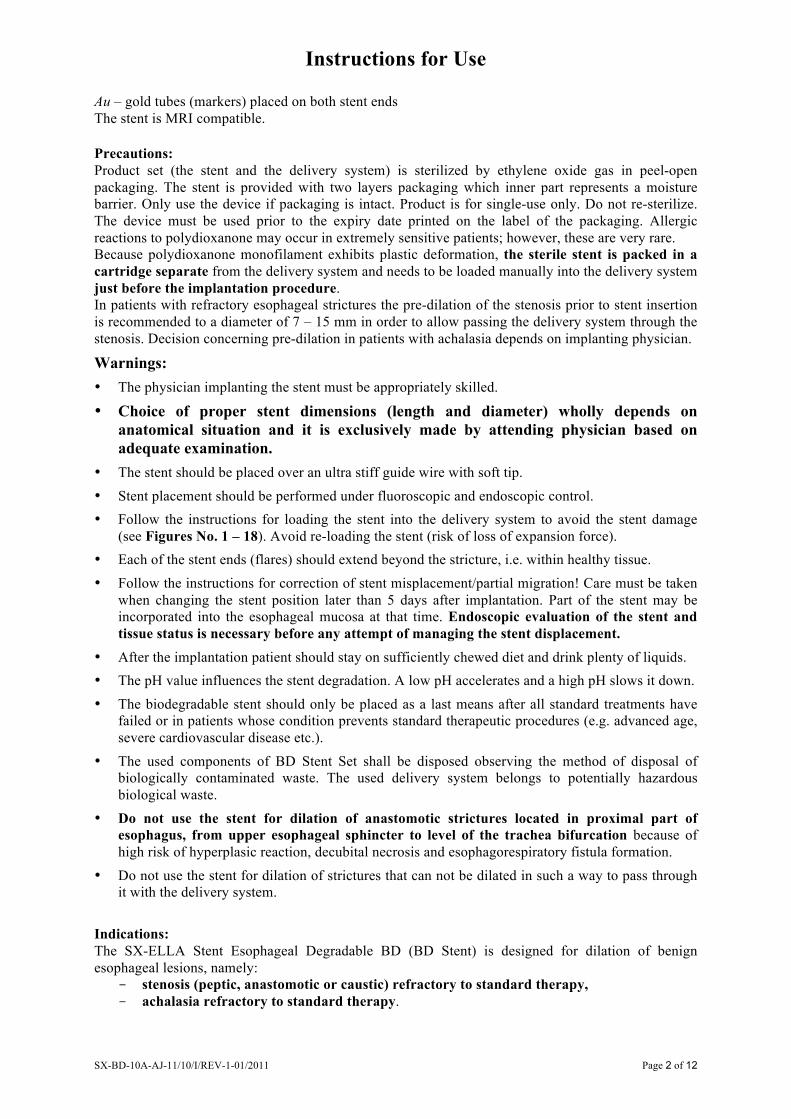
Figure No. 1 Insert the guide wire (ultra stiff 0.035") through the wire port into the delivery system.
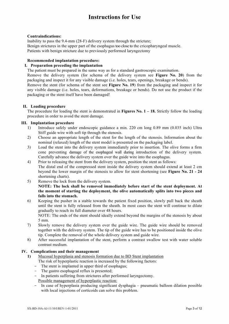
Figure No. 2 Remove the white pusher lock.
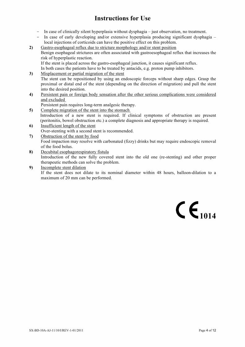
Figure No. 3 After removing the white lock pull the sheath handle. Detachable parts of the olive split automatically into two parts. Both detachable parts of the olive must be kept on sterile surface of the instruction table during the whole period of loading!
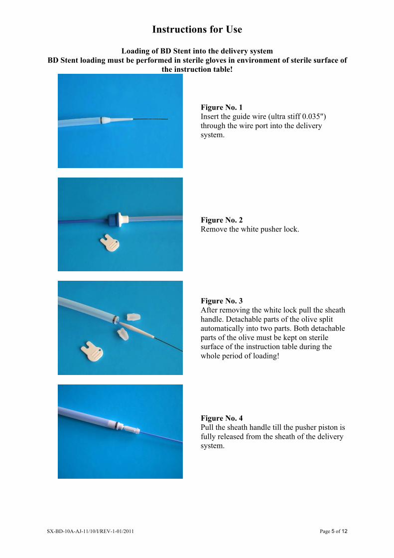
Figure No. 4 Pull the sheath handle till the pusher piston is fully released from the sheath of the delivery system.

Instructions for Use
SX-BD-10A-AJ-11/10/I/REV-1-01/2011 Page 6 of 12
Figure No. 5 Fix the pusher by the white lock. The white lock is inserted into notch of the pusher piston.
Figure No. 6 Put the compression tool and BD Stent on the shaft of the olive. The guide wire has to be safely positioned in the middle of the stent lumen in order to avoid stent’s mesh damage.
Figure No. 7 Compress the throat of BD Stent by using fingers and insert BD Stent into the compression tool. Do not screw or twist the stent!! Continue the BD Stent compression step by step. WHOLE radiopaque marker which is placed on the edge of the stent throat must be visible behind the opposite side of the compression tool. It means the stent throat with the whole radiopaque marker must be fully opened on the side of the compression tool closer to the pusher piston.
Figure No. 8 Move compression tool with BD Stent onto pusher piston of the delivery system.
Instructions for Use
SX-BD-10A-AJ-11/10/I/REV-1-01/2011 Page 7 of 12
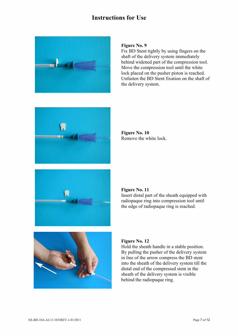
Figure No. 9 Fix BD Stent tightly by using fingers on the shaft of the delivery system immediately behind widened part of the compression tool. Move the compression tool until the white lock placed on the pusher piston is reached. Unfasten the BD Stent fixation on the shaft of the delivery system.
Figure No. 10 Remove the white lock.
Figure No. 11 Insert distal part of the sheath equipped with radiopaque ring into compression tool until the edge of radiopaque ring is reached.
Figure No. 12 Hold the sheath handle in a stable position. By pulling the pusher of the delivery system in line of the arrow compress the BD stent into the sheath of the delivery system till the distal end of the compressed stent in the sheath of the delivery system is visible behind the radiopaque ring.
Instructions for Use
SX-BD-10A-AJ-11/10/I/REV-1-01/2011 Page 8 of 12
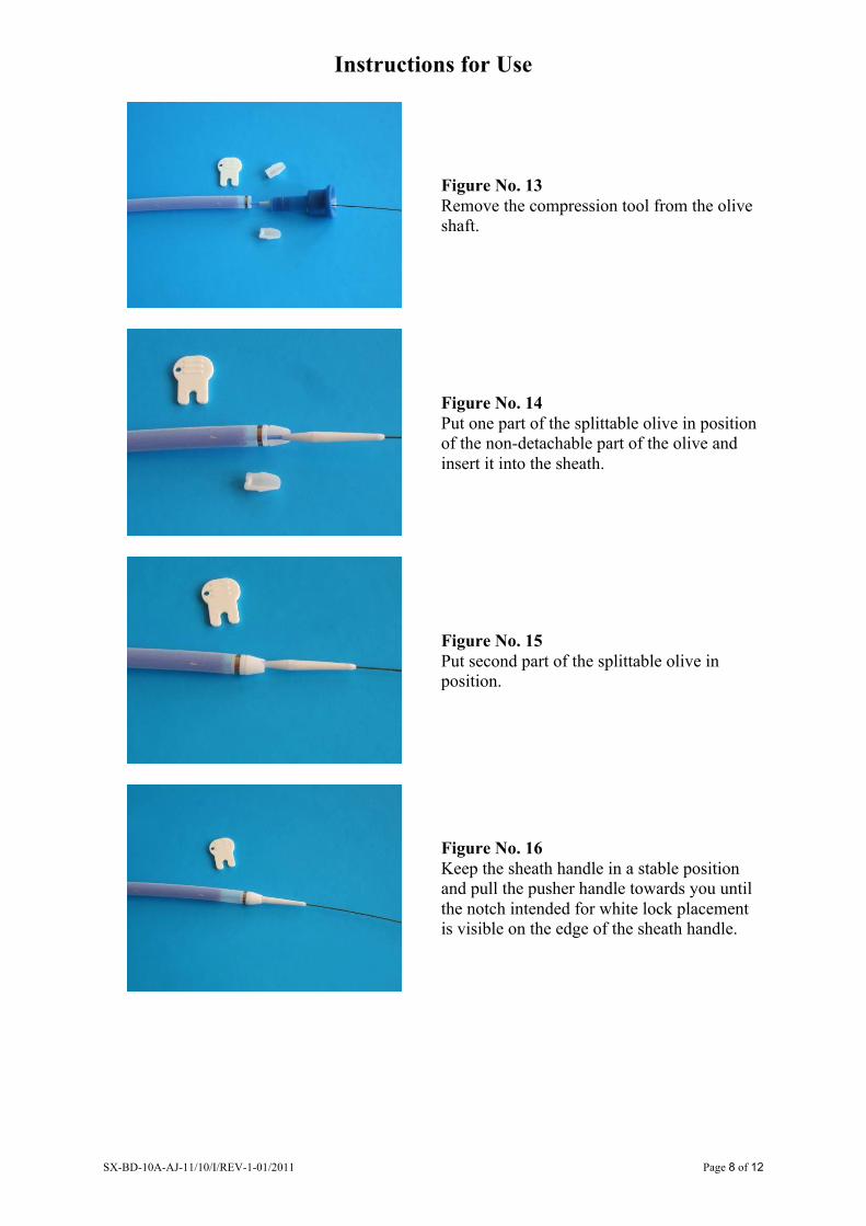
Figure No. 13 Remove the compression tool from the olive shaft.
Figure No. 14 Put one part of the splittable olive in position of the non-detachable part of the olive and insert it into the sheath.
Figure No. 15 Put second part of the splittable olive in position.
Figure No. 16 Keep the sheath handle in a stable position and pull the pusher handle towards you until the notch intended for white lock placement is visible on the edge of the sheath handle.
Instructions for Use
SX-BD-10A-AJ-11/10/I/REV-1-01/2011 Page 9 of 12
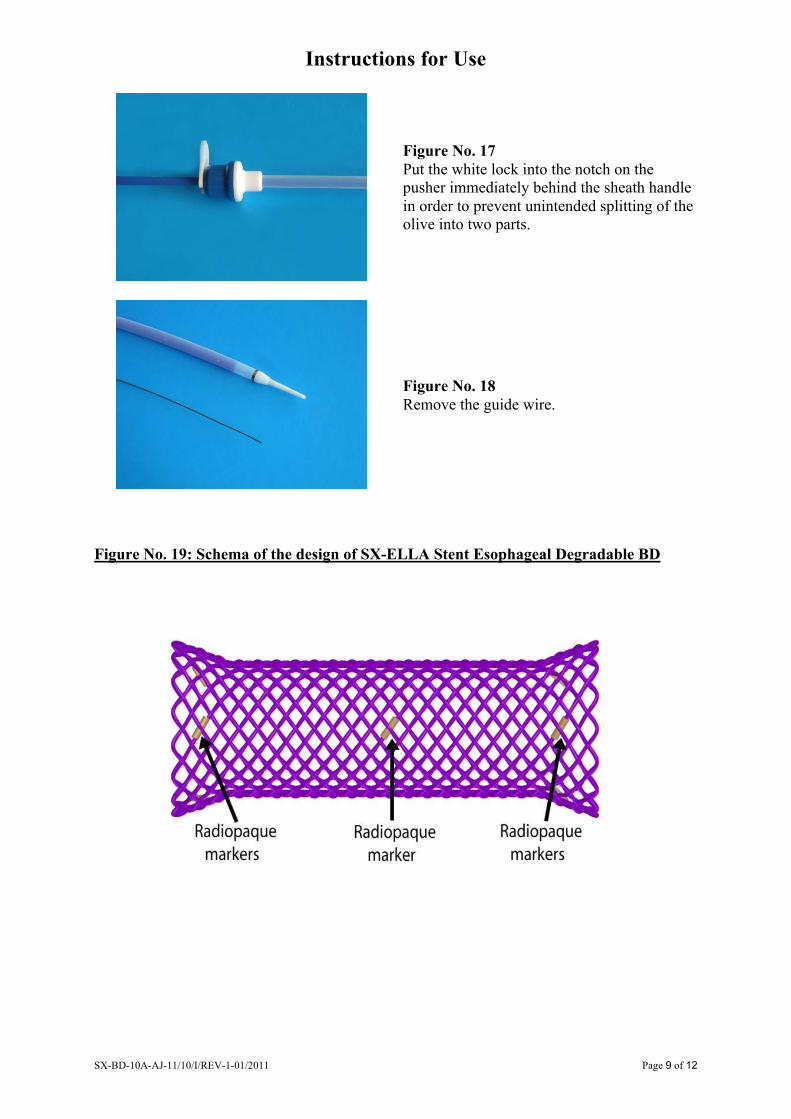
Figure No. 17 Put the white lock into the notch on the pusher immediately behind the sheath handle in order to prevent unintended splitting of the olive into two parts.
Figure No. 18 Remove the guide wire.
Figure No. 19: Schema of the design of SX-ELLA Stent Esophageal Degradable BD
Instructions for Use
SX-BD-10A-AJ-11/10/I/REV-1-01/2011 Page 10 of 12
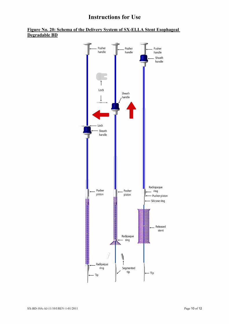
Figure No. 20: Schema of the Delivery System of SX-ELLA Stent Esophageal Degradable BD

Instructions for Use
SX-BD-10A-AJ-11/10/I/REV-1-01/2011 Page 11 of 12
Figure No. 21: Shortening chart of SX-ELLA Stent Esophageal Degradable BD 23/18/23 mm
Stent length [mm] Stent diameter [mm] L = 60 L = 80 L = 100
10 92 122 155 12 88 118 148 14 84 111 140 16 78 103 129 18 72 96 118 19 70 93 114 20 68 91 112 21 66 88 108 22 63 84 104 23 60 80 100
9.4/28F Delivery system
95 125 159
Figure No. 22: Shortening chart of SX-ELLA Stent Esophageal Degradable BD 25/20/25 mm
Stent length [mm] Stent diameter [mm] L = 60 L = 80 L = 100
10 101 135 168 11 99 132 164 12 97 129 161 13 95 127 158 14 93 124 154 16 88 116 144 18 81 107 131 20 76 98 116 22 68 92 110 25 60 80 100
9.4/28F Delivery system
104 137 171
Instructions for Use
SX-BD-10A-AJ-11/10/I/REV-1-01/2011 Page 12 of 12
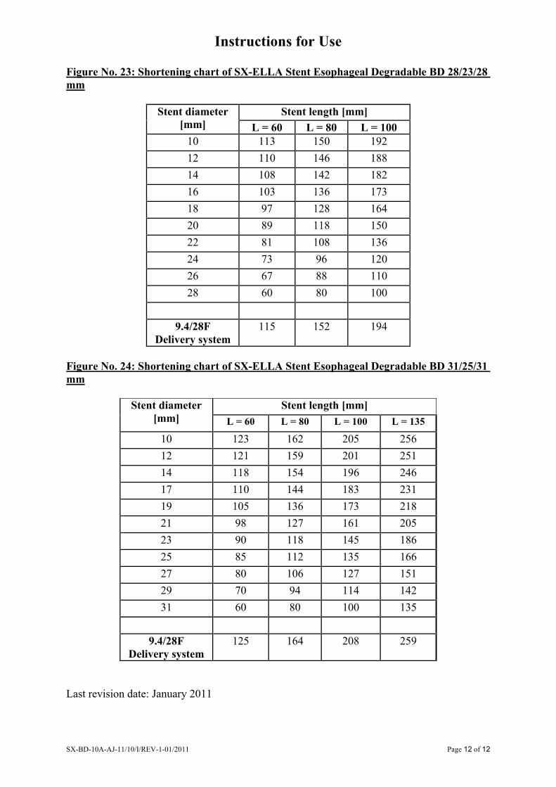
Figure No. 23: Shortening chart of SX-ELLA Stent Esophageal Degradable BD 28/23/28 mm
Stent length [mm] Stent diameter [mm] L = 60 L = 80 L = 100
10 113 150 192 12 110 146 188 14 108 142 182 16 103 136 173 18 97 128 164 20 89 118 150 22 81 108 136 24 73 96 120 26 67 88 110 28 60 80 100
9.4/28F Delivery system
115 152 194
Figure No. 24: Shortening chart of SX-ELLA Stent Esophageal Degradable BD 31/25/31 mm
Stent length [mm] Stent diameter [mm] L = 60 L = 80 L = 100 L = 135
10 123 162 205 256 12 121 159 201 251 14 118 154 196 246 17 110 144 183 231 19 105 136 173 218 21 98 127 161 205 23 90 118 145 186 25 85 112 135 166 27 80 106 127 151 29 70 94 114 142 31 60 80 100 135
9.4/28F Delivery system
125 164 208 259
Last revision date: January 2011