intech-cell responses to surface and architecture of tissue engineering scaffolds
TRANSCRIPT
8/3/2019 InTech-Cell Responses to Surface and Architecture of Tissue Engineering Scaffolds
http://slidepdf.com/reader/full/intech-cell-responses-to-surface-and-architecture-of-tissue-engineering-scaffolds 1/20
8/3/2019 InTech-Cell Responses to Surface and Architecture of Tissue Engineering Scaffolds
http://slidepdf.com/reader/full/intech-cell-responses-to-surface-and-architecture-of-tissue-engineering-scaffolds 2/20
Regenerative Medicine and Tissue Engineering - Cells and Biomaterials570
between cells and biomaterial scaffolds is called focal adhesion. To understand the factorsthat influence cell adhesive ability is a key in the development and application of new tissueengineering scaffold.Cell attachment is a complex process, affected by numerous aspects, such as cell behavior,
material surface properties, and environmental factors. Material surface properties comprisethe hydrophobicity, charge, roughness, softness and chemical composition of thebiomaterial surface itself.
2.1 Surface hydrophobicity
Biomaterial development has been focusing on surface modifications of biomaterials overyears in order to promote a greater understanding and control of the material characteristicsfor regulating biocompatibility. The surface hydrophobicity is well known as a key factor togovern cell response. The surface hydrophobicity can be assessed by measuring contactangle through water spread of a droplet on a surface. The lower the contact angle, the more
hydrophilic the surface is. Previous studies showed the more hydrophilic surface of materialfilms is the much more cell adhesion on the surface (Goddard & Hotchkiss, 2007; Xu, 2007).For example, osteoblast adhesion was reported decrease when the contact angle of surfaceincreased from 0° to 106°. Fibroblasts were found to have maximum adhesion when contactangles were between 60° and 80° (Tamada & Ikada, 1993; Wei et al., 2009). Interestingly,Vogler mentioned that the hydrophilic surface were suitable for the attachment of Madin-Darby Canine Kidney (MDCK) cells but more hydrophilic surfaces (contact angel θ< 65°)did not yield progressively high level of attachment efficiency (Vogler, 1999). Furthermore,surface hydrophobicity is related to the rate of cell spreading and differentiation. Onhydrophilic surfaces, cells generally showed good spreading, proliferation anddifferentiation. Mouse osteoblast-like cell line MC3T3-E1 showed more fractal morphology
on hydrophilic surface (contact angel θ= 0°) (Wei et al., 2009). 7F2 mouse osteoblasts onhydrophilic surface (contact angel θ= 24-31°) demonstrated accelerated metabolic activityand osteodifferentiation compared to their unmodified counterparts (contact angel θ= 72°)(Yildirim et al., 2010). The same phenomenon was observed in neuronal spreading andneurite outgrowth when the material surfaces reduced their hydrophobicity (Khorasani &Irani, 2008; Lee et al., 2003).
2.2 Protein adsorption
Since cell adhesion to material surface requires a series of cytoplasmic, transmembranal andextracellular proteins that assemble into stable contact sites (Geiger & Bendori, 1987), celladhesion and behaviors is likely involved the adsorption onto the material surface of serumand ECM proteins (Brynda & Andrade, 1990; Hattori et al., 1985). Many proteins, includingimmunoglobulins, vitronectin, fibrinogen, and fibronectin (Fn), adsorb onto implantsurfaces immediately upon contact with physiological fluids and modulate subsequentinflammatory responses. For example, adsorbed adhesive proteins mediate the attachmentand activation of neutrophils, macrophages, and other inflammatory cells. Many literaturestudies mentioned that different cell behaviors, related to different hydrophobicities, may bemediated by protein absorption, because surface wettability modified the sort and thequantity of adsorbed cell adhesion molecules. Hydrophobic surfaces tend to adsorb moreproteins, while hydrophilic surfaces tend to resist protein adsorption (Xu, 2007). Tamada etal. used bovine serum albumin (BSA), bovine γ-globulin and plasma Fn to study the protein
8/3/2019 InTech-Cell Responses to Surface and Architecture of Tissue Engineering Scaffolds
http://slidepdf.com/reader/full/intech-cell-responses-to-surface-and-architecture-of-tissue-engineering-scaffolds 3/20
Cell Responses to Surface and Architecture of Tissue Engineering Scaffolds 571
absorption onto various polymer substrates and the maximal protein absorption wasobserved on surfaces with water contact angle ranging from 60o to 80o (Tamada & Ikada,1993). Hence wetting has been discredited as an adequate predictor of protein adsorption.However, there is a growing concern that surface hydrophobicity does not guarantee the
protein adsorption. Certain hydrogel-like materials (e.g. oligo(ethylene glycol)) are resistantto protein adsorption even though these surfaces are (apparently) only modestly wettable(Noh & Vogler, 2006). Tamada reported that preadsorption of serum albumin prevented celladhesion of fibroblasts to all substrates, whereas preadsorbed Fn enhanced cell adhesion offibroblasts to all the substrates, independent of their water wettability. Withpreadsorption of Fn and BSA, similar pattern of cell attachment was investigated on aseries of N -isospropylacrylamide and N -tert-butylacrylamide based copolymer films(Allen et al., 2006). Therefore, the composition and conformation of the adsorbed proteinlayer is considered to be one of the major factors in determining the nature of cellinteraction with the materials.
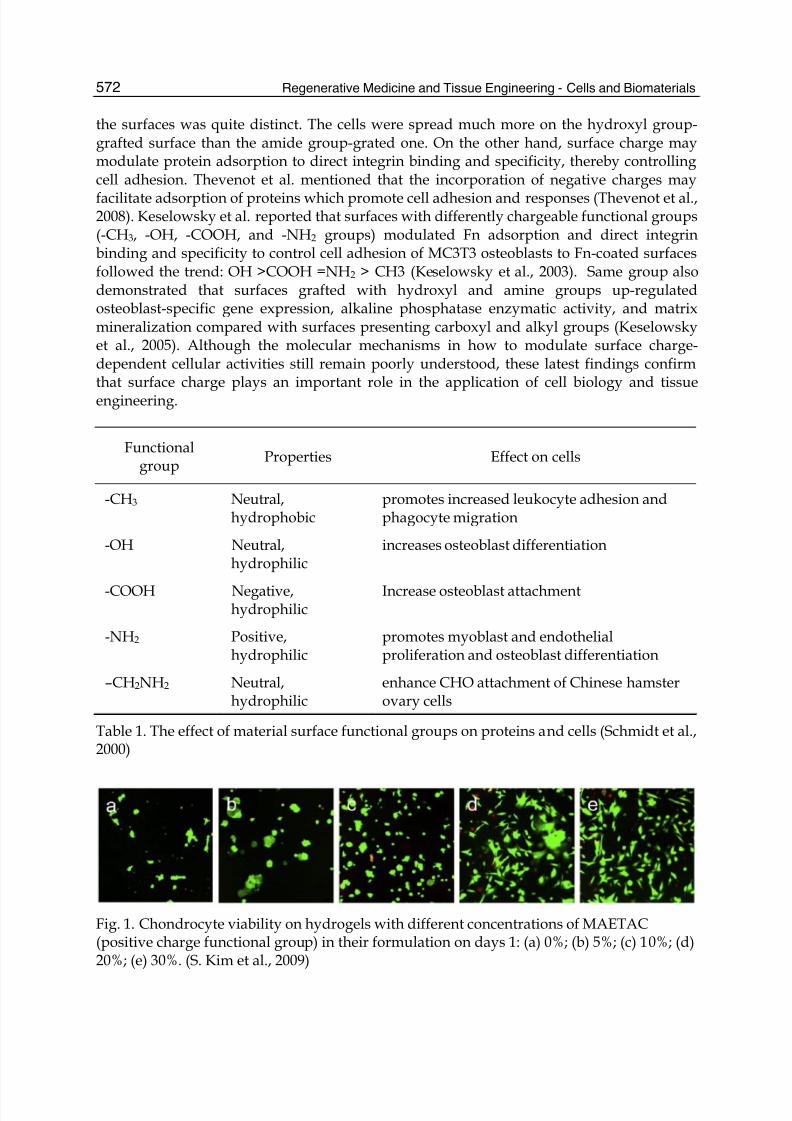
2.3 Surface chargeAfter surface hydrophobicity, surface charge has been recently described a lot in the cellattachment phenomenon. Firstly, the amount of surface charges can influence cell behavior(Ishikawa et al., 2007). As the degree of charge density of poly(styrene-ran-acrylic acid)increased, more cell adhesion and proliferation were observed (Jung et al., 2008). Fig. 1presented similar effect on 2-hydroxyethyl methacrylate (HEMA) and 2-methacryloxyethyltrimethyl ammonium chloride (MAETAC) copolymer hydrogels (Kim & Kihm, 2009).Secondly, many researchers reported the improved-biocompatibility, cell affinity and celldifferentiation on the implanted surfaces by using the positive ions and the negative ions(Bet et al., 2003). For instance, HEMA hydrogels incorporated with positive charges
supported significantly more cell attachment and spreading of osteoblasts and fibroblasts ascompared to negative or neutral charges (Schneider et al., 2004). Yaszemski’s groups alsoinvestigated that negatively charged oligo(poly(ethylene glycol) fumarate) hydrogelsincreased the extent of chondrocyte differentiation, such as collagen and glycosaminoglycanexpression, in comparison with that on the neutral or positively charged hydrogel scaffolds(Dadsetan et al., 2011). Similar pattern was also performed on neuronal growth anddifferentiation (Makohliso et al., 1993). Positively charged coating materials such aspolylysine improve neuronal attachment in vitro. On positive fluorinatedethylenepropylene (FEP) films, neurite outgrowth was significantly higher comparing tonegative and uncharged substrates. Finally, the surface charges can be used to modify cellbehvaior through the chemical functionalities of the polymer materials (Table 1). Lee et al.prepared polyethylene (PE) surfaces with differently chargeable functional groups (-COOH,-CH2OH, -CONH2 and –CH2NH2 groups) by corona discharge treatment, graftcopolymerization and substitution reaction to study the effect on cell behavior (J. H. Lee etal., 1994). Results indicated that Chinese hamster ovary (CHO) cells were more adhesive tothe functional group-grafted surfaces than the control PE surface due to the increasedwettability by grafting hydrophilic functional groups. The best cell adhesion, growth andspreading rate were recorded on polar and positively charged surfaces (amine group-grafted PE) while the negatively charged surface (carboxylic acid group-grafted PE) still hadpoor growth. Moreover, the surfaces grafted with neutral amide and hydroxyl groupsshowed a similar number of cell attachments; however; the morphology of cells attached on
8/3/2019 InTech-Cell Responses to Surface and Architecture of Tissue Engineering Scaffolds
http://slidepdf.com/reader/full/intech-cell-responses-to-surface-and-architecture-of-tissue-engineering-scaffolds 4/20
Regenerative Medicine and Tissue Engineering - Cells and Biomaterials572
the surfaces was quite distinct. The cells were spread much more on the hydroxyl group-grafted surface than the amide group-grated one. On the other hand, surface charge maymodulate protein adsorption to direct integrin binding and specificity, thereby controllingcell adhesion. Thevenot et al. mentioned that the incorporation of negative charges may
facilitate adsorption of proteins which promote cell adhesion and responses (Thevenot et al.,2008). Keselowsky et al. reported that surfaces with differently chargeable functional groups(-CH3, -OH, -COOH, and -NH2 groups) modulated Fn adsorption and direct integrinbinding and specificity to control cell adhesion of MC3T3 osteoblasts to Fn-coated surfacesfollowed the trend: OH >COOH =NH2 > CH3 (Keselowsky et al., 2003). Same group alsodemonstrated that surfaces grafted with hydroxyl and amine groups up-regulatedosteoblast-specific gene expression, alkaline phosphatase enzymatic activity, and matrixmineralization compared with surfaces presenting carboxyl and alkyl groups (Keselowskyet al., 2005). Although the molecular mechanisms in how to modulate surface charge-dependent cellular activities still remain poorly understood, these latest findings confirmthat surface charge plays an important role in the application of cell biology and tissueengineering.
Functionalgroup
Properties Effect on cells
-CH3 Neutral,hydrophobic
promotes increased leukocyte adhesion andphagocyte migration
-OH Neutral,hydrophilic
increases osteoblast differentiation
-COOH Negative,hydrophilic
Increase osteoblast attachment
-NH2 Positive,hydrophilic
promotes myoblast and endothelialproliferation and osteoblast differentiation
–CH2NH2 Neutral,hydrophilic
enhance CHO attachment of Chinese hamsterovary cells
Table 1. The effect of material surface functional groups on proteins and cells (Schmidt et al.,2000)
Fig. 1. Chondrocyte viability on hydrogels with different concentrations of MAETAC(positive charge functional group) in their formulation on days 1: (a) 0%; (b) 5%; (c) 10%; (d)20%; (e) 30%. (S. Kim et al., 2009)
8/3/2019 InTech-Cell Responses to Surface and Architecture of Tissue Engineering Scaffolds
http://slidepdf.com/reader/full/intech-cell-responses-to-surface-and-architecture-of-tissue-engineering-scaffolds 5/20
Cell Responses to Surface and Architecture of Tissue Engineering Scaffolds 573
2.4 Surface roughness
Material surface roughness (or topography) is another important factor influencing celladhesion and behavior. Indeed, roughness modulates the biological response of tissues incontact with the implant. Material surface roughness has a direct influence in vitro as well
as in vivo on cellular morphology, proliferation, and phenotype expression. Literaturepapers have been reported that cells grown on microrough surfaces, were stimulatedtowards differentiation; as shown by their gene expression in comparison with cellsgrowing on smooth surfaces. For instance, primary rat osteoblasts had higherproliferation and elevated alkaline phosphatase (ALP) activity and osteocalcin expressionon the rough surface (0.81 μm) in comparison with smooth one (Hatano et al., 1999). Inthe case of human foetal osteoblastic cells (hFOB 1.19), a similar increase in cell spreadingand proliferation on rough surfaces was reported (Lim, Hansen, Siedlecki, Runt, &Donahue, 2005). Depending on the scale of irregularities of the material surface, surfaceroughness can be divided to macroroughness (100 μm – milimeters), microroughness (100
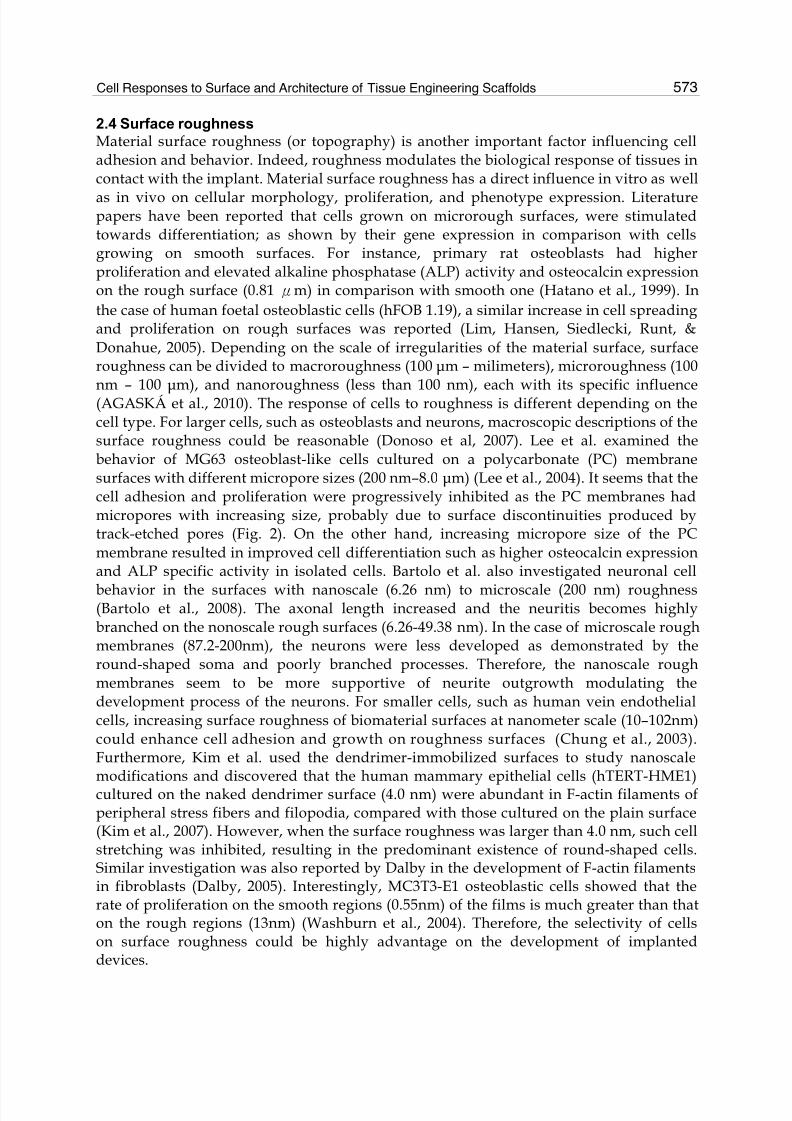
nm – 100 μm), and nanoroughness (less than 100 nm), each with its specific influence(AGASKÁ et al., 2010). The response of cells to roughness is different depending on thecell type. For larger cells, such as osteoblasts and neurons, macroscopic descriptions of thesurface roughness could be reasonable (Donoso et al, 2007). Lee et al. examined thebehavior of MG63 osteoblast-like cells cultured on a polycarbonate (PC) membranesurfaces with different micropore sizes (200 nm–8.0 μm) (Lee et al., 2004). It seems that thecell adhesion and proliferation were progressively inhibited as the PC membranes hadmicropores with increasing size, probably due to surface discontinuities produced bytrack-etched pores (Fig. 2). On the other hand, increasing micropore size of the PCmembrane resulted in improved cell differentiation such as higher osteocalcin expressionand ALP specific activity in isolated cells. Bartolo et al. also investigated neuronal cell
behavior in the surfaces with nanoscale (6.26 nm) to microscale (200 nm) roughness(Bartolo et al., 2008). The axonal length increased and the neuritis becomes highlybranched on the nonoscale rough surfaces (6.26-49.38 nm). In the case of microscale roughmembranes (87.2-200nm), the neurons were less developed as demonstrated by theround-shaped soma and poorly branched processes. Therefore, the nanoscale roughmembranes seem to be more supportive of neurite outgrowth modulating thedevelopment process of the neurons. For smaller cells, such as human vein endothelialcells, increasing surface roughness of biomaterial surfaces at nanometer scale (10–102nm)could enhance cell adhesion and growth on roughness surfaces (Chung et al., 2003).Furthermore, Kim et al. used the dendrimer-immobilized surfaces to study nanoscalemodifications and discovered that the human mammary epithelial cells (hTERT-HME1)cultured on the naked dendrimer surface (4.0 nm) were abundant in F-actin filaments ofperipheral stress fibers and filopodia, compared with those cultured on the plain surface(Kim et al., 2007). However, when the surface roughness was larger than 4.0 nm, such cellstretching was inhibited, resulting in the predominant existence of round-shaped cells.Similar investigation was also reported by Dalby in the development of F-actin filamentsin fibroblasts (Dalby, 2005). Interestingly, MC3T3-E1 osteoblastic cells showed that therate of proliferation on the smooth regions (0.55nm) of the films is much greater than thaton the rough regions (13nm) (Washburn et al., 2004). Therefore, the selectivity of cellson surface roughness could be highly advantage on the development of implanteddevices.
8/3/2019 InTech-Cell Responses to Surface and Architecture of Tissue Engineering Scaffolds
http://slidepdf.com/reader/full/intech-cell-responses-to-surface-and-architecture-of-tissue-engineering-scaffolds 6/20
8/3/2019 InTech-Cell Responses to Surface and Architecture of Tissue Engineering Scaffolds
http://slidepdf.com/reader/full/intech-cell-responses-to-surface-and-architecture-of-tissue-engineering-scaffolds 7/20
Cell Responses to Surface and Architecture of Tissue Engineering Scaffolds 575
3. Cell responses on architecture of tissue engineering scaffolds
On a macroscopic level, the overall shape of the scaffold provides boundaries for tissueregrowth. On a microscope level, the material provides a framework and capillary networks
for local cell growth and tissue organization, permitting cell attachment, distribution andproliferation within a controllable microenvironment (Saltzman, 2002). Apart from tissueengineered skin and vascular grafts that have been progressed into clinical use, the mostother tissue engineered human tissue or organs (e.g. liver and kidney) are still unsuccessful(Mikos et al., 2006). Simply produce a highly porous scaffold and cultivating it with theappropriate types of cells in most cases does not reproduce the desired feature of a normaltissue as tissue structure and function are known to be highly inter-related (Bhaia & Chen,1999). Many tissues have a hierarchical structure that varies over length scales of 0.1-1mm(Griffith, 2002). The subcellular structures (1-10µm) control cell-cell inter-relationships andsupracellular scale structures (100-1000µm) build the essential functional units of the tissue.In order to maintaining the activity of function cell, regulating cell behavior, and
reconstructing 3-dimentional multicellular masses, scaffold must be optimized to satisfy celland tissue growth including proper networks to provide fresh culture medium to all cellsand remove metabolites from the cells and maintain the hierarchical cellular architectures tomimic the functional cells living environment. Altering the micro-architecture, such as thematerial crystallinity or the microporosity, and/or the macro-architecture of the scaffold canbe achieved by changing the pores size, porosity, pore interconnectivity and tortuosity, tomatch the characteristics of the native tissue whilst retaining integrity (Hutmacher, 2001).A common problem encountered when using scaffolds in tissue engineering is the rapidcells attachment and proliferation on the outer edge of scaffold which restrict cellpenetration to the scaffold center, resulting in a necrotic core (Freed et al., 1999). This can beaddressed by altering the culture conditions use to growth tissue, for example using a flow
perfusion culture system (Botchwey et al., 2001), but it is only relevant to tissue engineeringin vitro. Another option or further method of addressing this is to design an optimizedscaffold that will improve nutrient and cell transfer to the scaffold center, both in vitro andin vivo. As discussed in above, characterization of surface wettability, charges and softness;modification of surface chemistry by coating with adhesion molecules together withoptimized the internal structure and architecture will all help to deal with this issue.Scaffold porosity in particular controls the key processes of nutrient supply to cells,metabolite dispersal, local pH stability and cell signaling. The size of the pores can affecthow close the cells are at the initial stages of cultivation (allowing for cell-cellcommunication in three dimensions), but also influences the amount of space the cells havefor 3-D organization in the later stages of tissue growth. In addition, a porous surface isknown to improve mechanical interlocking between the implanted scaffolds and thesurrounding natural tissue, providing greater mechanical stability at this critical interface(Karageorgiou & Kaplan, 2005). Cell seeding in the center of the scaffold and feeding theinner surfaces of the scaffolds are limited when the pores are too small whereas larger poresaffect the stability of the scaffold and its ability to provide physical support for the seededcells (Levenberg & Langer, 2004). To date cell seeding on 2-D scaffold surfaces has beenshown to be easy to perform but the preparation of 3-D cell-scaffold constructs forregeneration of organs is far more complex. For example, pores of adequate size allow cellsto migrate or adhere to the surface of a material, but interconnecting pores are necessary topermit cell growth into the scaffold interior.

8/3/2019 InTech-Cell Responses to Surface and Architecture of Tissue Engineering Scaffolds
http://slidepdf.com/reader/full/intech-cell-responses-to-surface-and-architecture-of-tissue-engineering-scaffolds 8/20
Regenerative Medicine and Tissue Engineering - Cells and Biomaterials576
3.1 Pore size of tissue engineering scaffold
Cell migration is modulated by a complex, spatiotemporally integrated set of biophysicalmechanisms that are influenced not only by the biochemistry of extracellular andintracellular signaling, but also by the biophysics of the surrounding extracellular
environment. Specific cells require different pore sizes for optimal attachment, growth andmotility (Table 2) (Ranucci et al, 2000). A recent study (Yang et al., 2010)on variable pore sizecollagen gel found that cell migration is hindered by small pore size that invasive distancewas not very sensitive in the pore size rang of 5-12µm. At small pore size, a variety offactors, including high ligand density in collagen gel that does not encourage the cellpolarity and release seen in mesenchymal migration likely contributes to the limitedinvasion (Ulrich et al., 2010) whilst very large pore size in scaffolds have insufficient tetherson which to generate traction would also limit cell migration. As a result, many researchesin tissue engineering are aimed at obtaining polymeric or bioceramic scaffolds with a veryhigh porosity and simultaneous good control over pore size and morphology (Hou et al.,2000). The presence of pores smaller than 160µm in PLA and PLGA scaffolds, produced by
salt leaching, has been reported to be optimal for attachment of human skin fibroblasts(Yang et al., 2002). Bony ingrowth was found to predominate in porous PMMA implanted inbone when the pore size was around 450µm (Ashman & Moss, 1977). Connective tissueformed when the pore size was below 100µm and extensive vascular infiltration was onlyobserved with pores around 10000µm. In the case of polyurethane meniscal implants,structures comprising macropores (150-300 µm), highly interconnected by micropores (<50µm) have been found to be conducive to ingrowth of fibrocartilaginous tissue (deGroot etal., 1996). The cell infiltration depth (120µm in 28 days) found in elastin scaffolds, forexample, probably results from the material’s high porosity and inter-connectivity (Lu,Ganesan et al., 2004). Osteoblasts was found to migrate faster inside the larger pore (100µm)of microcellular polyHIPE scaffolds; however, pore size did not affect cell penetration depthor mineralization extent (Akay et al., 2004). It has also been noticed in previous studies thatcell-scaffold binding can block pores of inadequate size and geometry (Freed & Vunjak-Novakovic, 1998; Yannas, 2000). High inter-connectivity of pores is also essential to supplynutrients and allows oxygen exchange in the inner regions of a scaffold to maintain cellviability, especially for complex tissue engineering of organs.
Cell/tissue type Optimal poresize (μm)
Scaffold material Reference
Human skin fibroblasts <160µm PLA/PLG (Yang.J et al., 2002)Bone 450µm PMMA (Ashman & Moss, 1977)
Fibrocartilaginous tissue 150-300µm Polyurethane (deGroot et al., 1996)Adult mammalian skincells
20-125µm Collagen-glycocaminoglycan
(Yannas, Lee, Orgill,SKrabut, & Murphy, 1989)
Osteogenic cells 100-150µm Collagen-GAG (O'Brien, Harley, Yannas,& Gibson, 2005a)
Smooth muscle cells 60-150µm PLA (Zeltinger, Sherwood,Graham, Mueller, &Griffith, 2001)
Endothelial cells <80µm Silicon nitride (Salem et al., 2002)
Table 2. Optimal pore size for cell infiltration and host tissue ingrowth

8/3/2019 InTech-Cell Responses to Surface and Architecture of Tissue Engineering Scaffolds
http://slidepdf.com/reader/full/intech-cell-responses-to-surface-and-architecture-of-tissue-engineering-scaffolds 9/20
Cell Responses to Surface and Architecture of Tissue Engineering Scaffolds 577
3.2 Porosity of tissue engineering scaffold
The porosity, that is, the percentage of void volume in the materials, is also use as a meansof quantifying the structure of a tissue engineering scaffolds. Researches have been focusingon the design of the scaffold to ensure appropriate porosity and porous structure for cells
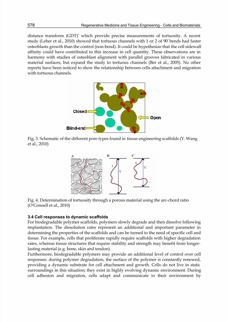
penetration and ingrowth. However, attempts to link scaffold porosity to cell performancehave not been particularly successful. Toth et al (Toth et al., 1995) report that improvementsin bone ingrowth occur with increasing porosity of macroporous biphasic calciumphosphate ceramic samples. However, they also report no discernible differences in boneunion after six months implantation between scaffolds that have 30%, 50% and 70%porosity. This observation could be attributed to the complicated internal structure ofscaffolds that consist of pores of different types (open, closed and blind-end pores), sizesand geometry, (Fig. 3). The presence of both random and anisotropic open porousarchitectures of PLA scaffolds were prepared using supercritical CO2 aims to find theoptimal channel diameter and geometry for osteosarcoma cell penetration. The results showthat cells penetrate into scaffolds containing aligned channels (400µm) more extensivelythan those that did not.
3.3 Connectivity and tortuosity of tissue engineering scaffold
The pore structures typically consist of irregularly shaped voids and connecting channels(connects) that can be difficult to defined due to merging of adjacent cavities, resulting inthe presence of fenestrations (windows) in the void walls. Beyond the fundamentalrequirements of adequate pore size and inter-connectivity, pore tortuosity also plays a keyrole in cells interaction with scaffolds. Tortuosity is defined as the ratio of the actual pathlength through connected pores to the Euclidean distance (shortest linear distance) (Fig.4).Tortuosity is another key factor in optimizing and designing tissue engineering scaffold
which is known to influence molecules and oxygen diffusion and cell migration rate. Silva etal reported (Silva et al., 2006) that aligned channel in both hyroxyapatite (HA) and poly(D,L-lactic acid) (PDLLA) scaffolds enhanced cell penetration and infiltration into the centralregion of the scaffold in comparison with tortuous channels. Analysis of humanosteosarcoma cell penetration into the aligned channels revealed that cell coverage increasedwith increasing channel diameter from around 22% in the 170µm diameter channel toapproximately 38% into the 420µm channel(Rose et al., 2004). In addition, cell penetrationinto 420µm channel was significantly greater than that observed within the 170µm channel.However, determination of tortuosity and cell responses on tortuous scaffold is still rarelyquoted in the literature. A common method to calculate tortuosity is via the results fromdissolution measurements. In this method, the tortuosity is calculated from severalparameters related to the dissolution of a molecules from a matrix (Desai et al., 1966; Foster& Parrott, 1990). This approach can result in unrealistic values of more than one thousand(Papadokostaki et al., 1998) or below one (Foster & Parrott, 1990). Tortuosity can also bemeasured from the porosity and diffusion coefficients obtained from spin echo NMRmeasurements (Wu et al., 2006). Mercury intrusion porosimetry has also been suggested fordetermining tortuosity. Another example of tortuosity calculating is to use the inflectioncount metric (ICM). This approach adds the number inflections of a 3-D framerepresentation of a pore connecting two points and multiplies this number by the pathlength (Bullitt et al., 2003). Wu et al (Wu et al., 2006) described a method to find the shortestroute through the pores in images of compacts using an algorithm called ‘grey-weighted
8/3/2019 InTech-Cell Responses to Surface and Architecture of Tissue Engineering Scaffolds
http://slidepdf.com/reader/full/intech-cell-responses-to-surface-and-architecture-of-tissue-engineering-scaffolds 10/20
Regenerative Medicine and Tissue Engineering - Cells and Biomaterials578
distance transform (GDT)’ which provide precise measurements of tortuosity. A recentstudy (Leber et al., 2010) showed that tortuous channels with 1 or 2 of 90°bends had fasterosteoblasts growth than the control (non-bend). It could be hypothesize that the cell sidewallaffinity could have contributed to this increase in cell quantity. These observations are in
harmony with studies of osteoblast alignment with parallel grooves fabricated in variousmaterial surfaces, but expand the study to tortuous channels (Ber et al., 2005). No otherreports have been noticed to show the relationship between cells attachment and migrationwith tortuous channels.
Fig. 3. Schematic of the different pore types found in tissue engineering scaffolds (Y. Wanget al., 2010)
Fig. 4. Determination of tortuosity through a porous material using the arc-chord ratio(O'Connell et al., 2010)
3.4 Cell responses to dynamic scaffolds
For biodegradable polymer scaffolds, polymers slowly degrade and then dissolve followingimplantation. The dissolution rates represent an additional and important parameter indetermining the properties of the scaffolds and can be turned to the need of specific cell andtissue. For example, cells that proliferate rapidly require scaffolds with higher degradationrates, whereas tissue structures that require stability and strength may benefit from longer-lasting material (e.g. bone, skin and tendon).Furthermore, biodegradable polymers may provide an additional level of control over cellresponses: during polymer degradation, the surface of the polymer is constantly renewed,providing a dynamic substrate for cell attachment and growth. Cells do not live in staticsurroundings in this situation; they exist in highly evolving dynamic environment. Duringcell adhesion and migration, cells adapt and communicate to their environment by
8/3/2019 InTech-Cell Responses to Surface and Architecture of Tissue Engineering Scaffolds
http://slidepdf.com/reader/full/intech-cell-responses-to-surface-and-architecture-of-tissue-engineering-scaffolds 11/20
Cell Responses to Surface and Architecture of Tissue Engineering Scaffolds 579
numerous methods ranging from differentiation, gene expression, growth, and apoptosis.Dynamic substrates that can alter the presentation of ligands to an attached cell willgenerate immediate opportunities for studies of scaffolds-cell adhesion, signaling, migrationand differentiation. Dynamic substrates may also be important for generating substrates that
can control the spatial and temporal interactions between two or more different populationsof attached cells in tissue engineering (Yousaf, 2009). However, much less effort has beeninvested in studying cell responses on dynamic substrates. Currently, there is no availablemethod to generate dynamic gradient substrates for studying cell polarity and directed cellmigration. One of recent studies found that programmed erasure of substrate topographycause a decrease in cell alignment as evidenced by an increase in angular dispersion withcorresponding remodeling of the actin cytoskeleton. Cell viability remained greater than95% before and after topography change (Davis et al., 2011).Beyond the biodegradability, mechanical input on scaffolds also significant influence thereorientation of cell shape (J. Wang et al., 2003), actin cytoskeleton remodeling (J. Wang et
al., 2000) and the synthesis of extraceullar matrix (Carver et al., 1991). Many studies showedthat cells are capable of surveying the external mechanical properties of their surroundingenvironment, respond to changes in the balance of intra- and extra- cellular forces, andregulate many important physiological proves (Pelham & Wang, 1997). As most tissueengineering scaffolds are made of biomaterials which have certain elasticity, rigidity andstretching-tension, cell responses to such scaffolds relate mechanical stimulation isbecoming more and more interesting. In case of fibroblasts differentiation, mechanicalstretching of silicone dishes can induce differentiation of fibroblasts into myofibrolasts,which is known to form scar tissue in vivo. Tock et al (Tock et al., 2003) reported thatmechanical loading impressed α-SMA expression, a marker of myofibroblasts hatmechanical tension in granulation tissues controls myofibroblast differentiation (Hinz et al.,
2001).
4. Challenges in studying cell behaviour on biomaterials and complex tissueengineering scaffolds
Cell adhesion to a material surface is an important phenomenon that controls the behaviorof cells, such as their morphology, migration, growth and differentiation. Counting the cellsadhered to material’s surface is the most common way to evaluate a material’s affinity to thecells but this method could not quantify the cell adhesion force on the material’s surface.Many studies used biological procedures to measure the characteristics of adhesion betweencells and biomaterials. For example, cell spreading and migration are often used as indirectindicators of adhesion strength and this lack of quantitative understanding of adhesionstrength limits the interpretation of functional studies of structural and signaling adhesivecomponents. Studying cell adhesion molecules such as focal adhesion kinase involved thebinding with biomaterial’s surface can provide more direct information about cell adhesionstrength. However, protein expression is working on a population of cells and not a singlecell due to the sensitivity of western blot or protein binding assay. Recently, a number oftechniques have been developed to study the cell adhesion behavior on materials frommechanical point of view. For instance, cell adhesion strength has been studied ascentrifugation force by centrifuge, tensile force by micropipette manipulation, shear force byparallel flow chamber and chemical binding force by atomic force microscope (Thoumine &

8/3/2019 InTech-Cell Responses to Surface and Architecture of Tissue Engineering Scaffolds
http://slidepdf.com/reader/full/intech-cell-responses-to-surface-and-architecture-of-tissue-engineering-scaffolds 12/20
Regenerative Medicine and Tissue Engineering - Cells and Biomaterials580
Ott, 1997; Truskey & Proulx, 1993; Leonenko et al., 2007). McClay and Lotz groupsdetermined the cell detachment force under different speed of centrifugation and Bouafsounet al. used flow chamber and jet impingement techniques to study cell detachment forces.However, centrifugation force by centrifuge and shear force by parallel flow chamber
quantify the cell-material adhesion strength for a population of cells but not for anindividual cell. Thus, tensile force by micropipette manipulation and chemical binding forceby atomic force microscope (AFM) are the more suitable techniques to study the celladhesion strength of a single cell on biomaterial surface. In 2009, Hung et al. utilized thedielectrophoresis force acting on the human bladder epithelial (ECV) cells to inducespatial movement for studying the cell adhesion strength. Dielectrophoresis is thephenomenon in which a particle, such as a living cell, is polarized and moved by theelectrical gravity in a non-uniform electric field (Jones, 2005). In our study,dielectrophoresis force was also used to determine cell adhesion strength of humanbladder epithelial cells on Fn and collagen type 1 coated surfaces and the cell adhesion
force was similar to the one measured by AFM but much smaller than the one measuredby flow chamber techniques. We suggested that the cell adhesion strength between asingle cell and biomaterial surface would be different from a population of cells.Therefore, new trends and possible long-term directions for determining both adhesionprocess and force are highlighted.For characterizing internal structure of tissue engineering scaffold and their correlation withcell behviour, a variety of techniques have been used to evaluate scaffold porosity includingtheoretical assessment, scanning electron microscopy (Flynn et al., 2006), mercuryporosimetry, gas pycnometry and adsorption. SEM analysis complements the theoreticalcalculations of porosity (Kellomake et al., 2000; Walsh et al., 2001; Zein et al., 2002) andallows direct measurements of pore size and wall thickness and cell morphology on the
surface. However, SEM cannot examine the scaffold interior without sample sectioningwhich introduces uncertainty due to unwanted material compression and edge effects andcells damage. Mercury porosimetry is a well known and established method, but it neithermeasures small mesopores (2-50nm pores) due to lack of mercury penetration nor measuresvery large pores as the mercury penetrates the structure before measurements can be made.The gas adsorption method is relevant to the study of porosity in nano-featured and nano-modified scaffolds (Ma, 2004), and is based on the electrical forces of attraction that bindatoms in solids. To counter the net inward attractive forces, surface atoms bindsurrounding gas molecules via Van der Waals and electrical forces. Researchers have usedgas adsorption to assess scaffolds with pore sizes ranging from 0.35-400 nm or 3.5 to
2000µm but the analysis does not evaluate closed pore content and cell proliferation ormigration.For analyzing the 3-D construction of tissue scaffolds and cell-material interactions, newimagining techniques such as micro-computed tomographic (micro-CT) have beendeveloped. Feldkamp et al (Feldkamp et al., 1989) pioneered micro-CT imaging technologyto analyze trabecular bone samples at a spatial resolution of 50µm. Since then, micro-CT hasbeen used extensively in the study of bone architecture and other tissue types. Micro-CTimages the specimen through exposure to small quantities of ionizing radiation andcorresponding measurements of absorption. The resulting grey-scale images form a series of2-D sequential slices which build up into a density map of the sample. With relevantcomputerized reconstruction, micro-CT provides precise quantitative and qualitative
8/3/2019 InTech-Cell Responses to Surface and Architecture of Tissue Engineering Scaffolds
http://slidepdf.com/reader/full/intech-cell-responses-to-surface-and-architecture-of-tissue-engineering-scaffolds 13/20
Cell Responses to Surface and Architecture of Tissue Engineering Scaffolds 581
information on the 3D morphology of specimens (Darling & Sun, 2004; Thurner et al., 2005;Thurner et al., 2004; Washburn et al., 2004; Williams et al., 2005) and the interior can bestudied in great detail without resorting to physical sectioning or the use of toxic chemicals.Williams et al (Williams et al., 2005) recently used Micro-CT to visualize PCL scaffolds
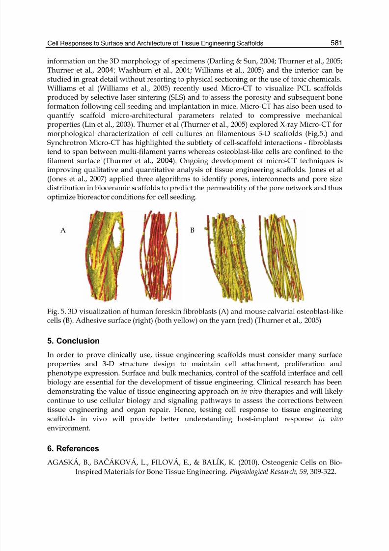
produced by selective laser sintering (SLS) and to assess the porosity and subsequent boneformation following cell seeding and implantation in mice. Micro-CT has also been used toquantify scaffold micro-architectural parameters related to compressive mechanicalproperties (Lin et al., 2003). Thurner et al (Thurner et al., 2005) explored X-ray Micro-CT formorphological characterization of cell cultures on filamentous 3-D scaffolds (Fig.5.) andSynchrotron Micro-CT has highlighted the subtlety of cell-scaffold interactions - fibroblaststend to span between multi-filament yarns whereas osteoblast-like cells are confined to thefilament surface (Thurner et al., 2004). Ongoing development of micro-CT techniques isimproving qualitative and quantitative analysis of tissue engineering scaffolds. Jones et al(Jones et al., 2007) applied three algorithms to identify pores, interconnects and pore sizedistribution in bioceramic scaffolds to predict the permeability of the pore network and thusoptimize bioreactor conditions for cell seeding.
Fig. 5. 3D visualization of human foreskin fibroblasts (A) and mouse calvarial osteoblast-likecells (B). Adhesive surface (right) (both yellow) on the yarn (red) (Thurner et al., 2005)
5. Conclusion
In order to prove clinically use, tissue engineering scaffolds must consider many surfaceproperties and 3-D structure design to maintain cell attachment, proliferation andphenotype expression. Surface and bulk mechanics, control of the scaffold interface and cell
biology are essential for the development of tissue engineering. Clinical research has beendemonstrating the value of tissue engineering approach on in vivo therapies and will likelycontinue to use cellular biology and signaling pathways to assess the corrections betweentissue engineering and organ repair. Hence, testing cell response to tissue engineeringscaffolds in vivo will provide better understanding host-implant response in vivo environment.
6. References
AGASKÁ, B., BAČÁKOVÁ, L., FILOVÁ, E., & BALÍK, K. (2010). Osteogenic Cells on Bio-Inspired Materials for Bone Tissue Engineering. Physiological Research, 59, 309-322.
A B
8/3/2019 InTech-Cell Responses to Surface and Architecture of Tissue Engineering Scaffolds
http://slidepdf.com/reader/full/intech-cell-responses-to-surface-and-architecture-of-tissue-engineering-scaffolds 14/20
Regenerative Medicine and Tissue Engineering - Cells and Biomaterials582
Agrawal, C. M., & Ray, R. B. (2001). Biodegradable polymeric scaffolds for musculoskeletaltissue engineering. Journal of Biomedical Materials Research, 55, 141-150.
Akay, G., Birch, M. A., & Bokhari, M. A. (2004). Microcellular polyHIPE polymer supportsosteoblast growth and bone formation in vitro. Bone, 24, 35-41.
Allen, L. T., Tosetto, M., Miller, I. S., O'Connor, D. P., Penney, S. C., Lynch, I., et al. (2006).Surface-induced changes in protein adsorption and implications for cellularphenotypic responses to surface interaction. Biomaterials, 27 , 3096-3108.
Ashman, A., & Moss, M. L. (1977). Implantation of porous polymethlmethacrylate resin fortooth and bone replacement. The Journal of Prosthetic Dentistry, 37 , 657-665.
Bartolo, L. D., Rende, M., Morelli, S., Salerno, S., A, P., Gordano, A., et al. (2008). Influence ofmembrane surface properties on the growth of neuronal cells isolated formhippocampus. Journal of Membrane Science, 325, 139-149.
Ber, S., Torun Koseb, G., & Hasircia, V. (2005). Bone tissue engineering on patternedcollagen films: an in vitro study. Biomaterials, 26, 1977-1986.
Bet, M. R., Goissis, G., Vargas, S., & Selistre-de-Araujo, H. S. (2003). Cell adhesion andcytotoxicity studies over polyanionic collagen surfaces with variable negativecharge and wettability. Biomaterials, 24, 131-137.
Bhaia, S. N., & Chen, C. S. (1999). Tissue engineering at the microscale. Biomedical
Microdevices, 2, 131-144.Botchwey, E. A., Dupree, M. A., Pollack, S. R., Levine, E. M., & Laurencin, C. T. (2001).
Tissue engineered bone: measurement of nutrient transport in three-dimensionalmatrices. Journal of Biomedical Materials Research, 67 , 357-367.
Brynda, E., Hlady, V., & Andrade, J. D. (1990). Protein packing in adsorbed layersstudied by excitation energy transfer. Journal of Colloid and Interface Science, 139,374-380.
Bullitt, E., Gerig, G., Aylward, S., Joshi, S., Smith, K., Ewendl, M., et al. (2003). Tortuosity. In Attributes and Malignant Brain Tumors In MICCAI 2003 proceedings LNCS 2878 (pp.671-679).
Carver, W., Nagpal, M. L., Nachigal, M., Borg, T. K., & Terrracio, L. (1991). Collagenexpression in mechanically stimulated cardiac fibroblasts. Circulation Research, 69,116-122.
Chung, T.-W., Liu, D.-Z., Wang, S.-Y., & Wang, S.-S. (2003). Enhancement of the growth ofhuman endothelial cells by surface roughness at nanometer scale. Biomaterials, 24,4655-4661.
Dadsetan, M., Pumberger, M., Casper, M. E., Shogren, K., Giuliani, M., Ruesink, T., et al.
(2011). The effects of fixed electrical charge on chondrocyte behavior. Acta
Biomaterialia.Dalby, M. J. (2005). Topographically induced direct cell mechanotransduction. Medical
Engineering and Physics, 27 , 730-742.Darling, A. L., & Sun, W. (2004). 3D microtomographic characterization of precision
extruded poly-epsilon-caprolactone scaffolds. Journal of Biomedical Materials
Research. Part B, Applied Biomaterials. 70B, 2, 311-317.Davis, K. A., Burke, K. A., Mather, P. T., & Henderson, J. H. (2011). Dynamic cell behavior
on shape memory polymer substrates. Biomaterials, 32, 2285-2293.
8/3/2019 InTech-Cell Responses to Surface and Architecture of Tissue Engineering Scaffolds
http://slidepdf.com/reader/full/intech-cell-responses-to-surface-and-architecture-of-tissue-engineering-scaffolds 15/20
Cell Responses to Surface and Architecture of Tissue Engineering Scaffolds 583
deGroot, J. H., de Vrijer, R., Pennings, A. J., Klompmaker, J., Veth, R. P. H., & Jansen, H. W.B. (1996). Use of porous polyurethanes for meniscal reconstruction and meniscalprostheses. Biomaterials, 17 , 163-173.
Desai, S. J., Singh, P., Simonelli, A. P., & Higuchi, W. I. (1966). Investigation of factors
influencing release of solid drug dispersed in inert matrices II. Journal of Pharmaceutical Sciences, 55, 1224-1229.
Donoso, M. G., Me´ndez-Vilas, A., Bruque, J. M., & Gonza´lez-Martin, M. L. (2007). On therelationship between common amplitude surface roughness parameters andsurface area: Implications for the study of cell–material interactions. International
Biodeterioration and Biodegradation, 59, 245-251.Engler, A. J., Sen, S., Sweeney, H. L., & Discher, D. E. (2006). Matrix elasticity directs stem
cell lineage specification. Cell 126, 677-689.Feldkamp, L. A., Goldstein, S. A., Parfitt, A. M., Jesion, G., & Kleerekope, M. (1989). The
direct examination of three-dimensional bone architecture in vitro by computed
tomography. Journal of Bone & Mineral Research, 4, 3-11.Flynn, L., Semple, J. L., & Woodhouse, K. A. (2006). Decellularized placental matrices foradipose tissue engineering. Journal of Biomedical Materials Research A, 79, 359-369.
Foster, T. P., & Parrott, E. L. (1990). Release of highly water-soluble medicinal compoundsfrom inert, heterogeneous matrixes. Journal of Pharmaceutical Sciences, 79, 806-810.
Freed, L. E., Martin, I., & Vunjak-Novakovic, G. (1999). Frontiers in tissue engineering. Invitro modulation of chondrogenesis. Clinical Orthopaedics & Related Research,
367 (58).Freed, L. E., & Vunjak-Novakovic, G. (1998). Culture of organized cell communities.
Advanced Drug Delivery Reviews, 33, 15-30.Geiger, B., Volk, T., Volberg, T., & Bendori, R. (1987). Molecular ineractions in adherens-type
contacts. Journal of Cell Science.Supplement, 8, 251-272.Goddard, J. M., & Hotchkiss, J. H. (2007). Polymer surface modification for the attachment of
bioactive compounds. Progress in Polymer Science, 32, 698-725.Griffith, L. G. (2002). Emerging design principles in biomaterials and scaffold for tissue
engineering. Annals of the New York Academy of Sciences, 961, 83-95.Hatano, K., Inoue, H., Kojo, T., Tsujisawa, T., Uchiyama, C., & Uchida, Y. (1999). Effect of
Surface Roughness on Proliferation and Alkaline Phosphatase Expression of RatCalvarial Cells Cultured on Polystyrene. Bone, 25, 439-445.
Hattori, S., Andrade, J. D., Hibbs, J. B., Gregonis, D. E., & King, R. N. (1985). Fibroblasts cellproliferation of charged hydroxyethyl methacrylate copolymers. Journal of Colloid
and Interface Science, 104, 73-78.Hinz, B., Mastrangelo, D., Iselin, C. E., Chaponnier, C., & Gabbiani, G. (2001). Mechanical
tension controls granulation tissue contractile activity and myofibroblastdifferentiation American Journal of Pathology, 159, 1009-1020.
Hou, Q., Grijpma, D. W., & Feijen, J. (2000). Porous polymeric structures for tissueengineering prepared by a coagulation, compression moulding and salt leachingtechnique. Biomaterials, 24(11), 1937-1947.
Hutmacher, D. W. (2001). Scaffold design and fabrication technologies for engineeringtissues--state of the art and future perspectives. Journal of biomaterials science, 12,107-124.
8/3/2019 InTech-Cell Responses to Surface and Architecture of Tissue Engineering Scaffolds
http://slidepdf.com/reader/full/intech-cell-responses-to-surface-and-architecture-of-tissue-engineering-scaffolds 16/20
Regenerative Medicine and Tissue Engineering - Cells and Biomaterials584
Ishikawa, J., Tsuji, H., Sato, H., & Gotoh, Y. (2007). Ion implantation of negative ions for cellgrowth manipulation and nervous system repair. Surface and coatings technology,201, 8083-8090.
Jiang, F. X., Yurke, B., Firesein, B. L., & Langrana, N. A. (2008). Neurite ougrowth on a DNA
crosslinked hyrogel with tunable stiffnesses. Annals of Biomeical Engineering, 36,1565-1579.
Jiang, F. X., Yurke, B., Schloss, R. S., Firesein, B. L., & Langrana, N. A. (2010). Therelationship between fibroblast growth and the dynamic stiffness of a DNAcorsslinked hydrogel. Biomaterials, 31, 1199-1212.
Jones, J. R., Poologasundarampillai, G., Atwood, R. C., Bernard, D., & Lee, P. D. (2007). Non-destructive quantitative 3D analysis for the optimisation of tissue scaffolds.Biomaterials, 28, 1404-1413.
Jung, H., Kwak, B., Yang, H. S., Tae, G., Kim, J.-S., & Shin, K. (2008). Attachment of cells topoly(styrene-co-acrylic acid) thin films with various charge densities. Colloids and
Surfaces A: Physiochemical and Engineering Aspect, 313, 562-566.Karageorgiou, V., & Kaplan, D. (2005). Porosity of 3D biomaterial scaffolds and oseogenesis.Biomaterials, 26, 5474-5491.
Kellomake, M., Niiranen, H., Puumanen, K., Ashammakhi, N., Waris, T., & tormala, P.(2000). Bioabsorbable scaffolds for guided bone regeneration and generation.Biomaterials, 21, 2495-2505.
Keselowsky, B. G., Collard, D. M., & García, A. J. (2005). Integrin binding specificityregulates biomaterial surface chemistry effects on cell differentiation. Proc Natl Acad
Sci U S A, 102, 5953-5957.Keselowsky, B. G., Collard, D. M., & J., G. A. (2003). Surface chemistry modulates
fibronectin conformation and directs integrin binding and specificity to control cell
adhesion. Jourmal of Biomedical Material Research A, 66, 247-259.Khatiwala, C. B., Peyton, S. R., Metzke, M., & Putnam, D. (2007). The regulation of
osteogenesis by ECM rigidity in MC3T3-E1 cells requires MAPK activation. Journal
of Cell Phsiology, 211, 661-672.Khorasani, M. T., Mirzadeh, H., & Irani, S. (2008). Plasma surface modification of poly(l-
lactic acid) and poly (lactic-co-glycolic acid) films for improvement of nerve cellsadhesion. Radiation Physics and Chemistry, 77 , 280-287.
Kim, M. H., Kino-Oka, M., Kawase, M., Yagi, K., & Taya, M. (2007). Response of humanepithelial cells to culture surfaces with varied roughnesses prepared byimmobilizing dendrimers with/without D-glucose display. Journal of Bioscience and
Bioengineering, 103, 192-199.Kim, S., E., E. A., & Kihm, K. D. (2009). Surface elasticity and charge concentration-
dependent endothelial cell attachment to copolymer polyelectrolyte hydrogel. Acta
Biomaterialia, 5, 144-151.Lahann, J., & Langer, R. (2005). Smart materials with dynamically controllable surfaces. MRS
Bull, 30, 185-188.Langer, R., & Vacanti, J. P. (1993). Tissue engineering. Science, 260, 920-927.Leber, C., Choi, H., Bose, S., & Bandyopadhyay, A. (2010). Micromachined Si channel width
and tortuosity on human osteoblast cell attachment and proliferation. Material
science and engineering, 30, 71-77.
8/3/2019 InTech-Cell Responses to Surface and Architecture of Tissue Engineering Scaffolds
http://slidepdf.com/reader/full/intech-cell-responses-to-surface-and-architecture-of-tissue-engineering-scaffolds 17/20
Cell Responses to Surface and Architecture of Tissue Engineering Scaffolds 585
Lee, J. H., Jung, H. W., Kang, I. K., & Lee, H. B. (1994). Cell behaviour on polymer surfaceswith different functional groups. Biomaterials, 15, 705-711.
Lee, S. J., Choi, J. S., Park, K. S., Khang, G., Lee, Y. M., & Lee, H. B. (2004). Response of MG63osteoblast-like cells onto polycarbonate membrane surfaces with different
micropore sizes. Biomaterials, 25, 4699-4707.Lee, S. J., Khang, G., Lee, Y. M., & Lee, H. B. (2003). The effect of surface wettability on
induction and growth of neurites from the PC-12 cell on a polymer surface. Journal
of Collid and Interface Science, 259, 228-235.Levenberg.S., & Langer, R. (2004). Advances in tissue engineering. Current Topics in
Developmental Biology, 61, 113-134.Lim, J. Y., Hansen, J. C., Siedlecki, C. A., Runt, J., & Donahue, H. J. (2005). Human foetal
osteoblastic cell response to polymer-demixed nanotopographic interfaces. Journal
of Royal Society Interface, 2, 97-108.Lin, A. S., Barrows, T. H., Cartmell, S. H., & Guldberg, R. E. (2003). Microarchitectural and
mechanical characterization of oriented porous polymer scaffolds. Biomaterials,24(3), 481-489.Lu, Q., Ganesan, K., Simionescu, D. T., & Vyavahare, N. R. (2004). Novel porous aortic
elastin and collagen scaffolds for tissue engineering. Biomaterials, 25(22), 5227-5237.
Ma, P. X. (2004). Scaffolds for tissue fabrication. Material today, 7 (5), 30-40.Makohliso, S. A., Valentini, R. F., & Aebischer, P. (1993). Magnitude and polarity of a
fluoroethylene propylene electret substrate charge influences neurite outgrowth invitro. Journal of Biomedical Materials Research, 27 , 1075-1085.
Marquez, J. P., Genin, G. M., Pryse, K. M., & Elson, E. L. (2006). Celular and matrixcontributions to tissue construct stiffness increase with cellular concentration.
Annals of Biomeical Engineering, 34, 1475-1482.Mikos, A. G., Herring, S. W., Ochareon, P., Elissenff, J., Lu, H. H., & Mooney, D. J. (2006).
Engineering complex tissues. Tissue Engineering, 12, 3307-3340.Mrksich, M. (2005). Dynamic substrates for cell biology. MRS Bull, 30, 180-184.Noh, H., & Vogler, E. A. (2006). Volumetric interpretation of protein adsorption: mass and
energy balance for albumin adsorption to particulate adsorbents withincrementally increasing hydrophilicity. Biomaterials, 27 , 5801-5812.
O'Brien, F. J., Harley, B. A., Yannas, I. V., & Gibson, L. J. (2005a). The effect of pore size oncell-adhesion in collagen-GAG scaffolds. Biomaterials, 26, 433-441.
O'Brien, F. J., Harley, B. A., Yannas, I. V., & Gibson, L. J. (2005b). The effect of pore size on
cell adhesion in collagen-GAG scaffolds.Biomaterials, 26
, 433-441.O'Connell, B., McGloughlin, T., & Walsh, M. T. (2010). Factors that affect mass transportfrom drug eluting stents into the artery wall. BioMedical Engineering, 9, 15.
Papadokostaki, K. G., Amarantos, S. G., & Petropoulos, J. H. (1998). Kinetics of release ofparticulate solutes incorporated in cellulosic polymer matrices as a function ofsolute solubility and polymer swellability. Journal of applied polymer science, 67 , 227-287.
Pelham, R. J., & Wang, Y. L. (1997). Cell locomotion and focal adhesions are regulated bysubstrate flexibility. Proc Natl Acad Sci U S A, 94, 136661 -136665.

8/3/2019 InTech-Cell Responses to Surface and Architecture of Tissue Engineering Scaffolds
http://slidepdf.com/reader/full/intech-cell-responses-to-surface-and-architecture-of-tissue-engineering-scaffolds 18/20
Regenerative Medicine and Tissue Engineering - Cells and Biomaterials586
Peyton, S. R., & Putnam, D. (2005). Extracellular matrix rigidity governs smoth muscle cellmotility in a biphasic fashion. Journal of Cell Phsiology, 204, 918-923.
Ranucci, C. S., Kumar, A., Batra, S. P., & Moghe, P. V. (2000). Control of hepatocyte functionon collagen foames: sizing matrix pores toward selective induction of 2-D and 3-D
cellular morphogenesis. Biomaterials, 21, 783-793.Rose, F. R., Cyster, L. A., Grant, P., Scotchford, C. A., Howdle, S. M., & Shakesheff, K. M.
(2004). In vitro assessment of cell penetration into porous hydroxyapatite scaffoldswith a central aligned channel. Biomaterials, 25, 5507-5514.
Salem, A. K., Stevens, R., Pearson, R. G., Davies, M. C., Tendler, S. J., & Roberts, C. J. (2002).Interactions of 3T3 fibroblasts and endothelial cells with defined pore features. Journal of Biomedical Materials Research, 61(2), 212-217.
Saltzman, W. M. (2002). Delivery of Molecular Agents in Tissue Engineering. In Tissue
Engineering (Vol. 17, pp. 23-30).Schmidt, D. R., Waldeck, H., & Kao, W. J. (2000). Protein adsorption to Biomaterials.
Schneider, G. B., English, A., Abraham, M., Zaharias, R., Stanford, C., & Keller, J. (2004).The effect of hydrogel charge density on cell attachment. Biomaterials, 25, 3023-3028.
Silva, M. M. C. G., Cyster, L. A., Barry, J. J. A., Yang, X. B., & Rose, F. R. A. J. (2006). Theeffect of anisotropic architecture on cell and tissue infiltration into tissueengineering scaffolds. Biomaterials, 27 , 5909-5917.
Tamada, Y., & Ikada, Q. (1993). Effect of preadsorbed proteins on cell adhesion to polymersurfaces. Journal of Colloid and Interface Science, 155, 334-339.
Thevenot, P., Hu, W. J., & Tang, L. P. (2008). Surface chemistry influences implantbiocompatibility. Current Topics in Medicinal Chemistry, 8, 270-280.
Thurner, P., Muller, R., Raeber, G., Sennhauser, U., & Hubbell, J. A. (2005). 3D morphology
of cell cultures: a quantitative approach using micrometer synchrotron lighttomography. Microscopy Research and Technique, 66(6), 289-298.
Thurner, P., Muller, R., Sennhauser, U., Hubbell, J. A., & Muller, B. (2004). Tomographystudies of biological cells on polymer scaffolds. Journal of Physical Condensed Matter,
16, S3499-3510.Tock, J., Van Putten, V., Stenmark, K. R., & Nemenoff, R. A. (2003). Induction of SM-alpha-
actin expression by mechanical strain in adult vascular smooth muscle cells inmediated through activation of JNK and p38 MAP kinase. Biochemical & Biophysical
Research Communications, 301, 1116-1121.Toth, J. M., An, H. S., Lim, T., Ran, Y., Weiss, N. G., Lundberg, W. R., et al. (1995).
Evaluation of porous biphasic calcim phosphate ceramics for anterior cervialinterbody fusion in a caprine model. Spine, 20, 2203-2210.Ulrich, T. A., Jain, A., Tanner, K., MacKay, J. L., & Kumar, S. (2010). Probing cellular
mechanobiology in three dimensional culture with collagen-agarose matrices.Biomaterials, 31, 1875-1884.
Vogler, E. A. (1999). Water and the acute biological response to surfaces. Journal of
Biomaterials Science Polymer Edition, 10, 1015-1045.Walsh, D., Furuzono, T., & Tanaka, J. (2001). Preparation of porous composite implant
materials by in situ polymerization of prous apatite containing caprolactone ormethyl methacrylate. Biomaterials, 22, 1205-1212.
8/3/2019 InTech-Cell Responses to Surface and Architecture of Tissue Engineering Scaffolds
http://slidepdf.com/reader/full/intech-cell-responses-to-surface-and-architecture-of-tissue-engineering-scaffolds 19/20
Cell Responses to Surface and Architecture of Tissue Engineering Scaffolds 587
Wang, J., Grood, E., Florer, J., & Wenstrup, R. (2000). Alignment and proliferation ofMC3T3-E1 osteoblasts in microgrooved silicone substrata subjected to cyclicstretching. Journal of Biomechanics, 33, 729-735.
Wang, J., Jia, F., Yang, G., Campbell, B., Stone, D., & Woo, S. (2003). Cell orientation
determines the alignment of cell-produced collagenous matrix. Journal of Biomechanics, 36, 97-102.
Wang, Y., Tomlins, P., Coombes, A., & Rides, M. (2010). On the determination of Darcypermeability coefficient for a porous tissue scaffold. Tissue Engineering Part C Method, 16(2), 281-289.
Washburn, N. R., Weir, M., Anderson, P., & Potter, K. (2004). Bone formation in polymericscaffolds evaluated by proton magnetic resonance microscopy and X-raymicrotomography. Journal of Biomedical Materials Research. Part A, 69(4), 738-747.
Washburn, N. R., Yamada, K. M., Simon, C. G., Jr., K., S. B. , & Amis, E. J. (2004). High-throughput investigation of osteoblast response to polymer crystallinity: influence
of nanometer-scale roughness on proliferation. Biomaterials, 25, 1215-1224.Wei, J., Igarashi, T., Okumori, N., Maetani, T., Liu, B. S., & Yoshinari, M. (2009). Influence ofsurface wettability on competitive protein adsorption and initial attachment ofosteoblasts. Biomedical Materials, 4, 045002.
Williams, J. M., Adewunmi, A., Schek, R., Flanagan, C. L., Krebsbach, P. H., Feiberg, S. E., etal. (2005). Bone tissue engineering using polycaprolactone scaffolds fabricated viaselective laser sintering. Biomaterials, 26, 4817-4827.
Wu, Y. S., Lucas, J., Frijlink, H. W., & Maarscalk, K. (2006). The determination of relativepath length as a measure for tortuosity in compacts using image analysis. European
Journal of Pharmaceutical Sciences, 28, 433-440.Xu, L. C. (2007). Effect of surface wettability and contact time on protein adhesion to
biomaterail surfaces. Biomaterials, 28, 3273-3283.Yang, Y., Motte, S., & Kaufman, L. J. (2010). Pore size variable type I collagen gels and their
interaction with glioma cells. Biomaterials, 31, 5678-5688.Yang.J, Shi, G., Bei, J., Wang, S., Cao, Y., Shang, Q., et al. (2002). Fabrication and surface
modification of macroporous poly(L-lactic acid) and poly(L-lactic co-glycolic acid)(70/30) cell scaffolds for human skin fibroblasts cell culture. Journal of Biomedical
Materials Research, 62, 438-446.Yannas, I. V. (2000). In vivo synthesis of tissues and organs. In R. p. Lanza, R. Langer & J. P.
Vacanti (Eds.), Principles of tissue engineering (pp. 167-178).Yannas, I. V., Lee, E., Orgill, D. P., SKrabut, E. M., & Murphy, G. F. (1989). Synthesis and
characterization of a model extracellular matrix that induces partial regeneration ofadult mammalian skin. Pro Natl cad Sci USA, 86(3), 933-937.Yildirim, E. D., Besunder, R., Pappas, D., Allen, F., Guceri, S., & Sun, W. (2010). Accelerated
differentiation of osteoblast cells on polycaprolactone scaffolds driven by acombined effect of protein coating and plasma modification. Biofabrication, 2,014019.
Yousaf, M. N. (2009). Model substrates for studies of cell mobility. Current Opinion in
Chemical Biology, 13, 697-704.

8/3/2019 InTech-Cell Responses to Surface and Architecture of Tissue Engineering Scaffolds
http://slidepdf.com/reader/full/intech-cell-responses-to-surface-and-architecture-of-tissue-engineering-scaffolds 20/20
Regenerative Medicine and Tissue Engineering - Cells and Biomaterials588
Zein, I., Hutmacher, D. W., Tan, K. C., & Teoh, S. H. (2002). Fused deposition modeling ofnovel scaffold architectures for tissue engineering applications. Biomaterials, 1169-1185.
Zeltinger, J., Sherwood, J. K., Graham, D. A., Mueller, R., & Griffith, L. G. (2001). Effect of
pore size and void fraction on cellular adhesion, proliferation, and matrixdepostion. Tissue Engineering, 7 (5), 557-572.