integrated plan for detection & management of neonatal ... · integrated plan for detection...
TRANSCRIPT

INTEGRATED PLANFOR DETECTION
& MANAGEMENTOF NEONATAL
JAUNDICE
MOH/K/ASA/85.17(GU)
(2ⁿd REVISION)


INTEGRATED PLAN FORDETECTION & MANAGEMENT
OFNEONATAL JAUNDICE
DIVISION OF FAMILY HEALTH DEVELOPMENTMINISTRY OF HEALTH MALAYSIA
2017 (2ⁿd Revision)

CONTENTSFOREWORD
1. INTRODUCTION What is neonatal jaundice (hyperbilirubinaemia)?Physiological jaundicePathological jaundiceProlonged neonatal jaundiceCauses of neonatal jaundiceRisk factors for severe jaundiceComplications of severe jaundiceMeasures to prevent severe neonatal jaundice
2. SCREENING & DETECTION OF NEONATAL JAUNDICE Antenatal careIntrapartum carePostnatal careHome visit during the postnatal period Clinical detection of jaundice
4. MANAGEMENT OF BABIES ADMITTED FOR NEONATAL JAUNDICE Assessment of babies with neonatal jaundice• History • Physical examination • Laboratory investigations
Treatment of neonatal jaundice • Phototherapy• Follow up• Exchange transfusion• Intravenous Immunoglobulin
5. SPECIAL AREAS/ ISSUES Breastfeeding and jaundice Rhesus & ABO IncompatibilityG6PD deficiencyTranscutaneous Bilirubinometer Parental reluctance or refusal for careProlonged neonatal jaundice
6. QUALITY ASSURANCESevere neonatal jaundice as QA Indicator
3. CRITERIA FOR ADMISSION FOR NEONATAL JAUNDICE
3-4
5
7
11
14
20
9
1
Integrated Plan For Detection & Managementof Neonatal Jaundice

7. MODEL OF GOOD CARE
8. APPENDICESAppendix 7.1 :
Appendix 7.2 :
Appendix 7.3 :
Appendix 7.4 :
Appendix 7.5 :
Appendix 7.6 :
Appendix 7.7 :
Appendix 7.8 :
Appendix 7.9 :
Appendix 7.10:
Appendix 7.11:
Appendix 7.12:
Appendix 7.13:
Appendix 7.14:
Appendix 7.15:
Appendix 7.16:
9. REFERENCES
ABBREVIATIONS
MEMBERS OF THE TECHNICAL WORKING GROUP
ACKNOWLEDGEMENTS
25
28
45
47
48
48
Parental Education Leaflet - Jaundice in babies
Capillary blood sampling for bilirubin testing
Cord blood sampling and collection of specimens for screening G6PD deficiency
Guidelines on the method for G6PD screening (blood collection and screening Form)
Guidelines for laboratory personnel (U/V fluorescence screening method for G6PD deficiency)
Notification of G6PD deficiency screening results
Standard Operating Procedure for detection of neonatal jaundice in hospitals with specialists
Standard Operating Procedure for detection of neonatal jaundice in hospitals without specialists
Checklist for the assessment of breastfeeding adequacy
BIND score
Flow Chart for the Management of Prolonged Neonatal Jaundice >35 weeks
Initial Assessment-Clerking and Referral Sheet for Prolonged Neonatal Jaundice in Health Clinics/ Hospitals Without Specialist
Clerking Sheet for Prolonged Neonatal Jaundice for Hospitals with Paediatricians
Workup for Conjugated Hyperbilirubinaemia
Infant Stool Colour Chart
Parental Education Leaflet on Prolonged Neonatal Jaundice
2
Integrated Plan For Detection & Managementof Neonatal Jaundice

3
7. MODEL OF GOOD CARE 24
Integrated Plan For Detection & Managementof Neonatal Jaundice
FOREWORD
Jaundice is one of the most common conditions that occur in newborns. There are many factors causing jaundice in newborns, both physiological and pathological. If not treated early, complications associated with jaundice can cause permanent neurological damage and lead to conditions such as cerebral palsy, deafness and intellectual impairment.
Thus, Ministry of Health has since the 1980’s, taken various measures to address the issue of jaundice in newborns. Emphasis on early detection and integrated management has always been the main focus. In an effort to further improve
management of jaundice, severe neonatal jaundice was taken up as a Quality Assurance Indicator in 1993
Guidelines have been developed to assist heath care professionals in primary care and hospitals in the management of jaundice and to ensure continuity of care of our newborns. Management guidelines have been reviewed periodically to ensure that the management being practiced are effective and in line with current practices.
This current guideline has been reviewed in line with the 2015 Ministry of Health’s Clinical Practice Guideline: Management of Neonatal Jaundice (second edition) and is meant for use as reference by staff from both hospital and health clinics.
I would like to congratulate the Child Health Sector from the Family Health Development Division for their hard work and commitment in ensuring this guideline is produced. My appreciation also to all who were involved in the development of this updated guideline.
Datuk Dr Noor Hisham AbdullahDirector General of Health Malaysia
4
Integrated Plan For Detection & Managementof Neonatal Jaundice
FOREWORD
Jaundice is one of the most common conditions requiring medical attention in newborns. Jaundice is a treatable condition and should not be allowed to cause morbidity and mortality if addressed in its early stages. In most babies, jaundice reflects a normal transitional phenomenon, however, in some babies the serum bilirubin level may rise excessively, which can be a cause for concern as it can result in death and lifelong neurologic sequelae in babies who survive. For these reasons, the presence of neonatal jaundice frequently results in diagnostic evaluation.
It is essential that a newborn’s jaundice be monitored closely by health care professionals as most healthy newborns require only a brief hospital stay. Health education of the population at risk, especially pregnant women, and early referral at primary health care level will reduce the burden of severe neonatal jaundice.
Previous efforts by the Ministry of Health have addressed the issue of jaundice with a special focus on G6PD deficiency. Many factors continue to cause the persistence of severe neonatal jaundice and in addition there are continuing difference between hospital and health in policy and communication. This is a combined document relating to neonatal jaundice that will be used by both hospital and health. Recommendations from this document call for closer vigilance of newborn for early detection of jaundice and effective treatment towards the prevention of kernicterus.
DR. SAFURAH BINTI JAAFARDirectorDivision of Family Health Development Ministry of Health Malaysia

5
Integrated Plan For Detection & Managementof Neonatal Jaundice
1. INTRODUCTION
What is neonatal jaundice (Hyperbilirubinaemia)?About 75% of normal newborns become clinically jaundiced sometime during the first week of life. Jaundice is apparent clinically, when the level of bilirubin in the serum rises above 85μmol/L (5mg/ dL).
Physiological jaundicePhysiological jaundice occurs as a result of excessive bilirubin formation and because the neonatal liver cannot clear bilirubin rapidly enough from the blood. In normal term babies, this unconjugated (indirect) hyperbilirubinaemia usually appears between 24-72 hours of age, reaches a maximum on the 4th -5th day and becomes undetectable after 14 days.
Pathological jaundiceJaundice is considered pathological if its appearance, duration, or pattern varies significantly from that of physiological jaundice.Features of pathological jaundice include:
Prolonged neonatal jaundice Prolonged neonatal jaundice refers to jaundice persisting beyond the 14 days of life in the term baby or 21 days of life in the preterm baby. Its causes include late onset breast milk jaundice, urinary tract infection, congenital hypothyroidism, biliary atresia and other uncommon conditions. Please refer to chapter 5 ‘Special areas/ issues – Prolonged Neonatal Jaundice’
Clinical jaundice appearing in the first 24 hours of age Rapid rise in the level of total bilirubin by:
Conjugated (Direct) hyperbilirubinaemia more than 34 μmol/L (2.0 mg/dL) or more than 15% of total bilirubin.
more than 103 μmol/L/24 hours (6 mg/dL/24 hours)which is indicated for phototherapymore than 8.5 μmol/L/hour (0.5 mg/dL/hour) which is indicated for exchange transfusion
Physiological jaundice Haemolysis due to ABO or Rh isoimmunisation, G6PD deficiency, microspherocytosis, drugs Cephalhaematoma, Subaponeurotic haemorrhage Polycythaemia Infection e.g. sepsis, meningitis, urinary tract infection and intra-uterine infection Breastfeeding and breastmilk jaundice Gastrointestinal tract obstruction: increase in enterohepatic circulation
Causes of neonatal jaundice
Prematurity (< 37 completed weeks) Low birth weight (< 2.5kg) Sepsis Baby of diabetic mother Onset of jaundice before 24 hours of life A sibling with severe neonatal jaundice or exchange transfusion Inadequate breastfeeding/ dehydration (as shown by >10% weight loss)Mothers with blood group O/ Rhesus negative G6PD deficiency Asphyxia Rapid rise of total serum bilirubinCephalhaematoma or bruises
Risk factors for severe jaundice
--
6
Integrated Plan For Detection & Managementof Neonatal Jaundice
Complications of severe jaundice Although most newborns with jaundice are otherwise healthy, they need to be monitored because bilirubin is potentially toxic to the central nervous system.Sufficiently elevated levels of bilirubin can lead to acute bilirubin encephalopathy and subsequently kernicterus. Kernicterus is associated with a high mortality, and survivors usually suffer sequelae like athetoid cerebral palsy, intellectual disability and high frequency hearing loss. The factors influencing bilirubin toxicity in the brain cells of the neonate are complex and incompletely understood. There is no specific level of total serum bilirubin above which kernicterus can be predicted to happen.
Measures to prevent severe neonatal jaundice
Health education to parents on NNJ : prevention, detection and treatment Identification of risk factors for severe NNJEarly detection of NNJEarly referral for further assessment or treatment
These are some measures to prevent severe NNJ that can be taken during the antenatal and postnatal visits:
1.
Breastfeeding should be encouraged. Inadequate breastfeeding in the first week may aggravate jaundice. Supportive measures should be present to promote successful breastfeeding. Supplementation may be needed temporarily to ensure adequate hydration, especially if there is more than 10% weight loss from birth weight. Supplementing with water or dextrose water does not lower bilirubin level in healthy, jaundiced and breast-feeding babies.
All babies should be screened for G6PD deficiency. Ideally G6PD status should be known before discharge. If G6PD deficient/ intermediate, it is recommended that the babies are observed for at least 4-5 days. If they are discharged earlier, they should be followed-up closely.
Babies of mothers with blood group “O”and with a sibling who had severe neonatal jaundice should be observed for at least the first 24 hours of life.
Predischarge screening in hospital using TcB may be considered.
2.
3.
4.
5.
Please refer to Chapter 5 ‘Special areas/ issues – Transcutaneous Bilirubinometer and Predischarge Screening’

7
Integrated Plan For Detection & Managementof Neonatal Jaundice
2. SCREENING AND DETECTION OF NEONATAL JAUNDICE
Neonatal jaundice screening and parental education begins from the antenatal period and extends into the early neonatal period.
Antenatal care
Education on neonatal jaundice should be provided for the expectant mother and a pamphlet should be given to her Please see Appendix 7.1 for Health education for parents – Jaundice in babies
All mothers should have blood taken for ABO and Rhesus group. Anti D Immunoglobulin (‘RhoGAM’) should be given by 28 weeks of gestation to all rh-negative mothers.Please refer to Chapter 5 on ‘Special areas/ issues – Rhesus and ABO Incompatibility’
Identify other risk factors for significant jaundice e.g. family history of severe neonatal jaundice, exchange transfusion and haemolytic diseases.
1.
2.
3.
Intrapartum care
Take cord blood for G6PD screening.
Attempts must be made to obtain G6PD results before the baby is discharged from hospital or as soon as possible in home deliveries.
G6PD results must be informed to parents and also be documented in the Home-based Child Health book and the G6PD register book.
If result shows G6PD deficiency/ Intermediate, the baby is to remain or be admitted to hospital for observation and monitoring for at least 4-5 days. Please refer to Chapter 5 on ‘Special areas/ issues – G6PD deficiency’
When the mother is Rh-negative, a direct Coombs’test, ABO blood type, Rh(D) type, bilirubin level and haemoglobin level (full blood count) from the baby’s (cord) blood are required.
1.
2.
3.
4.
5.
Postnatal care
Education on neonatal jaundice should be reinforced in the postnatal period.
Nursing personnel should actively look for signs of jaundice during routine care of the mother and baby.
Support the mother to breastfeed the baby adequately (especially preterm babies) to minimize the severity of neonatal jaundice. The adequacy of breastfeeding, weight and hydration status of all babies should be assessed during the first week of life. Babies with weight loss more than 7% of birth weight should be assessed for adequacy of breastfeeding and closely monitored for jaundice. Please refer to Chapter 5 ‘ Special areas/ issues - Breastfeeding and jaundice’.
All babies should be examined for jaundice before discharge. If jaundice is noted, transcutaneous bilirubin or serum bilirubin should be done before they leave the ward and managed appropriately. Special attention should be given to babies with risk factors.
1.
2.
3.
4.

8
Integrated Plan For Detection & Managementof Neonatal Jaundice
If jaundice is not detected on discharge, the mother should be given written instructions (Please see Appendix 7.1 for Health education for parents – Jaundice in babies) to inform the local health staff to review the baby the following day.
Home visit during the postnatal period
Home visit should be for all newborns on day 1, 2, 3, 4, 5, 6, 8,10, 15, and 20. Special attention for jaundice must be taken for the first five days of life.
If the jaundice is detected but not indicated for admission or phototherapy , daily visits should be conducted to monitor the severity of jaundice. Transcutaneous bilirubinometer, if available, could be used to measure the level of bilirubin.
For the baby with jaundice, sunlight exposure is not recommended as there is a risk of dehydration and sunburn.
1.
2.
3.
Neonatal jaundice first becomes visible on the face and forehead then gradually extends to the trunk and extremities. Jaundice can be detected by blanching the skin with finger pressure. The health care provider should examine the baby under good lighting for presence and severity of jaundice.The severity of the jaundice is determined clinically by the area of skin involved (see Table 1 and Figure 1).
Note: This may be difficult in dark skinned infants
Visual inspection of the baby, including Kramer’s rule, can only be used as a guide to the level of jaundice. There is a wide inter-observer error in the clinical estimation of the level of jaundice. If jaundice is detected clinically, total serum bilirubin (TSB) should be measured. Transcutaneous bilirubinometer (TcB) may be used to screen for jaundice, however if the levels exceed 200 umol/L (12mg/dL), TSB should be measured. Please refer to Chapter 5 on ‘Special areas/ issues – Transcutaneous Bilinomter and Predischarge Screening& Appendix 7.2 for the technique of capillary blood sampling for bilirubin testing.
Clinical detection of jaundice
Table 1 : Clinical assessment of neonatal jaundice (Kramer’s rule)
Area of the Body
Head & neckOver upper trunk above umbilicusLower trunk & thighs (below umbilicus)Arms & lower legsPalms & soles
68-13385-204
136-272187-306
>306
4-85-128-16
11-18> 18
μmol/L mg/ dL
Range of serum bilirubin

9
Integrated Plan For Detection & Managementof Neonatal Jaundice
Figure 1 : Diagram showing the degree of neonatal jaundice
umbilicusumbilicusumbilicus
(Ref. Kramer, L.I., Am. J. Dis. Childhood, 18: 454, 1969)
3. CRITERIA FOR ADMISSION FOR NEONATAL JAUNDICE
Babies with the following criteria should be referred and considered for admission:
Onset of jaundice within 24 hours.
Baby who require phototherapy based on Table 2 & Table 3.
Clinical jaundice below umbilicus.
Jaundice up to the level of the sole of the feet –likely to need exchange transfusion.
Rapid rise of serum bilirubin: more than 103 μmol/L/day (> 6mg/dL/day) - indication for phototherapy more than 8.5 μmol/L/hour (> 0.5mg/dL/hour) - indication for exchange transfusion
All G6PD deficient/ intermediate babies with or without jaundice (to keep for at least 5 days)
Other haemolytic disorders eg. ABO incompatibility and Rh isoimmunisation.
Clinical symptoms / signs suggestive of sepsis.
1.
2.
3.
4.
5.
6.
7.
8.
Table 2 : Guidelines for phototherapy in babies ≥38 weeks’ gestation
<2424487296
>5 days
7 (120)10 (171)12 (205)14 (239)15 (257)
9 (154)12 (205)15 (257)17 (291)18 (308)
Babies with any risk factor *
Hoursof Life
Total Serum Bilirubin levels µmol/L (mg/dL)
Healthy babies with no risk factorsBabies jaundiced at <24 hours of life are considered not healthy & require admission

10
Integrated Plan For Detection & Managementof Neonatal Jaundice
Table 3 : Guidelines for phototherapy in babies > 35 weeks’– 37 weeks + 6 days’ gestation
<2424487296
>5 days
5 (86)8 (137)10 (171)11 (188)12 (205)
7 (120)10 (171)12 (205)14 (239)15 (257)
Babies with any risk factor *
Hoursof Life
Total Serum Bilirubin levels µmol/L (mg/dL)
Healthy babies with no risk factors
*Risk factors are isoimmune haemolytic disease, G6PD deficiency, asphyxia, sepsis and low birth weight (<2.5kg)
Special Circumstances That May Require Admission:
Jaundice in a baby with logistic problems eg. remoteness / social reasonsBabies with logistic problems eg. remoteness / social reasons referred from the health centres for jaundice (even if mild), should be considered for admission, as it may have been difficult for health staff to persuade parents to come to hospital.
However, in cases where mothers are able to care for their babies and serum bilirubin can be measured, patients with mild jaundice can be allowed to go home and followed up by the local clinic.
Detection and management of NNJ at the postnatal ward in hospitals with and without specialists
Please refer to the flow diagrams in the Appendix 7.7 and 7.8 that outline care at these two locations.
Babies jaundiced at <24 hours of life are considered not healthy & require admission

11
Integrated Plan For Detection & Managementof Neonatal Jaundice
4. MANAGEMENT OF BABIES ADMITTED FOR NEONATAL JAUNDICE
A. ASSESSMENT OF BABIES WITH NEONATAL JAUNDICE
History
Day of onset of jaundice Previous siblings with hemolysis, G6PD deficiency, severe neonatal jaundice or exchange transfusion Mother’s blood group Gestation: the incidence of hyperbilirubinaemia increases with prematurity Symptoms of sepsis, apnoea, difficulty in feeding, feed intolerance and temperature instabilityAdequacy of breastfeeding
Physical examination
General condition, gestation, current weight and percentage of weight loss**, signs of sepsis or focal infection, hydration status Pallor, plethora, cephalhaematoma, subaponeurotic haemorrhage, bruises Signs of intrauterine infection e.g. petechiae, hepatosplenomegalyCephalo-caudal progression of severity of jaundice. Please refer to Table 1.In term babies with severe jaundice, look for signs of acute bilirubin encephalopathy e.g. lethargy, hypotonia, seizure, opisthotonus, high pitch cry. Please refer to Appendix 7.10: BIND score
Laboratory investigations
The two most important investigations in the approach of NNJ are:a) Total serum bilirubin b) G6PD status
There is no clinical benefit in conducting a full laboratory evaluation to identify causes of severe neonatal jaundice except in:a) Early onset neonatal jaundice (<24 hours)b) Rapid rise of TSB (> 8.5umol/L/h or > 0.5mg/dL/h)
The full laboratory investigations include the following:a) Mother’s and baby’s blood groupsb) Direct Coomb’s testc) Full blood count +/- peripheral blood pictured) Reticulocyte counte) Septic workup (if infection is suspected)
**Percentage of weight loss (% weight loss) can be calculated by using this formula:
birth weight – current weight x 100%birth weight
12
Integrated Plan For Detection & Managementof Neonatal Jaundice
B. TREATMENT OF NEONATAL JAUNDICE
Sunlight exposure is not recommended as there is a risk of dehydration and sunburn.
Phototherapy
Phototherapy is the mainstay of treatment. It should be started when TSB reaches the phototherapy levels for neonatal jaundice. Please refer to Table 4
Effective phototherapy consists of:a) Phototherapy in the blue light range (wavelengths of about 400-500nm)b) A minimum irradiance of 15uW/cm2/nm for conventional phototherapyc) A minimum irradiance of 30 uW/cm2/nm for intensive phototherapy.d) A distance of light source not more than 30-50cm from top surface of the babye) An adequately exposed baby
Care of babies during phototherapy:a) Babies should be regularly monitored for vital signs including temperature, hydration status and urine output. b) Babies’ eyes should be covered to prevent retinal damage.c) Breastfeeding should be continued.
Turn off photolights during feeding and blood taking.
Discontinue phototherapy when serum bilirubin is below the conventional phototherapy level. For healthy term babies who are 96 hours of life and older, discontinue phototherapy if serum bilirubin is below 280umol/L.
In babies without haemolytic disease, the average bilirubin increase of rebound jaundice after phototherapy is less than 17 μmol/L (1 mg/dL). Discharge from hospital need not be delayed in order to observe the baby for rebound jaundice but parental education is needed and they should be followed-up with readmission, if necessary.
When the baby is on phototherapy, visual assessment and TcB is not reliable in monitoring the bilirubin levels. Serum bilirubin levels should be measured to guide the management.
Table 4 : Guidelines for phototherapy and exchange transfusion (ET) in hospitalized babies of 35 or more weeks’ gestation
<24*24487296
>5 days
9 (154)12 (205)15 (257)17 (291)18 (308)
Hoursof Life
Age
CoventionalPhototheraphy -
TSB in mg/dL(μmol/L)
19 (325)22 (376)24 (410)25 (428)25 (428)
ET -TSB in mg/dL
(μmol/L)
LOW RISK>38 weeks and well
7 (120)10 (171)12 (205)14 (239)15 (257)
CoventionalPhototheraphy -
TSB in mg/dL(μmol/L)
17 (291)19 (325)21 (359)22.5 (385)22.5 (385)
ET -TSB in mg/dL
(μmol/L)
MEDIUM RISK>38 weeks with risk factors
or35 - 37 weeks + 6 days and
well
5 (86)8 (137)10 (171)11 (188)12 (205)
CoventionalPhototheraphy -
TSB in mg/dL(μmol/L)
15 (257)17 (291)18.5 (316)19 (325)19 (325)
ET -TSB in mg/dL
(μmol/L)
HIGH RISK35 - 37 weeks + 6 daysand with risk factors
Source : American Academy of Pediatrics. Pediatrics,2004:114:297-316

13
Integrated Plan For Detection & Management of Neonatal Jaundice
Start intensive phototherapy at TSB of 3 mg/dL (51 μmol/L) above the level for conventional phototherapy or when TSB increasing at >0.5 mg/dL (8.5 μmol/L) per hour.Risk factors are isoimmune haemolytic disease, G6PD deficiency, asphyxia and sepsis.Babies jaundiced at < 24 hours of life are not considered healthy and require further evaluation.
a)
b)c)
Exchange Transfusion
ET is indicated when the TSB is above the recommended levels. Please refer to Table 4
ET procedure should follow a standardised protocol and supervised by experienced personnel. Babies undergoing ET should be closely monitored.
Intravenous Immunoglobulin
High dose intravenous immunoglobulin (IVIG) (0.5 to 1 gm/kg single dose) has been used to reduce the need for exchange transfusions in Rh and ABO hemolytic disease, but its efficacy is inconclusive.
If used, it should be given after ET or as early as possible when the TSB is rising despite intensive phototherapy, even if ET is not as yet indicated.
All babies discharged less than 48 hours after birth should as far as possible be seen by a healthcare provider in an ambulatory setting or at home within 24 hours of discharge. On discharge, the local health worker should be informed and the parents advised accordingly.
Babies with severe/ pathological jaundice who are discharged in the first 5 days of life, early follow-up is needed to detect rebound jaundice.
Babies with acute bilirubin encephalopathy should have long-term follow-up to monitor for neurodevelopment sequelae.
Term and late preterm babies with TSB > 20mg/dL (342umol/L) and those who require exchange transfusion should have Auditory Brainstem Response (ABR) testing done, preferably before discharge or within the first month of life. They should continue the Audiology follow-up until 3 years old, to monitor for any development/ progression of hearing impairment.
Healthy term and late preterm babies with non-haemolytic hyperbilirubinemia, normal hearing assessment, and TSB < 25mg/dL (428umol/L) may be followed-up at the primary care level.
1.
2.
3.
4.
5.
Follow-up

14
Integrated Plan For Detection & Management of Neonatal Jaundice
5. SPECIAL AREAS/ ISSUES
BREASTFEEDING AND JAUNDICE
RHESUS & ABO INCOMPATIBILITY
Breastfeeding, because of its benefits, should be continued in the jaundiced babies. However, inadequate breastfeeding in the first week of life may aggravate jaundice. Supportive measures should be there to promote successful breastfeeding. Supplementation may be needed temporarily to ensure adequate hydration. In this instance, expressed breast milk should be given. If not available or inadequate,then only use formula milk.
Interruption of breastfeeding in healthy term newborn is discouraged and frequent breast-feeding should be continued. Supplementing with water or dextrose water does not lower bilirubin level in jaundiced, healthy, breast-feeding babies.
RHESUS & ABO INCOMPATIBILITY
Maternal antenatal testing should include ABO and Rh(D) typing. Both of these are associated with severe haemolytic neonatal jaundice. The Rh-negative mother is usually given anti-D immunoglobulin (‘RhoGAM’) at 28 weeks and at any other time if there is risk of sensitisation e.g. threatened abortion, antepartum haemorrhage etc.
A Rh-negative mother should ideally deliver in hospital. When the mother is Rh-negative, a direct Coombs’test, ABO blood type, Rh(D) type, bilirubin level and haemoglobin level (full blood count) on the baby’s (cord) blood are required. The baby should be referred to the paediatric team to decide on further management and should be discharged only if there is no risk of severe jaundice.
Babies of mothers with blood group “O”and with a sibling who had severe neonatal jaundice should be observed for at least the first 24 hours of life. If jaundiced, serum bilirubin should be performed on these babies and managed appropriately. If not jaundiced or sent home, the mother should be given written instructions to inform the local health staff to review the baby the next day. . Please see Appendix 7.1 for Health education for parents – Jaundice in babies
Table 5: Assessment of breastfeeding adequacy table
Urine outputAppearance and frequency of stoolsBaby’s colour, alertness and toneWeightNumber of feeds in the last 24 hoursBaby’s behavior during feedsSucking pattern during feeds
Length of feedEnd of the feed
Baby’s behavior after feeds
Parameter
At least 5-6 heavy wet nappies in 24 hoursAt least 2 in 24 hours; normal appearanceNormal skin colour, alert, good toneWeight loss not more than 10% of birth weightAt least 8 – 12 feedsGenerally calm and relaxedInitial rapid sucks changing to slower sucks with pauses and soft swallowingFeeding for 5 – 40 minutes at most feedsBaby lets go spontaneously, or does so when breast is gently liftedContent after most feeds
Normal
Source: Breastfeeding assessment form, Unicef, WHO

15
Integrated Plan For Detection & Management of Neonatal Jaundice
G6PD DEFICIENCY
Magnitude of problemGlucose-6-phosphate dehydrogenase (G6PD) deficiency is the most common human enzyme deficiency. It is an X-linked recessive condition. It affects 2.5% of newborns locally and is higher in boys (3.1%) and certain ethnic groups (Malays 2.2-3.5%, Chinese 3.1-4.5%, Dayaks 5%, Orang Asli 8- 23%). The contribution of G6PD deficiency to severe NNJ and kernicterus has declined over the years but it remains an important cause of haemolytic neonatal jaundice.
G6PD deficiency screening testThe screening test for G6PD deficiency is the semi-quantitative fluorescent spot test. It has the advantage of being simple and rapid. However, this test has a false positive rate of 13.3 % (sensitivity 97.9% and specificity 86.6%). See Appendix 7.4 and 7.5.
All babies must have G6PD screening done on cord blood. Ideally the G6PD status should be known before discharge. Any neonate who is discharged without the G6PD status should have the results obtained within 24 hours and if deficient/ intermediate, the parents should be notified and re-admitted as soon as possible. For home and clinic deliveries the G6PD status should be traced by the health staff.
ManagementAll babies with G6PD deficiency/ intermediate, including home deliveries, should be observed in hospital for at least 4-5 days. If jaundice is detected, treat accordingly. Term G6PD deficient babies with birth weights >2500 g may be discharged earlier on day four of life if the TSB is <160 μmol/L (9 mg/dL), and followed-up closely. Table 6 below outlines the list of drugs and foods/herbs to avoid in children with G6PD deficiency. All parents must be educated and given written guidelines on drugs and foods/herbs that should be avoided.
Table 6: Agents to be avoided in G6PD deficiency patients
4) Drugs that can be safely given in therapeutic doses - Paracetamol - Ascorbic acid - Aspirin - Chloramphenicol - Chloroquine - Colchicine - Diphendramine - Isoniazide - Phenacetin - Phenylbutazone - Phenytoin - Probenecid - Procainamide - Pyrimethamine - Quinidine - Streptomycin - Sulfisoxazole - Trimethoprim - Tripelennamine - Vitamin K - Mefloquine
1) Food and herbs to be avoided - Fava Beans (Kacang Parang) - Documented Chinese Herbs/medicine o Chuen Lin o San Chi o 12 herbs o 13 herbs Other traditional herbs/medications are also not to be taken unless with medical advice
2) Other chemicals to be avoided - Naphthalene (moth balls) - Mosquito coils and insect repellents which contains pyrethium
3) Drugs to be avoided or contraindicated - Acetanilide - Doxorubicin - Furazolidene - Methylene Blue - Nalidixic acid - Niridazole
- Nitrofurantoin - Phenozopyridine - Primaquine - Sulfamethoxazole - Bactrim

16
Integrated Plan For Detection & Management of Neonatal Jaundice
TRANSCUTANEOUS BILIRUBINOMETRYTranscutaneous bilirubinometer (TcB) is a hand-held device that measures the amount of bilirubin in the skin. Studies have found a good correlation between bilirubin levels measured by TcB and total serum bilirubin measurements.
However the difference between the two measurements are large at serum bilirubin levels more than 200µmol/L (12mg/dL). Thus if the TcB levels exceed 200µmol/L (12mg/dL), TSB should be measured. TcB should not be used to monitor bilirubin levels in babies on phototherapy but can be used after 24 hours of stopping phototherapy.
The two common types of TcB studied are JM103 and Bilicheck. The TcB is placed on either the baby’s forehead or sternum. Measurement over the sternum is preferred because it avoids the problem of obtaining a reading when the baby wrinkles the forehead when crying and the potential risk of injuring the eye if the baby struggles.
NormalPredischarge Screening for JaundicePredischarge screening for neonatal jaundice is a measure to prevent severe NNJ. It involves clinical risk factor* assessment and daily visual or TcB measurement (if available) until the baby is discharged. If jaundice is detected and TcB is not available, TSB should be measured.
If TcB is used, TSB should be done if the bilirubin level is more than 200µmol/L (12mg/dL). The TSB or TcB value is referenced against the AAP hour-specific bilirubin risk-zone nomogram (see Figure 2). Based on the risk-zone, the potential for the baby to develop severe NNJ can be predicted and treatment initiated appropriately.
*Risk factors include 1. Isoimmune (ABO or Rhesus) haemolytic disease, G6PD deficiency or other haemolytic diseases 2. Exclusive breastfeeding, if nursing is not going well, and/or weight loss is >8 - 10% 3. Previous sibling with jaundice 4. Cephalhaematoma or significant bruising 5. East Asian race
Figure 2: Nomogram for Designation of Risk at ≥36 Weeks’ Gestational Age with Birth Weight ≥2000 g or ≥35 Weeks’ Gestational Age with Birth Weight ≥2500 gSource: Bhutani VK, Johnson L, Sivieri EM. Predictive ability of a predischarge hour-specific serum bilirubin for subsequent significant hyperbilirubinemia in healthy term and near-term newborns. Pediatrics. 1999 Jan;103(1):6-14

17
Integrated Plan For Detection & Management of Neonatal Jaundice
Parental reluctance or refusal for careOccasionally parents of a baby with severe NNJ are reluctant for care or hospital admission. This is often a result of parents having transport problems, waiting for other relatives to arrive, preferring traditional treatment or a true reluctance to come for hospital care.
If parents refuse or are reluctant to bring the baby with severe NNJ to hospital, the following actions:
Call the FMS/ medical & health officer (M&HO) responsible for the area.The FMS/ M&HO has to advise the parents regarding the need to treat NNJ urgently. If this fails, the M&HO is to call the paediatric medical officer (MO) or paediatrician responsible for the area. The paediatric MO or paediatrician is to advise parents of the Child Act and get the baby to a hospital with the aid of the Welfare Department.
1.2.3.
4.

18
Integrated Plan For Detection & Management of Neonatal Jaundice
PROLONGED NEONATAL JAUNDICE
Definition of prolonged neonatal jaundiceVisible jaundice (SB >85 μmol/L or 5 mg/dL) that persists beyond 14 days of life in a term baby or 21 days in a preterm baby.
Causes of prolonged neonatal jaundiceIt may be unconjugated or conjugated hyperbilirubinaemia. Please refer to Table 7. Conjugated hyperbilirubinaemia is defined as the direct (conjugated) fraction of bilirubin more than 34 μmol/L (2mg/dL), or more than 15% of the total bilirubin.
Table 7 : Causes of Unconjugated and Conjugated Hyperbilirubinaemia
Septicaemia/ Urinary Tract Infection
HypothyroidismHaemolysisBreast milk jaundice (most common, benign condition)
Unconjugated Hyperbilirubinemia
Biliary AtresiaNeonatal hepatitis syndromeCholedochal CystSepticaemia/ Urinary Tract InfectionCongenital Infection (TORCHES)Inborn Error of Metabolism
TPN-induced cholestasis
Conjugated Hyperbilirubinemia*
*All babies with conjugated hyperbilirubinaemia must be referred to a paediatric department urgently to exclude biliary atresia.
Laboratory investigations
Total Serum Bilirubin with Direct/ indirect Bilirubin remains the most important laboratory investigation for prolonged neonatal jaundice.
TSB alone or heel prick capillary bilirubin is NOT helpful in the management of prolonged jaundice.
Other initial tests that are simple and helpful would be: FBC + reticulocyte count Free T4 & TSH Urine Dipstick and Microscopy
Further lab investigations are sometimes needed when the baby is referred to the Paediatric Team.
Management
All term babies at day 14 of life (or preterm babies at day 21 of life) MUST be visually assessed/screened for prolonged neonantal jaundice
Once prolonged neonatal jaundice is detected, the baby must be reffered to any medical officer the same day or the next working day.
The medical officer will then need to carry out thorough clinical assessment and send for a serum bilirubin with direct and indirect bilirubin. Thereafter, he/she will risk stratify the baby into high, moderate or low risk group and make a decision on further management or referrals. Please refer to Table 8.

19
Integrated Plan For Detection & Management of Neonatal Jaundice
Clinical Feature/
LabResults
High Risk
Ill/ Septic LookingRespiratory DistressPoor FeedingLethargyPoor Perfusion
Moderate Risk Low RiskNo features as in High or Moderate Risk category
To also consider:
ConjugatedHyperbilirbubinaemiaSevere Jaundice -TSB > 300μmol/LNew Onset of Jaundice (esp after Day 7)Pale Stool Dark Yellow Urine (that stains the diapers)Poor Weight GainHepatosplenomegaly
Bottle fed > 50%Jaundice > 1 mth not investigated before Other suspected medical conditionSignificant family history
Manage-ment
Stabilize ABC
Refer toPaediatrician Immediately
Could be managed and followed up at the primary care level or hospitals without specialist.
Jaundice at Day 14: o Serum Bilirubin with Direct/ Indirect bilirubin
If still jaundice at day 21: o Serum bilirubin with Direct/ Indirect bilirubin, o FBC + reticulocyteso UFEME + microscopy o Free T4 TSH
Refer to Paediatric Team
Same day or next working day
Well babies with good weight gain, exclusively breast fed (or >50%), bright yellow stool with normal clinical examination
Term babies (37 weeks):
Please refer to: Appendix 7.11: Flow Chart of the Management of Prolonged Neonatal Jaundice Appendix 7.12: Initial Assessment - Clerking and Referral Sheet for Prolonged Neonatal Jaundice in Health Clinics or Hospital without SpecialistAppendix 7.13: Clerking sheet for Prolonged Neonatal Jaundice for Hospitals with PaediatriciansAppendix 7.15: Infant Stool Colour ChartAppendix 7.16: Parental Education Pamphlet for Prolonged Jaundice
Table 8 : Initial Management of Neonatal Jaundice based on Risk Groups (at the point of diagnosis in any health facility
Preterm babies (>35 weeks to 37 weeks):To work up one week later than term babiesPlease refer to appendix 7.11
Note:Well, low risk babies with normal investigations results do not need repeated heel prick capillary bilirubin. *Warning signs and RME at 1, 2 months in health clinics, looking at the same clinical features will serve as a good safety netting.Refer to Peadiatric Team if conjugated hyperbilirubineamia,*warning signs, SB> 300μmol/L, abnormal lab results, jaundice more than 2 months or any features in the high or moderate risk category.*Warning signs for parents to seek help (at any stage): Unwell, pale stool, dark yellow urine, new onset of jaundice, persistent jaundice > 2 months.
1.
2.
3.

20
Integrated Plan For Detection & Management of Neonatal Jaundice
6. QUALITY ASSURANCE: SEVERE NEONATAL JAUNDICE AS QA INDICATOR
The QA indicator for severe neonatal jaundice (SNNJ) was designed in 1993 and reviewed in 2001 and 2006. Many districts have remained outliers despite efforts to reduce SNNJ. Among the factors relating to this include machine errors (wrong serum bilirubin levels), failure to detect jaundice at home visits and postnatal wards, limited home visits in some regions, failure to identify risk factors for severe NNJ before discharge from hospital and parental refusal for care.
Table 9 : Numbers of babies detected to have jaundice and type of treatment provided
Year No.Detected
No. ofsevere
NNJcases
ExchangeTransfusionPhototherapy
Neonatal Jaundice
Percentageof SNNJ
%detected
ActualLiveBirth
2007
2008
2009
2010
2011
2012
2013
2014
472,048
487,346
496,239
491,239
511,594
526,012
503,914
511,865
219,667
240,085
251,015
272,098
287,795
300,300
310,275
330,430
46.5
49.3
50.6
55.4
56.3
57.1
61.6
64.6
53,034
53,960
57,250
58,933
59,330
57,536
63,183
58,850
427
261
281
192
165
214
174
163
4523
3862
3513
2777
2607
2718
2816
2739
2.1
1.6
1.4
1.0
1.0
1.0
0.9
0.8
Definition of severe neonatal jaundiceSevere Neonatal Jaundice (SNNJ) is defined as neonate under 14 days of life with serum bilirubin levels > 340 μmol/L (> 20 mg/dL).
Rationale for selection of indicator
Formula for incidence rate of severe neonatal jaundiceIncidence rate of SNNJ = Total number of severe NNJ cases x 10,000 Total number of estimated live births
The rationale to monitor NNJ cases is to use it as a proxy in the prevention of bilirubin encephalopathy/kernicterus. It also serves as an indicator for overall neonatal care and morbidity. It is important that the complication of moderate to advanced acute bilirubin encephalopathy is being captured at the same time. One way is to report on the BIND score (see Table 10).
1)
2)3)

21
Integrated Plan For Detection & Management of Neonatal Jaundice
Hospitals with specialists - the relevant paediatrician, special care nursery sister, postnatal ward sister, public health matron/ sister of the district (coordinator).
Hospitals without specialists - the relevant senior medical officer of the paediatric unit/ family medicine specialist, hospital sister, public health matron/ sister of the district (coordinator).
1.
2.
Upper limitDistricts with rates exceeding 50 per 10,000 estimated live births are considered above the accepted level and have a shortfall in quality. These districts need to investigate why there are high rates of SNNJ and whether the care provided has been optimal (Figure 4).
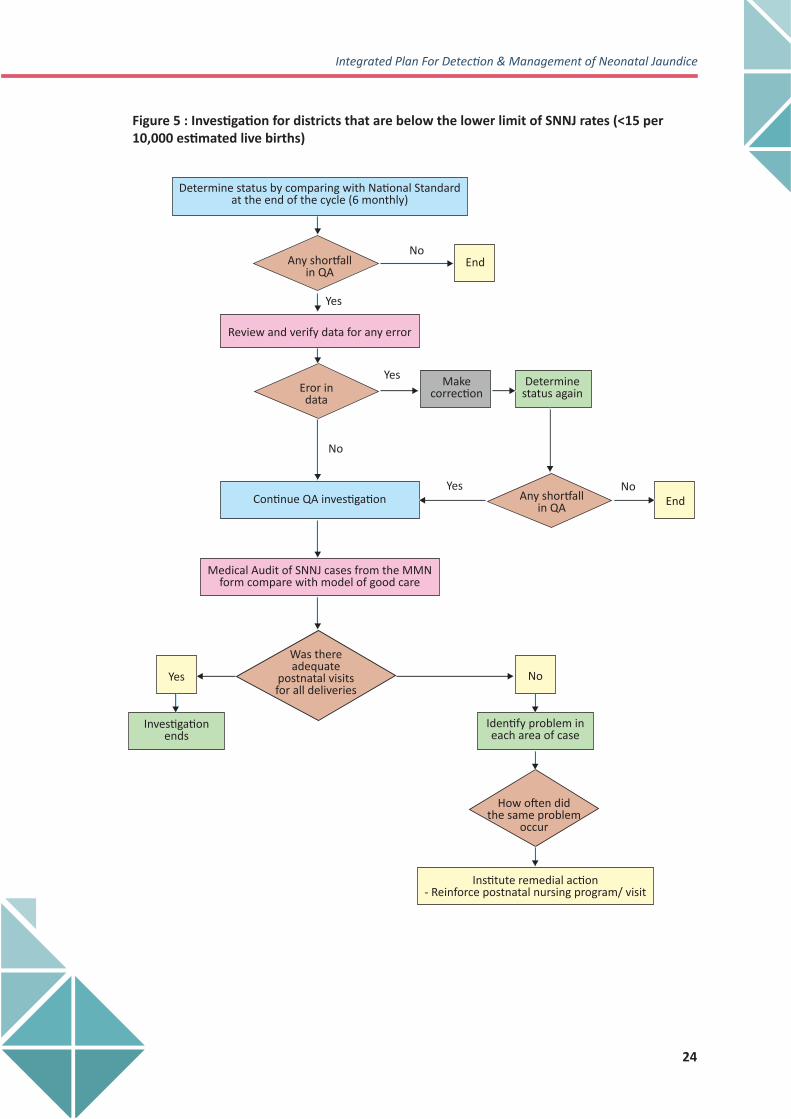
Lower limitDistricts with rates below 15 per 10,000 estimated live births are considered far below the accepted level and have a shortfall in quality. These districts need to investigate why there was such a low detection rate of SNNJ (Figure 5).
Staff responsible for investigation of severe neonatal jaundiceBoth hospital and health staff are jointly responsible for the prevention of SNNJ and hence will investigate and institute remedial measures. Regardless of whether the baby was or was not managed in the hospital the following individuals will be responsible for the investigation by the district:
Monitoring of babies with Acute Bilirubin Encephalopathy (ABE)ABE signifies the presence of changes in the mental (behavioural) status and muscle tone during the neonatal period when the baby is having severe NNJ.
These include drowsiness, poor feeding and hypotonia followed by hypertonia affecting extensor muscles in particular, resulting in retrocollis and opisthotonos. A BIND score could be used to assess for ABE in term babies who are admitted for severe NNJ. Moderate to advanced ABE signifies the potential of neurodevelopmental sequelae and needs long term follow-up and hearing assessment. Please Refer to Appendix 7.10
Data on severe NNJ should be analysed and presented twice yearly at the state perinatal mortality meetings.
STANDARD FOR SEVERE NEONATAL JAUNDICE RATE
Table 10: BIND Scoring
Mild ABEModerate ABEAdvanced ABE
1-34-67-9
Acute Bilirubin Encephalopathy Severity Total BIND score

22
Integrated Plan For Detection & Management of Neonatal Jaundice
Steps in investigation of SNNJFigure 5 shows the process of investigation and flow of information and data. An investigation should be conducted if there is a shortfall in quality
Figure 3 : Flow of data collection for NNJ
Monitoring of all newborns for NNJ
Newborn identified to have NNJ
Use the NNJ form to monitor, manage & notify(MMN form in 2 copies)
On going NNJ management at home, clinic, hospital.Data to be updated at all levels
Final outcome
Mild-Moderate NNJ
1. One copy of MMN form to be sent to health matron/ sister of the district2. One copy of MMN form to be kept with the child health card
Severe NNJ

23
Integrated Plan For Detection & Management of Neonatal Jaundice
Figure 4 : Investigation for districts that exceed the upper limit of SNNJ rates (>50 per 10,000 estimated live births)
Determine status by comparing with National Standardat the end of the cycle (6 monthly)
Any shortfallin QA
Error indata
Makecorrection
Any shortfallin QA
Review and verify data for any error
Continue QA Investigation
Was each step takenapproplately, timely &
adequately
Medical Audit of SNNJ cases from the MMNform compare with model of good care
End
End
No
No
No
No
Yes
Yes
Yes
Yes
Investigationends
Did not occur
Remedial measures for isolated case
Review 30 sample of NNJ using MMN forms
Occurred in sample problems identified
Why do theseproblems occur
Observe andinterview staff
Training Improve supervisoryprocedures
Revise organizationalprocess
Review clinicalprocedures
Healtheducation
Communitymobilisation
Study of avaiableinfrastructure
Interview sampleof mothers/community
Study/reviewenvironment/
logistics
Identify problem ineach area of case
How often didthe same problem
occur
Determine statusagain
24
Integrated Plan For Detection & Management of Neonatal Jaundice
Figure 5 : Investigation for districts that are below the lower limit of SNNJ rates (<15 per 10,000 estimated live births)
No
No
Yes
Yes
No
Identify problem ineach area of case
How often didthe same problem
occur
Institute remedial action- Reinforce postnatal nursing program/ visit
Yes
Investigationends
Was thereadequate
postnatal visitsfor all deliveries
Determine status by comparing with National Standardat the end of the cycle (6 monthly)
Makecorrection
Continue QA investigation
Medical Audit of SNNJ cases from the MMNform compare with model of good care
Determinestatus again
Review and verify data for any error
NoEnd
Yes
EndAny shortfallin QA
Eror indata
Any shortfallin QA

25
Integrated Plan For Detection & Management of Neonatal Jaundice
7. MODEL OF GOOD CARE
Education on neonatal jaundice provided for the expectant mother (pamphlet)All mothers should have blood taken for ABO and Rhesus group.Identify other risk factors for significant jaundice e.g. family history of severe neonatal jaundice, exchange transfusion and haemolytic diseases.
1.2.3.
Antenatal care
Cord blood for G6PD screening. Obtain G6PD screening results before baby is discharged from hospital or as soon as possible in home deliveries. If result shows G6PD deficiency/ Intermediate, the baby is to remain or be admitted to hospital for observation and monitoring for at least 4-5 days. Babies of Rh-negative mother should be managed at hospital.
1.
2.
3.
Intrapartum care
Examine all babies for jaundice before discharge and TcB/ serum bilirubin measured if jaundice is detected. Special attention should be given to babies with risk factors.Routine home visits as planned (Day 1, 2, 3, 4, 5, 6, 8,10, 15, and 20).Refer to clinic or hospital for admission or bilirubin testing and review based on ‘Criteria of Admission’
ManagementAppropriate management of NNJ in hospital.
Recording & Monitoring1. Use the “Monitor, Manage & Notify NNJ”form. Please see next page.2. Provide returns to health matron/ district sister.
1.
2.3.
Detection of neonatal jaundice postnatally
Prompt notification of postnatal mother to the nearest health centre.Support the mother to breastfeed the infant adequately and supplementation if needed. The adequacy of breastfeeding, weight and hydration status of all babies should be assessed during the first week of life. Babies with weight loss > 7% of birth weight should be assessed for adequacy of the breastfeeding and closely monitored for jaundice. Nursing personnel should actively look for signs of jaundice during routine care of the mother and baby.Identification of Risk Factors
Prematurity (< 37 weeks) Low birth weight (< 2.5kg) Sepsis Baby of diabetic mother Onset of jaundice before 24 hours of life A sibling with severe neonatal jaundice or exchange transfusionInadequate breastfeeding/ dehydration (as shown by > 7% weight loss)Mothers with blood group O / Rhesus negative G6PD deficiency Asphyxia Rapid rise of Total Serum BilirubinHigh pre discharge bilirubin levelCephalhaematoma or bruise
1.2.
3.
4.
Postnatal care

26
Integrated Plan For Detection & Management of Neonatal Jaundice
1 2 3 4 5 6 7 8 9 10 ≥ 11
Klinik…………..
Hospital…………
Kes SNNJ dikesan dari format QA FH2 : Kes Tidak dapat dikesan :
Infant of DM MotherInadequate feeding Hospital…………………
Klinik………………………
RUMUSAN KES (oleh Pegawai Perubatan & Kesihatan/PHN/JK) : Pelapor :Kes Neonatal Jaundis : Kes dalam Daerah :Kes SNNJ : Kes Dari Luar :
Sebab Kelewatan rujukan/kemasukan ke wad/menerima rawatan1. Faktor Pesakit : Nyatakan : 2. Faktor Perkhidmatan Kesihatan : Nyatakan : 3.Faktor Perkhidmatan Hospital : Nyatakan :
Paras SB Tertinggi(umol/l or g/dl): Fototerapi Hidup :Mengikut Borang QA FH2)BIND Score Tertinggi (0-9):
Fototerapi & Blood Exchange Mati :
Paras SB (umol/l or mg/dl)Discaj setelah dirawatParas SB (umol/l or mg/dl) Jika kes adalah SNNJ nyatakan Rawatan Outcome':
Jaundis dikesan dibawahparas umbilicusTSB dibuat di: KK(K)/Hosp (H) Swasta (S) Paras SB (umol/l or mg/dl)Rujuk ke hospitalParas SB (umol/l or mg/dl)Masuk wad (sekiranya kes
PERGERAKAN KES HARI POSTNATAL
Anak discajNotifikasi diterima oleh Lawatan Pertama & Berikutnya Jaundis Mula dikesan
G6PD-Deficient/intermediate Penjagaan Penjagaan
Lain-lain: Nyatakan……………………………
Prenatal Postnatal
H/O SNNJ in Siblings Kes disambut anggota kesihatan Anggota HospitalS & S Of Sepsis Tiada terima notifikasi
Tidak diketahuiCephalohematoma/bruises Tiada diketahui
Low Birth Weight(<2.5kg) Telefon Jururawat TerlatihPrematurity Saudara Penjaga
Mother's Blood Group O Surat Notifikasi BidanMother's Blood Group RH -ve Lawatan Rumah Jururawat Masyarakat
Lain-lain: Nyatakan…………… Lain-lain:Nyatakan…………..Faktor Risiko: Cara Nortifikasi Kelahiran: Anggota Yang Mengesan Jaundis
Bumi Sarawak: Hosp Kerajaan: Tidak diketahui:Bumi Sabah: Hospital Swasta:
Cina: ABC: 2--5:India: BBA: ≥ 6:
Kumpulan Etnik: Tempat Kelahiran: Gravida Ibu:Melayu: Rumah: Primigravida:
Nama: KP Ibu: Tarikh Lahir: - - Alamat Rumah:Jarak Rumah Dari Hospital Terdekat: km Jarak Rumah Dari Klinik Kesihatan Terdekat: km
MINISTRY OF HEALTH : MMN/NNJ 2016
MONITORING, MANAGEMENT AND NOTIFICATION OF NNJ / SNNJBulan/Tahun:Pejabat Kesihatan Daerah: Klinik : Hospital:
tidak masuk wad setelah dirujuk tulis TM pada tarikh rujukan)
Tarikh :

27
Integrated Plan For Detection & Management of Neonatal Jaundice
Sebaik sahaja NNJ dikesan – di klinik kesihatan atau hospital, borang ini hendaklah digunakan dan diisi.
Isikan dua (2) salinan. a. Salinan Pertama – dikepilkan di Buku Rekod Kesihatan Bayi dan Kanak-kanak 0-6 Tahun b. Salinan Kedua – disimpan hantar kepada Pejabat Kesihatan Daerah.
Ruangan PERGERAKAN KES:
1.
2.
3.
Salinan pertama yang telah lengkap perlu diambil semula selepas PN ke10, tetapi tentukan kes telah pulih.
Kedua-dua salinan perlu dihantar ke Pegawai Perubatan / Jururawat Kesihatan di KK untuk dibuat rumusan.
Jururawat Kesihatan kumpulkan semua borang Salinan Pertama dan hantar bersama reten bulanan setiap bulan ke Pegawai Kesihatan Daerah (Kesihatan Keluarga) / Penyelia Jururawat Kesihatan Daerah / Ketua Jururawat Kesihatan.
4.
5.
6.
Tarikh anak discaj. Tarikh notifikasi diterima. Tarikh lawatan postnatal pertama dan berikutnya. Tarikh pertama jaundis dikesan. Tarikh jaundis dikesan di bawah paras umbilicus. Tarikh TSB dijalankan dan tempat di mana ujian TSB dijalankan serta paras Serum TSB. (K = KK DAN JPL, H = HOSPITAL DAN A&E HOSPITAL, S = KLINIK ATAU HOSPITAL SWASTA)Tarikh rujukan ke Hospital iaitu (HOSPITAL DAN A&E HOSPITAL) dan paras TSB semasa rujukan.Tarikh masuk wad dan paras TSB semasa masuk wad. Sekiranya kes tidak masuk wad pada tarikh rujukan sila catatkan TM pada ruangan berkenaan.Tarikh discaj dan paras TSB semasa discaj.
i.ii.
iii.iv.v.
vi.
vii.
viii.
ix.
GARIS PANDUAN MENGISI BORANG PENGENDALIAN KES-KES NNJ DAN SNNJ
Pelapor:
Disemak:

28
Integrated Plan For Detection & Management of Neonatal Jaundice
PARE
NTA
L ED
UCA
TIO
N L
EAFL
ET
JAU
ND
ICE
IN B
ABI
ES
Wha
t is
jaun
dice
Jaun
dice
in
new
born
bab
ies
is s
een
as a
ye
llow
ness
of t
he s
kin
and
eyes
. Up
to 7
5% o
f al
l bab
ies
deve
lop
jaun
dice
.
Wha
t are
the
dang
er s
igns
?
Urg
ent t
reat
men
t sho
uld
be so
ught
if ja
undi
ced
babi
es d
evel
op th
e fo
llow
ing
sign
s:W
hat
caus
es ja
undi
ce?
In th
e hu
man
bod
y, n
ew b
lood
is b
eing
mad
e al
l the
tim
e an
d ol
d bl
ood
is b
eing
des
troy
ed.
One
of
the
brea
kdow
n pr
oduc
ts o
f bl
ood
is
‘bili
rubi
n’.
Bilir
ubin
is
norm
ally
pro
cess
ed i
n th
e liv
er a
nd is
elim
inat
ed f
rom
the
bod
y in
th
e st
ool
and
urin
e. F
or t
he fi
rst
few
day
s aft
er b
irth
, a
baby
’s l
iver
doe
s no
t w
ork
as
effici
ently
as
it do
es la
ter.
So t
here
ten
ds t
o be
a b
uild
up
of b
iliru
bin
in t
he b
lood
. Th
is
caus
es ja
undi
ce in
the
new
born
bab
ies.
Babi
es w
ho m
ay b
e pa
rticu
larl
y pr
one
to
seve
re ja
undi
ce in
clud
e:
Is ja
undi
ce h
arm
ful?
Seve
re ja
undi
ce c
an c
ause
• D
eath
•
Hea
ring
pro
blem
s (d
eafn
ess)
• Le
arni
ng d
ifficu
lties
• In
telle
ctua
l dis
abili
ties
• Ce
rebr
al p
alsy
• Pr
emat
ure
babi
es•
Babi
es w
ith in
fecti
on
• Ba
bies
with
G6P
D d
efici
ency
• Ba
bies
who
se b
lood
gro
up is
diff
eren
t fro
m
thei
r m
othe
r’s
o
Mot
hers
with
rhe
sus
nega
tive
o
Mot
hers
with
blo
od g
roup
O•
Babi
es w
ho d
o no
t rec
eive
ade
quat
e fe
eds
• Ba
by o
f dia
betic
mot
hers
• Ja
undi
ce v
isib
le w
ithin
24
hour
s aft
er b
irth
• Ja
undi
ce v
isib
le b
elow
the
umbi
licus
• N
ot a
ctive
, unw
ell o
r ha
ving
feve
r•
Not
feed
ing
wel
l•
Fits
or
stiff
ness
of t
he b
ody
• Ja
undi
ce p
ersi
sting
bey
ond
14 d
ays
• Pa
le c
olou
red
stoo
ls o
r te
a co
lour
ed u
rine
Whi
ch b
abie
s ge
t sev
ere
jaun
dice
?
Wha
t sho
uld
the
pare
nts
do if
thei
r ba
bies
de
velo
p ja
undi
ce?
Seek
ear
ly tr
eatm
ent a
t the
nea
rest
hea
lth
faci
lity
A b
lood
test
will
be
take
n to
det
erm
ine
the
leve
l of b
iliru
bin
in th
e ba
by’s
blo
odD
o no
t pu
t th
e ba
by u
nder
the
sun
, as
it w
ill c
ause
sun
burn
and
deh
ydra
tion.
How
can
par
ents
min
imis
e th
e se
veri
ty o
f ja
undi
ce in
my
baby
?
Ensu
re a
dequ
ate
brea
stfee
ding
(at
lea
st
8-12
tim
es e
very
24
hour
s)Av
oid
taki
ng
trad
ition
al
med
icati
on
if br
eastf
eedi
ng
Cont
act
Num
bers
of n
eare
st H
ealt
h Cl
inic
s /
Hos
pita
ls:_
____
____
Spec
ial
Not
e fo
r pa
rent
s of
bab
ies
born
to
mot
hers
wit
h bl
ood
grou
p O
:
Your
bab
y ha
s a
high
er r
isk
of
deve
lopi
ng s
ever
e ja
undi
ce.
Plea
se i
nfor
m t
he h
ealth
sta
ff o
f th
e ne
ares
t cl
inic
to
see
your
bab
y th
e da
y aft
er d
isch
arge
to e
xam
ine
for
jaun
dice
.
1. 2.
8.
APP
END
ICES
App
endi
x 7.
1

Appendix 7.2
CAPILLARY BLOOD SAMPLING FOR BILIRUBIN TESTING
Ensure baby is lying in a safe and secure position Hold the baby’s heel Hold the ankle with index and middle finger Use other fingers to steady the baby’s leg Partly encircle the baby’s heel with the thumb Clean the proposed puncture site with warm water and gauze. Alcohol impregnated wipes should not be used. Allow the area to dry. Gently compress the heel and hold the skin under tension. Puncture the skin in a steady manner. Relax tension and wipe away initial blood flow with cotton wool or gauze. While maintaining grip, hold the heel so that blood is allowed to hang.Gently but firmly compress the baby’s heel to form a large droplet of blood. Do not squeeze. Hold the capillary tube (or blood bottle) to the blood droplet and touch. Momentarily release pressure to collect subsequent blood then reapply pressure, allowing the blood to flow. Continue until sufficient blood has been obtained. Once the sample has been obtained, apply pressure to the site with gauze and maintain pressure until bleeding has stopped.
To obtain the sample:
Figure 6 : Diagram to show location where heel prick should be done
29
Integrated Plan For Detection & Management of Neonatal Jaundice

Appendix 7.3
CORD BLOOD SAMPLING & COLLECTION OF SPECIMENS FOR SCREENING G6PD DEFICIENCY
CollectionThe specimen is dried blood spots on filter paper. A specific type of filter paper is used and it see section on filter paper type and it is easy to transport to the laboratory from remote areas.
MethodBlood can also be obtained directly from the cut end of the maternal portion of cord. Sampling is carried out after the cord has been cut between the clamps or ties normally applied at delivery. Blood must not be drawn from the portion of cord still attached to the baby because of the serious risk of bleeding.
1.2.
3.
Release the clamp or tie on the maternal part of the cord. Using a gloved hand, gently squeeze blood along the cord to the end. Drop the blood into a sterile gully pot. A minimum of 1 ml of blood should be collected. Use a syringe or dropper to transfer the blood onto filter paper. Drop blood from the syringe or dropper directly onto the circle of filter paper, without touching the syringe or dropper to the paper.
1.2.3.4.5.6.
After cleaning
Note:Dropping the blood directly onto the filter paper from the end of the cord, results in poor control of the amount of blood going onto the paper (usually too much spilling over the edge of the circle). This alternative can be used if there is no other option.
30
Integrated Plan For Detection & Management of Neonatal Jaundice

Appendix 7.4
Before deliveryFill up the test request form with the name of mother, address, sex, ethnic group, telephone number, e-mail address, etc. (see below) Label G6PD screen filter paper strip with the registration number for cases delivered in hospital or use the Identification Card number (I/C) for cases delivered at Alternative Birthing Centers or home.
At delivery Completely wet one end of the filter paper strip with cord blood (minimum soak diameter 1 cm). Staple the other end of the filter paper strip to corresponding place on the G6PD screen form. Complete other relevant information required on the form. Put the form in the envelope and address it to the laboratory to which the form is to be sent for testing. For cases delivered in Alternative Birthing Centers or at home forms should be sent to nearby health centers with U/V box. Send the form by hand or post as soon as possible.
For babies who’s cord blood screening has not been done Blood is to be obtained by heel prick. Please refer to Appendix 7.2.Completely wet G6PD filter paper strip with heel blood at one end. Label the strip, staple it to the form, label the form, label the form and fill in the necessary particulars and send it to the laboratory as soon as possible.
2. Screening for G6PD enzyme deficiency in the laboratory All G6PD specimens must be treated as URGENT and processed on the day of arrival, even if there is only a single specimen to be tested. Record all details in the book for G6PD screening. Follow procedure for G6PD screening provided in the laboratory. Fill in the results on the G6PD form and send them back immediately. For guidelines on procedure for detection of G6PD enzyme deficiency by laboratory personnel, please refer to Appendix 7.5.
3. Notifications and Documentation of results - Hospital & Health See Appendix 7.6
GUIDELINES ON THE METHOD FOR G6PD SCREENING (BLOOD COLLECTION AND SCREENING FORM)
Blood for screening purposes should be obtained from the umbilical cord at birth. For babies not screened at birth, blood should be obtained by a heel prick as soon as possible.
1. Collection of blood for G6PD screening at birth
31
Integrated Plan For Detection & Management of Neonatal Jaundice
Appendix 7.5
PrincipleThis fairly simple and foolproof screening for G6PD deficiency is based on that of Beutler (1966). G6PD catalyses nicotinamide adenine dinucleotide phosphate (NADP) to it’s reduced form NADPH in erythrocytes. NADPH protects cells from oxidative damage. The conversion of NADP is the basic diagnostic testing for the deficiency. The screening method was modified by White (1972) to be used on samples collected on blotting paper or filter paper. The absence of fluorescence in U/V light would mean deficiency of the enzyme.
ReagentsAll reagents can be obtained from SIGMA
GUIDELINES FOR LABOATORY PERSONNEL (U/V FLUORESCENCE SCREENING METHOD FOR G6PD DEFICIENCY)
Oxidised glutathione (GSSG) 8 mmol/L M.W = 612.7 (49.016 mg/10 ml buffer)
Nicotinamide Adenine Dinucleotide Phosphate (NADP) 7.5 mmol/LM.W = 765.44Prepare fresh solution(5 7.4 05 mg/10ml buffer)The solution, if stored deep frozen, remains stable for a week
G-6-P (Gluocose-6-Phosphate) 10 mmol/L M.W =358.2 (35.82 mg/10ml buffer)
Tris-HCI Buffer, 0.75mol/L, pH = 7.8Tris (hydroxymethyl) aminomethane = 90.825 g/L (45.412 gm/500 ml).250 ml Tris + 33ml N HCI=0.75 M Tris –HCI Buffer(Accuracy of buffer solution should be adjusted to the first decimal place). It is essential to use a PH electrode, which is suitable for Tris.
1.
2.
3.
4.
32
Integrated Plan For Detection & Management of Neonatal Jaundice

Method
G6PD normal - normal follow upG6PD Deficient - admit for observation for at least 5 daysG6PD Intermediate
Confirmatory test should be sent after 6months old for G6PD intermediate & G6PD deficientTwo methods for confirmatory test; semi quantitative by ELISA or quantitative by immunoassay.Confirmatory test can be done at Ampang Hospital, Kuala Lumpur Hospital dan UKM Medical Centre, also known as Hospital Canselor Tuanku Muhriz .
0.1 ml working reagent is placed in a 10 x 75 min labeled test tube. Disc of 6 min punched from sample strip (using TOHO eyelet punch in similar) is placed into tube. Ideally the tube should be incubated at 37 ⁰C X 15 min. However the incubation can be done at room temperature for the same length of time. Using a capillary tube, spot the test mixture on a Whatman No.1 filter paper* and allow it to dry thoroughly, preferably using a hair drier. Examine under U/V lamp for fluorescence. Fluorescence + + + = No G6PD deficiency Fluorescence Nil = G6PD deficiency
1.2.3.
4.
5.
Work procedure
Large numbers of tests may be done at the same sitting. *Use “Chromatography Paper”Whatman No.1 (7.5 cm x 100m. Basic weight 87 gm/m, thickness 0.16 mm). Medium Flow Rate. The mixture of the working reagent should be prepared at district hospital and issued at weekly intervals to the peripheral clinics. This reagent should be transported in a ice-flask. In the peripheral clinics, the reagent should be stored in the freezer compartment. Filter paper should be made available to the peripheral clinics.
1.2.
3.
4.
Precautions
This method will miss some female heterozygotes but is reliable for detecting deficiencies among males. Check the expiration dates of commercial kits.
1.
2.
1.2.3.
Control
A known normal blood sample must be run as a control. Check the reagent daily before each batch of tests to make sure it is working properly.
Interpretation of results
Specimens from normal persons shows strong fluorescence. Red cells with less than 20% normal activity will show no fluorescence.
33
Integrated Plan For Detection & Management of Neonatal Jaundice
Confirmatory test:

Appendix 7.6
HospitalThe result should be made available before the baby is discharged. In a busy unit with many deliveries a day, some babies may discharged prior to receiving the laboratory results. There must be a recall system to call back the baby urgently to be admitted to Peadiatric Unit if the result is abnormal.
If the result is normal, the mothers should be notified (Fig. 7). The ward nurse in-charge should inform the public health nurse at the clinic nearest the parents home. The public health nurse should endorse the G6PD results in the Home Based Child Health Card.
Health CentreIf the result is abnormal, the nurse or the relevant health staff should be informed immediately, by the Medical Laboratory Technologist, by phone or fax. The baby should be referred to the hospital for admission.
NOTIFICATION OF G6PD DEFICIENCY SCREENING RESULTS
Figure 7 : Flow chart on notification of G6PD deficiency screening results
Ward nurse receives G6PD result
Admit baby to the ward
Abnormal Normal
Discharge
If the baby has been discharged, the ward nurse should contact the nearest public health nurse
by telephone or fax within 24 hours
The public health nurse will inform the neareststaff nurse/community nurse in the health clinic to do home visit and get the baby for admission
34
Integrated Plan For Detection & Management of Neonatal Jaundice

35
Integrated Plan For Detection & Management of Neonatal Jaundice
STA
ND
ARD
OPE
RATI
NG
PRO
CED
URE
FO
R D
ETEC
TIO
N O
F N
EON
ATA
L JA
UN
DIC
E IN
HO
SPIT
ALS
WIT
H S
PECI
ALI
STS
This
SO
P en
able
s im
med
iate
and
effe
ctive
acti
ons
thro
ugh
coop
erati
on b
etw
een
the
paed
iatr
ic a
nd o
bste
tric
tea
ms
for
early
det
ectio
n of
NN
J an
d to
av
oid
exch
ange
tran
sfus
ion.
App
endi
x 7.
7
With
no
risk
fact
ors:
Aft
er 7
2 ho
urs
of li
fe
BW
>2.5
kg
G
6PD
nor
mal
Mat
erna
l Rh
+ve
SB a
nd tr
ace
ASA
PSt
art p
hoto
ther
apy
whi
le a
wai
ting
resu
lt
SB a
bove
inte
nsiv
eph
otot
hera
py le
vel
SB a
bove
con
venti
onal
phot
othe
rapy
leve
l but
belo
w in
tens
ive
leve
l
SB b
elow
con
venti
onal
phot
othe
rapy
leve
l
Refe
r Pa
eds
imm
edia
tely
Conti
nue
phot
othe
rapy
Repe
at S
B ne
xt m
orni
ngO
ff p
hoto
ther
apy
Repe
at S
B ne
xt m
orni
ng (e
ither
inm
ater
nity
war
d if
mot
her
is s
till
war
ded
or a
t the
nea
rest
clin
ic if
mot
her
is d
isch
arge
24-7
2 ho
urs
of li
fe w
ith r
isk
fact
ors:
G6P
D d
efici
ency
Mat
erna
l Rh
-ve
Risk
of s
epsi
s
With
in 2
4 ho
urs
of li
fe(a
ll ca
ses
are
path
olog
ical
)
Refe
r Pa
eds
MO
imm
edia
tely
SB s
tat a
nd tr
ace
urge
ntly
Star
t Pho
toth
erap
y w
hile
aw
aitin
gre
sults
Dec
isio
n on
adm
issi
on &
car
eN
B: S
peci
al C
ases
:G
6PD
defi
cien
t bab
ies
- to
keep
for
4-5
days
(as
per
natio
nal C
PG)
Mat
erna
l Rh
-ve
babi
es -
ensu
re c
ord
bloo
d FB
C, S
B an
d Co
omb’
s ta
ken,
trac
ed u
rgen
tly. R
efer
Pae
ds
NO
TE:
All
mot
hers
and
bab
ies
disc
harg
ed fr
om th
e po
stna
tal
war
ds m
ust b
e no
tified
to th
e ne
ares
t hea
lth c
ente
r vi
a fa
x to
faci
litat
e ho
me
visi
ts
Refe
r Pa
eds
imm
edia
tely
Whi
le a
wai
ting
tran
sfer
to n
eona
tal w
ard
I
nten
sive
pho
toth
erap
y - i
rrad
ianc
e at
leas
t 30
S
B st
at a
nd tr
ace
urge
ntly
Doc
umen
t G6P
D s
tatu
s, m
ater
nal b
lood
gro
up a
nd p
ast
hist
ory
of s
ever
e N
NJ i
n re
ferr
al s
heet
Neo
nate
Clin
ially
Jaun
dice
TSB
stat
if a
vaila
ble
Aft
er24
hou
rs o
f life
STA
ND
ARD
OPE
RATI
NG
PRO
CED
URE
FO
R D
ETEC
TIO
N O
F N
EON
ATA
L JA
UN
DIC
E IN
HO
SPIT
ALS
WIT
HO
UT
SPEC
IALI
STS
Ratio
nale
: M
any
attem
pts
to r
educ
e th
e in
cide
nce
of S
ever
e N
NJ
in t
he p
ast
have
not
bro
ught
abo
ut s
igni
fican
t ch
ange
Thi
s SO
P is
to
enab
le e
arly
de
tecti
on o
f NN
J res
ultin
g in
imm
edia
te a
nd e
ffecti
ve a
ction
via
coo
pera
tion
betw
een
hosp
itals
with
out s
peci
alis
t with
the
prim
ary
care
team
App
endi
x 7.
8
36
Integrated Plan For Detection & Management of Neonatal Jaundice
SB
bel
owco
nven
tiona
lph
otot
hera
py le
vel
Not
ify H
ealth
Sta
ff
Adm
it ba
byS
tart
phot
othe
rapy
Do
SB
Rev
iew
SB
nex
tda
y &
clo
sely
supe
rvis
ed h
ome
visi
ts
Bef
ore
disc
harg
e lo
ok fo
r clin
ical
sig
ns o
f jau
ndic
e
Not
e 1:
NN
J pr
esen
ting
<24
hrs
of li
fe is
p
atho
logi
cal
Not
e 2:
Spe
cial
cas
esG
6PD
def
icie
nt b
abie
s - t
o ke
ep fo
r 5da
ys (a
s pe
r nat
iona
l CP
G)
Mat
erna
l Rh
-ve
babi
es -
ensu
re c
ord
bloo
d FB
C, S
B a
nd C
oom
b’s
test
take
n, tr
aced
urg
ently
. Ref
er
Pae
diat
rics
Kee
p al
l mot
hers
and
bab
ies
rout
inel
y fo
r 24
hour
s af
ter d
eliv
ery
(look
for j
aund
ice
at e
ach
shift
)
Clin
ical
ly n
o si
gn o
f jau
ndic
eC
linic
ally
jaun
dice
pre
sent
Che
ck s
erum
Bili
rubi
n
Bab
y w
ith h
igh
risk
fact
ors
Clo
se s
uper
vise
dho
me
visi
ts
Not
ify h
ealth
sta
ff
Hig
h R
isk
Fact
ors:
BW
< 2
.5kg
PO
G <
38
wks
Pre
viou
s si
blin
g w
ithS
NN
J or
ET
Cep
halo
haem
atom
aIn
fant
of D
MM
othe
r Blo
od G
roup
“O”
Ris
k of
sep
sis
Bab
y w
ith lo
wris
k fa
ctor
s
Not
ify H
ealth
Sta
ff
Rou
tine
Hom
e V
isits
If cl
inic
ally
jaun
dice
pre
sent
do S
erum
Bili
rubi
n
Ref
er s
peci
alis
tho
spita
l if S
B >
inte
nsiv
eph
otot
hera
py le
vel
SB
abo
veco
nven
tiona
lph
otot
hera
py le
vel
Hig
h R
isk
Fact
ors:
BW
< 2
.5kg
PO
G <
38
wks
Pre
viou
s si
blin
g w
ithS
NN
J or
ET
Cep
halo
haem
atom
aIn
fant
of D
MM
othe
r Blo
od G
roup
“O”
Ris
k of
sep
sis
Bab
y w
ith h
igh
risk
fact
ors
Bab
y w
ith lo
w ri
skfa
ctor
s
Adm
it ba
byS
tart
phot
othe
rapy
Do
SB

Appendix 7.9
CHECKLIST FOR THE ASSESSMENT OF BREASTFEEDING ADEQUACY
Penilaian Samada Bayi Mendapat Susu Ibu Dengan Secukupnya
Parameter Ya Tidak Catatan
Membuang air kecil - Menukar lampin basah sekurang-kurangnya 5-6 kali dalam 24 jam
1.
Najis- Kelihatan normal- Sekurang-kurangnya 2 kali dalam 24 jam
2.
Keadaan Am Bayi- Warna kulit normal- Aktif- Tona otot normal
3.
Penyusuan- Sekurang-kurangnya 8-12 kali penyusuan dalam 24 jam
5.
Kelakuan bayi semasa penyusuan- Tenang dan relaks
6.
Corak hisapan semasa penyusuan- Laju pada permulaannya dan - Lebih perlahan kemudian, diselang dengan penelanan yang lembut
7.
Jangkamasa penyusuan- Menyusu selama 5-40 minit setiap kali
8.
Akhir penyusuan- Bayi berhenti menyusu secara spontan
9.
Kelakuan bayi selepas penyusuan- Kelihatan puas dan kenyang
10.
Berat bayi- Kehilangan berat badan tidak lebih dari 10% berat lahir
4.
37
Integrated Plan For Detection & Management of Neonatal Jaundice
Appendix 7.10
BILIRUBIN-INDUCED NEUROLOGIC DYSFUNCTION (BIND) SCORE
38
Integrated Plan For Detection & Management of Neonatal Jaundice


Appendix 7.12
INITIAL ASSESSMENT – CLERKING AND REFERRAL SHEET FOR PROLONGED NEONATAL JAUNDICE IN HEALTH CLINICS/ HOSPITAL WITHOUT SPECIALIST
Adapted from ‘Ogundele MO et al. Implementation of a prolonged neonatal jaundice protocol supported by electronic software.Clinical Governance: An International Journal Vol. 15 No. 3, 2010 pp. 179-190’
To HospitalAppointment
Name :
Current age :(days of life)
Colour of stool: pale/ grey/ yellow(by inspection or history, pls state)
Feeding method:Exclusive BFBottle feedingMIxed feeding percentage of bottle feed____%
Colour of urine: clear/ light yellow/ dark yellow
Onset of Jaundice (Day of life):_______
Address :
Was Vitamin K given? : Yes No(If not given please consider giving)
Mother’s blood group :
(Please note that if cord TSH is not taken or unable to trace please repeat for reference)
G6PD : normal deficientCord TSH :
DOB/ Time of birthGestation :
Contact No.:
IC : Date :
::
Baby’s blood group (if known) :
Spoken to :
Family history:
Examination: General examination:
BW : kgAbdominal examination:
Other systems: Respiratory:
Prolonged jaundice Warning Sign given to parent’s? Yes No Please document the findings in the Child Health BookInvestigation taken prior to referral ( if any):
Impression :
Comments: Name & designationSignature & official stamp:
High risk(Refer STAT)
Intermediate risk(Refer STAT)
Low risk(Managed as per protocol)
CVS: CNS :Hepatomegaly cm Splenomegaly cm
CW : kg Pallor : Jaundice :
ConsanguinityBlood disorder (e.g. thalassemia SEA ovalocytosis, hemolytic diseses, etx;)Blood disorder (e.g congenital hypothyroidism, hyperthyroid mother on medication etc;)Liver disease (e.g biliary atresia, liver failure, congenital hepatis, ets;)Kidney disease (e.g UTI, VUR, juvenile renal failure)
New onset of jaundice? Yes No
40
Integrated Plan For Detection & Management of Neonatal Jaundice
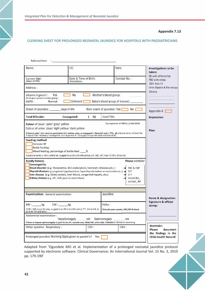
Appendix 7.13
Adapted from ‘Ogundele MO et al. Implementation of a prolonged neonatal jaundice protocol supported by electronic software. Clinical Governance: An International Journal Vol. 15 No. 3, 2010 pp. 179-190’
41
Integrated Plan For Detection & Management of Neonatal Jaundice
CLERKING SHEET FOR PROLONGED NEONATAL JAUNDICE FOR HOSPITALS WITH PAEDIATRICIANS

Appendix 7.14
Appendix A: Workup for Conjugated Hyperbilirubinaemia(mainly for biliary atresia and neonatal hepatitis)
FOR ALL BABIES SB with direct/ indirect’ FBC = reticulocyte countsFree T4 & TSH, urine Dipstick and Microscopy
Biochemistry Liver function test, SB with direct/ indirect, Gamma GT, CaPo4, Lipidprofile, coagulation profile, blood sugar
Microbiology VDRL, Blood C + S, Urine dipstick + microscopy, Urine culture
Endocrine Thyroid function test, Cortisol, Short synacthen test
Histology Liver biopsy
Others Opthalmology for embryotoxon/ chorioretinitis/ septooptic dysplasiaStool color observation for 3 days
Radiology Ultrasound HBS, CXR/ spine x-ray for butterfly veterbrae, HIDA scan,ECHO if murmur
Virology TORCHES, Hep B/C, Urine for CMV
Metabolic GAL-1-PUT, Alpha-1-antitrypsin, Plasma amino acids, Urine for reducingsugars, Urine amino acids, Urine organic acids, IRT/ sweat test
Areas Investigations
PLUS
42
Integrated Plan For Detection & Management of Neonatal Jaundice

43
Integrated Plan For Detection & Management of Neonatal Jaundice
Appendix 7.15
INFANT STOOL COLOUR CHART
Adapated from ‘Early Identification and Referral of Liver Disease in Infants’, Children’s Liver Disease Foundation, UK (www.childliverdisease.org)

44
Integrated Plan For Detection & Management of Neonatal Jaundice
Appendix 7.16
PARENTAL EDUCATION LEAFLET ON PROLONGED NEONATAL JAUNDICE

45
Integrated Plan For Detection & Management of Neonatal Jaundice
9. REFERENCES
Integrated Plan of Management for Detection & Management of Neonatal Jaundice. MOH 2009 (Revised).Guideline on Screening and Management of NNJ with Special Emphasis on G6PD Deficiency, MOH 1998CPG on Management of Neonatal Jaundice (second edition). MOH 2014. MOH/P/PAK/288.15 (GU). National Collaborating Centre for Women’s and Children’s Health. Neonatal jaundice. London: NCC-WCH; 2010. American Academy of Paediatrics Subcommittee on Hyperbilrubinemia. Management of hyperbilirubinemia in the newborn infant 35 or more weeks of gestation. Pediatrics, 2004. Jul;114:297-316.Paediatric Protocols for Malaysian Hospitals (3nd Edition), MOH 2012. Maisels MJ, Bhutani VK, Bogen D, et al. Hyperbilirubinemia in the newborn infant > or =35 weeks’ gestation: an update with clarifications. Pediatrics.2009Oct;124(4):1193-198.Yang WC, Zhao LL, Li YC, et al. Bodyweight loss in predicting neonatal hyperbilirubinemia 72 hours after birth in term newborn infants. BMC Pediatr. 2013 Sep;21(13):145.Chang RJ, Chou HC, Chang YH, et al. Weight loss percentage prediction of subsequent neonatal hyperbilirubinemia in exclusively breastfed neonates. Pediatr Neonatol. 2012 Feb;53(1):41-44.Institute of Health Economics. Transcutaneous Bilirubinometry for the Screening of Hyperbilirubinaemia in Neonates ≥35 Weeks’ Gestation. Edmonton AB:Institute of Health Economics; 2013Nagar G, Vandermeer B, Campbell S, et al. Reliability of transcutaneous bilirubin devices in preterm infants: a systematic review. Pediatrics. 2013 Nov;132(5):871-881Tan KL, Dong F. Transcutaneous bilirubinometry during and after phototherapy. Acta Paediatr. 2003;92(3):327-331.Johnson L, Bhutani VK, Karp K, et al. Clinical report from the pilot USA Kernicterus Registry (1992 to 2004). J Perinatol. 2009 Feb;29 Suppl 1:S25-45.Johnson L, Brown AK, Bhutani V. BIND - A clinical score for bilirubin induced neurologic dysfunction in newborns. Pediatrics 1999;104:Supplement 4 733-761Louis D, More K, Oberoi S, et al. Intravenous immunoglobulin in isoimmune haemolytic disease of newborn: an updated systematic review and meta-analysis. Arch Dis Child Fetal Neonatal Ed. 2014 Jul;99(4):F325-331Academy of Breastfeeding Medicine Protocol Committee. ABM clinical protocol #22: guidelines for management of jaundice in the breastfeeding infant equal to or greater than 35 weeks’ gestation. Breastfeed Med. 2010 Apr;5(2):87-93Bhutani VK, Johnson L, Sivieri EM. Predictive ability of a predischarge hour-specific serum bilirubin for subsequent significant hyperbilirubinaemia in healthy term and near-term newborns. Pediatrics. 1999 Jan;103(1):6-14Breastfeeding assessment form (Internet communication, 2 December 2014 at http://www.unicef.org.uk/BabyFriendly/Resources/Guidance-for-Health-Professionals/Formsand-checklists/Breastfeeding-assessment-form/)American Academy of Pediatrics (AAP) Subcommittee on Hyperbilirubinemia (2004), “Subcommittee on Hyperbilirubinemia Clinical practice guideline: management of hyperbilirubinemia in the newborn infant 35 or more weeks of gestation”, Pediatrics, Vol. 114 No. 1, pp. 297-316.Roberts KB; Subcommittee on Urinary Tract Infection, Steering Committee on Quality Improvement and Management. Urinary tract infection: clinical practice guideline for the diagnosis and management of the initial UTI in febrile infants and children 2 to 24 months. Pediatrics. 2011;128(3):595–610Ogundele MO et al.Implementation of a prolonged neonatal jaundice protocol supported by electronic software. Clinical Governance: An International Journal Vol. 15 No. 3, 2010 pp. 179-190Children’s Liver disease Foundation (CLDF) (2007), “Jaundice protocol: early identification and referral of liver disease in infants”, available at: www.childliverdisease.org/uploaded/File/ Jaundice_Prot_07.pdfTyrell, M., Hingley, S., Giles, C. and Menakaya, J.O. (2009), “Impact of delayed screening for prolonged jaundice in the newborn”, Archives of Disease in Childhood – Fetal and Neonatal Edition, Vol. 94 No. 2, p. F154.National Institute for Health and Clinical Excellence (NICE) (2007), “Urinary tract infection in children: diagnosis, treatment and long-term management”, available at: www.nice.org.uk/nicemedia/ pdf/CG54fullguideline.pdf
1.
2.3.4.
5.6.
7.
8.
9.
10.
11.
12.
13.
14.
15.
16.
17.
18.
19.20.21.
22.
23.
24.
46
Integrated Plan For Detection & Management of Neonatal Jaundice
Glissmeyer EW, Korgenski EK, Wilkes J, et al. Dipstick screening for urinary tract infection in febrile infants. Pediatrics. 2014;133(5). Available at: www.pediatrics. org/cgi/content/full/133/5/e1121 Schroeder AR et al. Diagnostic Accuracy of the Urinalysis for Urinary Tract Infection in Infants <3 Months of AgePEDIATRICS Volume 135, number 6, June 2015Vikas Satwik. An audit of urine culture results in well infants attending a prolong jaundice clinic. Arch Dis Child 2009; 94: 914.Ghaemi S et al. Late onset jaundice and UTI in neonates. Indian J Pediatr. 2007 Feb; 74(2): 139-41.Lee WS, Chai PF, Boey CM, Looi LM. Aetiology and outcome of neonatal cholestasis in Malaysia. Singapore Medical Journal. 51(5):434-9, 2010 May.Lee WS. Pre-admission consultation and late referral in infants with neonatal cholestasis. Journal of Paediatrics & Child Health. 44(1-2):57-61, 2008 Jan.Lee WS, Chai PF, Lim KS, Lim LH, Looi LM, Ramanujam TM. Outcome of biliary atresia in Malaysia: a single-centre study. Journal of Paediatrics & Child Health. 45(5):279-85, 2009 May.Lee WS, Chai PF. Clinical features differentiating biliary atresia from other causes of neonatal cholestasis.Annals of the Academy of Medicine, Singapore. 39(8):648-54, 2010 Aug.Yi-Chun Lin. Decreasing rate of Biliary Atresia in Taiwan 2004-2009. Pediatric 2011; 128 (3): 530-536Amar Singh. TSH screening of cord blood in Malaysia – Its Impact (The National Congenital Hypothyroid Screening programme in Malaysia). The Jounral of Endocrinology and Metabolism May 2010; 1(1)Fuziah Md Zain. Congenital hypothyroidism. MJPCH 2010 (Dec); Vol. 16; Supplementary 1.Zarina et al. Audit of Newborn Screening Programme for Congenital Hypothyroidism. Med J Malaysia Vol 63 No 4 October 2008S Kamameedi, Lim CT. Characteristics of Malaysian Infants with Biliary Atresia and Neonatal Hepatitis. Med J Malaysia 1997; 52(4)Sharad I et al. Prolonged Neonatal Jaundice and the Diagnosis of Biliary Atresia: A Single-Center Analaysis of Trends in Age at Diagnosis and Outcomes. Pediatrics 2008; 121: e1438
25.
26.
27.
28.29.
30.
31.
32.
33.34.
35.36.
37.
38.

47
Integrated Plan For Detection & Management of Neonatal Jaundice
ABBREVIATIONS
ABO
ABE
BIND
BW
C&S
CPG
ET
FBC
G6PD
IDM
IVIG
LB
MOH
MO
NIA
NNJ
QA
POG
PNNJ
Rh
SB
SNNJ
SOP
TcB
TORCHES
TSB
VDRL
Blood Group A, B, O
Acute bilirubin encephalopathy
Bilirubin Induced Neurologic Dysfunction
Birth weight
Culture & sensitivity
Clinical Practice Guidelines
Exchange transfusion
Full Blood Count
Glucose-6-Phosphate dehydrogenase
Infant of Diabetic Mother
Intravenous Immunoglobulins
Live birth
Ministry of Health
Medical officer
National Indicator Approach
Neonatal jaundice
Quality Assurance
Period of gestation
Prolonged neonatal jaundice
Rhesus
Serum bilirubin
Severe neonatal jaundice
Standard Operating Procedure
Transcutaneous bilirubinometry
Toxoplasmosis, Rubella, Cytomegalovirus, Herpes simplex, Syphilis
Total serum bilirubin
Venereal Disease Research Laboratory test
48
Integrated Plan For Detection & Management of Neonatal Jaundice
Dato’ Dr. Amar Singh HSS Senior Consultant Paediatrician (Community) Raja Permaisuri Bainun Hospital, Ipoh, Perak
Dr. Irene Cheah Guat SimSenior Consultant Paediatrician (Neonatology)Institute of Paediatrics, Kuala Lumpur Hospital
Dr. Aminah Bee Bt Mohd KassimPublic Health PhysicianFamily Health Development DivisionMinistry of Health Malaysia
Dr. Angeline Wan Seng Lian Consultant Paediatrician (Neonatology) Sultanah Fatimah Specialist Hospital, Muar, Johor
Dr. Rozita ZakariaConsultant Family Medicine SpecialistSultan Ismail Health Clinic, Johor Bahru, Johor
Datin Dr Baizurah Mohd HussainConsultant Chemical PathologyAmpang Hospital
Dr. Ang Ee LeeConsultant Paediatrician (Neonatology)Tengku Ampuan Rahimah Hospital, Klang, Selangor
Dr. Tan Hui SiuConsultant PaediatricianTeluk Intan Hospital, Perak
Dr. Maurice Steve UtapConsultant Family Medicine SpecialistTudan Miri Health Clinic, Miri, Sarawak
Pn. Aleijah bt AliHealth MatronSri Tanjong Health Clinic, Melaka
Pn. Nor Azizah bt Abd AzizHealth Matron Perak State Health Department, Ipoh, Perak
MEMBERS OF THE TECHNICAL WORKING GROUP
Dr. Aminah Bee Bt Mohd KassimPublic Health PhysicianFamily Health Development DivisionMinistry of Health Malaysia
Dr. Tan Hui SiuConsultant PaediatricianTeluk Intan Hospital, Perak
Dr. Amy Nur Diyana bt Mohamed NasirMedical OfficerFamily Health Development DivisionMinistry of Health Malaysia
EDITOR
ACKNOWLEDGEMENTS
We would like to acknowledge the committee members of the previous editions of ‘Integrated Plan of Action for the Detection and Management of Neonatal Jaundice’, the guidelines development group of the Malaysian CPG: Management of Neonatal Jaundice and the committee members of the ‘Guideline on screening and management of neonatal jaundice with special emphasis on G6PD enzyme deficiency’. We are also grateful to all those who gave feedback and suggestions during the development of this document.


DIVISION OF FAMILY HEALTH DEVELOPMENTMINISTRY OF HEALTH MALAYSIA
2017