intein-mediated fusion expression, high efficient...
TRANSCRIPT
www.elsevier.com/locate/yprep
Protein Expression and Purification 37 (2004) 361–367
Intein-mediated fusion expression, high efficient refolding,and one-step purification of gelonin toxin
Chenyun Guoa, Zhuoyu Lia, Yawei Shia, Mingqun Xub, John G. Wisec,Wolfgong E. Trommerc, Jingming Yuana,*
a Key Laboratory of Chemical Biology and Molecular Engineering of Ministry of Education, Institute of Biotechnology,
Shanxi University, Taiyuan 030006, PR Chinab New England Biolabs, Berverly, MA 01915, USA
c Department of Chemistry, University of Kaiserslautern, Kaiserslautern 67653, Germany
Received 5 April 2004, and in revised form 14 June 2004
Available online 6 August 2004
Abstract
An open reading frame of gelonin (Gel), one of ribosome inactivating proteins, was inserted into the vector pBSL-C which con-
tains the coding region of chitin binding domain (CBD)-intein, resulting in the fusion expression of CBD-intein–Gel in Escherichia
coli BL21 (DE3) by the induction of IPTG. The fusion product formed an aggregate of the misfolded protein, commonly referred to
as inclusion bodies (IBs). The IBs were denatured and then refolded by step-wise dialysis. About 69% fusion protein was in vitro
refolded to native state in the presence of GSSG and GSH as monitored by size-exclusion HPLC. The refolded CBD-intein–Gel
was loaded onto chitin beads column equilibrated with 10mM Tris buffer, 500mM NaCl, pH 8.5, and about 2.4mgGel/L culture
with 96% homogeneity was directly eluted from the captured column by incubation at 25 �C under pH 6.5 for 48h based on intein C-
terminal self-cleavage. Western blot, ELISA, and in vitro inhibition of protein synthesis demonstrated that the bioactivity of recom-
binant Gel was comparable to that of native Gel purified from seeds. This implied that the purified Gel by this method is biologically
active and suitable for further studies.
� 2004 Elsevier Inc. All rights reserved.
Keywords: Gelonin; Intein; In vitro Refolding; Self-cleavage; One-step purification; Bioactivity
Protein self-splicing is a post-translational processing
event in which an internal protein segment, the intein,
can catalyze its own excision from a precursor protein
and concomitantly ligate the flanking regions, the ex-
teins, to form a mature protein [1,2]. Since the mecha-
nism of protein splicing was elucidated, the research
and application of splicing element, intein, have been de-veloped in the field of protein engineering, purification
of recombinant proteins in particular [3,4]. A conven-
tional method for recombinant protein expression and
purification is to make the target protein to be a fusion
1046-5928/$ - see front matter � 2004 Elsevier Inc. All rights reserved.
doi:10.1016/j.pep.2004.06.037
* Corresponding author. Fax: +86 351 7018268.
E-mail address: [email protected] (J. Yuan).
product harboring an affinity tag, such as polyhistidine
(His-tag) [5], Escherichia coli maltose-binding protein
(MBP) [6], Schistosoma glutathione S-transferase
(GST) [7], Staphylococcus protein A [8], and so on.
However, all of these methods suffer a drawback that
a site-specific protease is necessary to cleave the target
protein from its affinity tag, the high cost and uncom-pleted cleavage of these proteases have limited their ap-
plication. In recent years, a rapid, simple protein
expression, and purification system has been performed
by using intein self-cleavage. The intein reactivity can be
controlled to reach the cleavage reaction at either its C-
terminus or its N-terminus. If C-terminal Asn of intein is
substituted to Ala, the cleavage of fusion protein will
362 C. Guo et al. / Protein Expression and Purification 37 (2004) 361–367
only occur at N-terminus of the intein. For example, hu-
man neurotrophin-3 (hNT-3) has been fused to N-termi-
nus of intein from Mycobacterium xenopi gyrA (Mxe
GyrA intein) and successfully purified on a chitin affinity
column by one-step manipulation, based on DTT induc-
ible peptide bond cleavage [9]. On the other hand, a tar-get protein can also be fused to the C-terminus of an
intein whose N-terminal CySH (Ser, Thr) was substitut-
ed to Ala, then the target protein can be purified by cy-
clization of the Asn residue at the C-terminus of intein
with a pH or temperature shift [10].
Gelonin (Gel) is one of the single chain plant ribo-
some inactivating proteins (RIPs). Due to lacking lec-
tin subunit, single chain RIPs are generally not ashighly toxic to intact cells as the double chain RIPs
such as ricin. Therefore, the single chain RIPs have
mostly been selected to construct the potent and spe-
cific immunoconjugates. Gel has been conjugated with
some monoclonal antibodies by gene fusion or chemi-
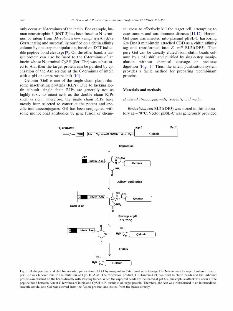
Fig. 1. A diagrammatic sketch for one-step purification of Gel by using inte
pBSL-C was blocked due to the mutation of CySH1–Ala1. The expression
proteins are washed off the beads directly with washing buffer. When the captu
peptide bond between Asn at C-terminus of intein and CySH at N-terminus o
succinic amide, and Gel was cleaved from the fusion product and eluted fro
cal cross to effectively kill the target cell, attempting to
cure tumors and autoimmune diseases [11,12]. Herein,
Gel gene was inserted into plasmid pBSL-C harboring
Ssp DnaB mini-intein attached CBD as a chitin affinity
tag and transformed into E. coli BL21(DE3). Then
pure Gel can be directly eluted from chitin beads col-umn by a pH shift and purified by single-step manip-
ulation without chemical cleavage or protease
digestion (Fig. 1). Thus, the intein purification system
provides a facile method for preparing recombinant
proteins.
Materials and methods
Bacterial strains, plasmids, reagents, and media
Escherichia coli BL21(DE3) was stored in this labora-
tory at �70 �C. Vector pBSL-C was generously provided
in C-terminal self-cleavage The N-terminal cleavage of intein in vector
product, CBD-intein–Gel, can bind to chitin beads and the unbound
red beads are incubated at pH 6.5, nucleophilic attack will occur at the
f target protein. Therefore, the Asn was transformed to an intermediate,
m the beads directly.

C. Guo et al. / Protein Expression and Purification 37 (2004) 361–367 363
by Dr. Xu in New England Biolabs (NEB, Beverly,
USA). PUC-Gel, native Gel, and mouse anti-Gel serum
are kindly provided by Prof. Dr. Trommer in Chemistry
Department of Kaiserslautern University, Germany. All
enzymes used, DNA and protein markers as well as chi-
tin beads are from NEB. Horseradish peroxidase-conju-gated goat anti-mouse immunoglobulin G was
purchased from Sigma (Missouri, USA). Agarose and
all other chemicals are of analytical grade. Cells were
grown on LB liquid or on LB solid medium as described
by Sambrook and Russell [13].
Construction of plasmid pBSL-Gel and expression of
recombinant in E. coli
To fuse the target gene to the C-terminus of CBD-in-
tein reading frame, the Gel gene (753bp) was amplified
from plasmid pUC-Gel by polymerase chain reaction
(PCR) with the following primers which, respectively,
contained unique restriction sites for AgeI (forward)
and PstI (reverse) to facilitate cloning: forward primer:
5 0-GGT GGT ACC GGT GGC CTG GAT ACCGTG AGC-3 0, reverse primer: 5 0-GGT GGT CTG
CAG TTA TTT CGG ATC TTT ATC G AC-3 0. The
conditions used were: 95 �C for 5min, 30 cycles of
(94 �C 30s, 52 �C 30s, and 72 �C 1min), and a final ex-
tension of 72 �C for 10min. The PCR product was puri-
fied and digested with corresponding enzymes,
subsequently inserted into the vector pBSL-C at AgeI
and PstI sites of MCS to form the recombinant plasmidpBSL-Gel [14]. After restriction enzyme digestion and
DNA sequencing verification, the pBSL-Gel was trans-
formed into competent E. coli strain BL21(DE3) by cal-
cium chloride method [13]. The resulting engineered
strain E. coli BL21(DE3)/pBSL-Gel was grown in LB
medium supplemented with 100g/mL ampicillin at
37 �C until the optical density (OD) at 600nm reached
0.5–0.6, and then induced by 0.5mM IPTG at 12 �Cfor 16h. The fusion expression product, CBD-intein–
Gel was confirmed by SDS–PAGE and Western blot
analysis.
Refolding of CBD-intein–Gel aggregate
The cultured cells were harvested by centrifugation at
6000rpm for 10min and the cell pellets from 1L IPTGinduced culture were suspended and sonicated in lysis
buffer A (10mM Tris–HCl, 500mM NaCl, 1mM
PMSF, pH 8.5, and 0.1% Triton X-100). The lysate
was then centrifuged at 12,000rpm for 30min. Because
the fusion protein almost completely existed in the form
of IBs by 10% SDS–PAGE analysis, the precipitate was
washed with buffer A containing 4M urea and centri-
fuged at 12,000rpm for 30min. This step was repeatedtwice to remove the cell debris and other impurities.
Then the IBs were dissolved in buffer B (10mM Tris–
HCl, 500mM NaCl, pH 8.5) containing 8M urea to
reach a protein concentration of about 0.3mg/mL. The
dissolved precipitate was kept for 2–3h at room temper-
ature to completely solubilize the aggregate. After cen-
trifugation at 12,000rpm for 30min, about 10mL
supernatant was step-wisely dialyzed against 1L bufferB containing 4, 2, and 1M urea sequentially at 4 �Cfor about 17–18h each. Finally, the protein solution
was dialyzed against 500mL buffer B containing
0.2mM GSH, 0.02mM GSSG, and 0.5M LL-Arg for in
vitro refolding at 4 �C for 24h. After centrifugation at
12,000rpm for 30min, the supernatant was further puri-
fied in the next step. The in vitro refolding process was
simultaneously monitored by size-exclusion HPLCequipped with a SW 300 gel column (7.8 · 300mm) with
10mM Tris buffer, pH 8.5, as the mobile phase at a flow
rate of 1.0mL/min, and absorbance at 280nm was mon-
itored with a UV detector.
One-step purification of Gel by intein C-terminal self-
cleavage
The refolded protein supernatant was applied to a
5mL chitin beads column pre-equilibrated with buffer
B at 4 �C. The captured column was washed with 20 col-
umn volumes of buffer B to remove unbound proteins
and flushed with 15mL cleavage buffer (10mM Tris–
HCl, 500mM NaCl, pH 6.5) before the outlet was
closed. After incubating the captured column at 25 �Cfor 48h, 5–10mL cleavage buffer was flowed throughthe column, the fractionation of Gel was collected ac-
cording to the absorbance at 280nm in LKB protein pu-
rification chromatography system and then examined by
12% SDS–PAGE. The chitin resin could be regenerated
with buffer C (10mM Tris–HCl, 500mM NaCl, and 1–
2% SDS, pH 8.5).
Determination of protein concentration and purity
Protein concentration was determined by Bradford
method using bovine serum albumin as the standard
[15]. Protein expression level and protein purity were es-
timated by comparing the intensity of Coomassie bril-
liant blue staining of samples run on SDS–PAGE. The
stained gel was quantified by gel document scanning
(GDS) with BIO-PROFIL Bio-ID V99.01 software.
Western blot and ELISA
Fusion protein CBD-intein–Gel and purified Gel
were run on 12% SDS–PAGE and transferred electro-
phoretically to a nitrocellulose membrane using a mini
trans-blot electrophoretic transfer cell (Bio-Rad) at
100V for 1h in transfer buffer (25mM Tris, 192mM gly-cine, and 0.025% SDS, pH 8.3). After blocking with 1%
BSA, the membrane was incubated with mouse anti-Gel

Fig. 2. SDS–PAGE analysis of the expression products from engi-
neered strain E. coli BL 21 (DE3)/pBSL-Gel. Lane 1, culture from host
strain; lane 2, uninduced culture from recombinant strain; lane 3,
induced culture from recombinant strain; lane 4, supernatant of the
cell lysate after centrifugation; and lane 5, precipitate of the cell lysate
after centrifugation.
364 C. Guo et al. / Protein Expression and Purification 37 (2004) 361–367
serum diluted 1:500 and the horseradish peroxidase-con-
jugated goat anti-mouse immunoglobulin G (1:10000).
Finally, the sample membrane was subjected to 3,3 0-di-
aminobenzidine tetrahydrochloride (DAB) solution to
visualize the antigen–antibody complex. For ELISA,
the color in the microtiter wells was developed withDAB as substrate for 40min and measured at 405nm us-
ing a microtiter plate reader.
Reticulocyte lysate activity
The bioactivity of Gel on inhibition protein synthesis
was assessed using a cell-free rabbit reticulocyte lysate
protein translation system purchased from GIBCO(Grand Island, NY, USA). After diluted in PBS, 5llGel sample and 40ll cell-free rabbit reticulocyte lysate
containing 10mM creatine phosphokinase, and 0.5mM
KClwere added into 96wells on themicrotiter plate, incu-
bating at 37 �C for 5min, then 10ll master mixture con-
taining 10mM creatine phosphate, 0.5mM MgCl2,
79mM KCl, 5lCi/mL [14C]valine, and amino acid mix-
tures were added and continuously incubated at 37 �Cfor 10min. Five microliter reaction mixture was taken
from the wells and added into 1mL ice chilled water con-
taining 500ll valine (1mg/mL), incubating 10min at
37 �C. The assay was repeated four times at each protein
concentration. The reaction was stopped by adding 25%
trichloroacetic acid. The protein obtained by the glassmi-
crofiber filter was dried and counted by liquid scintillator.
Results and discussion
Cloning and expression of Gel gene
It was shown from the results of double-enzymatic di-
gestion and DNA sequence analysis for plasmid pBSL-
Gel that Gel gene fragment was correctly inserted intovector pBSL-C and suitable for fusion expression in E.
coli (data not shown). The engineered strain E. coli
BL21(DE3)/pBSL-Gel can express the target product,
CBD-intein–Gel in the form of IBs only as estimated
by SDS–PAGE (Fig. 2) or Western blot analysis (data
not shown). To explore its soluble expression, various
factors were examined, including the optical density of
culture at induction (OD600nm 0.4–0.8), the concentra-tion of inducer IPTG (0.1–1.0 mM), induction tempera-
ture (37, 25, 16, and 12 �C) [16–18] as well as
co-expression with molecular chaperone GroESL [19].
Unfortunately, no significant amount of the soluble tar-
get product was observed under any of the above condi-
tions. Over-expression of a recombinant harboring the
gene of eukaryotic cells in host strain E. coli often results
in the formation of biologically inactive aggregates. For-tunately, it is possible to make the aggregates soluble by
some refolding methods [20].
Refolding of CBD-intein–Gel aggregate
It is well known that the IBs occurred during recom-
binant expression in bacteria as a random protein aggre-gate in an unfolded, partially folded or inactive
conformational state, which can be in vitro refolded to
partially recover its active and native state under the de-
fined conditions [20]. Due to the retention time of linear
and globular macromolecules being quite different on
size-exclusion chromatography, the whole process of in
vitro refolding was accurately monitored by size-exclu-
sion HPLC while the urea concentration was reducedin step-wise dialysis (Fig. 3). The IBs were initially
washed with 4M urea to remove the cell debris and
other impurities before completely solubilized in 8M
urea. As can be seen in Fig. 3, the protein was in linear
or random state under strong chaotropic solvent condi-
tion (purple trace, 2). There was little difference for the
chromatograms (purple and green traces, 2 and 3) in
the presence of 8 or 4M urea, which indicated that thecomplete unfolded polypeptide was still dominative with
4M urea [21]. However, when the characteristics of sol-
vent surrounded denatured protein were changed with
decreasing urea concentration, nucleation of protein
conformation seemed to be appearing after dialyzed
against 2M urea, as evidenced by the appearance of a
new peak at the retention time of 7.48min (turquoise
trace, 4). The unsymmetrical chromatogram profile indi-cated that the protein could partially be folded to an in-
termediate, and there existed a competition between the
first-order (correct) folding reaction and the higher-or-
der aggregation reaction. After dialyzed against 1M
urea, the majority of the target protein was refolded as
indicated by the main peak around 11min (pink trace,
5), and also a significant amount of protein seem to be
in a misfolded or aggregate state as indicated by thepeak around 5min (pink trace, 5). The misfolding or ag-
gregation of the protein could be from the association of

Fig. 3. Size-exclusion HPLC chromatograms of in vitro refolding process for CBD-intein–Gel. Conditions: Equipment, HPLC 1525 (Waters, USA);
Column, SW300 (7.8 · 300mm); Mobile phase, 10mM Tris–HCl, 0.5mM NaCl, pH 8.5; Flow rate, 1mL/min; Injected volume, 25lL; Detection:
A280nm; Black trace (1): buffer containing 8M urea; Purple trace (2): inclusion bodies solubilized in buffer with 8M urea; Green trace (3): fusion
protein after dialyzed against buffer with 4M urea; Turquoise trace (4): fusion protein after dialyzed against buffer with 2M urea; Pink trace (5):
fusion protein after dialyzed against buffer with 1M urea; and Brown trace (6): completely refolded fusion protein after dialyzed against buffer
without urea (0M urea).
Fig. 4. Effect of pH on the cleavage of CBD-intein–Gel. Lane 1,
expressed products (IBs); lane 2, in vitro refolded CBD-intein–Gel; and
lanes 3–7, cleavage products of lane 2 at pH 6.0, 6.5, 7.0, 7.5, and 8.0,
respectively. (CBD-intein–Gel, 55kDa; Gel, 28kDa).
C. Guo et al. / Protein Expression and Purification 37 (2004) 361–367 365
hydrophobic surfaces that were exposed in folding inter-
mediate or improper disulfide bridge formation [22]. The
competition of misfolding may kinetically limit the pro-
tein to be folded into its native state [23]. In our study,
the majority of the protein was shifted to the correct
conformation after urea was completely removed by di-alysis in the refolding buffer (10mM Tris–HCl, 500mM
NaCl, pH 8.5, 0.2mM GSH, 0.02mM GSSG, and 0.5M
LL-Arg) (brown trace, 6). Reduced and oxidized glutathi-
one (GSH and GSSG) are commonly used as oxido-
shuffling reagents, because thiol-disulfide exchange
reactions are rapidly reversible. The oxido-shuffling re-
agents can increase both the rate and the yield of correct
disulfide bond formation by rapid reshuffling of improp-er disulfide bonds [23]. LL-Arg contains a guanidino
group and it may play a role in suppressing aggregation
of the protein during refolding [24]. In conclusion, the
results from size-exclusion HPLC analysis figuratively
demonstrated in vitro refolding process during the
step-wise dialysis, which was also confirmed by follow-
ing bioactivity assay.
In vitro pH inducible cleavage of fusion protein
The target protein fused with intein can be cleaved at
the C-terminus of intein by pH shift based on the back-
ground of the recombinant plasmid. To investigate the
optimal pH for the cleavage reaction, the refolded
CBD-intein–Gel was incubated in vitro at five different
pH values, ranging from pH 6.0 to 8.0 at 25 �C. It wasshown from the results of SDS–PAGE in Fig. 4 that
the cleavage reaction was completely inhibited at
pHP7.5 and was gradually increased at either pH 6.0
or 7.0, while the optimal value for the yield and purity
of Gel seemed to be at pH 6.5. During the cleavage pro-
cess, three bands corresponding to fusion protein
(55kDa), CBD-intein (27kDa), and Gel (28kDa) should
occurred by SDS–PAGE analysis. However, the molec-
ular weights of Gel and its fusion partner are too close
to be separated on the gel plate. For confirming the tar-get protein Gel, a positive product at 28kDa band was
occurred by Western blot analysis (data not shown). It
has been speculated that pH sensitivity of the intein
aroused from protonation of the highly conserved pen-
ultimate histidine residue (pKa, approximately 6.5) of
the intein C-terminus [25]. Mutation of the penultimate
histidine inhibited intein C-terminal cleavage, which also
indicated the importance of the conserved histidine res-idue for intein C-terminal cleavage [26–28].
One-step purification of gelonin
The refolded CBD-intein–Gel was loaded onto a chi-
tin beads column at pH 8.5. After washing the captured
column with the equilibrated buffer, about 90% of
fusion protein was bound to the chitin beads (Fig. 5,lane 3). Pure Gel with 96% homogeneity was directly
eluted from the column after incubation at pH 6.5,

Fig. 5. SDS–PAGE for one-step purification of recombinant Gel on
chitin beads column. Lane 1, pellets after cell lysate; lane 2:
supernatant after renaturation and centrifugation; lane 3: flow-through
from chitin beads column; lane 4, recombinant Gel eluted from the
column after cleavage incubation at pH 6.5.
Table 2
ELISA analysis of recombinant Gel
Sample Recombinant Gel Native Gela BSAb
OD405nm 0.24 ± 0.01 0.30 ± 0.01 0.01 ± 0.01
Note. The data were the average values obtained by multiply tests.a Positive control.b Negative control.
Fig. 6. Comparison of inhibitory activity of native and recombinant
Gel in the cell-free protein synthesis assay. IC50 of native Gel is at
15pM; IC50 of recombinant Gel is at 20pM.
366 C. Guo et al. / Protein Expression and Purification 37 (2004) 361–367
25 �C for 48h (Fig. 5, lane 4). It is indicated from Table 1
that the refolding percentage of CBD-intein–Gel
reached about 69%, but Gel yield is only 5%, compared
with the quantity of inclusion bodies. The cleavage reac-
tion as described above was so incomplete that about
20% fusion product still stuck in the affinity column as
occurred by SDS–PAGE analysis with a small portion
of affinity beads. There was not much improvement inthe yield of Gel even if the cleavage reaction on-column
was extended to longer time. However, this system re-
quires no protease or chemical cleavage to obtain the
target protein from its fusion product, compared with
other affinity tag expression systems [29,30].
Bioactivity of recombinant Gel
It was demonstrated from ELISA (Table 2) and Wes-
tern blot analysis (data not shown) that the recombinant
fusion product or Gel only reveals the positive immuno-
reactivity. The bioactivity of recombinant Gel was fur-
ther confirmed with the functional analysis by using
inhibition assay of cell-free protein synthesis in rabbit
reticulocyte lysates. As shown in Fig. 6, native Gel in-
hibits cell protein synthesis by 50% at 15pM, whilstthe recombinant Gel is at 20pM with 50% inhibition.
The result indicated that the bioactivity of the recombi-
nant Gel was comparable to that of native Gel.
Table 1
Purification of recombinant Gel from 1L E. coli cell lysatea
Purification steps Total proteins (mg)
Inclusion bodies (in 8mol urea) 76
Refolding supernatant 51
Chitin column and intein-mediated cleavage 2.4
a Step and overall yields were calculated starting from the pure inclusion bo
method.b Purity is defined as the percentage of target protein in the purified prot
Conclusions
In this report, a fusion protein, CBD-intein–Gel was
over-expressed in the form of inclusion bodies in E. coli
and was successfully in vitro refolded through 8M urea
denaturation and step-wise dialysis. The refolding pro-
cess along the renaturation was concomitantly moni-
tored by size-exclusion HPLC, and the refoldingrecovery of the fusion product reaches about 69%.
About 2.4mgGel/L culture with 96% homogeneity was
obtained by single-step purification on chitin beads col-
umn, without any chemicals or protease treatment.
Moreover, immunoreactivity and functional assay also
demonstrated that the recombinant Gel possessed the
same properties as the natural Gel purified from seeds.
This intein-mediated purification scheme would provide
Target protein (mg) Purityb (%) Yield (%)
46 (CBD–intein–Gel) 60 100
32 (CBD–intein–Gel) 63 69
2.3 (Gel) 96 5
dy preparation and protein concentration was determined by Bradford
ein preparation.
C. Guo et al. / Protein Expression and Purification 37 (2004) 361–367 367
a convenient and economic method to prepare other
recombinant proteins.
Acknowledgments
This work was supported by the Natural Science
Foundation of China (NSFC: 30270292). We also thank
Dr. Tao Yuan in Aventis Pasteur, Canada, for his help-
ful suggestion.
References
[1] M.Q. Xu, M.W. Southworth, F.B. Mersha, L.J. Hornstra, F.B.
Perler, In vitro protein splicing of purified precursor and the
identification of a branched intermediate, Cell 75 (1993) 1371–
1377.
[2] F.B. Perler, E.O. Davis, G.E. Dean, F.S. Gimble, W.E. Jack, N.
Neff, C.J. Noren, J. Thorner, M. Belfort, Protein splicing
elements: inteins and exteins a definition of terms and the
recommended nomenclature, Nucleic Acids Res. 22 (1994) 1125–
1127.
[3] F.B. Perler, E. Adam, Protein splicing and its applications, Curr.
Opin. Biotechnol. 11 (2000) 377–383.
[4] S.F. Singleton, R.A. Simonette, N.C. Sharma, A.I. Roca, Intein-
mediated affinity-fusion purification of the Escherichia coli RecA
protein, Protein Express. Purif. 26 (2002) 476–488.
[5] M.W. Van Dyke, M. Sirito, M. Sawadogo, Single-step purifica-
tion of bacterially expressed polypeptides containing an oligo-
histidine domain, Gene 111 (1992) 99–104.
[6] C. Guan, P. Li, P.D. Riggs, H. Inouye, Vectors that facilitate the
expression and purification of foreign peptides in Escherichia coli
by fusion to maltose-binding protein, Gene 67 (1998) 21–30.
[7] D.B. Smith, K.S. Johnson, Single-step purification of polypeptides
expressed in Escherichia coli as fusions with glutathione S-
transferase, Gene 67 (1988) 31–40.
[8] B. Nilsson, L. Abrahmsen, Fusions to Staphylococcal protein A,
Mehtods Enzymol. 185 (1990) 144–161.
[9] Z.Y. Li, J.H. Fan, J.M. Yuan, Single-column purification of
recombinant human neurotrophin-3 (hNT3) by using the intein
mediated self-cleavage system, Biotechnol. Lett. 24 (2002) 1723–
1727.
[10] H.E. Humphries, M. Christodoulides, J.E. Heckels, Expression of
the class 1 outer-membrane protein of Neisseria meningitidis in
Escherichia coli and purification using a self-cleavable affinity tag,
Protein Express. Purif. 26 (2002) 243–248.
[11] M.G. Rosenblum, L.H. Cheung, Y. Liu, H.W. Marks, Design,
expression, purification, and characterization, in vitro and in vivo,
of an antimelanoma single-chain Fv antibody fused to the toxin
Gel, Cancer Res. 63 (2003) 3995–4002.
[12] I.L. Urbatsch, P.K. Sterz, K. Peper, W.E. Tommer, Antigen-
specific therapy of experimental myasthenia gravis with acetyl-
choline receptor–gelonin conjugates in vivo, Eur. J. Immunol. 23
(1993) 776–779.
[13] J. Sambrook, D.W. Russell, Molecular Cloning: A Laboratory
Manual, third ed., Cold Spring Harbor Laboratory Press, NY,
2001.
[14] S. Mathys, T.C. Evans, I.C. Chute, H. Wu, S. Chong, J. Benner,
X.Q. Liu, M.Q. Xu, Characterization of a self-splicing mini-intein
and its conversion into autocatalytic N- and C-terminal cleavage
elements: facile production of protein building blocks for protein
ligation, Gene 231 (1999) 1–13.
[15] M.M. Bradford, A rapid and sensitive method for the quantita-
tion of microgram quantities of protein utilizing the principle of
protein-dye binding, Anal. Biochem. 72 (1976) 248–254.
[16] C.H. Schein, H.M. Noteborn, Formation of soluble recombinant
proteins in Escherichia coli is favored by lower growth tempera-
ture, Bio/Technology 6 (1988) 291–294.
[17] S. Cabilly, Growth at sub-optimal temperature allows the
production of functional antigen-binding Fab fragments in
Escherichia coli, Gene 85 (1989) 553–557.
[18] E. Kopetzki, G. Schumacher, P. Buckel, Control of formation of
active soluble or inactive insoluble baker�s yeast alpha-glucosidasePI in Escherichia coli by induction and growth conditions, Mol.
Gen. Genet. 216 (1989) 149–155.
[19] Z. Zhang, L.P. Song, M. Fang, F. Wang, D. He, R. Zhao, J. Liu,
Z.Y. Zhou, C.C. Yin, Q. Lin, H.L. Huang, Production of soluble
and functional engineered antibodies in Escherichia coli improved
by FkpA, Biotechniques 35 (2003) 1041–1042.
[20] R. Rudolph, H. Lilie, In vitro folding of inclusion body proteins,
FASEB J. 10 (1996) 49–57.
[21] H. Yoshii, T. Furuta, T. Yonehara, D. Ito, Y.Y. Linko, P.
Linko, Refolding of denatured/reduced lysozyme at high concen-
tration with diafiltration, Biosci. Biotechnol. Biochem. 64 (2000)
1159–1165.
[22] J. Buchner, U. Brinkmann, I. Pastan, Renaturation of a single-
chain immunotoxin facilitated by chaperonins and protein disul-
fide isomerase, Bio/Technology 10 (1992) 682–685.
[23] A. Mitraki, J. King, Protein folding intermediates and inclusion
body formation, Bio/Technology 7 (1989) 690–697.
[24] T. Arakawa, K. Tsumoto, The effects of arginine on refolding of
aggregated proteins: not facilitate refolding, but suppress aggre-
gation, Biochem. Biophys. Res. Commu. 304 (2003) 148–152.
[25] D.W. Wood, W. Wu, G. Belfort, V. Derbyshire, M. Belfort, A
genetic system yields self-cleaving inteins for bioseparations, Nat.
Biotech. 17 (1999) 889–892.
[26] M.Q. Xu, F.B. Perler, The mechanism of protein splicing and its
modulation by mutation, EMBO J. 15 (1996) 5153–5164.
[27] S. Chong, S. Yang, H. Paulus, J. Benner, F.B. Perler, M.Q. Xu,
Protein splicing involving the Saccharomyces cerevisiae VMA
intein: the steps in the splicing pathway, side reactions leading to
protein cleavage and establishment of an in vitro splicing system,
J. Biol. Chem. 271 (1996) 22159–22168.
[28] S. Chong, K.S. Williams, C. Wotkowies, M.Q. Xu, Modulation
of protein splicing of the Saccharomyces cerevisiae vacuolar
membrane ATPase intein, J. Biol. Chem. 273 (1998) 10567–
10577.
[29] S. Chong, F.B. Mersha, D.G. Comb, M.E. Scott, D. Landry,
L.M. Vence, F.B. Perler, J. Benner, R.B. Kucera, C.A.
Hirvonen, J.J. Pelletier, H. Paulus, M.Q. Xu, Single-column
purification of free recombinant proteins using a self-cleavable
affinity tag derived from a protein splicing element, Gene 192
(1997) 271–281.
[30] S. Chong, G.E. Monotello, A. Zhang, E.L. Cantor, W. Liao,
M.Q. Xu, J. Benner, Utilizing the C-terminal cleavage activity
of a protein splicing element to purify recombinant proteins in
a single chromatographic step, Nucleic Acids Res. 26 (1998)
5109–5115.