interacting partners of the sunn symbiotic kinase
TRANSCRIPT

Clemson UniversityTigerPrints
All Dissertations Dissertations
12-2015
Interacting partners of the SUNN symbiotic kinaseAshley Dawn Zearfoss CrookClemson University, [email protected]
Follow this and additional works at: https://tigerprints.clemson.edu/all_dissertations
This Dissertation is brought to you for free and open access by the Dissertations at TigerPrints. It has been accepted for inclusion in All Dissertations byan authorized administrator of TigerPrints. For more information, please contact [email protected].
Recommended CitationCrook, Ashley Dawn Zearfoss, "Interacting partners of the SUNN symbiotic kinase" (2015). All Dissertations. 1849.https://tigerprints.clemson.edu/all_dissertations/1849

INTERACTING PARTNERS OF THE
SUNN SYMBIOTIC KINASE
A Dissertation
Presented to
the Graduate School of
Clemson University
In Partial Fulfillment
of the Requirements for the Degree
Doctor of Philosophy
Biochemistry and Molecular Biology
by
Ashley Dawn Zearfoss Crook
December 2015
Accepted by:
Dr. Julia A. Frugoli, Committee Chair
Dr. Douglas G. Bielenberg
Dr. Cheryl Ingram-Smith
Dr. Hong Luo
ii
ABSTRACT
The control of nodule number in legumes is primarily accomplished through a
complex systemic signaling pathway termed autoregulation of nodulation (AON). The
protein kinase SUNN, a leucine-rich repeat receptor-like kinase, exhibits shoot control of
AON. I show subcellular localization of SUNN to the plasma membrane and specifically
to the plasmodesmata, a prime location for systemic signaling. To aid in better
understanding the mechanisms of signaling in AON, I used different approaches to identify
potential protein-protein interactions involving SUNN, including candidate genes,
exploratory proteomics, and forward genetic screens. Candidate interactors, including
CLAVATA2 (CLV2), CORYNE (CRN) and guanine exchange factors of the RopGEF
family, were investigated using the bimolecular fluorescence complementation (BiFC)
assay with SUNN and mutant analysis for their role in AON. I report positive BiFC
interactions between SUNN and CLV2, CRN, RopGEF1, RopGEF2, and RopGEF5.
Mutant analysis of these candidate proteins provide additional support for a role in AON.
In legumes, mutations in either CLV2 or CRN, and RNAi of RopGEF1, RopGEF2, or
RopGEF5 in roots render hypernodulation phenotypes. Taken together, my results suggest
a signaling complex involving SUNN is formed in the shoots to transduce a signal from
the roots into a signal back to the roots through the activation of GTPases by RopGEFs,
ultimately regulating nodulation events. I also identified 22 novel putative interactors of
SUNN stemming from an exploratory proteomics experiment involving transgenic M.
truncatula carrying SUNN-YFP/HA that were subjected to co-immunoprecipitation to
isolate protein complexes that contain the transgene. The precipitated proteins were

iii
identified by LC-MS/MS and are presented here as putative interacting partners. In
addition, a forward genetics approach identified components of the AON signaling
pathway. Utilizing a genetic suppressor screen of sunn-1, we identified four independent
lines carrying mutations that suppress the supernodulation phenotype of sunn-1. The
mapping of one of these suppressor lines pinpointed the location of the lesion to an
approximately 150 kB region on Linkage Group 2 harboring about 26 annotated genes.

iv
DEDICATION
This dissertation is humbly dedicated to my family and friends for their unconditional love
and support throughout my career and especially to my loving husband, Andrew, whose
sacrifices made this work possible.

v
ACKNOWLEDGMENTS
I would like to thank my advisor Dr. Julia Frugoli for providing constant guidance and
support throughout my time at Clemson University. Her mentorship has helped shape me
as a professional and as a scientist.
A special thank you to members of the Frugoli lab (both former and current) for creating a
positive environment filled with lively discussions regarding science and life. To Elise
Schnabel for her innovative ideas and contributions to my projects, her patience and
willingness to teach me new techniques. I am grateful for the opportunity to learn from and
work alongside such incredible people.
Thank you to my graduate committee, Dr. Bielenberg, Dr. Ingram-Smith and Dr. Hong
Luo, for the guidance, discussions, ideas and critical reading of this manuscript.
This work was supported by NSF awards IOS#0950700, IOS#1146014, and a Clemson
University Wade Stackhouse Graduate Student Fellowship (2013-2015).

vi
TABLE OF CONTENTS
Page
TITLE PAGE .................................................................................................................... i
ABSTRACT ..................................................................................................................... ii
DEDICATION ................................................................................................................ iv
ACKNOWLEDGMENTS ............................................................................................... v
LIST OF TABLES ........................................................................................................ viii
LIST OF FIGURES ........................................................................................................ ix
CHAPTER
I. LITERATURE REVIEW .............................................................................. 1
The Nitrogen Problem.............................................................................. 1
The Basics of Symbioses ......................................................................... 5
Root Organogenesis and Nitrogen Sensing ............................................. 8
Establishment of the symbiosis ................................................................ 9
Nodule formation ................................................................................... 15
The Autoregulation of Nodulation Pathway .......................................... 19
Scope of Project ..................................................................................... 35
Literature Cited ...................................................................................... 37
II. Medicago truncatula SUNN, CLV2 AND CRN INTERACT
AND SYSTEMICALLY REGULATE NODULE NUMBER .............. 47
Abstract .................................................................................................. 47
Introduction ............................................................................................ 48
Results .................................................................................................... 52
Discussion .............................................................................................. 65
Materials and Methods ........................................................................... 70
Literature Cited ...................................................................................... 77
III. THE ROLE OF ROP GUANINE EXCHANGE FACTORS
IN NODULATION ................................................................................ 82

vii
Table of Contents (Continued)
Page
Abstract .................................................................................................. 82
Introduction ............................................................................................ 82
Results .................................................................................................... 85
Discussion .............................................................................................. 95
Materials and Methods ........................................................................... 99
Literature Cited .................................................................................... 104
IV. A PROTEOMICS APPROACH TO IDENTIFYING
INTERACTING PARTNERS OF SUNN ........................................... 106
Abstract ................................................................................................ 106
Introduction .......................................................................................... 106
Results .................................................................................................. 109
Discussion ............................................................................................ 123
Materials and Methods ......................................................................... 145
Literature Cited .................................................................................... 158
V. IDENTIFICATION OF GENETIC SUPPRESSORS
OF THE sunn-1 HYPERNODULATION PHENOTYPE ................... 162
Abstract ................................................................................................ 162
Introduction .......................................................................................... 162
Results .................................................................................................. 165
Discussion ............................................................................................ 178
Materials and Methods ......................................................................... 183
Literature Cited .................................................................................... 187
VI. SUMMARY ............................................................................................... 189
viii
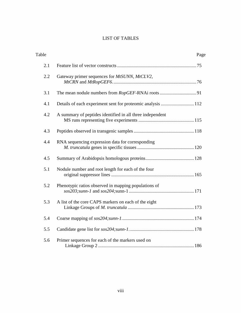
LIST OF TABLES
Table Page
2.1 Feature list of vector constructs ................................................................... 75
2.2 Gateway primer sequences for MtSUNN, MtCLV2,
MtCRN and MtRopGEF6. ...................................................................... 76
3.1 The mean nodule numbers from RopGEF-RNAi roots ............................... 91
4.1 Details of each experiment sent for proteomic analysis ............................ 112
4.2 A summary of peptides identified in all three independent
MS runs representing five experiments ............................................... 115
4.3 Peptides observed in transgenic samples ................................................... 118
4.4 RNA sequencing expression data for corresponding
M. truncatula genes in specific tissues ................................................ 120
4.5 Summary of Arabidopsis homologous proteins ......................................... 128
5.1 Nodule number and root length for each of the four
original suppressor lines ...................................................................... 165
5.2 Phenotypic ratios observed in mapping populations of
sos203;sunn-1 and sos204;sunn-1 ....................................................... 171
5.3 A list of the core CAPS markers on each of the eight
Linkage Groups of M. truncatula ........................................................ 173
5.4 Coarse mapping of sos204;sunn-1. ............................................................ 174
5.5 Candidate gene list for sos204;sunn-1. ...................................................... 178
5.6 Primer sequences for each of the markers used on
Linkage Group 2 ................................................................................. 186

ix
LIST OF FIGURES
Figure Page
1.1 Model of systemic AON signaling in M. truncatula ................................... 22
2.1 Rescue of AON phenotype in transgenic Medicago
truncatula carrying 35S::SUNN-YFP/HA construct .............................. 53
2.2 Subcellular localization of SUNN ............................................................... 54
2.3 SUNN co-localizes with the plasmodesmata in
N. benthamiana and transgenic M. truncatula ...................................... 54
2.4 SUNN, MtCLV2 and MtCRN form homomers in vivo
indicated by a bimolecular fluorescence complementation assay ......... 58
2.5 Heteromer formation between SUNN, MtCLV2 and MtCRN
in vivo using the bimolecular fluorescence complementation assay ..... 60
2.6 Mutation of CORYNE in M. truncatula causes
hypernodulation ..................................................................................... 64
3.1 Bimolecular fluorescence complementation assay shows
in vivo interactions between SUNN and RopGEF1,
RopGEF2, and RopGEF5 ...................................................................... 87
3.2 Lateral root phenotypes observed in RopGEF-RNAi lines ......................... 89
3.3 Representative root systems post-inoculation .............................................. 90
3.4 RNA interference of RopGEFs in A17 and sunn-4
hairy roots (per root phenotype) ............................................................ 92
3.5 RNA interference of RopGEFs in A17 and sunn-4
hairy roots (per root weight phenotype)................................................. 94
3.6 Hairy root transformations for RopGEF-RNAi ......................................... 101
4.1 Structural analysis of the LRR encoded by Medtr4g085480 ..................... 126

x
List of Figures (Continued)
Figure Page
4.2 An interaction network for At1G49750, a LRR protein,
homolog of Medtr4g085480 ................................................................ 132
4.3 Structure analysis of the transmembrane protein,
Medtr4g119380 .................................................................................... 135
4.4 An interaction network for At2G45820, a remorin
family protein ....................................................................................... 138
4.5 Multiple sequence alignment of Medtr3g009050 homologs ..................... 140
4.6 The structure of the LRR protein encoded by
Medtr3g009050 .................................................................................... 142
4.7 An interaction network for At1G33590, a LRR protein ............................ 143
4.8 Proteomics workflow ................................................................................. 146
4.9 The generations of transgenics used in the proteomics
analysis ................................................................................................. 147
5.1 Nodulation phenotype of sos F2’s ............................................................. 166
5.2 Generation of mapping crosses for sos203;sunn-1 and
sos204;sunn-1 ...................................................................................... 168
5.3 A color map representing the fine mapping efforts of
sos204;sunn-1 ...................................................................................... 176

1
CHAPTER ONE
LITERATURE REVIEW
“The destiny of world civilization depends upon providing a decent standard of living for
all mankind.”
– Dr. Norman Borlaug
The Nitrogen Problem
Regarded as an agricultural visionary and the father of the Green Revolution, Dr. Norman
Borlaug dedicated his life to preventing world hunger through his innovative breeding of
cereal crops and by providing adequate agricultural practices to many countries. The Green
Revolution resulted in increased yields of major crops due to the introduction of
nitrogenous fertilizers. By the late 1980’s and early 1990’s, it was recognized that the
introduction and perhaps more importantly, the inefficient use of nitrogen (N) fertilizers
along with related factors led to a significant alteration of the N cycle by humans as a result
of our race to feed a growing population. Throughout the 1990’s and 2000’s, the
environmental and socio-economical impacts associated with the N flux became apparent
(Fields, 2004; Vitousek, et al., 1997; Graham and Vance, 2000; Galloway, et al., 2008).
There is now a strong push to improve sources of natural N fixation (e.g. legume-Rhizobia
symbiosis) to help reverse and prevent negative impacts that are caused by the inefficient
use and over-application of fertilizers and to reduce the overall amount of fertilizers
needed.

2
The future and use of nitrogenous fertilizer
Millions have been fed worldwide through the addition of ammonia, created via the
Haber-Bosch process, as a N source in fertilizers resulting in higher crop yield. From 1960–
2010, the world population nearly doubled in size; however, the arable crop land increased
by about 13% (Food and Agriculture Organization, 2002). The ability to supply adequate
plant nutrients via artificially-made fertilizers increased crop yield and prevented wide-
spread famine from the 1970’s onward. The use of fertilizers (chemically fixed N) in
agricultural processes has increased to an all-time high – over 100 million tonnes consumed
per year (Good and Beatty, 2011; Glass, 2003). Once again, the agriculture industry is
feeling an intense pressure to meet food demands for a growing population expected to
soon reach 8 billion.
In a report in Science, prediction models based on trends in agriculture over the past
40 years projected fertilizer use, irrigation and total crop land for the years 2020 and 2050
(Tilman, et al., 2001). The models predicted a 2.7× increase in fertilizer use and a 1.9×
increase in irrigated land from 2000 to 2050 (Tilman, et al., 2001). Even with current
advances in agriculture, extraneous N applied as fertilizer is lost from agricultural systems
due to inefficient use or over-application in an attempt to increase yield. For example, it is
estimated that one half of the fertilizer used in China was lost due to volatilization and
leaching (Food and Agriculture Organization, 2002). Inefficient fertilizer use is both costly
to the grower and to the environment. The negative impacts of excess nitrogen on
atmospheric changes (i.e. global climate change), biodiversity and ecosystems, animal and
human health has received global attention.

3
The toxicity of excess nitrogen on the environment, plant and animal well-being
Shifts in the availability of nitrogen at any stage of the N cycle can have negative
socio-economical and environmental impacts. Economically, the use of natural gas in the
Haber-Bosch process is a point of concern. Accompanying the depletion of natural gas
reserves, the rise in production cost of fertilizers creates a strain on growers who are
struggling to meet demand. Ultimately, both the monetary and environmental price is borne
by the consumer. Another societal impact is the large differences in distribution of
available nitrogen, creating both N-rich and N-depleted soils. Often, N-depleted soils occur
in developing countries where growers are not able to afford the fertilizer needed to
supplement the soil (Food and Agriculture Organization, 2002). On the other hand, N-rich
soils can lead to and are often caused by the over-application of nitrogenous fertilizers
(Food and Agriculture Organization, 2002). In such cases, excess nitrogen leaches out of
the soil via rain or irrigation and into the groundwater and stream water supply (Vitousek,
et al., 1997).
Nitrates are regarded as toxic substances if present at >10 mg per liter of
groundwater (Tredoux, et al., 2009). In a survey of North American wells, almost every
state had wells (over 9% nationwide) with nitrate in excess of the limits allowable in
groundwater (Power and Schepers, 1989; Nolan, et al., 1997). The percentage of private
wells that exceeded 10 mg/L was four times higher in agricultural areas compared to public
water supplies (Nolan and Stoner, 2000). The toxicity of nitrate to humans is evident in
methaemoglobinaemia (Tredoux, et al., 2009) and thought to be linked to adverse

4
reproductive outcomes, increased possibility of developing certain cancers, diabetes and
several thyroid conditions (Ward, 2009).
In addition to human health concerns, leaching of nitrates into water sources causes
acidification of the soil and the depletion of crucial minerals from the soil (i.e. calcium and
magnesium), deforestation and depletion of wetlands (Vitousek, et al., 1997). The most
documented effect of leaching nitrates is the eutrophication of bodies of water including
estuaries, rivers, lakes and oceans. Increased nitrogen can lead to a reduction in oxygen
levels in bodies of water via toxic algal blooms (causing anoxia or hypoxia) which has
negative impacts on ecosystems and the biodiversity within the affected aquatic system
(Vitousek, et al., 1997; Rabalais, et al., 1996).
Natural forms of nitrogen fixation
Inert N2 can be naturally converted to a biologically relevant form of N via lightning
and microorganisms. Atmospheric N fixation occurs through the breakage of the triple
bond of N2 by the high energy of lightning and usable N is released into the biosphere via
rainfall as nitrates. Natural fixation through lightning is responsible for <10 Tg of N per
year (Borucki and Chameides, 1984).
Biological N fixation can be carried out by several genera of microorganisms
collectively called diazotrophs. The diazotrophs are able to chemically convert N2 into two
ammonia molecules using a unique enzyme called nitrogenase and are often found in
symbiotic relationships with plants where they provide a N source in exchange for a carbon
source and a conducive environment.

5
The Basics of Symbioses
The immobility of plants requires them to be dynamic and adaptable organisms
capable of self-sustainability with limited resources. They utilize carbon dioxide and light
to produce their own energy and form extensive root systems to scavenge for water and
nutrients to uptake from the immediate environment. Plants encounter a number of living
organisms throughout their lifetimes and when nutrients become scarce, some plants enter
into intricate relationships with other organisms. These relationships can be pathogenic,
with nutrient flow being unidirectional, symbiotic, where the exchange of nutrients is
beneficial to both organisms, or one benefitting and the other neutral, as with
commensalism. The ever-adapting plant has developed an arsenal of defense responses that
they utilize to ward off potential predators and some have mechanisms to allow them to
establish symbiotic relationships with fungi and/or bacteria.
Similarities between Mycorrhizal and Bacterial symbiosis
Arbuscular mycorrhizal (AM) fungi have been capable of establishing associations
with approximately 80% of plant species for nearly 400 million years exchanging
phosphorus scavenged from the soil for photoassimilates derived from plant processes
(Govindarajulu, et al., 2005). Similarly, a relationship between free-living soil bacteria
called Rhizobia and leguminous plants has been established to facilitate the fixation and
transport of N to the plant, in return, receiving photoassimilates from the plant. However,
unlike AM symbioses, which are ubiquitous, Rhizobia interact with only a small subset of
plants (Denarie, et al., 1992). This symbiosis is estimated to be only 60 million years old
and many of the genetic components and events that initiate the establishment of symbiosis

6
are borrowed from the more ancient AM symbiosis (Denison and Kiers, 2011; Oldroyd,
2013). The shared genetic components make up the common symbiotic pathway (CSP).
Beyond the CSP, the interactions between host and microbe result in very different
colonization events, mechanisms for nutrient exchange and nutritional needs.
During AM associations, initial signaling events between the fungus and the plant
result in the production of a specialized fungal cell called a haustorium. The haustorium
allows penetration into the inner cortex where the growth of fungal hyphae ensues. Once
inside the plant cells, nutrient exchange occurs through a modified periarbuscule
membrane that develops around the branched hyphal tissue (arbuscule). Similarly,
Rhizobia gain entry into the plant root; however, in response to initial signaling events
between the two symbionts, the plant root creates a new organ de novo for the bacteria to
reside and fix the nitrogen it will supply to the plant.
Biological nitrogen fixation
The fixation of N is possible in many members of the legume family, Fabaceae,
due to their ability to form these biologically important symbioses with diazotrophs from
the genus Rhizobium (for review, Oldroyd, et al., 2011; Ferguson, et al., 2010). Important
crop legumes, such as pea, soybean, peanut, chickpea and lentil, and the fodder and forage
legumes, alfalfa and clover, are estimated to account for 27% of primary crop production
worldwide grown on 15% of the world’s arable lands (Graham and Vance, 2003). With an
agricultural importance bested only by Graminiae, legumes are capable of providing 90 Tg
of N per year (Waggoner, et al., 2008) equivalent to more than $10 billion US dollars
(Graham and Vance, 2003).
7
Legumes receive biologically available forms of N fixed by the bacterial symbionts
living in unique root organs (called nodules). Nodules are formed de novo by the plant in
response to bacteria in the soil. The nodules provide an environment conducive for the
efficient running of the bacterial enzyme nitrogenase which is responsible for the
conversion of N2 to ammonia and other usable forms of N for the plant. To maintain a
favorable environment, the host plant provides an oxygen scavenger, leghemoglobin, to
reduce the oxygen concentration surrounding the differentiated bacteria once it is inside of
the nodule (Ott, et al., 2005). In addition to reduced oxygen levels, the bacteria are provided
carbon in the form of dicarboxylates (for review, Oldroyd, et al., 2011). The active,
nitrogen-fixing bacteroids shut off internal mechanisms that would normally integrate the
fixed N into amino acid stores for the bacteria and are instead able to supply that N directly
to the plant (Oldroyd, et al., 2011). After the conversion of N2 to ammonia, it is transported
out of the bacteroid and into the peribacteroid space within the symbiosome (Oldroyd, et
al., 2011).
Legumes differ in their transport of this N from the nodule to the rest of the plant.
In the temperate legumes, the ammonia is converted to amino acids, primarily glutamine
and asparagine before being transported out of the symbiosome and entering the xylem
(Oldroyd, et al., 2011). In Glycine max (soybean) and Phaseolus vulgaris (common bean),
it is converted to uriedes, mainly allantoin and allantoic acid and is subsequently
transported across the symbiosome membrane for long distance transport via the
vasculature (for review, Atkins and Smith, 2007). The “free” N is then available to the
legume as needed for plant development and to subsequent plants, making legumes useful

8
as agricultural rotations. Because establishing such a relationship is energy intensive for
the plant, it is only done under conditions of low nitrogen availability.
Root Organogenesis and Nitrogen Sensing
The plant is able to take up nitrate (NO3-) directly from the soil when available.
This is likely accomplished through sensing and uptake by nitrogen transporters, such as
NRT1 and NRT2 (for review, Wang, et al., 2012). The level of N available to the plant is
monitored remotely and systemically within the plant and can dictate organogenesis of
lateral roots and nodules (Wang, et al., 2012). Recent work has elucidated a long distance
signaling pathway responsible for decoding N needs. A family of small signaling peptides,
termed C-terminally encoded peptides (CEPs) regulated by nutritional status influence
lateral root formation in both Arabidopsis and Medicago, and nodulation in the latter (Imin,
et al., 2013; Ohyama, et al., 2008).
Specifically in M. truncatula, MtCEP1 is upregulated during periods of low N and
increases nodule number and size while limiting the formation of new lateral roots (Imin,
et al., 2013). Similar to the CLE peptides involved in the autoregulation of nodulation
pathway (discussed in detail later), CEPs produced in the root are translocated to the shoot
and interact with at least one leucine-rich repeat receptor-like kinase as part of a long
distance signaling pathway in Arabidopsis (Tabata, et al., 2014). In Medicago truncatula,
MtCRA2 was identified as the MtCEP receptor in the shoot (Huault, et al., 2014). The
control of root development from the shoot through a mobile peptide-receptor kinase
signaling pathway in response to nitrate levels is interesting because the plant uses these

9
signals to determine whether or not to nodulate, indicating crosstalk between the nitrogen
signaling pathway and the nodulation pathway. Furthermore, nodulating plants are
receiving N which would influence the MtCEPs and must at some point tie into regulating
nodule number. Indeed, in M. truncatula, overexpression of MtCEP1 resulted in increased
nodule numbers and size even under normal limiting conditions i.e., high nitrogen,
suggesting the production of MtCEP1 indicates N-deficiency (Imin, et al., 2013). MtCRA2
acts in both the shoot and the root in two pathways, one that is systemic positive regulation
of nodulation and the other is a local inhibition of lateral root growth (Huault, et al., 2014).
Under low nitrogen, the plant produces lateral roots and is also able to enter into
nodulation. Lateral roots are formed under nitrogen limiting conditions because they are
able to scavenge more area of soil for N. Under abundant nitrogen, either free in the soil or
from bacterial fixation, the plant halts the formation of lateral roots and the formation of
additional nodules. Plant hormones are involved in stimulating the formation of both lateral
roots and nodules, as with the root apical meristem. Cytokinin and auxin are two critical
hormones for development of the root apical meristem, lateral roots and nodules and will
be discussed later.
Establishment of the Symbiosis
Research involving the processes of the arbuscular mycorrhizal and the legume-
Rhizobia relationships in the past decade has broadened our understanding of symbioses.
Most of the studies related to legume nodulation make use of two model legumes,

10
Medicago truncatula and Lotus japonicus due to their small diploid genomes, self-
fertilization, vast seed production and available genetic resources (Cook, 1999).
The host plant and a compatible bacteria communicate prior to invasion of the plant
root using small signaling molecules. Flavonoid signals from the plant exude into the
rhizosphere, are detected by free-living bacteria in the soil, and this perception activates
bacterial gene expression to produce signaling molecules called Nod factors. The secreted
bacterial signals are recognized by host receptors in the root and result in the activation of
the common symbiosis pathway, leading to calcium oscillations and induced cell divisions
that create the nodule primordia (for review, Oldroyd and Downie, 2008; Kouchi, et al.,
2010; Oldroyd, et al., 2011; Oldroyd, 2013).
The bacteria enter the epidermal layer through root hair cells through an infection
thread with the aid of the plant. Eventually, the bacteria are released into the region of the
developing nodule just behind the growing tip where they will divide and differentiate into
nitrogen-fixing bacteroids (Brewin, 1991). At this point, they are surrounded by a plant-
derived membrane, forming an organelle-like structure with reduced function called the
symbiosome (Brewin, 1991). The symbiosome membrane is permeable and can facilitate
the movement of dicarboxylates, ammonia and other carbon and nitrogen sources
(Oldroyd, et al., 2011). Research within the field has identified some of the molecular
signals that the host and symbionts use to “recognize” each other, some of the mechanisms
by which they allow entry and in the case of nodulation, initiate the de novo development
of the nodule.

11
Plant- and bacterial-derived signals
Flavonoid secretion from a leguminous host is perceived by rhizobia in the
rhizosphere initiating the production of bacteria-derived lipo-chitooligosaccharides
(LCOs) called nodulation factors (NFs) (for review, see Gough and Cullimore, 2011).
These small signaling molecules are perceived by a LysM-like leucine rich repeat receptor-
like kinase (LRR-RLK) complex in the root (Limpens, et al., 2003). For Rhizobia, NFs
consist of four or five β1-4 linked N-acetyl glucosamine (GlcNAc) moieties with various
modifications that can dictate both host range and biological activity (Gough and
Cullimore, 2011).
The common symbiotic pathway
The LCO-type signals, although derived independently for bacteria and fungi, are
similar in structure to one another and are recognized by the same family of protein receptor
kinases in the plant. Perception of rhizobial NF triggers early nodulation events that are
required for successful infection. Because the same events are required in both nodulation
and mycorrhization, these processes and the proteins responsible, make up the common
symbiotic pathway. In Medicago and Lotus, four or eight proteins respectively, are known
to make up the CSP (Kouchi, et al., 2010). If the same set of events occur upon perception
of both microsymbionts, there must be a distinguishing factor that allows the plant to
determine which pathway to activate. Interesting work has recently suggested that this
regulation occurs at the beginning of the CSP with the makeup of the receptor complexes
intercepting the microsymbiont-derived signals at the epidermis (Antolín-Llovera, et al.,
2014; Ried, et al., 2014).

12
Current studies suggest a single receptor kinase is involved in multiple receptor
complexes, with the makeup of these complexes dictating their function and regulation of
processes. This developing paradigm for signaling events in plants creates added diversity
and novel complexity. Recent evidence involving the LysM receptor activity during the
CSP lends support to this model. Three genes encoding LysM receptor kinases, which are
indispensable for both nodulation and mycorrhization, include Nod Factor Perception
(NFP), LYK3 and Doesn’t Make Infections (DMI) 2 (Limpens, et al., 2003; Oldroyd, 2013;
Catoira, et al., 2000; Arrighi, et al., 2006). LYK3 has a functional kinase domain with
phosphorylation capabilities (Limpens, et al., 2003) and is postulated to act in a complex
with NFP to bind NFs (Broghammer, et al., 2012). However, evidence in L. japonicus
suggests that while this may be true, the recently identified role of LjSymRK/MtDMI2 and
the involvement of all three kinases in the CSP makes this a more complicated process than
a mere signaling cascade induction after ligand binding. Also, NF signaling continues
through the progression of the infection thread into the cortex and may vary depending on
the stage of infection, indicating there may be regulatory infractions placed on where
signaling kinases are localized (Den Herder, et al., 2007).
A series of studies has identified LjSymRK/MtDMI2 as a key regulator in NF
perception and nodule organogenesis and may dictate the symbiosis pathway that becomes
activated (Ried, et al., 2014; Antolín-Llovera, et al., 2014; Saha and DasGupta, 2015).
Mutants in the L. japonicus homologs of LYK3, NFP and DMI2 (Nod factor receptor
(NFR) 1, NFR5 and SymRK, respectively) show similar phenotypes consistent with being
upstream of Ca2+ spiking events. When NFR1 and NFR5 are overexpressed, an induction

13
of genes related to AM symbiosis is not observed, whereas if SymRK is overexpressed, an
induction in these genes is observed (Ried, et al., 2014; Antolín-Llovera, et al., 2014).
Interestingly, an interaction between NFR5 and SymRK was observed but only after the
cleavage of a malectin-like domain (MLD), indicating there is regulation on protein-protein
interactions involving SymRK (Antolín-Llovera, et al., 2014). The ectopic expression of
SymRK, as well as NFR1 and NFR5, produced spontaneous nodules without the presence
of Rhizobia, which suggests SymRK is an important receptor in both AM and Rhizobial
symbioses (Ried, et al., 2014). Signaling specificity seems to be dependent on these three
receptors and the complexes that are formed between them.
The perception of NFs by the corresponding kinase complex at the plasma
membrane induces root hair curling to facilitate invasion but is also linked to calcium
oscillations that occur in and around the nucleus. It is thought that the signal transduction
pathway that ensues after ligand binding to the LysM receptors triggers the production of
some secondary messenger. Recently, mevalonate was identified as the missing link
between Nod factor perception and Ca2+ signaling in the nucleus when an enzyme in the
mevalonate pathway was identified as an interacting protein with MtDMI2/SymRK
(Venkateshwaran, et al., 2015).
Genetic studies identified DMI1, a nuclear inner membrane-localized cation
channel (Ane, et al., 2004), which is thought to regulate an unidentified voltage-gated
calcium channel needed to pump Ca2+ from the ER to the nucleoplasm creating calcium
oscillations (for review, (Oldroyd, 2013). In L. japonicus, this process requires two cation
channels, CASTOR and POLLUX (Charpentier, et al., 2008). The calcium oscillations are

14
necessary for infection and occur first in the epidermal cells and then in the cortical cells
during infection, always proceeding the colonization of cells (Oldroyd, 2013).
The target of the calcium oscillations is to activate the nuclear localized protein,
DMI3 (LjCCaMK) (Oldroyd, 2013). DMI3 is a calcium and calmodulin-dependent
serine/threonine protein kinase that recognizes and binds either calcium, with three EF
finger domains, or calmodulin, via the calmodulin recognition domain (Levy, et al., 2004).
Constitutively active DMI3 is sufficient for the induction of nodulation in the absence of
Rhizobia, consistent with the idea that all previous signaling steps were solely to activate
DMI3 (Gleason, et al., 2006; Tirichine, et al., 2006). DMI3 associates and phosphorylates
the nuclear localized protein, IPD3, although the role IPD3 plays is largely unknown
(Messinese, et al., 2007).
The activation of DMI3 results in induction of the transcription factors Nodule
Specific Pathway (NSP) NSP1 and NSP2. These GRAS domain transcription factors are
required for nodulation (Smit, et al., 2005). Interestingly, NSP2 may be common between
AM and Rhizobial symbioses but its function depends on the partner protein (for review,
(Oldroyd, et al., 2011). In nodulation, NSP1/NSP2 complexes are required to target the
promoters of early nodulation genes (Hirsch, et al., 2009). A search in AM fungi for a
functional equivalent to NSP1 led to the identification of RAM1, which together with
NSP2 could target the promoters of AM inducible genes (Oldroyd, 2013), ending the
common symbiotic pathway.
NSP1 and NSP2 associate with the promoters of NF inducible genes promoting
their expression (Hirsch, et al., 2009). Two of these genes, Nodule Inception (NIN) and

15
Ethylene Response Factor Required for Nodulation (ERN1) are required for early cell
divisions leading to nodule primordia (Schauser, et al., 1999; Vernie, et al., 2008). In
addition, NIN is responsible for the initiation of infection threads which extend into the
cortical cells and allow colonization of the inner cell layers by the bacteria (Geurts, et al.,
2005). An additional role for NIN in the activation of small signaling peptides has also
been identified and will be discussed later (Soyano, et al., 2014).
Nodule Formation
There are two types of nodules formed by legumes, indeterminate and determinate.
Indeterminate nodulators, such as M. truncatula and pea, have a persistent meristem that
is maintained throughout the life of the nodule. The indeterminate nodule originates from
the inner cortical cells. Determinate nodulators, such as L. japonicus and soybean, have a
transient meristem and originate from the central cortex. Because these structural
differences exist, there are distinctions that need to be made in regards to nodule formation
and regulation between the indeterminate nodulators, i.e., M. truncatula, and the
determinate nodulators, i.e., L. japonicus. When appropriate, the differences will be noted.
Phytohormone signaling events in nodule formation
The first phytohormone identified in plants, auxin, regulates numerous and diverse
plant processes. Auxin directly binds the F-box protein TIR-1 in Arabidopsis, resulting in
the proteolysis of transcriptional repressors and the subsequent derepression of Auxin
Response Factor (ARF) transcription factors, thereby initiating changes in gene expression

16
(for review, Spartz and Gray, 2008). Auxin response and regulation is governed by similar
mechanisms in all dicotyledonous plants. Auxin often works very closely with another
phytohormone cytokinin, an alanine derivative, which regulates cell division and
differentiation. Together, auxin and cytokinin are responsible for the production of the
necessary cell divisions needed to form an organ.
During the development of other root organs like the lateral root, the apical
meristem, and the nodule, auxin accumulates via synthesis at the tip of the organ and drives
both cell division and growth. Cytokinin is also produced in these cells but its function is
suppressed by auxin (De Rybel, et al., 2014). However, unlike auxin which mainly relies
on active transport, cytokinin can diffuse to neighboring cells easily. This spatial separation
of cytokinin and auxin drives cell division and differentiation (De Rybel, et al., 2014).
Because the ratio of cytokinin to auxin in individual cells is important during
organogenesis, the transporters and receptors for auxin and cytokinin also play a
fundamental role in this balance.
Cytokinin signaling occurs through the histidine kinase, Cytokinin Receptor 1
(CRE1) in the cortex of M. truncatula (Gonzalez-Rizzo, et al., 2006). The induction of
cytokinin signaling is sufficient by itself to promote nodule primordia, as a gain of function
CRE1 spontaneously nodulates while a loss of function does not nodulate (Plet, et al., 2011;
Tirichine, et al., 2007). Cytokinin signaling through CRE1 is required for the expression
of the early nodulation transcription factors ERN, NIN and NSP2 (Plet, et al., 2011).

17
The low levels of auxin that are required during nodule formation are achieved
primarily through blocking polar auxin transport in a cytokinin-dependent manner (for
review, Ng, et al., 2015). Auxin is synthesized in the shoot and in the root tip, traveling to
other parts of the plant typically via cell-to-cell transport. This polar auxin transport from
cell-to-cell occurs through the PIN (PIN-FORMED) transporters which are inhibited by
the cytokinin-induced auxin response regulator, RR1, to produce a low auxin concentration
during the formation of a nodule (Ng, et al., 2015). The low auxin/high cytokinin gradient
initiates the nodule primordia and necessary cell divisions (Oldroyd, et al., 2011).
In Medicago sativa, pseudo-nodules are induced upon the addition of the synthetic
auxin transport inhibitors N-(1-naphthyl)phthalamic acid and 2,3,5-triiodobenzoic acid to
the root in the absence of bacteria, but this is not observed not in L. japonicus, indicating
auxin transport is important in nodulation but that the requirement may be different in
indeterminate and determinate nodules (Hirsch, et al., 1989). In addition, in M. truncatula
cre1 mutants, lack of nodulation was rescued by the addition of flavonoids and auxin
transport inhibitors. This suggests cytokinin signaling induces flavonoid production, which
are capable of influencing local auxin levels through regulation of transport (Ng, et al.,
2015).
Experiments in soybean applying the brassinosteroid, brassinolide, as a foliar
application or to the roots of hypernodulating mutants, resulted in decreased nodulation
(Terakado, et al., 2005). In addition, brassinosteroid inhibitor applications in wild type
plants led to significantly more nodules than untreated controls (Terakado, et al., 2005).
The synthetic strigolactone, GR24 can elicit increased nodulation when applied to alfalfa

18
(Medicago sativa) (Soto, et al., 2010). However, in pea, double mutant analysis of
hypernodulation mutants with plants deficient in either strigolactone or brassinosteroid
production did not show an altered nodulation phenotype when compared to single mutants
although the amount of strigolactones produced in one of the mutants was significantly
higher (Foo, et al., 2014). Based on these results, the authors suggest strigolactones and
brassinosteroids promote nodule formation but function independently of the
autoregulation of nodulation pathway (AON) (Foo, et al., 2014).
In pea, a mutant unable to produce gibberellic acid (GA) formed a limited number
of nodules, a phenotype that was rescued by the addition of GA to the roots (Ferguson, et
al., 2005). Grafting experiments with wild type shoots or wild type roots onto mutant GA
plants showed a restoration of phenotype, suggesting GAs were essential for nodulation
(Ferguson, et al., 2005). In L. japonicus, the effects of GA on early nodulation signaling
events show GA is a negative regulator of nodule organogenesis (Maekawa, et al., 2009).
The gaseous phytohormone ethylene inhibits nodulation in many legumes,
including M. truncatula, by regulating NF recognition and signaling events up to and
including the calcium oscillations (Oldroyd, et al., 2001). The addition of AVG
(aminoethoxyvinylglycine), an ethylene biosynthesis inhibitor, increased nodulation in M.
sativa (Peters and Crist-Estes, 1989), suggesting ethylene was involved in regulating
nodule number. Consistent with this hypothesis, the hypernodulating mutant sickle in M.
truncatula, was identified as ethylene insensitive and carrying a disruption in the EIN2
gene (Penmetsa and Cook, 1997; Penmetsa, et al., 2003; Penmetsa, et al., 2008). Clearly,
ethylene plays an important negative regulatory role in both formation and nodule number

19
regulation in M. truncatula, but plays a less significant role in regulating nodulation in
determinant nodulators, such as soybean (Penmetsa and Cook, 1997; Schmidt, et al., 1999;
Penmetsa, et al., 2003).
Ethylene signaling is not the only regulation imposed on nodule number in M.
truncatula and other legumes, as hypernodulation mutants that are not defective in ethylene
biosynthesis or sensing have been described (Penmetsa, et al., 2003). In the same
mutagenesis screen that produced sickle, another hypernodulating plant was identified that
did not show altered ethylene signaling but was clearly involved in regulation of nodule
number (Penmetsa, et al., 2003). Mtsunn (Super numerary nodules) plants exhibited 5-10
fold more nodules than wild type plants and double mutant analysis with Mtsunn and
Mtsickle showed an additive genetic effect, suggesting they were acting in separate genetic
pathways controlling nodulation (Penmetsa, et al., 2003). MtSUNN encodes a leucine-rich
repeat receptor-like kinase with highest similarity (aside from orthologs in legumes) to
CLAVATA1, the receptor kinase responsible for meristem maintenance in Arabidopsis
(Schnabel, et al., 2005). MtSUNN and its orthologs in other legumes are the key regulatory
kinases in the pathway controlling nodule number, AON (Krusell, et al., 2002; Nishimura,
et al., 2002; Searle, et al., 2003; Schnabel, et al., 2005).
The Autoregulation of Nodulation Pathway
Plants that enter into symbioses with nitrogen-fixing bacteria must regulate the
number of nodules that are formed. This balance is needed to ensure the energy intensive
relationship remains symbiotic rather than parasitic as the plant bears the cost of nodule

20
organogenesis in addition to supplying the nitrogenase enzyme with plant-derived
dicarboxylates. The plant utilizes a long distance signaling pathway, AON, to assess both
nitrogen status and symbiotic interactions, and to determine the extent of nodulation
needed.
Genetic screens have been useful in identifying components of the AON pathway.
Mutants deficient in regulating nodule number show hypernodulation or supernodulation
phenotypes, allowing 2-10x more nodules to form when compared to nodule number in
wild type plants. Grafting experiments with hypernodulating and wild type plants indicated
that in some cases, control of the root nodulation phenotype was determined by the
genotype of the shoot (Delves, et al., 1986; Penmetsa, et al., 2003), suggesting a long
distance signaling pathway. The analysis of hypernodulation and other mutants has given
a "broad brushstroke" structure of the AON pathway.
Most of the work on nodulation and AON has focused primarily on nodule
organogenesis and regulatory signaling from the root to the shoot. Once the signal
indicating nodule development is received from the root, it must be processed into a new
regulatory signal in the shoot, and that signal transported to the root to act in the root in
some fashion. This response factor and the events that take place in the root to halt
nodulation are still elusive; however, some key components that are involved in the
response signaling portion of AON have been elucidated (Figure 1.1).
Recent discoveries have placed genes within the pathway, providing us with clues
about mechanistic properties of AON, and about potential crosstalk with other signaling

21
pathways. Although there are still many more questions than answers, the continued use of
classic genetic screens, genomics, proteomics and systems biology will help us understand
how plants assess their nutritional status and how that influences symbiotic relationships.
In this section, I focus on recent discoveries in AON pathway analysis and develop a
current working model for AON signaling.
Overview of AON
Hormonal changes, particularly the balance of cytokinin and auxin, influence many
stages of AON (Figure 1.1). The interplay of these two hormones during initial nodulation
events (Figure 1.1, I) triggers the production of small mobile CLAVATA3/ESR-related
(CLE) peptides in the root that are modified and then transported to the shoot (Okamoto,
et al., 2013; Mortier, et al., 2010; Soyano, et al., 2014) (Figure 1.1, II & III). A LRR-RLK
(in M. truncatula, SUNN) in the shoot is a key regulator in the pathway as it intercepts the
input signals from the root (Okamoto, et al., 2013; Schnabel, et al., 2005) (Figure 1.1, IV).
Shoot interactions lead to the production of a proposed long distance shoot derived
inhibitor (SDI) that is translocated to the root to halt further nodulation events once the N
needs of the plant are met (V). The shoot to root signal, although not fully understood, may
be directed by hormonal signaling as it was demonstrated that ligand-receptor binding in
the shoot led to shoot-derived cytokinin production that was capable of halting nodule
formation in L. japonicus (Sasaki, et al., 2014) (Figure 1.1, V & VI). It is postulated that
cytokinin influences TML, a Kelch-repeat containing F-box protein, which regulates the
expression of CLEs and stops further cell divisions (Sasaki, et al., 2014; Soyano, et al.,
2014) (Figure 1.1, VII).

22
Figure 1.1 Model of systemic AON signaling in M. truncatula. The colonization of the
plant root by compatible rhizobia results in the production of a new organ, the nodule.
Systemic signaling events relay messages about N supply and demand from the root to the
shoot, promoting early nodulation events, (I). These include cytokinin signaling (through
the CRE1 receptor) and the expression of the transcription factor, NIN. NIN promotes the
expression of the small mobile signaling peptides, CLEs (II). Before movement through
the xylem, CLE peptides are modified by the addition of arabinose sugars by the
arabinosyltransferase, RDN1 (III). In the shoot, the binding of CLEs to the LRR receptor
like kinase, SUNN (IV), triggers the production of shoot-derived cytokinins (V) which
likely act as the shoot derived inhibitor. The signal from the shoot activates a Kelch-repeat
containing F-box protein, TML, through CRE1 signaling (VI). Ultimately, the positive
regulators of symbiosis and cell divisions, the CLEs and NIN are shut off (VII).
23
Phytohormone signaling in AON
The perception of Nod factor by receptors in the root triggers many changes in root
hormonal signaling and architecture. Because they are small and systemically mobile,
phytohormones have been investigated as the root- and shoot- derived signaling molecules
at play in AON. As mentioned previously, phytohormones control various plant processes,
including nodule organogenesis and some, but not all, have various regulatory roles in
nodule number control that are actively being investigated.
Auxin and cytokinin, and the ratio in which they are present, are important in AON
and will be mentioned throughout this section when appropriate. Other phytohormones,
gibberellic acid (GA), jasmonic acid (JA), strigolactones, brassinosteroids, abscisic acid
(ABA) and ethylene all play various roles in nodulation (for review, see (Ferguson and
Mathesius, 2014); (Nagata and Suzuki, 2014). While most play a role in nodule initiation
and have been discussed previously, others may have a role in AON, though many details
are still unknown and the roles of the hormones may be different between determinate and
indeterminate nodulators.
Nodulation events trigger AON
Bacterial LCOs induce many transcriptional changes in the plant root during NF
signaling. Recent work describes the accumulation of biologically active cytokinins (trans-
zeatin and isoentenyl adenine) in the infection zone following application of LCOs, a
process that was dependent on DMI3 (CCaMK) and regulated by ethylene in a feedback
loop (van Zeijl, et al., 2015). The accumulation of cytokinins in the infection zone during

24
rhizobial colonization are not only important in nodule organogenesis but may play a role
in triggering AON (Figure 1.1, I). One of the results of local changes in cytokinin
accumulation and signaling through the CRE1 receptor in the root is the activation of NIN,
a transcription factor that directly targets and upregulates the expression of small signaling
peptides that are 12-13 amino acids in length and belong to the CLE peptide family
(Soyano, et al., 2014) (Figure 1.1, II).
Root signals: The CLE peptides
The founding member of this family, CLAVATA3, has been extensively studied
for its role in controlling shoot apical meristem (SAM) and root apical meristem (RAM) in
Arabidopsis (Araya, et al., 2014; Fletcher, et al., 1999; Trotochaud, et al., 1999; Clark, et
al., 1995; Stone, et al., 1998; Clark, et al., 1993; DeYoung and Clark, 2001; Clark, et al.,
1997). The receptor for CLV3 is the well-studied Class XI LRR serine-threonine receptor
kinase CLV1, the Arabidopsis homolog of MtSUNN (Schnabel, et al., 2005; Clark, et al.,
1993). The structure of the ligand, CLV3, has also been studied extensively. The thirteen
amino acid peptide is processed from a longer prepropeptide by unknown proteases and
contains two hydroxyprolines (Ohyama, et al., 2009). The hydroxyprolines were
determined by LC-MS/MS to be arabinosylated with three arabinose moieties (Ohyama, et
al., 2009). It has not been determined if arabinosylation of the mature peptide happens
before or after processing but the modification increases the binding affinity of the peptide
to the CLV1 receptor kinase (Ogawa-Ohnishi, et al., 2013).

25
In M. truncatula, the CLE peptides MtCLE12 and MtCLE13 were identified as
being upregulated during nodulation events (Mortier, et al., 2010) and MtCLE13
upregulation requires an active cytokinin receptor (Mortier, et al., 2012) (Figure 1.1, II).
Constitutive expression of MtCLE12 and MtCLE13 reduced nodulation completely
whereas constitutive expression of a CLE not upregulated during nodulation, CLE4, did
not (Mortier, et al., 2010). Based on the role of MtCLE12 and MtCLE13 in AON, the
peptides are proposed to be the mobile root-derived signal that travels to the shoot and
interacts with MtSUNN (Mortier, et al., 2012; Okamoto, et al., 2013). Overexpression of
MtCLE12 or MtCLE13 suppressed nodulation in wild type and in weaker sunn mutants in
the allelic series; however, even excess peptide failed to regulate nodule number in sunn-3
and the null mutant sunn-4, which both encode truncated proteins, suggesting that the
regulation of nodulation by CLE peptides is dependent on a full length SUNN kinase
(Mortier, et al., 2010).
Root to shoot signaling: Modifications and transport of CLE peptides
The enzyme required for the addition of the first sugar moiety onto CLE peptides
is RDN1 (Figure 1.1, III). RDN1 (root determined nodulator 1) was identified as a
hypernodulation mutant in M. truncatula that controlled the nodulation phenotype from the
roots (Schnabel, et al., 2011). The rdn1 mutants displayed phenotypes similar to that of
other mutants defective in AON, with five times the amount of nodules and shorter roots
when compared to wild type (Schnabel, et al., 2011). The lesion responsible was identified
in a gene coding for a 357 amino acid protein of unknown function through genetic
mapping, and identity confirmed by phenotypic rescue analysis (Schnabel, et al., 2011).

26
The gene is a member of a three gene family (RDN1, 2, and 3) that is highly conserved in
all land plants (Schnabel, et al., 2011). Further, cross-species complementation studies
have shown RDN1 and RDN2, as well as, RDNs from poplar and rice are able to rescue the
hypernodulation phenotype of the rdn1 mutant in M. truncatula, but not RDN3, suggesting
RDN3 has a different function (Kassaw, 2012).
In 2013, while looking for proteins that were capable of adding the L-arabinose
moieties to the CLE peptide CLV3, Ogawa-Ohnishi, et al. (2013), purified a Golgi-
localized putative hydroxyproline O-arabinosyltransferase in Arabidopsis (termed an
HPAT) that was able to add the first arabinose to the hydroxyproline of a small peptide
(Ogawa-Ohnishi, et al., 2013). The HPAT gene identified was the Arabidopsis homolog of
MtRDN1. Additional hydroxyproline O-arabinosyltransferases were subsequently
identified in tomato (FIN series) (Xu, et al., 2015).
Prior to its function being identified, characterization of RDN1 by Schnabel et al.
(2011) showed that RDN1 is expressed in the vasculature using a promoter:GUS fusion in
Agrobacterium-transformed hairy roots (Schnabel, et al., 2011). This is in agreement with
the proposed function of RDN1 as a decorator of the CLE peptides, as the CLE peptides
are thought to be transported through the vascular tissues (Xu, et al., 2015) (Figure 1.1,
III). Subcellular localization studies of RDN1 in epidermal cells of tobacco show co-
localization with a Golgi marker and an association, but not localization with the plasma
membrane, indicating RDN1 is involved in protein trafficking and processing (Schnabel,
et al., 2011; Schnabel, et al., 2012; Kassaw, 2012), and consistent with the location
identified for HPATs in Arabidopsis (Ogawa-Ohnishi, et al., 2013). Given that the CLE

27
peptides are probably extracellular proteins located in the apoplastic space (Ohyama, et al.,
2009), it is likely they are trafficked through the trans-golgi network and would interact
with RDNs during this trafficking event. RDN1 mutants display a root controlled
phenotype. Grafting experiments using split roots to determine if RDN1 functioned in
sending the signal from the root, or if it was involved in receiving the signal, showed it was
the genotype of the “sending” root that determined the phenotype, confirming RDN1 had
a function in sending the root-derived signal (Kassaw and Frugoli, 2012). Again, the
proposed function of RDNs as O-arabinosyltransferases fits with this observation.
Based on the current data above, it is likely that RDN1 modifies MtCLE12 and/or
MtCLE13 and is responsible for adding at least one arabinose moiety to the
hydroxyprolines present in these peptides (Figure 1.1, III). Although there are no mutant
lines available to study RDN2 and RDN3, their functions are expected to be similar
although it is likely that they have different targets that are not exclusively CLE peptides.
Several questions emerge from the recent discovery of arabinosylated peptides and their
roles in long distance signaling. What role does the arabinosylation of hydroxyprolines
have in these peptides? Do the decorations allow for efficient translocation? Are they
needed for increased binding kinetics? A mechanistic model for CLE signaling (and small
peptide signaling, in general) is still in its infancy and with the diversity of processes
mediated by small signaling peptides, may be much more universal and complicated than
thought.
In Arabidopsis, the secretion of CLV3 from the L1-L2 layers in the meristem where
it is produced to the L3 layer where it interacts with its receptor has been shown (Brand, et

28
al., 2000; Fletcher, et al., 1999). This short distance movement of CLV3 is necessary for
function and is plausible as the CLE peptides studied thus far mainly exist in the apoplastic
space (Ohyama, et al., 2008). If the root-derived CLE peptides are the proposed long
distance signal interacting with the MtSUNN kinase in the shoot, the CLE peptides must
therefore be translocated from the root to the shoot. Supporting data for long distance
transport of CLE peptides has been obtained in legumes. The proposed AON CLEs in Lotus
japonicus, CLE-RSs, were constitutively expressed in soybean transgenic hairy roots and
then detected in xylem sap extracted from shoot tissues of the same plants during
symbiosis, indicating they are mobile and utilize the xylem for root to shoot transport
(Okamoto, et al., 2013).
Shoot regulation through the symbiotic kinase SUNN
The shoot receptor for CLE peptides, MtSUNN, and its orthologs in other legumes
(i.e. LjHAR1, GmNARK and PsSYM29), display hypernodulation phenotypes when
function is disrupted (Schnabel, et al., 2005; Schnabel, et al., 2003; Searle, et al., 2003;
Krusell, et al., 2002; Nishimura, et al., 2002) (Figure 1.1, IV). MtSUNN was originally
described as a mutant involved in a pathway regulating nodule numbers that was
independent of the ethylene signaling pathway after it was identified as a hypernodulation
mutant from a population derived from a ethylmethylsulfonate (EMS) mutagenesis
(Penmetsa, et al., 2003). Mutant sunn roots grafted to wild type shoots did not display a
nodulation phenotype; however, if sunn was mutated in the shoot, hypernodulation
occurred regardless of root phenotype, indicating SUNN acts in the shoot during AON
(Penmetsa, et al., 2003).

29
The lesion responsible for the phenotype was genetically mapped to a region on
Linkage Group 4 consisting of about 400 kB (Schnabel, et al., 2003). During the mapping
efforts of SUNN in M. truncatula, the cloning of NARK (Nishimura, et al., 2002) and HAR1
(Krusell, et al., 2002), in soybean and Lotus respectively, indicated a disruption in a LRR-
RLK was responsible for the hypernodulation phenotypes in those legumes. The
orthologous protein in M. truncatula was within the previously mapped region for the sunn
mutant and after sequencing, was confirmed to contain an amino acid substitution in the
kinase domain (Schnabel, et al., 2005). In addition to the amino acid substitution in the
kinase domain (R950K; referred to as sunn-1), three other alleles of SUNN were identified,
confirming that mutating this gene was responsible for the hypernodulation phenotype in
M. truncatula. They include a second amino acid substitution (S575R) that occurs in the
LRR region (sunn-2), a stop codon in the kinase domain (R923*; sunn-3) and a stop codon
just after the signal peptide in the sunn-4 mutant (S59*) (Schnabel, et al., 2005). While all
mutant alleles display hypernodulation, the null allele of sunn-4, shows the strongest
hypernodulation phenotype (Schnabel, et al., 2010).
SUNN encodes a Class XI LRR-RLK containing a short signaling peptide, a set of
21 leucine rich repeats, a single transmembrane domain and a cytoplasmic serine/threonine
kinase domain. Its closest homolog in Arabidopsis is the well characterized CLV1 protein
involved in meristem maintenance (Schnabel, et al., 2003); however, sunn and its legume
orthologs do not have the fasciation phenotype that Atclv1 mutants display (Schnabel, et
al., 2003; Krusell, et al., 2002; Nishimura, et al., 2002).

30
Quantitative RT-PCR was used to determine expression levels of SUNN transcript
in roots and shoots of wild type and in the sunn mutants. Although grafting experiments
indicated that SUNN acts to control nodule number in the shoot, the qRT-PCR results
indicated SUNN is expressed in both the root and the shoot. Furthermore, the expression
of SUNN transcript in the shoots of sunn-1 plants was slightly lower than in wild type plants
but was much reduced in the root when compared to wild type (Schnabel, et al., 2005)
suggesting SUNN activity in the shoots may influence expression of the receptor in the
root. In addition to hypernodulating, sunn mutants also display short root phenotypes
(Schnabel, et al., 2005), although the function of SUNN in the root has yet to be
determined. In orthologous Ljhar1 and in Gmnark mutants, an increase in lateral root
production was observed when compared to the respective wild types (Wopereis, et al.,
2000); however, no increase in lateral root production was seen in sunn mutants (Schnabel,
et al., 2005).
Promoter expression analyses using the GUS reporter system in M. truncatula
stably transformed plants revealed the SUNN promoter was active in the vascular tissue
and that this was true throughout the plant (Schnabel, et al., 2012). This is also consistent
with the observed expression of RDN1 (Schnabel, et al., 2012; Schnabel, et al., 2011).
Additionally, staining was detected in the vasculature of the nodule but was not observed
in either other nodule tissues or the shoot apical meristems (Schnabel, et al., 2012), similar
to expression of HAR1 in L. japonicus (Schnabel, et al., 2012; Nontachaiyapoom, et al.,
2007).

31
The extracellular LRR domain of SUNN is predicted to bind the CLE peptides
(Figure 1.1, IV). This hypothesis is supported by the structurally similar CLV1 functioning
as the receptor for CLV3 and other CLE peptides in Arabidopsis (Ogawa-Ohnishi, et al.,
2013; Ohyama, et al., 2009). In the same work identifying CLE peptides as arabinosylated,
binding studies with CLV1 showed an increase in biological activity with the addition of
each arabinose to the peptide, with the tri-arabinose chain conferring the highest activity
(Ohyama, et al., 2009). The CLE peptide thought to be involved in nodulation in L.
japonicus, CLE-RS2 is a 13 amino acid peptide that is also tri-arabinosylated and directly
binds the ortholog of SUNN, LjHAR1, in in vitro experiments (Okamoto, et al., 2013).
Also, vascular feeding experiments show the arabinosylated form of the CLE-RS2 peptide
(but not CLV3) is sufficient in halting nodulation when applied to the shoots of L.
japonicus wild type shoots but had no effect on the number of nodules made by a har1
mutant, indicating a functional HAR1 receptor kinase was needed for CLE control of AON
(Okamoto, et al., 2013).
Shoot signals and the root response
As with the root to shoot signal, phytohormones emerge as prime candidates for the
SDI signal that is sent from the shoot to the root. Auxin, not surprisingly, was the first to
be studied and its role in AON has been reviewed (van Noorden, et al., 2006). Early
experiments indicated rhizobial infected roots displayed higher auxin content in the roots
than wild type and that the flux of auxin from shoots to roots was reduced when exposed
to rhizobia, suggesting auxin transport from the root to the shoot is influenced by symbiotic
events (van Noorden, et al., 2006; Caba, et al., 2000). In AON mutants, the reduction of

32
auxin flux was not seen, indicating mutants in AON were unable to inhibit the transport of
auxin from the shoot to the root. (Caba, et al., 2000). When auxin transport was measured
in a sunn-1 mutant, the mutant displayed a higher rate of auxin transport (van Noorden, et
al., 2006). Thus, the reduction of auxin flux in response to rhizobial signaling occurs in a
SUNN dependent manner and is critical to reducing nodule number (van Noorden, et al.,
2006).
Support for cytokinin as the shoot derived inhibitor comes from experiments in L.
japonicus (Figure 1.1, V & VI). The binding of CLEs to the receptor kinase LjHAR1
(equivalent of MtSUNN) resulted in the production of shoot-derived cytokinins (Sasaki, et
al., 2014) (Figure 1.1, V). In addition, the expression of an adenylate
isopentenyltransferase (LjIPT3), involved in cytokinin biosynthesis, was increased and this
increase was dependent on LjHAR1 (Sasaki, et al., 2014) (Figure 1.1, V).
Too much love (TML), isolated as a hypernodulator in L. japonicus, regulates
nodule number from the root and is active after the signal is sent through the shoot
(Takahara, et al., 2013; Magori, et al., 2009). TML was cloned and characterized as a
Kelch-repeat containing F-box protein (Takahara, et al., 2013) and is presumably one of
the last steps in AON. The nature of F-box proteins suggests TML may function in
proteosomal degradation of its targets (Takahara, et al., 2013). The two TML genes present
in the M. truncatula genome are currently being assessed in the Frugoli laboratory for their
part in AON. Although the exact role TML plays in AON is still unclear, the expression of
both NIN and the CLEs are changed in a CRE1 dependent manner (Figure 1.1, VII) halting
nodule organogenesis (Figure 1.1, VIII).

33
Potential cross-talk between pathways
As mentioned previously, the double mutant analysis between Mtsunn and Mtsickle
indicates cross talk between the ethylene signaling pathway and AON (Penmetsa, et al.,
2003). However, this is not the only pathway feeding into AON. Mutants of sunn have a
nitrate insensitive phenotype where they are unable to perceive nitrogen (or the nitrogen
signal) and hypernodulate even in the presence of nitrate conditions that would normally
shut off nodulation. This evidence supports SUNN and AON events being tightly
correlated with the nitrogen sensing pathway of CEPs and MtCRA2. Also in Arabidopsis,
under nitrogen starved conditions, a separate set of CEP peptides were identified in
response to the nitrogen stress and they can interact with CLV1 in the root to produce
lateral roots (Araya, et al., 2014). This suggests that the LRR-RLKs may be capable of
binding various small signaling peptides depending on spatial and temporal restrictions of
the peptides, receptors and possibly co-receptors.
SymRK/DMI2, the kinase involved in NF signaling has been implicated to also
play an unidentified role in nodule organogenesis and regulation (Saha and DasGupta,
2015). As mentioned previously, constitutive expression of the SymRK/DMI2 kinase
domain produced spontaneous nodules in the absence of rhizobia, however, the formation
of the spontaneous nodules was dependent on SUNN, as they were not formed on roots of
sunn mutants overexpressing the kinase domain of SymRK/DMI2 (Saha and DasGupta,
2015).

34
As outlined above, nodulation research has identified several of the key constituents
that allow for efficient and specific communication between the two symbionts resulting
in the formation of nodules and bidirectional nutritional transport. In addition, the plant is
able limit the level of symbiosis by controlling the number of nodules that are formed based
on the availability and demand for N, partly through the AON pathway. Research in AON
has utilized multiple tools to provide a model of long distance signaling involving several
genetic components, outlined above. However, with major questions left to answer, the
pathway model is far from being complete.
Many questions remaining involve SUNN, the protein kinase that is essential for
the shoot decision to continue nodule initiation and development or to inhibit further
attempts to nodulate based on nitrate availability. Potential cross talk between other
pathways, i.e. nitrate signaling, has been demonstrated and raises the question of multiple
complexes involving SUNN. Does SUNN interact with additional receptor-like proteins or
LRR-RLKs to initiate the correct signaling pathway based on the nature of incoming
signals? Can SUNN respond to signals that are not CLE peptides? What is the nature of
the signaling cascades that ensue after binding of the ligand to SUNN? What are the targets
of downstream signaling? Additionally, SUNN is expressed in the root as well, though no
root function has been identified.

35
Scope of Project
The research contained within this dissertation was performed to explore potential
protein-protein interactions involving the SUNN protein kinase using two approaches and
a variety of techniques. In the first approach, we used information from other systems to
identify candidate proteins with potential for interacting with SUNN. Chapter 2
investigates the interactions between the kinase CORYNE (CRN), the receptor like protein
CLAVATA2 (CLV2) and SUNN. I demonstrate the formation of homomers and
heteromers involving SUNN with both CRN and CLV2 using bimolecular fluorescence
complementation (BiFC) assay. In addition, we show by mutant analysis that CRN directly
influences nodule number from the shoot. Subcellular localization of SUNN revealed
localization to the plasma membrane, and specifically to the plasmodesmata, locations that
are favorable for such interactions.
Some of the early nodulation signaling relies on Rop GTPases to target downstream
effectors. The link between signaling kinases and the activation of Rops were identified as
Rop guanine exchange factors (GEFs). The ability of RopGEFs, particularly RopGEF1 and
RopGEF5, to interact with the kinase domain of SUNN was demonstrated in a preliminary
yeast-two hybrid. In Chapter 3, we further investigate the interactions between SUNN and
the RopGEFs by BiFC. Our findings suggest that SUNN can interact with the two
candidates, RopGEF1 and RopGEF5, in addition to RopGEF2. Furthermore, RopGEF1-,

36
RopGEF2-, and RopGEF5- RNAi mutants display hypernodulation phenotypes in M.
truncatula roots indicative of roles in nodule number regulation.
In Chapters 4 and 5, additional interacting partners of SUNN are sought using an
exploratory approach. In Chapter 4, a population of transgenic plants carrying SUNN-
YFP/HA was used to conduct co-immunoprecipitation experiments to isolate SUNN-
YFP/HA with any interacting proteins in vivo. The proteins accompanying SUNN (putative
interactors) were identified by LC-MS/MS and investigated further. Several putative novel
interactors were recovered and their potential roles in nodulation and interactions with
SUNN are discussed. A genetic study in Chapter 5 utilizes a suppressor mutagenesis screen
of sunn-1 to identify genes involved in nodulation. Four mutants capable of suppressing
the hypernodulation phenotype of sunn-1 were identified. The genetic mapping efforts of
one mutant is discussed. The lesion was mapped to a region on Linkage Group 2 containing
26 genes.

37
Literature Cited
Ane JM, Kiss GB, Riely BK, Penmetsa RV, Oldroyd GE, Ayax C, Levy J, Debelle F,
Baek JM, Kalo P, Rosenberg C, Roe BA, Long SR, Denarie J, Cook DR (2004)
Medicago truncatula DMI1 required for bacterial and fungal symbioses in legumes.
Science 303: 1364-1367
Antolín-Llovera M, Ried MK, Parniske M (2014) Cleavage of the SYMBIOSIS
RECEPTOR-LIKE KINASE ectodomain promotes complex formation with Nod Factor
Receptor 5. Current Biology 24: 422-427
Araya T, Miyamoto M, Wibowo J, Suzuki A, Kojima S, Tsuchiya YN, Sawa S,
Fukuda H, von Wiren N, Takahashi H (2014) CLE-CLAVATA1 peptide-receptor
signaling module regulates the expansion of plant root systems in a nitrogen-dependent
manner. Proc Natl Acad Sci USA 111: 2029-2034
Arrighi J, Barre A, Amor BB, Bersoult A, Soriano LC, Mirabella R, de Carvalho-
Niebel F, Journet E, Ghérardi M, Huguet T (2006) The Medicago truncatula lysine
motif-receptor-like kinase gene family includes NFP and new nodule-expressed genes.
Plant Physiol 142: 265-279
Atkins CA, Smith PM (2007) Translocation in legumes: assimilates, nutrients, and
signaling molecules. Plant Physiol 144: 550-561
Borucki WJ, Chameides WL (1984) Lightning: Estimates of the rates of energy
dissipation and nitrogen fixation. Rev Geophys 22: 363-372
Brand U, Fletcher JC, Hobe M, Meyerowitz EM, Simon R (2000) Dependence of stem
cell fate in Arabidopsis on a feedback loop regulated by CLV3 activity. Science 289: 617-
619
Brewin NJ (1991) Development of the legume root nodule. Annu Rev Cell Biol 7: 191-
226
Broghammer A, Krusell L, Blaise M, Sauer J, Sullivan JT, Maolanon N, Vinther M,
Lorentzen A, Madsen EB, Jensen KJ, Roepstorff P, Thirup S, Ronson CW, Thygesen
MB, Stougaard J (2012) Legume receptors perceive the rhizobial lipochitin
oligosaccharide signal molecules by direct binding. Proc Natl Acad Sci USA 109: 13859-
13864
Caba JM, Centeno ML, Fernández B, Gresshoff PM, Ligero F (2000) Inoculation and
nitrate alter phytohormone levels in soybean roots: Differences between a supernodulating
mutant and the wild type. Planta 211: 98-104

38
Catoira R, Galera C, de Billy F, Penmetsa RV, Journet EP, Maillet F, Rosenberg C,
Cook D, Gough C, Denarie J (2000) Four genes of Medicago truncatula controlling
components of a nod factor transduction pathway. Plant Cell 12: 1647-1666
Charpentier M, Bredemeier R, Wanner G, Takeda N, Schleiff E, Parniske M (2008)
Lotus japonicus CASTOR and POLLUX are ion channels essential for perinuclear calcium
spiking in legume root endosymbiosis. Plant Cell 20: 3467-3479
Clark SE, Running MP, Meyerowitz EM (1995) CLAVATA3 is a specific regulator of
shoot and floral meristem development affecting the same processes as CLAVATA1.
Development 121: 2057-2067
Clark SE, Williams RW, Meyerowitz EM (1997) The CLAVATA1 gene encodes a
putative receptor kinase that controls shoot and floral meristem size in Arabidopsis. Cell
89: 575-585
Clark SE, Running MP, Meyerowitz EM (1993) CLAVATA1, a regulator of meristem
and flower development in Arabidopsis. Development 119: 397-418
Cook DR (1999) Medicago truncatula--a model in the making! Curr Opin Plant Biol 2:
301-304
De Rybel B, Adibi M, Breda AS, Wendrich JR, Smit ME, Novak O, Yamaguchi N,
Yoshida S, Van Isterdael G, Palovaara J, Nijsse B, Boekschoten MV, Hooiveld G,
Beeckman T, Wagner D, Ljung K, Fleck C, Weijers D (2014) Plant development.
Integration of growth and patterning during vascular tissue formation in Arabidopsis.
Science 345: 1255215
Delves AC, Mathews A, Day DA, Carter AS, Carroll BJ, Gresshoff PM (1986)
Regulation of the soybean-Rhizobium nodule symbiosis by shoot and root factors. Plant
Physiol 82: 588-590
Den Herder J, Vanhee C, De Rycke R, Corich V, Holsters M, Goormachtig S (2007)
Nod factor perception during infection thread growth fine-tunes nodulation. Mol Plant-
Microbe Interact 20: 129-137
Denarie J, Debelle F, Rosenberg C (1992) Signaling and host range variation in
nodulation. Annual Reviews in Microbiology 46: 497-531
Denison RF, Kiers ET (2011) Life histories of symbiotic rhizobia and mycorrhizal fungi.
Current Biology 21: R775-R785
DeYoung BJ, Clark SE (2001) Signaling through the CLAVATA1 receptor complex.
Plant Mol Biol 46: 505-513

39
Ferguson BJ, Mathesius U (2014) Phytohormone regulation of legume-rhizobia
interactions. J Chem Ecol 40: 770-790
Ferguson BJ, Indrasumunar A, Hayashi S, Lin MH, Lin YH, Reid DE, Gresshoff PM
(2010) Molecular analysis of legume nodule development and autoregulation. J Integr
Plant Biol 52: 61-76
Ferguson BJ, Ross JJ, Reid JB (2005) Nodulation phenotypes of gibberellin and
brassinosteroid mutants of pea. Plant Physiol 138: 2396-2405
Fields S (2004) Global nitrogen: cycling out of control. Environ Health Perspect 112:
A556-63
Fletcher JC, Brand U, Running MP, Simon R, Meyerowitz EM (1999) Signaling of
cell fate decisions by CLAVATA3 in Arabidopsis shoot meristems. Science 283: 1911-
1914
Foo E, Ferguson BJ, Reid JB (2014) The potential roles of strigolactones and
brassinosteroids in the autoregulation of nodulation pathway. Ann Bot 113: 1037-1045
Food and Agriculture Organization (2002) World Agriculture: Towards 2015/2030
Summary Report. http://www.fao.org/docrep/004/y3557e/y3557e00.htm
Galloway JN, Townsend AR, Erisman JW, Bekunda M, Cai Z, Freney JR, Martinelli
LA, Seitzinger SP, Sutton MA (2008) Transformation of the nitrogen cycle: recent trends,
questions, and potential solutions. Science 320: 889-892
Geurts R, Fedorova E, Bisseling T (2005) Nod factor signaling genes and their function
in the early stages of Rhizobium infection. Curr Opin Plant Biol 8: 346-352
Glass AD (2003) Nitrogen use efficiency of crop plants: physiological constraints upon
nitrogen absorption. Crit Rev Plant Sci 22: 453-470
Gleason C, Chaudhuri S, Yang T, Muñoz A, Poovaiah B, Oldroyd GE (2006)
Nodulation independent of rhizobia induced by a calcium-activated kinase lacking
autoinhibition. Nature 441: 1149-1152
Gonzalez-Rizzo S, Crespi M, Frugier F (2006) The Medicago truncatula CRE1
cytokinin receptor regulates lateral root development and early symbiotic interaction with
Sinorhizobium meliloti. Plant Cell 18: 2680-2693
Good AG, Beatty PH (2011) Fertilizing nature: a tragedy of excess in the commons. PLoS
Biol 9: e1001124

40
Gough C, Cullimore J (2011) Lipo-chitooligosaccharide signaling in endosymbiotic
plant-microbe interactions. Mol Plant Microbe Interact 24: 867-878
Govindarajulu M, Pfeffer PE, Jin H, Abubaker J, Douds DD, Allen JW, Bücking H,
Lammers PJ, Shachar-Hill Y (2005) Nitrogen transfer in the arbuscular mycorrhizal
symbiosis. Nature 435: 819-823
Graham PH, Vance CP (2003) Legumes: importance and constraints to greater use. Plant
Physiol 131: 872-877
Graham PH, Vance CP (2000) Nitrogen fixation in perspective: an overview of research
and extension needs. Field Crops Res 65: 93-106
Hirsch AM, Bhuvaneswari TV, Torrey JG, Bisseling T (1989) Early nodulin genes are
induced in alfalfa root outgrowths elicited by auxin transport inhibitors. Proc Natl Acad
Sci USA 86: 1244-1248
Hirsch S, Kim J, Munoz A, Heckmann AB, Downie JA, Oldroyd GE (2009) GRAS
proteins form a DNA binding complex to induce gene expression during nodulation
signaling in Medicago truncatula. Plant Cell 21: 545-557
Huault E, Laffont C, Wen J, Mysore KS, Ratet P, Duc G, Frugier F (2014) Local and
systemic regulation of plant root system architecture and symbiotic nodulation by a
receptor-like kinase. PLoS Genetics 10: e1004891
Imin N, Mohd-Radzman NA, Ogilvie HA, Djordjevic MA (2013) The peptide-encoding
CEP1 gene modulates lateral root and nodule numbers in Medicago truncatula. J Exp Bot
64: 5395-5409
Kassaw TK (2012) Molecular genetics of nodule number regulation: Cloning,
characterization and functional studies of the root determined nodulator1 (RDN1) gene in
Medicago truncatula. Dissertation. CLEMSON UNIVERSITY, 2012.
Kassaw TK, Frugoli JA (2012) Simple and efficient methods to generate split roots and
grafted plants useful for long-distance signaling studies in Medicago truncatula and other
small plants. Plant Methods 8: 38-4811-8-38
Kouchi H, Imaizumi-Anraku H, Hayashi M, Hakoyama T, Nakagawa T, Umehara Y,
Suganuma N, Kawaguchi M (2010) How many peas in a pod? Legume genes responsible
for mutualistic symbioses underground. Plant Cell Physiol 51: 1381-1397
Krusell L, Madsen LH, Sato S, Aubert G, Genua A, Szczyglowski K, Duc G, Kaneko
T, Tabata S, de Bruijn F (2002) Shoot control of root development and nodulation is
mediated by a receptor-like kinase. Nature 420: 422-426

41
Levy J, Bres C, Geurts R, Chalhoub B, Kulikova O, Duc G, Journet EP, Ane JM,
Lauber E, Bisseling T, Denarie J, Rosenberg C, Debelle F (2004) A putative Ca2+ and
calmodulin-dependent protein kinase required for bacterial and fungal symbioses. Science
303: 1361-1364
Limpens E, Franken C, Smit P, Willemse J, Bisseling T, Geurts R (2003) LysM domain
receptor kinases regulating rhizobial Nod factor-induced infection. Science 302: 630-633
Maekawa T, Maekawa‐Yoshikawa M, Takeda N, Imaizumi‐Anraku H, Murooka Y,
Hayashi M (2009) Gibberellin controls the nodulation signaling pathway in Lotus
japonicus. The Plant Journal 58: 183-194
Magori S, Oka-Kira E, Shibata S, Umehara Y, Kouchi H, Hase Y, Tanaka A, Sato S,
Tabata S, Kawaguchi M (2009) TOO MUCH LOVE, a root regulator associated with the
long-distance control of nodulation in Lotus japonicus. Mol Plant Microbe Interact 22:
259-268
Messinese E, Mun J, Yeun LH, Jayaraman D, Rougé P, Barre A, Lougnon G,
Schornack S, Bono J, Cook DR (2007) A novel nuclear protein interacts with the
symbiotic DMI3 calcium-and calmodulin-dependent protein kinase of Medicago
truncatula. Mol Plant Microbe Interact 20: 912-921
Mortier V, De Wever E, Vuylsteke M, Holsters M, Goormachtig S (2012) Nodule
numbers are governed by interaction between CLE peptides and cytokinin signaling. The
Plant Journal 70: 367-376
Mortier V, Den Herder G, Whitford R, Van de Velde W, Rombauts S, D'Haeseleer
K, Holsters M, Goormachtig S (2010) CLE peptides control Medicago truncatula
nodulation locally and systemically. Plant Physiol 153: 222-237
Nagata M, Suzuki A (2014) Effects of phytohormones on nodulation and nitrogen fixation
in leguminous plants. Advances in Biology and Ecology of Nitrogen Fixation. INTECH
Open Science. Ed. Takuji Ohyama. Pp. 978-953
Ng JL, Hassan S, Truong TT, Hocart CH, Laffont C, Frugier F, Mathesius U (2015)
Flavonoids and auxin transport inhibitors rescue symbiotic nodulation in the Medicago
truncatula cytokinin perception mutant cre1. The Plant Cell 27.8: 2210-2226
Nishimura R, Hayashi M, Wu G, Kouchi H, Imaizumi-Anraku H, Murakami Y,
Kawasaki S, Akao S, Ohmori M, Nagasawa M (2002) HAR1 mediates systemic
regulation of symbiotic organ development. Nature 420: 426-429
Nolan BT, Ruddy BC, Hitt KJ, Helsel DR (1997) Risk of Nitrate in groundwaters of the
United States a national perspective. Environ Sci Technol 31: 2229-2236

42
Nolan BT, Stoner JD (2000) Nutrients in groundwaters of the conterminous United States,
1992-1995. Environ Sci Technol 34: 1156-1165
Nontachaiyapoom S, Scott PT, Men AE, Kinkema M, Schenk PM, Gresshoff PM
(2007) Promoters of orthologous Glycine max and Lotus japonicus nodulation
autoregulation genes interchangeably drive phloem-specific expression in transgenic
plants. Mol Plant-Microbe Interact 20: 769-780
Ogawa-Ohnishi M, Matsushita W, Matsubayashi Y (2013) Identification of three
hydroxyproline O-arabinosyltransferases in Arabidopsis thaliana. Nature Chemical
Biology 9: 726-730
Ohyama K, Ogawa M, Matsubayashi Y (2008) Identification of a biologically active,
small, secreted peptide in Arabidopsis by in silico gene screening, followed by LC‐MS‐based structure analysis. The Plant Journal 55: 152-160
Ohyama K, Shinohara H, Ogawa-Ohnishi M, Matsubayashi Y (2009) A glycopeptide
regulating stem cell fate in Arabidopsis thaliana. Nature Chemical Biology 5: 578-580
Okamoto S, Shinohara H, Mori T, Matsubayashi Y, Kawaguchi M (2013) Root-
derived CLE glycopeptides control nodulation by direct binding to HAR1 receptor kinase.
Nature Communications 4: Article 2191
Oldroyd GE (2013) Speak, friend, and enter: signalling systems that promote beneficial
symbiotic associations in plants. Nature Reviews Microbiology 11: 252-263
Oldroyd GE, Downie JA (2008) Coordinating nodule morphogenesis with rhizobial
infection in legumes. Annu Rev Plant Biol 59: 519-546
Oldroyd GE, Murray JD, Poole PS, Downie JA (2011) The rules of engagement in the
legume-rhizobial symbiosis. Annu Rev Genet 45: 119-144
Oldroyd GE, Engstrom EM, Long SR (2001) Ethylene inhibits the Nod factor signal
transduction pathway of Medicago truncatula. Plant Cell 13: 1835-1849
Ott T, van Dongen JT, Gu C, Krusell L, Desbrosses G, Vigeolas H, Bock V,
Czechowski T, Geigenberger P, Udvardi MK (2005) Symbiotic leghemoglobins are
crucial for nitrogen fixation in legume root nodules but not for general plant growth and
development. Current Biology 15: 531-535
Penmetsa RV, Cook DR (1997) A Legume Ethylene-Insensitive Mutant Hyperinfected
by Its Rhizobial Symbiont. Science 275: 527-530

43
Penmetsa RV, Frugoli JA, Smith LS, Long SR, Cook DR (2003) Dual genetic pathways
controlling nodule number in Medicago truncatula. Plant Physiol 131: 998-1008
Penmetsa RV, Uribe P, Anderson J, Lichtenzveig J, Gish J, Nam YW, Engstrom E,
Xu K, Sckisel G, Pereira M (2008) The Medicago truncatula ortholog of Arabidopsis
EIN2, sickle, is a negative regulator of symbiotic and pathogenic microbial associations.
The Plant Journal 55: 580-595
Peters NK, Crist-Estes DK (1989) Nodule formation is stimulated by the ethylene
inhibitor aminoethoxyvinylglycine. Plant Physiol 91: 690-693
Plet J, Wasson A, Ariel F, Le Signor C, Baker D, Mathesius U, Crespi M, Frugier F
(2011) MtCRE1‐dependent cytokinin signaling integrates bacterial and plant cues to
coordinate symbiotic nodule organogenesis in Medicago truncatula. The Plant Journal 65:
622-633
Power J, Schepers J (1989) Nitrate contamination of groundwater in North America.
Agric Ecosyst Environ 26: 165-187
Rabalais NN, Turner RE, JustiĆ D, Dortch Q, Wiseman WJ, Gupta BKS (1996)
Nutrient changes in the Mississippi River and system responses on the adjacent continental
shelf. Estuaries 19: 386-407
Ried MK, Antolín-Llovera M, Parniske M (2014) Spontaneous symbiotic
reprogramming of plant roots triggered by receptor-like kinases. Elife 3: e03891
Saha S, DasGupta M (2015) Does SUNN-SYMRK Crosstalk occur in Medicago
truncatula for regulating nodule organogenesis? Plant Signaling & Behavior 10
Sasaki T, Suzaki T, Soyano T, Kojima M, Sakakibara H, Kawaguchi M (2014) Shoot-
derived cytokinins systemically regulate root nodulation. Nature Communications 5
Article 4983
Schauser L, Roussis A, Stiller J, Stougaard J (1999) A plant regulator controlling
development of symbiotic root nodules. Nature 402: 191-195
Schmidt JS, Harper JE, Hoffman TK, Bent AF (1999) Regulation of soybean nodulation
independent of ethylene signaling. Plant Physiol 119: 951-960
Schnabel E, Karve A, Kassaw T, Mukherjee A, Zhou X, Hall T, Frugoli J (2012) The
M. truncatula SUNN gene is expressed in vascular tissue, similarly to RDN1, consistent
with the role of these nodulation regulation genes in long distance signaling. Plant
Signaling & Behavior 7: 4-6

44
Schnabel E, Journet EP, de Carvalho-Niebel F, Duc G, Frugoli J (2005) The Medicago
truncatula SUNN gene encodes a CLV1-like leucine-rich repeat receptor kinase that
regulates nodule number and root length. Plant Mol Biol 58: 809-822
Schnabel E, Kulikova O, Penmetsa RV, Bisseling T, Cook DR, Frugoli J (2003) An
integrated physical, genetic and cytogenetic map around the sunn locus of Medicago
truncatula. Genome 46: 665-672
Schnabel E, Mukherjee A, Smith L, Kassaw T, Long S, Frugoli J (2010) The lss
supernodulation mutant of Medicago truncatula reduces expression of the SUNN gene.
Plant Physiol 154: 1390-1402
Schnabel EL, Kassaw TK, Smith LS, Marsh JF, Oldroyd GE, Long SR, Frugoli JA
(2011) The ROOT DETERMINED NODULATION1 gene regulates nodule number in
roots of Medicago truncatula and defines a highly conserved, uncharacterized plant gene
family. Plant Physiol 157: 328-340
Searle IR, Men AE, Laniya TS, Buzas DM, Iturbe-Ormaetxe I, Carroll BJ, Gresshoff
PM (2003) Long-distance signaling in nodulation directed by a CLAVATA1-like receptor
kinase. Science 299: 109-112
Smit P, Raedts J, Portyanko V, Debelle F, Gough C, Bisseling T, Guerts R (2005)
NSP1 of the GRAS protein family is essential for rhizobial nod factor-induced
transcription. Science 308: 1789-1791
Soto MJ, Fernández-Aparicio M, Castellanos-Morales V, García-Garrido JM,
Ocampo JA, Delgado MJ, Vierheilig H (2010) First indications for the involvement of
strigolactones on nodule formation in alfalfa (Medicago sativa). Soil Biol Biochem 42:
383-385
Soyano T, Hirakawa H, Sato S, Hayashi M, Kawaguchi M (2014) Nodule Inception
creates a long-distance negative feedback loop involved in homeostatic regulation of
nodule organ production. Proc Natl Acad Sci USA 111: 14607-14612
Spartz AK, Gray WM (2008) Plant hormone receptors: new perceptions. Genes Dev 22:
2139-2148
Stone JM, Trotochaud AE, Walker JC, Clark SE (1998) Control of meristem
development by CLAVATA1 receptor kinase and kinase-associated protein phosphatase
interactions. Plant Physiol 117: 1217-1225
Tabata R, Sumida K, Yoshii T, Ohyama K, Shinohara H, Matsubayashi Y (2014)
Perception of root-derived peptides by shoot LRR-RKs mediates systemic N-demand
signaling. Science 346: 343-346

45
Takahara M, Magori S, Soyano T, Okamoto S, Yoshida C, Yano K, Sato S, Tabata S,
Yamaguchi K, Shigenobu S, Takeda N, Suzaki T, Kawaguchi M (2013) Too much love,
a novel Kelch repeat-containing F-box protein, functions in the long-distance regulation of
the legume-Rhizobium symbiosis. Plant Cell Physiol 54: 433-447
Terakado J, Fujihara S, Goto S, Kuratani R, Suzuki Y, Yoshida S, Yoneyama T
(2005) Systemic effect of a brassinosteroid on root nodule formation in soybean as revealed
by the application of brassinolide and brassinazole. Soil Science & Plant Nutrition 51: 389-
395
Tilman D, Fargione J, Wolff B, D'Antonio C, Dobson A, Howarth R, Schindler D,
Schlesinger WH, Simberloff D, Swackhamer D (2001) Forecasting agriculturally driven
global environmental change. Science 292: 281-284
Tirichine L, Imaizumi-Anraku H, Yoshida S, Murakami Y, Madsen LH, Miwa H,
Nakagawa T, Sandal N, Albrektsen AS, Kawaguchi M (2006) Deregulation of a
Ca2+/calmodulin-dependent kinase leads to spontaneous nodule development.
Nature 441: 1153-1156
Tirichine L, Sandal N, Madsen LH, Radutoiu S, Albrektsen AS, Sato S, Asamizu E,
Tabata S, Stougaard J (2007) A gain-of-function mutation in a cytokinin receptor triggers
spontaneous root nodule organogenesis. Science 315: 104-107
Tredoux G, Engelbrecht J, Israel S (2009) Nitrate in Groundwater: Why is it a Hazard
and how to control it? Report to the Water Research Commission by CSIR. Natural
Resources and Environment, Stellenbosch, WRC report No. TT410/09.
Trotochaud AE, Hao T, Wu G, Yang Z, Clark SE (1999) The CLAVATA1 receptor-
like kinase requires CLAVATA3 for its assembly into a signaling complex that includes
KAPP and a Rho-related protein. Plant Cell 11: 393-406
van Noorden GE, Ross JJ, Reid JB, Rolfe BG, Mathesius U (2006) Defective long-
distance auxin transport regulation in the Medicago truncatula super numeric nodules
mutant. Plant Physiol 140: 1494-1506
van Zeijl A, den Camp, Rik HM Op, Deinum EE, Charnikhova T, Franssen H, den
Camp, Huub JM Op, Bouwmeester H, Kohlen W, Bisseling T, Geurts R (2015)
Rhizobium lipo-chitooligosaccharide signaling triggers accumulation of cytokinins in
Medicago truncatula roots. Molecular Plant 8: 1213-1226
Venkateshwaran M, Jayaraman D, Chabaud M, Genre A, Balloon AJ, Maeda J,
Forshey K, den Os D, Kwiecien NW, Coon JJ, Barker DG, Ane JM (2015) A role for
the mevalonate pathway in early plant symbiotic signaling. Proc Natl Acad Sci USA 112:
9781-9786

46
Vernie T, Moreau S, de Billy F, Plet J, Combier JP, Rogers C, Oldroyd G, Frugier F,
Niebel A, Gamas P (2008) EFD Is an ERF transcription factor involved in the control of
nodule number and differentiation in Medicago truncatula. Plant Cell 20: 2696-2713
Vitousek PM, Aber JD, Howarth RW, Likens GE, Matson PA, Schindler DW,
Schlesinger WH, Tilman DG (1997) Human alteration of the global nitrogen cycle:
sources and consequences. Ecol Appl 7: 737-750
Waggoner PE, Kinzig AP, Socolow RH (2008) Is nitrogen fertilizer use nearing a
balance? Phys Today 48: 74-75
Wang Y, Hsu P, Tsay Y (2012) Uptake, allocation and signaling of nitrate. Trends Plant
Sci 17: 458-467
Ward MH (2009) Too much of a good thing? Nitrate from nitrogen fertilizers and cancer.
Rev Environ Health 24: 357-363
Wopereis J, Pajuelo E, Dazzo FB, Jiang Q, Gresshoff PM, De Bruijn FJ, Stougaard
J, Szczyglowski K (2000) Short root mutant of Lotus japonicus with a dramatically altered
symbiotic phenotype. The Plant Journal 23: 97-114
Xu C, Liberatore KL, MacAlister CA, Huang Z, Chu Y, Jiang K, Brooks C, Ogawa-
Ohnishi M, Xiong G, Pauly M (2015) A cascade of arabinosyltransferases controls shoot
meristem size in tomato. Nat Genet 47: 784-792

47
CHAPTER TWO
Medicago truncatula SUNN, CLV2 AND CRN INTERACT AND
SYSTEMICALLY REGULATE NODULE NUMBER
*This chapter is prepared for submission to The Plant Journal with the following
authorship: Ashley D. Crook, Elise L. Schnabel, Julia A. Frugoli
Abstract
Autoregulation of nodulation (AON), a systemic signaling pathway in legumes, limits the
number of nodules formed by the legume in its symbiosis with rhizobia. Recent research
suggests a model for the systemic regulation in Medicago truncatula in which root
signaling peptides are translocated to the shoot where they bind to a shoot receptor complex
containing the leucine-rich repeat receptor-like kinase SUNN, triggering signal
transduction which terminates nodule formation in roots. Here we show that a tagged
SUNN protein that rescues the sunn-4 phenotype is localized to the plasma membrane and
is associated with the plasmodesmata. Using bimolecular fluorescence complementation
analysis we show that, like its sequence ortholog Arabidopsis CLV1, SUNN interacts with
homologous CLV1-interacting proteins MtCLAVATA2 and MtCORYNE. All three
proteins were also able to form homomers. A crn Tnt1 insertion mutant of M. truncatula
displayed a shoot controlled increased nodulation phenotype, similar to the clv2 mutants
of pea and Lotus japonicus, providing evidence that legume AON signaling could occur
through a multi-protein complex and that both MtCRN and MtCLV2 play roles in AON
together with SUNN.

48
Introduction
Interspecies signaling between rhizobia bacteria in the soil and the roots of nodule-
forming legume plants results in the formation of a nodule, a plant-derived organ wherein
rhizobia reside and actively fix nitrogen for the plant in a symbiotic relationship. A
complex series of signaling events results in recognition of the bacterial partner, allowing
access by the bacteria into the root tissue, the development of the nodule, and the delivery
and release of the bacteria into symbiosomes within the plant nodule cells (Oldroyd, 2013).
The symbiosis is also negatively regulated by a signaling pathway termed autoregulation
of nodulation (AON) which limits the number of nodules formed during the symbiosis
through short and long-distance signaling, involving molecules that act in both the root and
the shoot of the host plant (Soyano and Kawaguchi, 2014).
AON is induced in Medicago truncatula (Mt) within three days of inoculation with
compatible rhizobia (Kassaw and Frugoli, 2012) and this early regulation is abolished in
plants carrying mutations in a leucine-rich repeat receptor-like kinase (LRR-RLK), SUNN
(Super numerary nodules) (Penmetsa, et al., 2003; Schnabel, et al., 2005; Kassaw, et al.,
2015). Small peptides of the CLAVATA 3/ESR (CLE) family are the proposed ligands for
root signaling to SUNN in the shoot during AON (Okamoto, et al., 2013; Mortier, et al.,
2012). MtCLE12 and MtCLE13 are upregulated during early signaling events involving
Nod factor perception, consistent with the expression of MtCLE12 and MtCLE13 in
dividing cells of the growing nodule meristem (Mortier, et al., 2010). Ectopic expression
of these two CLE peptides systemically abolishes nodulation in wild type plants in a SUNN
dependent manner (Mortier, et al., 2010), suggesting SUNN is required for MtCLE12 and

49
MtCLE13 perception. Recently a Lotus japonicus (Lj) CLE peptide was reported to
relocate from the root to the shoot in composite soybean plants and is capable of binding
to LjHAR1 (the ortholog of SUNN) in an in vitro system (Okamoto, et al., 2013).
Therefore, it is likely that these CLE peptides are mobile root-derived ligands interpreted
in the shoot by the species-respective LRR-RLK. It is also possible that multiple signaling
roles for CLE peptides exist and the functional outcome of CLE signaling may be
influenced by the location and makeup of the receptor complex.
SUNN is a key regulatory kinase exerting control of nodulation events from the
shoot (Penmetsa, et al., 2003; Schnabel, et al., 2005; Schnabel, et al., 2010; Schnabel, et
al., 2012; Schnabel, et al., 2003). SUNN is expressed predominantly in the vascular tissues
in both the root and the shoot (Schnabel, et al., 2012). SUNN contains a signal peptide
followed by 21 LRRs, a membrane-spanning domain and a cytoplasmic domain containing
conserved serine/threonine kinase residues (Schnabel, et al., 2005). Plants with mutations
in SUNN exhibit short root length (Schnabel, et al., 2005) and the inability to regulate
nodule number resulting in a hypernodulation phenotype (5-10 fold increase in nodule
number compared to wild type) (Penmetsa, et al., 2003). Orthologs of SUNN in other
legumes include NARK (Glycine max), HAR1 (Lotus japonicus) and SYM29 (Pisum
sativum) and share 75-85% amino acid identity and similar mutant phenotypes (Krusell, et
al., 2002; Searle, et al., 2003; Nishimura, et al., 2002).
LRR-RLKs are major contributors to cellular communication in plants and control
diverse cellular processes (Dievart and Clark, 2004). SUNN and its orthologs have high
sequence similarity to CLAVATA1 (CLV1) (Searle, et al., 2003; Schnabel, et al., 2005;

50
Nishimura, et al., 2002; Krusell, et al., 2002), an LRR-RLK involved in distinct signaling
pathways controlling both root and shoot stem cell populations in Arabidopsis thaliana
(At) (Clark, et al., 1997; Stahl, et al., 2013). In the shoot apical meristem, CLV1 can form
a functional homomer to bind its small peptide ligand, CLV3. Other reports suggest CLV1
functions within a larger receptor complex with the receptor-like protein CLAVATA2
which may be needed for CLV1-containing complex stability (Jeong, et al., 1999). CLV2
forms a stable heterodimer with a membrane bound kinase, CORYNE (CRN), and is able
to function independently of CLV1 to regulate shoot apical meristem development (Muller,
et al., 2008; Zhu, et al., 2010; Guo, et al., 2010). Additionally, CLV2 requires CRN in
order to be transported from the endoplasmic reticulum (ER) to the plasma membrane (PM)
(Bleckmann, et al., 2010) and evidence for a larger complex consisting of CLV1, CLV2
and CRN for ligand reception also exists (Zhu, et al., 2010).
Consistent with a role in shoot apical meristem maintenance, clv1 mutants display
a fasciated meristem, a phenotype that has not been observed in mutants in SUNN or any
of its legume orthologs. However, a second receptor kinase in L. japonicus, KLAVIER,
also controls nodule number from the shoot but displays a fasciated meristem phenotype,
indicating the AON pathway may intersect the CLV pathway (Miyazawa, et al., 2010; Oka‐
Kira, et al., 2005) and therefore may share some of the same proteins and mechanisms.
Identification of clv2 mutants in L. japonicus and pea with hypernodulation and meristem
phenotypes support this hypothesis (Krusell, et al., 2011).
Control of root meristem activity is also coordinated through CLV1 signaling in A.
thaliana. CLE40 is perceived at the plasma membrane by a complex containing CLV1 and

51
the receptor kinase, CRINKLY4 (ACR4) (Stahl, et al., 2013). Interestingly, the
ACR4/CLV1 complex localized to plasmodesmata, which has emerged as a common
location for signaling complexes (Stahl, et al., 2013; Faulkner, 2013). A recent proteomics
analysis of plasmodesmata resident proteins in Arabidopsis showed an abundance of
membrane-associated proteins almost half of which were signaling kinases (Fernandez-
Calvino, et al., 2011). Work done with CLV1 and all of its known binding partners shows
that, in addition to the protein makeup of the signaling complex, the membrane distribution
for the receptors may also alter the outcome of ligand binding as well as the signal
transduction cascade activated (Stahl, et al., 2013).
In light of these data, we hypothesized that SUNN associates with orthologs of
CLV2 and CRN in the plasma membrane to initiate signaling cascades controlling nodule
number. We show that a tagged version of the SUNN LRR-RLK expressed in stable
transgenic M. truncatula plants carrying a null allele of SUNN is sufficient to restore the
nodulation and root length phenotypes to wild type. We used fluorescent microscopy in
these plants and tobacco to show that SUNN localizes to the plasma membrane and
specifically, to the plasmodesmata. We demonstrate that not only can SUNN form
homomers, it can form heteromers with both MtCRN and MtCLV2, suggesting the
formation of specific complexes for ligand specificity within the context of nodulation.
Additionally, since a CRN gene had not previously been implicated in AON, we
characterized a coryne mutant in M. truncatula with an increased nodulation phenotype
controlled from the shoot, evidence for the biological function of MtCRN/SUNN
interactions. Together the data can be interpreted to suggest a heteromeric signaling

52
complex in the shoot, similar to the meristematic AtCLV complex, which regulates nodule
number in the root.
Results
A tagged SUNN construct rescues nodulation and root length phenotypes in the sunn-4
mutant
A stable transgenic M. truncatula line expressing SUNN tagged with YFP and HA
in the sunn-4 mutant background was created (see Materials & Methods). Constitutive
expression of SUNN-YFP/HA (Figure 2.1, A) in the sunn-4 mutant restored nodule
numbers to wild type levels (Figure 2.1, B and C). Root length was partially restored to
wild type in transgenic plants with lengths significantly shorter than wild type (p<0.01,
Student’s t test) but significantly longer than sunn-4 (p<0.001, Student’s t test) (Figure 2.1,
B and D). Together, the data indicate expression of the tagged SUNN protein is sufficient
to restore AON pathway function in the sunn-4 mutant.

53
Figure 2.1 Rescue of AON phenotype in transgenic Medicago truncatula carrying
35S::SUNN-YFP/HA construct. (A) Diagram of construct used in this work. (B) Visual
comparison of nodulated roots 10 days post inoculation. (C) Mean nodule number and (D)
root length of aeroponically grown seedlings 10 days post-inoculation with S. medicae.
Error bars indicate Standard Error of the Mean. Significance of difference from A17, or
between sunn-4 and transgenic line where indicated, is determined by student’s t-test
p<0.01 (*) and p<0.001 (**). A17 n=3, sunn-4 n=4 and sunn-4/35S:SUNN-YFP/HA=19.
Scale bar = 2cm.

54
SUNN is localized to the plasma membrane and plasmodesmata.
To determine the subcellular localization of SUNN, the SUNN-YFP/HA construct
was transiently expressed in Nicotiana benthamiana epidermal cells using Agrobacterium
tumefaciens transformation (Figure 2.2). When expressed with organellar markers fused to
mCherry (Nelson, et al., 2007), the SUNN-YFP/HA protein (green; Figure 2.2, A) and the
plasma membrane marker AtPIP2A-mCherry (magenta; Figure 2.2, B) co-localized (light
pink; Figure 2.2, C) while the SUNN-YFP/HA protein did not co-localize with the Golgi
or ER markers (data not shown). A construct expressing SUNN-YFP/HA under the native
promoter used in (Schnabel, et al., 2012) (Figure 2.2, D) also co-localized with the plasma
membrane marker (Figure 2.2, E and F) and not with the Golgi or ER.
Figure 2.2 Subcellular localization of SUNN. Confocal transverse images of N.
benthamiana cells transiently co-expressing 35S:SUNN-YFP (A) or SUNNpro:SUNN-YFP

55
(D) and the plasma membrane marker AtPIP2A-mCherry (B and E respectively). In both
cases SUNN and the plasma membrane marker co-localize (C and F). See Materials and
Methods for microscopy settings. Bars = 20µm.
The expression of SUNN-YFP/HA in N. benthamiana epidermal cells either under
the constitutive promoter (Figure 2.3, A) or the native promoter (Figure 2.3, B) localized
in a punctate and discontinuous pattern around the membrane suggesting plasmodesmatal
localization. To investigate this possibility, we stained leaf tissue with aniline blue, which
selectively stains callose deposition within the tissue; plasmodesmata are marked by the
presence of callose deposits (Bell and Oparka, 2011). Punctate expression of SUNN-YFP
(Figure 2.3, D) and aniline blue stained callose deposits (Figure 2.3, E) co-localized in the
tobacco epidermal cells (Figure 2.3, F). We observed SUNN-YFP/HA in shoot vascular
cells of our whole plant transgenic from Figure 2.1 in order to determine if the observed
pattern was an artifact of the tobacco expression system or if this pattern also occurred in
the vascular cells of M. truncatula, where MtSUNN is normally expressed in wild type
plants (Schnabel, et al., 2012). The same punctate patterning along the plasma membrane
of shoot vascular cells was observed (Figure 2.3, C), suggestive of plasmodesmatal
localization (Levy, et al., 2007). As in N. benthamiana, SUNN-YFP/HA (Figure 2.3, G)
and aniline blue-stained callose deposits (Figure 2.3, H) co-localized in M. truncatula shoot
mesophyll cells (Figure 2.3, I), suggesting SUNN is concentrated in parts of the plasma
membrane specifically associated with the plasmodesmata.

56
Figure 2.3 SUNN co-localizes with the plasmodesmata in N. benthamiana and
transgenic M. truncatula. Confocal images of epidermal cells of N. benthamiana
transformed with 35S:SUNN-YFP (A), SUNNpro:SUNN-YFP (B), and vascular cells of
35S:SUNN-YFP M. truncatula (C) display a punctate expression pattern around the plasma
membrane. Aniline blue staining for callose deposits indicating plasmadesmata of the N.
benthamiana cells in (D) or the M. truncatula mesophyll cells in (G), displayed in (E) and
(H) respectively, co-localized with the SUNN signal (F and I respectively). Bars = 20µm.

57
SUNN, MtCRN and MtCLV2 form homomers and heteromers
Since CLV2 and CRN are both interacting partners with CLV1 in Arabidopsis
(Jeong, et al., 1999; Zhu, et al., 2010), we hypothesized their M. truncatula orthologs might
interact with SUNN. To determine if a molecular interaction could occur, we utilized a
bimolecular fluorescence complementation (BiFC) assay with the VENUS fluorophore
(Gehl, et al., 2009). The coding sequences for SUNN, MtCLV2 and MtCRN were cloned
into VYNE and VYCE vectors with either the N-terminus (aa 1-173) or the C-terminus (aa
156-239) of Venus fused to the C-terminus of the gene of interest (see Materials &
Methods). In addition, Rho of plants Guanine Exchange Factor 6 (MtRopGEF6) (Riely, et
al., 2011) was used as a negative control for SUNN interactions, since yeast two-hybrid
data indicated it did not interact with the SUNN kinase domain (Brendan Riely, personal
communication).
All possible protein pairs and orientations were tested. Since none of the results
depended on orientation (whether the protein carried the N or C terminus of VENUS), the
results of only one orientation per pair are displayed in Figures 2.4 and 2.5. All three
proteins, SUNN (Figure 2.4, A-C), MtCLV2 (Figure 2.4, D-F) and MtCRN (Figure 2.4, G-
I), are capable of forming homomers. Furthermore, SUNN also interacted with both
MtCLV2 (Figure 2.5, A-C) and MtCRN (Figure 2.5, D-F). Additionally, we observed an
interaction between MtCRN and MtCLV2 in our system (Figure 2.5, G-I), an expected
result given the report of the interactions of CRN and CLV2 in A. thaliana (Zhu, et al.,
2010; Muller, et al., 2008).

58
Figure 2.4 SUNN, MtCLV2 and MtCRN form homomers in vivo indicated by a
bimolecular fluorescence complementation assay. Confocal micrographs of N.
benthamiana cells transformed with two constructs, one containing the N-terminus of
Venus and the other the C-terminus of Venus fused to SUNN, MtCLV2 or MtCRN. See
Materials & Methods for details of image capture. (A, D, G) Venus expression in YFP
channel, observed at least five times in five independent experiments for A, four times in
one experiment for D, and six times in one experiment for G. (B, E, H) chlorophyll

59
autofluorescence at 740-790 nm, displayed because some chlorophyll autofluorescence is
emitted in the channel used to detect YFP. (C, F, I) Merged images of BiFC and
autofluorescence in which true Venus signal is green. Bars = 20µm.

60
Figure 2.5 Heteromer formation between SUNN, MtCLV2 and MtCRN in vivo using
the bimolecular fluorescence complementation assay. Confocal micrographs of N.
benthamiana cells transformed with two constructs, one carrying the N-terminus of Venus
fused to the protein indicated first, and the other the C-terminus of Venus fused to the

61
protein indicated second. (A, D, G, J) Venus expression in YFP channel observed at least
three times in three independent experiments for A, five times in two independent
experiments for D, and at least eight times in four independent experiments for G. (B, E,
H, K) chlorophyll autofluorescence at 740-790 nm, displayed because some chlorophyll
autofluorescence is emitted in the channel used to detect YFP (C, F, I, L) Merged images
of BiFC and autofluorescence in which true Venus signal is green. Bars = 20µm.
Like mutations in CLV2 in other legumes, Mtcrn mutants have increased nodule number
Since our data indicated that SUNN can interact with MtCRN and MtCLV2, we
proceeded to test whether these interactions affected the AON regulatory pathway. SUNN
regulates nodule number from the shoot (Penmetsa, et al., 2003) but is expressed
throughout the plant in the vasculature (Schnabel, et al., 2012). MtCRN and MtCLV2
transcripts occur in both shoots and roots according to the Medicago Gene Expression Atlas
(MtGEA) (Benedito, et al., 2008; He, et al., 2009), leaving the possibility that MtCLV2
and/or MtCRN could be interacting with SUNN in the shoot, the root, or both.
To investigate any effect of these interactions on AON and the location of the
interaction, we obtained M. truncatula populations containing Tnt1 insertions in MtCLV2
(NF5174) and MtCRN (NF3436) from the Noble Foundation
(http://bioinfo4.noble.org/mutant/), identified outcrossed progeny of the population
carrying insertions in the genes of interest, and observed phenotypes of homozygous
mutants. The line containing an insertion in the first exon of MtCRN (Figure 2.6, A)
displayed an AON defect (Figure 2.6, B and C). The Mtcrn mutant produced a more than
two-fold increase in nodule number compared to wild type (Figure 2.6, B). The phenotype

62
co-segregated with the insertion and analysis of the hypernodulation phenotype indicates a
recessive mutation (Figure 2.6, C). Plants homozygous for the crn insertion have
significantly more nodules than wild type (p<0.001, Student’s t test) while plants
heterozygous for the insertion have wild type nodulation (p=0.929, Student’s t test).
Grafting experiments between wild type and mutant shoots and roots determined that the
CRN genotype of the shoot was responsible for the nodule number of the root, since plants
carrying Mtcrn mutant shoots grafted onto wild type roots or Mtcrn roots had significantly
more nodules than wild type self grafted plants or plants with wild type shoots grafted onto
crn roots (p<0.001, Student’s t test) (Figure 2.6, D). These results are consistent with
grafting experiments on plants carrying mutations in SUNN (Penmetsa, et al., 2003). There
was no significant difference in nodule number between Mtcrn mutant shoots grafted onto
mutant or wild type roots (p=0.0874, Student’s t test).
In order to determine if MtCRN was expressed in the same tissue as SUNN, the
expression pattern of MtCRN was analyzed using a 3 kb fragment upstream of the start
codon fused to the uidA (GUS) reporter gene expressed in transgenic hairy roots. Although
we can only observe root expression in this system, expression was detected in the
vasculature, a pattern similar to that observed with the SUNN promoter (Schnabel, et al.,
2012), as well as in the lateral root cap (Figure 2.6, F and G). Combined with the MtGEA
detecting shoot expression of the gene, we hypothesize MtCRN is expressed in the shoot
vasculature. A floral phenotype was also occasionally observed; in these cases, multiple
pods formed in crn flowers, suggesting the presence of multiple pistils (Figure 2.6, E). This
phenotype is different from the M. truncatula bipistal mutant phenotype (Nair, et al., 2008)

63
or the L. japonicus klavier floral phenotype (Oka‐Kira, et al., 2005), but since the
phenotype was weakly penetrant we were not able to unequivocally link it to the crn
mutation.
The Mtclv2 line contained a Tnt-1 insertion upstream of the coding sequence, in
what is presumably the promoter. Plants homozygous for the insertion did not display a
nodulation phenotype. Because the location of the insertion in a non-coding region could
only affect expression levels, and MtCLV2 expression was easily detected in the mutant
(data not shown), the lack of a phenotype in this line is likely due to minimal disruption of
gene expression by the Tnt1 insertion. However, increased nodulation phenotypes have
been reported in clv2 mutants in L. japonicus and pea (Krusell, et al., 2011), confirming
that the gene is involved in AON. In these cases the authors noted that significant disruption
of the gene was required to observe a phenotype.

64
Figure 6. Mutation of CORYNE in M. truncatula causes hypernodulation. (A)
Schematic illustration of the Tnt1 insertion in the CRN gene (Mtr7g020850) which is 5334
bp including introns. Black arrows indicate exons. (B) Comparison of mean nodule number
10 days post inoculation between wild type (R108), crn (isolated from NF3436) and sunn-

65
5 (isolated from NF2262), n=10 for all genotypes. Significance from wild type indicated
by ** determined by Student’s t test, p<0.001. (C) Mean nodule number of plants
homozygous or heterozygous for crn, compared to wild type. Wild type n=15,
heterozygous n=49, homozygous n=18. Significance (indicated by **) determined by
Student’s t test (p<0.001) (D) Mean nodule number of grafted plants, displayed as shoot
genotype/root genotype. Mutant shoots, regardless of root genotype, are statistically
different from wild type determined by Student’s t test (**indicates p<0.001) but not each
other (p=0.0874). For this experiment, n=11 R108/R108, n=9 crn/crn, n=10 crn/R108, and
n=12 R108/crn. (E) occasional crn flower phenotype. (F) Histochemical staining of root
expressing a CRNpro:GUS fusion (see Materials and Methods). (G) Close up of root in (F).
Discussion
We have demonstrated that a tagged version of the SUNN LRR-RLK expressed in
stable transgenic M. truncatula sunn-4 plants is sufficient to restore the nodulation and root
length phenotypes (Figure 2.1). The SUNN protein localizes to the plasma membrane,
predominately to the plasmodesmata (Figure 2.2 and Figure 2.3). SUNN can form
homomers, as can both MtCRN and MtCLV2 (Figure 2.4). SUNN can interact with both
MtCRN and MtCLV2, and MtCRN and MtCLV2 interact with each other (Figure 2.5).
Mutations in CLV2 orthologs in legumes had previously been shown to affect nodule
number, but CRN had not previously been implicated in AON; we have shown mutation of
MtCRN co-segregates with increased nodule number and regulation of nodule number in
these mutants occurs from the shoot (Figure 2.6). While the shoot control of the phenotype
precludes a gene rescue in hairy roots, the absolute co-segregation of the phenotype with
plants homozygous for the insertion provides strong evidence for causality. Together the

66
data suggest nodule regulation from the shoot involves multiple proteins coming together
in a complex with the potential to respond to multiple ligands.
The rescue of the mutant nodule phenotype of the sunn-4 plant by insertion of a
tagged SUNN transgene into the plant’s genome demonstrates the tag used for localization
does not interfere with the function of the protein. Consistent with a signaling function,
SUNN localized primarily to the plasma membrane and was frequently observed in the
plasma membrane near or in the plasmodesmata. Plasmodesmatal localization is prevalent
with other LRR-RLKs involved in both short and long distance signaling pathways (Lucas
and Lee, 2004; Sagi, et al., 2005; Ishida, et al., 2014; Faulkner, 2013; Fernandez-Calvino,
et al., 2011; Stahl and Simon, 2013), including Arabidopsis CLV1/ACR4-CLE40 signaling
in the root apical meristem (Stahl, et al., 2013). Fernandez-Calvino, et al. (2011)
determined the plasmodesmata is populated with several receptor kinases involved in
various developmental activities (Fernandez-Calvino, et al., 2011), suggesting the
plasmodesmata is a prime location for signaling.
The short root length phenotype seen in sunn mutants could be attributed to altered
auxin flux from the shoot to the roots in these plants (van Noorden, et al., 2006). However,
in Arabidopsis, CLV1 plays an additional role in root apical meristem maintenance through
an interaction with the ACR4 kinase and the perception of AtCLE40 at plasmodesmata
(Stahl, et al., 2013). Although a role for SUNN in root meristems has not yet been
established and rescue of the root length phenotype was only partial in our transgenics,
interaction with proteins influencing meristem activity, leading to the determination of root
length, could be one of the roles of SUNN. In support of this, wild type shoot grafts, which

67
rescued the nodulation phenotype of sunn mutant plants, did not rescue the root length
phenotype of sunn mutants (Penmetsa, et al., 2003; Schnabel, et al., 2005), suggesting a
local role for SUNN in the roots.
Although grafting experiments demonstrated that SUNN regulates nodulation from
the shoot (Penmetsa, et al., 2003), SUNN is expressed in vasculature tissues of both the
root and the shoot (Schnabel, et al., 2012), compatible with roles in both local and long
distance signaling cascades. MtCRN and MtCLV2 are also expressed in both shoot and root
according to the MtGEA, and we were able to identify MtCRN expression specifically in
the vasculature (Figure 2.6, F and G), making it likely the three proteins are present in the
same cells. Since the grafting experiments with Mtcrn mutants (Figure 2.6, D) show shoot
control of nodule number, SUNN and MtCRN likely interact in the shoot. Because MtCRN
and MtCLV2 interact (Figure 2.5) a role for MtCLV2 in shoot signaling is also possible.
In Arabidopsis, CLV2 needs CRN in order to be trafficked to the plasma membrane from
the ER (Bleckmann, et al., 2010) and our experiments do not address the possibility that
the SUNN/MtCLV2 interaction may require MtCRN, as N. benthamiana cells used for
localization likely have a functional NbCRN gene. Of note, Krusell, et al. (2011) did not
observe an interaction in L. japonicus between the LjCLV2 protein and the L. japonicus
ortholog of SUNN, HAR1 (Krusell, et al., 2011). The authors attributed this to an observed
ER localization of a tagged LjCLV2 in N. benthamiana leaves. Given our interaction of
SUNN/MtCLV2 was not tested with a known organellar marker and could be at the plasma
membrane or the ER, we cannot comment on localization, but we did observe the
interaction at least five times in two independent experiments (Figure 2.5). Combined with

68
the data for CLV1/CLV2/CRN in Arabidopsis discussed below, we are surprised by the
lack of an observed interaction in L. japonicus.
Unlike Arabidopsis clv1 mutants, sunn mutant plants do not have floral meristem
defects (Schnabel, et al., 2005). However, AON signaling through SUNN parallels the
Arabidopsis CLV1 signaling in many respects. The actions of CLV1 signaling in
Arabidopsis ultimately lead to a restricted meristem size. As a balance to CLV1 signaling,
the LRR-RLKs of the Barely Any Meristem (BAM) family acts to promote meristem
growth through CLE signaling, and CLE signaling through SUNN is also part of AON
(Okamoto, et al., 2013; Mortier, et al., 2010). Because BAM kinases have a broader range
of CLE peptide binding and expression patterns than CLV1, BAM is hypothesized to
sequester CLE peptides, keeping them from entering the central meristematic zone and
preventing the activation of CLV1 signaling (Guo, et al., 2010; Deyoung and Clark, 2008).
A BAM kinase has been shown to interact with CLV1 by co-immunoprecipitation,
supporting the hypothesis that low expression levels of BAM within the central
meristematic zone can have a redundant role with CLV1 in this region (DeYoung, et al.,
2006; Guo, et al., 2010). The complexity of CLV1 signaling indicates binding partners,
local membrane environment, and expression patterns can influence the outcome of a
signal transduction pathway. The ability of many plant receptors to form functional
homomers or to be a part of a larger protein complex to receive and transmit signals is not
uncommon and could lead to ligand specificity and/or mediate different functions and
processes. As mentioned, meristematic development in Arabidopsis is controlled by
multiple signaling pathways - the perception of CLV3 through a functional CLV1

69
homomer (Trotochaud, et al., 1999) and perception through the formation of a heteromer
between the receptor-like protein CRN and CLV2 (Muller, et al., 2008; Zhu, et al., 2010)
as two examples - and may be influenced by membrane distribution of the various receptors
(Stahl, et al., 2013).
Using a split luciferase assay, Zhu, et al. (2010) found that CLV1 could interact
with the heteromer of CRN and CLV2 (Zhu, et al., 2010), which indicates a larger
heteromeric signaling complex. Since we have confirmed by BiFC assays that SUNN,
MtCRN and MtCLV2 are able to form homomers and that SUNN is able to interact with
MtCRN and MtCLV2, as well as MtCRN interacting with MtCLV2, we propose that a
heteromeric signaling complex similar to the meristematic AtCLV complex regulates
nodule number. The hypernodulation defect in the M. truncatula crn mutant adds
additional support to this hypothesis, and while we were not able to identify a clv2 mutant
in M. truncatula, the hypernodulation phenotypes of the pea and L. japonicus clv2 mutants
(Krusell, et al., 2011) are additional evidence for a role for MtCLV2 in AON. We have not
ruled out the intriguing possibility that SUNN, like AtCLV1, is a part of a fluid receptor
complex interacting with itself, MtCRN, MtCLV2 or both, and perhaps even more
molecules such as one of the MtBAMs, depending on signaling demands of the plant. We
propose a heteromeric signaling complex in M. truncatula shoots, similar to the
meristematic AtCLV complex, which regulates nodule number in the root.

70
Materials and Methods
Plant growth conditions and materials
For phenotypic analysis experiments, M. truncatula seeds were removed from the
pods and scarified in sulfuric acid for eight minutes, rinsed five times in dH2O, imbibed in
dH2O for two to six hours, cold treated for 48-72 hours in the dark, and allowed to
germinate one day at room temperature. Germinated seedlings were grown in an aeroponic
apparatus as previously described (Penmetsa and Cook, 1997). Plants were assessed after
13 days of growth following inoculation with Sinorhizobium medicae ABS7 (Bekki, et al.,
1987) on day three with the exception of panel B of Figure 2.6, where the inoculum was
Sinorhizobium meliloti 1021 (Meade, et al., 1982). Nodule numbers were determined by
visual inspection under an Olympus SZX12 stereoscopic system. Plants for reciprocal
grafting experiments were created, grown, and nodule numbers scored as in (Kassaw and
Frugoli, 2012).
M. truncatula for genetic crosses as well as N. benthamiana and M. truncatula for
imaging analysis were grown in a greenhouse, with supplemental lighting to create a 14:10
L:D cycle; a nightly minimum of 18ºC and daily maximum between 21 and 27ºC. N.
benthamiana was maintained on a light bench post-infiltration with (14:10 L:D) cycle and
temperatures between 21 and 24°C.
Depending on the experiment, wild type plants were either the A17 ecotype or the
R108 ecotype. The Mtclv2, Mtcrn and sunn-5 mutant lines were isolated from populations
of insertion lines obtained from the Nobel Foundation Medicago Mutant Database

71
(http://bioinfo4.noble.org/mutant/). Progeny from each line were grown and outcrossed to
isolate mutants homozygous in the insert of interest; Mtclv2 originated from NF5174,
Mtcrn from NF3436 and sunn-5 from NF2262, populations created through Tnt1
mutagenesis of the R108 ecotype (Tadege, et al., 2008).
Vector construction
Constructs for constitutive expression of tagged gene products were generated
using the GATEWAY system by amplifying coding sequences without stop codons using
primers carrying terminal attB sequences (Tables 2.1 and 2.2). For MtSUNN, first the 5’
and 3’ ends of the MtSUNN coding sequence were amplified by PCR from cloned genomic
and a kinase domain cDNA clone, respectively. The full-length SUNN coding sequence
fragment was then generated by overlap PCR. MtCLV2 (Medtr7g079550) and MtCRN
(Medtr7g020850) were amplified from cDNA isolated from the A17 ecotype. MtRopGEF6
was amplified from a Gateway donor vector (kindly provided by Brendan Riely). The
resulting fragments were transferred to the GATEWAY entry vector pDONR221 via the
BP reaction as per the manufacturer’s instructions (Life Technologies, Carlsbad, CA) and
the integrity verified by sequencing. Constructs were then moved into GATEWAY
destination vectors (promoter; C-terminal tag) pEarleyGate101 (35S;YFP/HA), BiFC-
VYNE (35S; N-terminus of VENUS) and BiFC-VYCE (35S; C-terminus of VENUS) as
described in Table 2.1 (Earley, et al., 2006; Gehl, et al., 2009).
A SUNNpro:SUNN-YFP/HA vector was created from pCAMBIA3300 carrying a
35S:SUNN cassette by replacing the 35S promoter with a 1.3 kb KpnI SUNN promoter
fragment from the vector used in (Schnabel, et al., 2012). The 3’ end of the SUNN cassette

72
was replaced by the 3’ end of SUNN-YFP/HA from the pEarleyGate101 vector using StuI
and PvuI. The final construct was verified by sequencing.
To create the CRNpro:GUS fusion, a 3 kb fragment from directly upstream of the
transcriptional start site of MtCRN was amplified by PCR and cloned upstream of
mGFP/GUS in pCAMBIA3304, replacing the 35S promoter, using BamHI and NcoI.
Generation of sunn-4 35S:SUNN-YFP/HA plants
The pEarleygate101 35S:SUNN-YFP/HA plasmid DNA was transferred into the
genome of M. truncatula cv. Jemalong A17 via Agrobacterium tumefaciens (EHA105)-
mediated tissue culture transformation. A phosphinothricin-resistant transformant that was
brightly YFP positive was analyzed by PCR to verify the presence of the transgene, and
allowed to set seed. A T1 plant carrying the transgene was crossed into the sunn-4 mutant
(Schnabel, et al., 2005) and the F1’s were allowed to self. All F3 plants used in the study
were screened by PCR for sunn-4 homozygosity and the presence of the transgene.
Transient expression in tobacco
A. tumefaciens (EHA105) mediated transient transformation of four week old N.
benthamiana seedlings was used for confocal microscopy as described by (Li, 2011). In
short, overnight cultures of EHA105 carrying the construct of interest were collected by
centrifugation at 1000 x g for two minutes. The pelleted bacteria were washed once in
dH2O and diluted with infiltration media containing 10 mM MgCl2 and 150 uM
acetosyringone to an OD600 of 0.4. The inoculum was allowed to rotate at room temperature
for two hours. For mixed inoculations, the two strains were combined in equal amount in

73
infiltration media to achieve an OD600 of 0.4. Leaves were infiltrated on the abaxial side of
the leaf using a 1 mL syringe with the needle removed and imaged two to four days after
infiltration.
Staining
Callose staining with Aniline Blue
Callose deposits were stained with aniline blue as described by (Levy, et al., 2007)
with modification. Stably expressing leaves of M. truncatula or transiently expressing leaf
discs of N. benthamiana were incubated for 16 hours in a 2:3 ratio (v/v) of 0.1% aniline
blue (Chem-Impex Int’l, Inc., Wood Dale, IL) and 1 M glycine mixed at least one day prior
to incubation. One microliter of Silwet-77 (Lehle Seeds, Round Rock, TX) was added to
minimize surface tension and facilitate uptake of the dye.
GUS staining of promoter fusion
The CRNpro:GUS fusion was expressed in the roots of A17 M. truncatula using
Agrobacterium rhizogenes-mediated hairy root transformation (Limpens, et al., 2004) with
strain ARqua1 (Quandt, et al., 1993). Plants were maintained on Harrison Modified
Fahreus media (Huo, et al., 2006) until expression patterns were observed by GUS staining
(Jefferson, et al., 1987) 21 days after transformation. Samples were mounted onto slides in
water and images were captured using a Nikon CoolPixId7000 camera mounted on a Zeiss
Axiostar Plus microscope under the 5x and 10x objective.

74
Confocal microscopy
Infiltrated leaf segments of approximately 1 cm2 were mounted onto a 1.5 mm glass
slide in 0.1 M phosphate buffer. For plasma membrane localization, images were collected
using a Zeiss LSM510 inverted confocal microscope with a 63x/1.4 oil-immersion
objective. Multi-track settings consisted of a 514 nm argon laser with a LP 530 emission
filter for the YFP track, a 543 nm He/Ne laser with a LP 560 for the mCherry track. Images
for callose staining (aniline blue) leaf segments were collected using a Nikon TiE inverted
confocal microscope equipped with a 100x oil immersion objective, 404 nm laser line and
a LP 425 emission filter. Images for BiFC were collected using a Leica SPE inverted
confocal microscope. Leaf segments were examined using an Apochromat 20x or 40x/1.15
oil-immersion objective using a 488 nm argon laser. Positive and negative controls were
provided with the vectors and were used to adjust the microscope settings for gain and laser
power, which remained the same across experiments. Images for the BiFC experiment were
generated with identical SPE settings (i.e. laser power, detector gain, average frames).
Multi-track settings used were as follows: a 505-550 nm emission filter was used for BiFC
and a 740-790 nm emission filter for chloroplast/red autofluorescence. A three-
dimensional image for each of the tracks was generated using the LAS-AF suite. The BiFC
track and the Chloroplast track were overlain to create the merged image.

75
Table 2.1 Feature list of vector constructs used in this study including the backbone, promoter, C-terminal tag, use in the study
and the primer pair used for cloning.
Vector Name Backbone Promoter Gene C-terminal
tag Use
Primer
paira Reference
SUNN-YFP/HA pEG101 35S MtSUNN YFP/HA
Whole plant
transgenic/
localization 1 Earley, et al., 2006
PSUNN:SUNN-YFP/HA pC3300 PSUNN MtSUNN YFP/HA Localization N/A N/A
VYNE_SUNN VYNE 35S MtSUNN Venus N BiFC assay 1 Gehl, et al., 2009
VYNE_CLV2 VYNE 35S MtCLV2 Venus N BiFC assay 2 Gehl, et al., 2009
VYNE_CRN VYNE 35S MtCRN Venus N BiFC assay 3 Gehl, et al., 2009
VYNE_RopGEF6 VYNE 35S MtRopGEF6 Venus N BiFC assay 4 Gehl, et al., 2009
VYCE_SUNN VYCE 35S MtSUNN Venus C BiFC assay 1 Gehl, et al., 2009
VYCE_CLV2 VYCE 35S MtCLV2 Venus C BiFC assay 2 Gehl, et al., 2009
VYCE_CRN VYCE 35S MtCRN Venus C BiFC assay 3 Gehl, et al., 2009
VYCE_RopGEF6 VYCE 35S MtRopGEF6 Venus C BiFC assay 4 Gehl, et al., 2009
a Primer sequences can be found in Table 2.2.

76
Table 2.2. Primer sequences representing the 5’ extensions (underlined) used to incorporate attB sites onto specific gene
primers (not underlined) are shown for MtSUNN, MtCLV2, MtCRN and MtRopGEF6.
Pair
designation Primer (5'-3')
1
MtSUNN-
Gateway attB1 GGGGACAAGTTTGTACAAAAAAGCAGGCTATGAAAAACATCACATGTTATT
MtSUNN-
Gateway attB2 GGGGACCACTTTGTACAAGAAAGCTGGGTCGAGATTAATCAAGTTGTGACTT
2
MtCLV2-
Gateway attB1 GGGGACAAGTTTGTACAAAAAAGCAGGCTCCATGACCCATCATCATTTTTGCT
MtCLV2-
Gateway attB2 GGGGACCACTTTGTACAAGAAAGCTGGGTAAGTTTTGGTTTGGAGAATGTACT
3
MtCRN-
Gateway attB1 GGGGACAAGTTTGTACAAAAAAGCAGGCTCCATGTCTTCTGAGCCTTCATCG
MtCRN-
Gateway attB2 GGGGACCACTTTGTACAAGAAAGCTGGGTAAAAACTATTGAGTTGTGTTAGCA
4
MtRopGEF6-
Gateway attB1 GGGGACAAGTTTGTACAAAAAAGCAGGCTCCATGGAGGCTTTATGTAATAAGAGTG
MtRopGEF6-
Gateway attB2 GGGGACCACTTTGTACAAGAAAGCTGGGTATTTTGTCTTGCTTTTCATGG
77
Literature Cited
Bekki A, Trinchant J, Rigaud J (1987) Nitrogen fixation (C2H2 reduction) by Medicago
nodules and bacteroids under sodium chloride stress. Physiol Plantarum 71: 61-67
Bell K, Oparka K (2011) Imaging plasmodesmata. Protoplasma 248: 9-25
Benedito VA, Torres‐Jerez I, Murray JD, Andriankaja A, Allen S, Kakar K,
Wandrey M, Verdier J, Zuber H, Ott T (2008) A gene expression atlas of the model
legume Medicago truncatula. Plant Journal 55: 504-513
Bleckmann A, Weidtkamp-Peters S, Seidel CA, Simon R (2010) Stem cell signaling in
Arabidopsis requires CRN to localize CLV2 to the plasma membrane. Plant Physiol 152:
166-176
Clark SE, Williams RW, Meyerowitz EM (1997) The CLAVATA1 gene encodes a
putative receptor kinase that controls shoot and floral meristem size in Arabidopsis. Cell
89: 575-585
DeYoung BJ, Bickle KL, Schrage KJ, Muskett P, Patel K, Clark SE (2006) The
CLAVATA1‐related BAM1, BAM2 and BAM3 receptor kinase‐like proteins are required
for meristem function in Arabidopsis. Plant Journal 45: 1-16
DeYoung BJ, Clark SE (2008) BAM receptors regulate stem cell specification and organ
development through complex interactions with CLAVATA signaling. Genetics 180: 895-
904
Dievart A, Clark SE (2004) LRR-containing receptors regulating plant development and
defense. Development 131: 251-261
Earley KW, Haag JR, Pontes O, Opper K, Juehne T, Song K, Pikaard CS (2006)
Gateway-compatible vectors for plant functional genomics and proteomics. Plant Journal
45: 616-629
Faulkner C (2013) Receptor-mediated signaling at plasmodesmata. Frontiers in Plant
Science 4: Article 521
Fernandez-Calvino L, Faulkner C, Walshaw J, Saalbach G, Bayer E, Benitez-Alfonso
Y, Maule A (2011) Arabidopsis plasmodesmal proteome. PLoS One 6: e18880

78
Gehl C, Waadt R, Kudla J, Mendel R, Hänsch R (2009) New GATEWAY vectors for
high throughput analyses of protein–protein interactions by bimolecular fluorescence
complementation. Molecular Plant 2: 1051-1058
Guo Y, Han L, Hymes M, Denver R, Clark SE (2010) CLAVATA2 forms a distinct
CLE‐binding receptor complex regulating Arabidopsis stem cell specification. Plant
Journal 63: 889-900
He J, Benedito VA, Wang M, Murray JD, Zhao PX, Tang Y, Udvardi MK (2009) The
Medicago truncatula gene expression atlas web server. BMC Bioinformatics 10: 441
Huo X, Schnabel E, Hughes K, Frugoli J (2006) RNAi phenotypes and the localization
of a protein: GUS fusion imply a role for Medicago truncatula PIN genes in nodulation. J
Plant Growth Regul 25: 156-165
Ishida T, Tabata R, Yamada M, Aida M, Mitsumasu K, Fujiwara M, Yamaguchi K,
Shigenobu S, Higuchi M, Tsuji H, Shimamoto K, Hasebe M, Fukuda H, Sawa S (2014)
Heterotrimeric G proteins control stem cell proliferation through CLAVATA signaling in
Arabidopsis. EMBO Rep 15: 1202-1209
Jefferson RA, Kavanagh TA, Bevan MW (1987) GUS fusions: beta-glucuronidase as a
sensitive and versatile gene fusion marker in higher plants. EMBO J 6: 3901-3907
Jeong S, Trotochaud AE, Clark SE (1999) The Arabidopsis CLAVATA2 gene encodes a
receptor-like protein required for the stability of the CLAVATA1 receptor-like kinase.
Plant Cell 11: 1925-1934
Kassaw T, Bridges JW, Frugoli J (2015) Multiple Autoregulation of Nodulation (AON)
signals identified through split root analysis of Medicago truncatula sunn and rdn1
mutants. Plants 4: 209-224
Kassaw TK, Frugoli JA (2012) Simple and efficient methods to generate split roots and
grafted plants useful for long-distance signaling studies in Medicago truncatula and other
small plants. Plant Methods 8: 38
Krusell L, Madsen LH, Sato S, Aubert G, Genua A, Szczyglowski K, Duc G, Kaneko
T, Tabata S, de Bruijn F (2002) Shoot control of root development and nodulation is
mediated by a receptor-like kinase. Nature 420: 422-426
Krusell L, Sato N, Fukuhara I, Koch BE, Grossmann C, Okamoto S, Oka-Kira E,
Otsubo Y, Aubert G, Nakagawa T, Sato S, Tabata S, Duc G, Parniske M, Wang TL,
Kawaguchi M, Stougaard J (2011) The Clavata2 genes of pea and Lotus japonicus affect
autoregulation of nodulation. Plant Journal 65: 861-871

79
Levy A, Erlanger M, Rosenthal M, Epel BL (2007) A plasmodesmata‐associated β‐1, 3‐glucanase in Arabidopsis. Plant Journal 49: 669-682
Li X (2011) Infiltration of Nicotiana benthamiana protocol for transient expression via
Agrobacterium. Bio-Protocols 1: 1-3
Limpens E, Ramos J, Franken C, Raz V, Compaan B, Franssen H, Bisseling T, Geurts
R (2004) RNA interference in Agrobacterium rhizogenes-transformed roots of Arabidopsis
and Medicago truncatula. J Exp Bot 55: 983-992
Lucas WJ, Lee J (2004) Plasmodesmata as a supracellular control network in plants.
Nature Reviews Molecular Cell Biology 5: 712-726
Meade HM, Long SR, Ruvkun GB, Brown SE, Ausubel FM (1982) Physical and genetic
characterization of symbiotic and auxotrophic mutants of Rhizobium meliloti induced by
transposon Tn5 mutagenesis. J Bacteriol 149: 114-122
Miyazawa H, Oka-Kira E, Sato N, Takahashi H, Wu GJ, Sato S, Hayashi M,
Betsuyaku S, Nakazono M, Tabata S, Harada K, Sawa S, Fukuda H, Kawaguchi M
(2010) The receptor-like kinase KLAVIER mediates systemic regulation of nodulation and
non-symbiotic shoot development in Lotus japonicus. Development 137: 4317-4325
Mortier V, De Wever E, Vuylsteke M, Holsters M, Goormachtig S (2012) Nodule
numbers are governed by interaction between CLE peptides and cytokinin signaling. Plant
Journal 70: 367-376
Mortier V, Den Herder G, Whitford R, Van de Velde W, Rombauts S, D'Haeseleer
K, Holsters M, Goormachtig S (2010) CLE peptides control Medicago truncatula
nodulation locally and systemically. Plant Physiol 153: 222-237
Muller R, Bleckmann A, Simon R (2008) The receptor kinase CORYNE of Arabidopsis
transmits the stem cell-limiting signal CLAVATA3 independently of CLAVATA1. Plant
Cell 20: 934-946
Nair RM, Peck DM, Dundas IS, Samac DA, Moore A, Randles JW (2008)
Morphological characterisation and genetic analysis of a bi-pistil mutant (bip) in Medicago
truncatula Gaertn. Sexual Plant Reproduction 21: 133-141
Nelson BK, Cai X, Nebenführ A (2007) A multicolored set of in vivo organelle markers
for co‐localization studies in Arabidopsis and other plants. Plant Journal 51: 1126-1136
Nishimura R, Hayashi M, Wu G, Kouchi H, Imaizumi-Anraku H, Murakami Y,
Kawasaki S, Akao S, Ohmori M, Nagasawa M (2002) HAR1 mediates systemic
regulation of symbiotic organ development. Nature 420: 426-429

80
Oka‐Kira E, Tateno K, Miura K, Haga T, Hayashi M, Harada K, Sato S, Tabata S,
Shikazono N, Tanaka A (2005) klavier (klv), a novel hypernodulation mutant of Lotus
japonicus affected in vascular tissue organization and floral induction. Plant Journal 44:
505-515
Okamoto S, Shinohara H, Mori T, Matsubayashi Y, Kawaguchi M (2013) Root-
derived CLE glycopeptides control nodulation by direct binding to HAR1 receptor kinase.
Nature Communications 4: Article 2191
Oldroyd GE (2013) Speak, friend, and enter: signalling systems that promote beneficial
symbiotic associations in plants. Nature Reviews Microbiology 11: 252-263
Penmetsa RV, Cook DR (1997) A legume ethylene-insensitive mutant hyperinfected by
its rhizobial symbiont. Science 275: 527-530
Penmetsa RV, Frugoli JA, Smith LS, Long SR, Cook DR (2003) Dual genetic pathways
controlling nodule number in Medicago truncatula. Plant Physiol 131: 998-1008
Quandt HJ, Puehler A, Broer I (1993) Transgenic root nodules of Vicia hirsuta: a fast
and efficient system for the study of gene expression in indeterminate-type nodules. MPMI
6: 699-706
Riely BK, He H, Venkateshwaran M, Sarma B, Schraiber J, Ané J, Cook DR (2011)
Identification of legume RopGEF gene families and characterization of a Medicago
truncatula RopGEF mediating polar growth of root hairs. Plant Journal 65: 230-243
Sagi G, Katz A, Guenoune-Gelbart D, Epel BL (2005) Class 1 reversibly glycosylated
polypeptides are plasmodesmal-associated proteins delivered to plasmodesmata via the
golgi apparatus. Plant Cell 17: 1788-1800
Schnabel E, Karve A, Kassaw T, Mukherjee A, Zhou X, Hall T, Frugoli J (2012) The
M. truncatula SUNN gene is expressed in vascular tissue, similarly to RDN1, consistent
with the role of these nodulation regulation genes in long distance signaling. Plant
Signaling & Behavior 7: 4-6
Schnabel E, Journet EP, de Carvalho-Niebel F, Duc G, Frugoli J (2005) The Medicago
truncatula SUNN gene encodes a CLV1-like leucine-rich repeat receptor kinase that
regulates nodule number and root length. Plant Mol Biol 58: 809-822
Schnabel E, Kulikova O, Penmetsa RV, Bisseling T, Cook DR, Frugoli J (2003) An
integrated physical, genetic and cytogenetic map around the sunn locus of Medicago
truncatula. Genome 46: 665-672

81
Schnabel E, Mukherjee A, Smith L, Kassaw T, Long S, Frugoli J (2010) The lss
supernodulation mutant of Medicago truncatula reduces expression of the SUNN gene.
Plant Physiol 154: 1390-1402
Searle IR, Men AE, Laniya TS, Buzas DM, Iturbe-Ormaetxe I, Carroll BJ, Gresshoff
PM (2003) Long-distance signaling in nodulation directed by a CLAVATA1-like receptor
kinase. Science 299: 109-112
Soyano T, Kawaguchi M (2014) Systemic regulation of root nodule formation. Advances
in Biology and Ecology of Nitrogen Fixation 89-109
Stahl Y, Grabowski S, Bleckmann A, Kühnemuth R, Weidtkamp-Peters S, Pinto KG,
Kirschner GK, Schmid JB, Wink RH, Hülsewede A (2013) Moderation of Arabidopsis
root stemness by CLAVATA1 and ARABIDOPSIS CRINKLY4 receptor kinase
complexes. Current Biology 23: 362-371
Stahl Y, Simon R (2013) Gated communities: apoplastic and symplastic signals converge
at plasmodesmata to control cell fates. J Exp Bot 64: 5237-5241
Tadege M, Wen J, He J, Tu H, Kwak Y, Eschstruth A, Cayrel A, Endre G, Zhao PX,
Chabaud M (2008) Large‐scale insertional mutagenesis using the Tnt1 retrotransposon in
the model legume Medicago truncatula. Plant Journal 54: 335-347
Trotochaud AE, Hao T, Wu G, Yang Z, Clark SE (1999) The CLAVATA1 receptor-
like kinase requires CLAVATA3 for its assembly into a signaling complex that includes
KAPP and a Rho-related protein. Plant Cell 11: 393-406
van Noorden GE, Ross JJ, Reid JB, Rolfe BG, Mathesius U (2006) Defective long-
distance auxin transport regulation in the Medicago truncatula super numeric nodules
mutant. Plant Physiol 140: 1494-1506
Zhu Y, Wang Y, Li R, Song X, Wang Q, Huang S, Jin JB, Liu C, Lin J (2010) Analysis
of interactions among the CLAVATA3 receptors reveals a direct interaction between
CLAVATA2 and CORYNE in Arabidopsis. Plant Journal 61: 223-233

82
CHAPTER THREE
THE ROLE OF ROP GUANINE EXCHANGE FACTORS
IN NODULATION
Abstract
Rho-like of plants guanine exchange factors (RopGEFs) have been identified as the link
between receptor kinase signaling and the activation of small Rop GTPases. After
activation through phosphorylation by a receptor kinase, the RopGEFs catalyze the
exchange of GDP for GTP on the Rop. Rops are active in the GTP-bound state and interact
with other effector molecules to complete signaling. The identification of Medicago
truncatula RopGEFs that mediate receptor signaling in the early stages of Nod factor
recognition led us to investigate whether SUNN, the central AON receptor kinase, could
interact with any of the RopGEFs, and if they played a role in controlling nodule number
in M. truncatula. We identified three RopGEFs, RopGEF1, RopGEF2, and RopGEF5,
capable of interacting with SUNN in a bimolecular fluorescence complementation assay.
RNA interference of RopGEF1, RopGEF2, and RopGEF5 in hairy roots of M. truncatula
resulted in an increase in the number of nodules formed by the plant. Together, our data
suggests RopGEFs are involved in root signaling to control nodule number and this could
occur through their interactions with the protein kinase, MtSUNN.
Introduction
The highly conserved Rho-like of plants (Rop) family of small GTPases are the
primary molecular switches implicated in mediating numerous signaling pathways in
plants, ranging from cell polarization and patterning to hormonal signaling (Berken, et al.,
2005; Berken, 2006; Gu, et al., 2006; Chang, et al., 2013; Wu, et al., 2011). Unlike fungi
and animals, Rops are the only family of small GTPases operating in plants. These

83
GTPases are mostly associated with the plasma membrane (reviewed in (Berken and
Wittinghofer, 2008). The Rops are capable of cycling between an inactive GDP-bound
state and an active GTP-bound state, spatially regulated largely through the activity of Rop
GTPase activating proteins (GAPs) or Rop guanine exchange factors (GEFs), respectively
(Berken and Wittinghofer, 2008). When activated by RopGEFs, the Rops are capable of
interacting with effector molecules, influencing downstream signaling events. In addition
to hydrolysis of the bound GTP to GDP by RopGAPs, Rops can be deactivated by the
direct binding of Rop guanine dissociation factors (RopGDIs) which prevent associations
with the plasma membrane, and thus, activation by RopGEFs (Berken and Wittinghofer,
2008).
In Medicago truncatula, seven Rop GTPases have been described. A phylogenetic
analysis of the Rop gene families from M. truncatula, soybean, Arabidopsis, and rice,
reveal four distinct groupings (I-IV) (Riely, et al., 2011). Overall analysis of Rop GTPase
expression in M. truncatula shows they are for the most part ubiquitously expressed in all
tissues examined (Riely, et al., 2011). In the uninoculated root, Rop3, Rop5, Rop6 and
Rop10 have the highest expression levels whereas, Rop7, Rop8 and Rop9 are expressed
but at much lower levels (Riely, et al., 2011). In the nodule, the Rop profile shows
expression of all Rops with the exception of Rop7 and Rop9 (Riely, et al., 2011). In the
shoot tissues (leaf, petiole, and stem), expression of six of the Rops was observed at
moderate levels, while Rop9 expression was low in these tissues (Riely, et al., 2011).
The first known plant RopGEF was identified in Arabidopsis using a Rop as bait in
a yeast-two hybrid screen to identify proteins capable of activating the Rops (Gu, et al.,

84
2006; Berken, et al., 2005). The plant RopGEFs differ from those found in other systems
as they contain a plant-specific Rop nucleotide exchanger (PRONE) domain that serves as
the primary catalytic core with variable N and C-terminal regulatory domains (Berken, et
al., 2005). The RopGEF family of M. truncatula consists of ten members, which activate
specific Rops depending on spatial and temporal restrictions (Riely, et al., 2011).
Plasma membrane-associated Rops are in a prime position to mediate signaling
provoked by receptor-ligand binding. Indeed, receptor-like kinases have been shown to
interact with RopGEFs. This interaction is considered the long-missing intermediary
between the receptor-like kinases and Rop GTPases in G-protein signaling cascades
(Berken, et al., 2005). Although several studies implicated RLKs as responsible for
upstream activation of Rops via RopGEFs, the mechanistic action has remained largely
unknown. In Arabidopsis, a receptor kinase controlling pollen tube growth, AtPRK2,
interacted directly with a specific RopGEF and Rop GTPase (Chang, et al., 2013). Using
this system, Chang et al. (2013) determined receptor kinases, like AtPRK2, could act as
positive regulators of G-protein signaling pathways through the phosphorylation of the
RopGEFs (Chang, et al., 2013). Further, auxin regulated Rop activation has been
demonstrated in a LRR-RLK-dependent manner in several pathways in Arabidopsis,
indicating a possible link between hormonal changes and GTPase signaling (reviewed in
(Miyawaki and Yang, 2014).
There have been numerous studies identifying Rops and the RopGEFs that regulate
them in various processes including pollen tube growth (Gu, et al., 2006; Chang, et al.,
2013), stomatal regulation (Li and Liu, 2012), root hair elongation (Riely, et al., 2011) and

85
nodulation events (Riely, et al., 2011; Ke, et al., 2012). Rop6 of Lotus japonicus was
identified as an interacting partner of NFR5, one of the Nod factor receptors (Ke, et al.,
2012), indicating nodulation events could be triggered by GTPase signaling. Another event
with direct ties to nodulation, root hair elongation, is mediated by one of the RopGEFs
(Riely, et al., 2011).
The possibility of plasma membrane bound LRR-RLKs playing a role in activation
of GTPases by the direct phosphorylation of RopGEFs to activate downstream signaling
events prompted us to investigate whether the SUNN LRR-RLK interacts with any of the
RopGEFs of M. truncatula. Preliminary positive results from a yeast-two hybrid
experiment indicated SUNN could interact with two RopGEFs in vitro. This led to
subsequent in vivo studies of these interactions using bimolecular fluorescence
complementation assays. Here, we provide evidence for interactions between SUNN and
RopGEF1, RopGEF2, and RopGEF5 in tobacco cells. Additionally, we investigate the role
RopGEF1, RopGEF2 and RopGEF5 play in nodulation through RNA interference studies
in roots of M. truncatula.
Results
SUNN interactions with RopGEFs from Medicago truncatula
The association of LjNFR5 and a Rop, in addition to the identification of RopGEFs
in M. truncatula and the role they play in mediating root hair growth, indicated a possible
role for GTPase signaling in early nodulation events (Ke, et al., 2012; Riely, et al., 2011).

86
A subsequent yeast-two hybrid screen using the kinase domain of SUNN as bait and the
ten RopGEFs in M. truncatula as prey revealed an interaction between SUNN and two
RopGEFs, RopGEF1 and RopGEF5 (B. K. Riely, unpublished data).
To assess whether SUNN can interact with RopGEF1 and RopGEF5 in vivo, we
employed the bimolecular fluorescence complementation assay. The complete coding
sequences for both RopGEF1 and RopGEF5 were cloned into the BiFC vectors, VYNE
and VYCE, containing either the N- or C-terminus of the split fluorophore VENUS,
respectively. Because the interaction was observed regardless of the orientation of vectors
used, only one VYNE/VYCE pair per interaction was imaged (Figures 3.1). The
reconstituted Venus fluorophore, indicative of protein interactions, emits in the YFP
channel (left panels). Chlorophyll autofluoresence at 740-790 nm, is displayed because
some chlorophyll autofluorescence is emitted in the channel used to detect YFP (middle
panels); however, merged images of YFP and Chlorophyll channels (right panels) clearly
show true Venus signal. Reconstitution of the fluorophore was seen in the epidermal cells
of tobacco after infiltration with SUNN and RopGEF1 combinations (Figure 3.1, C), as
well as SUNN and RopGEF5 combinations (Figure 3.1, I), suggesting SUNN can
physically interact with both RopGEF1 and RopGEF5 in planta. All interactions via BiFC
were observed in at least two independent experiments. Two other RopGEFs, RopGEF2
and RopGEF6, which were negative in the yeast-two hybrid assay were also analyzed by
BiFC for an interaction with SUNN. RopGEF2 and SUNN showed positive cells (Figure
3.1, F), however, the interactions were far less numerous than with SUNN and RopGEF1

87
or RopGEF5, but were observed in two independent experiments. RopGEF6 and SUNN
pairings were used in Chapter 2 as the negative control for BiFC (Figure 2.5, J-L).
Figure 3.1 Bimolecular fluorescence complementation assay shows in vivo
interactions between SUNN and RopGEF1, RopGEF2, and RopGEF5. SUNN can
form heteromers in vivo with three RopGEFs as indicated by a BiFC assay. Confocal
micrographs of N. benthamiana cells transformed with two constructs, one containing the
N-terminus of Venus and the other the C-terminus of Venus fused to either SUNN or
RopGEF1 (A-C), RopGEF2 (D-F), or RopGEF5 (G-I) shown as Venus expression in YFP

88
channel (left panels), chloroplast autofluorescence (middle panels), or the merged images
of BiFC and autofluorescence in which the true Venus signal is green (right panels). Scale
bars = 20 uM (A-C and G-I) and 50 uM (D-F).
Nodulation phenotypes of RopGEF1, RopGEF2 and RopGEF5 shown by RNA
interference
While the possible physical interaction between SUNN and the RopGEFs is
intriguing, it does not shed light on the role of the proteins in nodulation signaling, if any.
We further explored the role RopGEF1, RopGEF2 and RopGEF5 may play in nodulation
using RNA interference. To date, there are no M. truncatula lines available carrying
mutations in any of the RopGEFs of interest. Therefore, we utilized the hairy root
transformation system in M. truncatula to produce roots expressing RopGEF (1, 2 or 5)
RNA interference (RNAi) constructs and used these to assess nodulation phenotypes.
RNAi was performed for each of the three RopGEFs independently in both wild type (A17)
and sunn-4 mutant backgrounds. Because each RNAi construct was created in a different
binary vector, mock RNAi roots containing the corresponding empty vector controls for
each of the RopGEF vectors were also generated (see Materials and Methods).
During selection of transformed hairy roots, a lateral root phenotype was observed
in some of the RNAi roots for each construct, but not in the roots containing empty vectors.
RopGEF1-RNAi in A17 displayed an increase in lateral root formation resulting in a
“herringbone” appearance (Figure 3.2, A). The same phenotype was observed in
RopGEF2-RNAi, regardless of genetic background (Figure 3.2, B). A bumpy root

89
phenotype was observed in RopGEF5-RNAi in A17 which may be attributed to an increase
in lateral root primordia, although this is unclear (Figure 3.2, C).
Figure 3.2 Lateral root phenotypes observed in RopGEF-RNAi lines. Representative
images of the lateral root phenotypes observed in (A) RopGEF1-RNAi (A17) (B)
RopGEF2-RNAi (A17) and (C) RopGEF5-RNAi (A17). The increase in lateral root bumps
in (C) are indicated by the red arrow.
Two weeks post-inoculation with compatible Rhizobia, the plants were removed
from the perlite pots, washed and the nodule number and root weight for each plant was
documented. Microscopic analysis of the root systems showed RopGEF1-RNAi and
RopGEF5-RNAi plants consistently had greater than 90% of their roots transformed,
indicating the selection process was successful. However, RopGEF2-RNAi plants often

90
had a low percentage of their root systems transformed, ranging from 20-95%, which is
indicative of the difficulty seen with selecting for YFP roots.
The root systems obtained from the perlite grown plants (Figure 3.3, A) were too
large to facilitate accurate nodule counts and were therefore separated from the base of the
plant (Figure 3.3, B).This type of destructive counting was done on each individual root
system before being weighed and frozen for storage. During counting, it was noticed that
RopGEF2-RNAi roots were generally pink, a phenotype that was true regardless of genetic
background and was not seen in other RopGEF-RNAi roots or in any of the empty vector
controls (Figure 3.3, B; white arrows). Also, the previous lateral root phenotype was not
seen in post-inoculated root systems.
Figure 3.3 Representative root systems post-inoculation. For counting purposes, the
roots were separated by treatment and washed to remove the perlite. A representative A17
treatment is shown in (A). The plant in (B) was deconstructed to facilitate counting. Pink
roots are indicated by white arrows.

91
Nodulation was observed in all cases and the sample size for each treatment ranged
from 6 to 16 plants (Table 3.1). The mean nodule number per root system was statistically
higher in RopGEF2-RNAi A17 roots (p<0.037) and in RopGEF5-RNAi A17 roots
(p<0.024) when compared to the corresponding empty vector controls (Table 3.1; Figure
3.4, B and C). Nodule numbers were generally lower but not statistically different in any
of the RopGEF-RNAi lines when compared to empty vector controls in a sunn-4
background (Table 3.1; Figure 3.4, A-C).
Table 3.1 The mean nodule numbers from RopGEF-RNAi roots, displayed as per root
system or per gram of root, are shown in A17 and in sunn-4 genetic backgrounds.
Significant differences between the RopGEF-RNAi line and the empty vector were
determined by a Student’s t-test and the p-values are shown. The sample size for each
treatment is indicated in parenthesis (#).
Mean nodule number
per root system per gram of root
A17
sunn-4
A17
sunn-4
RopGEF1-RNAi 74.9 (16) 69.5 (13) 204 349
empty vector 44.4 (8) 91.7 (11) 86.4 312
p-value 0.18 0.58 0.032 0.76
RopGEF2-RNAi 57 (10) 56.9 (10) 117 250
empty vector 19 (6) 59.6 (8) 36.1 502
p-value 0.037 0.91 0.056 0.18
RopGEF5-RNAi 49 (12) 61 (9) 139 254
empty vector 21.7 (11) 141 (6) 64 449
p-value 0.024 0.081 0.034 0.15

92
Figure 3.4 RNA interference of RopGEFs in A17 and sunn-4 hairy roots. Boxplots
representing the nodule numbers per plant are shown for A17 or sunn-4 carrying
RopGEF1-RNAi (A), RopGEF2-RNAi (B), or RopGEF5-RNAi (C) and the corresponding
empty vectors. Asterisks indicate significant differences between the mean nodule number

93
observed in RopGEF-RNAi line and the empty vector (EV) control as determined by a
Student’s t-test and the p-values are given.
There were, however, obvious differences in the size of the root systems among all
twelve treatments and to account for this, the nodule counts are also represented as the
number of nodules per gram of root. When the amount of nodulation occurring with respect
to size of the root was taken into account, RopGEF1-RNAi and RopGEF5-RNAi roots in
A17 had significantly more nodules (p<0.032 and p<0.034, respectively) than the
respective empty vector controls (Table 3.1; Figure 3.5, A and C). Although not statistically
significant (p<0.05), RopGEF1-RNAi A17 roots had higher nodule numbers per root
system (Table 3.1; Figure 3.4) and RopGEF2-RNAi A17 roots generally had more nodules
per gram of root tissue than the empty vector controls which may be suggestive of a
biological significance. Again, there was no difference observed among RNAi roots and
empty vector controls in the sunn-4 background (Table 3.1; Figure 3.5, A-C). These results
indicate that RopGEF1, RopGEF2 and RopGEF5 have some role in nodule number
regulation.
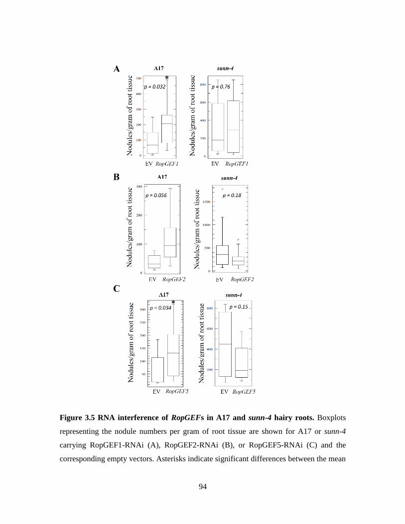
94
Figure 3.5 RNA interference of RopGEFs in A17 and sunn-4 hairy roots. Boxplots
representing the nodule numbers per gram of root tissue are shown for A17 or sunn-4
carrying RopGEF1-RNAi (A), RopGEF2-RNAi (B), or RopGEF5-RNAi (C) and the
corresponding empty vectors. Asterisks indicate significant differences between the mean

95
nodule number observed in RopGEF-RNAi line and the empty vector (EV) control as
determined by a Student’s t-test and the p-values are given.
Discussion
Rops and RopGEFs regulate many aspects of growth and development in plants
where they partner with a receptor kinase. The LRR-RLK responsible for nodule number
regulation, SUNN, had positive interactions with two RopGEFs, RopGEF1 and RopGEF5,
during a yeast-two hybrid experiment. Supporting the yeast-two hybrid results, we show
that SUNN can interact with RopGEF1, RopGEF5, and in addition, can interact with
RopGEF2 in vivo using the bimolecular fluorescence complementation (BiFC) assay. We
performed RNA interference against the three RopGEFs individually, RopGEF1,
RopGEF2 and RopGEF5, to deduce the role each played in nodulation. RNAi-RopGEF1,
-RopGEF2, and -RopGEF5 hairy root systems had an increased number of nodules formed
when compared to empty vectors, suggesting these specific RopGEFs are involved in
nodulation. In addition, lateral root phenotypes were also observed and reported.
Phylogenetic analysis of RopGEFs from Arabidopsis, M. truncatula, soybean, and
others, show four distinct groupings. The RopGEFs examined in this study include
RopGEF1, RopGEF2, and RopGEF5 from M. truncatula. Both RopGEF1 and RopGEF2
are expressed in tissues representing the shoot and the root; however, transcripts of
RopGEF5 were predominantly in the root and flower, and lower in other shoot tissues
(Riely, et al., 2011). SUNN is expressed in the vascular tissue in both aerial and root tissues
(Schnabel, et al., 2012). In AON, SUNN controls the number of nodules formed during

96
nodulation from the shoot, however SUNN is expressed in the root as well, and its root
function remains elusive (Schnabel, et al., 2012; Schnabel, et al., 2005; Penmetsa, et al.,
2003). Given the expression profile of SUNN and the RopGEFs, it is possible that the
RopGEF interactions with SUNN observed by BiFC could occur in either the root or the
shoot, with the exception of RopGEF5 which is expressed mainly in root tissues and
minimally in petioles and leaves.
Due to the difficulty of creating whole plant transgenic M. truncatula, we
investigated root phenotypes for the three RopGEFs using Agrobacterium rhizogenes-
mediated hairy root transformation to create transgenic root systems carrying RNAi
constructs to each of the three RopGEFs. This system allows us to silence each gene in a
specific genetic background in M. truncatula. In wild type A17 roots, we observed both
lateral root phenotypes and nodulation phenotypes suggesting that a single RopGEF can
regulate many processes. This is supported further with additional roles for RopGEF2 in
root hair formation (Riely, et al., 2011). Given the low number of RopGEFs and Rops
relative to the high number of pathways likely to require them in M. truncatula, multiple
functionality is plausible.
Riely, et al. (2011) previously silenced RopGEF2 in M. truncatula hairy roots using
the same construct as in this study looking specifically for root hair phenotypes (Riely, et
al., 2011). They concluded RopGEF2 had a role in root hair development and elongation
(Riely, et al., 2011). Root hairs are the primary mode of entry for the Rhizobia to enter the
root cells, so it is possible to have fewer nodules due to improperly elongated root hairs. In
fact, a number of genes isolated as suppressors of the har1 phenotype (the Lotus japonicus

97
orthologs of SUNN) affected root hair development (Murray, et al., 2006). Together with
the data suggesting SUNN interacts with the RopGEFs, we silenced the RopGEFs in a
sunn-4 background. The phenotypes observed in sunn-4 silenced roots, like those seen in
A17, were not suppressed. The increase in nodule number (>50%, with respect to empty
vectors) observed in A17 hairy roots was not seen in a sunn-4 background, making it
difficult to determine if the RopGEFs and SUNN function in the same pathway, as the
double mutants were not additive (Table 3.1)
RopGEF5-RNAi displayed a hypernodulation phenotype regardless of how the data
was analyzed. The hypernodulation phenotypes of RopGEF1-RNAi and RopGEF2-RNAi
depended on whether or not the size of the plant was taken into account. A17 wild type
roots will produce a certain number of nodules per root based on nitrogen need. The N
need, however, depends on the size of the plant. There were clear differences in the size of
the root systems among and within treatments and thus, root weight was determined for
each individual plant. Using the root weight, nodule counts per gram of tissue were
extracted. The hypernodulation phenotypes of RopGEF1-RNAi and RopGEF5-RNAi that
emerge when root weight is taken into account correlate with visual observations during
counting.
The lateral root phenotypes observed in silenced roots of all three RopGEF-RNAi
lines did not persist after inoculation with Rhizobia. In all cases, an increase in lateral roots
was seen when the RopGEFs were silenced, indicating a role in regulating the formation
of lateral roots, in addition to regulating the formation of new nodules. Interestingly, auxin
localization and transport is mediated by RopGEFs/Rop signaling for endocytosis and

98
trafficking of the PIN transporters (Xu, et al., 2010; Wu, et al., 2011; Huang, et al., 2014).
The local distribution and accumulation of auxin may also activate RopGEFs/Rops with
respect to their localization and association with the membrane (Wu, et al., 2011; Xu, et
al., 2010). In Arabidopsis, an accumulation of auxin at the site of root hair initiation
preceded RopGEF/Rop signaling with the RopGEF-interacting RLK, FERONIA (Wu, et
al., 2011; Xu, et al., 2010; Miyawaki and Yang, 2014; Duan, et al., 2010). The lateral root
phenotype and/or the hypernodulation phenotype displayed in the RopGEF silencing lines
may be due to changes in local auxin gradients influenced by the RopGEFs dependent
manner. This is especially interesting because of the changes in auxin response displayed
in a sunn mutant (van Noorden, et al., 2006).
The constructs, RopGEF2-RNAi and the EV(RopGEF2)-RNAi, used for hairy root
transformations contain a eYFP gene driven by the pUBIQ10 promoter for microscopic
analysis and for selection of transgenic roots. The YFP fluorescence was hard to see under
the microscope used for root selection. When the roots were ready to be counted, a post-
inoculation microscopy analysis was done on all plants. Due to the difficulty incurred with
selection via YFP, RopGEF2 plants had a consistently low percentage of transformed roots
when viewed post-inoculation, likely making the nodulation counts lower than they would
have been if most of the roots were transformed. Therefore, efforts to repeat RopGEF2
using a DsRed construct are underway.
RopGEF2 did not show up as an interacting protein during yeast-two hybrid studies
using the SUNN kinase domain but did interact in vivo studies using BiFC. As mentioned
previously, RopGEFs can be directly phosphorylated by RLKs. It is possible that SUNN

99
can phosphorylate RopGEF1 and RopGEF5 but relies on a partner kinase to phosphorylate
RopGEF2. In the tobacco system, this presumed partner kinase may exist, resulting in the
interaction between SUNN and RopGEF2, but only in vivo.
Interactions between the LRR-RLK, SUNN, and the guanine exchange factors,
RopGEF1, RopGEF2, and RopGEF5, were observed using a BiFC assay and a yeast-two
hybrid analysis. In addition to the interactions with SUNN, the silencing of RopGEF1 and
RopGEF5, and possibly RopGEF2, led to significant increases in nodule number. cDNA
collected from the roots of the RNAi lines is currently being analyzed via quantitative PCR
analysis to determine the level of gene knockdown. The results of this study indicate a role
for RopGEFs in nodulating roots of M. truncatula.
Materials and Methods
Plant materials and growth conditions
Seed of Medicago truncatula genotypes A17 and sunn-4 to be used for RNAi were
scarified and imbibed as described previously (Chapter 2) and allowed to germinate
overnight at room temperature. Seed of Nicotiana benthamiana were germinated in 3M
potting mix as described previously (Chapter 2). Four-week old tobacco plants were used
for bimolecular complementation assays.
Vector construction
A pBluescript cloning vector containing two multiple cloning sites interrupted by
an intron and driven by the constitutive 35S promoter was used as an intermediate cloning

100
vector. A RopGEF1-specific 150 bp fragment located in the 3’ UTR of MtRopGEF1 was
amplified from A17 genomic DNA. To create the sense fragment, the PCR primers were
modified to contain XhoI and KpnI restriction enzyme sites on the 5’ and 3’ end,
respectively. The antisense fragment was created using primers that added ClaI and BamHI
restriction enzyme sites on the 5’ and 3’ end, respectively, facilitating directional cloning
into the RNAi construct. Once completed, the fragment containing the 35S promoter, sense
fragment, intron, antisense fragment and the terminator was excised out of the pBluescript
cloning vector using restriction enzymes XhoI and XbaI and used to directionally clone
into the binary vector pCambia DSRed/35S (Figure 3.6, A) for plant transformation. The
binary vector pCDsRed/35S containing the RNAi construct was confirmed by sequencing
and was subsequently transformed into electrocompetent Agrobacterium rhizogenes strain
ArQUA1 to facilitate hairy root transformations.
RNAi constructs containing small fragments specific to RopGEF2 and RopGEF5
were obtained from our collaborator, Dr. Muthu Venkateschwaran from the University of
Wisconsin-Platteville. The binary vectors pK7GWIWG2(II)-R:MtRopGEF5-RNAi (spec)
and pH7GWIWG2(II)-YC3.6-MtRopGEF2-RNAi (spec) were received, in addition to the
empty vector constructs for each, in Agrobacterium rhizogenes strain MSU440 and
subcultured appropriately.
The complete coding sequences of MtRopGEF1 (Medtr4g075640), MtRopGEF2
(Medtr8g030850) and MtRopGEF5 (Medtr3g069590) were cloned from A17 genomic
DNA and were used to create donor plasmids using the vector pDONR201 (zeocin) as per
manufacturer’s instructions (Thermo Fisher Scientific, Waltham, MA). The donor vectors

101
were confirmed by sequencing and used to clone the genes into the BiFC binary vectors,
containing either the N-terminal (VYNE) or the C-terminal fragment (VYCE) of the
VENUS fluorophore, by the LR reaction of the gateway system (Thermo Fisher Scientific,
Waltham, MA). VYNE and VYCE vectors containing RopGEF1 and RopGEF5 were
transformed into Agrobacterium tumefaciens strain EHA105 and used in BiFC assays.
RopGEF6 was cloned into VYNE and VYCE as described in Chapter 2.
Figure 3.6 Hairy root transformations for RopGEF-RNAi. The binary vector pCambia
DSRed/35S containing the RNAi fragment, hairy roots at two weeks post-inoculation and
plants post-selection of transformed roots in perlite. The binary vector (A) contains the
RNAi fragment (sense-intron-antisense) driven by the 35S promoter, in addition to a
fluorescent marker driven by the UBIQ10 promoter. Transformed hairy roots (B) were
selected by microscopic analysis and the plants were transferred to the perlite system (C).

102
Bimolecular complementation assay in tobacco
The analysis of protein-protein interactions using the bimolecular fluorescence
complementation system in N. benthamiana was done as described previously (Chapter 2).
Briefly, bacterial cultures containing either VYNE-SUNN or VYCE-SUNN were mixed 1:1
with bacterial cultures containing either VYNE-RopGEF or VYCE-RopGEF to obtain
VYNE/VYCE combinations. Here, we use the VYNE-RopGEF1/VYCE-SUNN and
VYNE-SUNN/VYCE-RopGEF5 combinations to show interactions.
Agrobacterium-mediated hairy root transformations for RNA interference
Two days prior to inoculation of seedlings, lawn plates were created by plating 200
uL of overnight culture onto TY agar plates for each of the six A. rhizogenes cultures
carrying the RNAi constructs and the corresponding empty vectors. Hairy root
transformations were performed for twenty A17 and twenty sunn-4 plants for each vector,
combining for a total of twelve independent treatments, as described in (Chapter 2). After
inoculation with A. rhizogenes, the plants were placed on small square (100 x 100 x 15
mm) petri dishes containing modified HMF (Huo, et al., 2006). The plants were incubated
at 20°C for seven days to facilitate transformation and then moved to incubation at 25°C
to facilitate growth of transformed roots for two weeks (Figure 3.6, B).
In addition to the RNAi construct, the T-DNA transferred to the plant cells during
transformation also included a fluorescent selectable marker, either DsRED (RopGEF1-
RNAi and RopGEF5-RNAi) or YFP (RopGEF2-RNAi). To obtain only transgenic root
systems for nodulation purposes, non-transformed (non-fluorescent) roots were removed

103
prior to being planted in perlite (Figure 3.6, C). Transformed roots were selected using
either a GFP or DsRED cube on an Olympus SZH-10 stereoscopic system. The plants
received 10:20:10 fertilizer solution during watering for a week before being switched to
watering with caisson media without nitrate.
After five days of growth under nitrogen-limiting conditions, the RNAi plants were
inoculated with 6 mL of Sinorhizobia meliloti strain ABS7M (Bekki, et al., 1987) diluted
to a concentration of OD600=0.3 with culture media, applied to the crown of the root system.
Nodulation was assessed two weeks following inoculation by destructive counting with the
aid of 2.75x magnification optivisor. Nodule number and root weight were determined and
the root tissue was frozen for each individual plant.

104
Literature Cited
Bekki A, Trinchant J, Rigaud J (1987) Nitrogen fixation (C2H2 reduction) by Medicago
nodules and bacteroids under sodium chloride stress. Physiol Plantarum 71: 61-67
Berken A (2006) Rops in the spotlight of plant signal transduction. Cellular and Molecular
Life Sciences CMLS 63: 2446-2459
Berken A, Thomas C, Wittinghofer A (2005) A new family of RhoGEFs activates the
Rop molecular switch in plants. Nature 436: 1176-1180
Berken A, Wittinghofer A (2008) Structure and function of Rho-type molecular switches
in plants. Plant Physiol Bioch 46: 380-393
Chang F, Gu Y, Ma H, Yang Z (2013) AtPRK2 promotes ROP1 activation via RopGEFs
in the control of polarized pollen tube growth. Molecular Plant 6: 1187-1201
Duan Q, Kita D, Li C, Cheung AY, Wu HM (2010) FERONIA receptor-like kinase
regulates RHO GTPase signaling of root hair development. Proc Natl Acad Sci USA 107:
17821-17826
Gu Y, Li S, Lord EM, Yang Z (2006) Members of a novel class of Arabidopsis Rho
guanine nucleotide exchange factors control Rho GTPase-dependent polar growth. Plant
Cell 18: 366-381
Huang JB, Liu H, Chen M, Li X, Wang M, Yang Y, Wang C, Huang J, Liu G, Liu Y,
Xu J, Cheung AY, Tao LZ (2014) ROP3 GTPase contributes to polar auxin transport and
auxin responses and is important for embryogenesis and seedling growth in Arabidopsis.
Plant Cell 26: 3501-3518
Huo X, Schnabel E, Hughes K, Frugoli J (2006) RNAi phenotypes and the localization
of a protein:: GUS fusion imply a role for Medicago truncatula PIN genes in nodulation. J
Plant Growth Regul 25: 156-165
Ke D, Fang Q, Chen C, Zhu H, Chen T, Chang X, Yuan S, Kang H, Ma L, Hong Z,
Zhang Z (2012) The small GTPase ROP6 interacts with NFR5 and is involved in nodule
formation in Lotus japonicus. Plant Physiol 159: 131-143
Li Z, Liu D (2012) RopGEF1 and RopGEF4 are functional regulators of ROP11 GTPase
in ABA-mediated stomatal closure in Arabidopsis. FEBS Lett 586: 1253-1258

105
Miyawaki KN, Yang Z (2014) Extracellular signals and receptor-like kinases regulating
ROP GTPases in plants. Frontiers in plant science 5: Article 449
Murray J, Karas B, Ross L, Brachmann A, Wagg C, Geil R, Perry J, Nowakowski K,
MacGillivary M, Held M (2006) Genetic suppressors of the Lotus japonicus har1-1
hypernodulation phenotype. Mol Plant-Microbe Interact 19: 1082-1091
Penmetsa RV, Frugoli JA, Smith LS, Long SR, Cook DR (2003) Dual genetic pathways
controlling nodule number in Medicago truncatula. Plant Physiol 131: 998-1008
Riely BK, He H, Venkateshwaran M, Sarma B, Schraiber J, Ané J, Cook DR (2011)
Identification of legume RopGEF gene families and characterization of a Medicago
truncatula RopGEF mediating polar growth of root hairs. The Plant Journal 65: 230-243
Schnabel E, Karve A, Kassaw T, Mukherjee A, Zhou X, Hall T, Frugoli J (2012) The
M. truncatula SUNN gene is expressed in vascular tissue, similarly to RDN1, consistent
with the role of these nodulation regulation genes in long distance signaling. Plant
Signaling & Behavior 7: 4-6
Schnabel E, Journet EP, de Carvalho-Niebel F, Duc G, Frugoli J (2005) The Medicago
truncatula SUNN gene encodes a CLV1-like leucine-rich repeat receptor kinase that
regulates nodule number and root length. Plant Mol Biol 58: 809-822
van Noorden GE, Ross JJ, Reid JB, Rolfe BG, Mathesius U (2006) Defective long-
distance auxin transport regulation in the Medicago truncatula super numeric nodules
mutant. Plant Physiol 140: 1494-1506
Wu HM, Hazak O, Cheung AY, Yalovsky S (2011) RAC/ROP GTPases and auxin
signaling. Plant Cell 23: 1208-1218
Xu T, Wen M, Nagawa S, Fu Y, Chen J, Wu M, Perrot-Rechenmann C, Friml J, Jones
AM, Yang Z (2010) Cell surface-and rho GTPase-based auxin signaling controls cellular
interdigitation in Arabidopsis. Cell 143: 99-110

106
CHAPTER FOUR
A PROTEOMICS APPROACH TO IDENTIFYING
INTERACTING PARTNERS OF SUNN
Abstract
Proteomic studies to reveal protein-protein interactions can be powerful tools to further our
understanding of signaling networks in plants. Additionally, the functions of specific proteins can
be investigated by their associations with other proteins. The identification of novel protein
interactors of known bait proteins can be determined when protein pull-downs are coupled with
mass spectrometry. To better understand nodulation signaling through the SUNN kinase, we
utilized transgenic M. truncatula to isolate interacting partners in both root and shoot tissues under
various nodulation conditions. Through five independent experiments, we identify unique peptides
representing 22 M. truncatula proteins. Public RNA sequencing databases, information on
Arabidopsis homologs and structure analyses were consulted to elucidate possible roles the putative
interactors may have in nodulation and in other aspects of plant development.
Introduction
Protein-protein interactions (PPIs) drive cellular function in all domains of life.
Identifying PPIs that involve a protein of interest provides insight on the role such proteins
play in a given biological process. While there are several methods to study and identify
PPIs (i.e., split systems like BiFC and yeast-two hybrid), they usually require expression
in a non-native environment and preceding knowledge of candidate proteins (see Chapters
2 & 3 for candidate protein approaches). Co-immunoprecipitation of a protein used in
conjunction with mass spectrometry-based techniques is a powerful method for identifying
candidate PPIs. Often, these experiments are able to be performed under native conditions.

107
In addition, a treatment (such as biotic or abiotic stressors) can be applied to the system to
view plasticity within PPIs. Modern mass spectrometry (MS) techniques and
instrumentation allows for the processing of high complexity samples with a high enough
sensitivity to identify proteins of low abundance. Also, a co-immunoprecipitation coupled
LC-MS/MS approach eliminates the need for prior knowledge of PPIs or a candidate
protein list and is valuable in identifying novel proteins.
In Chapter 2, we showed that the LRR receptor kinase, SUNN, demonstrates
plasticity as a receptor kinase. Two proteins, CORYNE and CLAVATA2, known to be
involved in meristem development and maintenance in Arabidopsis, are capable of
interacting with SUNN to form probable signaling complexes in respect to nodule number
regulation (AON) in Medicago truncatula. Grafting experiments for SUNN and CRN
indicate a shoot control of nodulation by these proteins suggesting a functional signaling
complex in the shoot (Schnabel, et al., 2003; Schnabel, et al., 2010; Schnabel, et al., 2005;
Schnabel, et al., 2012; Penmetsa, et al., 2003).
The results from Chapter 2 suggest SUNN is part of a fluid receptor complex and
could change binding partners to facilitate response to different ligands depending on the
signaling needs of the plant. Recently, one ligand of HAR1, the Lotus japonicus equivalent
of SUNN, was found to be a small signaling peptide belonging to the CLE family. Beyond
this, very little is known about the signaling complexes that form to receive ligands during
AON, where these complexes are formed, if this is the only ligand, or what downstream
signaling events that occur to halt the formation of new nodules.

108
Due to the subcellular localization of SUNN to the plasma membrane (Chapter 2),
the interactions with the ligands and downstream signaling likely occurs there. The plasma
membrane is the central zone for cell-to-cell communication and transport. Membrane
proteins are notoriously hard to isolate and generally require advanced enrichment,
solubilization using detergents and subcellular fractionation to obtain pure components.
However, with system specific tweaks, proteomic analysis of plant plasma membranes has
been done in many plant systems (for review, see Komatsu, et al., 2007).
There are many instances where perception of the ligand induces endocytosis of the
receptor, targeting it for a new location or ubiquitin-targeted degradation by the vacuole
(Russinova, et al., 2004; Robatzek, et al., 2006). In these cases, the signaling cascades that
alter the specific biological process occurs while in transit, at the new location, or just by
the process of being recycled and the protein no longer performing its original task.
Although the parallelisms found between SUNN signaling in M. truncatula and the CLV1
signaling pathway in Arabidopsis (see Chapters 1 and 2) suggest SUNN’s signaling targets
are at the plasma membrane, it is possible that some downstream effects may depend on
the trafficking of SUNN.
Here, we identify novel protein interactions with the SUNN protein kinase during
and after the onset of AON. To find proteins that interact with SUNN at the plasma
membrane and during intracellular trafficking events, we utilized a total protein extraction
protocol with buffers containing a non-ionic detergent to facilitate the solubilization of
membrane components. Co-immunoprecipitation of the tagged SUNN protein kinase
followed with LC MS/MS to identify proteins precipitating with SUNN yielded several

109
new proteins as candidate interactors that may provide insight into how the signaling
cascade of AON works, including two novel LRR proteins.
Results
Experiments.
A total of five independent experiments were sent in three sets (MS Runs I-III) to
North Carolina State University (NCSU) for MS. Experiment 1 was processed and
analyzed by MS as a single run (MS Run I). Experiments 2 and 3 together constitute MS
Run II and experiments 4 and 5 were paired for both sample processing and analysis (MS
Run III). The experiments have not been replicated and are not intended to be viewed as
replicates of each other, but rather standalone experiments, as they each differ in the
generation of transgenic plants used, the days post-inoculation, and some environmental
conditions (Table 4.1). The conditions of each experiment and the progression of logic for
each change is detailed as follows:
Experiment 1. An F2 plant from the original sunn-4 transgenic in Chapter 2, F25,
was genotyped by PCR as a sunn-4 homozygote and determined by PCR and microscopy
to be carrying the transgene. A seed collection of progeny from F25 were used as a
transgenic tissue source. The purpose of this experiment was to determine the best elution
method, comparing competitive or acidic elution to the boiling method. The plants were
harvested at 10 days post inoculation (dpi) as in nodulation experiments in Chapter 2 and
the entire plant was processed.

110
Since identification of the transgene by Western blot was not always reliable with
the F25 progeny, the seed of an F2 population was used for Experiments 2 and 3. The shoot
tissue was separated from the root tissue and the samples were processed separately for all
remaining experiments.
Experiment 2. The F2 seed were collected after 7 dpi and the tissue separated as
previously described. Due to environmental issues unclear to us, the growth apparatus did
not produce any plants with nodules. The shoot samples gave a strong signal for the
transgene by Western blot, whereas the root samples’ signals were faint.
Experiment 3. F2 seed were grown, nodulated and collected at 4 dpi, rather than 7
or 10 dpi. This time point was chosen to coincide with the demonstrated onset of AON
(Kassaw and Frugoli, 2012). When blotted to detect the transgene, no bands were visible
in the root sample, though the shoot sample showed a faint band corresponding to SUNN.
Due to the faint transgene bands on Westerns and the number of peptides identified in MS,
we decided to maximize the tissue amount per experiment and to extend our dpi, to allow
the plants to grow larger. Concurrently, we realized that our samples could be diluted due
to the F2 generation segregating non-transgenic plants containing a native SUNN protein,
which would compete with the transgene for binding partners.
Experiment 4. Seed from an F4 population collected from three F3 plants that were
progeny of the F25 plant used previously and verified to be sunn-4 was grown, nodulated,
and plant material collected at 7 dpi. Western analysis was negative for the transgene.

111
Experiment 5. Because transgene copy number could be a factor limiting signal
strength and ultimately partner binding capacity in Experiments 3 and 4, we used progeny
of F2’s which may carry more copies of the transgene than F25. The F3 population was
grown, nodulated and collected at 7 dpi. Again, the Western analysis was negative for the
transgene.

112
Table 4.1 Details of each experiment sent for proteomic analysis. Details of each Experiment, including the transgenic
generation used, days post-inoculation, whether the whole plant transgenic (WPT) or wild type (A17) sample was whole or
separated, and the amount of tissue used. Information regarding co-immunoprecipitation and sample preparation include the
bead source for the pull-down, elution method and volume, as well as, the volume and method used for peptide extraction.
Transgenic
Generation DPI Samples
Tissue
amounts Bead Source Elution type
Eluate
amount
Processed
eluate
amount
MSF
Protocol
1 F3 10
WPT 0.57g Protein G + α-HA HA peptide 200uL 10ug In-solution
WPT Protein G + α-HA 0.1% TFA 100uL 10ug In-solution
WPT Protein G + α-HA Boil 100uL 30uL In-gel
A17 0.55g Protein G + α-HA HA peptide 200uL 10ug In-solution
A17 Protein G + α-HA 0.1% TFA 100uL 10ug In-solution
A17 Protein G + α-HA Boil 100uL 30uL In-gel
2 F2 7
WPT-SHOOT 1.2g α-HA conjugated Boil 100uL 25uL In-gel
WPT-ROOT 1.5g α-HA conjugated Boil 100uL 25uL In-gel
A17-SHOOT 1.2g α-HA conjugated Boil 100uL 25uL In-gel
A17-ROOT 1.2g α-HA conjugated Boil 100uL 25uL In-gel
3 F2 4
WPT-SHOOT 1.0g α-HA conjugated Boil 100uL 25uL In-gel
WPT-ROOT 2.55g α-HA conjugated Boil 100uL 25uL In-gel
A17-SHOOT 1.0g α-HA conjugated Boil 100uL 25uL In-gel
A17-ROOT 1.35g α-HA conjugated Boil 100uL 25uL In-gel
4
F4
{T38, T35,
T33} : T25 :
T12
7
WPT-SHOOT 2.36g α-HA conjugated Boil 100uL 50uL In-gel
WPT-ROOT 1.06g α-HA conjugated Boil 100uL 50uL In-gel
A17-SHOOT 2.3g α-HA conjugated Boil 100uL 50uL In-gel
A17-ROOT 1.06g α-HA conjugated Boil 100uL 50uL In-gel
5 F3 7
WPT-SHOOT 2.0g α-HA conjugated Boil 100uL 75uL In-gel
WPT-ROOT 1.5g α-HA conjugated Boil 100uL 75uL In-gel
A17-SHOOT 2.0g α-HA conjugated Boil 100uL 75uL In-gel
A17-ROOT 1.5g α-HA conjugated Boil 100uL 75uL In-gel

113
Identification of peptides in samples.
Peptides were identified by mass spectrometry for all experimental samples eluted
by the boil method. There were no peptides identified in samples where proteins were either
competitively eluted with excess HA peptide or eluted by a pH shift through treatment with
0.1% trifluoroacetic acid (see Experiment 1, Table 4.1). The target bait protein, SUNN,
was identified in all three MS runs and in all experiments with the exception of the root
samples of Experiments 2 and 5 or the shoot sample of Experiment 4. The number of tryptic
peptides identified within SUNN varied with protein coverage ranging from 1.03% to
6.88% among experiments (Table 4.2). The total number of peptides identified for each
MS run were 39, 712, and 965 for Runs I, II, and III, respectively, as summarized in Table
4.2. There were 26, 167, and 192 groups of protein candidates for MS Run I, II, and III,
respectively, based on the ability to link peptides identified to proteins in M. truncatula
(Table 4.2).
A total of 2, 9, 3, 2, and 12 (corresponding to Experiments 1-5) unique peptides
(identified in the transgenic pull down and not the wild type control) were identified in
shoot samples, root samples or both, and are summarized in Table 4.3. The peptide
sequences were blasted against the newest version of the Medicago truncatula genome (v.
Mt4.0) to determine all annotated Mt genes corresponding to a 100% identity across the
entire query length of the identified peptide and results are summarized in Table 4.3. Most
peptides had at least one hit with a maximum of four hits (homologous proteins). However,
three peptides did not display identity to any annotated gene in the genome at this 100%
identity cutoff. One of these peptides, GFGFITPDDGSEELFVHQSQIQTDGFR, has 58%

114
aa identity with Medtr1g026290, annotated as a ‘scythe protein ubiquitin-like domain
protein’ and 96% aa identity with the uncharacterized protein Glyma11g11290 of Glycine
max. The other two, SNNNVYNATATGIVNK and YSEITFPILSPDPATKR have 81%
and 88% aa identity, respectively, to scaffold Medtr0002s0690 annotated as
‘apocytochrome protein F’.

115
Table 4.2 A summary of peptides identified in all three independent MS runs representing five experiments.
MS RUN 1 MS RUN II MS RUN III
Total Peptides
Detected 39 712 965
Total Unique
Peptides 34 402 475
Total Number of
Groups 26 167 192
SUNN transgene
present?
Whole Shoot Root Shoot Root Shoot Root Shoot Root
Yes Yes No Yes Yes No Yes Yes No
% Coverage of
transgene detected 1.54% 6.67% n/a 2.98% 1.03% n/a 6.88% 1.03% n/a
Experiment 1 Experiment 2 Experiment 3 Experiment 4 Experiment 5

116
Peptides and corresponding proteins identified at 10 dpi in a nodulating F3 population
(Experiment 1).
Two peptides were unique to the transgenic sample in Experiment 1 corresponding
to Medtr8g086070 {annotated: dicarboxylate carrier} and Medtr4g085480 {annotated:
Serine/Threonine kinase, plant-type protein, putative}. The RNA sequencing database at
JCVI reported expression of these two genes in multiple tissues with an increased
abundance of the transcripts in rhizobial infected roots (Table 4.4). Medtr4g085480
represents a LRR protein which is analyzed and discussed in further detail below.
Peptides and corresponding proteins identified at 7 dpi in a non-nodulating F2 population
(Experiment 2).
The previously addressed unknown peptides corresponding to ‘apocytochrome f’
were the only two peptides unique to the shoot sample. The root sample submitted for
Experiment 2 contained peptides that corresponded to seven potential SUNN interacting
proteins including (as annotated in Mt4.0) a beta-hydroxyacyl-ACP dehydratase, subtilisin-
like serine protease (Arabidopsis homolog is AIR, auxin induced in roots), a peptidyl-
prolyl cis-trans isomerase, an inactive purple acid phosphatase-like protein, the heat shock
protein B1-2, as well as an albumin-2 protein (Table 4.3). All of the corresponding Mt
genes were expressed in root tissues and in most cases, transcripts had higher abundance
in roots treated with Nod factor and or infected with Rhizobia. However, it is important to
note that in this sample, the transgene was not detected by MS at levels above the filters
for peptide identification (see Materials and Methods). In addition, the samples used in this

117
experiment did not display obvious nodules seven days post-inoculation although it is
possible that the infection process, including Nod factor signaling, had begun.
Peptides and corresponding proteins identified at 4 dpi in a nodulating F2 population
(Experiment 3).
The peptide annotated “scythe protein ubiquitin-like domain protein” discussed
previously was identified in the root sample of Experiment 3; also identified in the root
sample was a peptide matching Medtr7g080370, annotated as “calreticulin” (Table 4.3).
RNA sequencing data in JBrowse recorded expression of this gene in all tissues for which
the datasets are available, with high transcript abundance in roots, including a control, Nod
factor treated, and Rhizobia infected samples (Table 4.4). In the shoot sample, a peptide
corresponding to Medtr4g119380 was identified and this protein is annotated as a
“transmembrane protein, putative” (Table 4.3) and will be analyzed and discussed further
below.
Peptides and corresponding proteins identified at 7 dpi in a F4 population (Experiment 4).
A “pre-rRNA-processing TSR1-like protein” (Medtr3g115250) and a “preprotein
translocase subunit SecA” (Medtr3g088555 or Medtr1g086050) were two potential SUNN
interacting proteins unique to the root sample (Table 4.3). There were no proteins identified
in the shoot sample that were not found in other datasets. The corresponding Mt genes were
expressed in all tissue datasets available on JBrowse RNA sequencing data (Table 4.4).

118
Table 4.3 Peptides observed in transgenic samples. A list of observed peptide sequences detected in the transgenic samples,
including which experimental sample the peptide was isolated in (cross-hatched shading) and a description of the corresponding
protein in M. truncatula.
Expt. 1 Expt. 2 Expt. 3 Expt. 4 Expt. 5
Description (Mt4.0) Peptide sequence Observed WPT Shoot Root Shoot Root Shoot Root Shoot Root
Unknown SNNNVYNATATGIVNK
YSEITFPILSPDPATKR
dicarboxylate carrier
protein IQLGQGSAASVTSTML
Serine/Threonine
kinase, plant-type
protein, putative TLTEVLFLGNK
transmembrane protein,
putative IASLIPKDVK
beta-hydroxyacyl-ACP
dehydratase FPAFPTILDINQIR
VIEYNPGVSAVAIK
subtilisin-like serine
protease SVAEGQEALSAGAK
peptidyl-prolyl cis-
trans isomerase VFFDMSVGGNPAGR
inactive purple acid
phosphatase-like
protein LVSISNFIAFANPK
heat shock protein
81-2 GIVDSEDLPLNISR

119
Table 4.3 continued.
Expt. 1 Expt. 2 Expt. 3 Expt. 4 Expt. 5
Description (Mt4.0) Peptide sequence Observed WPT Shoot Root Shoot Root Shoot Root Shoot Root
albumin-2 protein IAEMFPFLEGTGFEHGIDAAFR
calreticulin QVVDEVFANR
Unknown GFGFITPDDGSEELFVHQSQIQTDGFR
legume lectin beta
domain protein FSTIAQIVDLK
LRR receptor-like
kinase FSGNIPLSISSLYPTLR
nascent protein
associated complex
alpha chain LGLKPVTGVSR
SPTSDTYIIFGEAK
DEAD-box ATP-
dependent RNA
helicase VLITTDLLAR
tonoplast intrinsic
protein NIAVGTPQEATHPDTLK
pre-rRNA-processing
TSR1-like protein VADLLVFVASAR
PLAT-plant-stress
protein GNLDIFSGR
carboxy-terminal
region remorin AALEAQLR
eukaryotic translation
initiation factor 5A4 KLEDIVPSSHNCDVPHVNR
preprotein translocase
subunit SecA RIDNQLR

120
Table 4.4 RNA sequencing data for corresponding M. truncatula genes in specific tissues. Peptide sequences observed only
in transgenic samples as they are described by Mt v4.0 JBrowse with the corresponding Medtr gene identifier and expression
pattern based on RNA sequencing data. Tissues represented are nodules (N), root (control, RC), root treated with Nod factor
(RNF), root infected with Rhizobia (RI), blade (Bl), bud (Bu), seed pod (SP) and open flower (OF).
Potential protein
matches (Medtr #)
Expression in M. truncatula
Description Peptide sequence Observed RNA seq (Jbrowse)
Unknown (apocytochrome F) SNNNVYNATATGIVNK n/a
YSEITFPILSPDPATKR n/a
dicarboxylate carrier protein IQLGQGSAASVTSTML Medtr8g086070 N, RC, R4, RNF, RI, Bl, Bu, SP, OF
Serine/Threonine kinase, plant-
type protein, putative TLTEVLFLGNK Medtr4g085480 N, RC, R4, RNF, RI, Bl, Bu, SP, OF
transmembrane protein, putative IASLIPKDVK Medtr4g119380 N, R4, Bl, Bu, SP, OF
beta-hydroxyacyl-ACP dehydratase FPAFPTILDINQIR Medtr4g094265 N, RC, R4, RNF, RI, Bl, Bu, SP, OF
VIEYNPGVSAVAIK Medtr4g094265
subtilisin-like serine protease SVAEGQEALSAGAK Medtr7g081750 N, RC, R4, RNF, RI, Bl, Bu, SP, OF
Medtr7g081500 N, RC, R4, RNF, RI, Bl, Bu, SP, OF
Medtr7g081420 N, RC, R4, RNF, RI, Bu, SP, OF
peptidyl-prolyl cis-trans isomerase VFFDMSVGGNPAGR Medtr4g075290 N, RC, R4, RNF, RI, Bl, Bu, SP, OF
inactive purple acid phosphatase-
like protein LVSISNFIAFANPK Medtr3g074930 N, RC, R4, RNF, RI, Bl, Bu, SP, OF
heat shock protein 81-2 GIVDSEDLPLNISR Medtr5g096430 N, RC, R4, RNF, RI, Bl, Bu, SP, OF
Medtr5g096460 N, RC, R4, RNF, RI, Bl, Bu, SP, OF
albumin-2 protein IAEMFPFLEGTGFEHGIDAAFR Medtr0057s0100 N, RC, R4, RNF, RI, OF
calreticulin QVVDEVFANR Medtr7g080370 N, RC, R4, RNF, RI, Bl, Bu, SP, OF

121
Table 4.4 continued.
Potential protein
matches (Medtr #)
Expression in M. truncatula
Description Peptide sequence Observed RNA seq (Jbrowse)
Unknown GFGFITPDDGSEELFVHQSQIQTDGFR n/a n/a
legume lectin beta domain protein FSTIAQIVDLK Medtr3g027280 N, Bl, Bu
Medtr1g090957 N, Bl
LRR receptor-like kinase FSGNIPLSISSLYPTLR Medtr3g009050 N, RC, R4, RNF, RI , Bl, Bu, SP, OF
nascent protein associated complex
alpha chain
LGLKPVTGVSR Medtr3g092970 N, RC, R4, RNF, RI, Bl, Bu, SP, OF
SPTSDTYIIFGEAK Medtr7g088680 N, RC, R4, RNF, RI, Bl, Bu, SP, OF
DEAD-box ATP-dependent RNA
helicase
VLITTDLLAR Medtr4g125970 N, RC, R4, RNF, RI, Bl, Bu, SP, OF
Medtr2g039960 N, RC, R4, RNF, RI, Bl, Bu, SP, OF
Medtr8g018400 N, RC, R4, RNF, RI, Bl, Bu, SP, OF
Medtr2g099620 N, RC, R4, RNF, RI, Bl, Bu, SP, OF
tonoplast intrinsic protein NIAVGTPQEATHPDTLK Medtr7g103030 N, RC, R4, RNF, RI, Bl, Bu, SP, OF
pre-rRNA-processing TSR1-like
protein VADLLVFVASAR Medtr3g115250 N, RC, R4, RNF, RI, Bl, Bu, SP, OF
PLAT-plant-stress protein GNLDIFSGR Medtr8g090315 N, RC, R4, RNF, RI, Bl, Bu, SP, OF
Medtr3g087490 N, RC, R4, RNF, RI, Bl, Bu, SP, OF
carboxy-terminal region remorin AALEAQLR Medtr8g031370 N, RC, R4, RNF, RI, Bl, Bu, SP, OF
eukaryotic translation initiation
factor 5A4
KLEDIVPSSHNCDVPHVNR Medtr4g109130 N, RC, R4, RNF, RI, Bl, Bu, SP, OF
Medtr3g437680 N, RC, R4, RNF, RI, Bl, Bu, SP, OF
Medtr5g042990 N, RC, R4, RNF, RI, Bl, Bu, SP, OF
Medtr4g130917 N, RC, R4, RNF, RI, Bl, Bu, SP, OF
preprotein translocase subunit
SecA
RIDNQLR Medtr3g088555 N, RC, R4, RNF, RI, Bl, Bu, SP, OF
Medtr1g086050 N, RC, R4, RNF, RI, Bl, Bu, SP, OF

122
Peptides and corresponding proteins identified at 7 dpi in a F3 population (Experiment 5).
Five peptides unique to transgenic shoot tissue and six peptides unique to transgenic
root tissue were identified in the fifth experiment using a F3 population (Table 4.3). In
addition to the “preprotein translocase subunit SecA” found in Experiment 4 root samples,
the shoot sample of Experiment 5 included peptides corresponding to a legume “lectin beta
domain protein” (Medtr3g027280 or Medtr1g090957) (Table 4.3). The corresponding
legume lectin transcripts are both present in low amounts in the nodule and blade;
additionally, Medtr3g027280 had detectable transcript in the bud (Table 4.4).
Two peptides corresponding to Medtr3g092970 and Medtr7g088680 were
identified and both proteins are annotated as “a nascent protein associated complex alpha
chain” (Table 4.3). The expression pattern for both of these transcripts are similar and they
are highly abundant in root tissues, including roots treated with Nod factor and infected
with Rhizobia (Table 4.4). Interestingly, like the transgenic sample in Experiment 1, a
peptide FSGNIPLSISSLYPTLR (Medtr3g009050) annotated as a “LRR receptor-like
kinase” was identified in the shoot sample (Table 4.3).
The caveat for the root sample in Experiment 5 is that like the root sample of
Experiment 2 and the shoot sample of Experiment 4, the transgene was not represented by
any of the peptides identified in the sample in correspondence with selection guidelines
(Table 4.2; see Materials and Methods). Nonetheless, six peptides were identified within
the sample that were not present in wild type samples (Table 4.3). One of the peptides
present in the shoot sample of Experiment 5 corresponding to “nascent protein associated

123
complex alpha chain” (Medtr7g088680) was also found in the root sample (Table 4.3). In
addition, a “DEAD-box ATP-dependent RNA helicase”, a “eukaryotic translation
initiation factor 5A4”, and a “tonoplast intrinsic protein” (Medtr7g103030) were identified
in the sample (see Table 4.3 for Mt genes).
A peptide corresponding to a “carboxy-terminal region remorin” (Medtr8g031370)
was also found in this sample (Table 4.3). The transcript of this potential SUNN-interacting
protein was found in all tissues but had higher abundance in the root tissues, including the
Nod factor treated and Rhizobia infected roots according to the RNA sequencing database
(Table 4.4). In this case, the Arabidopsis homolog to Medtr8g031370, At3g61260, a
“remorin family protein” (59% aa identity over 94% of the protein queried), was not the
best match to the peptide itself. When blasted, the peptide had 89% identity to At2g45820,
also annotated as a “remorin family protein”.
Discussion
Our proteomics analysis identified many potential novel SUNN-protein
interactions. To isolate the transgene, a plasma membrane resident protein from the plasma
membrane, we refined our protein extraction protocols by introducing a detergent
solubilization step facilitating the release of detergent-soluble membrane resident proteins.
Using this protocol, we were able to effectively isolate SUNN-YFP. The extraction process
also allowed for the isolation of SUNN as it transited the ER and Golgi, which could
provide insight on what happens to SUNN if it is involved in ligand induced endocytosis
or whether it interacts with certain proteins during transport, however the design of the

124
study did not allow these distinctions to be made. It is possible that SUNN was isolated in
the study as it was being processed or trafficked, and not just residing in the plasma
membrane. However, the final list of proteins reside in various places, including the cell
wall, apoplast, plasma membrane, cytosol and even the plasmodesmata; in addition, the
identified LRR proteins are non-cytoplasmic and probably located in the apoplast.
The experimental method employed has some caveats. Because there is the
potential for false positives, all proteins identified as putative interactors will need to be
further validated by additional methods. Second, the use of the non-ionic detergent Triton
X-100 for protein solubility means that the detergent insoluble protein fraction was left
behind. There could be a loss of SUNN and interacting partners if SUNN interacts with a
protein that would be resident in this insoluble fraction or if SUNN becomes detergent
insoluble upon some interactions.
Nonetheless, useful information was obtained from the five experiments. The use
of homologs from other systems expanded the repertoire of tools available to investigate
the probable functions of the different proteins. The wealth of information in the
LegumeIP, JCVI, NCBI and TAIR databases was a valuable resource for elucidating
protein function and localization and can be indispensable in the future for data mining.
Experiment 1 - 10 dpi, nodulating plants. Putative interactors that emerged from
Experiment 1 included a dicarboxylate carrier and a LRR protein. The isolation of a peptide
corresponding to a dicarboxylate carrier is interesting because it is dicarboxylates

125
(particularly malate) that are supplied to the bacteroid across the symbiosome membrane
(see Chapter 1); however, its interaction with the receptor kinase cannot be explained.
The Arabidopsis homolog of Medtr4g085480, At1g49750, encodes a LRR-
containing protein (Table 4.5). Because of this, Medtr4g085480 was analyzed further
using an LRR search tool, a signal peptide prediction program TargetP, and a
transmembrane prediction program, Phobius. The protein is predicted to have six LRRs
(conserved β-strand motifs) and does not contain a kinase domain or a transmembrane
domain (Figure 4.1, A) (Bej, et al., 2014; http://lrrsearch.com/index.php?page=tool). A
signal peptide to the secretory pathway is predicted by TargetP (score = 0.956)
(Emanuelsson, et al., 2007). This signal peptide constitutes amino acids 1-28 as determined
by Phobius with a non-cytoplasmic domain from 29-366aa (Stockholm Bioinformatics
Center; http://phobius.sbc.su.se/; Käll, et al., 2004) (Figure 4.1, B). The gene ontology
terms associated with this gene include protein binding (0005515) and endomembrane
system (0012505) as indicated on the LegumeIP page from the Samuel Roberts Noble
Foundation (http://plantgrn.noble.org/LegumeIP/).
Given that the whole plant was used as the transgenic sample of Experiment 1, it is
impossible to determine where the interaction between Medtr4g085480 and SUNN would
occur. Nonetheless, STRING analysis of the At homolog yielded intriguing results (Figure
4.2). Surprisingly, the At homolog interacts with a calmodulin-like protein 10, a potential
calcium sensor (Figure 4.2). Even more intriguing was the ligand for such LRRs, the rapid
alkalization protein, RALF-like small signaling peptide 34 (Figure 4.2). If the LRR does
form a complex with SUNN we could speculate that RALFL peptides may be a ligand for

126
a specific signaling pathway. In 2008, a RALFL peptide in Medicago truncatula
(MtRALFL1) was implicated in nodulation as being upregulated during the onset of
symbiosis (Combier, et al., 2008). Furthermore, the overexpression of RALFL1 led to
decreased nodulation by means of aborted infection threads (Combier, et al., 2008).
Figure 4.1 Structural analysis of the LRR encoded by Medtr4g085480. The structure
of the LRR protein encoded by Medtr4g085480. (A) A cartoon schematic of
Medtr4g085480 depicting the locations of the proposed signal peptide (SP, red) and the
conserved LRR domains (yellow). Created using IBS (Liu, et al., 2015). (B) A prediction

127
profile of Medtr4g085480 for a signal peptide (red), cytoplasmic domains (green), non-
cytoplasmic domains (blue) and transmembrane domains (grey) across the full length
protein. (Stockholm Bioinformatics Center; http://phobius.sbc.su.se/; Käll, et al., 2004)

128
Table 4.5 Summary of Arabidopsis homologous proteins. Peptide sequences unique to transgenic samples, their Medtr gene
identifier and Mt descriptor are listed. The Arabidopsis homolog of either the full length Mt protein or the peptide sequence as
identified by MS was determined by blasting against the Arabidopsis protein database was identified and the percent coverage
and identity is listed. (--) indicates the entry is the same as the one above.
Mt Descriptor Peptide sequence Observed
Potential
protein matches
(Medtr #)
At Homolog At descriptor %
coverage
%
identity
Unknown SNNNVYNATATGIVNK n/a
YSEITFPILSPDPATKR n/a
dicarboxylate
carrier protein IQLGQGSAASVTSTML Medtr8g086070 At5g19760
Dicarboxylate/tricarbox
ylate carrier 100 81
Serine/Threonine
kinase, plant-
type protein,
putative
Medtr4g085480 At1g49750 Leucine-rich repeat-
containing protein 90 80
TLTEVLFLGNK At4g06744 Leucine-rich repeat-
containing protein 90 90
transmembrane
protein, putative
Medtr4g119380 n/a
IASLIPKDVK At3g09870 SAUR-like auxin-
responsive protein 90 78
beta-
hydroxyacyl-
ACP
dehydratase
Medtr4g094265 At2g22230 putative 3-hydroxyacyl-
ACP dehydratase 78 83
FPAFPTILDINQIR -- -- 100 86
VIEYNPGVSAVAIK -- -- 100 93
subtilisin-like
serine protease
Medtr7g081750 At2g04160
Subtilisin-like serine
endopeptidase family
protein
97 47
Medtr7g081500 -- -- 98 49
Medtr7g081420 -- -- 98 49
SVAEGQEALSAGAK n/a

129
Table 4.5 continued.
Mt Descriptor Peptide sequence Observed
Potential
protein matches
(Medtr #)
At Homolog At descriptor %
coverage
%
identity
peptidyl-prolyl
cis-trans
isomerase
Medtr4g075290 At2g16600 Encodes cytosolic
cyclophilin ROC3 98 84
VFFDMSVGGNPAGR At2g38730 Peptidyl-prolyl cis-trans
isomerase CYP22 100 79
inactive purple
acid
phosphatase-like
protein
Medtr3g074930 At5g50400 purple acid phosphatase
27 95 73
LVSISNFIAFANPK -- -- 100 64
heat shock
protein 81-2
Medtr5g096430 At5g56010 Heat shock protein 90-3 100 92
Medtr5g096460 -- -- 100 92
GIVDSEDLPLNISR -- -- 100 100
albumin-2
protein IAEMFPFLEGTGFEHGIDAAFR Medtr0057s0100 n/a
calreticulin
Medtr7g080370 At1g08450 Calreticulin-3 94 79
QVVDEVFANR AT3G23680
putative F-box
associated
ubiquitination effector
protein
80 88
Unknown
GFGFITPDDGSEELFVHQSQIQTDGFR At2g17870
Cold shock domain-
containing protein 100 81
legume lectin
beta domain
protein
Medtr3g027280 At5g55830
Probable L-type lectin-
domain containing
receptor kinase S.7
81 32
Medtr1g090957 At4g02410
L-type lectin-domain
containing receptor
kinase IV.3
64 36
FSTIAQIVDLK n/a

130
Table 4.5 continued.
Mt Descriptor Peptide sequence Observed
Potential
protein matches
(Medtr #)
At Homolog At descriptor %
coverage
%
identity
LRR receptor-
like kinase
Medtr3g009050 At1g33590 leucine-rich repeat
(LRR) family protein 94 67
FSGNIPLSISSLYPTLR At1g33670 leucine-rich repeat
family protein 94 75
nascent protein
associated
complex alpha
chain
Medtr3g092970 At3g49470
nascent polypeptide-
associated complex
subunit alpha-like
protein 2
100 67
Medtr7g088680 At3g12390
Nascent polypeptide-
associated complex
subunit alpha-like
protein 1
99 75
LGLKPVTGVSR At3g49470 -- 100 91
SPTSDTYIIFGEAK At3g12390 -- 100 86
DEAD-box
ATP-dependent
RNA helicase
Medtr4g125970 n/a
Medtr2g039960 At3g13920 Eukaryotic initiation
factor 4A-1 100 92
Medtr8g018400 At3g13920 -- 100 91
Medtr2g099620 At1g54270 -- 100 93
VLITTDLLAR matches
both above -- 100 100
tonoplast
intrinsic protein
Medtr7g103030 At2g36830 Aquaporin TIP1-1 100 80
NIAVGTPQEATHPDTLK At2g36830 -- 100 76
pre-rRNA-
processing
TSR1-like
protein
Medtr3g115250 At1g42440
VADLLVFVASAR

131
Table 4.5 continued.
Mt Descriptor Peptide sequence Observed
Potential
protein matches
(Medtr #)
At Homolog At descriptor %
coverage
%
identity
PLAT-plant-
stress protein
Medtr8g090315 AT2G22170
PLAT-plant-stress
domain-containing
protein
69 67
Medtr3g087490 AT2G22170 -- 68 72
GNLDIFSGR -- -- 100 100
carboxy-terminal
region remorin
Medtr8g031370 At3g61260 remorin family protein 94 59
AALEAQLR At2g45820 -- 100 89

132
Figure 4.2 An interaction network for At1G49750, a LRR protein, homolog of
Medtr4g085480. Interactions include RALFL34, ralf-like 34; At5G37670, heat shock
protein 15.7; SVL2, SHV3-like 2; At3G26560, ATP-dependent RNA helicase
DHX8/PRP22; At2G41090, calmodulin-like protein 10; APL1, ADP glucose
pyrophosphorylase large subunit 1; At5G38980, uncharacterized protein; PIRL1, plant
intracellular ras group-related LRR 1; PIRL7, plant intracellular ras group-related LRR 7;
and PIRL3, plant intracellular ras group-related LRR 3.

133
Experiment 2 - 7 days post-inoculation, non-nodulating plants. In the root samples,
several proteins were identified as putative SUNN interactors and among them were a
subtilisin protease, a peptidyl-prolyl cis-trans isomerase, and a purple acid phosphatase.
This sample is intriguing in that nodulation did not occur. Therefore, it is easy to speculate
that the interactions seen within this sample relate to additional roles SUNN may play in
the root. Purple acid phosphatases are non-specific phosphatases and mostly involved in
the uptake and recycling of phosphorus for the plant. In soybean, nodulation-induced
expression of purple acid phosphatases has been shown under Pi-limiting conditions (Li, et
al., 2012), indicating some role during symbioses.
The functions listed on TAIR for the At homolog of the serine subtilisin protease
include lateral root formation and proteolysis (Neuteboom, et al., 1999a; Swarup, et al.,
2008; Neuteboom, et al., 1999b) (Table 4.5). This At gene was identified as upregulated in
response to auxin in root cultures, named for the phenotype (AIR3), and required for auxin-
induced lateral root formation; it also accumulates at the site of lateral root emergence
(Neuteboom, et al., 1999a).
Another protein related to auxin signaling is the cyclophilin, peptidyl-prolyl cis-
trans isomerase, which may have various roles in plant development. Recently, roles for
cyclophilin in defense response have been demonstrated, and particularly, roles in signal
transduction (Afzal, et al., 2013). The tomato homolog of the cyclophilin isolated here,
Cyp1, regulates root development (including lateral roots) by activating auxin-response
pathways and is capable of traveling along the phloem (Spiegelman, et al., 2015). Other
cyclophilins have demonstrated phloem-travel and the ability to interact with LRR

134
receptors (Afzal, et al., 2013). Therefore, it is possible that cyclophilins may interact with
the LRR domain of SUNN in the root, influencing root growth and development.
Experiment 3 - 4 dpi, nodulating plants. Calreticulin, a Ca2+ binding ER resident
protein that has a number of functions related to calcium signaling was identified in the
root sample. A peptide with 58% identity to a “scythe protein ubiquitin-like domain
protein” was also detected in the root sample of these nodulating plants and may relate
directly to the calreticulin protein observed. Recently, a Scythe_N Ubiquitin-like domain
protein, CIP73, was identified in a yeast two-hybrid screen for interacting partners of the
calcium, calmodulin-dependent protein kinase (CCaMK) in Lotus japonicus (homolog of
MtDMI3) (Kang, et al., 2011). The expression of CIP73 was mainly in the root tissues and
was significantly decreased upon Rhizobial infection of the roots. In subsequent studies,
CIP73 interacted with a heat-shock protein that negatively regulated nodule number (Kang,
et al., 2015). An interaction network based on the soybean homolog Glyma11G11290
indicates it can also interact with GmAGL15, Agamous-like15, a TF responsible for
ethylene response (Zheng, et al., 2013). High ethylene controls nodule number locally by
inhibiting early nodulation events, including Ca2+ spiking and root hair curling (Mortier, et
al., 2012). These studies indicate similar proteins, such as the one identified in this study,
may play roles in the nodulation pathway.
In addition, the shoot sample contained a peptide sequence that when blasted to the
M. truncatula genome, matches a transmembrane protein, Medtr4g119380 (Table 4.3).
Interestingly, when the full length protein of Medtr4g119380 is used as the blast query to

135
the Arabidopsis protein database, there is no obvious homolog; however, if the peptide
sequence is used instead, there is a 78% identity to At3g09870, annotated as ‘SAUR-like
auxin responsive protein’, over 90% of the queried sequence (Table 4.5).
Probius analysis of the protein sequence of Medtr4g119380 predicted a cytoplasmic
region spanning amino acids 1-183 (which could contain a non-cytoplasmic region and two
additional transmembrane regions), a transmembrane region (184-204) and a short non-
cytoplasmic tail (205-210) (Figure 4.3). There was no predicted signal peptide when
analyzed by TargetP. A conserved domain of the Ribosomal L18e-like superfamily was
found at amino acids 100-192. The peptide that was identified by MS, IASLIPKDVK,
contains three amino acids which are conserved in the ribosomal L18e-like superfamily
(underlined).
Figure 4.3 Structure analysis of the transmembrane protein, Medtr4g119380. A
prediction profile of Medtr4g119380 for cytoplasmic domains (green), non-cytoplasmic

136
domains (blue), and transmembrane domains (grey) across the full-length protein.
Obtained from Stockholm Bioinformatics Center; http://phobius.sbc.su.se/; Käll, et al.,
2004).
Experiment 4 - 7 dpi, nodulating plants. Very little information was obtained from
root and shoot samples from this experiment. Two peptides were identified in the
transgenic samples that were not identified in the wild type samples. One corresponds to
the M. truncatula gene that encodes the pre-protein translocase subunit SecA, responsible
for transport of proteins into and across the membranes. In addition, a pre-rRNA-
processing TSR1-like protein, required for ribosomal assembly, was also identified but its
function suggests this may be a false positive interactor of SUNN.
Experiment 5 - 7 dpi, nodulating plants. The samples in this experiment produced
the most putative SUNN interactors. In addition to the remorin discussed below, several
other proteins were identified in the root sample of experiment 5. These included a DEAD
box ATP-dependent RNA helicase, a nascent protein associated complex alpha chain, and
a eukaryotic translation initiation factor 5A4 which because of their functions, are also
likely to be false positive interactors of SUNN. A PLAT-plant-stress protein was also
among the proteins in the root sample. Very little information is available regarding the
possible function of this protein, however members of this family have been implicated as
regulators in stress signaling and may have a role in abscisic acid signaling (Hyun, et al.,
2014).

137
There is no information in the literature on the carboxy-terminal region remorin for
Medicago truncatula, however the two At homologs have been studied (Table 4.5). Data
contained in TAIR indicates subcellular localization of these proteins to the plasma
membrane and the plasmodesmata with a function in DNA and protein binding (Fernandez-
Calvino, et al., 2011; Nuhse, et al., 2003). In addition, At2g45820 is expressed more highly
in apical tissues and data show an induction of the gene by auxin (Alliotte, et al., 1989).
From TAIR database information, At2g45820 is induced approximately 10-fold in
response to auxin but not cytokinin and was identified in the late 1980’s as an auxin
regulated DNA-binding protein (Alliotte, et al., 1989), then later classified as a remorin.
Interestingly, the STRING analysis of At2g45820 indicates an interaction with the
response regulators, ARR3 and ARR4. ARR3 and ARR4 are Type A response regulators
implicated in negative regulation of cytokinin signaling (To, et al., 2004) (Figure 4.4).
Direct binding of ARR4 to At2g45820 has been demonstrated by yeast two hybrid studies
(Yamada, et al., 1998). ARR4, in contrast to At2g45820, is rapidly induced by cytokinin
treatment (Yamada, et al., 1998).
In AON, the shoot derived inhibition of nodulation is correlated with changes in
cytokinin and auxin levels, and in the root, the changes in cytokinin and auxin levels drive
the formation of nodules. As described in Chapter 1, the direct binding of CLE peptides to
SUNN in the shoot is thought to trigger the production of shoot-derived cytokinins.
Cytokinins have been implicated in reducing polar auxin transport. A decrease in auxin
flux has been observed after Rhizobial inoculation in M. truncatula and it is possible that
this is caused by the increase in shoot-derived cytokinins. Supporting this, in a sunn-1

138
mutant defective in downstream signaling, the auxin flux from shoot to root is much higher
than that of wild type plants (van Noorden, et al., 2006; Mortier, et al., 2012). Given the
functions of the remorin protein in Arabidopsis and the relationships seen between
auxin/cytokinin and SUNN in M. truncatula, a similar mechanism could be possible in the
root system where SUNN could function as a regulatory phosphor donor, allowing the
interaction of remorin and possibly ARRs to mandate auxin/cytokinin signaling balance as
needed.
Figure 4.4 An interaction network for At2G45820, a remorin family protein.
Interactions include At1G45207, remorin-like protein; At1G67590, remorin family
protein; At4G36970, remorin family protein; At3G06390, uncharacterized protein;
At5G14150, uncharacterized protein; At5G53650, uncharacterized protein; At3G11800,

139
uncharacterized protein; At2g38480, uncharacterized protein; ARR3 and ARR4, Type A
regulatory response proteins discussed in the text.
As shown in Chapter 2, SUNN could participate in a dynamic receptor complex
with evidence that the binding partners could change to meet the signaling demands of the
cell. Therefore, it should not be surprising that two LRR proteins were found in our
proteomics study. These proteins could be co-receptors with SUNN, conferring altered
ligand specificity. These two proteins do not contain a transmembrane domain and they are
secreted outside of the cell where the LRR domain of SUNN would also be located, making
them able to interact to form a functional signaling receptor complex.
The second LRR protein identified was detected in the shoot sample of Experiment
5. Structure analysis indicated the presence of LRR-RI domains in addition to the
conserved LRR domains (Figure 4.5 and Figure 4.6, A). To deduce the exact number of
LRR regions present in Medtr3g009050, a LRR search tool was utilized
(http://lrrsearch.com/index.php?page=tool) (Bej, et al., 2014). This search tool predicts 13
conserved β-strand motifs (underlined, Figure 4.5; Figure 4.6, A). Some of the LRR repeats
in this protein are similar to the LRR-(ribonuclease inhibitor-like) superfamily.

140
Figure 4.5 Multiple sequence alignment of Medtr3g009050 homologs. Alignments are
shown for Cicer arietinum (chickpea), Lotus japonicus, Glycine max (soybean) and
Arabidopsis thaliana using ClustalW. Conserved residues are marked with an asterisks (*).
Conserved substitutions and semi-conserved substitutions are also marked (.) and (:),

141
respectively. LRR-RI domains are highlighted by boxes and the conserved LRR β-domains
are underlined. Sequence identity to Medtr3g009050 are chickpea (82.77%), Lotus
(75.11%), soybean (69.39%) and Arabidopsis (64.57%).
The hallmark of LRR (RI-like) domains is that they are arranged in a β-strand-turn-
α-helix orientation giving rise to a horseshoe conformation with a rigid interior (where the
β-strands are located) and an exterior of α-helices (Matsushima and Miyashita, 2012).
These domains consist of the variable region motif of SGxIPxxLxxLxx where, S = S or T;
G = G or S; I = I or L; L = L, I, V, F or M followed by a conserved domain of
LxxLxLxxNxL where, L = L, I, V or F; N = N, T, S or C; C = C, S or N (Matsushima and
Miyashita, 2012). The LRR-(RI-like) domains are conserved in homologous proteins in the
legumes when aligned by protein sequence, Glycine max (Soybean; GenBank Accession:
ACM89594), Cicer arietinum (Chickpea; Genbank Accession: XP_004499669), Lotus
japonicus (Lotus; GenBank Accession: AFK38506) and in Arabidopsis thaliana
(Arabidopsis; GenBank Accession: AEE31606) highlighted in Figure 4.5. Eight full
repeats of the variable domain and conserved domain were observed in the amino acid
sequence (boxes; Figure 4.5). Other instances of the conserved domain (β-strands) are
present throughout the sequence (underlined; Figure 4.6, A).
Protein sequence analysis using the signal peptide and transmembrane domain
predictor, Phobius (Stockholm Bioinformatics Center; http://phobius.sbc.su.se/; Käll, et
al., 2004), predicts amino acids 1-26 as a signal peptide to the secretory pathway (score of

142
0.957) and the remainder of the protein to be non-cytoplasmic with no transmembrane
domain (Figure 4.6, B).
Figure 4.6 The structure of the LRR protein encoded by Medtr3g009050. (A) A
cartoon schematic of Medtr3g009050 depicting the locations of the proposed signal peptide
(SP, red), LRR-RI domains (cyan) and the conserved LRR domains (yellow). Created with
IBS (Liu, et al., 2015). (B) A prediction profile of Medtr3g009050 for cytoplasmic domains
(green), non-cytoplasmic domains (blue) and transmembrane domains (grey) across the
full length protein. (Stockholm Bioinformatics Center; http://phobius.sbc.su.se/; Käll, et
al., 2004)

143
RNA transcript analysis of nodulating plants treated with nitrate indicated
Medtr3g009050 is induced by nitrate (Cabeza, et al., 2014). It is possible that in response
to nitrate, SUNN partners with the LRR protein to relay the “stop nodulation” signal from
the shoot. The Arabidopsis homolog, At1g33590, an LRR family protein may be associated
with lateral root formation and is induced upon auxin treatment (Table 4.5) (Laskowski, et
al., 2006). STRING analysis of At1G33590 revealed high confidence interactions with a
number of proteins including protein phosphatase C, a protein kinase, map 4 kinase, an
additional LRR protein, a cyclase associated protein, and several G-proteins (for relevance
here, see Chapter 3) (Figure 4.7).
Figure 4.7 An interaction network for At1G33590, a LRR protein. Interactions include
At1G33600, LRR-containing protein; At2G20050, protein phosphatase 2C and cyclic

144
nucleotide-binding/kinase domain-containing protein; SHD, SHEPHERD; At3G15220,
putative protein kinase; MAP4K1, map 4 kinase alpha 1; At1G13600, LRR-containing
protein; CAP1, cyclase associated protein 1; At3G26560, ATP-dependent RNA helicase
DHX8/PRP22; XLG2, extra-large GTP-binding protein 2; GPA1, G protein alpha subunit
1; XLG1, extra-large G-protein 1.
Proteins known to interact with the At homolog included a map 4 kinase,
SHEPHERD and several G-proteins. In Chapter 3, we discussed the evidence for G-protein
signaling in nodulation and interactions with SUNN. The possible role of G-protein
signaling in the onset of nodulation has a genetic basis in that Choudhury and colleagues
determined that the expression profiles of G-proteins changes in soybean nodules in
response to Rhizobia (Choudhury and Pandey, 2013). Specifically, they found expression
to be higher in hypernodulating mutants (nts382, a mutation in NARK - the homolog of
SUNN) and lower in non-nodulating mutants with respect to wild type when challenged
with bacteria, suggesting a clear role of G-proteins in nodulation signaling and also, in
AON (Choudhury and Pandey, 2013). Therefore, it is possible that SUNN and its putative
interacting LRR receptor Medtr3g009050 are coupled to a G-protein for signaling.
Interestingly but perhaps not surprising, the molecular chaperone SHEPHERD
(SHD) was also identified as an interactor of the LRR protein, Medtr3g007050. SHD, an
ER resident heat shock protein has been shown to play a role in the folding of CLAVATA1
(CLV1) proteins and/or in the production of complexes involving CLV1 during shoot
apical meristem maintenance (Ishiguro, et al., 2002). Its requirement in CLV1 signaling

145
pathways suggests that it may be important in SUNN signaling due to the parallels in the
mechanisms of signaling between the two pathways (see Chapter 2).
In this study, we have demonstrated the use of co-immunoprecipitation and LC-
MS/MS to identify novel putative interactors of the SUNN kinase. A total of 28 unique
peptides across five independent experiments from both shoot and root tissues were
identified and investigated in preliminary data mining. The many interesting proteins and
relationships identified in this work will need to be studied in further detail. The work
forms the basis of many hypotheses detailed above, which can be developed and tested to
help understand the role SUNN plays in both the shoot and the roots during nodulation and
to further elucidate its role in AON.
Materials and Methods
Five independent experiments were conducted. Each experiment varied in type of
tissue (root/shoot, whole plant), transgenic generation (F2, F3 or F4), type of elution used,
volume of column elution and amount of elution used for analysis. A summary of the five
experiments is found in Table 4.1. The methods for protein extraction, mass spectrometry
sample preparation and the instrumentation used in analysis were consistent throughout all
experiments. The general workflow for the experiments is documented in Figure 4.8.

146
Figure 4.8 Proteomics workflow. A schematic representation of the overall workflow of
the proteomics study from plant growth to identification of peptides after co-
immunoprecipitation via the transgene SUNN.
Generation of transgenic plant material
Populations of Medicago truncatula sunn-4 containing the transgene SUNN-
YFP/HA (see Chapter 2) were used for Co-IP analysis. Seed were collected individually
from F1 progeny, grown and allowed to self, generating the F2 and subsequent populations.
The populations of plants used in this study are diagrammed in Figure 4.9. Information on

147
the specific generation used in a given experiment is found in Table 4.1 and discussed in
the text.
Figure 4.9 The generations of transgenics used in the proteomics analysis. A schematic
representation of the progeny from 35S:SUNN-YFP/HA x sunn-4 parents including the
segregation of wild type SUNN and the transgene within each successive generation as
determined by PCR (sunn-4 and the trangene) and microscopic analysis of transgene
expression.

148
Plant growth conditions
M. truncatula seeds from transgenic and wild type (A17) plants were scarified and
imbibed as in Chapter 2 and were grown aeroponically on a caisson as previously described
(Penmetsa and Cook, 1997). Sinorhizobium medicae, strain ABS7 (Bekki, et al., 1987) was
used as inoculum three days after loading. Plants were removed and inspected visually for
rescue of phenotype when applicable (after the F2 generation) and for the presence of the
transgene, as detected by an Olympus SZH-10 stereoscopic system equipped with a GFP
cube. In the cases where root and shoot tissue were processed and analyzed separately, the
plant was bisected where the hypocotyl and root join.
Protein extraction
Fresh tissue was ground in liquid nitrogen using a chilled mortar and pestle before
being transferred into 5 mL of homogenization buffer (50 mM HEPES-KOH, pH 7.5; 10
mM EDTA, pH 8.0; 25 mM sucrose; 0.6% polyvinylpyrrolidone) supplemented with a
protease inhibitor cocktail (cOmplete™, Roche Life Science, Indianapolis, IN). Triton X-
100 was added to a final concentration of 0.5% to help with solubilization of membranes
(Luche, et al., 2006). The material was further homogenized using a hand held tissue
homogenizer (Tissue Tearor™) with a 7 mm x 75 mm open slotted adaptor operating at
30,000 rpm. The tissue slurry was placed on a rotating mixer for overnight (12-16 h)
incubation at 4°C to facilitate complete solubilization of membranes. The resulting slurry
was cleared of debris by centrifugation at 10,000 x g in a table top centrifuge for ten
149
minutes or until fully cleared. The supernatant was collected and is referred to as the
“lysate”.
Co-immunoprecipitation
Co-immunoprecipitation was carried out using magnetic beads either equipped
with Protein G (Life Technologies, Carlsbad, CA) or conjugated to the hemagluttinin (HA)
primary antibody (Life Technologies, Carlsbad, CA). In cases where Protein G beads were
used, the HA primary antibody was conjugated to the Protein G beads using the coupling
reagent dimethyl pimelimidate DMP (Thermo Scientific, Waltham, MA) as per
manufacturer’s protocol. In short, for each sample, 50 uL of beads were washed and
incubated with 10 uL α-HA antibody (1 mg/mL) for 40 minutes at room temperature
shaking at 230 rpm. After washing, the antibody was chemically coupled to the beads by
suspension in 40 mM DMP (in 0.2 M triethanolamine buffer, pH 8.2) with gentle mixing
for one hour at room temperature. The cross-linking reaction was terminated by the
addition of 50 mM Tris, pH 7.5 for fifteen minutes and washed thoroughly with phosphate-
buffered saline (PBS) with 0.05% Tween-20. For cases where magnetic beads were
purchased already conjugated to the HA antibody, the beads were prepared as per
manufacturer’s instructions (Pierce, ThermoFisher Scientific, Waltham, MA). In short, 25
uL of beads were washed with Tris-buffered saline (TBS) with 0.05% Tween-20 for a total
of three times before used for each co-IP sample. A summary of the experiments using
Protein G/α-HA magnetic beads versus α-HA magnetic beads can be found in Table 4.1.

150
After the final wash, the magnetic beads were incubated with 4 mL of sample lysate
at 4°C for 12-16 h to allow antibody binding to the target protein. In all cases, A17 without
the transgene served as a control for off-target or non-specific binding of proteins to the
beads or to the antibody. The beads were collected for 30 minutes with use of a high
strength magnet at 4°C and the supernatant collected for Western blot analysis as the “flow
through” sample. The beads were washed three times with TBS with 0.05% Tween-20.
The proteins were eluted either by a sharp increase in acidity, competitively with excess
HA peptide, or by boiling in Laemmli buffer (3X, New England Biolabs, Ipswich, MA)
containing β-mercaptoethanol (β-ME). For samples in Experiment 1, the beads were first
suspended in 100 uL of HA peptide (2 mg/mL; Pierce, ThermoFisher Scientific, Waltham,
MA) and then incubated at 37°C for 15 minutes on a platform shaker, twice. The total
eluate (200 uL) was collected and used in analysis as “HA elution”. The same beads were
extracted a second time in 100 uL of 0.1% trifluoroacetic acid (TFA) and mixed for 10
minutes. The supernatant was extracted, neutralized using 15 uL of 2 M Tris-HCl, pH 8.0,
and used for both WB analysis and in-solution digests for MS analysis as “TFA elution”.
To ensure all proteins were eluted from the beads, the beads were suspended in 100 uL of
Laemmli buffer and boiled at 95°C for five minutes. The supernatant was collected as the
“boil elution”. For all other experiments, the proteins were eluted only by boiling the beads
in Laemmli buffer (+β-ME) as described, as this method yielded the best results.
Western blot analysis
The protein eluates were analyzed individually by Western blot using 20 uL eluate
and 10 uL 3X Laemmli buffer (+ βME) to constitute a 30 uL sample. For boil samples, 20

151
uL was loaded for WB analysis. Samples were loaded onto a 6% SDS polyacrylamide gel
with a 5% stacking gel and run at 100 V in a Mini-PROTEAN gel electrophoresis tank
(Bio-Rad, Hercules, CA) with electrophoresis buffer (25 mM Tris, 192 mM Glycine, 0.1%
SDS) for two hours or until the dye front reached the bottom of the gel. The Rainbow ECL
protein marker was used as the standard (GE Life Sciences, Pittsburgh, PA). The gels were
soaked in Western transfer buffer (25 mM Tris, 192 mM Glycine, 10% (v/v) methanol) for
five minutes prior to being blotted onto polyvinylidene fluoride (PVDF) membrane
overnight at 4°C and 20 V using a wet transfer system (Bio-Rad, Hercules, CA). Following
transfer, the membranes were fixed in 8% acetic acid, rotating for 1 hour. The membranes
were blocked using 3-4% nonfat dry milk dissolved in TBS with 0.05% Tween-20, rotating
at room temperature for 1-3 hours. Blocked membranes were incubated in a 1:4,000
dilution of primary antibody (α-HA, mouse monoclonal; Pierce, ThermoFisher Scientific,
Waltham, MA), rinsed and incubated in a 1:10,000 dilution of secondary antibody (Cy5
conjugated α-mouse, goat monoclonal (Lot: 9658403); Pierce, ThermoFisher Scientific,
Waltham, MA). An Amersham Biosciences Typhoon Imager 9400 (GE Healthcare,
Piscataway, NJ, USA), equipped with a 635nm laser line for excitation of Cy5 and an LPR
665 emission filter, was used for imaging.
Mass spectrometry preparation of samples
In-gel digestion. The samples intended for MS analysis were loaded and run on a
1.5 mm 10% SDS-PAGE acrylamide gel at 70 V until the dye front was approximately 25
mm into the gel (for a description of volumes added for each experiment, see Table 4.1).
The gel was subjected to fixing with 4% (v/v) ethanol and 10% acetic acid (v/v) before

152
staining overnight (12-16 h) in Coomassie Brilliant Blue G-250 stain (0.1% Coomassie
Brilliant Blue G-250; 2% phosphoric acid; 10% ammonium sulfate; 45% methanol). The
gel lanes for each sample were cut into 2 mm x 1.5 mm x 5 mm pieces and placed in a
acetonitrile (ACN; Fisher Scientific, Waltham, MA)-rinsed 2 mL Eppendorf tube. The
samples were then destained in a 50:50 mixture (v/v) of 100 mM ammonium bicarbonate
(NH4HCO3; Sigma-Aldrich, St. Louis, MO) buffer: ACN changing the mixture as needed
until all blue coloring was gone. The samples were all subjected to reduction, alkylation
and tryptic digest as per the protocol suggested by North Carolina State University Mass
Spectrometry Facility. The gel plugs were dehydrated in ACN before a 30 minute
incubation at 56°C in 10 mM dithiothreitol (DTT; Acros, ThermoFisher Scientific,
Waltham, MA) in 100 mM for reduction of disulfide bonds. Plugs were dehydrated again
in ACNneat. Alkylation of cysteine residues was accomplished using 55 mM iodoacetamide
(IAA; Sigma-Aldrich, St. Louis, MO) in 100 mM NH4HCO3 at room temperature in the
dark. After another dehydration step, the plugs were dried in a rotovap speed vacuum
under vacuum. The plugs were reconstituted in a small amount of 100 mM NH4HCO3
buffer and placed on ice. Two hundred microliters of Trypsin (20 ng/uL; Sigma-Aldrich,
St. Louis, MO) was added to each tube of plugs and placed at 37°C for 16 hours. A mixture
of 5% formic acid (v/v):ACN (1:2) was added to the tryptic digest and incubated at 37°C
for 30 minutes with periodic vortexing to release the digested peptide fragments. The
supernatant was collected in a new acetonitrile rinsed 1.5 mL Eppendorf tube and the plugs
were dehydrated. A second elution step was done with 100 mM NH4HCO3 buffer for 15
minutes with constant vortexing and the supernatant was extracted to the corresponding

153
sample tube. A final dehydration step yielded the last volume of supernatant to be added
to the sample tube. The tubes containing tryptic peptides were spun to dryness in a rotovap
under vacuum and sent to North Carolina State University Mass Spectroscopy Facility
(MSF) for further analysis.
In-solution digestion
Protein eluates from HA peptide competitive elutions and 0.1% TFA elutions were
subjected to in-solution digestion. Protein concentrations of 10 ug were used as
recommended by the NCSU MSF protocol that was followed. In short, 50 mM NH4HCO3
buffer was added to the protein samples. Reduction of disulfide bonds was achieved by the
addition of DTT (100 mM, in H2O) and the sample was incubated at 95°C for five minutes.
Following reduction, the samples were subjected to alkylation by IAA (100 mM, in H2O)
in the dark. Trypsin (0.1 ug) was added for digestion of proteins at 37°C for three hours.
An additional 0.1 ug of trypsin was added to the samples and they were allowed to digest
overnight (16 hours) under the same conditions. Formic acid (5%) was added to the
samples and they were spun to dryness in the rotovap under vacuum before being sent to
NCSU MSF.
Mass spectrometry
The five independent experiments were analyzed via mass spectrometry using the
same instrumental setup at North Carolina State University Mass Spectrometry Facility by
Dr. Vamsikrishna Khandi. The samples were resuspended in 50 uL of Mobile Phase A

154
(98% H2O/2% ACN/0.1% HCOOH) with the exception of samples for Experiments 4 and
5, which were resuspended in 30 uL of Mobile Phase A.
Liquid chromatography and mass spectrometry were performed on a Eksigent
(Dublin, CA) nanoLC-1D+ and Nanoflex cHiPLC with an autosampler coupled to a hybrid
LTQ-Orbitrap XL mass spectrophotometer (Thermo Scientific Inc, San Jose, CA). The
nanoLC was operated in reversed-phase mode with a PicoTip Emitter column packed with
Magic C18- stationary phase (particle size, 5 µm and 120 Å). Three technical replicate runs
of 10 uL sample were loaded onto the column and subsequently subjected to MS/MS.
Analytical separations were done at 350 nL/min on the nano-flow pump. Methodology for
the mass spectrum acquisition consisted of a precursor scan followed with eight data
dependent tandem MS scans consistent with methods aimed to obtain coverage of low and
high abundance peptides/proteins. The MS and MS/MS data were investigated using
MASCOT Distiller (Matrix Science, Beachwood, OH). Searches were performed against
a Medicago truncatula proteomics database compiled from different sources
(http://more.biotech.wisc.edu/) and against a protein database generated from v3.5 of the
Medicago truncatula genome. The MSF generated a Pearl script to render a reverse
sequence database using the same genome information to determine and minimize false
discovery rate (<1%). Along with the false discovery rate of 1%, the variable amino acid
modifications (M, oxidation; N, Q, deamidation) and the fixed modification (C,
carbamidomethylation), a maximum of two missed cleavages, and the peptide charges 1+,
2+ and 3+ were all allowed during database searching.

155
Data analysis
The data were compiled into a ProteoIQ file (Premier Biosoft International) for
viewing. ProteoIQ compiles a list or grouping of proteins using the M. truncatula database
that contains a unique peptide(s) identified from the given MS/MS data. Groups can
contain one or more potential hits to the protein database depending on the number of
proteins present that are a match to the peptide(s) (homologous proteins) and each group
is given an ID number by the software. In some instances, the group contains proteins that
have more than one peptide that was identified (i.e. two peptide fragments from the same
protein). Group assignments were filtered within ProteoIQ using a cutoff probability filter
for peptides of >0.9. The filtered dataset generated was used to identify proteins unique to
samples containing the transgene.
Bioinformatics analysis.
Lists representing groups of protein candidates (referred to as Group ID) were
visually compared within and among MS runs. In an effort to eliminate false positives
generated from the pull-down method, we compared the protein groups generated from the
A17 sample with the protein groups generated from the transgenic sample for each
experiment and did not further pursue any protein groups that were represented in either
the A17 sample alone or both the A17 sample and the transgenic sample, as these are false
positive interactors (total of 385). This subtraction method allowed us to get rid of any false
positives that may be in the transgenic sample, focusing our efforts on protein groups that
showed up only in transgenic tissues (total of 28). These protein groups were further

156
analyzed on an experiment to experiment basis and constitute our potential interactor list.
The peptide that underlies each group, rather than any description and/or protein
identification given by the database search, was used in all other analyses to deter any
misassignments due to comparisons of older versions of the genome database.
To aid in the determining the function of the putative interactors, we took advantage
of RNA expression datasets in M. truncatula and homolog information available in other
systems, including protein-protein interaction databases. RNA sequencing datasets of
several tissue types and nodulation conditions were viewed in the Medicago truncatula
genome browser, JBrowse (J. Craig Venter Institute, Rockville, MD) for each of the Mt
genes to determine an expression profile. The RNA sequencing data includes datasets for
nodules (N), root (control, RC), root treated with Nod factor (RNF), root infected with
Rhizobia (RI), blade (Bl), bud (Bu), seed pod (SP) and open flower (OF). The expression
profile of each Mt gene describes where transcripts were detected and gives an indication
for levels of expression (bold, at least one standard deviation above mean; normal, near
mean; italics, below mean) (Table 4.4).
The Arabidopsis Information Resource (TAIR) database is more extensive than the
Medicago databases (LegumeIP or JCVI) with regard to data on functions and expression
analyses of genes and proteins. Therefore, we took advantage of this resource where there
was an Arabidopsis homolog of the Medicago protein to assist in understanding function.
The MS identified peptide sequence and the full length protein sequence of each Mt gene
was used in a BLAST search against the Arabidopsis thaliana (TaxID: 3702) proteome
using the BLASTp program at NCBI to determine the closest homolog in A. thaliana for

157
each of the peptides and Mt genes (Table 4.5). The sequence coverage and identity are also
given in Table 4.5. To gain further insight into function, the homologous Arabidopsis
proteins, and if available, soybean homologs were used as queries to develop interaction
networks based on co-expression, experimental evidence and data mining among others
using v10 of STRING (Jensen, et al., 2009) since no such database exists for Medicago
truncatula.

158
Literature Cited
Afzal AJ, Srour A, Goil A, Vasudaven S, Liu T, Samudrala R, Dogra N, Kohli P,
Malakar A, Lightfoot DA (2013) Homo-dimerization and ligand binding by the leucine-
rich repeat domain at RHG1/RFS2 underlying resistance to two soybean pathogens. BMC
Plant Biol 13: 43-2229-13-43
Alliotte T, Tire C, Engler G, Peleman J, Caplan A, Van Montagu M, Inze D (1989)
An Auxin-Regulated Gene of Arabidopsis thaliana Encodes a DNA-Binding Protein. Plant
Physiol 89: 743-752
Bej A, Sahoo BR, Swain B, Basu M, Jayasankar P, Samanta M (2014) LRRsearch: an
asynchronous server-based application for the prediction of leucine-rich repeat motifs and
an integrative database of NOD-like receptors. Comput Biol Med 53: 164-170
Bekki A, Trinchant J, Rigaud J (1987) Nitrogen fixation (C2H2 reduction) by Medicago
nodules and bacteroids under sodium chloride stress. Physiol Plantarum 71: 61-67
Cabeza R, Koester B, Liese R, Lingner A, Baumgarten V, Dirks J, Salinas-Riester G,
Pommerenke C, Dittert K, Schulze J (2014) An RNA sequencing transcriptome analysis
reveals novel insights into molecular aspects of the nitrate impact on the nodule activity of
Medicago truncatula. Plant Physiol 164: 400-411
Choudhury SR, Pandey S (2013) Specific subunits of heterotrimeric G proteins play
important roles during nodulation in soybean. Plant Physiol 162: 522-533
Combier J, Küster H, Journet E, Hohnjec N, Gamas P, Niebel A (2008) Evidence for
the involvement in nodulation of the two small putative regulatory peptide-encoding genes
MtRALFL1 and MtDVL1. Mol Plant Microbe Interact 21: 1118-1127
Emanuelsson O, Brunak S, von Heijne G, Nielsen H (2007) Locating proteins in the cell
using TargetP, SignalP and related tools. Nature Protocols 2: 953-971
Fernandez-Calvino L, Faulkner C, Walshaw J, Saalbach G, Bayer E, Benitez-Alfonso
Y, Maule A (2011) Arabidopsis plasmodesmal proteome. PLoS One 6: e18880
Hyun TK, Van Der Graaff E, Albacete A, Eom SH, Großkinsky DK, Böhm H,
Janschek U, Rim Y, Ali WW, Kim SY (2014) The Arabidopsis PLAT domain protein1
is critically involved in abiotic stress tolerance. PLoS One 9: e112946
Ishiguro S, Watanabe Y, Ito N, Nonaka H, Takeda N, Sakai T, Kanaya H, Okada K
(2002) SHEPHERD is the Arabidopsis GRP94 responsible for the formation of functional
CLAVATA proteins. EMBO J 21: 898-908

159
Jensen LJ, Kuhn M, Stark M, Chaffron S, Creevey C, Muller J, Doerks T, Julien P,
Roth A, Simonovic M, Bork P, von Mering C (2009) STRING 8-a global view on
proteins and their functional interactions in 630 organisms. Nucleic Acids Res 37: D412-
6
Käll L, Krogh A, Sonnhammer EL (2004) A combined transmembrane topology and
signal peptide prediction method. J Mol Biol 338: 1027-1036
Kang H, Xiao A, Huang X, Gao X, Yu H, He X, Zhu H, Hong Z, Zhang Z (2015) A
Lotus japonicus co-chaperone protein interacts with the ubiquitin-like domain protein
cip73 and plays a negative regulatory role in nodulation. Mol Plant-Microbe Interact 28:
534-545
Kang H, Zhu H, Chu X, Yang Z, Yuan S, Yu D, Wang C, Hong Z, Zhang Z (2011) A
novel interaction between CCaMK and a protein containing the Scythe_N ubiquitin-like
domain in Lotus japonicus. Plant Physiol 155: 1312-1324
Kassaw TK, Frugoli JA (2012) Simple and efficient methods to generate split roots and
grafted plants useful for long-distance signaling studies in Medicago truncatula and other
small plants. Plant Methods 8: 38
Komatsu S, Konishi H, Hashimoto M (2007) The proteomics of plant cell membranes. J
Exp Bot 58: 103-112
Laskowski M, Biller S, Stanley K, Kajstura T, Prusty R (2006) Expression profiling of
auxin-treated Arabidopsis roots: toward a molecular analysis of lateral root emergence.
Plant Cell Physiol 47: 788-792
Li C, Gui S, Yang T, Walk T, Wang X, Liao H (2012) Identification of soybean purple
acid phosphatase genes and their expression responses to phosphorus availability and
symbiosis. Ann Bot 109: 275-285
Liu W, Xie Y, Ma J, Luo X, Nie P, Zuo Z, Lahrmann U, Zhao Q, Zheng Y, Zhao Y,
Xue Y, Ren J (2015) IBS: an illustrator for the presentation and visualization of biological
sequences. Bioinformatics 31: 3359-3361
Luche S, Santoni V, Rabilloud T (2006) Evaluation of nonionic and zwitterionic
detergents as membrane protein solubilizers in two-dimensional electrophoresis. arXiv
preprint q-bio/0611076
Matsushima N, Miyashita H (2012) Leucine-rich repeat (LRR) domains containing
intervening motifs in plants. Biomolecules 2: 288-311

160
Mortier V, Holsters M, Goormachtig S (2012) Never too many? How legumes control
nodule numbers. Plant Cell Environ 35: 245-258
Neuteboom LW, Ng JM, Kuyper M, Clijdesdale OR, Hooykaas PJ, van der Zaal, Bert
J (1999a) Isolation and characterization of cDNA clones corresponding with mRNAs that
accumulate during auxin-induced lateral root formation. Plant Mol Biol 39: 273-287
Neuteboom LW, Veth-Tello LM, Clijdesdale OR, Hooykaas PJ, van der Zaal BJ
(1999b) A novel subtilisin-like protease gene from Arabidopsis thaliana is expressed at
sites of lateral root emergence. DNA Res 6: 13-19
Nuhse TS, Stensballe A, Jensen ON, Peck SC (2003) Large-scale analysis of in vivo
phosphorylated membrane proteins by immobilized metal ion affinity chromatography and
mass spectrometry. Mol Cell Proteomics 2: 1234-1243
Penmetsa RV, Cook DR (1997) A legume ethylene-insensitive mutant hyperinfected by
its Rhizobial symbiont. Science 275: 527-530
Penmetsa RV, Frugoli JA, Smith LS, Long SR, Cook DR (2003) Dual genetic pathways
controlling nodule number in Medicago truncatula. Plant Physiol 131: 998-1008
Robatzek S, Chinchilla D, Boller T (2006) Ligand-induced endocytosis of the pattern
recognition receptor FLS2 in Arabidopsis. Genes Dev 20: 537-542
Russinova E, Borst JW, Kwaaitaal M, Cano-Delgado A, Yin Y, Chory J, de Vries SC
(2004) Heterodimerization and endocytosis of Arabidopsis brassinosteroid receptors BRI1
and AtSERK3 (BAK1). Plant Cell 16: 3216-3229
Schnabel E, Karve A, Kassaw T, Mukherjee A, Zhou X, Hall T, Frugoli J (2012) The
M. truncatula SUNN gene is expressed in vascular tissue, similarly to RDN1, consistent
with the role of these nodulation regulation genes in long distance signaling. Plant
Signaling & Behavior 7: 4-6
Schnabel E, Journet EP, de Carvalho-Niebel F, Duc G, Frugoli J (2005) The Medicago
truncatula SUNN gene encodes a CLV1-like leucine-rich repeat receptor kinase that
regulates nodule number and root length. Plant Mol Biol 58: 809-822
Schnabel E, Kulikova O, Penmetsa RV, Bisseling T, Cook DR, Frugoli J (2003) An
integrated physical, genetic and cytogenetic map around the sunn locus of Medicago
truncatula. Genome 46: 665-672
Schnabel E, Mukherjee A, Smith L, Kassaw T, Long S, Frugoli J (2010) The lss
supernodulation mutant of Medicago truncatula reduces expression of the SUNN gene.
Plant Physiol 154: 1390-1402

161
Spiegelman Z, Ham B, Zhang Z, Toal TW, Brady SM, Zheng Y, Fei Z, Lucas WJ,
Wolf S (2015) A tomato phloem‐mobile protein regulates the shoot‐to‐root ratio by
mediating the auxin response in distant organs. The Plant Journal 83: 853-863
Swarup K, Benková E, Swarup R, Casimiro I, Péret B, Yang Y, Parry G, Nielsen E,
De Smet I, Vanneste S (2008) The auxin influx carrier LAX3 promotes lateral root
emergence. Nat Cell Biol 10: 946-954
To JP, Haberer G, Ferreira FJ, Deruere J, Mason MG, Schaller GE, Alonso JM,
Ecker JR, Kieber JJ (2004) Type-A Arabidopsis response regulators are partially
redundant negative regulators of cytokinin signaling. Plant Cell 16: 658-671
van Noorden GE, Ross JJ, Reid JB, Rolfe BG, Mathesius U (2006) Defective long-
distance auxin transport regulation in the Medicago truncatula super numeric nodules
mutant. Plant Physiol 140: 1494-1506
Yamada H, Hanaki N, Imamura A, Ueguchi C, Mizuno T (1998) An Arabidopsis
protein that interacts with the cytokinin-inducible response regulator, ARR4, implicated in
the His-Asp phosphorylay signal transduction. FEBS Lett 436: 76-80
Zheng Q, Zheng Y, Perry SE (2013) AGAMOUS-Like15 promotes somatic
embryogenesis in Arabidopsis and soybean in part by the control of ethylene biosynthesis
and response. Plant Physiol 161: 2113-2127

162
CHAPTER FIVE
IDENTIFICATION OF GENETIC SUPPRESSORS
OF THE sunn-1 HYPERNODULATION PHENOTYPE
Abstract
The use of mutagenesis for both forward and reverse genetic studies has provided an
unequivocal tool for unraveling genetic pathways. Many genes involved in regulating
nodule numbers in legumes have been identified through mutant analysis and mapping
efforts. Here, we screened mutagenized seed of sunn-1, a weak allele of SUNN, for mutants
that suppress the hypernodulation phenotype. Four suppressors of sunn-1 (SOS) lines were
isolated, selfed and the progeny were verified to suppress the sunn-1 phenotype. Mapping
populations were created for two of the four sos mutants. The mapping efforts for
sos204;sunn-1 has identified a region of approximately 150kB on Linkage Group 2 that
likely harbors the lesion. This region contains 26 annotated genes, some of which have
been sequenced fully. Although the source of the mutation conferring suppression is not
known, significant progress in the genetic mapping efforts of two sos lines is presented.
Introduction
The ability to manipulate the genomes of organisms facilitates our understanding
of genetic components in diverse and complex biological processes. A variety of tools are
available to study genes and to assign function. Targeted mutagenesis tools, i.e. RNAi and
TILLING (target-induced local lesions in genomes), are available for functional studies
when a gene of interest is known (Limpens, et al., 2004). Forward genetics approaches are
utilized to identify new genetic components of a pathway by generating individual mutants
or mutant populations by random mutagenesis that are subsequently screened

163
phenotypically and later characterized genetically. Relatively recent advances in deriving
mutant populations includes genome tagging techniques using retrotransposons (e.g. Tnt1
from tobacco) which are not only sufficient in creating the mutant population but also aid
in subsequent cloning of genes of interest (Tadege, et al., 2008). In addition to the above
methods, a large scale, cost-effective mutagenesis can be achieved in Medicago truncatula
by seed treatments in ethyl-methyl sulfonate (EMS), a strong alkylating agent which results
in heritable point mutations constituted primarily of C/G to T/A changes (Penmetsa and
Cook, 2000).
Genes involved in all aspects of nodulation (the common symbiotic pathway,
organogenesis and autoregulation) were identified largely through genetic mutant screens
in the model legumes Lotus japonicus and M. truncatula, as well as in pea. For example,
the doesn’t make infections series of proteins (dmi1, dmi2, and dmi3) were identified along
with nsp1 (nodulation signaling pathway) from an EMS mutant screen in M. truncatula
wild type A17 (Levy, et al., 2004; Limpens, et al., 2003; Endre, et al., 2002; Ane, et al.,
2004; Catoira, et al., 2000).
Mutants deficient in autoregulation of nodulation were also identified through EMS
mutagenesis. The screening of a large population of EMS mutagenized A17 seed generated
by the Cook lab (Penmetsa and Cook, 2000) to identify mutants that may be involved in
AON led to the identification of SUNN (sunn-1; (Penmetsa, et al., 2003)) which was
subsequently mapped and genetically characterized (Schnabel, et al., 2003; Schnabel, et
al., 2012; Schnabel, et al., 2005). In total, four mutant alleles of SUNN, described in
(Schnabel, et al., 2005) were found in mutant screens: two (sunn-1 and sunn-3) in EMS

164
screens (Penmetsa and Cook, 2000; Charron, et al., 2004) and two (sunn-2 and sunn-4)
from gamma-irradiation screens (Sagan, et al., 1995). The general phenotype of sunn
alleles include short root length (Schnabel, et al., 2005) and the inability to regulate the
number of nodules formed resulting in a hypernodulation phenotype (5-10 fold increase)
when compared to wild type A17 (Penmetsa, et al., 2003).
The mutagenesis approaches used to identify the mutants described above were
performed in a wild type background. However, other genes, especially those with
regulatory functions, may be underrepresented in such genetic screens. Suppressed
nodulation mutants may have a mild phenotype or be virtually indistinguishable from wild
type and are, therefore, difficult to identify in a wild type genetic background. Here, we
employed a different strategy by screening a population where the hypernodulation mutant,
sunn-1, was used as the genetic background for mutagenesis. The mutation constituting
this allele is an amino acid substitution in the kinase domain allowing only partial kinase
function. Performing the suppressor screen using the weak allele sunn-1, as opposed to a
null or non-functional SUNN kinase, facilitates the identification of proteins that may
interact directly or in the same pathway as SUNN. Through screening the remaining M2
plants of a mutagenized population to completion, I identified four additional suppressed
sunn-1 mutants beyond the two previously identified by other lab members. These mutants
are termed suppressors of sunn-1 (sos) mutants, and are defined by restoring nodule
numbers closer to wild type levels in a sunn-1 background. I then used genetic-based
mapping strategies to identify the causative loci.

165
Results
Suppressor screening of EMS sunn-1 plants
In this study, a suppressor mutation is defined as any mutation that counters the
hypernodulation phenotype of sunn-1 resulting in nodule numbers that are close to wild
type. Four suppressed sunn-1 lines were identified through screening M2 seed remaining
from the original EMS treated sunn-1 seed on an aeroponic growth chamber. Each of these
four lines, designated sos (suppressor of sunn-1) 203, 204, 205, and 206, had wild type
level nodulation when compared to sunn-1, which has >30 nodules on average. A cleaved
amplified polymorphic sequence (CAPS) marker for the sunn-1 allele was used to verify
the SUNN status of the suppressor lines. The sunn-1 mutation creates a HaeIII restriction
enzyme recognition site allowing for easy screening via PCR and restriction digestion for
the sunn-1 mutation. All four suppressed lines were homozygous for the sunn-1 allele. The
nodule counts of the founding M2 plants identified are found in Table 5.1, along with root
length data.
Table 5.1 Nodule number and root length for each of the four original suppressor
lines. The nodule count and root length (cm) of EMS mutagenized M. truncatula sunn-1
plants grown and nodulated on a caisson. A17 and sunn-1 data are represented as mean ±
SD.
Nodule
count Root length
(cm)
sos203;sunn-1 7 12
sos204;sunn-1 2 11
sos205;sunn-1 6 9.5
sos206;sunn-1 11 15
A17 5.5 ± 3.3 29.3 ± 4.4
sunn-1 32.7 ± 11.9 18.8 ± 2.8

166
The four sos lines were allowed to set seed and were used as the parents for crosses
to create the mapping populations. Of the four lines, the mapping crosses for sos204;sunn-
1 and sos203;sunn-1 to ecotype A20 were successful and mapping efforts will be
discussed; crossing attempts for sos205;sunn-1 and sos206;sunn-1 to ecotype A20 have not
yet been successful. Progeny of the four individual sos lines were phenotyped in regards to
nodulation and maintained a suppressed phenotype when compared to sunn-1 (Figure 5.1).
Figure 5.1 Nodulation phenotype of sos F2’s. Nodule counts of caisson grown F2’s of
(A) sos203;sunn-1, (B) sos204;sunn-1, (C) sos205;sunn-1, and (D) sos206;sunn-1
demonstrate a suppression phenotype when compared to sunn-1 alone. Significance was
determined by Student’s t-test (p<0.005). Sample size is shown in parenthesis. n=11-21 (A
and C); n=5-11 (B); n=9-21 (D)

167
Generation of sos203;sunn-1 and sos204;sunn-1 mapping populations
The sos203;sunn-1 and sos204;sunn-1 lines were successfully crossed to the
mapping parent A20. The F1 plants generated from this cross were confirmed as crosses
using the sunn-1 CAPS marker. The primers CLV-Q and CLV-R were used to amplify
across the sunn-1 lesion in all plants and the fragments were digested with the restriction
enzyme HaeIII to determine true crosses. The digest yields a product of 617 bp from a
sunn-1 allele and two products of 398 bp and 219 bp from the wild type SUNN allele. A
confirmed A17/A20 heterozygote at the sunn-1 marker locus would be sunn-1/SUNN
generating a sunn-1 undigested fragment of 617 bp and the SUNN digested fragments of
398 bp and 219 bp (the smaller fragment is sometimes not visible). The A20 x sos203
sunn-1 cross yielded 3 heterozygotes out of 5 F1 plants tested (a-e; identified by asterisks)
and A20 x sos204;sunn-1 cross yielded 3 true heterozygotes out of 3 F1 plants tested (a-c)
(Figure 5.2). The homozygous sunn-1 plants obtained from the A20 x sos203;sunn-1 cross
(a and c) were likely derived from mixed pollination of hand emasculated sos203;sunn-1
flowers. All verified F1 plants were allowed to set seed which were collected as the
mapping populations for the respective lesions.

168
Figure 5.2 Generation of mapping crosses for sos203;sunn-1 and sos204;sunn-1. The
sunn-1 CAPS marker was used to confirm crosses for A20 x sos203;sunn-1 and A20 x
sos204;sunn-1. Wild type SUNN and sunn-1 amplified with CLV-Q and CLV-R primers
was subjected to a HaeIII digestion. SUNN contains a restriction site which is altered in
sunn-1, resulting in the restriction pattern of sunn-1/sunn-1=617 bp fragment;
SUNN/SUNN=398 bp and a 219 bp fragment (not shown); and heterozygotes=a 617 bp,
398 bp, and 219 bp fragment. Asterisks (*) represent confirmed crosses.
A mapping cross of A20 (SUNN/SUNN, SOS/SOS) to sos;sunn-1 (sunn-1/sunn-1,
sos/sos in the A17 ecotype) should result in a genotypic segregation of 9:3:3:1 in the F2
population after self-fertilization; where 9/16 of the plants carry at least one wild type allele
of SUNN and SOS, 3/16 plants carry a wild type allele but are homozygous for sos, 3/16
plants are homozygous sunn-1 and contain at least one wild type copy of SOS and 1/16
plants are homozygous for both sunn-1 and sos. Although four genotypic classes exist, it
is impossible for us to distinguish phenotypically between wild type nodulation and
suppressed hypernodulation due to sos/sos, resulting in only two phenotypic classes; those
169
plants who are SUNN/SUNN or SUNN/sunn-1 and have wild type nodulation or those plants
who are sunn-1/sunn-1 and have suppressed hypernodulation, and those plants who are
sunn-1/sunn-1 and have hypernodulation (summarized in Table 5.2).
In both mapping populations, A20 x sos203; sunn-1 and A20 x sos204; sunn-1, the
observed number of sunn-1 plants (including both hypernodulators and suppressed plants)
was lower than the expected frequency for a single recessive gene of 0.25 (12.9% and
12.9% respectively; Table 5.2). Seven sunn-1 plants with suppressed nodulation
phenotypes (0.86%) and 97 hypernodulating plants (12.00%) were identified from a total
F2 mapping population of 808 individuals for sos203; sunn-1 (Table 5.2). The observed
ratios of genotype distributions were significantly different than expected values (9:3:3:1)
as determined by a chi-square goodness-of-fit test (Χ2= 72.93; p<<0.001).
Due to the low recovery of suppressed plants in the population, finding mapping
plants proved challenging and delayed the start of coarse mapping for sos203;sunn-1.
However, markers across all eight chromosomes have been tested on at least two of the
suppressed plants and eight hypernodulators. Despite preliminary efforts, linkage to a
chromosome has not been determined for sos203, and therefore only mapping of sos204
sunn-1 will be discussed.
Mapping efforts to identify the sos204 lesion
A total of 503 F2 plants from the mapping population of A20 x sos204;sunn-1 were
screened for nodulation phenotype and genotyped for SUNN/sunn-1 status. Of the 503
plants, 56 hypernodulators and 9 suppressors were identified; however, the observed ratios

170
were significantly different than the expected ratio of 9:3:3:1 (Χ2= 41.35; p<0.001) (Table
5.2). Nevertheless, DNA extracted from the nine suppressors was used to genotype at
markers spanning the genome for coarse mapping of the sos204 lesion (Table 5.3).
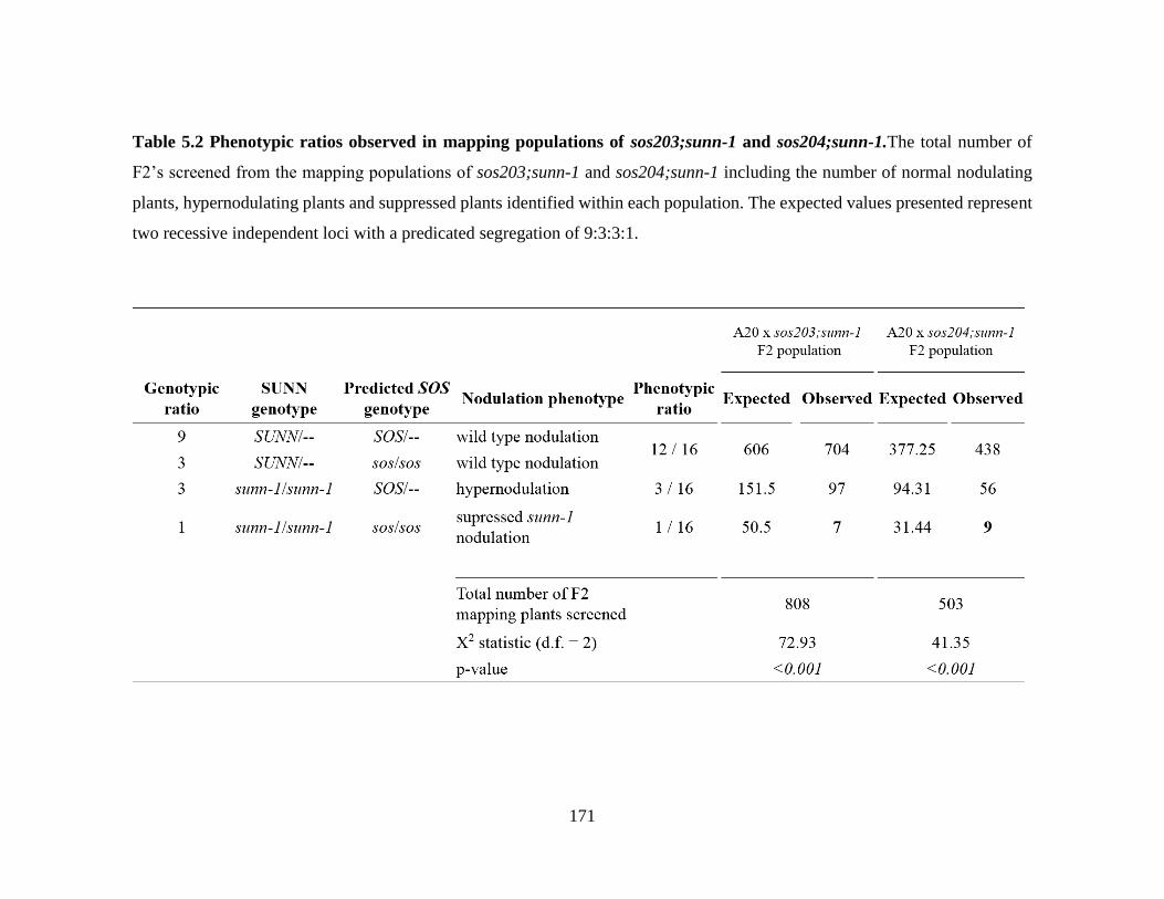
171
Table 5.2 Phenotypic ratios observed in mapping populations of sos203;sunn-1 and sos204;sunn-1.The total number of
F2’s screened from the mapping populations of sos203;sunn-1 and sos204;sunn-1 including the number of normal nodulating
plants, hypernodulating plants and suppressed plants identified within each population. The expected values presented represent
two recessive independent loci with a predicated segregation of 9:3:3:1.

172
A set of genetic markers distributed across all eight Linkage Groups of M.
truncatula were used to map the lesion responsible for the sos204 phenotype (Table 5.3).
The CAPS markers mostly represent single nucleotide polymorphisms, existing between
the two parental genomes A17 (contains sunn-1 and sos) and A20 (crossing parent), which
either create or destroy a restriction enzyme recognition site. By determining the parental
genotype at each of the marker loci in our mapping plants, we can infer the probable
location of our lesion. Recombination between the two parents creates three possibilities
at each marker locus; the plant either has both alleles from A17, both alleles from A20 or
one allele from each parent, A17/A20. In our sos;sunn-1 mapping plants, regions of
Linkage Groups containing alleles from both parents (A17/A20; het) or from A20 (A20;
homozygous) cannot harbor our sos mutation and therefore, can be ruled out. Likewise, the
supernodulating plants (sunn-1/sunn-1;SOS/SOS or SOS/sos) can also be used to rule out
regions; however, in this case, regions that are A17 homozygous do not contain the
suppressor lesion.
The plants were tested for genotype, either A17, A20 or heterozygous, for each of
the markers using a restriction digest unique to the genotype (Table 5.3). Using these core
markers, we identified DK490L on Linkage Group (LG) 2 as linked to the suppression
phenotype, since all plants tested had the A17 parental genotype at this locus (Table 5.4).
When tested at the markers to the north, PFK, and to the south, PGK1, the suppressed
mapping plants were either homozygous A17 or heterozygous A17/A20 (Table 5.4) setting
the initial boundaries for fine mapping between 8,869,991 bp and 27,710,628 bp on LG 2.

173
Table 5.3 A list of the core CAPS markers on each of the eight Linkage Groups of M.
truncatula that may be used in coarse mapping of suppressor genes.
Linkage
Group
Name RE Type A17
fragment
A20
fragment
1 CysPr2 BcuI CAPS 240 210+30
1 DSI SspI CAPS 510 410+100
2 DK407L AluI CAPS 320 250+70
2 PFK SspI CAPS 500 420+80
2 DK490L HinfI CAPS 350 250+100
2 PGK1 DraI CAPS 550 210+340
3 DK258L XbaI CAPS 470 100+360
3 TRPT DdeI CAPS 290 230+60
3 MtEIL1 EcoRI CAPS 255+55 310
4 EPS BglII CAPS 233 digested
4 ES63B19R AluI CAPS 306+132 438
5 DK018R length 220 230
5 DK013L RsaI CAPS 110+300 410
5 DK139L EcoRV CAPS 260 130+130
5 WPK4 SpeI CAPS 220 190+30
6 CrS length 750 950
6 AW774849 EcoRV CAPS 258 176+82
7 DK322L DdeI CAPS 175+90 145+90+30
7 ENOL SphI CAPS 450+1050 1500
7 DK427 BsmAI CAPS 440+60 500
8 DK455L BspHI CAPS 330 220+90
8 GLO3 HincII CAPS 210+30 240
8 DENP length 195 222

174
Table 5.4 Coarse mapping of sos204;sunn-1. The genotypes at each marker are indicated for suppressed mapping plants of
sos204;sunn-1 used for mapping; A = A17/A17, H = heterozygous (A17/A20), B = A20/A20, and displayed as a color map
(Kiss, et al., 1998).
Chromosome Marker RE A17 fragment A20 fragment Suppressed sos204;sunn-1 mapping plants
5 46 61 112 138 207 265 308 318
1 CysPr2 SpeI 240 30+210
1 DSI SspI 510 100+410 H A H H H B B
2 DK407L AluI 320 250+70 A A A A A A A A H
2 PFK SspI 500 80+420 A A A A A A A A/H H
2 DK490L HinfI 350 100+250 A A A A A A A A A
2 PGKI DraI 550 210+340 H H H A A H
3 DK258L XbaI 470 100+360 A A H H H A
3 TRPT DdeI 290 60+230 A A A H A A B
3 MtEIL1 EcoRI 310 255+55 H H A A B H B
4 EPS BglII 1620 1050+570 B H H B B
4 sunn-1 HaeIII A A A A A A A A A
5 DK018R length 230 220 A B B A H A
5 DK013L RsaI 110 + 300 410 H H H A A A
5 WPK4 SpeI 220 190+30 H H H H A H B
6 CrS length 750 950
6 AW774849 EcoRV 258 82+176 H A B H H B
7 DK322L DdeI 90 + 175 90+30+145 H B A H B H
7 DK427 BSMAI 440+60 500 A H A H H
8 DK455L BspHI 330 220+90
8 GLO3 HincII 210 + 30 240 A H H A H A
8 DENP length 195 222 A A A A A A

175
Five suppressed plants (5, 46, 61, 207, and 318) with informative recombination
events in the region of interest on LG2 were used to fine map the sos204 lesion by
continuing to rule out portions of the chromosome that could not contain the suppressor.
Of these plants, one was heterozygous at a marker on the northern boundary (PFK) and
four plants were heterozygous at marker PGKI on the southern boundary with all five
plants showing linkage (homozygous A17) to marker DK490L. The region of interest
contained two additional CAPS markers, RBBP and ES50020-C/D, which were tested in
the five informative mapping plants (Figure 5.3). To aid in eliminating more of the region,
eight additional markers were created (see Materials and Methods) that were either used as
CAPS markers or directly sequenced in mapping plants of interest. Their genotypes, along
with other markers along LG 2, can be seen in Figure 5.3. The suppressed mapping plants
alone were adequate in defining the region of interest to just above 3.7 Mb (from
12,017,650 bp to 15,773,942 bp).
Because our mapping population is small when just suppressors are considered, 16
hypernodulating plants were genotyped at subsequent markers, when needed. This allowed
for regions that are genotyped as A17 to be ruled out because they cannot contain the lesion
responsible for suppression of nodulation or they would, in fact, themselves be suppressed.
Four of these hypernodulators had informative recombination events, narrowing the region
(Figure 5.3) from 3.7 Mb to the final region of approximately 150 kB (from 12,017,650 bp
to 12,166,318 bp) flanked by markers 2g031720 and 2g032030.

176
Figure 5.3. A color map representing the fine mapping efforts of sos204;sunn-1. The markers used to fine map sos204 and
their location along Chromosome 2 are shown. The borders generated from coarse mapping are shown as bold lines. The
genotypes at each marker are indicated for suppressed mapping plants and the hypernodulators used for mapping; A = A17/A17,
H = heterozygous (A17/A20). Green areas can contain the suppression lesion while yellow cannot. Each band (either grey or
white) represents 5 Mb.

177
The region between Medtr2g031720 and Medtr2g032030 contains 26 candidate
genes inclusive of the two border genes (Table 5.5). Two relatively small candidate genes
within the region were sequenced in sos204;sunn-1 to determine if they contained the
causative lesion. Each of these, Medtr2g031820 and Medtr2g032000, in sos204;sunn-1 had
a 100% nucleotide match to the reference sequence across the region sequenced.
Additionally, these genes were sequenced in A20 to render a marker; however, there were
no SNPs found between A17 and A20 in the regions sequenced.

178
Table 5.5. Candidate gene list for sos204;sunn-1. A list of annotated genes between and
including Medtr2g031720 and Medtr2g032030 on Chromosome 2 (Medicago truncatula
genome version 4.0). Genes highlighted in green have bee sequenced and are wild type in
nucleotide sequence.
Discussion
Genetic screens, including suppressor screens, are valuable tools in identifying
novel genetic components of a given pathway. A forward genetics approach was taken to
Gene ID Annotation
Medtr2g031720 F-box protein interaction domain protein
Medtr2g031730 hypothetical protein
Medtr2g031740 nucleic acid binding protein
Medtr2g031750 transmembrane amino acid transporter family protein
Medtr2g031760 50S ribosomal protein L7/L12
Medtr2g031770 folate/biopterin transporter
Medtr2g031780 folate/biopterin transporter
Medtr2g031790 folate/biopterin transporter
Medtr2g031810 folate/biopterin transporter
Medtr2g031820 hypothetical protein
Medtr2g031830 hypothetical protein
Medtr2g031870 myb transcription factor
Medtr2g031880 myb transcription factor
Medtr2g031890 hypothetical protein
Medtr2g031910 DUF4283
Medtr2g031920 cytochrome p450/ent-kaurenoic acid oxidase
Medtr2g031930 cytochrome p450/ent-kaurenoic acid oxidase
Medtr2g031960 50S ribosomal protein L7/L12
Medtr2g031980 annexin D8
Medtr2g031990 short-chain dehyrogenase/reductase family protein
Medtr2g032000 transmembrane protein, putative
Medtr2g032010 DNA topoisomerase, putative
Medtr2g032020 DNA topoisomerase, putative
Medtr2g032030 WRKY family TF

179
isolate suppressors of the sunn-1 hypernodulation phenotype in Medicago truncatula. At
least two suppressors were identified from this EMS sunn-1 population prior to this study.
One, cre1-4, constitutes a new allele of the cytokinin receptor CRE1 located at the bottom
of Chromosome 8 and the manuscript by Elise Schnabel and Julia Frugoli characterizing
that mutant is in revision. The isolation of cre1-4 and a second suppressor, sos16, which is
currently being mapped, validates the effectiveness of our suppressor screen. In this study,
we identify four new suppressors, sos203, sos204, sos205, and sos206, by screening of the
remaining seed stock.
Suppressor screens generate a population with a diverse array of phenotypes. In a
similar hypernodulation suppressor screen in Lotus japonicus of har1, the authors report
the screening of 150,000 M2 seed for several phenotypes (Murray, et al., 2006). Of this
screen, they identified 61 suppressors and further characterized the lines by genetic
mapping and microscopy (Murray, et al., 2006). They found a majority of these suppressors
were new alleles of genes known to be involved in the CSP, nodule organogenesis, and
AON (Murray, et al., 2006). Similar to the har1 suppressor screen, we expected different
levels of suppression within our population from few nodules to no nodules, and even
enhanced nodulators. In addition, we expected disruption in genes involved in early
nodulation events producing little to no infection, as well as, genes involved in AON
limiting the number of nodules formed to closer to wild type. We have seen both of these
conditions throughout the screening process, but chose to focus on mutants that reduced
nodule number, but did not eliminate nodulation. However, with the exception of cre1-4,

180
we cannot comment on the novelty of our mutants as we do not know the identity of the
genes containing the lesions responsible for the suppression phenotypes.
The four putative suppressors were homozygous for the original sunn-1 mutation
on LG 4 as tested by a CAPS marker specific for the sunn-1 locus. Therefore, we assume
the suppression phenotype is caused by at least one second site mutation elsewhere in the
genome. The identity of this locus was sought by classical mapping efforts aided by CAPS
markers specific to the mapping population created between ecotypes A17 and A20
(Penmetsa and Cook, 2000). In a previous study focused on nodulation mutants after EMS
treatment in A17, Penmetsa and Cook (2000) validated the use the M. truncatula ecotype
A20 as a crossing parent with A17 as it is similar in nodulation patterns but polymorphic
enough to obtain markers.
F2 progeny from the successful crosses of A20 x sos203;sunn-1 and A20 x
sos204;sunn-1 did not hold true to the 9:3:3:1 segregation ratio expected of two
independent recessive loci (sunn-1 and the suppressor sos). In fact, the representation of
sunn-1 alleles overall was lower in both populations (12.9%) than the expected 25%. The
mapping cross used to identify the gene responsible for the sunn-1 phenotype also had
fewer mutants than expected segregating in the F2 (20% versus 25%, Elise Schnabel,
personal communication) which is thought to be caused by a translocation between LG 4
(near the sunn locus) and the bottom of LG 8 (Kamphuis, et al., 2007). Therefore, it is
possible that the location of sunn and the location of the suppressor influence the
recombination rates, resulting in fewer hypernodulating plants than expected in a cross
between A17 and A20.

181
In addition to low numbers of sunn-1 plants, a low number of suppressed plants
were also observed in the two crosses. The location where the lesion is thought to reside
could play a role in the observed low frequency of suppressed plants within the mapping
populations, as an incompatible locus between A17 and A20 could be in play. The
translocation event in A17, mentioned previously, creates many problems for outcrossing
and mapping, including low germination rates and albinism, both of which can skew
segregation data for certain gene combinations (Kamphuis, et al., 2007).
Albinism results from the interaction between two genes, one from A17 and the
other from A20. If the sos lesion is linked to the locus in A17 contributing to albinism, then
plants carrying the suppressor lesion can be lost as albino plants. In fact, F3 progeny from
a hypernodulating plant heterozygous throughout the region of interest on LG 2 showed an
increase in the number of albino plants (nearly a quarter of the population) compared to
the normal 6% observed in an A17/A20 cross (Penmetsa and Cook, 2000) (data not shown).
Also, sos203;sunn-1 plants were generally small; this short stature could be the result of an
additional mutation in a lethal recessive gene segregating with the sos203 locus.
The screening of over 800 individuals led to the discovery of seven sos203;sunn-1
mapping plants, six of which are living and will greatly assist the mapping efforts for
sos203;sunn-1. Nine suppressed plants and four hypernodulators facilitated mapping of
sos204;sunn-1 to a region of 150,000 bases in the middle of LG 2, annotated in Mt Genome
version 4 as containing 26 genes. Information obtained from data mining on these
candidates suggests three genes should be examined first through sequencing efforts, as
they have the greatest potential to affect nodule regulation, discussed below.

182
The cytochrome p450/ent-kaurenoic acid oxidase (KAO; Medtr2g031930) encodes
an enzyme similar to AtKAO, responsible for catalyzing three major steps in the
conversion of ent-kaurenoic acid to GA12 in the gibberellin biosynthetic pathway (for role
of GA in nodulation, see Chapter 1). In pea, a mutant in one of the two PsKAO genes
(PsKAO1; likely homolog of Medtr2g031930) resulted in a lower nodule number
phenotype when compared to wild type (Davidson, et al., 2003). Two other top candidate
genes in this region include Medtr2g031750, a transmembrane amino acid transporter
family protein, and Medtr031980, an annexin D8. The transmembrane amino acid
transporter homolog in Arabidopsis, AAP1, is responsible for the transport amino acids,
particularly L-glutamate and neutral amino acids, in the roots of Arabidopsis (Perchlik, et
al., 2014). In addition, this transporter is reported to be upregulated in giant cells in
response to nematode infection, suggesting a role in nutrient transport and acquisition
during plant-microbe interactions (Hammes, et al., 2005). Annexin D8 promotes vesicle
docking to the plasma membrane during trafficking events and is therefore a likely
candidate given our knowledge of signal transduction events occurring at the plasma
membrane (Chapters 1 and 2).
Although much progress was made with sos204 and a mapping population has been
established with sos203, the project remains ongoing. Mapping populations for the sos205
and sos206 lesions have yet to be developed. After the identities of the disrupted genes are
known, complementation studies will need to be completed to validate the findings.
Furthermore, placing the genes in the pathway will likely require additional
characterization, including both double mutant and grafting analyses.

183
Materials and Methods
Plant growth conditions
Medicago truncatula seed used in this study were scarified, imbibed and
germinated as described in Chapter 2. M. truncatula used for genetic crosses or seed
collection were grown in a greenhouse with a 14:10 L:D cycle and a temperature range
between 18°C-27°C as described in Chapter 2. When assayed for nodulation, the plants
were grown on an aeroponic chamber, inoculated with Sinorhizobia medicae strain
ABS7M three days after germination, and assayed 7-10 days post-inoculation as in
(Penmetsa and Cook, 1997). Plants were then planted in 3M Farfard potting mix and kept
under a light table (14:10, L:D) at 21°C-24°C until DNA extraction.
sunn-1 suppressor screen using a Ethylmethyl sulfonate (EMS) mutant population and
generation of mapping populations
More than 300 seed of an existing population of EMS treated sunn-1 (in ecotype
A17) M2 seed were screened for nodulation phenotypes and identified suppressors were
used in mapping crosses. The Jemalong ecotype A20 was used as a pollen donor for
flowers of two suppressor lines, sos203;sunn-1 and sos204;sunn-1 (ecotype A17) as
described in (Penmetsa and Cook, 2000). F1 plants for crosses to both sos203;sunn-1 and
sos204;sunn-1, which would be heterozygous at the SUNN gene (SUNN/sunn-1), were
allowed to self. The F2 seed collected from each cross was used to map the sos203 or
sos204 lesion. Crossing attempts for creating a mapping population for sos205 and sos206
were unsuccessful.

184
DNA isolation
Leaflets from each plant were transferred to a Plant Card (Whatman, GE
Healthcare) via manufacturer’s instructions for DNA isolation. Plant Cards contain a
specialized matrix to capture the DNA and are approved for long term storage. A small
piece of matrix containing DNA was excised from the Plant Card following the
manufacturer’s instructions and used directly for PCR. For plants used extensively for
mapping, DNA isolation was completed using a Qiagen DNA extraction kit (Qiagen,
Germantown, MD) with 100 mg of fresh tissue as per manufacturer’s instructions.
Screening and genotyping of F2 mapping populations
The F2 plants stemming from A20 x sos203;sunn-1 and A20 x sos204;sunn-1
crosses were phenotyped for nodule number. Because the F2 population was segregating
with respect to the sunn-1 locus, DNA was collected from each plant and used for
genotyping at the sunn-1 cleaved amplified polymorphic sequence (CAPS) marker
described in the text using primers CLV-Q (5’-TCCATCCCATTACGAGACTTACTA-
3’) and CLV-R (5’-ACACGACATTGTGAAATTCAAACCTAT-3’) in combination with
the restriction enzyme HaeIII. Only F2 plants that were sunn-1 homozygous with normal
nodule counts (suppressors) or hypernodulators were used to test marker loci.
PCR analysis of marker loci
Marker loci used in this study generally consisted of small amplified sequences
containing single nucleotide polymorphisms between the two parental ecotypes A17 and
A20 (Table 5.6). These were used to determine the genotype represented at each of the loci

185
for suppressed mapping plants to identify a region of the genome that is linked to the
suppression phenotype. In some cases, hypernodulating plants were used to identify
regions of the genome not linked to the suppression phenotype that could be ruled out. The
SNPs, in the case of CAPS markers, were located in restriction enzyme sites resulting in
different digestion patterns between A17, A20 and A17/A20 heterozygotes (Table 5.3). In
other cases, there were length differences between the amplified fragments depending on
the parental genotype at that locus (Table 5.3). The marker set used to coarse map
sos203;sunn-1 and sos204;sunn-1 consisted of 18 CAPS markers and three length markers
(Table 5.3). Each of the eight chromosomes was represented by at least two markers during
coarse mapping.
Identification of new marker loci
Additional markers had to be developed on Chromosome 2 in order to facilitate fine
mapping efforts for sos 204;sunn-1. Intronic regions, usually 500-1000 bp, within an area
of interest were amplified from both A17 and A20 ecotypes. Sequencing data was
generated and analyzed for SNPs. A SNP was either directly used as a CAPS marker or the
genotype of mapping plants at the marker was determined by sequencing using SNPs
present (Table 5.6).

186
Table 5.6 Primer sequences for each of the markers used on Linkage Group 2.
Marker Forward Primer (5’-3’) Reverse Primer (5’-3’) h2_15a20a TTGGTGCACCGTATTTTGAG CCAGGCATCCTTTTCTTTTC
1N1R CATATTGTTAGATTTGTGG GTGAGCGTTAAGTTGGTAGAG
DK407L TTAATTTTATCAACCCACCATATTAGTCAA CCAGTGCTGGAAAAGACAATCAATC
h2_178m20a ATCCCACATCGGGAAGTTTT AAAGAGCGCTAACAGTGGGA
PFK TCCCACTGCAAATCATGTCAAAAC ACACAAGTGGATATTGATGGTTAGAC
2g028610 ACATGTTCACCGGTCCATTT GCACACAACTATCACTGCCA
2g030640 GGTAGGGCGGGATTCACTAT TGGTTTTAATTTTGGTCCCCTA
2g031380 TTTGTCTAGTTTGAGGCCAACA TCAGCGCAACAGTGATTTTC
2g031240 GGTCGAGGGAAAGTTGAAAA TATCCTGCCCGCAGATTATT
2g031720 TGGTGTAGAAGCCGAACTAAAC AGGATCAAAACCGAAGCCGTA
2g032030 GGCCAACACAAAGAAATGCC CTGAGGAACCAAAGCATGCA
2g033590 AACCCAAAGTTTCTCCAGATCA TGTGTCCACATCTTGTCCCTA
DK490L TGGTTCCAAATTCCACTCAAAAGC AAAAATTGTGTTGTGGTTTAGTGGTAGAC
AZ1A03 TGCTTTGGCGTCCTATTTTTATTTA TGTGTTGGAGAAGATGAAGCAGAA
RBBP CAAGAGGACGCAAACCTAAACC CACAATTCGCAATCACCAAAGTAT
ES50020-C/D CACCTTCTTATAACTTCTTCATTG GTTTCCAGTTGTCTTCTTCTCCTC
PGKI GATGACTGTATTGGCGAGGAAGT GTTCGACACGGCTCCAACTA

187
Literature Cited
Ane JM, Kiss GB, Riely BK, Penmetsa RV, Oldroyd GE, Ayax C, Levy J, Debelle F,
Baek JM, Kalo P, Rosenberg C, Roe BA, Long SR, Denarie J, Cook DR (2004)
Medicago truncatula DMI1 required for bacterial and fungal symbioses in legumes.
Science 303: 1364-1367
Catoira R, Galera C, de Billy F, Penmetsa RV, Journet EP, Maillet F, Rosenberg C,
Cook D, Gough C, Denarie J (2000) Four genes of Medicago truncatula controlling
components of a nod factor transduction pathway. Plant Cell 12: 1647-1666
Charron D, Pingret JL, Chabaud M, Journet EP, Barker DG (2004) Pharmacological
evidence that multiple phospholipid signaling pathways link Rhizobium nodulation factor
perception in Medicago truncatula root hairs to intracellular responses, including Ca2+
spiking and specific ENOD gene expression. Plant Physiol 136: 3582-3593
Davidson SE, Elliott RC, Helliwell CA, Poole AT, Reid JB (2003) The pea gene NA
encodes ent-kaurenoic acid oxidase. Plant Physiol 131: 335-344
Endre G, Kereszt A, Kevei Z, Mihacea S, Kaló P, Kiss GB (2002) A receptor kinase
gene regulating symbiotic nodule development. Nature 417: 962-966
Hammes UZ, Schachtman DP, Berg RH, Nielsen E, Koch W, McIntyre LM, Taylor
CG (2005) Nematode-induced changes of transporter gene expression in Arabidopsis
roots. Mol Plant-Microbe Interact 18: 1247-1257
Kamphuis LG, Williams AH, D'Souza NK, Pfaff T, Ellwood SR, Groves EJ, Singh
KB, Oliver RP, Lichtenzveig J (2007) The Medicago truncatula reference accession A17
has an aberrant chromosomal configuration. New Phytol 174: 299-303
Kiss G, Kereszt A, Kiss P, Endre G (1998) Colormapping: a non-mathematical procedure
for genetic mapping. Acta Biol Hung 49: 125-142
Levy J, Bres C, Geurts R, Chalhoub B, Kulikova O, Duc G, Journet EP, Ane JM,
Lauber E, Bisseling T, Denarie J, Rosenberg C, Debelle F (2004) A putative Ca2+ and
calmodulin-dependent protein kinase required for bacterial and fungal symbioses. Science
303: 1361-1364
Limpens E, Franken C, Smit P, Willemse J, Bisseling T, Geurts R (2003) LysM domain
receptor kinases regulating rhizobial Nod factor-induced infection. Science 302: 630-633

188
Limpens E, Ramos J, Franken C, Raz V, Compaan B, Franssen H, Bisseling T, Geurts
R (2004) RNA interference in Agrobacterium rhizogenes-transformed roots of Arabidopsis
and Medicago truncatula. J Exp Bot 55: 983-992
Murray J, Karas B, Ross L, Brachmann A, Wagg C, Geil R, Perry J, Nowakowski K,
MacGillivary M, Held M (2006) Genetic suppressors of the Lotus japonicus har1-1
hypernodulation phenotype. Mol Plant-Microbe Interact 19: 1082-1091
Penmetsa RV, Cook DR (2000) Production and characterization of diverse developmental
mutants of Medicago truncatula. Plant Physiol 123: 1387-1398
Penmetsa RV, Frugoli JA, Smith LS, Long SR, Cook DR (2003) Dual genetic pathways
controlling nodule number in Medicago truncatula. Plant Physiol 131: 998-1008
Perchlik M, Foster J, Tegeder M (2014) Different and overlapping functions of
Arabidopsis LHT6 and AAP1 transporters in root amino acid uptake. J Exp Bot 65: 5193-
5204
Sagan M, Morandi D, Tarenghi E, Duc G (1995) Selection of nodulation and
mycorrhizal mutants in the model plant Medicago truncatula (Gaertn.) after γ-ray
mutagenesis. Plant Science 111: 63-71
Schnabel E, Karve A, Kassaw T, Mukherjee A, Zhou X, Hall T, Frugoli J (2012) The
M. truncatula SUNN gene is expressed in vascular tissue, similarly to RDN1, consistent
with the role of these nodulation regulation genes in long distance signaling. Plant
Signaling & Behavior 7: 4-6
Schnabel E, Journet EP, de Carvalho-Niebel F, Duc G, Frugoli J (2005) The Medicago
truncatula SUNN gene encodes a CLV1-like leucine-rich repeat receptor kinase that
regulates nodule number and root length. Plant Mol Biol 58: 809-822
Schnabel E, Kulikova O, Penmetsa RV, Bisseling T, Cook DR, Frugoli J (2003) An
integrated physical, genetic and cytogenetic map around the sunn locus of Medicago
truncatula. Genome 46: 665-672
Tadege M, Wen J, He J, Tu H, Kwak Y, Eschstruth A, Cayrel A, Endre G, Zhao PX,
Chabaud M (2008) Large‐scale insertional mutagenesis using the Tnt1 retrotransposon in
the model legume Medicago truncatula. The Plant Journal 54: 335-347

189
CHAPTER SIX
SUMMARY
The production and maintenance of nodule structures on the roots of leguminous
plants is energetically costly to the plant during symbioses with Rhizobia. To ensure a
balanced symbiotic relationship, a long distance signaling pathway called autoregulation
of nodulation (AON) controls the number of nodules that are formed by the plant root
depending on nitrogen needs. This pathway relies on mobile signals that are derived from
nodulation events occurring in the root, their transduction in the shoot into a decision and
subsequent signaling back down to the root to halt the formation of additional nodules
under conditions when N demand is met. SUNN is the central receptor kinase in the shoot
responsible for receiving the root-derived signal and orchestrating the necessary response
during AON. In this study, I investigated protein-protein interactions of the SUNN kinase
by identifying candidate interactors based on other systems and preliminary data, as well
as discovering novel putative interactors using exploratory proteomics and forward genetic
screens.
First, I determined the subcellular localization of SUNN. SUNN co-localized with
a plasma membrane protein, AtPIP2, when transiently expressed in tobacco cells under
both constitutive and native promoters. Microscopic analysis of stem vascular cells in a
transgenic Medicago truncatula plant carrying 35S:SUNN-YFP/HA also showed
localization to the plasma membrane. A punctate patterning along the plasma membrane
which co-localized with aniline blue staining of callose deposits was observed, suggesting
SUNN localized to parts of the plasma membrane associated with plasmodesmata.

190
The strong hypernodulation phenotype and short root phenotype of the sunn-4 null
mutant were rescued with the addition of 35S:SUNN-YFP/HA in stable transgenic M.
truncatula. Nodule number was completely restored in plants containing the transgene,
suggesting this tagged version of SUNN is capable of performing the function of wild type.
The root length phenotype was partially rescued in plants containing the transgene. The
ability of the transgene to restore function is important because this transgenic line was
used for exploratory proteomics.
The receptor-like protein CLAVATA2 and the pseudo-kinase CORYNE were
chosen as candidates for interactions studies due to the structural and amino acid sequence
similarity between SUNN and the well-studied CLAVATA1 protein responsible for SAM
maintenance in Arabidopsis, a protein with which both CLV2 and CRN interact. I showed
SUNN is capable of forming homomers in vivo, and forms heteromers with both CLV2
and CRN. Since CLV2 mutants in both pea and Lotus japonicus display hypernodulation
phenotypes, a role in AON for the M. truncatula gene is likely. In addition, we showed a
CRN mutant in M. truncatula is disrupted in AON, producing more nodules than wild type,
and this phenotype is dictated by the genotype of the shoot. Taken together, these data
suggests the possibility of a heteromeric signaling complex influencing nodule number
from the shoot.
The nature of the downstream signaling that takes place after SUNN (or a complex
containing SUNN) receives the signal from the root has remained largely unknown. The
identification of RopGEFs, guanine exchange factors, in M. truncatula raised the
possibility that SUNN could signal through Rop GTPases. The kinase domain of SUNN

191
interacted with RopGEF1 and RopGEF5 in a yeast-two hybrid experiment performed by a
collaborator. In vivo interactions of SUNN and RopGEF1, RopGEF2, and RopGEF5 were
demonstrated using a bimolecular fluorescence complementation assay in tobacco cells.
RNAi against RopGEF1, RopGEF2, and RopGEF5 individually in M. truncatula hairy
roots resulted in hypernodulation phenotypes, indicating these RopGEFs affect nodule
number in the root. While a function for SUNN has not been demonstrated directly in the
root, it is expressed in root tissues. The discovery that SUNN interacts with the RopGEFs
in vivo and the apparent role of RopGEFs in nodulation, suggests SUNN signals through
RopGEF activation of RopGTPases in either the root or the shoot or both. Further
experiments will be required to determine this.
To identify novel interactors of SUNN, I used co-immunoprecipitation coupled
with LC-MS/MS of the tagged SUNN transgene in our transgenic M. truncatula. This
exploratory proteomics approach identified over twenty putative interacting proteins
throughout different stages of nodulation including a remorin, two LRR proteins, a
cyclophilin, a phosphatase, a subtilisin protease, and a N-scythe ubiquitin protein. For some
of the proteins featured in this study, their function and even the role they may play in
nodulation was inferred from other systems. Structural analysis of the LRR proteins
showed they do not contain a transmembrane domain and are likely localized in the
extracellular space, where they could interact with the LRR region of SUNN. Furthermore,
the experimental design allowed for the isolation of protein-protein interactions occurring
in the shoot to be separated from those occurring in the root, which will allow future
dissection of the root and shoot functions of SUNN.

192
Lastly, I used a forward genetics screen to identify mutants capable of suppressing
the hypernodulation phenotype of sunn-1. I isolated and characterized four suppressed
sunn-1 mutants. Mapping populations for sos203;sunn-1 and sos204;sunn-1 were created
and the F2 screened for suppressors. Mapping efforts for sos204;sunn-1 indicate the lesion
for sos204 lies in a region on Linkage Group 2 that is approximately 150 kB and contains
26 annotated genes. Of these genes, three are prime candidates: a transmembrane amino
acid transporter, an annexin D8, and an ent-kauronic acid oxidase. These genes are
currently being sequenced in sos204;sunn-1 mutants.
My study of protein-protein interactions involving the central kinase controlling
AON allows us to understand the mechanisms of the pathway better. I conclude from these
analyses that SUNN is a dynamic receptor kinase that likely forms complexes with a
variety of receptor like proteins, LRR proteins, protein kinases, and possibly other LRR-
RLKs to promote the events of specific signaling cascades depending on the needs of the
plant. In addition, RNAi and BiFC analysis of RopGEFs indicate SUNN may trigger
GTPase signaling. Although many question remain regarding AON and nodulation in
general, this work places several crucial pieces in the puzzle.