interaction between bluetongue virus outer capsid protein vp2 and
TRANSCRIPT

BioMed CentralVirology Journal
ss
Open AcceResearchInteraction between Bluetongue virus outer capsid protein VP2 and vimentin is necessary for virus egressBishnupriya Bhattacharya, Rob J Noad and Polly Roy*Address: Department of Infectious and Tropical Diseases, London School of Hygiene and Tropical Medicine, Keppel Street, London, WC1E 7HT, UK
Email: Bishnupriya Bhattacharya - [email protected]; Rob J Noad - [email protected]; Polly Roy* - [email protected]
* Corresponding author
AbstractBackground: The VP2 outer capsid protein Bluetongue Virus (BTV) is responsible for receptorbinding, haemagglutination and eliciting host-specific immunity. However, the assembly of this outercapsid protein on the transcriptionally active viral core would block transcription of the virus. Thusassembly of the outer capsid on the core particle must be a tightly controlled process during virusmaturation. Earlier studies have detected mature virus particles associated with intermediatefilaments in virus infected cells but the viral determinant for this association and the effect ofdisrupting intermediate filaments on virus assembly and release are unknown.
Results: In this study it is demonstrated that BTV VP2 associates with vimentin in both virusinfected cells and in the absence of other viral proteins. Further, the determinants of vimentinlocalisation are mapped to the N-terminus of the protein and deletions of aminio acids betweenresidues 65 and 114 are shown to disrupt VP2-vimentin association. Site directed mutation alsoreveals that amino acid residues Gly 70 and Val 72 are important in the VP2-vimentin association.Mutation of these amino acids resulted in a soluble VP2 capable of forming trimeric structuressimilar to unmodified protein that no longer associated with vimentin. Furthermore,pharmacological disruption of intermediate filaments, either directly or indirectly through thedisruption of the microtubule network, inhibited virus release from BTV infected cells.
Conclusion: The principal findings of the research are that the association of mature BTV particleswith intermediate filaments are driven by the interaction of VP2 with vimentin and that thisinteraction contributes to virus egress. Furthermore, i) the N-terminal 118 amino acids of VP2 aresufficient to confer vimentin interaction. ii) Deletion of amino acids 65–114 or mutation of aminoacids 70–72 to DVD abrogates vimentin association. iii) Finally, disruption of vimentin structuresresults in an increase in cell associated BTV and a reduction in the amount of released virus frominfected cells.
BackgroundBluetongue virus (BTV), an insect (Culicoides sp) transmit-ted double-layered non-enveloped virus, is the type spe-cies virus of the genus Orbivirus, in the family Reoviridae.
BTV is an economically important virus and causes hem-orrhagic disease in sheep and other ruminants [1]. Thevirus has a genome consisting of ten double stranded (ds)RNA segments, located in a core particle made up of two
Published: 15 January 2007
Virology Journal 2007, 4:7 doi:10.1186/1743-422X-4-7
Received: 11 December 2006Accepted: 15 January 2007
This article is available from: http://www.virologyj.com/content/4/1/7
© 2007 Bhattacharya et al; licensee BioMed Central Ltd. This is an Open Access article distributed under the terms of the Creative Commons Attribution License (http://creativecommons.org/licenses/by/2.0), which permits unrestricted use, distribution, and reproduction in any medium, provided the original work is properly cited.
Page 1 of 12(page number not for citation purposes)

Virology Journal 2007, 4:7 http://www.virologyj.com/content/4/1/7
major proteins (VP3 and VP7) and three minor proteins(VP1, VP4 and VP6). In the mature particle, this core iscoated by two outer capsid proteins, VP2 (110 kDa) andVP5 (60 kDa) (see schematic, Fig. 1). The most exposedprotein on the mature virion, VP2, is responsible forreceptor binding [2-4], haemagglutinating activity [5] andelicits host specific immunity [6-8]. The smaller protein,VP5, is involved in cell penetration during the initialstages of infection [9,10]. After entry into cells, the virus isuncoated by removal of VP2 and VP5 to yield the core par-ticle that is transcriptionally active. This complex is anend-point in virus disassembly and protects the viraldsRNA genome from intracellular surveillance mecha-nisms.
Like other members of Reoviridae, BTV replicates in thecytoplasm of infected cells. Electron microscopic analysisof thin sections of BTV infected cells have revealed a largenumbers of virus-specific tubules, and juxtanuclear inclu-sions bodies containing virus-like particles in addition tosmall numbers of intracellular virus particles (15). Whilethe tubules are multimers of NS1 [11], inclusion bodiesare predominantly formed by NS2 [12,13]. Similar torotavirus inclusion bodies [14], it has been speculatedthat during BTV replication and assembly NS2 binds coreproteins, viral single-stranded (ssRNA) [15,16] andrecruits these components to the inclusion bodies [17].Additionally, it has been hypothesised that the viralgenome interacts with the minor structural proteins toform the transcriptase complex which is subsequentlyencapsulated by a single shell of VP3 to form the subcoreof the assembling virus [18]. The subcore acts as a scaffold
for the deposition of VP7 trimers, thus forming a stablecore structure [19-21]. However, the outer capsid pro-teins, VP2 and VP5 have not been localised to the viralinclusion bodies and it is still not clear when and wherein the cell are they assembled on to the core. As the outercapsid proteins, particularly VP2, would effectively blockthe channels in the core that are used during transcriptionof the viral genome it is unlikely that the coating of coreswould be an uncontrolled process.
Previous electron microscopy studies have indicated thatBTV particles can be found attached to vimentin interme-diate filaments [22]. Negative staining of intact, infectedcells labelled with anti-VP2 antibody revealed that BTVparticles were also present under the cell membrane aswell as on the cell surface [23]. In addition, both virusaggregates and single virion particles retain an associationwith the cortical layer of the cytoskeleton following celllysis. This has lead to speculation that there may be inter-action of the particle with the actin-rich cortical layerunderlying the cell membrane [23]. Recent studies haveshown that NS3, the only virus-coded glycoprotein ofBTV, interacts with VP2 as well as Tsg101 and the p11 sub-unit of the heterotetrameric calpactin II complex, therebyfacilitating virus release [24,25]. Thus, in addition to itsrole in virus attachment, VP2 is emerging as a key playerin the control of BTV assembly and egress from infectedcells. A detailed understanding of the control of VP2 asso-ciation with the newly assembled core is therefore animportant step in understanding virus assembly andegress.
In this manuscript we demonstrate that normal subcellu-lar distribution of VP2 relies on an association withvimentin intermediate filaments, and identify key resi-dues in this interaction located in the N-terminus of theprotein. Furthermore, disruption of the vimentin filamentnetwork using pharmacological inhibitors lead to a dis-ruption of virus egress.
ResultsIn infected cells VP2 localises to punctuate areas within the cytoplasmIn order to assess the intracellular localisation of VP2 invirus infection, cells were first infected with BTV-10 andthen VP2 detected by immunofluorescence microscopy.At 4 hours post infection VP2 was found to be restricted topunctuate areas within the cytoplasm (Fig. 2A). A similardistribution of VP2 was observed when cells were trans-fected with plasmids encoding VP2 or a VP2-GFP fusionprotein where the GFP was located at the C-terminus ofVP2 (Fig. 2B and 2C, respectively). However, fusion ofGFP to the N-terminus of VP2 altered the subcellularlocalisation of the protein and resulted in a more diffusepattern of fluorescence (Fig. 2D). Western blot analysis of
Schematic of the mature BTV particleFigure 1Schematic of the mature BTV particle. Organisation of the major structural proteins VP2, VP5, VP3 and VP7 in the architecturally complex BTV particle. On entry into cells the outer capsid proteins VP2 and VP5 are lost, releasing a tran-scriptionally active core particle.
VP2
VP5VP7VP3
Genomic RNA
and enzymatic
proteins
Page 2 of 12(page number not for citation purposes)

Virology Journal 2007, 4:7 http://www.virologyj.com/content/4/1/7
the VP2 GFP fusion proteins confirmed that they wereexpressed in cells as full-length proteins (Fig. 2F).
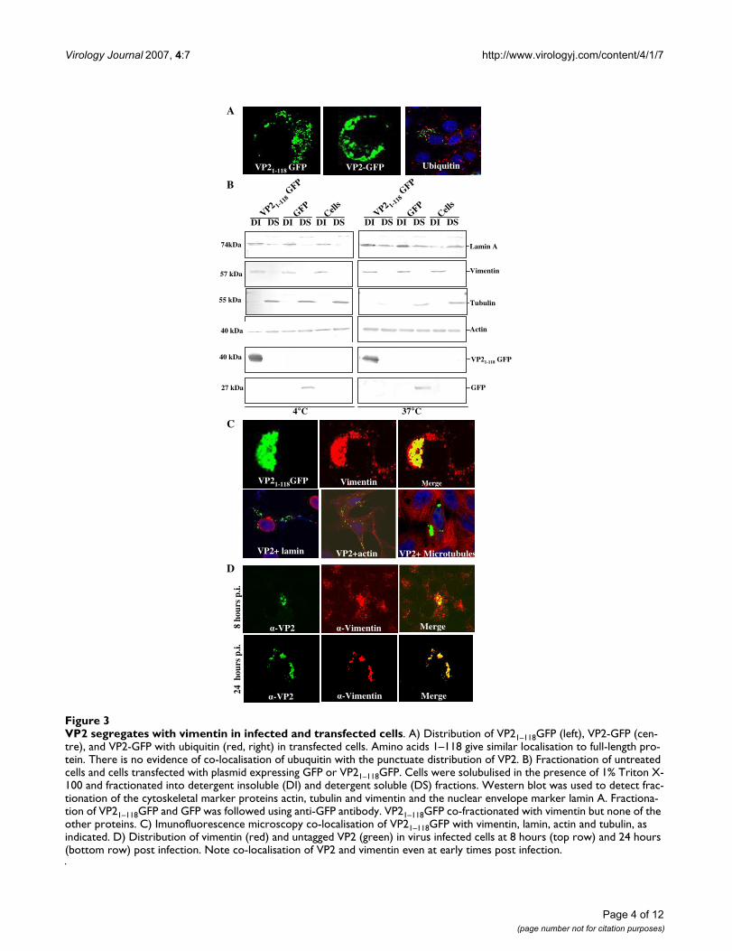
The N-terminus of VP2 is sufficient to direct sub-cellular localisationSince the fusion of GFP to the N-terminus but not the C-teriminus of VP2 altered its sub-cellular localisation wehypothesised that the N-terminus of the protein con-tained a signal necessary for intracellular localisation thatwas disrupted in the fusion protein. To test this possibil-ity, we expressed an N-terminal fragment of VP2 consist-ing of the first 118 residues of the protein fused at its C-terminus to GFP (Fig. 3A, VP21–118GFP). The distribution
of this deletion mutant was indistinguishable from that ofthe full-length VP2-GFP protein (Fig. 3A, VP2 GFP), sug-gesting that it contained the signals necessary for the sub-cellular localisation of VP2. The punctuate pattern ofVP21–118GFP accumulation was distinct from what wouldbe expected if the chimeric protein had been incorporatedinto aggresomes as there was no evidence of co-localisa-tion with ubiquitin (Fig. 3A, ubiquitin).
VP2 in virus infected cells and VP21–118GFP associate with vimentinEarlier electron microscopic studies of BTV infected cellshave suggested that the virus associates with the cell mem-brane and with vimentin intermediate filaments [22,26].In order to test where VP21–118GFP was localised withincells, subcellular fractionation studies were carried out inthe presence of detergents at both 4°C and 37°C (Fig. 3B).These studies were designed to separate soluble cytoplas-mic proteins and lipid raft associated proteins fromcytoskeletal and nuclear fractions of the cell. As expected,actin was present as both soluble and cytoskeletal associ-ated insoluble forms under the conditions of the study.Tubulin and untagged GFP were present predominantlyin the detergent soluble cytosolic fraction. The nuclearmembrane protein lamin was present predominantly inthe detergent insoluble fractions at both 4°C and 37°C,although at the higher temperature the proportion of sol-ubulised lamin was increased. Vimentin and VP2 co-seg-regated in these fractionation studies, and were presentonly in the detergent insoluble fractions (Fig. 3B). Thus,fractionation studies supported association with VP2 withdetergent insoluble cell fractions containing vimentin.Immunofluorescence was performed with each of the pro-teins in the cell fractionation studies in Vero cells express-ing VP21–118GFP. While there was no evidence that eitheractin, tubulin or lamin co-localised with the VP2 deletionmutant, all of the VP21–118GFP was detected in areas of thecell rich in vimentin (Fig. 3C). Thus, in both cell fraction-ation and colocalisation studies VP21–118GFP and vimen-tin were detected together.
In order to confirm the association between vimentin andfull-length VP2, the distribution of vimentin and VP2 invirus infected cells was monitored by immunoflorescence.BTV infection of mammalian cells in tissue culture leadsto rapid changes in cell morphology with apoptosis,which is triggered during virus entry [27]. Thus the distri-bution of VP2 and vimentin was assessed at early (8hours) and late (24 hours) timepoints following virusinfection (Fig. 3D). At 8 hours post infection vimentinwas distributed throughout the cell but was also present inconcentrated foci within the cytoplasm. At the same time-point, VP2 was detected in concentrated foci within thecells. Intriguingly, the largest foci of VP2 co-localised withthe largest foci of vimentin. All the VP2 in detected in
Distribution of VP2 within infected and transfected Vero cells by fluorescence microscopyFigure 2Distribution of VP2 within infected and transfected Vero cells by fluorescence microscopy. A) Cells infected with BTV-10, B-E) transfected cells expressing B) VP2, C) VP2-GFP, D) GFP-VP2 and E) GFP only. VP2 in A and B were detected with anti VP2 monoclonal antibody (rabbit) and either FITC (A) or TRITC (B) conjugated sec-ondary antibody. C-E were visualised based on GFP fluores-cence. Expression of full-length, tagged VP2 variants was confirmed by western blot using an anti-GFP antibody (F).
VP
2-G
FP
GFP-V
P2
137kDa
F
27 kDa
GF
P
A B C
D E
Page 3 of 12(page number not for citation purposes)
Virology Journal 2007, 4:7 http://www.virologyj.com/content/4/1/7
Page 4 of 12(page number not for citation purposes)
VP2 segregates with vimentin in infected and transfected cellsFigure 3VP2 segregates with vimentin in infected and transfected cells. A) Distribution of VP21–118GFP (left), VP2-GFP (cen-tre), and VP2-GFP with ubiquitin (red, right) in transfected cells. Amino acids 1–118 give similar localisation to full-length pro-tein. There is no evidence of co-localisation of ubuquitin with the punctuate distribution of VP2. B) Fractionation of untreated cells and cells transfected with plasmid expressing GFP or VP21–118GFP. Cells were solubulised in the presence of 1% Triton X-100 and fractionated into detergent insoluble (DI) and detergent soluble (DS) fractions. Western blot was used to detect frac-tionation of the cytoskeletal marker proteins actin, tubulin and vimentin and the nuclear envelope marker lamin A. Fractiona-tion of VP21–118GFP and GFP was followed using anti-GFP antibody. VP21–118GFP co-fractionated with vimentin but none of the other proteins. C) Imunofluorescence microscopy co-localisation of VP21–118GFP with vimentin, lamin, actin and tubulin, as indicated. D) Distribution of vimentin (red) and untagged VP2 (green) in virus infected cells at 8 hours (top row) and 24 hours (bottom row) post infection. Note co-localisation of VP2 and vimentin even at early times post infection.
A
VP21-118 GFP VP2-GFP Ubiquitin
D
f cf cf cf c
8 ho
urs
p.i.
24 h
ours
p.i.
α-VP2 α-Vimentin Merge
α-VP2 α-Vimentin Merge
C
VP21-118GFP Vimentin Merge
VP2+ lamin VP2+actin VP2+ Microtubules
B
4°C 37°C
VP21-118 GFP
Actin
40 kDa
GFP
Vimentin57 kDa
40 kDa
27 kDa
GFP
DI DS DI DS DI DSVP2 1-1
18GFP
Cells
GFP
DI DS DI DS DI DSVP2 1-1
18GFP
Cells
74kDa
Tubulin
Lamin A
55 kDa
Virology Journal 2007, 4:7 http://www.virologyj.com/content/4/1/7
virus infected cells was found associated with vimentin,although not all the vimentin at this timepoint was asso-ciated with VP2 (Fig. 3D). In contrast, at 24 hours postinfection, by which time there are substantial virus-induced changes in cell morphology almost all the vimen-tin and VP2 present in the cell colocalised (Fig. 3D). Thus,data from virus infected cells confirm the VP2-vimentinassociation detected with the VP21–118GFP deletionmutant.
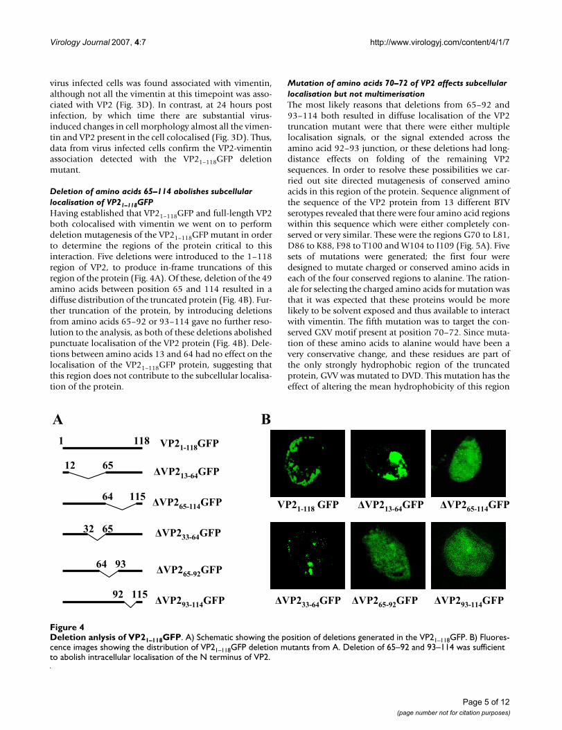
Deletion of amino acids 65–114 abolishes subcellular localisation of VP21–118GFPHaving established that VP21–118GFP and full-length VP2both colocalised with vimentin we went on to performdeletion mutagenesis of the VP21–118GFP mutant in orderto determine the regions of the protein critical to thisinteraction. Five deletions were introduced to the 1–118region of VP2, to produce in-frame truncations of thisregion of the protein (Fig. 4A). Of these, deletion of the 49amino acids between position 65 and 114 resulted in adiffuse distribution of the truncated protein (Fig. 4B). Fur-ther truncation of the protein, by introducing deletionsfrom amino acids 65–92 or 93–114 gave no further reso-lution to the analysis, as both of these deletions abolishedpunctuate localisation of the VP2 protein (Fig. 4B). Dele-tions between amino acids 13 and 64 had no effect on thelocalisation of the VP21–118GFP protein, suggesting thatthis region does not contribute to the subcellular localisa-tion of the protein.
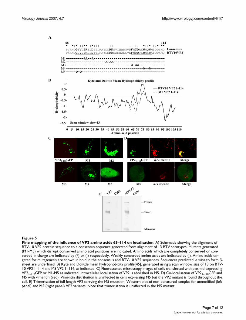
Mutation of amino acids 70–72 of VP2 affects subcellular localisation but not multimerisationThe most likely reasons that deletions from 65–92 and93–114 both resulted in diffuse localisation of the VP2truncation mutant were that there were either multiplelocalisation signals, or the signal extended across theamino acid 92–93 junction, or these deletions had long-distance effects on folding of the remaining VP2sequences. In order to resolve these possibilities we car-ried out site directed mutagenesis of conserved aminoacids in this region of the protein. Sequence alignment ofthe sequence of the VP2 protein from 13 different BTVserotypes revealed that there were four amino acid regionswithin this sequence which were either completely con-served or very similar. These were the regions G70 to L81,D86 to K88, F98 to T100 and W104 to I109 (Fig. 5A). Fivesets of mutations were generated; the first four weredesigned to mutate charged or conserved amino acids ineach of the four conserved regions to alanine. The ration-ale for selecting the charged amino acids for mutation wasthat it was expected that these proteins would be morelikely to be solvent exposed and thus available to interactwith vimentin. The fifth mutation was to target the con-served GXV motif present at position 70–72. Since muta-tion of these amino acids to alanine would have been avery conservative change, and these residues are part ofthe only strongly hydrophobic region of the truncatedprotein, GVV was mutated to DVD. This mutation has theeffect of altering the mean hydrophobicity of this region
Deletion anlysis of VP21–118GFPFigure 4Deletion anlysis of VP21–118GFP. A) Schematic showing the position of deletions generated in the VP21–118GFP. B) Fluores-cence images showing the distribution of VP21–118GFP deletion mutants from A. Deletion of 65–92 and 93–114 was sufficient to abolish intracellular localisation of the N terminus of VP2.
BA
VP21-118GFP
32 65 ΔVP233-64GFP
ΔVP293-114GFP
64 93
92 115
ΔVP265-92GFP
1181
12 65ΔVP213-64GFP
ΔVP265-114GFP64 115
ΔVP233-64GFP ΔVP265-92GFP ΔVP293-114GFP
VP21-118 GFP ΔVP213-64GFP ΔVP265-114GFP
Page 5 of 12(page number not for citation purposes)

Virology Journal 2007, 4:7 http://www.virologyj.com/content/4/1/7
of the truncated VP2, such that the hydrophobic patchnormally present in this region of protein has meanhydrophilic properties (Fig. 5B).
On expression, all of the four alanine mutations had sim-ilar punctate accumulation patterns to the unmodifiedVP2 sequence (Fig. 5C). In contrast, the mutation of theGVV to DVD at positions 70–72 resulted in a diffuse pat-tern of expression similar to that seen with the ΔVP265–
92GFP and ΔVP293–114GFP deletion mutants. Vimentintargeted immunofluorescence with cells expressing theDVD (M5) mutant revealed that the association betweenvimentin and VP2 had been broken (Fig. 5D). Because theM5 mutation was designed to alter the hydrophobicityprofile of the vimentin binding region of VP2 it was pos-sible that the changes in accumulation seen were a resultof global changes in the folding of the VP2 protein. Inorder to address this possibility, the M5 mutation wasintroduced into the full-length VP2 protein, and this pro-tein expressed in insect cells using the baculovirus expres-sion system. Unmodified VP2 multimerises to form atrimeric triskelion structure present on the outer surface ofthe mature virus particle [28]. By separating purified,unmodified VP2 under native conditions it is possible todetect monomeric, dimeric and trimeric forms of the pro-tein (Fig. 5E). Separation of the full-length M5 VP2mutant under the same conditions resulted in the forma-tion of a similar proportion of each of the multimericstates of the VP2 protein as the unmodified protein. Thusthe mutation that prevented the association of VP2 withvimentin did not prevent the formation of higher ordermultimers of the VP2 protein. This suggests that while it ispossible that the local protein architecture was affected bythe M5 mutation the overall folding of the protein wassimilar enough to the unmodified protein that multimer-isation was unaffected.
Pharmacological disruption of vimentin intermediate filaments inhibits virus releaseThe vimentin intermediate filament network has a radialorganisation, extending outwards from the cell centre.This localization partially overlaps with that of microtu-bules there is evidence that the two filament systems inter-act [29,30]. Indeed, vimentin structures move alongmicrotubules [29,31-33]. Therefore, if the vimentin-VP2interaction was important for BTV assembly and/or egresswe predicted that pharmacological disruption of eithermicrotubules or vimentin would result in changes of theamount of virus produced or the amount of virus releasedfrom infected cells. To test this possibility we carried outpharmacological experiments with colchicine and acryla-mide which specifically disrupt the microtubule andvimentin intermediate filament networks respectively.Consistent with our previous experiments, in untreatedcells vimentin was detected throughout the cytoplasm and
this distribution was unaffected by the expression of full-length, untagged VP2. However, in the presence of colch-icine, as expected, there was a redistribution of vimentinand VP2 towards the nucleus (Fig. 6A). This observationsupports other observations that vimentin intermediatefilaments are organised by the microtubule network[29,30]. The redistribution of vimentin was even moredramatic on the addition of acrylamide which resulted ina collapse of the intermediate filament network and achange in cell morphology to a more rounded shape (Fig.6A). To test the effect of these changes in vimentin distri-bution on virus replication and release, virus wasadsorbed to cells and then cells were treated with eithercolchicine (Fig. 6B) or acrylamide (Fig. 6B). Indirect dis-ruption of vimentin with colchicine resulted in 2.5 timesmore cell associated virus and a five-fold reduction in theamount of the released virus detected compared tountreated control cells. Addition of acrylamide resulted ina 50 fold reduction in the amount of released virus and a250 fold increase in the amount of cell associated virus at24 hours post infection. These data suggest that disrup-tion of vimentin either directly or indirectly inhibits thetrafficking of mature virus particles out of virus infectedcells.
DiscussionThe specific roles of the two proteins in the orbivirus outercapsid in the processes of virus attachment and mem-brane penetration have now been elucidated [4,9,10].Intriguingly, one of these proteins, VP2, also plays a rolein virus egress from infected cells as it binds a viral non-structural protein that acts as a molecular bridge betweenthe virus particle and cellular export and release factors[24,25,34]. Thus, VP2 is important to both the infectivityand egress of BTV particles. The proteins that are requiredto form the transcriptionally active orbivirus core struc-ture have all been localised to inclusion bodies withinvirus infected cells [17,35,36]. However, the outer capsidproteins do not localise to inclusions and therefore thesite of assembly of mature virus particles before transportout of infected cells is unknown. Members of other generain the Reoviridae cannot be used as a model for BTV inouter capsid assembly, as this information is either notavailable, or the outer capsid and assembly process isquite distinct. For example, unlike BTV, significant stepsin the outer capsid assembly of rotavirus are sequesteredin the endoplasmic reticulum [37]. Thus, in order to gainfurther insight into the assembly and intracellular traffick-ing of BTV, the localisation of VP2 in virus infected cellswas followed using immunofluorescence microscopy. Invirus infected cells VP2 was located in punctuate distribu-tion throughout the cytoplasm even at quite early timespost infection (Fig. 2). This distribution could be mim-icked in cells transfected with plasmid expressing VP2 pro-tein, full-length VP2 fused at its C-terminus to GFP, or the
Page 6 of 12(page number not for citation purposes)
Virology Journal 2007, 4:7 http://www.virologyj.com/content/4/1/7
Page 7 of 12(page number not for citation purposes)
Fine mapping of the influence of VP2 amino acids 65–114 on localisationFigure 5Fine mapping of the influence of VP2 amino acids 65–114 on localisation. A) Schematic showing the alignment of BTV-10 VP2 protein sequence to a consensus sequence generated from alignment of 13 BTV serotypes. Mutants generated (M1-M5) which disrupt conserved amino acid positions are indicated. Amino acids which are completely conserved or con-served in charge are indicated by (*) or (:) respectively. Weakly conserved amino acids are indicated by (.). Amino acids tar-geted for mutagenesis are shown in bold in the consensus and BTV-10 VP2 sequences. Sequences predicted in silico to form β-sheet are underlined. B) Kyte and Dolittle mean hydrophobicity profile[45], generated using a scan window size of 13 on BTV-10 VP2 1–114 and M5 VP2 1–114, as indicated. C) Fluorescence microscopy images of cells transfected with plasmid expressing VP21–118GFP or M1-M5 as indicated. Intracellular localisation of VP2 is abolished in M5. D) Co-localisation of VP21–118GFP and M5 with vimentin (red). Vimentin distribution is unaffected in cells expressing M5 but the VP2 mutant is found throughout the cell. E) Trimerisation of full-length VP2 carrying the M5 mutation. Western blot of non-denatured samples for unmodified (left panel) and M5 (right panel) VP2 variants. Note that trimerisation is unaffected in the M5 mutant.
A
VP21-118GFP α-Vimentin Merge
M5 α-Vimentin Merge
C
B
M1VP21-118GFP
M3 M4 M5
M2
11465
---------AA--A------------------------------------
--------------------A-AA--------------------------
---------------------------------A-AA-------------
---------------------------------------A--A-------
-----D–D------------------------------------------
M3
M1M2
M4M5
* *:* ::** :*::: :: .: :. *::* :.* **
PVRNDGIVLPRILDITLRAYDDRKSVINARGVDFHTDAKWMQWMIDDSMD Consensus
BTV10VP2
Amino acid position
1
0.5
0
-0.5
-1
-1.5
-2.5
-2
0 5 10 15 20 25 30 35 40 45 50 55 60 65 70 75 80 85 90 95 100 105 110
Kyte and Dolittle Mean Hydrophobicity profile
Scan window size=13
BTV10 VP2 1-114
M5 VP2 1-114
Hy
dro
ph
ob
icit
y
D
Trimer
Dimer
Monomer
VP2
E Cells
M5VP2
Cells
PKRNDGVVVPRLLDITLRAYDNRKSAKNAKGVEFMTDTKWMKWAIDDKMD

Virology Journal 2007, 4:7 http://www.virologyj.com/content/4/1/7
Page 8 of 12(page number not for citation purposes)
Disrupting vimentin inhibits virus release in BTV infected cellsFigure 6Disrupting vimentin inhibits virus release in BTV infected cells. A) Co-localization of untagged VP2 with vimentin in transfected cells. Top, untreated cells; Middle, cells treated with colchicine to disrupt microtubules; bottom, cells treated with acrylamide to disrupt vimentin. In each row VP2 and vimentin localisation are shown in green and red respectively. As expected, disruption of microtubules with colchicine causes a rearrangement in the localisation of vimentin. B) Effect of treat-ing cells with colchicine on the release of virus from infected cells. Cells and culture medium were harvested at 4, 8 and 24 hours post infection with BTV-10 in untreated and colchicine (5 μg/ml) treated cells. Control samples are labelled 4c, 8c and 24c, respectively. The titre of cell associated and released virus for each timepoint normalised to 100% for untreated cells. Col-chicine treatment resulted in a time dependent increase in the titre of cell associated virus and a time-dependent decrease in the titre of virus in the culture medium. D) As C but for cells treated with acrylamide (50 μM) to directly disrupt the vimentin network. Effect of acrylamide was similar but much faster and more dramatic than that observed by indirect disruption of vimentin through disruption of microtubules using colchicine. For C and D error bars indicate the standard error of three rep-licates of the experiments.
e f h
i
g
j k l
Ch
olc
hic
ine
Acr
yla
mid
e
A
a
Un
trea
ted
B
C
4 4c 8 8c 24 24c
Rel
ati
ve
titr
e
Rel
ati
ve
titr
e
4 4c 8 8c 24 24c
Hours post infectionHours post infection
4 4c 8 8c 24 24c
Hours post infection4 4c 8 8c 24 24c
Hours post infection
Rel
ati
ve
titr
e
Rel
ati
ve
titr
eCell associated virus Released virus
Virus in the media.Untreated, infected cells Cell associated virus.

Virology Journal 2007, 4:7 http://www.virologyj.com/content/4/1/7
N-terminal 118 amino acids of VP2 fused to GFP (Fig. 2;Fig. 3). Furthermore, it was found that VP2 co-fraction-ated with the intermediate filament protein vimentin infractionation studies, and co-localised with vimentin inimmunofluorescence of VP2 in transfected and virusinfected cells (Fig. 3). These data are consistent with ear-lier electron microscopy studies that have found maturevirus particles in linear arrays along intermediate fila-ments in infected cells [26]. Furthermore they demon-strate for the first time that the determinant for BTV-vimentin association is the VP2 protein.
The vimentin associated localisation of VP2 in virusinfected and VP2 transfected cells was quite distinct fromthe aggresome structure that has been noted following theoverexpression of certain proteins. There was no evidenceof the cage-like vimentin arrangement around VP2 whichis observed with aggresomes [38,39]. Nor was there anyevidence that ubiquitin co-localised with VP2 in trans-fected cells (Fig. 3).
Using a deletion mutagenesis strategy we were able tolocalise the vimentin localisation signal within VP2 to theregion encompassing the amino acids 65–114 (Fig. 4).This was confirmed by systematic site-directed mutagene-sis of conserved amino acids within this region to identifykey residues in VP2-vimentin association. Of five sets ofmutants, which changed a total of 13 conserved aminoacids, only one, mutation of GVV to DVD at postions 70–72, prevented co-localisation of VP2 with vimentin (Fig.5). This mutant was specifically designed to alter thehydrophobicity profile of the VP2 region from aminoacids 63–80 to make it more hydrophilic. Thus, it was ini-tially unclear whether the disruption of the associationbetween VP2 and vimentin was due to global changes inthe folding of the protein. However, when the DVD muta-tion was introduced into full-length VP2 the ability of therecombinant protein to assemble into trimeric VP2 struc-tures was equivalent to that of unmodified protein. Thus,if there are changes to the structure of the protein inducedby the DVD mutation, they occur at a local as opposed toa global level within the protein. These data indicate theDVD mutation either disrupts vimentin-VP2 associationbecause the conserved GXV motif at position 70–72 isnecessary for vimentin interaction or, like the deletions ofaminio acids 65–92 or 93–114, mutation of this motifresults in local rearrangement of the structure of VP2. At aprimary sequence level, there is no apparent homologybetween this region of VP2 and vimentin, or any otherknown vimentin interacting protein. Therefore, in combi-nation with the identity of the completely conservedamino acid (Glycine) and our mutagenesis data it is likelythat VP2-vimentin interaction involving amino acids 65–114 is conformationally dependent.
Within the cell vimentin is emerging as a key player inintracellular trafficking. Vimentin structures have beenshown to move along microtubules, and two candidatemotors had been identified for this motility: conventionalkinesin, and more recently, cytoplasmic dynein [29,31-33]. Significantly for VP2-vimentin interactions, themotility of vimentin intermediate filaments is principallyfrom the nucleus towards the cell surface in BHK-21 cells[33]. Indeed, in this study when vimentin intermediate fil-aments were disrupted pharmacologically either directly,or indirectly by disrupting microtubules, there was a dra-matic reduction in the amount of virus released frominfected cells (Fig. 6). This reduction in release was notentirely due to a decrease in overall virus synthesis as, par-ticularly in the case of acrylamide treatment, decrease inBTV release corresponded to an increase in cell associatedvirus (Fig. 6B). A plausible explanation for these observa-tions would be that VP2-vimentin interaction is involvedin the transport of mature virus particles. This hypothesiswould correlate well with recent evidence that the viralnon-strucutral protein NS3, which interacts with VP2 at itsC-terminus [24], interacts at its N-terminus with the cellu-lar release factor Tsg101 [25]. In this model, mature virusparticles would be assembled from cores released fromjuxtanuclear inclusions, trafficked to the cell surfacethrough vimentin association and released through theinteraction of NS3 and Tsg101. Future work will focus ontesting the validity of this model.
ConclusionPrincipal conclusions from this study are: i)The interac-tion that has been observed between mature BTV particlesand vimentin intermediate filaments [26] is dependenton outer capsid protein VP2. ii) Amino acids 1–118 ofVP2 are sufficient to determine vimentin association andwithin this region deletion of residues 65–114 or muta-tion of amino acids 70–72 to DVD is sufficient to abolishvimentin interaction. iii) Disruption of the vimentinintermediate filament network leads to an accumulationof intracellular virus particles and a reduction in virusrelease possibly because vimentin is involved in BTV traf-ficking to the cell surface.
MethodsCells and virusesA cell culture-adapted BTV serotype 10isolate (BTV-10;Colorado isolate) was used. The virus was propagated inVero (African Green Monkey Kidney) cells and its titre wasdetermined by plaque assay in BSR (Baby Hamster Kid-ney) cells. Vero and BSR cells were were incubated at 37°Cin Dulbecco's modified Eagle's medium (Gibco BRL) con-taining 10% foetal calf serum (FCS), 100U penicillin/ml,and 100 μg streptomycin/ml (Sigma-Aldrich ChemicalCo., St. Louis, Mo.). Recombinant baculovirus expressingbluetongue virus VP2 (AcMNPV-VP2) was propagated in
Page 9 of 12(page number not for citation purposes)

Virology Journal 2007, 4:7 http://www.virologyj.com/content/4/1/7
Spodoptera frugiperda (Sf21) cells grown in suspension cul-tures at 28°C in TC100 medium (Sigma-Aldrich) supple-mented with 10% foetal calf serum. Purification ofrecombinant VP2 was completed as described [4,40].
Antibodies and pharmacological reagentsMonoclonal antibody against BTV-10 VP2 was a gift fromN J. MacLachlan (School of Vet. Med., Davis, CA). Poly-clonal antibodies against vimentin (goat), actin (goat)and GFP (rabbit) were purchased from Santa Cruz Bio-tech, CA. Polyclonal antibodies against ubiquitinin, tubu-lin and nuclear lamin were purchased from Abcam(Cambridge UK). Tetramethyl rhodamine isothiocyanate(TRITC) and fluorescein isothiocyanate (FITC)-conju-gated secondary antibodies were purchased from Sigma-Aldrich. For pharmacological experiments, moleculargrade acrylamide and cholchicine were purchased fromSigma-Aldrich.
Construction of fusion proteins and VP2 mutantsPlasmids expressing VP2 were constructed by PCR ampli-fying the full-length L2 gene from BTV-10 and ligating itdownstream of the Pol II or T7 RNA polymerase promot-ers in the pCAG-GS (The CABRI Consortium) or pCITE-2avectors (Novagen), respectively.
To express the N-terminal GFP tagged VP2 fusion protein(GFP-VP2), GFP was amplified by PCR without its stopcodon and inserted in-frame upstream of the VP2 protein.To express C-terminal GFP tagged VP2 (VP2-GFP) the nor-mal VP2 stop codon was mutated to a restriction site usingthe QuickChange™ mutagenesis system (Stratagene, LaJolla, CA) and the GFP gene was excised from the pEGFPplasmid and ligated in-frame to the modified VP2 gene.VP21–118GFP was produced by cloning EGFP as a blunt-end fragment from pEGFP (Clontech, Mountain View,CA) into a plasmid containing the full-length BTV-10 VP2gene cut with AflII. For deletion analyses, site-directedmutagenesis was used to introduce unique restriction sitesin VP21–118GFP plasmid and internal deletions were intro-duced by cutting at the inserted restriction sites and reli-gating the plasmid. Mutations were designed to introduceminimal changes to the amino acid sequence of the result-ing deletion mutants. The QuickChange™ system (Strata-gene) was used to generate amino acid substitutionmutants in the VP21–118GFP plasmid according to themanufacturer's protocol.
To express full-length VP2 with the DVD mutation atamino acids 70–72 the transfer vector pAcYM10.2 [40]expressing wild-type VP2 was mutated using the sameprimers and protocol as to introduce the mutation intothe VP21–118GFP plasmid. The orientation of constructs inall the plasmids was examined by restriction enzyme anal-ysis, and the authenticity of each construct was confirmed
by DNA sequencing using the ABI Prism® Big Dye™Termi-nator cycle sequencing ready reaction kits (Applied Bio-systems, Foster City, CA).
Isolation of recombinant baculoviruses expressing mutant VP2 proteinRecombinant baculovirus expressing the DVD mutant forVP2 was produced using standard baculovirus recombina-tion procedures as described [41]. Recombinant baculovi-ruses were plaque purified and propagated in Sf21 cells asdescribed elsewhere [41].
Expression of proteins from transfected plasmids and virus infectionVero cells were plated in six well plates and plasmid DNAwas transfected with Lipofectamine Plus™ transfectionreagent (Invitrogen, Carlsbad, CA) following the manu-facturer's instructions. Expression was analysed at 12–72hours post-transfection as indicated in the text.
Confocal microscopyBTV infected or plasmid transfected cells were fixed oncoverslips by incubation for 30 min at room temperaturewith 4% (w/v) paraformaldehyde in phosphate-bufferedsaline (PBS). Cover slips were then incubated for 15 minat room temperature with 1% Triton X in PBS (pH 7.5)and blocked for an hour with PBS containing 1% bovineserum albumin. Cells were incubated with primary anti-bodies diluted (1:100) in blocking buffer for 1 h, washed5 times with PBS then incubated with appropriate second-ary antibodies conjugated to TRITC (1:64) or FITC(1:128) prior to washing with PBS and mounting inVectashield mounting media (Vector Laboratories, Burl-ingham, CA). Coverslips were examined with a Zeiss Axio-vert 200 M laser-scanning microscope fitted with ahelium-argon laser. Images were acquired and analysedusing LSM 510 confocal software (Zeiss). For GFP imag-ing, cells grown on cover slips were transfected with thedifferent GFP chimeras, washed and fixed at 12 hour inter-vals, from 0 to 48 hours post transfection as described pre-viously.
Isolation of Detergent Soluble (DS) and Insoluble (DI) fractionsFractions were essentially prepared as described [42], withsome minor modifications. In brief, 72 hours post trans-fection, cells were rinsed twice with ice-cold PBS, re-sus-pended and centrifuged at 1000 g for 5 minutes (SanyoMicro Centaur, Jepson Bolton & Co Ltd, Watford, UK).The cell pellet was lysed for 30 min at 4°C in 0.2 ml of a25 mM Tris-HCl (pH 7.5) buffer, containing 150 mMNaCl, 5 mM EDTA, and 1% Triton X-100, followed by 30strokes in a dounce homogeniser. detergent soluble andinsoluble fractions were separated by centrifugation for 15minutes at 12,000 g (4°C) and resolved by SDS/10%
Page 10 of 12(page number not for citation purposes)
Virology Journal 2007, 4:7 http://www.virologyj.com/content/4/1/7
Polyacrylamide gel electrophoresis (PAGE) [43]. In someexperiments, cell lysis was carried out at 37°C instead of4°C, and 1% Nonidet P-40 (NP40) was used instead of1% Triton X-100 in the lysis buffer, as indicated in thetext. SDS PAGE gels were stained with Coomassie brilliantblue as standard.
Analysis of the oligomeric nature of VP2Sf21 cell monolayers were infected with recombinant bac-uloviruses (pAcNPV.VP2 or pAcNPV.VP2M1) at a MOI of3. Cells were harvested at 72 hours post infection, washedwith PBS and lysed in 20 mM sodium phosphate buffercontaining 150 mM NaCl [pH 7.5], 1% [v/v] Triton X andprotease inhibitors (Sigma-Aldrich) at 4°C for 10 min-utes. The lysate was clarified by centrifugation for 10 minat 10,000 rpm (Sanyo Micro Centaur, Jepson Bolton & CoLtd, Watford, United Kingdom) and sample buffer addedto a final concentration of (1% SDS, 15% glycerol, 10 mMTris-HCl [pH 6.8], 0.02% [wt/vol] bromophenol blue)without 1% β-mercaptoethanol, without heating, andresolved by SDS-7% PAGE followed by western blotting.
Western blot analysisSDS-PAGE separated proteins were transferred onto aHybond enhanced chemiluminescence nitrocellulosemembrane (GE Healthcare, Uppsala Sweden) asdescribed previously [44] and were probed with appropri-ate antibodies to GFP, vimentin, tubulin, lamin and actin.Subsequently, the blots were incubated with alkalinephosphatase conjugated secondary antibodies and devel-oped with BCIP-NBT substrate (Sigma-Aldrich).
Treatment of the cells with cytoskeleton inhibitorsCells were washed 48 hours post transfection and incu-bated in medium with 50 μM of acrylamide (24 hours) or5 μg/ml of colchicine (2 hours). They were then processedfor immunofluorescence staining as described above. Toexamine the effect of the inhibitors on viral yield at differ-ent time points, the cells were first infected as describedpreviously and then pharmacologically treated. Cellsupernatant and cell pellets were collected and were sub-jected to three freeze/thaw cycles to disrupt cells and torelease virus. All samples were collected, the viral titer ofeach sample was determined in triplicate by virus plaqueassay in BSR cell monolayer cultures. Infectivity titers werecalculated as log10 pfu/ml, and the mean and standarderror of the reduction mediated by each inhibitor was cal-culated (Sigma-AldrichPlot 2000, SYSTAT Software Inc.).All the pharmacological treatments were performed induplicate and the plaque assay in triplicate.
Competing interestsThe author(s) declare that they have no competing inter-ests.
Authors' contributionsBB carried out the confocal microscopy, mutagenesis andfractionation studies. RN conceived the study, made theinitial observations on the localisation of GFP-tagged VP2and wrote a draft of the manuscript. PR contributed ideasto the design and co-ordination of the study and securedfunding for the project. All authors read and approved thefinal manuscript.
AcknowledgementsStaff completed the work described in this manuscript were supported by grants from the BBSRC (RN), and the NIH (BB and PR).
References1. Roy P: Orbiviruses and their replication. In Fields' Virology Volume
2. Fourth edition. Edited by: Fields BN. Philadelphia , Lippincott-RavenPublishers; 2001:1835-1869.
2. Huismans H, Van der Walt NT, Cloete M, Erasmus BJ: The bio-chemical and immunological characterization of bluetonguevirus outer capsid polypeptides. In Double-Stranded RNA VirusesEdited by: Compans RW, Bishop DHL. New York , Elsevier;1983:165-172.
3. Eaton BT, Crameri GS: The site of bluetongue virus attachmentto glycophorins from a number of animal erythrocytes. J GenVirol 1989, 70(Pt 12):3347-3353.
4. Hassan SH, Roy P: Expression and functional characterizationof bluetongue virus VP2 protein: Role in cell entry. J Virol1999, 73(12):9832-9842.
5. Cowley JA, Gorman BM: Genetic reassortants for identificationof the genome segment coding for the bluetongue virushemagglutinin. J Virol 1987, 61(7):2304-2306.
6. Marshall JJ, Roy P: High level expression of the two outer capsidproteins of bluetongue virus serotype 10: their relationshipwith the neutralization of virus infection. Virus Res 1990,15(3):189-195.
7. Urakawa T, French TJ, Adachi Y, Fukusho A, LeBlois H, Flamand M,Mertens P, Roy P: Synthesis of recombinant baculovirusesexpressing the outer capsid protein VP2 of five BTV sero-types and the induction of neutralizing antibodies to homol-ogous and heterologous BTV serotypes [published errataappear in Virus Res 1994 Oct;34(1):93 and 1994Nov;34(2):187]. Virus Research 1994, 31(2):149-161.
8. Pierce CM, Rossitto PV, MacLachlan NJ: Homotypic and hetero-typic neutralization determinants of bluetongue virus sero-type 17. Virology 1995, 209(1):263-267.
9. Hassan SH, Wirblich C, Forzan M, Roy P: Expression and func-tional characterization of bluetongue virus VP5 protein: rolein cellular permeabilization. J Virol 2001, 75(18):8356-8367.
10. Forzan M, Wirblich C, Roy P: A capsid protein of nonenvelopedBluetongue virus exhibits membrane fusion activity. Proc NatlAcad Sci U S A 2004, 101(7):2100-2105.
11. Urakawa T, Roy P: Bluetongue virus tubules made in insectcells by recombinant baculoviruses: expression of the NS1gene of bluetongue virus serotype 10. J Virol 1988,62(11):3919-3927.
12. Hyatt AD, Eaton BT: Ultrastructural distribution of the majorcapsid proteins within bluetongue virus and infected cells. JGen Virol 1988, 69(Pt 4):805-815.
13. Thomas CP, Booth TF, Roy P: Synthesis of bluetongue virus-encoded phosphoprotein and formation of inclusion bodiesby recombinant baculovirus in insect cells: it binds the single-stranded RNA species. J Gen Virol 1990, 71(Pt 9):2073-2083.
14. Silvestri LS, Taraporewala ZF, Patton JT: Rotavirus replication:plus-sense templates for double-stranded RNA synthesis aremade in viroplasms. J Virol 2004, 78(14):7763-7774.
15. Lymperopoulos K, Noad R, Tosi S, Nethisinghe S, Brierley I, Roy P:Specific binding of Bluetongue virus NS2 to different viralplus-strand RNAs. Virology 2006, 25:25.
16. Lymperopoulos K, Wirblich C, Brierley I, Roy P: Sequence specifi-city in the interaction of Bluetongue virus non-structuralprotein 2 (NS2) with viral RNA. J Biol Chem 2003,278(34):31722-31730.
Page 11 of 12(page number not for citation purposes)

Virology Journal 2007, 4:7 http://www.virologyj.com/content/4/1/7
Publish with BioMed Central and every scientist can read your work free of charge
"BioMed Central will be the most significant development for disseminating the results of biomedical research in our lifetime."
Sir Paul Nurse, Cancer Research UK
Your research papers will be:
available free of charge to the entire biomedical community
peer reviewed and published immediately upon acceptance
cited in PubMed and archived on PubMed Central
yours — you keep the copyright
Submit your manuscript here:http://www.biomedcentral.com/info/publishing_adv.asp
BioMedcentral
17. Modrof J, Lymperopoulos K, Roy P: Phosphorylation of Blue-tongue Virus Nonstructural Protein 2 Is Essential for Forma-tion of Viral Inclusion Bodies. J Virol 2005, 79(15):10023-10031.
18. Kar AK, Ghosh M, Roy P: Mapping the assembly of Bluetonguevirus scaffolding protein VP3. Virology 2004, 324(2):387-399.
19. Tanaka S, Roy P: Identification of domains in bluetongue virusVP3 molecules essential for the assembly of virus cores. JVirol 1994, 68(5):2795-2802.
20. Grimes JM, Jakana J, Ghosh M, Basak AK, Roy P, Chiu W, Stuart DI,Prasad BV: An atomic model of the outer layer of the blue-tongue virus core derived from X-ray crystallography andelectron cryomicroscopy. Structure 1997, 5(7):885-893.
21. Limn CH, Staeuber N, Monastyrskaya K, Gouet P, Roy P: Functionaldissection of the major structural protein of bluetonguevirus: identification of key residues within VP7 essential forcapsid assembly. J Virol 2000, 74(18):8658-8669.
22. Eaton BT, Hyatt AD: Association of bluetongue virus with thecytoskeleton. Subcell Biochem 1989, 15:233-273.
23. Eaton BT, Hyatt AD, Brookes SM: The replication of bluetonguevirus. Curr Top Microbiol Immunol 1990, 162:89-118.
24. Beaton AR, Rodriguez J, Reddy YK, Roy P: The membrane traf-ficking protein calpactin forms a complex with bluetonguevirus protein NS3 and mediates virus release. Proc Natl AcadSci U S A 2002, 99(20):13154-13159.
25. Wirblich C, Bhattacharya B, Roy P: Non-structural protein 3 ofBluetongue virus assists virus release by recruiting theESCRT I protein Tsg101. J Virol 2006, 80(1):460-473.
26. Mortola E, Noad R, Roy P: Bluetongue virus outer capsid pro-teins are sufficient to trigger apoptosis in mammalian cells.J Virol 2004, 78(6):2875-2883.
27. Nason E, Rothnagel R, Muknerge SK, Kar AK, Forzan M, Prasad BVV,Roy P: Interactions between the Inner and Outer Capsids ofBluetongue Virus. J Virol 2004, 78(15):8059-8067.
28. Prahlad V, Yoon M, Moir RD, Vale RD, Goldman RD: Rapid move-ments of vimentin on microtubule tracks: kinesin-dependentassembly of intermediate filament networks. J Cell Biol 1998,143(1):159-170.
29. Yoon M, Moir RD, Prahlad V, Goldman RD: Motile properties ofvimentin intermediate filament networks in living cells. J CellBiol 1998, 143(1):147-157.
30. Liao G, Gundersen GG: Kinesin is a candidate for cross-bridgingmicrotubules and intermediate filaments. Selective bindingof kinesin to detyrosinated tubulin and vimentin. J Biol Chem1998, 273(16):9797-9803.
31. Gyoeva FK, Gelfand VI: Coalignment of vimentin intermediatefilaments with microtubules depends on kinesin. Nature 1991,353(6343):445-448.
32. Helfand BT, Mikami A, Vallee RB, Goldman RD: A requirement forcytoplasmic dynein and dynactin in intermediate filamentnetwork assembly and organization. J Cell Biol 2002,157(5):795-806.
33. Beaton A: Identifying interactions between host cell proteinsand bluetongue virus non-structural using the yeast two-hybrid system. Thesis, Oxford University 1999.
34. Hyatt AD, Brookes SM, Gould AR, Eaton BT: Morphogenesis ofbluetongue viruses: development of a model for the site ofvirus synthesis, translocation and release from infected tis-sue culture cells. In Bluetongue, Afircan Horse Sickness and RelatedOrbiviruses Edited by: Walton TEOBI. Boca Raton, FL , CRC Press;1992:358-365.
35. Kar AK, Iwatani N, Roy P: Assembly and intracellular localiza-tion of the bluetongue virus core protein VP3. J Virol 2005,79(17):11487-11495.
36. Chwetzoff S, Trugnan G: Rotavirus assembly: an alternativemodel that utilizes an atypical trafficking pathway. Curr TopMicrobiol Immunol 2006, 309:245-261.
37. Johnston JA, Ward CL, Kopito RR: Aggresomes: a cellularresponse to misfolded proteins. J Cell Biol 1998,143(7):1883-1989.
38. Garcia-Mata R, Bebok Z, Sorscher EJ, Sztul ES: Characterizationand dynamics of aggresome formation by a cytosolic GFP-chimera. J Cell Biol 1999, 146(6):1239-1254.
39. Inumaru S, Roy P: Production and characterization of the neu-tralization antigen VP2 of bluetongue virus serotype 10 usinga baculovirus expression vector. Virology 1987, 157(2):472-479.
40. King LA, Possee RD Eds.: The Baculovirus Expression System:A Laboratory Guide. London , Chapman and Hall; 1992.
41. Roper K, Corbeil D, Huttner WB: Retention of prominin inmicrovilli reveals distinct cholesterol-based lipid micro-domains in the apical plasma membrane. Nat Cell Biol 2000,2(9):582-592.
42. Laemmli UK: Cleavage of structural proteins during theassembly of the head of the bacteriophage T4. Nature (London)1970, 227:680-685.
43. Sambrook J, Russell DW: Molecular Cloning: A LaboratoryManual. Third edition. Cold Spring Harbor, New York , Cold SpringHarbor Laboratory Press; 2001.
44. Kyte J, Doolittle RF: A simple method for displaying the hydro-pathic character of a protein. J Mol Biol 1982, 157(1):105-132.
Page 12 of 12(page number not for citation purposes)