interactions with antimalarial drugs extracted from first
TRANSCRIPT

Page 1/16
First Principle Study of Silver NanoparticlesInteractions with Antimalarial Drugs Extracted fromArtemisia Annua PlantMahmood Akbari ( [email protected] )
University of South AfricaRazieh Morad
University of South AfricaMalik Maaza
University of South Africa
Research Article
Keywords: Silver Nanoparticle, Artemisia Annua Plant, Artemisinin, Artemether, Artesunate, DensityFunctional Theory, Molecular Dynamics Simulations, Drug Delivery
Posted Date: August 12th, 2020
DOI: https://doi.org/10.21203/rs.3.rs-56961/v1
License: This work is licensed under a Creative Commons Attribution 4.0 International License. Read Full License
Version of Record: A version of this preprint was published on October 27th, 2020. See the publishedversion at https://doi.org/10.1007/s11051-020-05058-4.

Page 2/16
AbstractSilver nanoparticles have a great potential in a broad range of applications such as drug-delivery carriersbecause of their antiviral and antibacterial properties. In this study, the coating properties of silvernanoparticle with three common anti-malarial drugs Artemisinin, Artemether, and Artesunate have beenstudied by using the quantum mechanical and classical atomistic molecular dynamics simulation inorder to use as the drug delivery to treat Malaria and COVID-19 diseases. The optimized structure,frequencies, charge distribution and the electrostatic potential maps of three drug molecules weresimulated by using the density functional theory (DFT) at the B3LYP/6-311++g(d,p) level of theory. Thenmolecular dynamics simulation was used to study the coating of AgNP with each of these drugs. Thea�nity of interaction was obtained as; Artesunate > Artemether > Artemisinin which is in agreement withthe DFT results on the adsorption of drugs on the Ag(111) slab.
IntroductionFor millennia, herbal folk medicines in Asia, Africa, and South America have been used to treat infectiousdiseases [1,2]. Amongst them, the Artemisia annua comprises a group of plants known as wormwoodshave been used for some medicinal purposes, including malaria, for centuries [3]. Artemisinin which is animportant bioactive component found in Artemisia annua leaves and �owers is extracted from this plantand is the basis for the WHO‐recommended anti‐malaria combination therapies used in millions of adultsand children each year with few if any, side effects [4]. Artesunate and artemether are the two mostsigni�cant artemisinin derivatives and promising novel drugs to treat pulmonary �brosis by inhibiting pro-�brotic molecules associated with pulmonary �brosis [5,6]. Artemisinin and its derivatives exhibitsigni�cant properties such as a high antioxidant activity (due to its high phenolic content) [7] andcontains sterols that show virus inhibitory potential [8]. The most interesting intension in testing theantioxidant and anti�brotic effects of Artemisinin and its derivatives is considering their key role in lung�brosis [5]. The choice of which oral Artemisia annua’s extract to use in different clinical situations hasbeen largely empirical. Furthermore, this plant has a history of being safe, cheap, and widely available fortherapies although �nal technical analysis is demanded. Besides malaria, artemisinin and its derivativeshave e�ciency against some viral and parasitic diseases i.e. hepatitis B, schistosomiasis, Chagasdisease, African sleeping sickness, and treatment of some cancers [9-11]. In addition, recently, someresearch groups at the Max Planck Institute of Colloids and Interfaces in Germany [12] and University ofSwat in Pakistan [13] have shown in laboratory studies that aqueous and ethanolic extracts of speciallybred sweet wormwood plants (Artemisia annua) are active against the new coronavirus that has causedthe COVID‐19 pandemic. These studies motivate scientists again to pay attention to this miracletraditional medicinal plant.
Drug delivery has long been a concern in nanomedicine [14,15]. It has been shown that the besttherapeutic effect can be achieved by loading the drug onto the nanocarriers such as metal nanoparticlesthat can accumulate near the target cells. This method can enhance permeability and retention effect ofthe drug [16]. Metal nanoparticles, such as silver and gold with the size of 1−100 nm, possess different

Page 3/16
chemical, physical, and optical properties compared to those of their bulk structures while due torelatively high surface areas could exhibit various properties and applications in diverse �elds [17]. Inparticular, silver nanoparticles (AgNPs) have attracted impressive attention toward the biomedicine-related assessment as unconventional antimicrobial agents [18–20]. Conjugation of drugs onto silvernanoparticles can protect them against the body’s immune system, thereby extending their bloodcirculation time [21]. The unique properties of AgNPs make these nanoparticles as a potential therapeuticfor the treatment of infectious diseases [21]. The nanoparticles with smaller size show more stability andcan interact with biomolecules both at the surface and inside cells [22]. Silver nanoparticles engineeredfor biomedical applications must meet a series of conditions, such as being stable and not aggregate,biocompatible, selective to target cells or tissues, non-toxic and affordable [21, 23, 24]. Recently, theantiviral and immunomodulatory properties of silver nanoparticles have been studied extensively [25-30].For instance, Prusty and Swain synthesized a polyacrylamide/ dextran nano-hydrogels hybrid systemwith covalently attached silver nanoparticles for the release of ornidazole [31]. Also Garofalodemonstrated in vivo antiviral activity of AgNPs during respiratory syncytial virus (RSV) infection [32].
In the context of computational materials science, �rst principle Density Functional Theory (DFT)calculations allow the prediction and calculation of material behaviour on the basis of quantummechanical considerations, without requiring higher-order parameters such as fundamental materialproperties [33-35]. Alongside DFT, Molecular Dynamics (MD) simulation is a powerful computationalmethod that can analyse the interactions between species in a system in atomistic level and revealsproblems that are not observable experimentally [36,37]. However, recourse to MD simulation was neededto evaluate the interaction of drug and AgNP to predict the a�nity sites of the drug to interact which is auseful concept in designing effective combination for drug delivery purposes [38,39].
In this study, the interactions of Artemisinin, Artemether, and Artesunate drugs with slab of sliver areinvestigated at the DFT level of theory, and the optimized geometrical and charge distributions are usedinput for molecular dynamic simulation to �nd out the coating a�nity of silver nanoparticle as the drugvehicle to e�cient usage of these drugs and deliverer to the target agent.
Computational MethodsThe geometry optimization, frequencies, and electronic structure calculations including the chargedistribution and the electrostatic potential maps of three drug molecules were performed by using thedensity functional theory (DFT) at the B3LYP/6-311++g(d,p) level of theory with the Gaussian softwarepackage, version 09 [40]. Having no imaginary frequencies for optimized geometries con�rms the trueminima on the potential-energy surfaces. The adsorption of three drug molecules on a periodic slab ofthe Ag(111) surface were simulated within the generalized gradient approximation (GGA) in theformulation of PBE (Perdew–Burke- Ernzerhof) [41] implemented in PWscf code of QUANTUM-ESPRESSOpackage (version 6.4.1) [42]. The D3-Grimme dispersion correction [43] was considered and the electron-ion interaction was described by the ultra-soft pseudopotential with scalar relativistic and non-linear corecorrections for Ag atoms from the Quantum espresso pseudopotential library. The energy cut-off of the

Page 4/16
plane-wave basis set was set to 1088 eV and the convergence threshold of energy and force were set to10-4 eV and 10-3 eV/ Å. Two layers of Ag atoms were considered with a total number of 64 atoms perlayer. During the calculations, the internal slab atoms were kept �xed at the bulk positions while theatoms in the top layer and molecule were allowed to relax. In order to avoid the interaction of slab with itsreplica under the periodic boundary condition, 15 Å of vacuum considered in the z-direction(perpendicular to the slab). A gamma centred k-point sampling of Monkhorst–Pack grids in the �rstBrillouin zone of the supercell and a Gaussian smearing with a width of σ = 0.01 eV were used in thecalculations. The convergences of the energies with respect to the energy cutoff, size of the unit cell andk-point sampling were tested before the structure optimization.
The interaction of icosahedral silver clusters, (the most stable particles in the range of fewer than 1130silver atoms) with the drug molecules were simulated using molecular dynamics (MD) in a cubic box withsides of 60 × 60 × 60 A˚ by GROMACS 2019 software [44] and using the CHARMM36 force �eld [45]under the periodic boundary conditions (PBCs). Water molecules were simulated with the TIP3P model[45,46]. Silver nanocluster consists of 147 silver atoms (diameter of 1.6 nm) were �xed at the centre ofthe simulation box while surrounded randomly with 12 molecules of each drug (Artemisinin, orArtemether, or Artesunate) and water. H-bond lengths were kept constant using the LINCS routine [47] andelectrostatic interactions were simulated with the particle mesh Ewald (PME) [48] approach using thelong-range cut-off of 1 nm. The steepest descent minimization algorithm has been utilized to minimizethe energy of the system for all atoms [49]. For equilibration �rst, each system was equilibrated in an NVTensemble (constant number of particles (N), volume (V), and temperature (T)) for 100 ps, and then in anNPT ensemble (constant number of particles (N), pressure (P), and temperature (T)) for 200 ps. Finally,the molecular dynamics simulation was performed for each system for 100 ns under constant conditionsof 1 atm and 300 K with a time step of 2 fs. The trajectory information was analysed using GROMACSutilities and molecular graphics and visualization were performed using VMD 1.9.3 [50]. The force �eldparameters of the Artemisinin, Artemether, and Artesunate were obtained from CHARMM CGenFF [51] andthe Lennard Jones parameters for Ag nanoparticles were obtained from already published data [52,53].
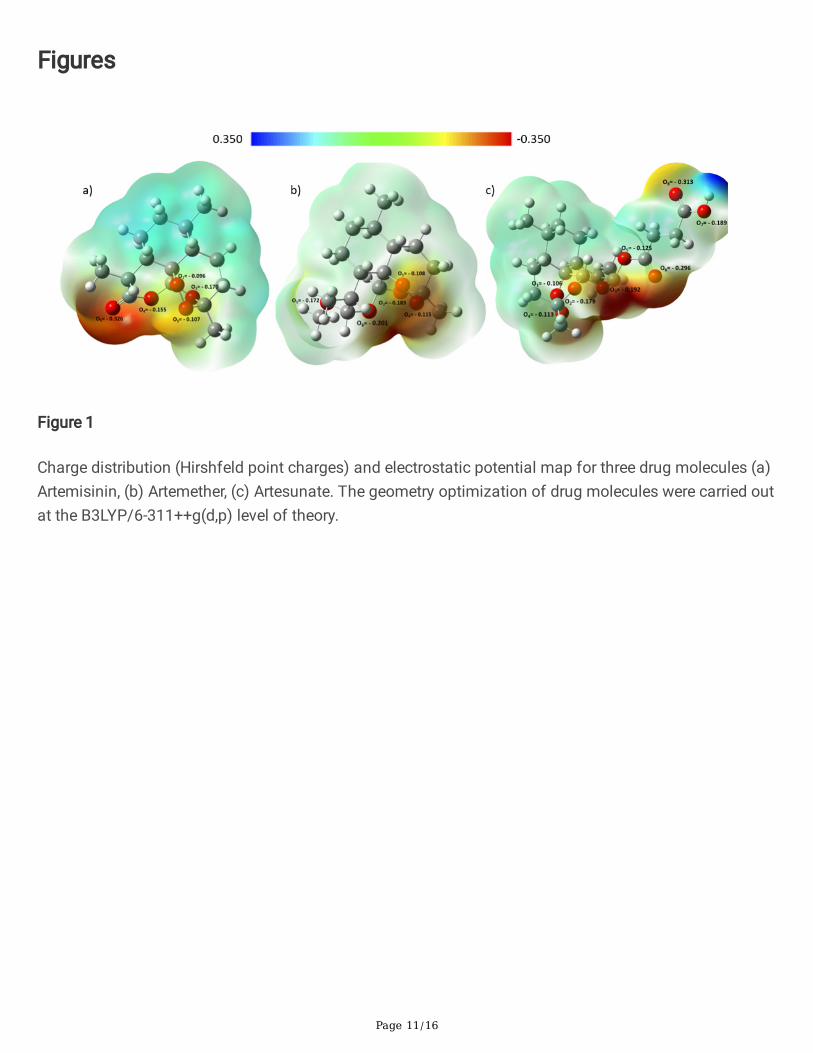
Results And DiscussionsFigure 1 displays the charge distribution and the electrostatic potential map of three drug moleculesArtemisinin, Artemether, and Artesunate. The DFT Hirschfeld point charges and electrostatic potentialmaps are obtained for the optimized molecules in water (as the solvent). The O-groups are the electronrich site of each molecule which can donate the electron density via their lone pairs to 4d and 5s orbitalsof the silver atom [34,36]. Therefore, the reddish areas in the charge distribution maps in Figure 1demonstrate the active sites of the molecules which have the most a�nity to interact with the AgNPs.
The optimized structures and charge density difference of drug molecules on Ag(111) surface are
presented in Figure 2. The adsorption energy for these structures were de�ned as
where is the total energy of the drug molecule (Artemisinin/Artemether/Artesunate) adsorbed on

Page 5/16
the Ag(111) surface, while are the total energy of the Ag(111) slab and isolated drug molecule,respectively. The results for the adsorption energy are as follow;
As expected from the electrostatic potential maps,the drugs interacts with the Ag surface through oxygen atoms, and Artemisinin interacts weakly with theAg surface, with interaction energy of 0.81 eV.
The charge density difference of drug molecules adsorbed on Ag(111) slab with a charge density iso-surface value of 0.0005 e/a.u.3 is plotted in Figure 2. This quantity accounts for the charge redistributiondue to the interaction between the drug molecule and the Ag surface. The yellow color indicates anincrease in the electron density after adsorbing, and the blue color indicates an electron density loss. This�gure con�rms the higher a�nity of Artesunate to interact with Ag and also indicates clearly that the drugmolecules interact with Ag atoms via their oxygens as discussed based on their electrostatic potentialmap.
Figure 3 displays the spectra of the root mean square displacement (RMSD) of Artemisinin, Artemether,and Artesunate, respectively. The plot reveals that all systems have reached their equilibrium state before20 ns of simulation time where the �uctuations of RMSDs have reduced signi�cantly. Therefore, the MDtrajectories extracted after 20 ns are used for further analysis.
Figure 4 describes the RDF, g(r), for the active sites of Artemisinin, Artemether, and Artesunate moleculesto interact with the Ag (147-atom icosahedral) nanoparticles. The radial distribution function (RDF)describes how the density of surrounding molecule varies as a function of the distance from a referencepoint. In this part, the arrangement of three drug molecules and their a�nities to interact with AgNP hasbeen investigated using RDF analysis. For Artemisinin, the highest peak of atom O5 (-0.326 |e|) in Figure4(a) shows the highest a�nity of this atom to interact with AgNP due to having more negative charges tointeract with the silver nanoparticle on the surface. In Figure 4(b), the atoms O3 (-0.201 |e|) of Artemether,and in Figure 4(c), O6 (-0.296 |e|) of Artesunate demonstrate the highest a�nity to interact with AgNP.These results are in agreement with DFT calculations (see Figure 1 and 2).
The RDF results for the drug molecules with respect to AgNP individually, Figure 5, and the calculatedvdW interaction energy, Figure 6(a), demonstrate that the Artesunate has a higher a�nity to interact withAgNP compared to the Artemether and Artemisinin which is also in agreement with DFT calculations forthe adsorption energy of drug molecules toward Ag (111) surface (See Figure 2). The average interactionenergy (vdW) based on MD simulation for Artemisinin, Artemether, and Artesunate are -1477.77 ± 11,-1523.62 ± 6.2, and -1854.35 ± 8.4 KJ/mol, respectively. Moreover as can be seen from Figure 6, theinteraction energy �uctuations become steady and constant at the time about 17 ns which means all 12molecules coated the surface of AgNP at this time. Figure 6(b) illustrates the coating process of AgNPwith Artesunate molecules in terms of interaction energy.

Page 6/16
ConclusionThe coating of silver nanoparticle with three common anti-malarial drugs Artemisinin, Artemether, andArtesunate have been investigated by using the density functional theory and molecular dynamicssimulations. The interaction energies of drugs with Ag(111) slab clearly indicates the strength of theinteraction as Artesunate > Artemether > Artemisinin which is con�rmed by the molecular dynamicssimulation of coating 12 drug molecules on the Ag nanocluster. This work suggest that the AgNP withlow toxicity and antiviral activity can be used as a drug delivery vehicle to enhance permeability andretention (EPR) effect of these drugs again malaria and recent COVID-19 disease.
DeclarationsCompeting Interests
The authors declare no competing interests.
Acknowledgment
The authors acknowledge the UNESCO UNISA ITHEMBA-LABS/NRF Africa Chair in Nanosciences &Nanotechnology (U2ACN2) and the Centre for High Performance Computing (CHPC), South Africa forproviding computational resources and facilities for this research project. The authors also would like tothank Dr Zahra Jamshidi for her useful comments and discussions.
References[1] Stojanoski N (1999) Development of health culture in Veles and its region from the past to the end ofthe 20th century. Veles: Society of science and art. 1999: 13–34.
[2] Petrovska BB (2012) Historical review of medicinal plants’ usage. Pharmacognosy Reviews 6(11): 1-5.https://doi.org/10.4103/0973-7847.95849.
[3] Dalrymple DG (2013) Artemisia Annua, Artemisinin, ACTs & Malaria Control in Africa: Tradition,Science and Public Policy. Politics & Prose Bookstore.
[4] Wong HN, Padín-Irizarry V, van der Watt ME, Reader J, Liebenberg W, Wiesner L, Smith P, Eribez K,Winzeler EA, Kyle DE, Birkholtz L-M, Coertzen D and Haynes RK (2020) Optimal 10-AminoartemisininsWith Potent Transmission-Blocking Capabilities for New Artemisinin Combination Therapies–ActivitiesAgainst Blood Stage P. falciparum Including PfKI3 C580Y Mutants and Liver Stage P. berghei Parasites.Front. Chem. 7: 901. https://doi.org/10.3389/fchem.2019.00901.
[5] Wang C, Xuan X, Yao W, Huang G Jin J (2015) Anti-pro�brotic effects of artesunate on bleomycin-induced pulmonary �brosis in Sprague Dawley rats. Molecular Medicine Reports 12(1): 1291–1297.https://doi.org/10.3892/mmr.2015.3500.

Page 7/16
[6] Suputtamongkol Y, Newton PN, Angus B, Teja-Isavadharm P, Keeratithakul D, Rasameesoraj M,Pukrittayakamee S, White NJ (2001) A comparison of oral artesunate and artemether antimalarialbioactivities in acute falciparum malaria. Br J Clin Pharmacol 52(6): 655–661.https://doi.org/10.1046/j.1365-2125.2001.01458.x.
[7] Ferreira JF, Luthria DL, Sasaki T, Heyerick A (2010) Flavonoids from Artemisia annua L. asantioxidants and their potential synergism with artemisinin against malaria and cancer. Molecules 15(5):3135–3170. https://doi.org/10.3390/molecules15053135.
[8] Abid Ali Khan MM, Jain DC, Bhakuni RS, Zaim M, Thakur RS (1991) Occurrence of some antiviralsterols in Artemisia annua. Plant Science 75(2): 161–165. https://doi.org/10.1016/0168-9452(91)90230-6.
[9] Duffy PE, Mutabingwa TK (2006) Artemisinin combination therapies. Lancet (London, England)367(9528): 2037-2039. https://doi.org/10.1016/s0140-6736(06)68900-9.
[10] White NJ (2008) Qinghaosu (artemisinin): the price of success. Science 320(5874): 330-334.
https://doi.org/10.1126/science.1155165.
[11] Efferth T (2007) Schwabe Award 2006: antiplasmodial and antitumor activity of artemisinin—frombench to bedside. Planta Med. 73(4): 299–309. https://doi.org/10.1055/s-2007-967138.
[12] Gilmore K, Osterrieder K, Seeberger PH (2020) "Artemisia annua Plant Extracts are Active AgainstSARS-CoV-2 In Vitro". https://www.fu-berlin.de/en/presse/informationen/fup/2020/fup_20_107-beifuss-corona/index.html. submitted for publication.
[13] Ul Haq F, Roman M, Ahmad K, Ur Rahman S, Ali Shah SM, Suleman N, Ullah S, Ahmad I, Ullah W(2020) Artemisia annua: Trials are needed for COVID-19. Phytotherapy Research 1–2.https://doi.org/10.1002/ptr.6733.
[14] Patra JK, Das G, Fraceto LF et al (2018) Nano based drug delivery systems: recent developments andfuture prospects. J Nanobiotechnol 16(71): 1-33. https://doi.org/10.1186/s12951-018-0392-8.
[15] Ivanova N, Gugleva V, Dobreva M, Pehlivanov I, Stefanov S, Andonova V (2018) Silver Nanoparticlesas Multi-Functional Drug Delivery Systems. https://doi.org /10.5772/intechopen.80238.
[16] Bazban-shotorbani S, Hasani-sadrabadi MM, Karkhaneh A, Serpooshan V (2015) Revisiting structure-property relationship of pH-responsive polymers for drug delivery applications, J. Contr. Release 253: 46-63. https://doi.org/10.1016/j.jconrel.2017.02.021.
[17] Aderibigbe BA (2017) Metal-Based Nanoparticles for the Treatment of Infectious Diseases. Molecules22(8): 1370. https://doi.org/10.3390/molecules22081370.

Page 8/16
[18] Alexander JW (2009) History of the medical use of silver. Surgical Infections 10(3): 289–292.https://doi.org/10.1089/sur.2008.9941.
[19] Nedelcu I-A, Ficai A, Sonmez M, Ficai D, Oprea O, Andronescu E (2014) Silver Based Materials forBiomedical Applications. Current Organic Chemistry 18(2): 173-184.https://doi.org/10.2174/13852728113176660141.
[20] Burdușel A-C, Gherasim O, Mihai Grumezescu A, Mogoantă L, Ficai A, Andronescu E (2018)Biomedical Applications of Silver Nanoparticles: An Up-to-Date Overview. Nanomaterials 8(9): 681;https://doi.org/10.3390/nano8090681.
[21] Mody VV, Siwale R, Singh A, Mody H.R (2010) Introduction to metallic nanoparticles. J. Pharm. Bioall.Sci. 2(4): 282–289. https://doi.org/10.4103/0975-7406.72127.
[22] Rezaee P, Akbari M, Morad R, Koochaki A, Maaza M, Jamshidi Z (2020) First Principle Simulation ofCoated Hydroxychloroquine on Ag, Au and Pt Nanoparticle as a Potential Candidate for Treatment ofSARS-CoV-2 (COVID-19), First Principle Simulation of Coated Hydroxychloroquine on Ag, Au and PtNanoparticle as a Potential Candidate for Treatment of SARS-CoV-2 (COVID-19). arXiv:2006.02343.
[23] Choi O, Deng KK, Kim NJ, Ross L, Surampalli RY, Hu ZQ (2008) The inhibitory effects of silvernanoparticles, silver ions, and silver chloride colloids on microbial growth. Water Res. 42: 3066–3074.https://doi.org/10.1016/j.watres.2008.02.021.
[24] Feng QL, Wu J, Chen GQ, Cui FZ, Kim TN, Kim JO (2000) A mechanistic study of the antibacterialeffect of silver ions on Escherichia coli and Staphylococcus aureus. J. Biomed. Mater. Res. 52: 662–668.https://doi.org/10.1002/1097-4636(20001215)52:4<662::aid-jbm10>3.0.co;2-3.
[25] Geraldo DA, Needham P, Chandia N, Arratia-Perez R, Mora GC, Villagra NA (2016) Green synthesis ofpolysaccharides-based gold and silver nanoparticles and their promissory biological activity. BiointerfaceResearch in Applied Chemistry 6(3): 1263–1271. http://repositorio.unab.cl/xmlui/handle/ria/1139.
[26] Chowdhury NR, MacGregor-Ramiasa M, Zilm P, Majewski P, Vasilev K (2016) Chocolate silvernanoparticles: Synthesis, antibacterial activity and cytotoxicity. Journal of Colloid and Interface Science482: 151–158. https://doi.org/10.1016/j.jcis.2016.08.003.
[27] Tavaf Z, Tabatabaei M, Khala�-Nezhad A, Panahi F (2017) Evaluation of antibacterial, antibo�lm andantioxidant activities of synthesized silver nanoparticles (AgNPs) and casein peptide fragments againstStreptococcus mutans. European Journal of Integrative Medicine 12: 163–171.https://doi.org/10.1016/j.eujim.2017.05.011.
[28] Henke P, Kirakci K, Kubt P, Fraiberk M, Forstov J, Mosinger J (2016) Antibacterial, Antiviral, andOxygen-Sensing Nanoparticles Prepared from Electrospun Materials. ACS Applied Materials & Interfaces8(38): 25127–25136. https://doi.org/10.1021/acsami.6b08234.

Page 9/16
[29] Galdiero S, Falanga A, Vitiello M, Cantisani M, Marra V Galdiero M (2011) Silver Nanoparticles asPotential Antiviral Agents. Molecules 16(10): 8894–8918. https://doi.org/10.3390/molecules16108894.
[30] Villeret B, Dieu A, Straube M et al (2018) Silver Nanoparticles Impair Retinoic Acid-Inducible Gene I-Mediated Mitochondrial Antiviral Immunity by Blocking the Autophagic Flux in Lung Epithelial Cells. ACSNano 12(2): 1188–1202. https://doi.org/10.1021/acsnano.7b06934.
[31] Prusty K, Swain SK (2018) Nano silver decorated polyacrylamide/dextran nanohydrogels hybridcomposites for drug delivery applications, Materials Science & Engineering C 85: 130-141.https://doi.org/10.1016/j.msec.2017.11.028.
[32] Morris D, Ansar M, Speshock J, Ivanciuc T, Qu Y, Casola A, Garofalo R (2019) Antiviral andImmunomodulatory Activity of Silver Nanoparticles in Experimental RSV Infection. Viruses 11(8): 732.https://doi.org/10.3390/v11080732.
[33] Pakiari AH, Jamshidi Z (2007) Interaction of Amino Acids with Gold and Silver Clusters. The Journalof Physical Chemistry A 111(20): 4391–4396. https://doi.org/10.1021/jp070306t.
[34] Granatier J, Urban M, Sadlej AJ (2007) Van der Waals Complexes of Cu, Ag, and Au with HydrogenSul�de. The Bonding Character. The Journal of Physical Chemistry A 111(50): 13238–13244.https://doi.org/10.1021/jp0757098.
[35] Aliakbari Tehrani Z, Jamshidi Z, Jebeli Javan M, Fattahi A (2012) Interactions of GlutathioneTripeptide with Gold Cluster: In�uence of Intramolecular Hydrogen Bond on Complexation Behavior. TheJournal of Physical Chemistry A 116(17): 4338–4347. https://doi.org/10.1021/jp2080226.
[36] Antuek A, Urban M, Sadlej AJ (2003) Lone pair interactions with coinage metal atoms: Weak van derWaals complexes of the coinage metal atoms with water and ammonia. The Journal of ChemicalPhysics 119(14): 7247–7262. https://doi.org/10.1063/1.1605936.
[37] Sambasivam A, Sangwai AV, Sureshkumar R (2016) Self-Assembly of Nanoparticle SurfactantComplexes with Rodlike Micelles: A Molecular Dynamics Study. Langmuir 32(5): 1214–1219.https://doi.org/10.1021/acs.langmuir.5b03689.
[38] Yousefpour A, Modarress H, Goharpey F, Amjad-Iranagh S (2018) Interaction of drugs amlodipine andparoxetine with the metabolizing enzyme CYP2B4: a molecular dynamics simulation study, J. Mol.Model. 24(3): 67. https://doi.org/10.1007/s00894-018-3617-8.
[39] Kordzadeh A, Amjad-Iranagh S, Zarif M, Modarress H (2019) Adsorption and encapsulation of thedrug doxorubicin on covalent functionalized carbon nanotubes: A scrutinized study by using moleculardynamics simulation and quantum mechanics calculation, Journal of Molecular Graphics and Modelling88: 11-22. https://doi.org/10.1016/j.jmgm.2018.12.009.
[40] Frisch MJ, Trucks GW, Schlegel HB et al (2009) "Gaussian 09", Wallingford CT.

Page 10/16
[41] Perdew JP, Burke K, Ernzerhof M (1996) Generalized Gradient Approximation Made Simple, Physicalreview letters 77(18): 3865-3868. https://doi.org/10.1103/PhysRevLett.77.3865.
[42] Giannozzi P, Baroni S, Bonini N, Calandra M et al (2009) QUANTUM ESPRESSO: a modular and open-source software project for quantum simulations of materials. J. Phys.: Condens. Matter 21(39): 395502.https://doi.org/10.1088/0953-8984/21/39/395502.
[43] Grimme S, Ehrlich S, Goerigk L (2011) Effect of the damping function in dispersion corrected densityfunctional theory. Journal of computational chemistry 32(7): 1456– 65.https://doi.org/10.1002/jcc.21759.
[44] Abraham M, Van der Spoel D, Lindahl E, Hess B (2019) " GROMACS" 2019.
[45] Huang J, MacKerell Jr AD (2013) " CHARMM36 all‐atom additive protein force �eld: Validation basedon comparison to NMR data". J Comput Chem 34(25): 2135-2145. https://doi.org/10.1002/jcc.23354.
[46] Jorgensen WL, Chandrasekhar J, Madura JD, Impey RW, Klein ML (1983) Comparison of simplepotential functions for simulating liquid water. J. Chem. Phys. 79: 926.https://doi.org/10.1063/1.445869.
[47] Hess B, Bekker H, Berendsen HJ, Fraaije JG (1997) LINCS: a linear constraint solver for molecularsimulations. Journal of Computational Chemistry 18(12): 1463-1472.https://doi.org/10.1002/(SICI)1096-987X(199709)18:12<1463::AID-JCC4>3.0.CO;2-H.
[48] Essmann U, Perera L, Berkowitz ML, Darden T, Lee H, Pedersen LG (1995) A smooth particle meshEwald method. J. Chem. Phys. 103(19): 8577. https://doi.org/10.1063/1.470117.
[49] Adcock SA, McCammon JA (2006) Molecular dynamics: survey of methods for simulating theactivity of proteins. Chem. Rev. 106(5): 1589–1615. https://doi.org/10.1021/cr040426m.
[50] Humphrey W, Dalke A, Chulten K (1996) VMD: visual molecular dynamics. Journal of MolecularGraphics 14(1): 33-38. https://doi.org/10.1016/0263-7855(96)00018-5.
[51] Vanommeslaeghe K, MacKerell Jr AD (2012) Automation of the CHARMM General Force Field(CGenFF) I: Bond Perception and Atom Typing. Journal of chemical information and modelling 52(12):3144-3154. https://doi.org/10.1021/ci300363c.
[52] Sohraby F, Soltanabad MH, Bagheri M, Javan MB, Moghadam MJ, Baghkheirati EK, Bagherieh NajjarMB (2020) Application of Molecular Dynamics in Coating Ag-Conjugated Nanoparticles with PotentialTherapeutic Applications. Nano Biomed. Eng. 12(1): 90-98. https://doi.org/10.5101/nbe.v12i1.p90-98.
[53] Kyrychenko A, Pasko DA, Kalugin ON (2017) Poly (vinyl alcohol) as a water protecting agent for silvernanoparticles: the role of polymer size and structure. Physical Chemistry Chemical Physics 19: 8742–8756. https://doi.org/10.1039/C6CP05562A.
Page 11/16
Figures
Figure 1
Charge distribution (Hirshfeld point charges) and electrostatic potential map for three drug molecules (a)Artemisinin, (b) Artemether, (c) Artesunate. The geometry optimization of drug molecules were carried outat the B3LYP/6-311++g(d,p) level of theory.

Page 12/16
Figure 2
The charge density difference of adsorbed (a) Artemisinin (b) Artemether (c) Artesunate molecules onAg(111) slab. The blue and yellow means the negative and positive values corresponding to the loss andgain of electrons. The isovalue sets at 0.0005 e/a.u.3

Page 13/16
Figure 3
RMSD plots of drug molecules: Artemisinin, Artemether, and Artesunate versus simulation time. The MDtrajectories after 20 ns have been taken for further analysis.

Page 14/16
Figure 4
RDF plots for the active sites of (a) Artemisinin, (b) Artemether, and (c) Artesunate with AgNP.

Page 15/16
Figure 5
RDF plots of Artemisinin, Artemether, and Artesunate with respect to AgNP.

Page 16/16
Figure 6
(a) Interaction energies (vdW) between Artemisinin, Artemether, and Artesunate and AgNP. (b) The processof coating AgNP with 12 Artesunate molecules in terms of interaction energy.