international journal of infectious diseases · in september 2015, due to the persistence of pain...
TRANSCRIPT
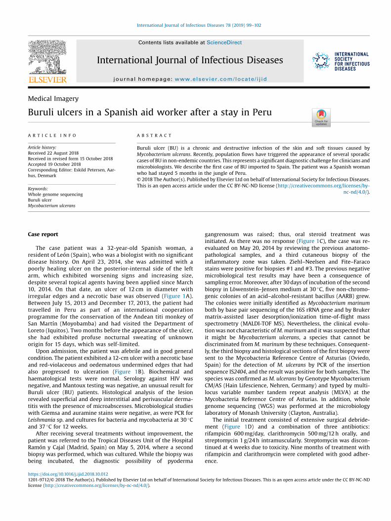
International Journal of Infectious Diseases 78 (2019) 99–102
Medical Imagery
Buruli ulcers in a Spanish aid worker after a stay in Peru
A R T I C L E I N F O
Article history:Received 22 August 2018Received in revised form 15 October 2018Accepted 19 October 2018Corresponding Editor: Eskild Petersen, Aar-hus, Denmark
Keywords:Whole genome sequencingBuruli ulcerMycobacterium ulcerans
A B S T R A C T
Buruli ulcer (BU) is a chronic and destructive infection of the skin and soft tissues caused byMycobacterium ulcerans. Recently, population flows have triggered the appearance of several sporadiccases of BU in non-endemic countries. This represents a significant diagnostic challenge for clinicians andmicrobiologists. We describe the first case of BU imported to Spain. The patient was a Spanish womanwho had stayed 5 months in the jungle of Peru.© 2018 The Author(s). Published by Elsevier Ltd on behalf of International Society for Infectious Diseases.This is an open access article under the CC BY-NC-ND license (http://creativecommons.org/licenses/by-
nc-nd/4.0/).
Contents lists available at ScienceDirect
International Journal of Infectious Diseases
journal home page: www.elsevier .com/ locat e/ i j id
Case report
The case patient was a 32-year-old Spanish woman, aresident of León (Spain), who was a biologist with no significantdisease history. On April 23, 2014, she was admitted with apoorly healing ulcer on the posterior-internal side of the leftarm, which exhibited worsening signs and increasing size,despite several topical agents having been applied since March10, 2014. On that date, an ulcer of 12 cm in diameter withirregular edges and a necrotic base was observed (Figure 1A).Between July 15, 2013 and December 17, 2013, the patient hadtravelled in Peru as part of an international cooperationprogramme for the conservation of the Andean titi monkey ofSan Martín (Moyobamba) and had visited the Department ofLoreto (Iquitos). Two months before the appearance of the ulcer,she had exhibited profuse nocturnal sweating of unknownorigin for 15 days, which was self-limited.
Upon admission, the patient was afebrile and in good generalcondition. The patient exhibited a 12-cm ulcer with a necrotic baseand red-violaceous and oedematous undermined edges that hadalso progressed to ulceration (Figure 1B). Biochemical andhaematological tests were normal. Serology against HIV wasnegative, and Mantoux testing was negative, an unusual result forBuruli ulcer (BU) patients. Histological analysis of the lesionrevealed superficial and deep interstitial and perivascular derma-titis with the presence of microabscesses. Microbiological studieswith Giemsa and auramine stains were negative, as were PCR forLeishmania sp. and cultures for bacteria and mycobacteria at 30 �Cand 37 �C for 12 weeks.
After receiving several treatments without improvement, thepatient was referred to the Tropical Diseases Unit of the HospitalRamón y Cajal (Madrid, Spain) on May 5, 2014, where a secondbiopsy was performed, which was cultured. While the biopsy wasbeing incubated, the diagnostic possibility of pyoderma
https://doi.org/10.1016/j.ijid.2018.10.0121201-9712/© 2018 The Author(s). Published by Elsevier Ltd on behalf of International Solicense (http://creativecommons.org/licenses/by-nc-nd/4.0/).
gangrenosum was raised; thus, oral steroid treatment wasinitiated. As there was no response (Figure 1C), the case was re-evaluated on May 20, 2014 by reviewing the previous anatomo-pathological samples, and a third cutaneous biopsy of theinflammatory zone was taken. Ziehl–Neelsen and Fite–Faracostains were positive for biopsies #1 and #3. The previous negativemicrobiological test results may have been a consequence ofsampling error. Moreover, after 30 days of incubation of the secondbiopsy in Löwenstein–Jensen medium at 30 �C, five non-chromo-genic colonies of an acid–alcohol-resistant bacillus (AARB) grew.The colonies were initially identified as Mycobacterium marinumboth by base pair sequencing of the 16S rRNA gene and by Brukermatrix-assisted laser desorption/ionization time-of-flight massspectrometry (MALDI-TOF MS). Nevertheless, the clinical evolu-tion was not characteristic of M. marinum and it was suspected thatit might be Mycobacterium ulcerans, a species that cannot bediscriminated from M. marinum by these techniques. Consequent-ly, the third biopsy and histological sections of the first biopsy weresent to the Mycobacteria Reference Centre of Asturias (Oviedo,Spain) for the detection of M. ulcerans by PCR of the insertionsequence IS2404, and the result was positive for both samples. Thespecies was confirmed as M. ulcerans by Genotype MycobacteriumCM/AS (Hain Lifescience, Nehren, Germany) and typed by multi-locus variable number tandem repeat analysis (MLVA) at theMycobacteria Reference Centre of Asturias. In addition, wholegenome sequencing (WGS) was performed at the microbiologylaboratory of Monash University (Clayton, Australia).
The initial treatment consisted of extensive surgical debride-ment (Figure 1D) and a combination of three antibiotics:rifampicin 600 mg/day, clarithromycin 500 mg/12 h orally, andstreptomycin 1 g/24 h intramuscularly. Streptomycin was discon-tinued at 4 weeks due to toxicity. Nine months of treatment withrifampicin and clarithromycin were completed with good adher-ence.
ciety for Infectious Diseases. This is an open access article under the CC BY-NC-ND
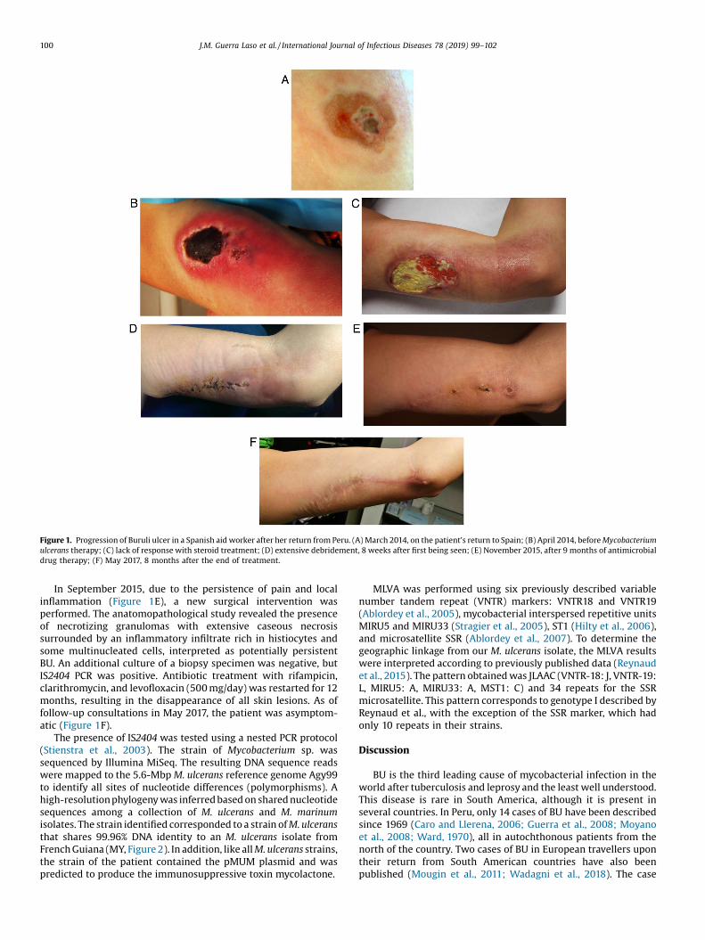
Figure 1. Progression of Buruli ulcer in a Spanish aid worker after her return from Peru. (A) March 2014, on the patient’s return to Spain; (B) April 2014, before Mycobacteriumulcerans therapy; (C) lack of response with steroid treatment; (D) extensive debridement, 8 weeks after first being seen; (E) November 2015, after 9 months of antimicrobialdrug therapy; (F) May 2017, 8 months after the end of treatment.
100 J.M. Guerra Laso et al. / International Journal of Infectious Diseases 78 (2019) 99–102
In September 2015, due to the persistence of pain and localinflammation (Figure 1E), a new surgical intervention wasperformed. The anatomopathological study revealed the presenceof necrotizing granulomas with extensive caseous necrosissurrounded by an inflammatory infiltrate rich in histiocytes andsome multinucleated cells, interpreted as potentially persistentBU. An additional culture of a biopsy specimen was negative, butIS2404 PCR was positive. Antibiotic treatment with rifampicin,clarithromycin, and levofloxacin (500 mg/day) was restarted for 12months, resulting in the disappearance of all skin lesions. As offollow-up consultations in May 2017, the patient was asymptom-atic (Figure 1F).
The presence of IS2404 was tested using a nested PCR protocol(Stienstra et al., 2003). The strain of Mycobacterium sp. wassequenced by Illumina MiSeq. The resulting DNA sequence readswere mapped to the 5.6-Mbp M. ulcerans reference genome Agy99to identify all sites of nucleotide differences (polymorphisms). Ahigh-resolution phylogeny was inferred based on shared nucleotidesequences among a collection of M. ulcerans and M. marinumisolates. The strain identified corresponded to a strain of M. ulceransthat shares 99.96% DNA identity to an M. ulcerans isolate fromFrench Guiana (MY, Figure 2). In addition, like all M. ulcerans strains,the strain of the patient contained the pMUM plasmid and waspredicted to produce the immunosuppressive toxin mycolactone.
MLVA was performed using six previously described variablenumber tandem repeat (VNTR) markers: VNTR18 and VNTR19(Ablordey et al., 2005), mycobacterial interspersed repetitive unitsMIRU5 and MIRU33 (Stragier et al., 2005), ST1 (Hilty et al., 2006),and microsatellite SSR (Ablordey et al., 2007). To determine thegeographic linkage from our M. ulcerans isolate, the MLVA resultswere interpreted according to previously published data (Reynaudet al., 2015). The pattern obtained was JLAAC (VNTR-18: J, VNTR-19:L, MIRU5: A, MIRU33: A, MST1: C) and 34 repeats for the SSRmicrosatellite. This pattern corresponds to genotype I described byReynaud et al., with the exception of the SSR marker, which hadonly 10 repeats in their strains.
Discussion
BU is the third leading cause of mycobacterial infection in theworld after tuberculosis and leprosy and the least well understood.This disease is rare in South America, although it is present inseveral countries. In Peru, only 14 cases of BU have been describedsince 1969 (Caro and Llerena, 2006; Guerra et al., 2008; Moyanoet al., 2008; Ward, 1970), all in autochthonous patients from thenorth of the country. Two cases of BU in European travellers upontheir return from South American countries have also beenpublished (Mougin et al., 2011; Wadagni et al., 2018). The case
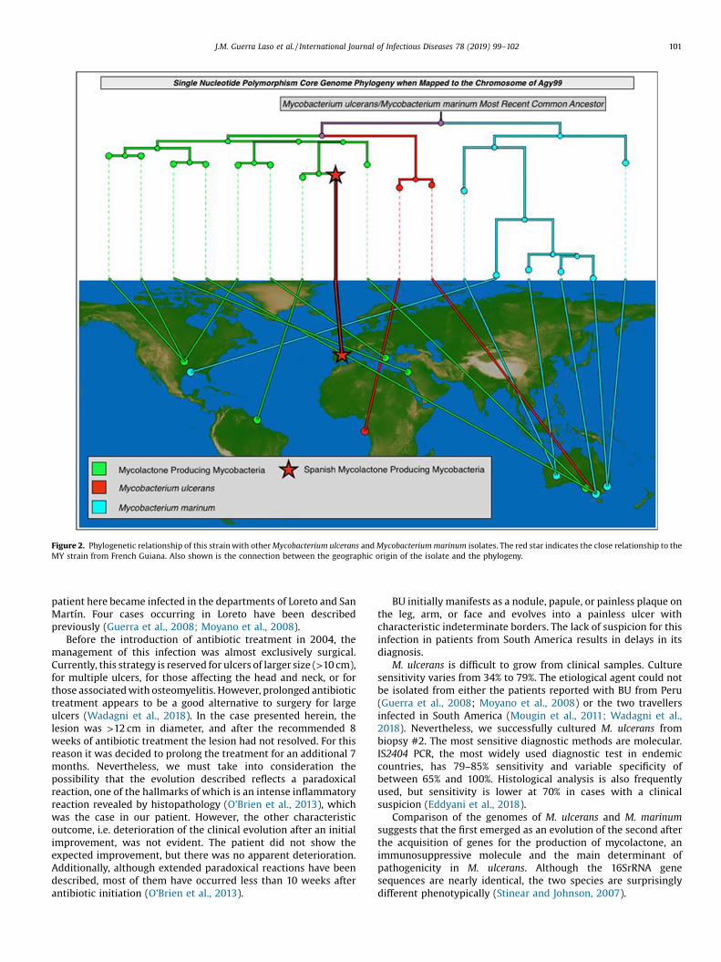
Figure 2. Phylogenetic relationship of this strain with other Mycobacterium ulcerans and Mycobacterium marinum isolates. The red star indicates the close relationship to theMY strain from French Guiana. Also shown is the connection between the geographic origin of the isolate and the phylogeny.
J.M. Guerra Laso et al. / International Journal of Infectious Diseases 78 (2019) 99–102 101
patient here became infected in the departments of Loreto and SanMartín. Four cases occurring in Loreto have been describedpreviously (Guerra et al., 2008; Moyano et al., 2008).
Before the introduction of antibiotic treatment in 2004, themanagement of this infection was almost exclusively surgical.Currently, this strategy is reserved for ulcers of larger size (>10 cm),for multiple ulcers, for those affecting the head and neck, or forthose associated with osteomyelitis. However, prolonged antibiotictreatment appears to be a good alternative to surgery for largeulcers (Wadagni et al., 2018). In the case presented herein, thelesion was >12 cm in diameter, and after the recommended 8weeks of antibiotic treatment the lesion had not resolved. For thisreason it was decided to prolong the treatment for an additional 7months. Nevertheless, we must take into consideration thepossibility that the evolution described reflects a paradoxicalreaction, one of the hallmarks of which is an intense inflammatoryreaction revealed by histopathology (O’Brien et al., 2013), whichwas the case in our patient. However, the other characteristicoutcome, i.e. deterioration of the clinical evolution after an initialimprovement, was not evident. The patient did not show theexpected improvement, but there was no apparent deterioration.Additionally, although extended paradoxical reactions have beendescribed, most of them have occurred less than 10 weeks afterantibiotic initiation (O’Brien et al., 2013).
BU initially manifests as a nodule, papule, or painless plaque onthe leg, arm, or face and evolves into a painless ulcer withcharacteristic indeterminate borders. The lack of suspicion for thisinfection in patients from South America results in delays in itsdiagnosis.
M. ulcerans is difficult to grow from clinical samples. Culturesensitivity varies from 34% to 79%. The etiological agent could notbe isolated from either the patients reported with BU from Peru(Guerra et al., 2008; Moyano et al., 2008) or the two travellersinfected in South America (Mougin et al., 2011; Wadagni et al.,2018). Nevertheless, we successfully cultured M. ulcerans frombiopsy #2. The most sensitive diagnostic methods are molecular.IS2404 PCR, the most widely used diagnostic test in endemiccountries, has 79–85% sensitivity and variable specificity ofbetween 65% and 100%. Histological analysis is also frequentlyused, but sensitivity is lower at 70% in cases with a clinicalsuspicion (Eddyani et al., 2018).
Comparison of the genomes of M. ulcerans and M. marinumsuggests that the first emerged as an evolution of the second afterthe acquisition of genes for the production of mycolactone, animmunosuppressive molecule and the main determinant ofpathogenicity in M. ulcerans. Although the 16SrRNA genesequences are nearly identical, the two species are surprisinglydifferent phenotypically (Stinear and Johnson, 2007).
102 J.M. Guerra Laso et al. / International Journal of Infectious Diseases 78 (2019) 99–102
In conclusion, the differential diagnosis of infection by M.ulcerans should be included for patients from tropical areaspresenting with ulcers, especially if such ulcers are painless,exhibit rapid growth, and are located on the extremities. A lack offamiliarity with the disease results in delays in diagnosis andsubsequent treatment, leading to severe deformities and dis-abilities.
Acknowledgements
Dr Enrique Gómez Mampaso, Hospital Ramón y Cajal, Madrid,for his support in the handling of the sample and growth ofMycobacterium ulcerans and Dr Octavio Rivero-Lezcano for helpfuldiscussions.
Funding: None of the authors received funding from any source.Ethical approval: The patient gave her approval for the
publication of her clinical case.Conflict of interest: None of the authors has a conflict of interest.
References
Ablordey A, Swings J, Hubans C, Chemlal K, Locht C, Portaels F, et al. Multilocusvariable-number tandem repeat typing of Mycobacterium ulcerans. J ClinMicrobiol 2005;43:1546–51.
Ablordey A, Fonteyne PA, Stragier P, Vandamme P, Portaels F. Identification of a newvariable number tandem repeat locus in Mycobacterium ulcerans for potentialstrain discrimination among African isolates. Clin Microbiol Infect2007;13:734–6.
Caro F, Llerena G. Ulcer of Buruli in Tumbes. Case report and literature review. Foliadermatol Peru 2006;17:76–81.
Eddyani M, Sopoh GE, Ayelo G, Brun LVC, Roux JJ, Barogui Y, et al. Diagnosticaccuracy of clinical and microbiological signs in patients with skin lesionsresembling Buruli ulcer in an endemic region. Clin Infect Dis 2018;67:827–34,doi:http://dx.doi.org/10.1093/cid/ciy197.
Guerra H, Palomino JC, Falconí E, Bravo F, Donaires N, Van Marck E, et al.Mycobacterium ulcerans disease, Peru. Emerg Infect Dis 2008;14:373–7.
Hilty M, Yeboah-Manu D, Boakye D, Mensah-Quainoo E, Rondini S, Schelling E, et al.Genetic diversity in Mycobacterium ulcerans isolates from Ghana revealed by tonewly identified locus containing to variable number of tandem repeats. JBacteriol 2006;188:1462–5.
Mougin B, Avenel-Audran M, Hasseine L, Martin L, Cottin J, Pomares C, et al. Acutaneous ulcer resulting from Mycobacterium ulcerans - Leishmania brazilensiscoinfection in South America. Am J Trop Med Hyg 2011;5:897–9.
Moyano LM, Chero JC, Gonzalvez GE, Cisticercosis Working Group in Peru. Buruliulcer. Am J Trop Med Hyg 2008;79:3.
O’Brien DP, Robson M, Friedman ND, Walton A, McDonald A, Callan P, et al.Incidence, clinical spectrum, diagnostic features, treatment and predictors ofparadoxical reactions during antibiotic treatment of Mycobacterium ulceransinfections. BMC Infect Dis 2013;13:416, doi:http://dx.doi.org/10.1186/1471-2334-13-416.
Reynaud Y, Millet J, Couvin D, Rastogi N, Brown C, Couppié P, et al. Heterogeneityamong Mycobacterium ulcerans from French Guiana revealed by multilocusvariable number tandem repeat analysis (MLVA). PLoS One 2015;10:e0118597,doi:http://dx.doi.org/10.1371/journal.pone.0118597.
Stienstra Y, van der Werf TS, Guarner J, Raghunathan PL, Spotts Whitney EA, van derGraaf WT, et al. Analysis of an IS 2404 -based nested PCR for diagnosis of Buruliulcer disease in regions of Ghana where the disease is endemic. J Clin Microbiol2003;41:794–7.
Stinear T, Johnson P. From Marinum to Ulcerans: a mycobacterial human pathogenemerges. Microbe 2007;2:187–94.
Stragier P, Ablordey A, Meyers WM, Portaels F. Genotyping Mycobacterium ulceransand Mycobacterium marinum by using mycobacterial interspersed repetitiveunits. J Bacteriol 2005;187:1639–47.
Wadagni AC, Barogui YT, Johnson RC, Sopoh GE, Affolabi D, van der Werf TS, et al.Delayed versus standard assessment for excision surgery in patients with Buruliulcer in Benin: a randomized controlled trial. Lancet Infect Dis 2018;18:650–6,doi:http://dx.doi.org/10.1016/S1473-3099(18)30160-9.
Ward DE. Buruli ulcer. Br Med J 1970;3:346.
Jose Manuel Guerra Lasoa
Teresa Nebreda Mayoralb,*Juan José Palacios Gutiérrezc
Élia Samaniego Gonzálezd
Bárbara Rodríguez Martíne
Leticia Barrio Rodrígueze
Nieves Alonso Orcajof
Elena Magaz Garcíaa
Timothy Stinearg
Andrew H. Buultjensh
Jose Antonio Pérez MolinaiaInternal Medicine Service, Complejo Asistencial Universitario de
León, León, Spain
bMicrobiology Service, Complejo Asistencial Universitario de León,Avda Altos de Nava s/n, 47075 León, Spain
cRegional Reference Unit of Mycobacteria, Microbiology Service,Hospital Universitario Central de Asturias, Oviedo, Spain
dDermatology Service, Complejo Asistencial Universitario de León,Universidad de Leon, León, Spain
ePlastic Surgery Service, Complejo Asistencial Universitario de León,León, Spain
fPathological Anatomy Service, Complejo Asistencial Universitario deLeón, León, Spain
gDepartment of Microbiology, Monash University, Clayton, Victoria,Australia
hDepartment of Microbiology and Immunology, The Peter DohertyInstitute for Infection and Immunity, The University of Melbourne,
Melbourne, Australia
iInfectious Diseases Service, Hospital Ramón y Cajal, Madrid, Spain
* Corresponding author.E-mail address: [email protected] (T. Nebreda May-
oral).
Corresponding Editor: Eskild Petersen, Aarhus, Denmark
Received 22 August 2018Received in revised form 15 October 2018
Accepted 19 October 2018