international journal of innovative pharmaceutical ... › sites › default › files › articles...
TRANSCRIPT

REVIEW ARTICLE Ahsan Habib et.al / IJIPSR / 3 (9), 2015, 1372-1388
Department of Pharmacy ISSN (online) 2347-2154
Available online: www.ijipsr.com September Issue 1372
HYDROGEL: A NOBLE APPROACH TO HEALTHCARE
1Md. Ahsan Habib,
2Mst. Asma Akter,
1Md. Ershad Alam,
3Ziaul Karim,
4Md. Tanzir Ahmed Joarder
1M.Pharm, Department of Pharmacy, University of Asia Pacific, Dhaka, BANGLADESH
2M.Pharm, Department of Pharmacy, Stamford University, Dhaka, BANGLADESH
3M.Pharm, Department of Pharmacy, State University of Bangladesh, BANGLADESH
4B.Pharm, Department of Pharmacy, Manarat International University, Dhaka, BANGLADESH
Corresponding Author Md. Ahsan Habib
M.Pharm (Pharmaceutical Technology)
Department of Pharmacy,
University of Asia Pacific,
Dhaka, BANGLADESH
E-mail: [email protected]
Phone: +88-01713865486
International Journal of Innovative
Pharmaceutical Sciences and Research www.ijipsr.com
ABSTRACT
Hydrogels are crosslinked polymeric networks, which have the ability to hold water within
the spaces available among the polymeric chains. Hydrogels are water swollen polymer
matrices, with a huge tendency to absorb water. Their ability to swell, under physiological
conditions, makes them an ideal material for biomedical applications. The hydrophilicity of
the network is due to the presence of chemical residues such as hydroxylic, carboxylic,
amidic, primary amidic, sulphonic and others that can be found within the polymer
backbone or as lateral chains. The hydrogels have been used extensively in various
biomedical applications, viz. drug delivery, cell carriers and/or entrapment, wound
management, tissue engineering, growing new body part, contact lens applications etc.
Hydrogels can serve as scaffolds that provide structural integrity to tissue constructs,
control drug and protein delivery to tissues and cultures, and serve as adhesives or barriers
between tissue and material surfaces. This review presents an overview to the advances in
hydrogel based drug delivery that have become the interest of most researchers.
Key words: Hydrogels, biomedical applications, cross linked polymer.

REVIEW ARTICLE Ahsan Habib et.al / IJIPSR / 3 (9), 2015, 1372-1388
Department of Pharmacy ISSN (online) 2347-2154
Available online: www.ijipsr.com September Issue 1373
INTRODUCTION
Hydrophilic gels called hydrogels are three-dimensional (3D), cross-linked materials absorbing
large quantities of water without dissolving. Softness, smartness, and the capacity to store water
make hydrogels unique materials [1]. The ability of hydrogels to absorb water arises from
hydrophilic functional groups attached to the polymer backbone while their resistance to
dissolution arises from cross-links between network chains. Water inside the hydrogel allows free
diffusion of some solute molecules, while the polymer serves as a matrix to hold water together.
Another aspect of hydrogel is that the gel is a single polymer molecule, that is, the network chains
in the gel are connected to each other to form one big molecule on macroscopic scale. It is natural
to expect that the conformational transitions of the elastically active network chains become
visible on the macroscopic scale of hydrogel samples. The gel is a state that is neither completely
liquid nor completely solid. These half liquid-like and half solid-like properties cause many
interesting relaxation behaviors that are not found in either a pure solid or a pure liquid. From the
point of view of their mechanical properties, the hydrogels are characterized by an elastic
modulus which exhibits a pronounced plateau extending to times at least of the order of seconds,
and by a viscous modulus which is considerably smaller than the elastic modulus in the plateau
region. The amount of water absorbed in hydrogels is related to the presence of specific groups
such as –COOH, –OH, –CONH2, –CONH–, and –SO3H. Capillary effect and osmotic pressure
are other variables that also influence the equilibrium water uptake of hydrogels [2].
History of hydrogels
The word polymer has been derived from the Greek words polys (meaning many) and meros (part
or unit). The polymers (e.g. proteins and celluloses) form the basic building block of life. For over
fifty years hydrogels have been used in numerous biomedical disciplines, in ophthalmology as
contact lenses and in surgery as absorbable sutures, as well as in many other areas of clinical
practice to cure such illnesses as diabetes mellitus, osteoporosis, asthma, heart diseases and
neoplasms. It was in 1955 that Professors Lim and Wichterle of Prague, Czech Republic,
synthesized the first hydrogel with potential biomedical uses. That was synthetic poly-2-
hydroxyethyl methacrylate, used – soon after its discovery – in contact lens production. The main
advantage of that revolutionary biomaterial was its stability under varying pH, temperature and
tonicity conditions. In the 1980s hydrogels were modified for other applications. Lim and Sun
obtained calcium alginate microcapsules for cell engineering, and Yannas’ group modified
REVIEW ARTICLE Ahsan Habib et.al / IJIPSR / 3 (9), 2015, 1372-1388
Department of Pharmacy ISSN (online) 2347-2154
Available online: www.ijipsr.com September Issue 1374
synthetic hydrogels with some natural substances, such as collagen and shark cartilage to obtain
novel dressings, providing optimal conditions for healing burns. Nowadays, hydrogels continue to
interest scientists. They are obtained from new materials using the latest techniques to make them
safe and non-toxic. The final hydrogel product is present in very advanced applications, e.g. tissue
engineering and regeneration, where they can be applied in a non-invasive manner. They can
serve in the prevention of thrombosis, post-operative adhesion formation, drug delivery systems,
coatings for biosensors and cell transplantation.
What are Hydrogels?
According to the latest medical and pharmaceutical encyclopaedias, there is still no precise and
limiting definition of the term hydrogel. Most often, a hydrogel is considered to be a material
made when a water-insoluble polymer absorbs a large amount of water, or else it is simply a
water-swollen polymer network. Polymer hydrogels can be of either synthetic or natural origin,
homopolymers or copolymers [3].
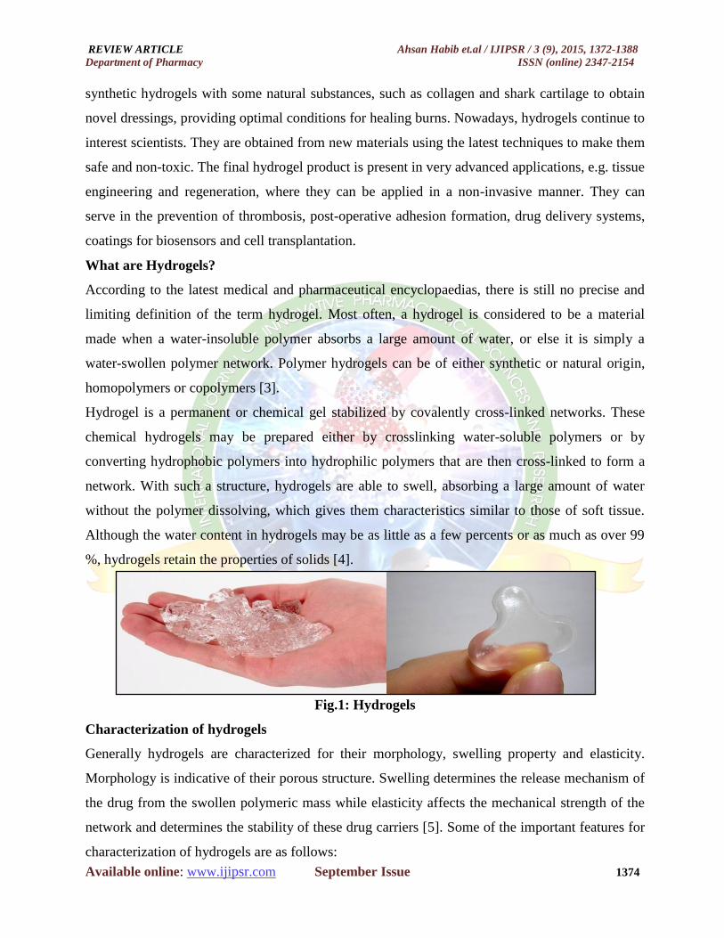
Hydrogel is a permanent or chemical gel stabilized by covalently cross-linked networks. These
chemical hydrogels may be prepared either by crosslinking water-soluble polymers or by
converting hydrophobic polymers into hydrophilic polymers that are then cross-linked to form a
network. With such a structure, hydrogels are able to swell, absorbing a large amount of water
without the polymer dissolving, which gives them characteristics similar to those of soft tissue.
Although the water content in hydrogels may be as little as a few percents or as much as over 99
%, hydrogels retain the properties of solids [4].
Fig.1: Hydrogels
Characterization of hydrogels
Generally hydrogels are characterized for their morphology, swelling property and elasticity.
Morphology is indicative of their porous structure. Swelling determines the release mechanism of
the drug from the swollen polymeric mass while elasticity affects the mechanical strength of the
network and determines the stability of these drug carriers [5]. Some of the important features for
characterization of hydrogels are as follows:
REVIEW ARTICLE Ahsan Habib et.al / IJIPSR / 3 (9), 2015, 1372-1388
Department of Pharmacy ISSN (online) 2347-2154
Available online: www.ijipsr.com September Issue 1375
X-ray diffraction
It is also used to understand whether the polymers retain their crystalline structure or they get
deformed during the processing pressurization process [6] [7].
In-vitro release study for drugs
Since hydrogels are the swollen polymeric networks, interior of which is occupied by drug
molecules, therefore, release studies are carried out to understand the mechanism of release over a
period of application [6,7].
FTIR (Fourier Transform Infrared Spectroscopy)
FTIR (Fourier Transform Infrared Spectroscopy) is a useful technique for identifying chemical
structure of a substance. It is based on the principle that the basic components of a substance, i.e.
chemical bonds, usually can be excited and absorb infrared light at frequencies that are typical of
the types of the chemical bonds. The resulting IR absorption spectrum represents a fingerprint of
measured sample. This technique is widely used to investigate the structural arrangement in
hydrogel by comparison with the starting materials [8].
Scanning Electron Microscopy (SEM)
SEM can be used to provide information about the sample's surface topography, composition, and
other properties such as electrical conductivity. Magnification in SEM can be controlled over a
range of up to 6 orders of magnitude from about 10 to 500,000 times. This is a powerful
technique widely used to capture the characteristic ‘network’ structure in hydrogels [9].
Light scattering
Gel permeation chromatography coupled on line to a multi angle laser light scattering (GPC-
MALLS) is a widely used technique to determine the molecular distribution and parameters of a
polymeric system. Hydrogel in a polymeric system can be quantified using this technique. This
technique is widely used in quantifying the hydrogels of several hydrocolloids such as gum
arabic, gelatine and pullulan [10].
Rheology
Hydrogels are evaluated for viscosity under constant temperature of usually 4°C by using Cone
Plate type viscometer [11].
Swelling behavior of hydrogels
When a hydrogel in its initial state is placed in an aqueous solution, water molecules will
penetrate into the polymer network. The entering molecules are going to occupy some space, and
as a result some meshes of the network will start expanding, allowing other water molecules to
REVIEW ARTICLE Ahsan Habib et.al / IJIPSR / 3 (9), 2015, 1372-1388
Department of Pharmacy ISSN (online) 2347-2154
Available online: www.ijipsr.com September Issue 1376
enter within the network. Evidently, swelling is not a continual process; the elasticity of the
physically (e.g. hydrogen bonding) or chemically (covalent, atomic, ionic) cross-linked network
will counter-balance the infinite stretching of the network to prevent its destruction. Thus, by
balancing these two opposite forces, a net force, known as the swelling pressure (Psw) is produced,
which is equal to zero at equilibrium obtained with pure water, and that can be expressed as [12]:
Psw = k × Cn
Where, k and n are constants, and C is the polymer concentration. At the equilibrium there is no
additional swelling.
In the case of ionic polymers, the swelling equilibrium of the polymeric matrix is more
complicated as it heavily depends also on the ionic strength.
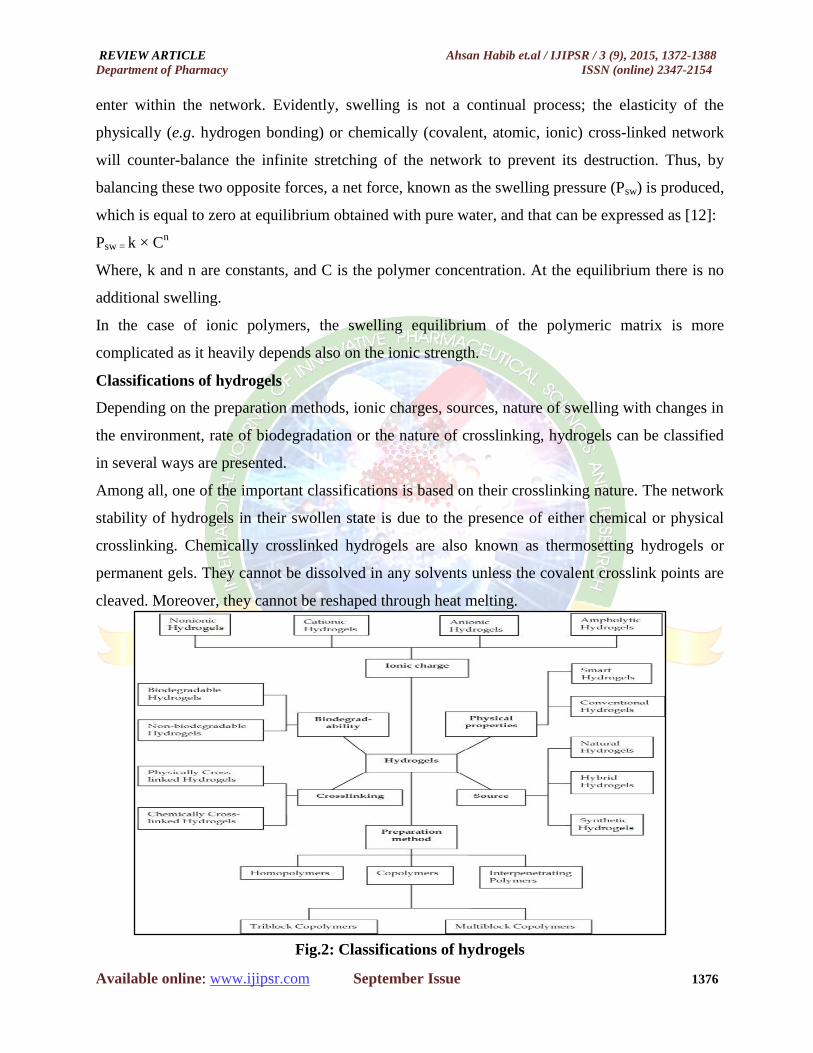
Classifications of hydrogels
Depending on the preparation methods, ionic charges, sources, nature of swelling with changes in
the environment, rate of biodegradation or the nature of crosslinking, hydrogels can be classified
in several ways are presented.
Among all, one of the important classifications is based on their crosslinking nature. The network
stability of hydrogels in their swollen state is due to the presence of either chemical or physical
crosslinking. Chemically crosslinked hydrogels are also known as thermosetting hydrogels or
permanent gels. They cannot be dissolved in any solvents unless the covalent crosslink points are
cleaved. Moreover, they cannot be reshaped through heat melting.
Fig.2: Classifications of hydrogels

REVIEW ARTICLE Ahsan Habib et.al / IJIPSR / 3 (9), 2015, 1372-1388
Department of Pharmacy ISSN (online) 2347-2154
Available online: www.ijipsr.com September Issue 1377
Table 1: Stimuli-sensitive Hydrogel
Structure and bonding
Scientists still do not fully understand how hydrogels manage to absorb so much water, and there
is still plenty of ongoing research into their properties and uses. Understanding the structure and
bonding of these advanced materials helps to explain these properties; this in turn helps chemists
to design new hydrogels which can perform different functions. Many hydrogels are polymers of
carboxylic acids. The acid groups stick off the main chain of the polymer. When these polymers
are put into water, the hydrogen atoms react and come off as positive ions. This leaves negative
ions along the length of the polymer chain. When polymer chains are in solution, they tend to coil
up. However, the hydrogel now has lots of negative charges down its length.
Fig.3: The polymer chain of a hydrogel
Fig.4: A polymer chain coiled up in solution
Fig.5: A hydrogel polymer chain with lots of negative charges along its length
Type of Stimuli-Sensitive
Hydrogel Key Points
Thermo Release of medicine occurs, through abrupt decrease in surface area due to
temperature change.
pH Swelling controlled through the interactions between protons in solution and
ions within hydrogel
Electro Drug release occurs when electric filed acts on the hydrogel
Enzyme After swelling occurs, from increase in pH, enzymes degrade hydrogel,
therefore releasing medicine
REVIEW ARTICLE Ahsan Habib et.al / IJIPSR / 3 (9), 2015, 1372-1388
Department of Pharmacy ISSN (online) 2347-2154
Available online: www.ijipsr.com September Issue 1378
Preparation of Hydrogels
Hydrogels are usually prepared from polar monomers. According to their starting materials, they
can be divided into natural polymer hydrogels, synthetic polymer hydrogels and combinations of
the two classes. Hydrogels can be classified as physically and chemically cross-linked gels. In the
first case the networks are held together by physical forces, including ionic, H-bonding or
hydrophobic forces, while in the second case the gel has covalently crosslinked networks.
Hydrogels are prepared by various methods. Some of the important methods are discussed below:
Isostatic ultra high pressure (IUHP)
Here the suspension of natural biopolymers like starch, are subjected to ultrahigh pressure of 300-
700 MPa for 5 or 20 min in a chamber which brings about changes in the morphology of the
polymer (i.e. gelatinization of starch molecules occur). It is different from heat-induced
gelatinization where a change in ordered state of polymer occurs. Usually the temperature within
the chamber varies from 40 to 52°C [6].
Use of cross linkers
Since hydrogels are the polymers which swell in presence of water and they entrap drug within
their pores; therefore, to impart sufficient mechanical strength to these polymers, cross linkers are
incorporated like glutaraldehyde, calcium chloride and oxidized konjac glucomannan (DAK).
These cross linkers prevent burst release of the medicaments. Hydrogels of gelatin has been
prepared with DAK. Some researchers have reported in situ hydrogel formation by incorporating
lactose along with sodium azide that results in formation of azide groups along with amino groups
in polymers like chitosan and thus a photocrosslinkable chitosan (Az-Ch-LA) is formed which has
desired integrity [13,14].
Use of water and critical conditions of drying
Aerogels of carbon have been prepared by super critically controlling the drying conditions.
Aerogels of resorcinol formaldehyde hydrogels have also been prepared by using water as solvent
and sodium carbonate as pH regulator. The final texture of hydrogel is governed by molar ratio of
resorcinol to sodium carbonate. This method of preparation leads to porous hydrogels with no
shrinkage during drying process. The method is expensive but leads to formation of xerogels with
sufficient mechanical strength [15].
Use of nucleophilic substitution reaction
Hydrogels of N-2-dimethylamino ethyl-methacryalmide (DMAEMA), a pH and temperature
sensitive hydrogel has been prepared by nucleophilic substitution reaction between methacyloyl

REVIEW ARTICLE Ahsan Habib et.al / IJIPSR / 3 (9), 2015, 1372-1388
Department of Pharmacy ISSN (online) 2347-2154
Available online: www.ijipsr.com September Issue 1379
chloride and 2-dimethylamino ethylamine. The synthesized hydrogel was characterized for its
swelling behaviour [16].
Use of gelling agents
Gelling agents like glycophosphate, 1-2 propanediol, glycerol, trehalose, mannitol, etc, have been
used in formation of hydrogels. Usually the problem of turbidity and presence of negative charged
moieties which are associated with this method pose problem of interaction with the drug [16].
Use of irradiation and freeze thawing
Hydrogels prepared by chemical methods (i.e. use of crosslinkers, gelling agents or reaction
initiators) are having problems of removal of residue or unnecessary charged moieties present.
Irradiation method is suitable and convenient but the processing is costly. The mechanical
strength of such hydrogels is less. However with freeze thawing method, the hydrogels so formed
have sufficient mechanical strength and stability but are opaque in appearance with a little
swelling capacity. However, hydrogels prepared by microwave irradiation are more porous than
conventional methods [11].
Natural polymers and synthetic monomers used in hydrogel
Table 2: Natural polymers and synthetic monomers used in hydrogel fabrication
Natural polymers Synthetic monomers/polymers
Chitosan Hydroxyethylmethacryate (HEMA)
Alginate N-(2-Hydroxy propyl)methacrylate (HPMA)
Fibrin N-Vinyl-2-pyrrolidone (NVP)
Collagen N-Isopropylacrylamide (NIPAMM)
Gelatin Vinyl acctate (VAc)
Hyaluronic acid Acryolic acid (AA)
Dextran
Methacrylic acid (MAA)
Polyethylene glycol acrylate/methacrylate (IPEGA/PEGMA)
Polyethylene glycol diacrylate/dimethacrylate (PEGDA/PEGDMA)
Applications of hydrogels
Certain important properties of hydrogels for their applications as biomaterials can be tabulated as
follows:
Superior biocompatibility
Good oxygen permeability
Low protein adsorption and cell adhesion
Aqueous surface environment to protect cells and therapeutic drugs (peptides, proteins,
oligonucleotides, DNA)
REVIEW ARTICLE Ahsan Habib et.al / IJIPSR / 3 (9), 2015, 1372-1388
Department of Pharmacy ISSN (online) 2347-2154
Available online: www.ijipsr.com September Issue 1380
Minimal frictional irritation within the surrounding tissues upon implantation
Soft and tissue-like physical properties
Micro-porous structure for additional transport channels
Ease of surface modification with specific biomolecules
Can be injected in vivo as a solution that gels at body temperature
These properties of hydrogels made them ideal biomaterials for applications in drug delivery
system, cell encapsulation, contact lenses, scaffolds for tissue engineering, biosensors, intelligent
cell culture substrates, wound dressing, soft tissue replacement and many more.
Hydrogels in drug delivery
Well-designed drug delivery systems must control solute release over time. Various biomaterials
have been investigated to control drug release; however, among them, hydrogels show two
distinct advantages. (i) Drugs can easily diffuse out through the hydrogels. The rate of drug
release can be controlled in many ways such as by changing the crosslinking density, preparing
the hydrogel with monomers of controlled hydrophilicity and/or controlling the ratio of
hydrophilic to hydrophobic monomers. (ii) Compared with hydrophobic materials, hydrogels may
interact less strongly with drugs; consequently, a larger fraction of active molecules of drug,
especially proteins and peptides, can be released through hydrogel carriers [17].
Bioresponsive hydrogels in drug delivery systems
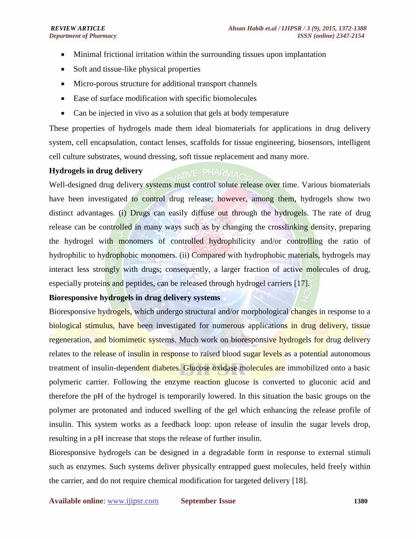
Bioresponsive hydrogels, which undergo structural and/or morphological changes in response to a
biological stimulus, have been investigated for numerous applications in drug delivery, tissue
regeneration, and biomimetic systems. Much work on bioresponsive hydrogels for drug delivery
relates to the release of insulin in response to raised blood sugar levels as a potential autonomous
treatment of insulin-dependent diabetes. Glucose oxidase molecules are immobilized onto a basic
polymeric carrier. Following the enzyme reaction glucose is converted to gluconic acid and
therefore the pH of the hydrogel is temporarily lowered. In this situation the basic groups on the
polymer are protonated and induced swelling of the gel which enhancing the release profile of
insulin. This system works as a feedback loop: upon release of insulin the sugar levels drop,
resulting in a pH increase that stops the release of further insulin.
Bioresponsive hydrogels can be designed in a degradable form in response to external stimuli
such as enzymes. Such systems deliver physically entrapped guest molecules, held freely within
the carrier, and do not require chemical modification for targeted delivery [18].
REVIEW ARTICLE Ahsan Habib et.al / IJIPSR / 3 (9), 2015, 1372-1388
Department of Pharmacy ISSN (online) 2347-2154
Available online: www.ijipsr.com September Issue 1381
Fig.6: Schematic representation of the glucose-sensitive hydrogel membrane consisting of a
poly (amine) and glucose-oxidase-loaded membrane [20].
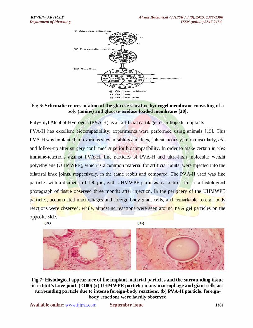
Polyvinyl Alcohol-Hydrogels (PVA-H) as an artificial cartilage for orthopedic implants
PVA-H has excellent biocompatibility; experiments were performed using animals [19]. This
PVA-H was implanted into various sites in rabbits and dogs, subcutaneously, intramuscularly, etc.
and follow-up after surgery confirmed superior biocompatibility. In order to make certain in vivo
immune-reactions against PVA-H, fine particles of PVA-H and ultra-high molecular weight
polyethylene (UHMWPE), which is a common material for artificial joints, were injected into the
bilateral knee joints, respectively, in the same rabbit and compared. The PVA-H used was fine
particles with a diameter of 100 μm, with UHMWPE particles as control. This is a histological
photograph of tissue observed three months after injection. In the periphery of the UHMWPE
particles, accumulated macrophages and foreign-body giant cells, and remarkable foreign-body
reactions were observed, while, almost no reactions were seen around PVA gel particles on the
opposite side.
Fig.7: Histological appearance of the implant material particles and the surrounding tissue
in rabbit’s knee joint. (×100) (a) UHMWPE particle: many macrophage and giant cells are
surrounding particle due to intense foreign-body reactions. (b) PVA-H particle: foreign-
body reactions were hardly observed
REVIEW ARTICLE Ahsan Habib et.al / IJIPSR / 3 (9), 2015, 1372-1388
Department of Pharmacy ISSN (online) 2347-2154
Available online: www.ijipsr.com September Issue 1382
Hydrogels for cell encapsulation
Cell encapsulation technology provides a promising therapeutic modality for diabetes,
hemophilia, cancer and renal failure [21].
The selection of a suitable biomaterial as a membrane for encapsulating cells is the major
challenge towards the success of cell encapsulation therapy. Biocompatibility, microporous
structure and minimal surface irritation within the surrounding tissues of hydrogels attracted them
for this application. They can be designed with required porosity that resists any entrance of
immune cells and allows stimuli, oxygen, nutrients and/or waste transfer through the pores.
Genetically modified alginates and polyethylene oxide based hydrogels [22] have been studied as
cell encapsulation systems.
Hydrogels in tissue engineering
Tissue engineering (TE) is a multidisciplinary approach and involves the expertise of materials
science, medical science and biological science for the development of biological substitutes
(tissue/ organ). It is emerging as an important field in regenerative medicine.
It has got three basic components namely, cells/tissues, scaffolds and implantation and/or
grafting. The principles of TE have been used extensively to restore the function of a traumatized/
malfunctioning tissues or organs. In practice, the patient’s cells are generally combined with a
scaffold for generating new tissue. A scaffold can be made up of either ceramic or polymer, which
can be either permanent or resorbable. The pore size of the scaffolds should be >80 μm [23].
This is necessary for the cell migration into the core of the scaffolds, angiogenesis, and supply of
nutrients to the cells and to take away the metabolic products away from the cells. The scaffolds
made up of polymers are generally hydrogels.
Every year thousands of people are victims of tissue loss and organ failure caused either due to
disease or trauma. Also, there is a shortage of organ donors because of the religious beliefs and/or
medical complications. Recently the use of resorbable hydrogels in TE has gained much
importance because (a) it is easy to process the polymers; (b) the properties of the hydrogels can
be tailored very easily; and (c) resorbable polymers like polylactic acid (PLA), polyglycolic acid
(PGA), and their co-polymers (PLA-co-PGA; PLGA) are being used for biomedical application.
Sterilization of the hydrogels is very tricky, which may alter the characteristics of the scaffold.
Hence, due consideration on the sterilization method should be given before selecting a particular
sterilization method [24].

REVIEW ARTICLE Ahsan Habib et.al / IJIPSR / 3 (9), 2015, 1372-1388
Department of Pharmacy ISSN (online) 2347-2154
Available online: www.ijipsr.com September Issue 1383
Fig.8: Schematic diagram showing multidisciplinary approach of tissue engineering
Hydrogels for contact lens application
The cornea of the eye is a precisely formed transparent structure of protein fibers containing about
80% water and 20% formed materials making it a natural hydrogel. Synthetic hydrogels have
found to be suitable in contact lens applications when the refractive power of cornea is
compromised. In addition to their biocompatibility and softness, inter-connected microstructures
of hydrogels help oxygen diffusitivity to the epithelial layer of the cornea. Certain hydrogels
possess high refractive index, modulus, and transparency, required to fit for this application. Since
no single hydrophilic polymer structure provides all required properties, copolymers developed
from a group of hydrophilic monomers like dimethylacrylamide, N-vinyl pyrrolidone and
methacrylic acid and hydrophobic monomers like perfluoro polyethers, methyl methacrylate and
silicon-containing monomers are utilized to design contact lenses [25].
Hydrogels in wound healing
Hydrogel is a crosslinked polymer matrix which has the ability to absorb and hold water in its
network structure. Hydrogels act as a moist wound dressing material and have the ability to
absorb and retain the wound exudates along with the foreign bodies, such as bacteria, within its
network structure. In addition to this, hydrogels have been found to promote fibroblast
proliferation by reducing the fluid loss from the wound surface and protect the wound from
external noxae necessary for rapid wound healing. Hydrogels help in maintaining a micro-climate
for biosynthetic reactions on the wound surface necessary for cellular activities. Fibroblast
proliferation is necessary for complete epithelialisation of the wound, which starts from the edge
of the wound. Since hydrogels help to keep the wound moist, keratinocytes can migrate on the
surface.

REVIEW ARTICLE Ahsan Habib et.al / IJIPSR / 3 (9), 2015, 1372-1388
Department of Pharmacy ISSN (online) 2347-2154
Available online: www.ijipsr.com September Issue 1384
Hydrogels may be transparent, depending on the nature of the polymers, and provide cushioning
and cooling/soothing effects to the wound surface. The main advantage of the transparent
hydrogels includes monitoring of the wound healing without removing the wound dressing. The
process of angiogenesis can be initiated by using semi-occlusive hydrogel dressings, which is
initiated due to temporary hypoxia. Angiogenesis of the wound ensures the growth of granulation
tissue by maintaining adequate supply of oxygen and nutrients to the wound surface. Hydrogel
sheets are generally applied over the wound surface with backing of fabric or polymer film and
are secured at the wound surface with adhesives or with bandages [4].
Fig.9: Hydrogels in burn treatment
Medical application of hydrogels
Hydrogels have also been designed for augmenting vocal cords, prevention of scar formation after
surgery and as coverings for perforated ear drums and rhinoplasty. These applications were the
driving force that initiated a detailed study on the relationship between the structure of cross-
linked hydrophilic polymers and their biocompatibility [26]. These results were translated into
clinical applications: one of the successful examples was the use of HEMA-based hydrogels in
rhinoplasty, which produced long-term biocompatibility and excellent cosmetic results [27].
Fig.10: Use of hydrogels (copolymers of HEMA with EDMA) in rhinoplasty:
(a) patient before surgery; (b) patient after surgery [28]
REVIEW ARTICLE Ahsan Habib et.al / IJIPSR / 3 (9), 2015, 1372-1388
Department of Pharmacy ISSN (online) 2347-2154
Available online: www.ijipsr.com September Issue 1385
Growing new body parts
The technology is still a long way off, but scientists are hopeful that some time in the future it will
be possible to grow replacement body parts in hydrogels. Cells of the required tissue will be
added to the hydrogel and injected into the body where they are needed. The hydrogel will take
the place of the damaged tissue and also allow nutrients to pass through it to the cells inside. Over
time, the cells will grow and the hydrogel be degraded by the body until new tissue is in place to
repair the damage.
Hydrogels are still a long way from being used in hospitals and whole organs grown on hydrogels
are even further away, but researchers in the USA recently managed to grow lung tissue in a
hydrogel which demonstrated that this idea has potential.
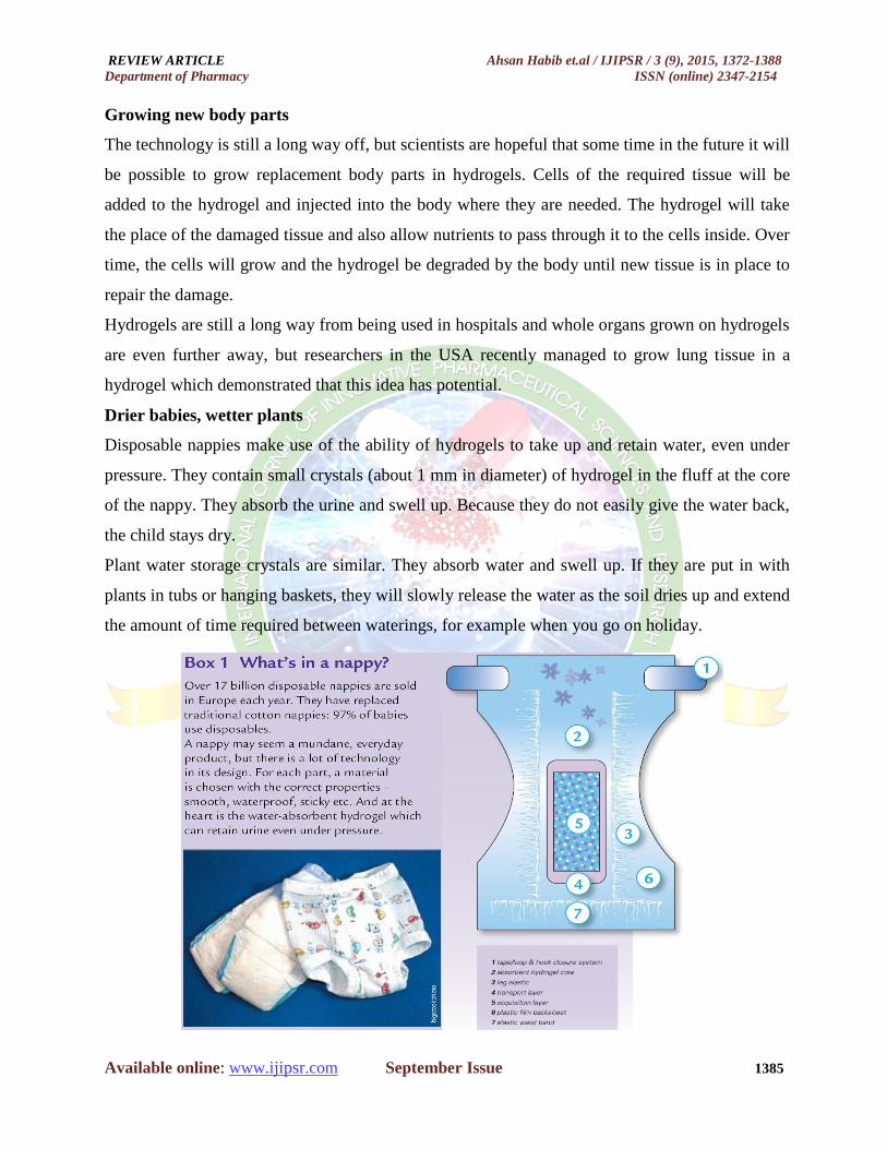
Drier babies, wetter plants
Disposable nappies make use of the ability of hydrogels to take up and retain water, even under
pressure. They contain small crystals (about 1 mm in diameter) of hydrogel in the fluff at the core
of the nappy. They absorb the urine and swell up. Because they do not easily give the water back,
the child stays dry.
Plant water storage crystals are similar. They absorb water and swell up. If they are put in with
plants in tubs or hanging baskets, they will slowly release the water as the soil dries up and extend
the amount of time required between waterings, for example when you go on holiday.
REVIEW ARTICLE Ahsan Habib et.al / IJIPSR / 3 (9), 2015, 1372-1388
Department of Pharmacy ISSN (online) 2347-2154
Available online: www.ijipsr.com September Issue 1386
CONCLUSION
During past decades, hydrogels have played a very essential role in biomedical applications. New
synthetic methods have been used to prepare homo- and co-polymeric hydrogels for a wide range
of drugs, peptides, and protein delivery applications. Recent enhancements in the field of polymer
science and technology have led to the development of various stimuli sensitive hydrogels. Either
pH-sensitive and/or temperature-sensitive hydrogels can be used for site-specific controlled drug
delivery. Hydrogels that are responsive to specific molecules, such as glucose or antigens, can be
used as biosensors as well as drug delivery systems. Polymer solutions in water (sol phase) that
transform into a gel phase on changing the temperature (thermo-gelation) offer a very exciting
field of research. Recent advances in the development of novel hydrogels for drug delivery
applications have focused on several aspects of their synthesis, characterization and behaviour.
Obviously, drug release from hydrogel networks is controlled by a complex combination of
different mechanisms, such as matrix swelling, drug dissolution/diffusion and hydrogel erosion.
Successful design of drug delivery systems relies not only on proper network design but also on
precise description of hydrogel behaviour as well as mathematical modelling of drug release
profiles. As more advanced release devices, such as in-situ forming hydrogels are developed more
rigorous mathematical modeling approaches are needed to describe the complete mechanisms
governing drug release from these systems.
REFERENCE
1. 1.Shibayama M and Tanaka T. Phase transition and related phenomena of polymer gels.
Advance Polymer Science. 1993; 109: 1–62
2. 2.Dergunov S and Mun G. Gamma-irradiated chitosan-polyvinyl pyrrolidone hydrogels as
pH-sensitive protein delivery system. Radiation Physics and Chemistry. 2009; 78 (1): 65-
68.
3. 3.Peppas NA and Langer R. Advances in Biomaterials, Drug Delivery, and
Bionanotechnology. Bioengineering, Food and Natural Products. 2003; 49(12): 2990-
3006.
4. 4.Qiu and Park K. Environment-sensitive hydrogels for drug delivery. Advanced Drug
Delivery Reviews. 2001; 53 (3): 321-339.
5. 5.Khare AR and Peppas NA. Swelling/deswelling of anionic copolymer gels.
Biomaterials. 1995; 16: 559-567.

REVIEW ARTICLE Ahsan Habib et.al / IJIPSR / 3 (9), 2015, 1372-1388
Department of Pharmacy ISSN (online) 2347-2154
Available online: www.ijipsr.com September Issue 1387
6. 6.Szepes A, Makai Z, Blumer C, Mader K, Kasa P and Revesz PS. Characterization and
drug delivery behaviour of starch based hydrogels prepared via isostatic ultrahigh
pressure. Carbohydrate Polymer. 2008; 72: 571-575.
7. 7.Yu H and Xiao C. Synthesis and properties of novel hydrogels from oxidized Konjac
glucomannan cross linked gelation for in-vitro drug delivery. Carbohydrate Polymer.
2008; 72: 479-489.
8. 8.Mansur HS, Orefice RL and Mansur. Characterization of poly(vinyl
alcohol)/poly(ethylene glycol) hydrogels and PVA-derived hybrids by small-angle Xray
scattering and FTIR spectroscopy. Polymer. 2004; 45 (21): 7193-7202.
9. Pourjavadi A and Kurdtabar M. Collagen-based highly porous hydrogel without any
porogen: Synthesis and characteristics. European Polymer Journal. 2007; 43: 877-889.
10. Al-Assaf S, Phillips GO and Williams PA. Controlling the molecular structure of food
hydrocolloids. Food Hydrocolloids. 2006; 20: 369-377.
11. Schuetz YB, Gurny R and Jordan O. A novel thermoresponsive hydrogel of chitosan.
European Journal of Pharmaceutics and Biopharmaceutics. 2008; 68: 19-25.
12. Baroli B. Hydrogels for tissue engineering and delivery of tissue-inducing substances.
Journal of Pharmaceutical Science. 2007; 96: 2197-2223.
13. Ta HT, Dass CR and Dunstan DE. Injectable chitosan hydrogels for localized cancer
therapy. Journal of Control Release. 2008; 126: 205-216.
14. Singh B, Sharma N and Chauhan N. Synthesis, characterization and swelling studies of
pH responsive Psyllium and methacrylamide based hydrogels for the use in colon specific
drug delivery. Carbohydrate Polymers. 2007; 69: 631-643.
15. Leonard A, Blacher S, Crine M and Jomaa W. Evolution of mechanical properties and
final textural properties of resorcinolformaldehyde xerogels during ambient air drying.
Journal of Non-Crystalline Solids. 2008; 354 (10-11): 831-838.
16. Wang M, Xu L, Hu H, Zhai M, Peng J, Nho Y, Li J and Wei G. Radiation synthesis of
PVP/ CMC hydrogels as wound dressing. Nuclear Instruments and Methods in Physics
Research B. 2007; 265: 385-389.
17. Silva A, Richard C, Bessodes M, Scherman D and Merten O. Growth factor delivery
approaches in hydrogels. Biomacromolecules. 2009; 10 (1): 9-18.
18. Ulijn RV, Bibi N, Jayawarna V, Thornton PD, Todd SJ, Mart RJ, Smith AM and Gough
JE. Bioresponsive hydrogels. Materials Today. 2007; 10 (4): 40-48.

REVIEW ARTICLE Ahsan Habib et.al / IJIPSR / 3 (9), 2015, 1372-1388
Department of Pharmacy ISSN (online) 2347-2154
Available online: www.ijipsr.com September Issue 1388
19. Noguchi T, Yamamuro T and Oka M. Poly(vinyl alcohol) hydrogel as an artificial
articular cartilage: evaluation of biocompatibility. Journal of Applied Biomaterials. 1991;
2: 101–107.
20. Ling YD and Lu MG. Fabrication of poly(N-isopropylacrylamide-co-itaconic acid)
hydrogels in DMSO/water mixtures and their characterization, Iranian Polymer Journal.
2008; 17 (2): 155-166.
21. Orive G, Hernández R, Gascón A, Calafiore R, Chang T, Hortelano G, Hunkeler D and
Pedraz J. History, challenges and perspectives of cell microencapsulation. Trends in
Biotechnology. 2004; 22 (2): 87-92.
22. 22.Miura S, Teramura Y and Iwata H. Encapsulation of islets with ultra-thin polyion
complex membrane through poly(ethylene glycol)-phospholipids anchored to cell
membrane. Biomaterials. 2006; 27 (34): 5828-5835.
23. 23.Pal K, Bag S and Pal S. Development of Porous Ultra High Molecular Weight
Polyethylene scaffolds for the fabrication of orbital implant. Journal of Porous Materials.
2008; 15: 53-59.
24. Hoffman AS. Hydrogels for biomedical applications. Advanced Drug Delivery Reviews.
2002; 54 (1): 3-12.
25. Groot J, Spaans C, Calck R, Beijma F, Norrby S and Pennings A. Hydrogels for an
accommodating intraocular lens. Biomacromolecules. 2003; 4 (3): 608-616.
26. Kopecek L, Šprincl H, Bažilová and Vacìk J. Biological tolerance of poly(N-substituted
acrylamides). Journal of Biomedical Materials Research. 1973; 7 (1): 111-121.
27. Sprincl J, Vacìk and Kopeček J. Biological tolerance of ionogenic hydrophilic gels.
Journal of Biomedical Materials Research. 1973; 7 (1): 123-136.
28. Voldřich Z, Tománek J, Vacìk and Kopeček J. Long-term experience with poly(glycol
monomethacrylate) gel in plastic operations of the nose. Journal of Biomedical Materials
Research. 1975; 9 (6): 675-685.