international journal of nanomedicine dovepress · open access full text article ... digital...
TRANSCRIPT
© 2013 Lelli et al, publisher and licensee Dove Medical Press Ltd. This is an Open Access article which permits unrestricted noncommercial use, provided the original work is properly cited.
International Journal of Nanomedicine 2013:8 307–314
International Journal of Nanomedicine
Different corrosive effects on hydroxyapatite nanocrystals and amine fluoride-based mouthwashes on dental titanium brackets: a comparative in vitro study
Marco Lelli1
Olivia Marchisio2
Ismaela Foltran1
Annamaria Genovesi2
Giulia Montebugnoli1
Massimo Marcaccio1
Ugo Covani2
Norberto Roveri1
1Alma Mater Studiorum, University of Bologna, Department of Chemistry, Bologna, Italy; 2University of Pisa, Istituto Stomatologico Tirreno, Lido di Camaiore, Lucca, Italy
Correspondence: Norberto Roveri Department of Chemistry “G Ciamician”, Alma Mater Studiorum University of Bologna, Via Selmi 2, I-40126 Bologna, Italy Tel +39 05 1209 9486 Fax +39 05 1209 9593 Email [email protected]
Abstract: Titanium plates treated in vitro with a mouthwash containing amine fluoride
(100 ppm F−) and another containing zinc-substituted carbonate–hydroxyapatite have been
analyzed by scanning electron microscopy and atomic force microscopy to evaluate the modi-
fication of the surface roughness induced by treatment with these two different mouthwashes.
The treatment with F−-based mouthwash produces a roughness characterized by higher
peaks and deeper valleys in the streaks on the titanium bracket surface compared with those
observed in the reference polished titanium plates. This effect causes a mechanical weakness
in the metallic dental implant causing bacterial growth and therefore promotes infection and
prosthesis contamination. However, the in vitro treatment with a mouthwash containing zinc-
substituted carbonate–hydroxyapatite reduced the surface roughness by filling the streaks
with an apatitic phase. This treatment counteracts the surface oxidative process that can affect
the mechanical behavior of the titanium dental implant, which inhibits the bacterial growth
contaminating prostheses.
Keywords: mouthwash, titanium brackets, corrosion, hydroxyapatite, aminic fluoride
IntroductionThe use of titanium in dental implant materials is based on the low thermal conductivity
of this metal, besides the fact that it doesn’t release toxic ions and has an appropriate
elasticity module, which helps bone to heal when in close contact with the titanium
implant surface.1
Titanium is highly ductile and resistant to cyclic forces in the oral environment.
These cyclic forces, known as fatigue, are produced by masticatory movements, which
are capable of causing internal microcracks in titanium-based implants.2 Titanium
constantly undergoes mechanical and thermal stresses in an aggressive host oral
environment.
Mouthwashes and toothpastes containing fluoride ions are among the most
important products in the oral care market. In fact, fluoride has been considered
the main method to reduce the enamel dissolution and prevent caries. Fluoride ions
have the ability to interact with the hydroxyapatite crystals forming fluoridated
hydroxyapatite (Ca10
(PO4)
6(OH)F) or fluorapatite (Ca
10(PO
4)
6F
2). These minerals have
greater lattice energy, higher crystallinity, and better resistance to dissolution than
hydroxyapatite. Fluoride also increases the seeding rate of apatite crystals.1
Dovepress
submit your manuscript | www.dovepress.com
Dovepress 307
O R I G I N A L R E S E A R C h
open access to scientific and medical research
Open Access Full Text Article
http://dx.doi.org/10.2147/IJN.S35245
International Journal of Nanomedicine 2013:8
The in vivo treatment using fluoride toothpastes may lead
to a partial substitution of the hydroxylic groups with fluoride
ions in the native enamel hydroxyapatite.3,4
The bone-promoting activity of F− has been extensively
documented in the literature.16 Titanium fluoride forms a stable
layer on hydroxyapatite surfaces where titanium shares the
oxygen atoms of phosphate, which becomes covalently bound
to the hydroxyapatite surface. Fluoride is essential in this reac-
tion because it allows phosphate ions to react directly with
titanium to form a firm and stable connection.1 The oxygen
and phosphate in the tissue fluids may replace the fluoride
and the phosphate becomes covalently bound to the titanium
surface. Such a reaction may induce a bone formation where
the phosphate in the bone is firmly (covalently) bound to the
titanium implant. This process may catalyze bone formation
and facilitate deposition of particularly well-mineralized bone
tissue close to the implant surface.5,6
Oxidative processes can thicken and condense the
titanium oxide layer on the surface, which improves the
anticorrosion stability of the underlying titanium.7,8 On
the other hand, reductive agents, such as fluoride (F−), may
have the opposite effect and attack this layer.8 The corrosion
process is caused by electrochemical reactions. Titanium
is known to present high resistance to corrosion against
attack by solutions containing strong mineral acids, such as
hydrochloric acid or sulfuric acid. However, when titanium
is placed in contact with a fluorinated medium at pH below
3.5, its titanium oxide layer is damaged and degraded. This
phenomenon is macroscopically visible by the loss of surface
brightness.9,10 The fluorides may also cause corrosion, which
increases roughness on the surfaces exposed to the oral cavity,
making it easier for microorganisms to grow where they are
protected from oral hygiene mechanisms (brushing, swal-
lowing, and flow of crevicular fluid).2,11,12
Anodic polarization and immersion tests indicated that
the titanium corrosion in solutions containing F− depends on
the concentration of hydrofluoric acid (HF). The passivation
film on titanium was destroyed when the HF concentration
in the solution was .30 ppm.
A rough surface favors plaque accumulation13 in the peri-
implant crevices of the gingiva, which is an undesirable effect
in this very sensitive region of the implant. Accordingly, to
avoid pathogenic plaque accumulation,14 the neck (abutment)
of an implant must be polished. It is very important to ensure
that these surfaces receive continuous care.
Many studies shown the high risk for fluoride to produce
fluorosis, especially in children, and bone diseases in
the elderly.15–21 The European Food Safety Authority (EFSA)
scientific panel has advised that there is a risk of fluoride
intake from ingested oral care products and toothpastes that
can be fluoridated with a maximum level of 1500 mg/Kg.
The EFSA considers that the maximum fluoride intake should
be 0.1 mg fluoride/kg/day in children aged 1–8 years which
is equivalent to 1.5 and 2.5 mg fluoride per day in children
aged 1–3 years and 4–8 years, respectively.22,23
Innovative commercial mouthwashes and toothpastes
in the last decade have replaced fluoride with biomimetic
hydroxyapatite nanocrystals as a remineralizing agent to
avoid the dangerous effects of fluoride in human health,
which have been ascribed to the amount of fluoride ingested
daily.16
In the inorganic phase of bone and dental hard tissues,
hydroxyapatite [Ca10
(PO4)
6(OH)
2] (HA) has been widely
studied as a bone filler, prosthetic coating, and implantable
biomaterial in dentistry and orthopedic surgery because of its
biocompatibility, bioactivity, and osteoconductivity.17
Biological HA is not stoichiometric according to the
ideal formula Ca10
(PO4)
6(OH)
2, but at low quantities Ca2+ is
replaced by other ions such as Na+, K+, Zn2+, Mg2+, Sr2+, while
PO4
3− and OH− can be partially substituted by other anions
like CO3
2−, HPO42−, P
2O
74−, SiO
44−.17,24−26
Carbonate is the prevalent foreign anion present into bone-
hydroxyapatite structure and represents about 4–8 wt%.27,28
The substitution of CO32− groups into the PO
43− sites (type B
carbonate apatite) is prevalent in children, while the carbon-
ate replacement to OH− groups (type A carbonate apatite)
increases with the age of the individual.29
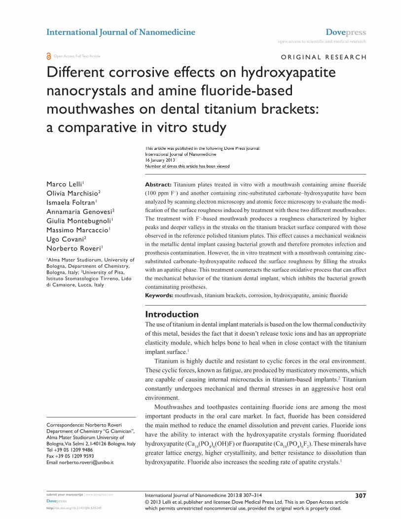
Biomimetic carbonate–hydroxyapatite nanocrystals
(CHA) have been synthesized nearly stoichiometrically in
bulk Ca/P molar ratio of about 1.7 and contains 4 ± 1 wt%
of carbonate ions that replaced prevalently phosphate groups.
Biomimetic CHA nanocrystals have been synthesized from
20 nm to 100 nm (Figure 1) with both acicular and plate-like
morphology to deliver drugs30–33 with a surface infused with
amino acids and proteins.26,34–36
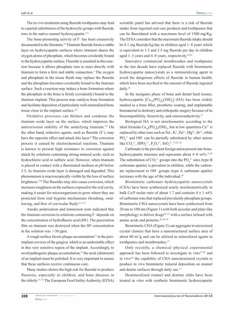
Biomimetic CHA (Figure 2) can aggregate in microsized
crystal clusters that have a nanostructured surface area of
about 80 m2/g and can be utilized as mineralized agents in
toothpastes and mouthwashes.37
Only recently, a chemical–physical experimental
approach has been followed to investigate in vitro37,38 and
in vivo3,4 the capability of CHA nanostructured crystals to
produce in vivo biomimetic mineral deposition on enamel
and dentin surfaces through daily use.11
Demineralized enamel and dentine slabs have been
treated in vitro with synthetic biomimetic hydroxyapatite
submit your manuscript | www.dovepress.com
Dovepress
Dovepress
308
Lelli et al
International Journal of Nanomedicine 2013:8
The aim of this work is to evaluate the action of a
nanostructured CHA-based mouthwash on titanium implants,
in comparison with a F−-based mouthwash.
In this work, we have performed an in vitro analysis on
the effect of F− and CHA on the surface of titanium brackets,
using scanning electron microscopy (SEM) and atomic
force microscopy (AFM), which allows a direct quantitative
characterization of the surface roughness.
Materials and methodIn vitro testWe considered four types of titanium resulting from dif-
ferent processes: polished, blasted, machined, and acid-
etched. All samples were titanium grade plates 3 mm thick
and 6 mm diameter. In vitro tests were carried out using
six samples for each type of titanium. Polished titanium
brackets were tested at room temperature in nanostructured
microcrystal CHA-based mouthwash for comparison to
an amine fluoride F−-based mouthwash with a pH ranging
between 4.3 and 5.5. The stoichiometric composition of
biomimetic CHA, which represents the active component
in the used mouthwash, is Ca(10-x)
Znx(PO
4)
(6-y)(CO
3)
y (OH)
2
where x is 0.1 and y is 0.4.
Each plate sample was dipped in mouthwash for
60 seconds, twice a day for 30 days to simulate a proper oral
hygiene rinse. After each treatment, the titanium plates were
washed under running water to replicate in vivo the usual
teeth rinse procedure.
Morphological characterizationSEM observations were carried out by means of a Carl-
Zeiss EVO, 40 XVP (Oberkochen, Germany) equipped
with an energy dispersive detector (EDAX) Inca 250
(Oxford, UL), using secondary electrons at 25 KV and
different magnifications.
AFM was carried out directly on the titanium plate
samples’ substrates. AFM imaging was performed on a
Digital Instruments Nanoscope IIIa Multimode SPM (Digi-
tal Instruments, Santa Barbara, CA, USA). The advanced
analog and digital circuit designs and innovative software
and hardware provide superior multitasking control for
scanning probe microscopes (SPMs).42,43 The samples in
contact mode using a J scanner and silicon nitride tips
(200 lm long with nominal spring constant 0.06 N/m).
ResultsThe surface morphologies of different polished, blasted,
machined, and acid-etched titanium plates have been charac-
Figure 1 TEM image of biomimetic ChA nanocrystals.Abbreviations: ChA, carbonate–hydroxyapatite; TEM, transmission electron microscopy.
Figure 2 TEM image of aggregated biomimetic ChA nanocrystals.Abbreviations: ChA, carbonate–hydroxyapatite; TEM, transmission electron microscopy.
nanocrystals for a few minutes and the surfaces chemically
and physically characterized. CHA induces surface remin-
eralization, which forms a biomimetic apatite coating on
enamel and dentin surfaces. Enamel remineralization occurs
quickly due to the specific chemical–physical characteris-
tics of innovative nanostructured hydroxyapatite particles,
which closely resemble the natural hydroxyapatite present
in enamel.39 Therefore, our experimental results suggest that
the deterioration of teeth can be prevented.
CHA synthesized with tailored biomimetic character-
istics for composition, structure, size, and morphology can
chemically bind themselves on the surfaces of hard tissues
in teeth, filling in the scratches, and producing a bound
biomimetic apatitic coating to protect the enamel surface
structure.3,40,41
There are some commercial products for oral care, like
toothpastes and mouthwashes, based on CHA, which come
into contact with titanium prostheses.
submit your manuscript | www.dovepress.com
Dovepress
Dovepress
309
Corrosive effects on dental titanium brackets
International Journal of Nanomedicine 2013:8
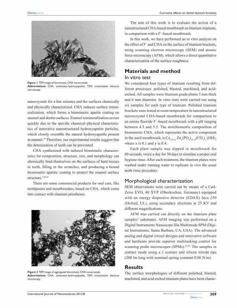
Figure 3 SEM images of the different polished, blasted, machined, and acid-etched titanium disk surfaces used in preliminary selection: (A) polished titanium surface, (B) blasted titanium surface, (C) machine-treated titanium surface, and (D) etched titanium surface.Abbreviation: SEM, scanning electron microscopy.
terized by SEM and the images are reported in Figure 3A–D
respectively.
Figure 3 shows that blasted, machine-treated, and acid-
etched titanium plates have an inhomogeneous surface, while
the polished plate surface (Figure 3A) has low grooves and
thus a smooth surface (Figure 3C). Polished titanium plates
that have a homogeneous surface with a very small roughness
appear to be the more suitable titanium plates to be utilized
in this study.
Comparable samples of polished titanium plates have been
surface characterized by SEM before and after a daily in vitro
treatment with two different mouthwashes. A mouthwash con-
taining an amine fluoride (100 ppm F−) and another containing
zinc-substituted CHA have been utilized to investigate their
different effects on the titanium plate surface.
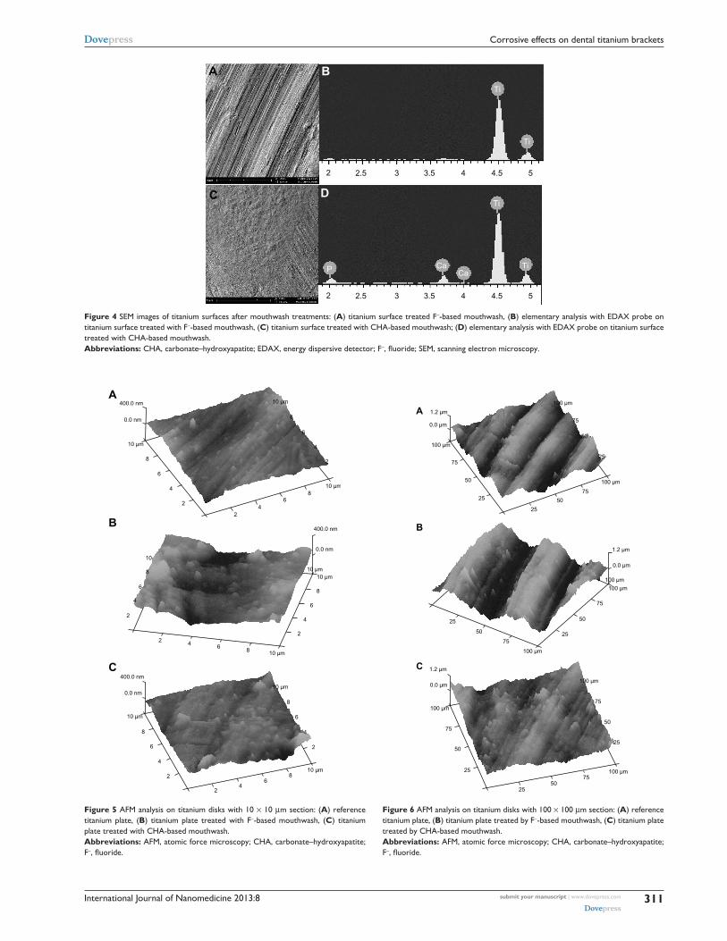
After the in vitro treatment with the two different
mouthwashes, SEM analysis of the titanium plates showed
an appreciable difference between the plates treated with
amine F−-based mouthwash (Figure 4A) and those treated
with the CHA-based mouthwash (Figure 4C). In fact, the
surface roughness present on titanium surface increases
after the treatment with amine F−-based mouthwash
compared to that observed on the reference titanium plate
(Figure 3A).
Contrarily, the CHA-based mouthwash seems to cover
valleys and grooves present on the titanium surface, which
appreciably reduces the roughness present on the reference
titanium plates.
The EDAX elemental analysis performed on the surface
of the titanium after treatment with a fluoride rinse showed
only one peak related to titanium (Figure 4B). If the EDAX
was performed after treatment with CHA-based mouthwash,
the same survey grade showed peaks attributable to the cal-
cium and phosphate ions present in the right stoichiometric
molar ratio of hydroxyapatite, 1.7 (Figure 4D).
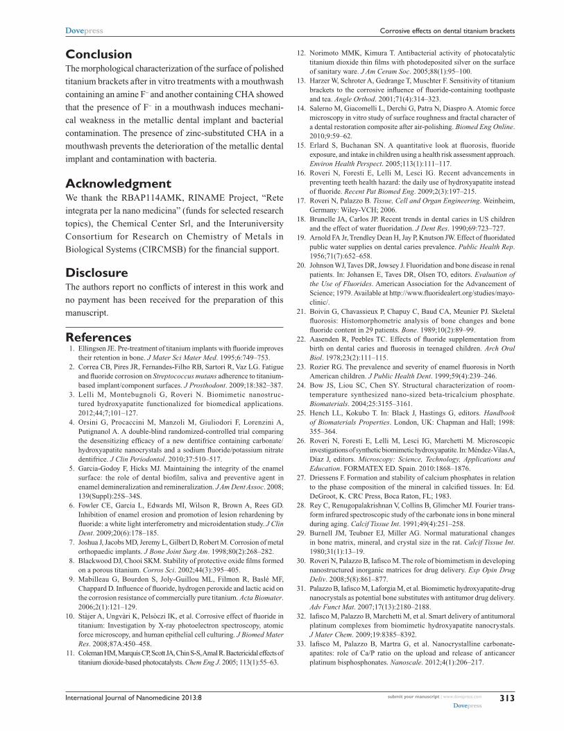
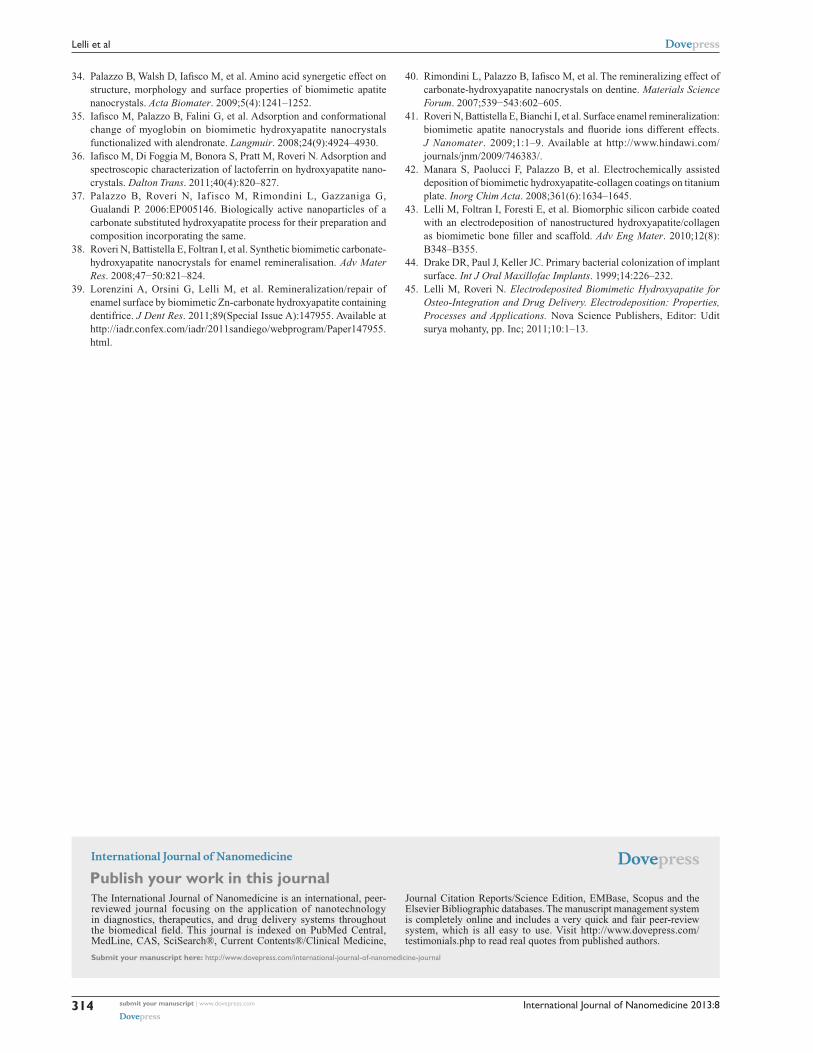
For more detailed results, the titanium brackets treated
with the two different mouthwashes were investigated by
AFM microscopy.
submit your manuscript | www.dovepress.com
Dovepress
Dovepress
310
Lelli et al
International Journal of Nanomedicine 2013:8
400.0 nm
0.0 nm
10 µm
400.0 nm
0.0 nm
400.0 nm
0.0 nm
10 µm
10 µm10 µm
10 µm
10 µm
10 µm
4
6
8
2
8642
86
42
2
4
6
8
2
4
6
8
10
8
6
4
2
8
6
4
2
A
B
C
810 µm
10 µm
8
6
4
2
64
2
Figure 5 AFM analysis on titanium disks with 10 × 10 µm section: (A) reference titanium plate, (B) titanium plate treated with F−-based mouthwash, (C) titanium plate treated with CHA-based mouthwash.Abbreviations: AFM, atomic force microscopy; ChA, carbonate–hydroxyapatite; F−, fluoride.
1.2 µm
1.2 µm
0.0 µm
100 µm
100 µm
1.2 µm
0.0 µm
100 µm
75
25 50
50 2575
25
75
50
25
5075
100 µm
100 µm
100 µm
75
50
25
75
50
25
100 µm
0.0 µm
100 µm
75
50
25
25
50
75
100 µm
A
B
C
Figure 6 AFM analysis on titanium disks with 100 × 100 µm section: (A) reference titanium plate, (B) titanium plate treated by F−-based mouthwash, (C) titanium plate treated by CHA-based mouthwash.Abbreviations: AFM, atomic force microscopy; ChA, carbonate–hydroxyapatite; F−, fluoride.
240 µm
2.5
BA
DC
3.5 4
Ti
Ti
TiCaCaP
Ti
4.5 53
2 2.5 3.5 4 4.5 53
Figure 4 SEM images of titanium surfaces after mouthwash treatments: (A) titanium surface treated F−-based mouthwash, (B) elementary analysis with EDAX probe on titanium surface treated with F−-based mouthwash, (C) titanium surface treated with CHA-based mouthwash; (D) elementary analysis with EDAX probe on titanium surface treated with CHA-based mouthwash.Abbreviations: ChA, carbonate–hydroxyapatite; EDAX, energy dispersive detector; F−, fluoride; SEM, scanning electron microscopy.
submit your manuscript | www.dovepress.com
Dovepress
Dovepress
311
Corrosive effects on dental titanium brackets

International Journal of Nanomedicine 2013:8
Table 1 AFM analysis of different titanium disks, 10 × 10 µm section
10 × 10 μm sections Root mean square (Rq)
Highest peak (Rp)
Deepest valley (Rv)
Polished titanium disk (reference)
93 nm 350 nm −140 nm
F−-based mouthwash treated disk
92 nm 460 nm −230 nm
CHA-based mouthwash treated disk
56 nm 300 nm −120 nm
Abbreviations: AFM, atomic force microscopy; ChA, carbonate–hydroxyapatite; F−, fluoride.
Table 2 AFM analysis of different titanium disks, 100 × 100 µm section
100 × 100 μm Root mean square (Rq)
Highest peak (Rp)
Deepest valley (Rv)
Polished titanium disk (reference)
280 nm 700 nm −740 nm
F−-based mouthwash treated disk
460 nm 840 nm −940 nm
CHA-based mouthwash treated disk
235 nm 620 nm −520 nm
Abbreviations: AFM, atomic force microscopy; ChA, carbonate–hydroxyapatite; F−, fluoride.
Titanium plates treated with the two different mouthwashes
were investigated using titanium sections of different
dimensions. We have examined titanium plate sections of
10 × 10 µm and 100 × 100 µm wide. Figures 5 and 6 show
AFM images obtained on the surface of 10 × 10 µm and
100 × 100 µm sections, respectively. In fact, in the 10 × 10 µm
samples, AFM measures the surface nanoroughness with
high accuracy, while in the 100 × 100 µm samples, it is
possible to evaluate the roughness extension with high
accuracy.
In Figures 5A and 6A, the AFM images of the titanium
reference surface are reported, in Figures 5B and 6B, the
AFM images of F− mouthwash-treated titanium surface
are reported, while in Figures 5C and 6C, the AFM images
show the effect of CHA mouthwash on the titanium surface.
The numerical analysis carried out on the AFM images is
reported in Tables 1 and 2. The root mean square (Rq),
highest peak (Rp), and deepest valley (Rv) were measured
on 10 × 10 µm sections of reference titanium surface
and after treatment with mouthwashes containing F− and
CHA (Table 1). The Rq, Rp, and Rv were measured on
100 × 100 µm sections of reference titanium surface and
after treatment with the mouthwashes containing F− and
CHA (Table 2).
DiscussionA morphological characterization was performed on the
surface of polished, titanium brackets after in vitro treatment
with mouthwashes containing amine F− and CHA. Titanium
plates treated with the two different mouthwashes were
analyzed by SEM and AFM using titanium sections of two
different dimensions (10 × 10 μm and 100 × 100 μm) for a
well-defined highly accurate evaluation of the modification
of surface nanoroughness and its extension (respect the value
Rq, Rp and Rv reported in table 2) induced by the treatment
with the two different mouthwashes.
The treatment with the F−-based mouthwash induces a
roughness characterized by streaking with higher peaks and
deeper valleys on the titanium bracket surface compared
with those observed in the reference polished titanium plates.
Contrarily, the treatment with the CHA-based mouthwash
induces a roughness characterized by streaking with
smoothed peaks and less deep valleys on the titanium bracket
surface compared with those observed in the reference
polished titanium disc. These findings can be explained
by the hydroxyapatite deposition filling the streaks on the
titanium surface and therefore reducing surface roughness.
The EDAX analysis revealed the presence of calcium and
phosphorus in a molar ratio of 1.7, which was characteristic
of the CHA present in the mouthwash.
The increased roughness induced by the in vitro
treatment with the mouthwash containing amine F−
(Tables 1 and 2) modify the mechanical behavior of the
titanium brackets, which promotes a mechanical weakness
of the metallic dental implant. The increased depth of the
streaks encourage bacterial growth promoting infection
and prosthesis contamination and mobility.44 Contrarily,
the in vitro treatment with the CHA-based mouthwash
reduces the surface roughness by filling in the streaks
(Tables 1 and 2). This hydroxyapatite deposition protects
against the surface oxidative process, which can damage
the mechanical behavior of the titanium brackets. The
depositation of hydroxyapatite coating on titanium surface
is able to prevent the bacterial growth contaminating the
prosthesis. Furthermore, when bacterial plaque, by its
acidity, solubilizes the zinc-substituted CHA present on
the titanium implant.40,41,45 On one hand, our data shows
that the presence of F− in mouthwashes should be avoided
to prevent any degradation of the metallic dental implant
and subsequent contamination with bacteria. On the other
hand, the CHA-based mouthwash prevents the degradation
of the metallic dental implants and the formation of bacterial
film.
submit your manuscript | www.dovepress.com
Dovepress
Dovepress
312
Lelli et al
International Journal of Nanomedicine 2013:8
ConclusionThe morphological characterization of the surface of polished
titanium brackets after in vitro treatments with a mouthwash
containing an amine F− and another containing CHA showed
that the presence of F− in a mouthwash induces mechani-
cal weakness in the metallic dental implant and bacterial
contamination. The presence of zinc-substituted CHA in a
mouthwash prevents the deterioration of the metallic dental
implant and contamination with bacteria.
AcknowledgmentWe thank the RBAP114AMK, RINAME Project, “Rete
integrata per la nano medicina” (funds for selected research
topics), the Chemical Center Srl, and the Interuniversity
Consortium for Research on Chemistry of Metals in
Biological Systems (CIRCMSB) for the financial support.
DisclosureThe authors report no conflicts of interest in this work and
no payment has been received for the preparation of this
manuscript.
References 1. Ellingsen JE. Pre-treatment of titanium implants with fluoride improves
their retention in bone. J Mater Sci Mater Med. 1995;6:749–753. 2. Correa CB, Pires JR, Fernandes-Filho RB, Sartori R, Vaz LG. Fatigue
and fluoride corrosion on Streptococcus mutans adherence to titanium-based implant/component surfaces. J Prosthodont. 2009;18:382–387.
3. Lelli M, Montebugnoli G, Roveri N. Biomimetic nanostruc-tured hydroxyapatite functionalized for biomedical applications. 2012;44;7;101–127.
4. Orsini G, Procaccini M, Manzoli M, Giuliodori F, Lorenzini A, Putignanol A. A double-blind randomized-controlled trial comparing the desensitizing efficacy of a new dentifrice containing carbonate/hydroxyapatite nanocrystals and a sodium fluoride/potassium nitrate dentifrice. J Clin Periodontol. 2010;37:510–517.
5. Garcia-Godoy F, Hicks MJ. Maintaining the integrity of the enamel surface: the role of dental biofilm, saliva and preventive agent in enamel demineralization and remineralization. J Am Dent Assoc. 2008; 139(Suppl):25S–34S.
6. Fowler CE, Garcia L, Edwards MI, Wilson R, Brown A, Rees GD. Inhibition of enamel erosion and promotion of lesion rehardening by fluoride: a white light interferometry and microidentation study. J Clin Dent. 2009;20(6):178–185.
7. Joshua J, Jacobs MD, Jeremy L, Gilbert D, Robert M. Corrosion of metal orthopaedic implants. J Bone Joint Surg Am. 1998;80(2):268–282.
8. Blackwood DJ, Chooi SKM. Stability of protective oxide films formed on a porous titanium. Corros Sci. 2002;44(3):395–405.
9. Mabilleau G, Bourdon S, Joly-Guillou ML, Filmon R, Baslé MF, Chappard D. Influence of fluoride, hydrogen peroxide and lactic acid on the corrosion resistance of commercially pure titanium. Acta Biomater. 2006;2(1):121–129.
10. Stàjer A, Ungvàri K, Pelsòczi IK, et al. Corrosive effect of fluoride in titanium: Investigation by X-ray photoelectron spectroscopy, atomic force microscopy, and human epithelial cell culturing. J Biomed Mater Res. 2008;87A:450–458.
11. Coleman HM, Marquis CP, Scott JA, Chin S-S, Amal R. Bactericidal effects of titanium dioxide-based photocatalysts. Chem Eng J. 2005; 113(1):55–63.
12. Norimoto MMK, Kimura T. Antibacterial activity of photocatalytic titanium dioxide thin films with photodeposited silver on the surface of sanitary ware. J Am Ceram Soc. 2005;88(1):95–100.
13. Harzer W, Schroter A, Gedrange T, Muschter F. Sensitivity of titanium brackets to the corrosive influence of fluoride-containing toothpaste and tea. Angle Orthod. 2001;71(4):314–323.
14. Salerno M, Giacomelli L, Derchi G, Patra N, Diaspro A. Atomic force microscopy in vitro study of surface roughness and fractal character of a dental restoration composite after air-polishing. Biomed Eng Online. 2010;9:59–62.
15. Erlard S, Buchanan SN. A quantitative look at fluorosis, fluoride exposure, and intake in children using a health risk assessment approach. Environ Health Perspect. 2005;113(1):111–117.
16. Roveri N, Foresti E, Lelli M, Lesci IG. Recent advancements in preventing teeth health hazard: the daily use of hydroxyapatite instead of fluoride. Recent Pat Biomed Eng. 2009;2(3):197–215.
17. Roveri N, Palazzo B. Tissue, Cell and Organ Engineering. Weinheim, Germany: Wiley-VCH; 2006.
18. Brunelle JA, Carlos JP. Recent trends in dental caries in US children and the effect of water fluoridation. J Dent Res. 1990;69:723–727.
19. Arnold FA Jr, Trendley Dean H, Jay P, Knutson JW. Effect of fluoridated public water supplies on dental caries prevalence. Public Health Rep. 1956;71(7):652–658.
20. Johnson WJ, Taves DR, Jowsey J. Fluoridation and bone disease in renal patients. In: Johansen E, Taves DR, Olsen TO, editors. Evaluation of the Use of Fluorides. American Association for the Advancement of Science; 1979. Available at http://www.fluoridealert.org/studies/mayo-clinic/.
21. Boivin G, Chavassieux P, Chapuy C, Baud CA, Meunier PJ. Skeletal fluorosis: Histomorphometric analysis of bone changes and bone fluoride content in 29 patients. Bone. 1989;10(2):89–99.
22. Aasenden R, Peebles TC. Effects of fluoride supplementation from birth on dental caries and fluorosis in teenaged children. Arch Oral Biol. 1978;23(2):111–115.
23. Rozier RG. The prevalence and severity of enamel fluorosis in North American children. J Public Health Dent. 1999;59(4):239–246.
24. Bow JS, Liou SC, Chen SY. Structural characterization of room- temperature synthesized nano-sized beta-tricalcium phosphate. Biomaterials. 2004;25:3155–3161.
25. Hench LL, Kokubo T. In: Black J, Hastings G, editors. Handbook of Biomaterials Properties. London, UK: Chapman and Hall; 1998: 355–364.
26. Roveri N, Foresti E, Lelli M, Lesci IG, Marchetti M. Microscopic investigations of synthetic biomimetic hydroxyapatite. In: Méndez-Vilas A, Díaz J, editors. Microscopy: Science, Technology, Applications and Education. FORMATEX ED. Spain. 2010:1868–1876.
27. Driessens F. Formation and stability of calcium phosphates in relation to the phase composition of the mineral in calcified tissues. In: Ed. DeGroot, K. CRC Press, Boca Raton, FL; 1983.
28. Rey C, Renugopalakrishnan V, Collins B, Glimcher MJ. Fourier trans-form infrared spectroscopic study of the carbonate ions in bone mineral during aging. Calcif Tissue Int. 1991;49(4):251–258.
29. Burnell JM, Teubner EJ, Miller AG. Normal maturational changes in bone matrix, mineral, and crystal size in the rat. Calcif Tissue Int. 1980;31(1):13–19.
30. Roveri N, Palazzo B, Iafisco M. The role of biomimetism in developing nanostructured inorganic matrices for drug delivery. Exp Opin Drug Deliv. 2008;5(8):861–877.
31. Palazzo B, Iafisco M, Laforgia M, et al. Biomimetic hydroxyapatite-drug nanocrystals as potential bone substitutes with antitumor drug delivery. Adv Funct Mat. 2007;17(13):2180–2188.
32. Iafisco M, Palazzo B, Marchetti M, et al. Smart delivery of antitumoral platinum complexes from biomimetic hydroxyapatite nanocrystals. J Mater Chem. 2009;19:8385–8392.
33. Iafisco M, Palazzo B, Martra G, et al. Nanocrystalline carbonate-apatites: role of Ca/P ratio on the upload and release of anticancer platinum bisphosphonates. Nanoscale. 2012;4(1):206–217.
submit your manuscript | www.dovepress.com
Dovepress
Dovepress
313
Corrosive effects on dental titanium brackets
International Journal of Nanomedicine
Publish your work in this journal
Submit your manuscript here: http://www.dovepress.com/international-journal-of-nanomedicine-journal
The International Journal of Nanomedicine is an international, peer-reviewed journal focusing on the application of nanotechnology in diagnostics, therapeutics, and drug delivery systems throughout the biomedical field. This journal is indexed on PubMed Central, MedLine, CAS, SciSearch®, Current Contents®/Clinical Medicine,
Journal Citation Reports/Science Edition, EMBase, Scopus and the Elsevier Bibliographic databases. The manuscript management system is completely online and includes a very quick and fair peer-review system, which is all easy to use. Visit http://www.dovepress.com/ testimonials.php to read real quotes from published authors.
International Journal of Nanomedicine 2013:8
34. Palazzo B, Walsh D, Iafisco M, et al. Amino acid synergetic effect on structure, morphology and surface properties of biomimetic apatite nanocrystals. Acta Biomater. 2009;5(4):1241–1252.
35. Iafisco M, Palazzo B, Falini G, et al. Adsorption and conformational change of myoglobin on biomimetic hydroxyapatite nanocrystals functionalized with alendronate. Langmuir. 2008;24(9):4924–4930.
36. Iafisco M, Di Foggia M, Bonora S, Pratt M, Roveri N. Adsorption and spectroscopic characterization of lactoferrin on hydroxyapatite nano-crystals. Dalton Trans. 2011;40(4):820–827.
37. Palazzo B, Roveri N, Iaf isco M, Rimondini L, Gazzaniga G, Gualandi P. 2006:EP005146. Biologically active nanoparticles of a carbonate substituted hydroxyapatite process for their preparation and composition incorporating the same.
38. Roveri N, Battistella E, Foltran I, et al. Synthetic biomimetic carbonate-hydroxyapatite nanocrystals for enamel remineralisation. Adv Mater Res. 2008;47−50:821–824.
39. Lorenzini A, Orsini G, Lelli M, et al. Remineralization/repair of enamel surface by biomimetic Zn-carbonate hydroxyapatite containing dentifrice. J Dent Res. 2011;89(Special Issue A):147955. Available at http://iadr.confex.com/iadr/2011sandiego/webprogram/Paper147955.html.
40. Rimondini L, Palazzo B, Iafisco M, et al. The remineralizing effect of carbonate-hydroxyapatite nanocrystals on dentine. Materials Science Forum. 2007;539−543:602–605.
41. Roveri N, Battistella E, Bianchi I, et al. Surface enamel remineralization: biomimetic apatite nanocrystals and fluoride ions different effects. J Nanomater. 2009;1:1–9. Available at http://www.hindawi.com/journals/jnm/2009/746383/.
42. Manara S, Paolucci F, Palazzo B, et al. Electrochemically assisted deposition of biomimetic hydroxyapatite-collagen coatings on titanium plate. Inorg Chim Acta. 2008;361(6):1634–1645.
43. Lelli M, Foltran I, Foresti E, et al. Biomorphic silicon carbide coated with an electrodeposition of nanostructured hydroxyapatite/collagen as biomimetic bone filler and scaffold. Adv Eng Mater. 2010;12(8): B348–B355.
44. Drake DR, Paul J, Keller JC. Primary bacterial colonization of implant surface. Int J Oral Maxillofac Implants. 1999;14:226–232.
45. Lelli M, Roveri N. Electrodeposited Biomimetic Hydroxyapatite for Osteo-Integration and Drug Delivery. Electrodeposition: Properties, Processes and Applications. Nova Science Publishers, Editor: Udit surya mohanty, pp. Inc; 2011;10:1–13.
submit your manuscript | www.dovepress.com
Dovepress
Dovepress
Dovepress
314
Lelli et al