international journal of pharma and bio sciences · molecular docking studies of 1, ... belongs to...
TRANSCRIPT

This article can be downloaded from www.ijpbs.net
B - 68
International Journal of Pharma and Bio Sciences
MOLECULAR DOCKING STUDIES OF 1, 2 DISUBSTITUTED IDOPYRANOSE
FROM VITEX NEGUNDO WITH ANTI-DIABETIC ACTIVITY OF TYPE 2 DIABETES
Co Authors
1ARUMUGAM SUDHA, 2RAMAR MANIKANDAN AND
3CHINNASAMY ARULVASU
2Department of Animal Health & Management, Alagappa University, Karaikudi – 630 003, India
3Department of Zoology, University of Madras, Chennai – 600 025
RESEARCH ARTICLE
ARTICALTICLE
BIOINFORMATICS
Corresponding Author
PAPPU SRINIVASAN
1Department of Bioinformatics, Alagappa University, Karaikudi –
630 003, India
ABSTRACT Type 2 diabetes is one of the major life threatening diseases worldwide these cases are progressing at an incremental rate every year and number of research works are going on to control the disease by targeting its enzymes or proteins. The medicinal properties of Vitex negundo and one of its compounds, 1, 2 disubstituted idopyranose (C23H28O12) was checked against diabetes mellitus by molecular docking with various proteins which are involved in the carbohydrate metabolism. The inhibitor, 1, 2 disubstituted idopyranose was found to be increased insulin sensitivity and normalize blood glucose level by binding in the active sites of the protein and thus useful therapeutic agent for the herbal therapy of diabetes. The docking method to explore the ability of 1, 2 disubstituted idopyranose bound to the active binding site in the proteins and showed that the inhibitor is the best binder of the present study. We concluded that the natural products with interesting biological properties and structural diversity have often served as valuable lead drug candidates for the treatment of human diseases and also it replaces the chemically synthesized drugs which cause side effects.

This article can be downloaded from www.ijpbs.net
B - 69
KEYWORDS
Diabetes mellitus, Vitex negundo, 1, 2 disubstituted idopyranose, Docking, Glide
INTRODUCTION Type 2 diabetes mellitus (T2DM) is a genetically heterogeneous, polygenic disease with a complex inheritance pattern and is caused by genetic predisposition and environmental factors (LeRoith, 2002). The disease is characterized by altered expression of many genes and their products in several tissue types (Gerich, 1998; Gloyn, 2003). Number of proteins was considered as a target to control the diabetes mellitus. In the present study, four proteins such as human maltase-glucoamylase (MGA), dipeptidyl peptidase-4 (DPP-4), aldose reductase (AR) and glycogen synthase kinase-3 (GSK-3) are considered as targets for the inhibitor, 1, 2 disubstituted idopyranose (C23-
H28O12), derived from the leaves of the medicinal plant, Vitex negundo Linn (Verbanaceae). MGA is an enzyme responsible for catalyzing the last glucose releasing step in starch digestion. MGA accounts for all glucoamylase activity, 20% of the maltase activity and 1% of the sucrase activity (Semenza et al., 1989). Human MGA encoded by the gene MGAM (Buford et al., 2003; Roy et al., 2002) is an alpha glucosidase responsible for hydrolysis of α-1, 4- linkages from the non-reducing end of the maltose oligosaccharides (Nichols et al., 1998) and belongs to glycoside hydrolase family 31. It is type II membrane protein, 1857 amino acids in length anchored in the brush border epithelial cells of the small intestine. Dipeptidyl peptidase-IV (DPP-IV) is a multifunctional protein involved in many physiological processes such as binding protein, receptor and proteolytic enzyme. DPP-IV was discovered in the late 80’s, as a serine peptidase belonging to the S9b protein family (Ogata et al., 1989). This ecto-enzyme exists in
two forms, i.e., as a soluble homo-dimer and as a type II integral plasma membrane glycoprotein which is abundantly expressed on a variety of cell surfaces (Bjelke et al., 2004; Abbott and Gorrell, 2002; Lambeir et al., 1997; Duke-Cohan et al., 1996). DPP-IV deactivates the incretin hormones GLP-1 and GIP by cleaving the penultimate proline or alanine from the N-terminal (P1-position) of the peptide, since the intact N-terminal ends of both GLP-1 and GIP are essential for biological activity (Adelhorst et al., 1994; Rolin et al., 2004). The cleavage of the N-terminal dipeptide segment by DPP-IV plays an important role in maintaining glucose homeostasis. Thus, shielding incretin hormones from catastrophic effects of DPP-IV enzyme by the administration of inhibitors will maintain the concentrations of incretin hormones and prolong their proficient antidiabetic action. Aldose reductase (AR) is the first enzyme of the polyol pathway and is widely distributed in mammalian tissues. Due to increased aldose reductase activity, the accumulation of intracellular sorbitol is also raised. It implicates the development of various secondary complications of diabetes mellitus (Muthenna et al., 2009). Glycogen synthase kinase-3 (GSK-3) is a unique multifunctional serine/threonine kinase and it was inactivated by phosphorylation. In response to insulin binding, PKB/AKT phosphorylates GSK-3 on serine 9, which prevents the enzyme from phosphorylating glycogen synthase (Frame et al., 2001; Mukai et al., 2002; Doble and Woodgett, 2003). Unphosphorylated glycogen synthase is active and able to synthesize

This article can be downloaded from www.ijpbs.net
B - 70
glycogen. Thus it plays a key role in the transduction of regulatory and proliferative signals arising out at the cell membrane in the insulin signalling pathway, leading to potential modulation of blood glucose levels (McManus et al., 2005). GSK-3 is also unique and it requires a substrate that has been phosphorylated by a distinct kinase before it can phosphorylate the substrate. Computational Biology and bioinformatics have the potential not only of speeding up the drug discovery process thus reducing the costs, but also of changing the way drugs are designed. Rational drug design (RDD) helps to facilitate and speedup the drug designing process, which
involves variety of methods to identify novel compounds. One such method is the docking of the drug molecule with the receptor (target). The site of drug action, which is ultimately responsible for the pharmaceutical effect, is a receptor and docking is the process by which two molecules fit together in 3D space. A bioactive compound 1, 2 disubstituted idopyranose (Fig.1) has been already reported by us from the leaves of medicinal plant, Vitex negundo Linn (Verbanaceae) and this compound helps in regeneration of damaged pancreatic ß-cells and hyperglycaemic nature against streptozotocin-induced diabetes in in vitro studies (Manikandan et al., 2009).
Figure 1 1, 2 disubstituted idopyranose
The aim of the present study is to investigate the inhibitory activity of the compound, 1, 2 disubstituted idopyranose on type 2 diabetes by molecular docking studies and to analyze the ADME/T properties of the compound for drug like candidates by using the Schrodinger software 9.0 and hence it would serve as to design drug alternative to diabetes.
MATERIALS AND METHODS Docking studies were performed for 1, 2 disubstituted idopyranose (C23H28O12) with target proteins by Glide 5.5 module of Schrodinger suite.
Computational methods with Glide Version 5.5 All computational studies were carried out using Glide version 5.5, installed in a single machine running on Intel CoreTM 2 Duo processor with 1GB RAM and 160 GB hard disk with Red Hat Linux Enterprise version 5.0 as the operating system. Ligand preparation The structure of the compound, 1, 2 disubstituted idopyranose (C23H28O12) was drawn by using ChemSketch (ACDLABS 12.0) and converted to 3D structure with the help of 3D optimization tool. By using the LigPrep (2.3)

This article can be downloaded from www.ijpbs.net
B - 71
module (Ligprep, Version 2.3, 2009), the drawn ligand was geometry optimized by using the Optimized Potentials for Liquid Simulations-2005 (OPLS-2005) force field with the Steepest Descent followed by truncated Newton Conjugate gradient protocol. Partial atomic charges were computed using the OPLS-2005 force field. The LigPrep is a utility in Schrodinger software suite that combines tools for generating 3D structures from 1D (Smiles) and 2D (SDF) representation, searching for tautomers and steric isomers and geometry minimization of ligands. Finally, 32 poses had been prepared with different tautomeric and steric features for docking studies. Protein preparation The X-ray crystal structures of all the proteins viz., maltase glucoamylase (MGA), dipeptidyl peptidase-4 (DPP-4), aldose reductase and glycogen synthase kinase-3 (GSK-3) were obtained from the RCSB protein data bank (http://www.rcsb.org/pdb). After evaluating numbers of entries, the best proteins were selected by analyzing the protein with Ramachandran plot and ProCheck using SAVS server based on ligand and number of disallowed regions (Laskowaski et al., 1993; Morris and MacArthur, 1992; Ramachandran et al., 1963). After selection, Protein preparation wizard of Schrodinger suite has been used to prepare protein. The proteins were preprocessed separately by deleting the substrate cofactor as well as the crystallographically observed water molecules (water without H bonds), correcting the mistakes in PDB file, optimizing hydrogen bonds. After assigning charge and protonation state finally energy minimization with root mean square deviation (RMSD) value of 0.30Å was done using OPLS2005 force field. Validation of the docking protocol in Glide The most suitable method of evaluating the accuracy of a docking procedure is to determine how closely the lowest energy pose predicted by
the scoring function resembles an experimental binding mode as determined by X-ray crystallography. In the present study, the docking of proteins with their already presented ligand was performed to test the reliability and reproducibility of the docking protocol for our study. The root mean square deviations (RMSD) between the predicted conformation and the observed X-ray crystallographic conformation of the ligand by Glide (3 Å) was analyzed. This indicates the reliability of the docking method in reproducing the experimentally observed binding mode for target proteins. Docking studies The docking studies were done for all the prepared proteins separately. Docking studies on compounds prepared through LigPrep were carried out in the active site of the protein. Receptor Vander Waals scaling for the non polar atoms was set to 0.9 which makes the protein site “roomier” by moving back the surface of non Polar Regions of the protein and ligand. This kind of adjustments emulate to some extent the effect of breathing motion to the protein site, it is a kind of giving breathing to the receptor, this approach softens the active site region of the receptor making it flexible (Taverna and Goldstein, 2002). The prepared protein and the ligand were employed to build energy grids using the default value of protein atom scaling (1.0 Å) within a cubic box, centered around the centroid of the X-ray ligand pose. After Grid generation, the ligand was docked with the protein by using Glide 5.5 module (Glide, Version 5.5, 2009) in extra precision mode (XP) which uses MCSA (Monte Carlo Based Simulated Algorithm) based minimization. The best docked pose (with lowest Glide Score value) obtained from Glide (Hamilton-Miller, 1995; Friesner et al., 2004; Friesner et al., 2006; Halgren et al., 2004) was analysed. The binding energy was calculated by Liaison module (Liaison, Version 5.5, 2009).

This article can be downloaded from www.ijpbs.net
B - 72
ADME/T property analysis The above prepared ligands were then neutralized and checked for their ADME/T properties using QikProp 2.3 module (QikProp, Version 3.2, 2009). QikProp helps in analyzing the pharmacokinetics and pharmacodynamics of the ligand by accessing the drug like properties. Predicted significant ADME/T properties such as Molecular weight (MW), permeability through MDCK Cells (QPlogMDCK), Qik Prop predicted log IC50 value for blockage of K+ channels (QPlogHERG), QikProp predicted gut-blood barrier (QPPCaco) and violations of the Lipinski’s rule of five (LROF) are reported here .
RESULTS AND DISCUSSION The docking simulation technique was performed using Glide module (Schrodinger suite) with plant-derived compound 1, 2 disubstituted idopyranose and it was docked into each of four different targets. 2qmj for maltase glucoamylase (Fig. 2), 3f8s for dipeptidyl peptidase-4 (Fig. 3), 3f7z for glycogen synthase kinase-3 (Fig. 4) and 3g5e for aldose reductase (Fig. 5) were selected after evaluating number of geometries from protein data bank (PDB) for docking studies. For validating the software, the proteins were redocked with the already bound ligand.
Figure 2 Figure 3 Maltase Glucoamylase (2qmj) Dipeptidyl peptidase-4 (3f8s)

This article can be downloaded from www.ijpbs.net
B - 73
Figure 4 Figure 5 Glycogen synthase kinase-3(3f7z) Aldose Reductase (3g5e)
By docking the known ligand acarbose with maltase glucoamylase (Sim et al., 2008), PF2 with dipeptidyl peptidase-4 (Ammirati et al., 2009), IDD740 with aldose reductase (Van et al., 2009) and oxadiozole with GSK-3 (Saitoh, M., 2009), RMSD were checked for the reliability of docking method in reproducing the experimentally observed binding mode of the proteins. The ligand, 1, 2 disubstituted idopyranose (C23H28O12) prepared with 32 poses using LigPrep (Fig. 2 6, 7) were docked with
four target proteins, maltase glucoamylase (PDB ID: 2qmj), dipeptidyl peptidase-4 (PDB ID: 3f8s), aldose reductase (PDB ID: 3g5e) and glycogen synthase kinase-3 (PDB ID: 3f7z) separately. In that 32 poses, the best 10 poses (1 to 10) were selected according to the Glide XP score and lowest energy docked conformation and subjected to the energy minimization using Liasion module. Table 1 summarizes the result of the docking study presented as Glide score and Glide energies.
Figure 6 1, 2 disubstituted idopyranose- 32 poses

This article can be downloaded from www.ijpbs.net
B - 74
Figure 7 (1-12)
1, 2 disubstituted idopyranose with 32 Poses

This article can be downloaded from www.ijpbs.net
B - 75
Figure 7 (13-24) 1, 2 disubstituted idopyranose with 32 Poses
This article can be downloaded from www.ijpbs.net
B - 76
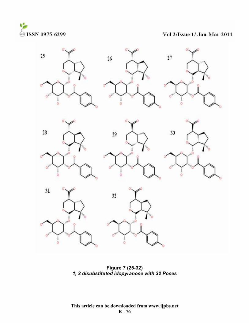
Figure 7 (25-32) 1, 2 disubstituted idopyranose with 32 Poses

This article can be downloaded from www.ijpbs.net
B - 77
Table 1 Docking result of the ligand, 1, 2 disubstituted idopyranose with 32 Poses
2qmj 3f8s 3g5e 3f7z Ligand
Poses
Glide Score (XP)
Glide Energy (kcal/mol)
Glide Score (XP)
Glide Energy (kcal/mol)
Glide Score (XP)
Glide Energy (kcal/mol)
Glide Score (XP)
Glide Energy (kcal/mol)
1. -8.19 -40.40 -10.06 -49.44 -6.78 -40.04 -10.26 -48.86
*2. -7.35 -47.02 -9.86 -52.25 -6.65 -40.11 -9.41 -41.16
*3. -6.42 -47.53 -8.76 -52.41 -6.51 -39.36 -9.56 -51.00
4. -6.41 -47.25 -8.76 -52.08 -6.26 -38.67 -9.50 -52.81
5. -6.40 -43.11 -8.53 -48.87 -6.21 -40.97 -9.27 -39.79
6. -6.34 -44.27 -8.22 -45.77 -5.99 -43.16 -9.22 -52.95
7. -6.31 -44.76 -8.09 -47.19 -5.83 -38.62 -9.16 -49.72
8. -6.23 -43.07 -8.07 -50.26 -5.77 -38.04 -9.04 -51.22
9. -6.17 -33.50 -7.95 -50.55 -5.76 -42.30 -8.90 -54.53
10 -6.01 -47.21 -7.94 -50.14 -5.63 -37.04 -8.89 -47.75 11 -5.99 -43.25 -7.94 -47.27 -5.60 -33.41 -8.76 -44.62 12. -5.90 -41.33 -7.92 -50.70 -5.53 -38.00 -8.72 -48.40 13. -5.87 -40.19 -7.83 -47.20 -5.45 -39.82 -8.71 -53.48 14. -5.87 -47.73 -7.82 -52.02 -5.42 -43.79 -8.67 -46.66 15. -5.87 -42.05 -7.75 -42.59 -5.21 -35.75 -8.38 -46.48 16. -5.86 -44.46 -7.72 -53.03 -5.18 -37.62 -8.19 -48.74 17. -5.77 -43.74 -7.58 -42.36 -5.12 -37.03 -8.11 -46.84 18. -5.68 -37.29 -7.55 -46.38 -5.05 -31.68 -8.10 -46.36
19. -5.59 -45.92 -7.50 -46.44 -5.04 -37.89 -8.09 -43.08
20. -5.57 -38.37 -7.38 -49.24 -4.98 -39.10 -8.07 -48.70
21. -5.37 -43.31 -7.37 -46.03 -4.97 -32.95 -7.97 -49.62
22. -5.37 -41.20 -7.22 -48.40 -4.95 -37.34 -7.89 -47.73
23. -5.33 -39.98 -7.19 -49.96 -4.92 -35.64 -7.80 -42.88
24. -5.32 -39.14 -7.06 -49.44 -4.81 -37.47 -7.66 -52.88
25. -5.31 -41.33 -6.94 -39.09 -4.79 -37.14 -7.65 -44.84
26. -5.03 -35.53 -6.84 -46.95 -4.46 -42.87 -7.63 -50.65
27. -5.03 -45.48 -6.82 -50.24 -4.43 -38.66 -7.61 -46.76
28. -4.89 -42.23 -6.63 -45.19 -4.38 -38.80 -7.58 -43.77
29. -4.81 -44.49 -6.37 -46.39 -4.36 -38.83 -7.41 -48.49
30. -4.75 -44.72 -6.26 -43.90 -4.23 -35.80 -7.30 -49.76
31. -4.48 -36.71 -6.21 -42.03 -4.17 -37.17 -7.02 -48.97
32. -3.87 -36.76 -6.06 -46.30 -3.92 -42.59 -6.93 -41.15
* Ligand pose showing high glide score and low bound energy

This article can be downloaded from www.ijpbs.net
B - 78
According to the docking result, the ligand was selected with best dock score and low bound energy. In that, the ligand pose 2 had the good glide score and energy compared to other poses for 2qmj (Glide Score, -7.35 and Glide Energy, -47.02 kcal/mol) (Fig. 8), 3f8s (Glide Score, -9.86 and Glide Energy, -52.25 kcal/mol) (Fig. 9), and 3g5e (Glide Score, -6.65 and Glide Energy, -40.11 kcal/mol) (Fig. 11) and ligand pose 3 for
3f7z (Glide Score, -9.56 and Glide Energy, -51.00 kcal/mol) (Fig. 10).The docking interactions between the selected natural compound and the known inhibitor of each target were compared and from the result it was revealed that the idopyranose possess better score for 3f8s and 3f7z than the previously bound one (Table. 2).
Figure 8 Figure 9 1, 2 disubstituted idopyranose 1, 2 disubstituted idopyranose
bound with 2qmj bound with 3f8s
Figure 10 Figure 11 1, 2 disubstituted idopyranose 1, 2 disubstituted idopyranose
bound with 3f7z bound with 3g5e

This article can be downloaded from www.ijpbs.net
B - 79
Table 2 Comparison of best ligand score between 1, 2 disubstituted idopyranose and known
inhibitors
Idopyranose Known Inhibitors Pdb Id
Glide Score (XP)
Glide Energy (kcal/mol)
No of Hydrogen bond interactions
Glide Score (XP)
Glide Energy (kcal/mol)
No of Hydrogen bond interactions
2qmj -7.35 -47.02 3 -10.99 -57.72 6
3f8s -9.86 -52.25 6 -5.96 -42.83 2
3g5e -6.65 -40.11 5 -6.16 -75.02 6
3f7z -9.56 -51.00 5 -8.80 -55.94 3
The bioactive compound also interacted the proteins with more number of hydrogen bonds than known inhibitors, six hydrogen bond interactions (Arg125, Asn710, Arg125, Glu205, Glu205 and Glu206) with the protein DPP-4(3f8s), five hydrogen bond interactions
(Gln185,Gln185,Arg 125,Arg141and Pro136) with GSK-3(3f7z), in their binding pocket. The Liasion values (-80.34 kcal/mol for DPP-4 and -99.47 kcal/mol for GSK-3) also confirmed the result with good binding energy (Table. 3).
Table 3 Liaison binding energy for protein-ligand complex
Liaison Values Ligand Poses 2qmj 3f8s 3g5e 3f7z
1 -18.51 -71.06 -76.67 -55.16
2* -13.05 -80.34 -56.55 -99.47
3 -14.58 -70.61 -42.59 -55.32
4 -9.98 -62.91 -94.47 -77.12
5 -9.62 -84.33 -68.83 -75.58
6 -33.24 -66.36 -65.44 -78.75
7 1.61 -86.73 -61.29 -58.83
8 -33.70 -26.73 -41.03 4.27
9 15.20 -71.13 -101.19 -47.90
10 11.45 -37.42 -72.60 -15.97
* Ligand pose with good liaison score

This article can be downloaded from www.ijpbs.net
B - 80
Several reports revealed that the extract of the medicinal plant, Vitex negundo possessed anti-diabetic properties (Irene et al., 2006). The anti-diabetic activity of 1, 2 disubstituted idopyranose had been determined in Wistar rats (Manikandan et al., 2009). The ADME/T prediction of 1, 2 disubstituted idopyranose (C23H28O12) shows good result with least number of stars and least number of violations (Table. 4). From this result, we can suggest that 1, 2 disubstituted idopyranose is a potent inhibitor of dipeptidyl peptidase-4 and glycogen synthase kinase-3 than the commercially available drugs.
Table 4
ADME/T properties of 1, 2 disubstituted idopyranose
Descriptors/Properties Value
Mol_MW 498.483
QPlogMDCK 1.921
QPlogHERG -3.135
LROF 2
QPPCaco 4.709
Stars 2
QPlogKp -5.288
QPlogS -2.779
QPlogBB -2.889
QPpolrz 42.933
CONCLUSION
The Protein-Ligand interaction plays a significant role in structural based drug designing. In the present work we have docked the ligand, 1, 2 disubstituted idopyranose (C23H28O12) from the medicinal plant with the proteins that are used as the target for Type 2 diabetes. The analysis of the docking result allowed us to know the efficiency of the natural bioactive compound to control the diabetes. The docking study revealed the binding orientation of the natural ligand 1, 2
disubstituted idopyranose in the target protein’s active site (dipeptidyl peptidase-4 and glycogen synthase kinase-3) which resulted in inhibition of enzyme activity. The binding energy of the ligand-protein interactions also confirmed that the ligand tightly fit to the macromolecule, protein. From the results obtained, it will be essential to understand the important structural features required to enhance the inhibitory activities and further it will help to produce augmented inhibitory compounds.

This article can be downloaded from www.ijpbs.net
B - 81
REFERENCES 1. Abbott, C. A and Gorrell, M. D. 2002.
Ectopeptidases: CD13/Aminopeptidase N and CD26/Dipeptidylpeptidase IV. In: Langner, J., Ansorge, S., (eds.), Medicine and Biology Kulwer/Plenum Publishing Corp., New York, pp. 171-184.
2. Adelhorst, K., Hedegaard, B.B., Knudsen, L.B., Kirk, O. 1994. Structure activity studies of glucagon like peptide-1. J Biol Chem 269: 6275–6278.
3. Ammirati, M.J., Andrews, K.M., Boyer, D.D., Brodeur, A.M., Danley, D.E., Dordan, S.D., Hulin, B., Liu, S., McPherson, R.K., Orena, S.J., Parker, J.C., Polevkova, J., Qiu, X, Soglia, C.B., Treadway, J.L., VanVolkenburg, M.A., Wilder, D.C., Piotrowski, D.W. 2009. 3,3-Difluoro-pyrrolidin-1-yl)-[(2S,4S)-(4-(4-pyrimidin-2-yl-piperazin-1-yl)-pyrrolidin-2yl]-methane: a potent, selective, orally active dipetptidyl peptidase IV inhibitor. Bioorg Med Chem Lett 19: 1991-1995.
4. Bjelke, R.J., Christensen, J., Branner, S., Wagtmann, N., Olsen, C., Kanstrup, A.B., Rasmussen, H.B. 2004. Tyrosine 547 constitutes an essential part of the catalytic mechanism of Dipeptidyl peptidase-4. J Biol Chem 279: 34691– 34697.
5. Buford, L., Nichols., Stephen Avery., Partha Sen., Dallas M. Swallow., Dagmar Hahn., Erwin Sterchi. 2003. The maltase-glucoamylase gene: common ancestry to sucrase-isomaltase with complementary starch digestion activities. Proc Natl Acad Sci 100(3): 1432–1437.
6. Doble, B. W., Woodgett, J. R. 2003. GSK-3: Tricks of the trade for a multi-tasking kinase. J Cell Sci 116: 1175–1186.
7. Duke-Cohan, J.S., Morimoto, C., Rocker J.A., Schlossman, S.F. 1996. Serum high molecular weight dipeptidyl peptidase-4 (CD26) is similar to a novel antigen DPPT-L
released from activated T-cells. J Immunol 156: 1714–1721.
8. Frame, S., Cohen, P., Biondi, R. M. 2001. A common phosphate binding site explains the unique substrate specificity of GSK3 and its inactivation by phosphorylation. Mol Cell : 1321– 1327.
9. Friesner, R.A., Richard, A., Robert, B., Murphy, R.A., Matthew, P. Repasky., Leah L. Frye., Jeremy R. Greenwood., Thomas A. Halgren., Paul C. Sanschagrin., Daniel T. Mainz. 2006. Extra Precision glide – Docking and scoring incorporating a model of hydrophobic enclosure for protein-ligand complexes based on a new theory of molecular recognition. J Med Chem 49: 6177–6196.
10. Friesner, R. A., Banks, J. L., Murphy, R. B., Halgren, T. A., Klicic, J. J., Mainz, D. T. 2004. A New Approach for Rapid, Accurate Docking and Scoring. 1. Method and Assessment of Docking Accuracy. J Med Chem 47: 1739–1749.
11. Gerich, J.E. 1998. The genetic basis of type 2 diabetes mellitus: impaired insulin secretion versus impaired insulin sensitivity. Endocr Rev 19: 491-503.
12. Gloyn, A.L. 2003. The search for type 2 diabetes genes. Ageing Res Rev 2:111-127.
13. Glide, Version 5.5, (2009) Schrodinger, LLC, New York, NY.
14. Halgren, T. A., Murphy, R. B., Friesner, R. A., Beard, H. S., Frye, L. L. Pollard, W. T., Banks, J. L. 2004. Glide: A new approach for rapid, accurate docking and scoring. 2. Enrichment factors in database screening. J Med Chem 47: 1750–1759.
15. Hamilton-Miller, J. M. T. 1995. Antimicrobial Properties of Tea (Camellia

This article can be downloaded from www.ijpbs.net
B - 82
sinensis L.) Antimicrob. Agents and Chemother 39: 2375–2377.
16. Irene, M., Villasenor, M. R., Lamadrid, A. 2006 J Ethnopharmacol 104:129-131.
17. Lambeir, A.M., Diaz Pereira, J.F., Chacon, P., Vermeulen, G., Heremans, K., Devreese, B., Van Beeumen, J., De Meester, I., Scharpe, S. 1997. A Prediction of DPP-IV/CD26 domain structure from a physico-chemical investigation of Dipeptidyl Peptidase-IV (CD26) from human seminal plasma. Biochem Biophys Acta. 1340(2): 215-226.
18. Laskowaski, R.A., MacArthur, M.W., Moss, D.S., Thornton, J.M. 1993. PROCHECK: a program to check the stereochemical quality of protein structures. J Appl Cryst 26: 283-291.
19. LeRoith, D. 2002. Beta-cell dysfunction and insulin resistance in type 2 diabetes: role of metabolic and genetic abnormalities. Am J Med 113, 3S-11S.
20. Liaison, Version 5.5, (2009) Schrodinger, LLC, New York, NY.
21. Ligprep, Version 2.3, (2009) Schrodinger, LLC, New York, NY.
22. Manikandan, R., Sundaram, R., Srinivasan, P., Beulaja, S., Arulvasu, C. 2009. Isolation of 1, 2 di-substituted idopyranose from Vitex negundo and its effects on diabetic rats. In J Pharma Analysis 1 (2): 4-10.
23. McManus, E. J., Sakamoto, K., Armit, L. J., Ronaldson, L., Shapiro, N., Marquez, R., Alessi, D. R. 2005. Role that phosphorylation of GSK3 plays in insulin and Wnt signalling defined by knockin analysis. Embo J 24:1571–1583.
24. Morris, A.L., MacArthur, M.W. 1992. Stereochemical quality of protein structure coordinates. Proteins 12:345-364.
25. Mukai, F., Ishiguro, K., Sano, Y., Fujita, S. C. 2002. Alternative splicing isoform of tau protein kinase I/glycogen synthase kinase 3β. J Neurochem 81: 1073–1083.
26. Muthenna, P., Suryanarayana, P., Gunda Shravan, K., Petrash J Mark., Reddy G Bhanuprakash. 2009. Inhibition of aldose reductase by dietary antioxidant curcumin: mechanism of inhibition, specificity and significance. FEBS letters 583(22): 3637-3642.
27. Nichols, B.L., Eldering, J., Avery, S., Hahn, D., Quaroni, A., Sterchi, E. 1998. Human small intestinal maltase-glucoamylase cDNA cloning: homology to sucrase-isomaltase. J Biol Chem 273: 3076–3081.
28. Ogata, S., Misumi, Y., Ikehara, Y. 1989. Primary structure of rat liver dipeptidyl peptidase IV reduced from its cDNA and identification of the NH2-terminal signal sequence as the membrane-anchoring domain. J Biol Chem 264(6): 3596–3601.
29. QikProp Version 3.2, (2009) Schrodinger, LLC, New York, NY.
30. Ramachandran, G.N., Ramakrishnan, C., Sasisekharan, V. 1963. Stereochemistry of polypeptide chain configurations. J Mol Biol 7: 95-99.
31. Rolin, B., Deacon, C.F., Carr, R.D., Ahrén, B. 2004. Inhibiton of Dipeptidyl peptidase-4 (DPP-4)-A novel approach to treat Type 2 Diabetes. Eur J Pharmacol 494: 283–288.
32. Roy, R., Gautier, M., Hayes, H., Laurent, P., Zaragoza, P., Eggen, A., Rodellar, C. 2002. Assignment of maltase glucoamylase (MGAM) gene to bovine chromosome 4q34 by in situ hybridization and confirmation by radiation hybrid mapping. Cytogenet Genome Res. 98: 311.
33. Saitoh, M., Kunitomo, J., Kimura, E., Hayase, Y., Kobayashi, H., Uchiyama, N., Kawamoto, T., Tanaka, T., Mol, C.D., Dougan, D.R., Textor, G.s., Snell, G.P., Itoh, F. 2009. Design, synthesis and structure-activity relationships of 1,3,4-oxadiazole derivatives as novel inhibitors

This article can be downloaded from www.ijpbs.net
B - 83
of glucogen synthase kinase-3beta. Bioorg Med Chem 17: 2017-2029.
34. Semenza, G., Auricchio, S. 1989. The Metabolic Basis of Inherited Disease II. McGraw-Hill Inc, New York, 2975–2997.
35. Sim, L., Quezada-Calvillo, R., Sterchi, E.E., Nichols, B.L., Rose, D.R. 2008. Human intestinal maltase-glucoamylase: crystal structure of the N-terminal catalytic subunit and basis of inhibition and substrate specificity. J Mol Biol 375: 782-792.
36. Taverna, D.M and Goldstein, R.A. 2002. Why are proteins marginally stable. Proteins 46: 105.
37. Van Zandt, M.C., Doan, B., Sawicki, D.R., Sredy, J., Podjarny, A.D. 2009. Discovery of [3-(4,5,7-trifluoro-benzothiazol-2-ylmethyl)-pyrrlol [2,3-b] pyridine-1-yl] acetic acids as highly potent and selective inhibitors of alsdose reductase for treatment of chronic diabetic complications. Bioorg Med Chem Lett 19: 2006-2008