international journal of surgery case reports ge junction 2 incisura 1 fundus 4.4days defining a...
TRANSCRIPT
Og
HT
a
ARRAA
KGLGL
1
tfbamofdpvlcobsm(nr
a
h2o
CASE REPORT – OPEN ACCESSInternational Journal of Surgery Case Reports 25 (2016) 91–96
Contents lists available at ScienceDirect
International Journal of Surgery Case Reports
journa l h omepage: www.caserepor ts .com
ptimal management of GIST tumors located near theastroesophageal junction: Case report and review of the literature
ishaam Ismael ∗, Yury Ragoza, James Caccitolo, Steven Coxhe University of Texas Health Science Center at Tyler, Tyler, TX, USA
r t i c l e i n f o
rticle history:eceived 6 May 2016eceived in revised form 27 May 2016ccepted 13 June 2016vailable online 17 June 2016
eywords:
a b s t r a c t
INTRODUCTION: The safety and oncologic outcome of laparoscopic gastric GIST resection is well estab-lished especially for lesions <5 cm in diameter. The optimal management of GIST tumors near the GEjunction remains unclear.METHODS: We present a case-report of a 4.7 cm GIST tumor near the GE junction managed byendoscopically-assisted laparoscopic wedge resection (EAWR). We present a review of the literaturehighlighting the various combined laparo-endoscopic techniques available.
IST tumorsaparo-endoscopic resectionastroesophageal junction tumorsaparoscopic wedge resection
RESULTS: We used the non-touch lesion-lifting method to laparoscopically resect the GIST tumor underendoscopic guidance. There were no complications and the patient was discharged on postoperative day3.CONCLUSIONS: Endoscopically-assisted laparoscopic wedge resections are feasible and safe for GISTtumors near the GE junction.
© 2016 Published by Elsevier Ltd on behalf of IJS Publishing Group Ltd. This is an open access articlehe CC
under t. Introduction
Gastrointestinal Stromal Tumors (GISTs) are the most frequentumors of the gastric submucosa [1]. These tumors are derivedrom the interstitial cells of Cajal and have been shown to har-or gain-of-function mutations in the cell-surface KIT receptor inpproximately 90% of cases [2]. Surgical resection with negativeargins is the standard of care. The safety and oncologic outcome
f laparoscopic gastric GIST resection is well established especiallyor lesions <5 cm in diameter [3]. Laparoscopy has been shown toecrease intraoperative blood loss, postoperative pain and hos-ital stay [3]. It has the added advantage of allowing improvedisualization during surgery to rule out disease spread to theiver or peritoneum. Although the National Comprehensive Can-er Network Clinical Practice Guidelines for Optimal Managementf patients with GIST suggests that laparoscopic techniques shoulde limited to tumors <5 cm [4], many groups do not consider tumorize as a contraindication for the laparoscopic approach [5]. Theanagement of tumors located near or at the gastro-esophageal
GE) junction poses a particular challenge. The esophagus and vaguserves are at risk for injury, and the GE junction is at risk for nar-
owing or dysfunction if the resection is not well-planned.Several surgical procedures have been proposed for man-gement GE junction GIST tumors. They use a combination of
∗ Corresponding author.E-mail address: [email protected] (H. Ismael).
ttp://dx.doi.org/10.1016/j.ijscr.2016.06.006210-2612/© 2016 Published by Elsevier Ltd on behalf of IJS Publishing Group Ltd. This irg/licenses/by-nc-nd/4.0/).
BY-NC-ND license (http://creativecommons.org/licenses/by-nc-nd/4.0/).
laparoscopic and endoscopic techniques and include: enucleation,endoscopically assisted wedge resection (EAWR), laparoscopicallyassisted endoscopic resection (LAER), laparoscopic and endoscopiccooperative surgery (LECS) and transgastric resection for tumorslocated posteriorly. The treatment of GIST tumors at the GE junctionhas traditionally been an esophagectomy [6]. Recent studies haveproposed enucleation with or without fundoplication as a reason-able alternative for small tumors with low mitotic rates [7,8]. Onthe other hand, there are several treatment options for tumors near,not at, the GE junction. One of the major limitations of the laparo-scopic approach is accurately localizing these lesions and definingtheir anatomic relationship to the GE junction. This can be eas-ily addressed by adding intraoperative endoscopy. The non-touchlesion-lifting technique, first described by Ohgami et al. in 1996, forlaparoscopic wedge resection has gained widespread acceptanceand is our method of choice for resecting GIST tumors near the GEjunction [1,9].
In this case-report, we describe the management of a GISTtumor located near the GE junction. We employ both endoscopyand laparoscopy to facilitate resection using the non-touch lesion-lifting technique. Consent in accordance with our institutionalregulations was obtained.
2. Case report
64 year old gentleman referred for a general surgical evaluationof an umbilical hernia. A CT scan of the abdomen was ordered toassess the defect size and its contents. The CT demonstrated an
s an open access article under the CC BY-NC-ND license (http://creativecommons.

CASE REPORT – OPEN ACCESS92 H. Ismael et al. / International Journal of Surgery Case Reports 25 (2016) 91–96
GIST
ivhssmw(c
sasilddtttloeTos
Fig. 1. CT scan findings demonstrating a 4.7 cm
ncidental 4.7 cm mass involving the gastric fundus and lesser cur-ature 2 cm from the GE junction and a fat containing umbilicalernia (Fig. 1). EGD/EUS were performed and demonstrated a 5 cmubmucosal hypoechoic nodule located within 2 cm of the gastroe-ophageal junction along the anterior wall of the stomach. It hadultiple cystic areas suggestive of necrosis. Fine-needle aspirationas performed and demonstrated spindle cells, DOG-1 and CD117
C-kit) positive, consistent with a GIST. A lesion-lifting endoscopi-ally assisted laparoscopic wedge resection was planned.
The patient was taken to the operating room and placed in aupine position. An incision was made over his umbilical herniand the hernia sac was dissected down to the fascia. The herniaac was excised and a 12-mm balloon tipped trocar was insertednto the abdomen. The abdomen was insufflated and a diagnosticaparoscopy was performed. The liver and peritoneum were free ofisease. Three more trocars were inserted: a 5-mm right parame-ian, 12-mm left mid-clavicular and a 5-mm left anterior axillaryrocar. An Endo-Harmonic (Ethicon US, LLC.) was used to mobilizehe left lateral segment of the liver and a laparoscopic liver retrac-or was placed through the right paramedian port. The tumor wasocated on the anterior lesser curvature of the stomach within 2 cmf the GE junction. The Endo-Harmonic was used to mobilize the
sophageal fat pad to define the superior extent of our resection.he gastro-hepatic ligament was incised and the medial borderf the stomach was dissected. An endoscope was passed into thetomach and retroflexed identifying the tumor and its relationshipwithin 2 cm of the gastroesophageal junction.
to the GE junction. A laparoscopic 60 mm linear stapler was usedto lift the tumor anteriorly and close below it. This was done underendoscopic guidance. The endoscope was used to make sure thestaple line was below the tumor and away from the GE junction.The resection was completed using 2 Covedien black reloads withTri-Staple technology (Medtronic-Covedien, Minneapolis US). Anair-leak test was performed at the end of the case by endoscopi-cally insufflating the stomach and submerging the staple line undernormal saline. There was no leak. The operative time was 90 minwith minimal blood loss.
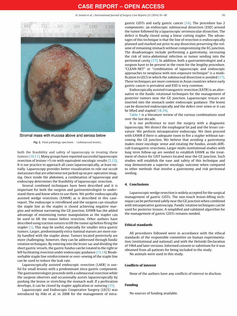
The patient underwent a water soluble followed by thin Bar-ium upper GI study on the first postoperative day which did notshow a leak. He was placed on a clear liquid diet and advanced to aregular diet the following day. He was discharged on the third post-operative day. Pathology demonstrated a 4.7 cm GIST with negativemargins and a mitotic rate of <5/50 HPF (Fig. 2). His case was pre-sented at our institutional tumor board and no adjuvant treatmentwas recommended. He underwent a surveillance CT scan 6 monthsafter surgery which showed no evidence of disease. He remainssymptom free without pain or iron-deficiency anemia.
3. Discussion and literature review
There are several endoscopic, laparoscopic and open surgicalprocedures available for the management of GIST tumors near theGE junction. Over the last decade, multiple reports have described
CA
SE
RE
PO
RT
– O
PE
N A
CC
ES
SH
. Ism
ael et
al. /
International Journal
of Surgery
Case R
eports 25
(2016) 91–96
93
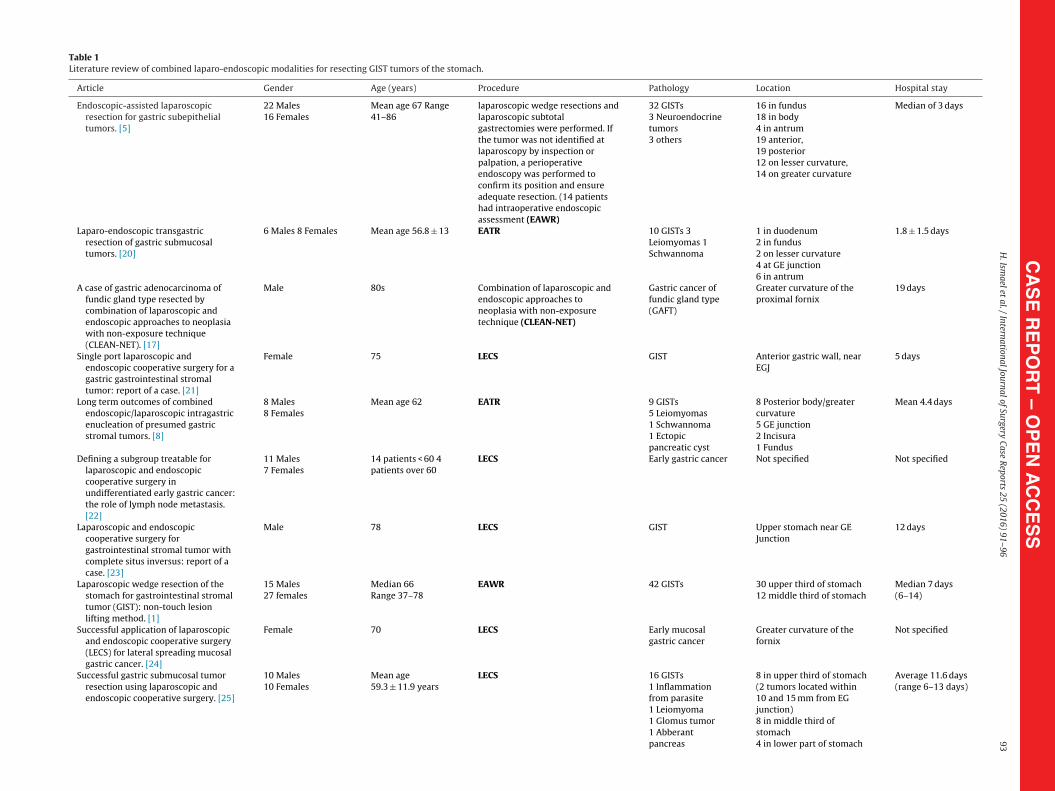
Table 1Literature review of combined laparo-endoscopic modalities for resecting GIST tumors of the stomach.
Article Gender Age (years) Procedure Pathology Location Hospital stay
Endoscopic-assisted laparoscopicresection for gastric subepithelialtumors. [5]
22 Males16 Females
Mean age 67 Range41–86
laparoscopic wedge resections andlaparoscopic subtotalgastrectomies were performed. Ifthe tumor was not identified atlaparoscopy by inspection orpalpation, a perioperativeendoscopy was performed toconfirm its position and ensureadequate resection. (14 patientshad intraoperative endoscopicassessment (EAWR)
32 GISTs3 Neuroendocrinetumors3 others
16 in fundus18 in body4 in antrum19 anterior,19 posterior12 on lesser curvature,14 on greater curvature
Median of 3 days
Laparo-endoscopic transgastricresection of gastric submucosaltumors. [20]
6 Males 8 Females Mean age 56.8 ± 13 EATR 10 GISTs 3Leiomyomas 1Schwannoma
1 in duodenum2 in fundus2 on lesser curvature4 at GE junction6 in antrum
1.8 ± 1.5 days
A case of gastric adenocarcinoma offundic gland type resected bycombination of laparoscopic andendoscopic approaches to neoplasiawith non-exposure technique(CLEAN-NET). [17]
Male 80s Combination of laparoscopic andendoscopic approaches toneoplasia with non-exposuretechnique (CLEAN-NET)
Gastric cancer offundic gland type(GAFT)
Greater curvature of theproximal fornix
19 days
Single port laparoscopic andendoscopic cooperative surgery for agastric gastrointestinal stromaltumor: report of a case. [21]
Female 75 LECS GIST Anterior gastric wall, nearEGJ
5 days
Long term outcomes of combinedendoscopic/laparoscopic intragastricenucleation of presumed gastricstromal tumors. [8]
8 Males8 Females
Mean age 62 EATR 9 GISTs5 Leiomyomas1 Schwannoma1 Ectopicpancreatic cyst
8 Posterior body/greatercurvature5 GE junction2 Incisura1 Fundus
Mean 4.4 days
Defining a subgroup treatable forlaparoscopic and endoscopiccooperative surgery inundifferentiated early gastric cancer:the role of lymph node metastasis.[22]
11 Males7 Females
14 patients < 60 4patients over 60
LECS Early gastric cancer Not specified Not specified
Laparoscopic and endoscopiccooperative surgery forgastrointestinal stromal tumor withcomplete situs inversus: report of acase. [23]
Male 78 LECS GIST Upper stomach near GEJunction
12 days
Laparoscopic wedge resection of thestomach for gastrointestinal stromaltumor (GIST): non-touch lesionlifting method. [1]
15 Males27 females
Median 66Range 37–78
EAWR 42 GISTs 30 upper third of stomach12 middle third of stomach
Median 7 days(6–14)
Successful application of laparoscopicand endoscopic cooperative surgery(LECS) for lateral spreading mucosalgastric cancer. [24]
Female 70 LECS Early mucosalgastric cancer
Greater curvature of thefornix
Not specified
Successful gastric submucosal tumorresection using laparoscopic andendoscopic cooperative surgery. [25]
10 Males10 Females
Mean age59.3 ± 11.9 years
LECS 16 GISTs1 Inflammationfrom parasite1 Leiomyoma1 Glomus tumor1 Abberantpancreas
8 in upper third of stomach(2 tumors located within10 and 15 mm from EGjunction)8 in middle third ofstomach4 in lower part of stomach
Average 11.6 days(range 6–13 days)
CA
SE
RE
PO
RT
– O
PE
N A
CC
ES
S94
H
. Ism
ael et
al. /
International Journal
of Surgery
Case R
eports 25
(2016) 91–96
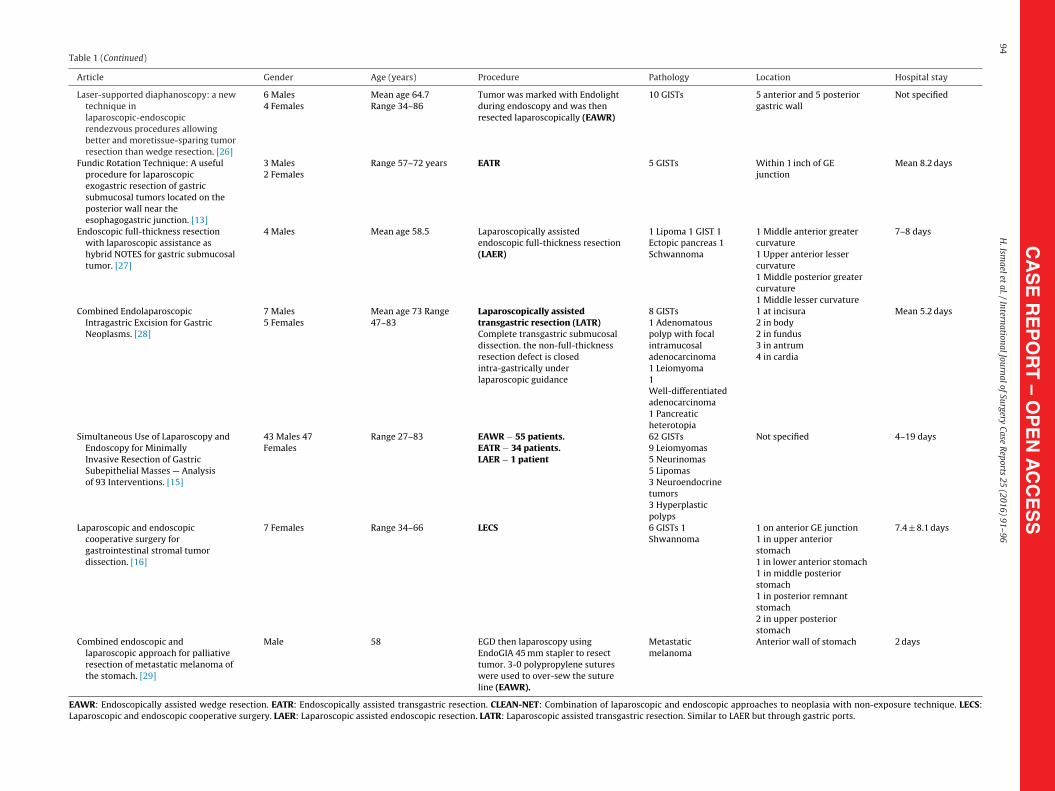
Table 1 (Continued)
Article Gender Age (years) Procedure Pathology Location Hospital stay
Laser-supported diaphanoscopy: a newtechnique inlaparoscopic-endoscopicrendezvous procedures allowingbetter and moretissue-sparing tumorresection than wedge resection. [26]
6 Males4 Females
Mean age 64.7Range 34–86
Tumor was marked with Endolightduring endoscopy and was thenresected laparoscopically (EAWR)
10 GISTs 5 anterior and 5 posteriorgastric wall
Not specified
Fundic Rotation Technique: A usefulprocedure for laparoscopicexogastric resection of gastricsubmucosal tumors located on theposterior wall near theesophagogastric junction. [13]
3 Males2 Females
Range 57–72 years EATR 5 GISTs Within 1 inch of GEjunction
Mean 8.2 days
Endoscopic full-thickness resectionwith laparoscopic assistance ashybrid NOTES for gastric submucosaltumor. [27]
4 Males Mean age 58.5 Laparoscopically assistedendoscopic full-thickness resection(LAER)
1 Lipoma 1 GIST 1Ectopic pancreas 1Schwannoma
1 Middle anterior greatercurvature1 Upper anterior lessercurvature1 Middle posterior greatercurvature1 Middle lesser curvature
7–8 days
Combined EndolaparoscopicIntragastric Excision for GastricNeoplasms. [28]
7 Males5 Females
Mean age 73 Range47–83
Laparoscopically assistedtransgastric resection (LATR)Complete transgastric submucosaldissection. the non-full-thicknessresection defect is closedintra-gastrically underlaparoscopic guidance
8 GISTs1 Adenomatouspolyp with focalintramucosaladenocarcinoma1 Leiomyoma1Well-differentiatedadenocarcinoma1 Pancreaticheterotopia
1 at incisura2 in body2 in fundus3 in antrum4 in cardia
Mean 5.2 days
Simultaneous Use of Laparoscopy andEndoscopy for MinimallyInvasive Resection of GastricSubepithelial Masses — Analysisof 93 Interventions. [15]
43 Males 47Females
Range 27–83 EAWR − 55 patients.EATR − 34 patients.LAER − 1 patient
62 GISTs9 Leiomyomas5 Neurinomas5 Lipomas3 Neuroendocrinetumors3 Hyperplasticpolyps
Not specified 4–19 days
Laparoscopic and endoscopiccooperative surgery forgastrointestinal stromal tumordissection. [16]
7 Females Range 34–66 LECS 6 GISTs 1Shwannoma
1 on anterior GE junction1 in upper anteriorstomach1 in lower anterior stomach1 in middle posteriorstomach1 in posterior remnantstomach2 in upper posteriorstomach
7.4 ± 8.1 days
Combined endoscopic andlaparoscopic approach for palliativeresection of metastatic melanoma ofthe stomach. [29]
Male 58 EGD then laparoscopy usingEndoGIA 45 mm stapler to resecttumor. 3-0 polypropylene sutureswere used to over-sew the sutureline (EAWR).
Metastaticmelanoma
Anterior wall of stomach 2 days
EAWR: Endoscopically assisted wedge resection. EATR: Endoscopically assisted transgastric resection. CLEAN-NET: Combination of laparoscopic and endoscopic approaches to neoplasia with non-exposure technique. LECS:Laparoscopic and endoscopic cooperative surgery. LAER: Laparoscopic assisted endoscopic resection. LATR: Laparoscopic assisted transgastric resection. Similar to LAER but through gastric ports.
CASE REPORT – OH. Ismael et al. / International Journal of Su
btrItmie
isartgabdstimrslsc
fTtpd
i
Fig. 2. Final pathology specimen − (submucosal lesion).
oth the feasibility and safety of laparoscopy in treating theseumors [10,11]. Many groups have reported successful laparoscopicesection of lesions >5 cm with equivalent oncologic results [5,12].t is our practice to approach all cases laparoscopically, at least ini-ially. Laparoscopy provides better visualization to rule out occult
etastases that are otherwise not picked up on pre-operative imag-ng. Once inside the abdomen, a combination of laparoscopy andndoscopy determines the feasibility of laparoscopic resection.
Several combined techniques have been described and it ismportant for both the surgeon and gastroenterologist to under-tand them and know when to use them. We prefer endoscopicallyssisted wedge resections (EAWR) as is described in this case-eport. The endoscope is retroflexed and the surgeon can visualizehe staple line as the stapler is closed achieving negative mar-ins and without narrowing the GE junction. EAWR has the addeddvantage of minimizing tumor manipulation as the stapler cane used to lift the tumor before resection. Other authors haveescribed using traction sutures to lift the tumor up before firing thetapler [1]. This may be useful, especially for smaller intra-gastricumors. Larger, predominantly extra-luminal masses are more eas-ly handled with the stapler alone. Tumors located posteriorly are
ore challenging; however, they can be addressed through fundicotation techniques. By entering into the lesser sac and dividing thehort gastric vessels, the gastric fundus can be rotated to the right oreft facilitating resection under endoscopic guidance [13,14]. Bioab-orbable staple line reinforcement or over-sewing of the staple linean be used to reduce the leak rate.
Laparoscopically assisted endoscopic resection (LAER) is use-ul for small lesions with a predominant intra-gastric component.he gastroenterologist proceeds with a submucosal resection whilehe surgeon observes and occasionally assists laparoscopically by
ushing the lesion or stretching the stomach wall. If a perforationevelops, it can be closed by stapler application or suturing [15].Laparoscopic and Endoscopic Cooperative Surgery (LECS) wasntroduced by Hiki et al. in 2008 for the management of intra-
PEN ACCESSrgery Case Reports 25 (2016) 91–96 95
gastric GISTs and early gastric cancer [16]. The procedure has 2components: an endoscopic submucosal dissection (ESD) aroundthe tumor followed by a laparoscopic seromuscular dissection. Thedefect is finally closed using a linear cutting stapler. The advan-tages of this technique is that the line of resection is endoscopicallyplanned and marked out prior to any dissection preserving the vol-ume of remaining stomach without compromising the EG junction.The disadvantages include performing a gastrotomy, increasingthe risk of intra-abdominal infection or tumor seeding into theperitoneal cavity [17]. In addition, both a gastroenterologist and asurgeon have to be present in the room for the lengthy procedure.“CLEAN-NET” or “combination of laparoscopic and endoscopicapproaches to neoplasia with non-exposure technique” is a modi-fication to LECS in which the submucosal dissection is avoided [17].These techniques are more common in Asian countries where earlygastric cancer is prevalent and ESD is very common.
Endoscopically assisted transgastric resection (EATR) is an alter-native to the fundic rotational techniques for the management ofposterior tumors near the GE junction. Laparoscopic trocars areinserted into the stomach under endoscopic guidance. The lesioncan be dissected endoscopically and the defect over sewn or it canbe lifted and stapled [18,19].
Table 1 is a literature review of the various combinations usedover the last decade.
It is our preference to start the surgery with a diagnosticlaparoscopy. We dissect the esophageal fat pad and the lesser cur-vature. We perform intraoperative endoscopy. We then proceedwith EAWR if there is adequate room to fire a stapler without nar-rowing the GE junction. We believe that avoiding a gastrotomymakes more oncologic sense and rotating the fundus, avoids diffi-cult transgastric resections. Larger multi-institutional studies withlong term follow-up are needed to establish EAWR as the treat-ment of choice for GIST tumors located near the GE junction. Suchstudies will establish the ease and safety of this technique andmay demonstrate a superior oncologic outcome when comparedto other methods that involve a gastrotomy and risk peritonealseeding.
4. Conclusions
Laparoscopic wedge resection is widely accepted for the surgicalmanagement of gastric GISTs. The non-touch lesion-lifting tech-nique can be performed safely near the GE junction when combinedwith intraoperative gastroscopy. Fundic rotation techniques can beused for posterior lesions. A simplified and validated algorithm forthe management of gastric GISTs remains needed.
Ethical standards
All procedures followed were in accordance with the ethicalstandards of the responsible committee on human experimenta-tion (institutional and national) and with the Helsinki Declarationof 1964 and later versions. Informed consent or substitute for it wasobtained from all patients for being included in the study.
No animals were used in this study.
Conflicts of interest
None of the authors have any conflicts of interest to disclose.
Funding
No sources of funding available.
– O9 l of Su
E
C
b
A
G
R
[
[
[
[
[
[
[
[
[
[
[
[
[
[
[
[
[
[
[
[
OTpc
CASE REPORT6 H. Ismael et al. / International Journa
thical approval
Ethical approval has been given.
onsent
Written consent in accordance with the ethics committee haseen obtained.
uthor contribution
Hishaam Ismael: Study concept and writing the paper.Yury Ragoza: Data collection.James Caccitolo: Assisted in surgery and reviewed the paper.Steven Cox: Data interpretation and paper.
uarantor
Hishaam Ismael MD.
eferences
[1] H. Kiyozaki, M. Saito, H. Chiba, et al., Laparoscopic wedge resection of thestomach for gastrointestinal stromal tumor (GIST): non-touch lesion liftingmethod, Gastric Cancer 17 (April (2)) (2014) 337–340.
[2] J. Correa-Cote, C. Morales-Uribe, A. Sanabria, Laparoscopic management ofgastric gastrointestinal stromal tumors, World J. Gastrointest. Endosc. 6 (July(7)) (2014) 296–303.
[3] Y.X. Koh, A.Y. Chok, H.L. Zheng, et al., A systematic review and meta-analysiscomparing laparoscopic versus open gastric resections for gastrointestinalstromal tumors of the stomach, Ann. Surg. Oncol. 20 (October (11)) (2013)3549–3560.
[4] G.C. Demetri, R.S. Benjamin, C.D. Blanke, et al., NCCN task force report:management of patients with gastrointestinal stromal tumor (GIST) −updateof the NCCN clinical practice guidelines, J Natl Compr Canc Netw 5 (Suppl 2)(2007) S1-S29, quiz S30.
[5] J.S. Davilla, D. Momblan, A. Gines, et al., Endoscopic-assisted laparoscopicresection for gastric subepithelial tumors, Surg. Endosc. 30 (2016) 199–203.
[6] F.B. Zhang, H.C. Shi, Y.S. Shu, et al., Diagnosis and surgical treatment ofesophageal gastrointestinal stromal tumors, World J. Gastroenterol. 21 (May(18)) (2015) 5630–5634.
[7] F. Coccolini, F. Catena, L. Ansaloni, et al., Esophagogastric junctiongastrointestinal stromal tumor: resection vs: enucleation, World J.Gastroenterol. 16 (September (35)) (2010) 4374–4376.
[8] J.S. Mino, A.D. Guerron, R. Monteriro, et al., Long-term outcomes of combinedendoscopic/laparoscopic intragastric enucleation of presumed gastric stromaltumors, Surg. Endosc. (August (15)) (2015).
[9] M. Ohgami, Y. Otani, T. Kubota, et al., Laparoscopic curative surgery for earlygastric cancer, Nippon Rinsho 54 (1999) 1307–1311.
10] K.L. Huguet, R.M. Rush, D.J. Tessier, et al., Laparoscopic gastric gastrointestinal
stromal tumor resection: the mayo clinic experience, Arch. Surg. 143 (2008)587–590.11] J. Nishimura, K. Nakajima, T. Omori, et al., Surgical strategy forgastrointestinal stromal tumor: laparoscopic vs open resection, Surg. Endosc.21 (2007) 875–878.
pen Accesshis article is published Open Access at sciencedirect.com. It is distribermits unrestricted non commercial use, distribution, and reproductredited.
PEN ACCESSrgery Case Reports 25 (2016) 91–96
12] T. Takahashi, K. Nakajima, Y. Miyazaki, et al., Surgical strategy for the gastricgastrointestinal stromal tumors (GISTs) larger the 5 cm: laparoscopic surgeryis feasible, safe and oncologically acceptable, Surg. Laparosc. Endosc.Percutan. Tech. 25 (April (2)) (2015) 114–118.
13] H. Matusi, K. Nabeshima, Y. Okamoto, et al., Fundic rotation technique: auseful procedure for laparoscopic exogastric resection of gastric submucosaltumors located on the posterior wall near the esophagogastric junction, TokaiJ. Exp. Clin. Med. 36 (4) (2011) 152–158.
14] Z.W. Ke, D.L. Chen, J.L. Cai, et al., Extraluminal laparoscopic-wedge resectionof submucosal tumors on the posterior wall of the gastric fundus close to theesophagocardiac junction, J. Laparoendosc. Adv. Surg. Tech. A 19 (2009)741–744.
15] D. Wilhelm, V. Delius, M. Burian, et al., Simultaneous use of laparoscopy andendoscopy for minimally invasive resection of gastric subepithelialmasses—analysis of 93 interventions, World J. Surg. 32 (2008) 1021–1028.
16] N. Hiki, Y. Yamamoto, T. Fukunaga, et al., Laparoscopic and endoscopiccooperative surgery for gastrointestinal tumor dissection, Surg. Endosc. 22(2007) 1729–1735.
17] M. Kato, T. Uraoka, Y. Isobe, et al., A case of gastric adenocarcinoma of fundicgland type resected by combination of laparoscopic and endoscopicapproaches to neoplasia with non-exposure technique, (CLEAN-NET), Clin. J.Gastroenterol. 8 (December (6)) (2015) 393–399, http://dx.doi.org/10.1007/s12328-015-0619-2, Epub 2015 Nov 28.
18] C. Gayer, D. Edelman, B. Curtis, et al., Combined endoscopic and laparoscopicapproach to gastroesophageal tumor, JSLS 15 (April–June (2)) (2011) 228–231.
19] R.M. Singaporewalla, B.H. Ganesan, T.D. Lee, Laprondoscopic removal of abenign gastric stromal tumor at the cardia, JSLS 10 (January–March (1))(2006) 117–121.
20] J.S. Barajas-Gamboa, G. Acosta, T.J. Savides, et al., Laparo-endoscopictransgastric resection of gastric submucosal tumors, Surg. Endosc. 29 (2015)2149–2157.
21] T. Obuchi, A. Sasaki, S. Baba, Single-port laparoscopic and endoscopiccooperative surgery for a gastric gastrointestinal stromal tumor: report of acase, Surg. Today 45 (2015) 641–646.
22] H. Li, L. Chen, Z. Huo, et al., Defining a subgroup treatable for laparoscopic andendoscopic cooperative surgery in undifferentiated early gastric cancer: therole of lymph node metastasis, J. Gastrointest. Surg. 19 (2015) 1763–1768.
23] M. Mori, K. Shuto, A. Hirano, et al., Laparoscopic and endoscopic cooperativesurgery for gastrointestinal stromal tumor with complete situs inversus:report of a case, Surg. Case Rep. 1 (2015) 72.
24] S. Nunobe, N. Hiki, T. Gotoda, et al., Successful application of laparoscopic andendoscopic cooperative surgery (LECS) for a lateral spreading mucosal gastriccancer, Gastric Cancer 15 (2012) 338–342.
25] H. Tsujimoto, Y. Yaguchi, I. Kumano, et al., Successful gastric submucosaltumor resection using laparoscopic and endoscopic cooperative surgery,World J. Surg. 36 (2012) 327–330.
26] M. Patrzyk, K. Ludwig, C.D. Heidecke, et al., Laser-supported diaphanoscopy: anew technique in laparoscopic-endoscopic rendezvous procedures allowingbetter and more tissue-sparing tumor resection than wedge resection, Surg.Endosc. 25 (2011) 2023–2028.
27] N. Abe, H. Takeuchi, O. Yanagida, et al., Endoscopic full-thickness resectionwith laparoscopic assistance as hybrid NOTES for gastric submucosal tumor,Surg. Endosc. 23 (2009) 1908–1913.
28] D. Wong, S. Wong, A. Leung, et al., Combined endolaparoscopic intragastricexcision for gastric neoplasms, JLAST 19 (6) (2009) 765–770.
29] R.S. Date, E.A. Griffiths, S.A. Pritchard, et al., Combined endoscopic andlaparoscopic approach for palliative resection of metastatic melanoma of thestomach, World J. Surg. Oncol. 4 (20) (2006).
uted under the IJSCR Supplemental terms and conditions, whichion in any medium, provided the original authors and source are