international symposium on rna-based regulation & ribosome ... · the two c-di-gmp riboswitches...
TRANSCRIPT
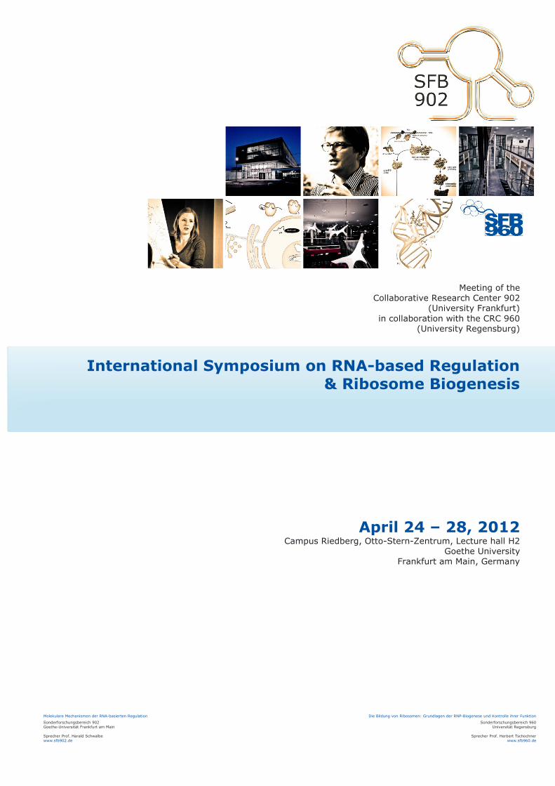
Meeting of the Collaborative Research Center 902
(University Frankfurt) in collaboration with the CRC 960
(University Regensburg)
International Symposium on RNA-based Regulation & Ribosome Biogenesis
April 24 – 28, 2012 Campus Riedberg, Otto-Stern-Zentrum, Lecture hall H2
Goethe University Frankfurt am Main, Germany
Molekulare Mechanismen der RNA-basierten Regulation
Sonderforschungsbereich 902 Goethe-Universität Frankfurt am Main Sprecher Prof. Harald Schwalbe www.sfb902.de
Die Bildung von Ribosomen: Grundlagen der RNP-Biogenese und Kontrolle ihrer Funktion
Sonderforschungsbereich 960 Universität Regensburg
Sprecher Prof. Herbert Tschochner
www.sfb960.de
- 2 - International RNA Symposium, April 24 – 28, 2012
Table of Contents
LOCATION ......................................................................................................................................................... 4
SCIENTIFIC PROGRAM ...................................................................................................................................... 5
Tuesday, 24 April ............................................................................................................................................................................... 6
Wednesday, 25 April .......................................................................................................................................................................... 7
Thursday, 26 April .............................................................................................................................................................................. 9
Friday, 27 April .................................................................................................................................................................................. 10
Saturday, 28 April ............................................................................................................................................................................. 12
LECTURE ABSTRACTS ...................................................................................................................................... 13 Strobel, Scott A. ............................................................................................................................................................................ 14 Pan, Tao ......................................................................................................................................................................................... 15 Ke, Ailong ...................................................................................................................................................................................... 16 D'Souza, Victoria ........................................................................................................................................................................... 17 Groisman, Eduardo A. ................................................................................................................................................................... 18 Wagner, Rolf ................................................................................................................................................................................. 19 Westhof, Eric ................................................................................................................................................................................ 20 Woodson, Sarah ............................................................................................................................................................................ 21 Meister, Gunter ............................................................................................................................................................................. 22 Izaurralde, Elisa .............................................................................................................................................................................23 Karbstein, Katrin .......................................................................................................................................................................... 24 Wolin, Sandra ................................................................................................................................................................................25 Tschochner, Herbert .................................................................................................................................................................... 26 Linder, Patrick ............................................................................................................................................................................... 27 Yusupov, Marat ............................................................................................................................................................................ 28 Stark, Holger ................................................................................................................................................................................ 29 Suess, Beatrix ............................................................................................................................................................................... 30 Wöhnert, Jens ............................................................................................................................................................................... 31 Rossi, John J. .................................................................................................................................................................................32 Hermann, Thomas ........................................................................................................................................................................ 33 Sattler, Michael ............................................................................................................................................................................ 34 Hoskins, Aaron Andrew ............................................................................................................................................................... 35 Fedor, Martha J. ........................................................................................................................................................................... 36 Längst, Gernot ............................................................................................................................................................................. 37 Bohnsack, Markus ........................................................................................................................................................................ 38 Hartig, Jörg S. ............................................................................................................................................................................... 39 Höbartner, Claudia ....................................................................................................................................................................... 40 Scott, William G. ............................................................................................................................................................................ 41 Bindereif, Albrecht ....................................................................................................................................................................... 42
- 3 - POSTER ABSTRACTS ....................................................................................................................................... 43
Andreou, Alexandra (Promoted Poster Talk) ............................................................................................................................. 44 Aregger, Regula (Promoted Poster Talk) ................................................................................................................................... 45 Brecht, Michael ............................................................................................................................................................................ 46 Cevec, Mirko ................................................................................................................................................................................. 47 Dueck, Anne (Promoted Poster Talk) ......................................................................................................................................... 48 Fürtig, Boris (Promoted Poster Talk) .......................................................................................................................................... 49 Hafidh, Said (Promoted Poster Talk) .......................................................................................................................................... 50 Hamperl, Stephan ......................................................................................................................................................................... 51 Heinrich, Eva-Marie .......................................................................................................................................................................52 Hierlmeier, Thomas (Promoted Poster Talk) .............................................................................................................................. 53 Katari, Venkata Subbaraju ........................................................................................................................................................... 54 Kiosze, Kristin ............................................................................................................................................................................... 55 Klinkert, Birgit (Promoted Poster Talk) ...................................................................................................................................... 56 Koschinat, Melanie....................................................................................................................................................................... 57 Lieblein, Anna Lena (Promoted Poster Talk) .............................................................................................................................. 58 Malchau, Manja ............................................................................................................................................................................ 59 Manelyte, Laura ........................................................................................................................................................................... 60 Martin, Roman .............................................................................................................................................................................. 61 Merkl, Philipp ............................................................................................................................................................................... 62 Nasiri, Amir ................................................................................................................................................................................... 63 Ochs, Meike Julia (Promoted Poster Talk) ................................................................................................................................. 64 Ohmayer, Uli ................................................................................................................................................................................. 65 Reining, Anke ............................................................................................................................................................................... 66 Rinnenthal, Jörg ........................................................................................................................................................................... 67 Sauert, Martina (Promoted Poster Talk) .................................................................................................................................... 68 Steinert, Hannah .......................................................................................................................................................................... 69 Stempfl, Thomas .......................................................................................................................................................................... 70 Strieder, Nicholas .......................................................................................................................................................................... 71 Tretbar, Sandy (Promoted Poster Talk) ....................................................................................................................................... 72 Urban, Marc ................................................................................................................................................................................. 73 Wacker, Anna ............................................................................................................................................................................... 74 Wawrzyniak, Katarzyna Marianna (Promoted Poster Talk) ...................................................................................................... 75 Weigand, Julia E. .......................................................................................................................................................................... 76 Wittmann, Alexander ....................................................................................................................................................................77
CONTACTS ....................................................................................................................................................... 78
APPENDIX ........................................................................................................................................................88
How to reach us ............................................................................................................................................................................... 89
Map of the public transportation network Frankfurt am Main ................................................................................................... 90
Impressum ........................................................................................................................................................................................ 91
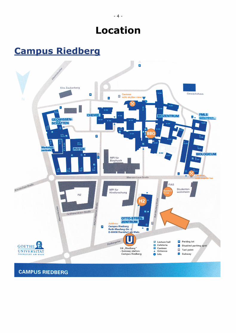
- 4 -
Location
Campus Riedberg
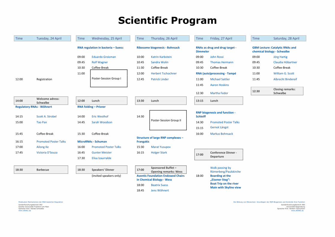
Scientific Program Time Tuesday, 24 April Time Wednesday, 25 April Time Thursday, 26 April Time Friday, 27 April Time Saturday, 28 April
RNA regulation in bacteria – Suess: Ribosome biogenesis - Bohnsack RNAs as drug and drug target - Dimmeler
GBM Lecture: Catalytic RNAs and chemical biology - Schwalbe
09:00 Eduardo Groisman
10:00 Katrin Karbstein
09:00 John Rossi
09:00 Jörg Hartig
09:45 Rolf Wagner
10:45 Sandra Wolin
09:45 Thomas Hermann
09:45 Claudia Höbartner
10:30 Coffee-Break
11:30 Coffee-Break
10:30 Coffee-Break
10:30 Coffee-Break
11:00
Poster-Session Group I 12:00 Herbert Tschochner
RNA (auto)processing - Tampé
11:00 William G. Scott
12:00 Registration
12:45 Patrick Linder
11:00 Michael Sattler
11:45 Albrecht Bindereif
11:45 Aaron Hoskins
12:30 Martha Fedor 12:30 Closing remarks:
Schwalbe 14:00 Welcome adress:
Schwalbe 12:00 Lunch 13:30 Lunch 13:15 Lunch
Regulatory RNAs - Wöhnert RNA folding – Prisner
14:15 Scott A. Strobel
14:00 Eric Westhof
14:30 Poster-Session Group II
RNP biogenesis and function - Schleiff
15:00 Tao Pan
14:45 Sarah Woodson
14:30 Promoted Poster Talks
15:15 Gernot Längst
15:45 Coffee-Break
15:30 Coffee-Break
16:00 Markus Bohnsack
16:15 Promoted Poster Talks MicroRNAs - Schuman Structure of large RNP complexes – Frangakis
17:00 Ailong Ke
16:00 Promoted Poster Talks 15:30 Marat Yusupov 17:45 Victoria D'Souza
16:45 Gunter Meister
16:15 Holger Stark
17:00 Conference Dinner - Departure
17:30 Elisa Izaurralde
18:30 Barbecue 18:30 Speakers‘ Dinner 17:00 Sponsored Buffet –
Opening remarks: Wess Walk passing by Römerberg/Paulskirche
(invited speakers only)
Aventis Foundation Endowed Chairs in Chemical Biology - Wess
18:00 Boarding at the „Eisener Steg“:
18:00 Beatrix Suess
Boat Trip on the river Main with Skyline view
18:45 Jens Wöhnert
Molekulare Mechanismen der RNA-basierten Regulation
Sonderforschungsbereich 902 Goethe-Universität Frankfurt am Main Sprecher Prof. Harald Schwalbe www.sfb902.de
Die Bildung von Ribosomen: Grundlagen der RNP-Biogenese und Kontrolle ihrer Funktion
Sonderforschungsbereich 960 Universität Regensburg
Sprecher Prof. Herbert Tschochner www.sfb960.de
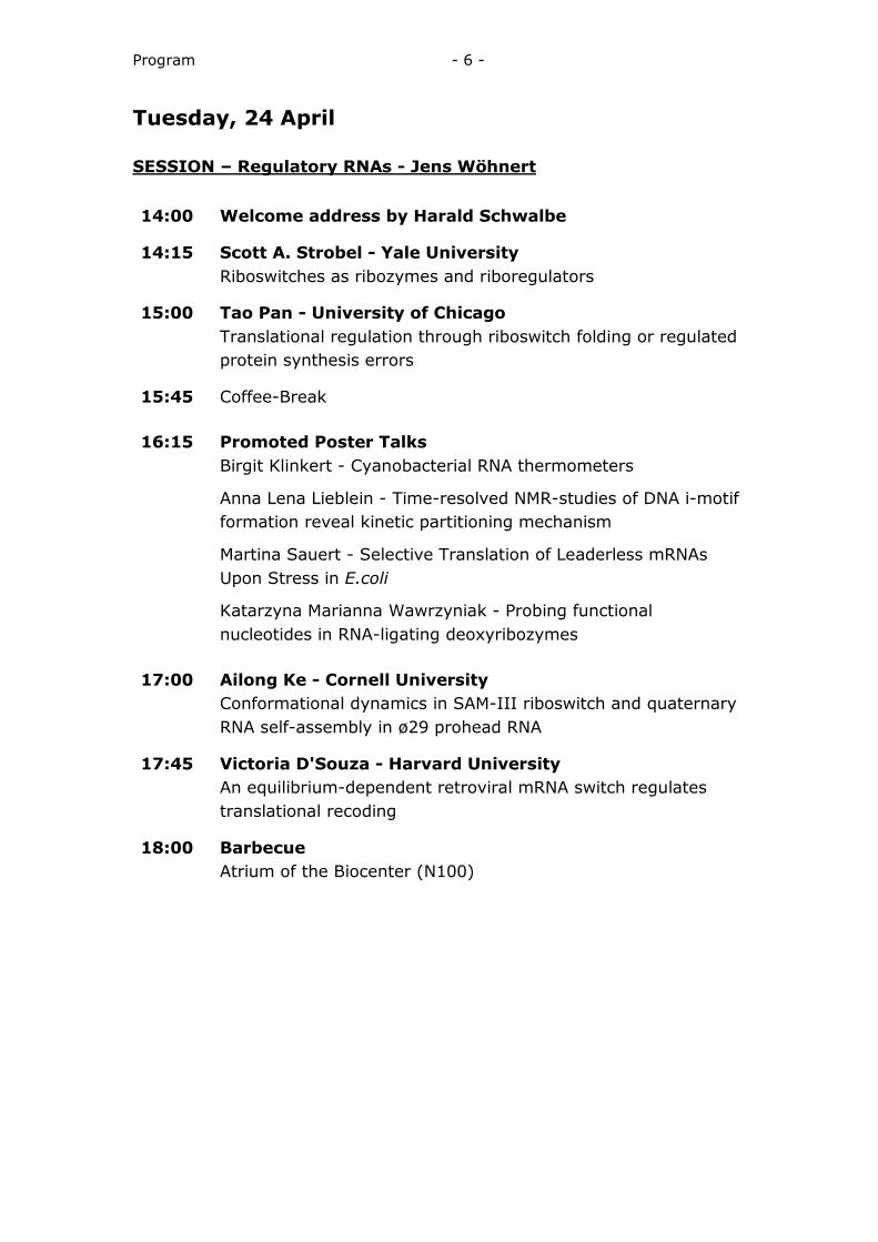
Program - 6 -
Tuesday, 24 April SESSION – Regulatory RNAs - Jens Wöhnert 14:00 Welcome address by Harald Schwalbe
14:15 Scott A. Strobel - Yale University Riboswitches as ribozymes and riboregulators
15:00 Tao Pan - University of Chicago Translational regulation through riboswitch folding or regulated
protein synthesis errors
15:45 Coffee-Break 16:15 Promoted Poster Talks Birgit Klinkert - Cyanobacterial RNA thermometers
Anna Lena Lieblein - Time-resolved NMR-studies of DNA i-motif formation reveal kinetic partitioning mechanism
Martina Sauert - Selective Translation of Leaderless mRNAs Upon Stress in E.coli
Katarzyna Marianna Wawrzyniak - Probing functional nucleotides in RNA-ligating deoxyribozymes
17:00 Ailong Ke - Cornell University Conformational dynamics in SAM-III riboswitch and quaternary
RNA self-assembly in ø29 prohead RNA
17:45 Victoria D'Souza - Harvard University An equilibrium-dependent retroviral mRNA switch regulates
translational recoding
18:00 Barbecue Atrium of the Biocenter (N100)
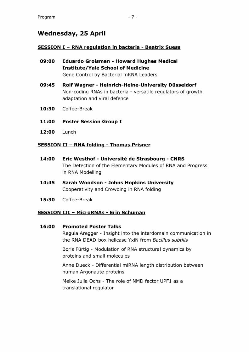
Program - 7 -
Wednesday, 25 April SESSION I – RNA regulation in bacteria - Beatrix Suess 09:00 Eduardo Groisman - Howard Hughes Medical
Institute/Yale School of Medicine Gene Control by Bacterial mRNA Leaders
09:45 Rolf Wagner - Heinrich-Heine-University Düsseldorf Non-coding RNAs in bacteria - versatile regulators of growth
adaptation and viral defence
10:30 Coffee-Break 11:00 Poster Session Group I
12:00 Lunch SESSION II – RNA folding - Thomas Prisner 14:00 Eric Westhof - Université de Strasbourg - CNRS The Detection of the Elementary Modules of RNA and Progress
in RNA Modelling
14:45 Sarah Woodson - Johns Hopkins University Cooperativity and Crowding in RNA folding
15:30 Coffee-Break SESSION III – MicroRNAs - Erin Schuman 16:00 Promoted Poster Talks Regula Aregger - Insight into the interdomain communication in
the RNA DEAD-box helicase YxiN from Bacillus subtilis
Boris Fürtig - Modulation of RNA structural dynamics by proteins and small molecules
Anne Dueck - Differential miRNA length distribution between human Argonaute proteins
Meike Julia Ochs - The role of NMD factor UPF1 as a translational regulator

Program - 8 -
SESSION III – MicroRNAs - Erin Schuman (continued) 16:45 Gunter Meister – University of Regensburg - No title
17:30 Elisa Izaurralde - Max Planck Institute for Developmental Biology
Interaction of GW182 proteins with PABPC1 and deadenylase complexes is required for miRNA-mediated gene silencing
18:30 Speakers’ Dinner (invited speakers only) at the Faculty Lounge of the Frankfurt Institute for Advanced
Studies (FIAS)
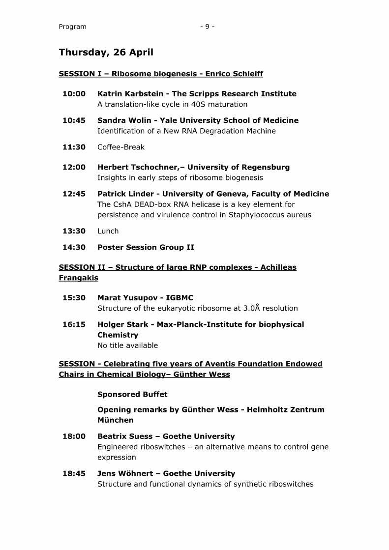
Program - 9 -
Thursday, 26 April SESSION I – Ribosome biogenesis - Enrico Schleiff 10:00 Katrin Karbstein - The Scripps Research Institute A translation-like cycle in 40S maturation
10:45 Sandra Wolin - Yale University School of Medicine Identification of a New RNA Degradation Machine
11:30 Coffee-Break 12:00 Herbert Tschochner,– University of Regensburg Insights in early steps of ribosome biogenesis
12:45 Patrick Linder - University of Geneva, Faculty of Medicine The CshA DEAD-box RNA helicase is a key element for
persistence and virulence control in Staphylococcus aureus
13:30 Lunch
14:30 Poster Session Group II SESSION II – Structure of large RNP complexes - Achilleas Frangakis 15:30 Marat Yusupov - IGBMC Structure of the eukaryotic ribosome at 3.0Å resolution
16:15 Holger Stark - Max-Planck-Institute for biophysical Chemistry
No title available SESSION - Celebrating five years of Aventis Foundation Endowed Chairs in Chemical Biology– Günther Wess Sponsored Buffet
Opening remarks by Günther Wess - Helmholtz Zentrum München
18:00 Beatrix Suess – Goethe University Engineered riboswitches – an alternative means to control gene
expression
18:45 Jens Wöhnert – Goethe University Structure and functional dynamics of synthetic riboswitches
Program - 10 -
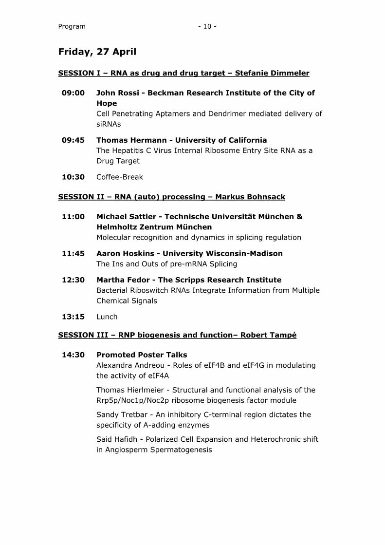
Friday, 27 April SESSION I – RNA as drug and drug target – Stefanie Dimmeler 09:00 John Rossi - Beckman Research Institute of the City of
Hope Cell Penetrating Aptamers and Dendrimer mediated delivery of
siRNAs
09:45 Thomas Hermann - University of California The Hepatitis C Virus Internal Ribosome Entry Site RNA as a
Drug Target
10:30 Coffee-Break SESSION II – RNA (auto) processing – Markus Bohnsack 11:00 Michael Sattler - Technische Universität München &
Helmholtz Zentrum München Molecular recognition and dynamics in splicing regulation
11:45 Aaron Hoskins - University Wisconsin-Madison The Ins and Outs of pre-mRNA Splicing
12:30 Martha Fedor - The Scripps Research Institute Bacterial Riboswitch RNAs Integrate Information from Multiple
Chemical Signals
13:15 Lunch SESSION III – RNP biogenesis and function– Robert Tampé 14:30 Promoted Poster Talks Alexandra Andreou - Roles of eIF4B and eIF4G in modulating
the activity of eIF4A
Thomas Hierlmeier - Structural and functional analysis of the Rrp5p/Noc1p/Noc2p ribosome biogenesis factor module
Sandy Tretbar - An inhibitory C-terminal region dictates the specificity of A-adding enzymes
Said Hafidh - Polarized Cell Expansion and Heterochronic shift in Angiosperm Spermatogenesis
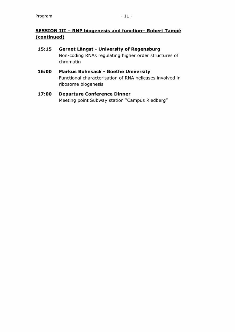
Program - 11 -
SESSION III – RNP biogenesis and function– Robert Tampé (continued) 15:15 Gernot Längst - University of Regensburg Non-coding RNAs regulating higher order structures of
chromatin
16:00 Markus Bohnsack - Goethe University Functional characterisation of RNA helicases involved in
ribosome biogenesis
17:00 Departure Conference Dinner Meeting point Subway station “Campus Riedberg”
Program - 12 -
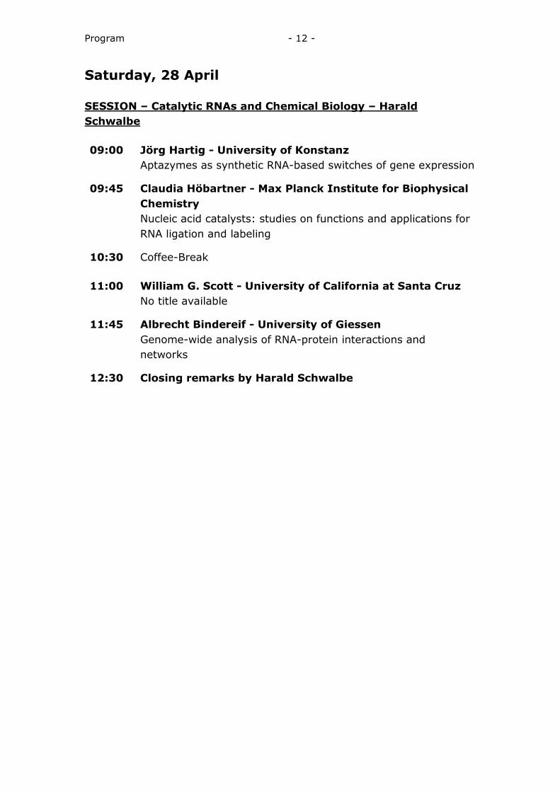
Saturday, 28 April SESSION – Catalytic RNAs and Chemical Biology – Harald Schwalbe 09:00 Jörg Hartig - University of Konstanz Aptazymes as synthetic RNA-based switches of gene expression
09:45 Claudia Höbartner - Max Planck Institute for Biophysical Chemistry
Nucleic acid catalysts: studies on functions and applications for RNA ligation and labeling
10:30 Coffee-Break 11:00 William G. Scott - University of California at Santa Cruz No title available
11:45 Albrecht Bindereif - University of Giessen Genome-wide analysis of RNA-protein interactions and
networks
12:30 Closing remarks by Harald Schwalbe

Lecture Abstracts
Lecture Abstracts - 14 -
Strobel, Scott A. (Tuesday, 14:15)
Riboswitches as ribozymes and riboregulators Affiliation: Yale University. E-mail: [email protected]
RNA elements capable of binding small molecules are now recognized as a common regulatory element. The small molecule ligands are quite variable as are the mechanisms by which the RNAs provide regulation. This presentation will focus on two systems: the glmS ribozyme/riboswitch and the two classes of c-di-GMP riboswitches. The glmS riboswitch regulates gene expression through a self-cleavage activity. The reaction is catalyzed with the assistance of the metabolite cofactor glucosamine-6-phosphate (GlcN6P), whose amino group is proposed to serve as the general acid during the reaction. GlcN6P, like other pyranose sugars, undergoes spontaneous and rapid interconversion between the α and β-anomers at the C1 position. The riboswitch selectively binds the α-anomer of GlcN6P in a pH dependent manner. The data support a model where the reaction pKa corresponds to that of the GlcN6P amine and the amine’s pKa is reduced about 1.5 pH units. This observation has broader relevance for considering how the microenvironment of an RNA, despite its anionic character, can reduce the pKas of functional groups for use in catalysis. The two c-di-GMP riboswitches are macromolecular targets in the c-di-GMP second messenger signaling pathway. They regulate many genes related to c-di-GMP metabolism as well as genes involved in bacterial motility, virulence and biofilm formation. We have determined structures of both riboswitch classes and developed compounds that selectively target one riboswitch class over the other. We have also identified cyclic-dinucleotide analogs that are highly resistant to hydrolysis by the metabolizing enzyme but can still bind known c-di-GMP target molecules, including RNA and proteins. These stable c-di-GMP analogs can inhibit biofilm formation. These data suggest that nuclease resistant second messenger analogs could be useful chemical tools for dissecting mechanisms of biofilm growth as well as a powerful strategy for modulating bacterial biofilm formation.
Lecture Abstracts - 15 -
Pan, Tao (Tuesday, 15:00)
Translational regulation through riboswitch folding or regulated protein synthesis errors Authors: Pan, Tao; Perdrizet, George A; Artsimovitch, Irina; Sosnick, Tobin R; Weisberg, Chloe; Jones, Thomas E. Affiliation: University of Chicago. E-mail: [email protected]
Translational regulation is of fundamental importance in gene expression and protein function. I will discuss two different processes that cells employ in translational regulation: riboswitch folding during transcription which controls translational initiation, and mis-translation as a potential, new mechanism of stress response. Riboswitches are cis acting elements that regulate gene expression by affecting transcriptional termination or translational initiation in response to binding of a metabolite. RNA polymerase pausing is a fundamental property of transcription that can influence RNA folding. We show that pausing plays an important role in the folding and conformational rearrangement of the E. coli btuB riboswitch during transcription by the E. coli RNA polymerase. This riboswitch consists of a ~200 nucleotide, coenzyme B12 binding aptamer domain and a ~40 nucleotide expression platform that controls translational initiation. Transcriptional pauses at strategic locations block the formation of alternate structures and play a chaperoning role that couples folding of both domains. Pausing at strategic locations may be a general mechanism for coordinated folding and conformational rearrangements of riboswitch structures that underlie their response to environmental cues. A central pillar of biology is the high fidelity of replication, transcription and translation. We recently discovered that ~1% of methionylated tRNAs in human cells are non-cognate tRNAs, and the mis-methionylation levels increase up to 10-fold when cells are subjected to oxidative stress such as elevated levels of reactive oxygen species (ROS, Nature 462, 522-6). These regulated changes in protein synthesis errors involving only methionine (Met) substitution at non-Met positions in proteins at a frequency of approximately 1 in 500-5000 amino acids. We demonstrated that methionyl-tRNA synthetase (MRS) misacylates specific non-cognate tRNAs, and MRS is the only tRNA synthetase yet known that exhibits a high and regulated level of misacylation in cells. Oxidative stress is a wide spread biological process and has been implicated in many processes in human health and disease. As Met residues are known to protect proteins against ROS-mediated damage, we propose that regulated mis-translation represent a new stress response mechanism which allows for better adaptation of the cell to oxidative stress. We are investigating how mis-translation is regulated in cells and its functional consequences.
Lecture Abstracts - 16 -
Ke, Ailong (Tuesday, 17:00)
Conformational dynamics in SAM-III riboswitch and quaternary RNA self-assembly in ø29 prohead RNA Authors: Ke, Ailong. Affiliation: Cornell University. E-mail: [email protected]
SAM-III (SMK box) translational riboswitches regulates S-adenosyl-L- methionine (SAM) synthetase metK genes in members of the Lactobacillales. The ability of these riboswitches to repeatedly switch back and forth between two alternative conformations suggests that the ligand-induced folding dynamics is governed by thermodynamics principles. We previously reported the crystal structure of the SAM-bound E. fae SAM-III riboswitch. Using SHAPE chemical probing analysis, we show that SAM switches off the SAM-III riboswitch through a conformation selection scheme. We reveal that while the majority of the apo SAM-III RNA molecules exist in an alternatively base paired (ON) conformation, a subset of them pre-organize into a SAM-bound-like (READY) conformation, which upon SAM exposure is selectively stabilized into the SAM-bound (OFF) conformation through an induced-fit mechanism. Mutagenesis showed that the ON state is only slightly more stable than the READY state, as point mutations in a hypervariable region outside the SAM-binding core can alter the folding landscape to favor the READY state. The conformation selection model likely applies to other translational riboswitches. Prohead RNA: A common theme in nature is for folded proteins to further self-assemble into larger quaternary structures. Given its prevalence in the protein world, it is perplexing to notice the apparent absence of a similar mechanism in the RNA world. In fact, the only naturally occurring example of a multimeric RNA self-assembly system is found in prohead RNA (pRNA) of the Bacillus subtilis bacteriophage ø29 packaging motor. Here we report the quaternary pRNA crystal structure at 3.5 Å resolution. pRNA protomers assemble in a head-to-tail fashion through intermolecular base pairing interactions into a tetrameric ring in the crystal lattice. Tertiary interactions are weak within each pRNA protomer, but strong between adjacent protomers, and inherent flexibilities are built-in to allow different oligomerization states in solution and on the packaging motor. EM docking, chemical probing, and mutagenesis showed that pentameric RNA ring formation on the phage packaging motor is directed by specific interactions with the surrounding prohead capsid. pRNA therefore plays a chaperoning role by instructing the ring-ATPase formation and anchoring it directly onto the phage prohead.
Lecture Abstracts - 17 -
D'Souza, Victoria (Tuesday, 17:45)
An equilibrium-dependent retroviral mRNA switch regulates translational recoding Authors: Houck-Loomis, Brian; Durney, Michael A.; Salguero, Carolina; Shankar, Neelaabh; Nagle, Julia M.; Goff, Stephen P.; D'Souza, Victoria M.. Affiliation: Harvard University. E-mail: [email protected]
Most retroviruses require translational recoding of a viral messenger RNA stop codon to maintain a precise ratio of structural (Gag) and enzymatic (Pol) proteins during virus assembly1,2. Pol is expressed exclusively as a Gag–Pol fusion either by ribosomal frameshifting or by read-through of the gag stop codon3. Both of these mechanisms occur infrequently and only affect 5–10% of translating ribosomes, allowing the virus to maintain the critical Gag to Gag–Pol ratio4–8. Although it is understood that the frequency of the recoding event is regulated bycis RNAmotifs, nomechanistic explanation is currently available for how the critical protein ratio is maintained. Here we present the NMR structure of the murine leukaemia virus recoding signal and show that a protonation-dependent switch occurs to induce the active conformation. The equilibrium is such that at physiological pH the active, read-through permissive conformation is populated at approximately 6%: a level that correlates within vivo protein quantities. The RNA functions by a highly sensitive, chemomechanical coupling tuned to ensure an optimal read-through frequency. Similar observations for a frameshifting signal indicate that this novel equilibrium-based mechanism may have a general role in translational recoding.
Lecture Abstracts - 18 -
Groisman, Eduardo A. (Wednesday, 09:00)
Gene Control by Bacterial mRNA Leaders Authors: Groisman, Eduardo A. Affiliation: Howard Hughes Medical Institute/Yale School of Medicine. E-mail: [email protected]
The leader regions of many bacterial mRNAs can adopt alternative stem-loop structures that dictate whether the associated coding region(s) are transcribed and/or translated. Which stem-loop structure forms is determined by ions or metabolites the levels of which are modified by the protein(s) specified in the coding region(s) subjected to regulation. Metabolites can be sensed directly by mRNA leaders designated riboswitches or by the effect the metabolites have on the coupling of transcription of the mRNA leader with translation of a short open reading frame(s) in the mRNA leader. I will discuss a novel general mechanism by which riboswitches control transcription of the associated coding regions. I will also describe the identification of complex mRNA leaders that utilize distinct mechanisms to sense different signals.
Lecture Abstracts - 19 -
Wagner, Rolf (Wednesday, 09:45)
Non-coding RNAs in bacteria - versatile regulators of growth adaptation and viral defence Authors: Wagner, Rolf. Affiliation: Heinrich-Heine-University Düsseldorf. E-mail: [email protected]
The abundant existence of small non-coding RNAs (ncRNAs) in bacteria has recently attracted increasing interest. It was shown that many ncRNAs are involved in the regulation of gene expression thereby modulating cell growth and metabolism. ncRNAs act by a variety of different mechanisms, including direct base pairing, protein binding or structural mimicry with other nucleic acids. Our insight into the function of these molecules has increased considerably in the past and for some ncRNAs mechanistic details of their functions have been characterized. In this talk I will focus on two types of bacterial ncRNAs, namely 6S RNA and crRNAs, which are involved in quite different cellular functions. In the first part I will summarize studies performed to understand the molecular mechanism of 6S RNA-dependent transcriptional regulation. 6S RNA is a small regulatory RNA with widespread distribution in bacteria. It acts as transcriptional regulator, which accumulates during stationary phase and inhibits transcription from many promoters due to stable association with σ70-containing RNA polymerase. Surprisingly. this small RNA acts as template for RNA polymerase giving rise to de novo transcripts, termed pRNAs, which terminate transcriptional inhibition and thus contribute to growth adaptation. In part two of the talk a short introduction to the recently discovered defence system against phages and foreign genetic elements will be given. The system is known as CRISPR (Clustered Regularly Interspaced Short Palindromic Repeats) and represents an RNA-based immune system, which is found in many bacteria and almost all archaea. In E. coli the CRISPR system is silenced by the DNA-binding protein H-NS and can be activated by the transcriptional regulator LeuO. Studies performed to understand the complex regulation of the CRISPR defence in E.coli will be presented.
Lecture Abstracts - 20 -
Westhof, Eric (Wednesday, 14:00)
The Detection of the Elementary Modules of RNA and Progress in RNA Modelling Authors: Westhof, Eric. Affiliation: Université de Strasbourg - CNRS. E-mail: [email protected]
RNA architecture can be viewed as the hierarchical assembly of preformed double-stranded helices defined by Watson-Crick base pairs and RNA modules maintained by non-Watson-Crick base pairs. RNA modules are recurrent ensemble of ordered non-Watson-Crick base pairs. Such RNA modules constitute a signal for detecting non-coding RNAs with specific biological functions. It is, therefore, important to be able to recognize such genomic elements within genomes. Through systematic comparisons be-tween homologous sequences and x-ray structures, followed by automatic clustering, the whole range of sequence diversity in recurrent RNA modules has been character-ized. These data permitted the construction of a computational pipeline for identifying known 3D structural modules in single and multiple RNA sequences in the absence of any other information. Any module can in principle be searched, but four can be searched automatically: the G-bulged loop, the Kink-turn, the C-loop and the tandem GA loop. The present pipeline can be used for RNA 2D structure refinement, 3D model assembly, and for searching and annotating structured RNAs in genomic data. Following the recent dramatic advances in tools aimed at RNA 3D modelling, a first, collective, blind experiment in RNA three-dimensional structure prediction has been performed. The goals are to assess the leading edge of RNA structure prediction tech-niques, compare existing methods and tools, and evaluate their relative strengths, weak-nesses, and limitations in terms of sequence length and structural complexity. The re-sults should give potential users insight into the suitability of available methods for dif-ferent applications and facilitate efforts in the RNA structure prediction community in their efforts to improve their tools.

Lecture Abstracts - 21 -
Woodson, Sarah (Wednesday, 14:45)
Cooperativity and Crowding in RNA folding Authors: Woodson, Sarah A; Kilburn, Duncan; Behrouzi, Reza; Roh, Joon Ho; Briber, R. M.. Affiliation: Johns Hopkins University. E-mail: [email protected]
Many non-coding RNAs fold into specific three-dimensional structures in order to act as catalysts or regulatory elements. In the cell, the stability of the folded RNA depends on its sequence motifs and its interactions with metal ions and with many other solutes. Using small angle X-ray scattering to measure the folding of a bacterial group I ribozyme, we first show that molecular crowding equivalent to what is present in real cells stabilizes RNA tertiary structures by the excluded volume effect. Crowder molecules favor more compact structures, and compression of the native state ensemble correlates with higher catalytic activity. Consequently, the ribozyme reaches its catalytically active structure at much lower Mg2+ concentrations in a crowded milieu than in a dilute solution. Second, to understand how non-coding RNAs fold uniquely despite a small number of tertiary interaction motifs, we mutated the major tertiary interactions and measured perturbations to folding free energies. Double and triple mutant cycles show that core and peripheral structural motifs are cooperatively linked in near-native folding intermediates, and this cooperativity depends on the native orientation of the core helices. We also find that the emergence of a cooperative interaction network at an early stage of folding suppresses non-native structures and guides the search for the native state.

Lecture Abstracts - 22 -
Meister, Gunter (Wednesday, 16:45)
No title available Authors: . Affiliation: Universität Regensburg. E-mail: [email protected]
No abstract available
Lecture Abstracts - 23 -
Izaurralde, Elisa (Wednesday, 17:30)
Interaction of GW182 proteins with PABPC1 and deadenylase complexes is required for miRNA-mediated gene silencing Authors: Huntzinger, Eric; Braun, Joerg; Kuzuoğlu-Öztürk, Duygu; Izaurralde, Elisa. Affiliation: Max Planck Institute for Developmental Biology. E-mail: [email protected]
miRNA-mediated gene silencing in animal cells requires interaction of Argonaute proteins with GW182 family proteins, which in turn confer binding to PABPC1 as well as to the PAN2-PAN3 and CCR4-NOT deadenylase complexes. Human GW182 proteins interact directly with PABPC1, PAN3 and NOT1 through their C-terminal silencing domains (SDs). Although the GW182 protein interaction network is conserved in Drosophila melanogaster (Dm), the Dm SD is not sufficient for deadenylase complex binding and requires additional N-terminal sequences. Functional studies show that GW182 mutants that bind deadenylation factors but not PABPC1, fail to rescue silencing in cells depleted of endogenous GW182, indicating that PABPC1 binding is required for silencing. However, PABPC1 binding alone is not sufficient for silencing. We have defined the minimal requirements for a functional GW182 protein and show that binding to both PABPC1 and deadenylase complexes is necessary for miRNA-mediated gene silencing in vivo.
Lecture Abstracts - 24 -
Karbstein, Katrin (Thursday, 10:00)
A translation-like cycle in 40S maturation Authors: Karbstein, Katrin; Strunk, Bethany; Novak, Megan; Young, Crystal. Affiliation: The Scripps Research Institute. E-mail: [email protected]
Assembly factors (AFs) prevent premature translation initiation on small (40S) ribosomal subunit assembly intermediates by blocking ligand binding. However, it is unclear how AFs are displaced from maturing 40S ribosomes, if or how maturing subunits are assessed for fidelity, and what prevents premature translation initiation once AFs dissociate. Here we show that maturation involves a translation-like cycle whereby the translation factor eIF5B promotes joining of large (60S) subunits with pre-40S subunits to give 80S-like complexes, which are subsequently disassembled by the termination factor Rli1. The AFs Tsr1 and Rio2 block the mRNA channel and initiator tRNA binding sites and thus 80S-like complexes lack mRNA or initiator tRNA. After Tsr1 and Rio2 dissociate from 80S-like complexes Rli1-directed displacement of 60S subunits allows for translation initiation. This cycle thus provides a functional test of 60S subunit binding and the GTPase site before ribosomes enter the translating pool, and establishes a new paradigm for understanding ribosome maturation.
Lecture Abstracts - 25 -
Wolin, Sandra (Thursday, 10:45)
Identification of a New RNA Degradation Machine Authors: Wolin, Sandra; Chen, Xinguo; Taylor, David; Wang, Hong-Wei. Affiliation: Yale University School of Medicine. E-mail: [email protected]
Although the functions of many RNA-protein complexes (RNPs) are well understood, the roles of others are still being elucidated. One class whose function remains under investigation is the Ro class of RNPs. The major protein component, the Ro 60 kDa protein, is present in many animal cells and also ~5% of bacterial genomes. In all characterized species, Ro binds ~100 nt noncoding RNAs called Y RNAs. In the only bacterium where Ro has been studied, the radiation-resistant Deinococcus radiodurans, the ortholog Rsr functions with 3’ to 5’ exoribonucleases to modulate RNA metabolism during some types of environmental stress. Rsr and two 3’ to 5’ exoribonucleases, RNase II and RNase PH, are required for efficient 23S rRNA maturation during heat stress. In stationary phase, Rsr functions with the 3’ to 5’ exoribonuclease polynucleotide phosphorylase (PNPase) to degrade rRNAs. Rsr and PNPase are also found together in immunoprecipitates, with the interaction increasing in stationary phase. To understand how Rsr influences the function of an exoribonuclease, we purified the Rsr/PNPase complex from D. radiodurans. This revealed that Rsr and the Y RNA adapt PNPase for degrading structured RNA. Characterization of this RNA degradation machine will be presented.
Lecture Abstracts - 26 -
Tschochner, Herbert (Thursday, 12:00)
Insights in early steps of ribosome biogenesis Authors: Tschochner, Herbert; Jakob, Steffen; Ohmayer, Uli; Hierlmeier, Thomas; Perez-Fernandez, Jorge; Neueder, Andreas; Reiter, Alarich; Hamperl, Stephan; Seitz, Hannah; Merkl, Philipp; Gerber, Jochen; Németh, Attila; Deutzmann, Rainer; Gadal, Olivier; Léger, Isabelle; Griesenbeck, Joachim; Milkereit, Philipp. Affiliation: Universität Regensburg. E-mail: [email protected]
Ribosome production starts with the synthesis of the ribosomal precursor RNAs by RNA polymerase I (Pol I) and III (Pol III) and the co-transcriptional assembly of many ribosomal proteins and numerous transiently interacting ribosome biogenesis factors. Data will be presented focusing on two aspects of early events in ribosome biogenesis in the yeast S. cerevisiae: First, assembly of ribosomal proteins of the small ribosomal subunit (SSU) and, second, termination of pre-rRNA synthesis. Early assembly We have investigated the interdependency of ribosomal protein assembly events and the transient interactions of ribosome biogenesis factors with early pre-ribosomal intermediates. We observed that components of the small subunit (SSU) processome UTP-A and UTP-B sub-modules were recruited to early pre-ribosomes independently of all tested r-proteins. On the other hand, groups of SSU processome components were identified whose association with early pre-ribosomes was affected by specific r-protein assembly events in the head-platform interface of the SSU. One of these components, Noc4p, appeared to be itself required for robust incorporation of r-proteins into the SSU head domain. Our results indicate that some SSU processome components are transient primary pre-rRNA binders in vivo whereas other factors of this group play a role in the assembly of major SSU domains. Transcription termination Several DNA cis-elements and trans-acting factors were described to be involved in transcription termination and to release the elongating RNA polymerases from the template. Different models for the molecular mechanism of transcription termination have been suggested for eukaryotic RNA polymerase I from results of in vitro and in vivo experiments. To analyse the molecular requirements for yeast RNA polymerase I termination an in vivo approach was used in which efficient termination resulted in growth inhibition. This led to the identification of a Myb-like protein, Ydr026c, as bona fide termination factor (recently designated Nsi1 (for “NTS1 silencing protein 1”)). Possible Nsi1 functions in regard to the mechanism of transcription termination are discussed.
Lecture Abstracts - 27 -
Linder, Patrick (Thursday, 12:45)
The CshA DEAD-box RNA helicase is a key element for persistence and virulence control in Staphylococcus aureus Authors: Oun, Stella; Redder, Peter; Didier, Jean-Philippe; Giraud, Caroline; Corvaglia, Anna; François, Patrice; Buttazzoni, Elena; Schrenzel, Jacques; Linder, Patrick. Affiliation: University of Geneva, Faculty of Medicine. E-mail: [email protected]
Staphylococcus aureus is an opportunistic pathogen that can cause life-threatening diseases. Of particular concern in S. aureus infections is the formation of biofilms on catheters and prosthetic devices since the bacteria become almost impossible to eradicate by the immune system and antibiotic treatment. This persistence state of growth relies on the expression of surface encoded proteins that allow attachment to various surfaces and contrasts to the dispersal mode of growth that relies on the secretion proteins such as toxins and proteases. The switch is mainly mediated by the Staphylococcal agr quorum sensing system. DEAD-box RNA helicases are present in almost all living organisms from prokaryotes to eukaryotes. These proteins are ATP-dependent RNA binding proteins that most often function in multi-subunit complexes. ATP binding and hydrolysis induce conformational changes that allow the dynamic formation and rearrangements of RNA-protein complexes and induce local strand separation. In bacteria, DEAD-box proteins are involved in ribosome biogenesis, translation initiation, and RNA turnover allowing rapid adaptations of gene expression to environmental changes and stress conditions. We show that an inactivation of the cshA RNA helicase gene from S. aureus, causes an increase of agr mRNA levels as a result of an increased stability of this mRNA. As a consequence, biofilm formation is decreased and hemolysis increased. Inactivation of the agrA gene in the cshA mutant restores the original biofilm and hemolysis phenotypes, clearly indicating that control of the abundance and stability of the agr mRNA through CshA activity is primordial for virulence factor expression control.
Lecture Abstracts - 28 -
Yusupov, Marat (Thursday, 15:30)
Structure of the eukaryotic ribosome at 3.0Å resolution Authors: Yusupov, Marat. Affiliation: IGBMC. E-mail: [email protected]
We present here the complete structure of the full 80S ribosome from S. cerevisiae at a resolution of 3Å. The model includes nearly all the rRNA sequences as well as all ribosomal proteins, with the single exception of protein L1. The eukaryotic 80S and bacterial 70S ribosome shares 34 common proteins and eukaryotic ribosome has additional 45 unique proteins and bacterial ribosome has 22 additional unique proteins. The majority of eukaryotic specific elements are located on the periphery of the conserved core thus broadening the surface of interactions between the two subunits through additional eukaryotic bridges. The molecular interactions creating these bridges together with their eukaryotic-specific components can now be described in details. Our crystals capture the ribosome in two different conformations which are believed to reflect intermediate states in course of mRNA and tRNA translocation. The structural comparison of these states, which differ by the degree of rotation of the small subunit and the swiveling of its head with respect to the large subunit, provides a detailed description of conformational rearrangements as well as coordinated movements of intersubunit bridges.

Lecture Abstracts - 29 -
Stark, Holger (Thursday, 16:15)
No title available Authors: . Affiliation: Max-Planck-Institute for biophysical Chemistry. E-mail: [email protected]
No abstract available
Lecture Abstracts - 30 -
Suess, Beatrix (Thursday, 18:00)
Engineered riboswitches – an alternative means to control gene expression Authors: Suess, Beatrix. Affiliation: Goethe University. E-mail: [email protected]
Numerous synthetic RNA-based control devices, so called engineered riboswitches, have been developed in the last years. We have engineered riboswitches by insertion of in vitro selected, small molecule binding aptamers into untranslated regions of mRNAs, exploiting the fact that upon ligand binding the RNA structure interferes either with translation initiation or pre-mRNA splicing in yeast. An advantage of these regulators is that they can be designed in principle to any non-toxic, cell-permeable ligand of choice. In addition, the direct RNA-ligand interaction renders auxiliary protein factors unnecessary. While many RNA aptamers have been identified that bind to a plethora of small molecules, only very few are capable of acting as riboswitch. Using a screening approach we identified a set of aptamers which confer neomycin-dependent regulation, however, to a various degree. In a combination of genetic, biochemical and structural studies we addressed the molecular basis for these differences. We demonstrated that a destabilized and open ground state accompanied by extensive structural changes upon ligand binding is necessary for regulation while inactive aptamers are already prestructured without the ligand. We identified a switching element responsible for the destabilization of the ligand free form without compromising the bound form. We will exploit this knowledge for the further development of RNA-based devises for the conditional control of gene expression in various organisms including humans. We aim to control all steps of gene expression including alternative splicing, mRNA stability and miRNA processing.
Lecture Abstracts - 31 -
Wöhnert, Jens (Thursday, 18:45)
Structure and functional dynamics of synthetic riboswitches Authors: Wöhnert, Jens. Affiliation: Johann-Wolfgang-Goethe-Universität. E-mail: [email protected]
We investigated the structure, the ligand specificity and the functional dynamics of two synthetic riboswitches which are triggered by binding of neomycin and related aminoglycosides and tetracycline, respectively, using solution NMR-spectroscopy. The neomycin sensing riboswitch revealed a ligand binding mechanism based on conformational capture. Furthermore, we could show that ligand specificity is not based on structural differences between different aminoglycoside-riboswitch complexes but on differential dynamics. In contrast, the tetracycline riboswitch recognizes its ligand based on a largely preformed tertiary structure pre-stabilized by Mg2+-ions. The different ligand binding mechanisms and their consequences for the function of the two riboswitches are reminiscent of the mechanisms found for natural riboswitches and have implications for the design of new synthetic riboswitches.
Lecture Abstracts - 32 -
Rossi, John J. (Friday, 09:00)
Cell Penetrating Aptamers and Dendrimer mediated delivery of siRNAs Authors: Rossi, John J.. Affiliation: Beckman Research Institute of the City of Hope. E-mail: [email protected]
We have been interested in evolving aptamers as therapeutic agents and for cell specific delivery of small interfering RNAs (siRNAs). Four examples of aptamers selected for specific use will be described. The first is an aptamer selected against the HIV envelope protein, which both neutralizes HIV and delivers siRNAs into HIV infected cells. The second example is an aptamer targeting the B-cell specific receptor BAFF R1, which is also dual function in that inhibits Baff ligand signaling and also delivers siRNAs into BAFF R1 expressing cells. Both of these aptamers were isolated using a purified ligand to select the aptamers. Cell based SELEX is conceptually different than ligand based SELEX. In this process the library is toggled between cells that do not express the ligand of interest and then cells that do express the ligand. We have used cell based SELEX to select aptamers that bind to the SIGN R receptor on macrophages and dendritic cells as well as aptamers that are selectively internalized into pancreatic cancer cells. The cell based SELEX selected aptamers are all internalized, and can also be used to deliver cargoes such as siRNAs and mRNAs. An alternative siRNA delivery approach is the use of PAMAM dendrimers. We have applied a cocktail of siRNAs in a PAMAM dendrimer for the treatment of HIV infection in a humanized mouse model. Several logs of viral inhibition were achieved with a single injection of 10 micrograms of siRNAs. This talk will highlight the dual function aspects of aptamers and the delivery efficacy of the dendrimer.
Lecture Abstracts - 33 -
Hermann, Thomas (Friday, 09:45)
The Hepatitis C Virus Internal Ribosome Entry Site RNA as a Drug Target Authors: Hermann, Thomas. Affiliation: University of California, San Diego. E-mail: [email protected]
The internal ribosome entry site (IRES) in the genomic RNA of hepatitis C virus (HCV) is highly conserved across viral genotypes and therefore provides an attractive target for antiviral drugs. We have investigated the molecular mechanism of action of translation inhibitors targeting the HCV IRES and suppressing the subgenomic viral replicon. It is demonstrated that selective conformational induction at a structured domain of the IRES is responsible for the inhibition of viral translation in cells infected with HCV. A high-resolution crystal structure for the IRES RNA target in complex with a viral translation inhibitor has been determined, which provides a roadmap for structure-guided discovery of novel HCV IRES inhibitors.
Lecture Abstracts - 34 -
Sattler, Michael (Friday, 11:00)
Molecular recognition and dynamics in splicing regulation Authors: Sattler, Michael. Affiliation: Technische Universität München & Helmholtz Zentrum München. E-mail: [email protected]
Molecular recognition of RNA plays a crucial for many essential RNA processing events during gene regulation. Two studies will be presented that highlight the role of conformational dynamics during molecular recognition: i) the recognition of poly-pyrimidine tract RNA by the essential splicing factor U2AF65 during spliceosome assembly and ii) the recognition of dimethyl-arginines by Tudor domains in snRNP maturation.
Lecture Abstracts - 35 -
Hoskins, Aaron Andrew (Friday, 11:45)
The Ins and Outs of pre-mRNA Splicing Authors: Hoskins, Aaron; Crawford, Daniel; Shcherbakova, Inna; Friedman, Larry; Gelles, Jeff; Moore, Melissa. Affiliation: University Wisconsin-Madison. E-mail: [email protected]
In eukaryotes, pre-mRNA transcripts must be processed by a number of cellular machines to generate mRNAs. Removal of introns and exon ligation are carried out by a multi-MegaDalton complex of 5 snRNAs and ~ 100 proteins called the spliceosome. Amazingly, the spliceosome is a single turnover enzyme and must be assembled anew on each intron prior to splicing. This dynamic and compositional complexity has made biochemical analysis of the spliceosome quite challenging. Using recently developed single molecule fluorescence techniques, we can now follow spliceosome assembly and catalysis in realtime in a whole cell extract. Key to these experiments is the labeling of endogenous spliceosome components in yeast with fluorophores using a combination of genetic engineering and chemical biology. We have previously shown that this approach can reveal new insight into spliceosome assembly (Hoskins, et al., Science, 2011). We have now extended these results in several directions including a single molecule analysis of the conservation of the spliceosome assembly pathway among many different pre-mRNAs, the use of single molecule FRET to define pre-mRNA conformation during specific stages of splicing, and directly monitoring the action of trans-acting factors functioning during proofreading or spliceosome recycling. Together, these results illustrate the utility and versatility of single molecule methods in studying complex enzymes in cell extracts to provide a broad range of new biochemical insights.
Lecture Abstracts - 36 -
Fedor, Martha J. (Friday, 12:30)
Bacterial Riboswitch RNAs Integrate Information from Multiple Chemical Signals Authors: Fedor, Martha J.; Watson, Peter Y.. Affiliation: The Scripps Research Institute. E-mail: [email protected]
Riboswitches regulate expression of many bacterial genes in response to intracellular metabolite levels. Riboswitches have been the subject of extensive biochemical and structural investigations in vitro, which have revealed the molecular details of small ligand recognition by these highly structured regulatory RNAs. However, the complexity of the intracellular environment can be difficult to recapitulate in vitro, making investigation of intracellular riboswitch activity important for full understanding of their function. The glmS riboswitch self-cleaves upon binding glucosamine-6-phosphate, the metabolic product of glucosamine-6-phosphate synthase, the GlmS gene product. Therefore, self-cleavage reports directly and quantitatively on folding of the ligand-bound riboswitch. By expressing the glmS riboswitch in a yeast model system, rather than its normal gram-positive bacterial host, we measured the ligand concentration dependence of riboswitch self-cleavage for the first time and learned that this riboswitch responds to inhibitory as well as activating metabolites in vivo. Reporter gene experiments in B. subtilis confirmed that the glmS riboswitch is subject to inhibition as well as activation in its natural biological context. Previous work focused on riboswitch recognition of single cognate ligand. Our work extends these studies by demonstrating that the glmS riboswitch can sense the concentrations of multiple intracellular metabolites that share functional groups with the cognate ligand and have the capacity to form a subset of the full complement of ligand-RNA interactions. Based our in vivo studies, we propose a new model in which a single riboswitch can integrate information from multiple chemical signals to regulate gene expression in response to overall metabolic state of the cell. Conventional riboswitches function by adopting different conformations in the presence and absence of bound ligands. Our ongoing studies test the hypothesis that conventional riboswitches also exhibit the ability to sense a variety of chemical signals.
Lecture Abstracts - 37 -
Längst, Gernot (Friday, 15:15)
Non-coding RNAs regulating higher order structures of chromatin Authors: Längst, Gernot. Affiliation: University of Regensburg. E-mail: [email protected]
Packaging of DNA into nucleosomes and the formation of higher order chromatin structures determine DNA accessibility and the activity of genome domains. We identified a novel RNA-dependent mechanism that establishes and maintains the open chromatin structure within euchromatic regions in Drosophila cells. The mechanism of reversible chromatin opening was reconstituted in vitro and the molecular components were identified and characterized. We show that this effect depends on the Drosophila decondensation factor 31 (Df31) that specifically binds to RNA and was shown to localize to euchromatic regions. Df31 is capable to tether a heterogeneous pool of short, single-stranded RNA molecules to chromatin. This novel class of chromatin-associated RNA (caRNA) is stably linked to chromatin and is largely composed of snoRNAs. Biophysical studies show that Df31 does preferentially bind to the snoRNAs and links them to chromatin. We suggest that the Df31 mediated linkage of snoRNAs and chromatin forms a RNA-chromatin network resulting in the establishment of open chromatin domains. Analysis of caRNAs in human cells reveals as well a strong enrichment of snoRNA molecules, implying a conserved and important role for snoRNAs in the modulation of higher order structures of chromatin and genome accessibility.
Lecture Abstracts - 38 -
Bohnsack, Markus (Friday, 16:00)
Functional characterisation of RNA helicases involved in ribosome biogenesis Authors: Bohnsack, Markus. Affiliation: Goethe University. E-mail: [email protected]
The synthesis of cytoplasmic ribosomes in Eukaryotes is best understood in the yeast Saccharomyces cerevisiae. Here, the pathway requires more than 180 non-ribosomal proteins, such as GTPases, nucleases and RNA helicases. In addition, 75 small nucleolar RNAs (snoRNAs) base-pair with pre-ribosomal RNA and are involved pre-rRNA processing and modification, and many are predicted to require helicase activities for their release. Since the knowledge on the 19 RNA helicases involved in ribosome biogenesis is still limited and molecular and regulatory functions of these enzymes seem to be diverse, the elucidation of their roles has remained a challenging task. We are studying RNA helicase function in S. cerevisiae. Using the UV cross-linking and analysis of cDNA (CRAC) approach, Solexa deep sequencing and bioinformatics, we have identified RNA binding sites of helicases involved in ribosome biogenesis. We have recently focused on the DEAH-box RNA helicase Prp43 and the DEAD-box protein Rok1. Prp43 has at least 5 major binding sites on pre-rRNA where it fulfills diverse functions, such as release of several snoRNAs from pre-60S and facilitation of 20S pre-rRNA cleavage at site D in pre-40S. For Rok1, we identified putative binding sites on the 18S rRNA where Rok1 is required for release of the snoRNA snR30 from pre-ribosomes.
Lecture Abstracts - 39 -
Hartig, Jörg S. (Saturday, 09:00)
Aptazymes as synthetic RNA-based switches of gene expression Authors: Hartig, Jörg S.. Affiliation: University of Konstanz. E-mail: [email protected]
RNAs play pivotal roles in regulating gene expression. An increasing number of ligand-dependent, RNA-based mechanisms are discovered. Besides the wide-spread occurrence of riboswitches in nature, artificial systems for controlling gene expression in biotechnology and synthetic biology applications have been constructed successfully in recent years. We have engineered synthetic riboswitches based on aptamer-ribozyme conjugates (aptazymes) that rely on controlling RNA self-cleavage activity as expression platform. Aptazymes enable control over a range of RNA functions such as the translation of mRNAs, maturation of tRNAs, as well as the integrity of the 16S rRNA. The synthetic RNA switches are highly modular with respect to the applied RNA class as well as the ligands used for triggering changes of gene expression. Apart from small molecules, ribozyme-based switches can be redesigned in order to react to RNA or temperature as triggers of changed gene expression. They can be applied in many organisms ranging from bacteria to mammalian cells. In addition, combinations of individual switches allow for constructing Boolean logic operations in bacteria. An overview of some novel applications and an outlook towards utilizing these RNA regulators for higher information processing and in therapeutic strategies will be provided.
Lecture Abstracts - 40 -
Höbartner, Claudia (Saturday, 09:45)
Nucleic acid catalysts: studies on functions and applications for RNA ligation and labeling Authors: Höbartner, Claudia. Affiliation: Max Planck Institute for Biophysical Chemistry. E-mail: [email protected]
Biochemical and biophysical investigations of RNA folding pathways, RNA-ligand, and RNA-protein interactions often require site-specifically modified RNA, which is usually obtained by chemical synthesis in combination with enzymatic ligation strategies. Alternative approaches involve postsynthetic labeling of RNA. Synthetic DNA is a promising chemical tool for posttranscriptional manipulation of RNA. In the form of deoxyribozymes (also known as DNA enzymes), DNA combines in one molecule the ability of Watson-Crick base-pairing for specific substrate binding and the ability to catalyse chemical transformations. Such active DNA sequences are identified by in vitro selection from random sequence DNA pools, and have proven useful for various practical applications. For example, deoxyribozymes catalyze the ligation of functionalized RNA substrates in linear and branched topologies, thereby providing access to RNA structures that are difficult to synthesize by other methods. Expanding the substrate scope and engineering of DNA enzymes for broader applications in RNA research requires a more detailed understanding of their molecular functions. The current knowledge about the catalytic mechanism and the molecular principles of DNA catalysis is still very limited. Structural probing of the active site and the identification of essential functional groups of core nucleotides can provide deeper insight into deoxyribozyme function. With our recently developed combinatorial DNA probing methods, we have identified the most important nucleotides and their functional groups involved in formation of catalytically competent active sites of RNA-ligating deoxyribozymes.[1, 2] Functional group requirements and tolerance for modifications at the RNA nucleotides involved in the ligation reaction were also analyzed. The results of these studies will be presented, and the potential of the investigated deoxyribozymes as tools for site-specific labeling of RNA will be discussed. [1] F. Wachowius, F. Javadi-Zarnaghi, C. Höbartner, Combinatorial mutation interference analysis reveals functional nucleotides required for DNA catalysis. Angew. Chem. Int. Ed. 2010, 49, 8504. [2] F. Wachowius, C. Höbartner, Probing essential nucleobase functional groups in aptamers and deoxyribozymes by nucleotide analogue interference mapping of DNA. J. Am. Chem. Soc. 2011, 133, 14888.

Lecture Abstracts - 41 -
Scott, William G. (Saturday, 11:00)
No title available Authors: . Affiliation: University of California at Santa Cruz. E-mail: [email protected]
No abstract available
Lecture Abstracts - 42 -
Bindereif, Albrecht (Saturday, 11:45)
Genome-wide analysis of RNA-protein interactions and networks Authors: Bindereif, Albrecht. Affiliation: University of Giessen. E-mail: [email protected]
To analyze global activities of RNA-binding proteins, we perform genomewide individual-nucleotide resolution crosslinking-immunoprecipitation in combination with deep-sequencing (iCLIP-Seq). We have recently applied this approach in two projects, focussing, first, on the U1C protein in the trypanosome system, and, second, on the human heterogeneous nuclear ribonucleoprotein L (hnRNP L). The U1C protein is important in the first step of spliceosome assembly, the precise recognition of the 5’ splice site by the U1 snRNP. Using iCLIP-Seq in trypanosome cells, we demonstrate that U1C participates in both cis- and trans-splicing. HnRNP L, a multifunctional RNA-binding protein, specifically recognizes CA-repeat and CA-rich RNA elements and participates in diverse functions of mRNA metabolism. HnRNP L can regulate alternative splicing either as an activator or a repressor. Based on iCLIP-Seq and genomic mapping, we derived a comprehensive genomewide map of hnRNP L RNA interaction sites in HeLa cells. Analysis of the crosslink sites revealed that hnRNP L RNA-binding followed distinct patterns for exons activated or repressed by hnRNP L, suggesting a positional code and a predictive RNA map.

Poster Abstracts

Poster Abstracts 44
Andreou, Alexandra (Promoted Poster Talk) (Talk on Friday 14:30, Poster No. 1, Poster-Session Group I)
Roles of eIF4B and eIF4G in modulating the activity of eIF4A Authors: Andreou, Alexandra; Klostermeier, Dagmar. Affiliation: University of Münster. E-mail: [email protected]
Eukaryotic translation initiation factor 4A (eIF4A) is a DEAD box protein that is involved in unwinding mRNA secondary structure during translation initiation. In order to carry out its function, eIF4A depends on interactions with other translation initiation factors eIF4G, eIF4B and eIF4H, which enhance its otherwise minimal ATPase and helicase activity. The mechanism by which this stimulation is accomplished is still to a large extent unknown. Extensive studies have already focused on the interaction between S. cerevisiae eIF4A and the eIF4A binding domain of eIF4G (eIF4G572-853). A proposal has been put forward that eIF4G572-853 stabilizes a “half-open” conformation of eIF4A, which allows faster phosphate and RNA release. Here we demonstrate that eIF4G572-950, a construct carrying the C-terminal R/S rich tail of S. cerevisiae eIF4G, induces the same conformation of eIF4A as eIF4G572-853 does, as evidenced from a change in FRET efficiency of eIF4A labeled with donor and acceptor dyes on the two RecA domains. The construct containing the C-terminal tail is interestingly a more potent activator of eIF4A´s ATPase activity. Furthermore, an additional stimulatory effect on the ATPase activity that is observed upon combined presence of eIF4G572-853 and Tif3, the yeast homologue of eIF4B, is greatly reduced for eIF4G containing the tail. In addition, in smFRET experiments with labeled eIF4A we observed that only upon presence of both eIF4G572-950 and Tif3 in combination with the substrates RNA and ATP is it possible to observe a shift in FRET efficiency distribution to higher values, which could be indicative of the closed active eIF4A conformer. These results highlight the importance of the 100 C-terminal amino acids of eIF4G in regulating eIF4A ATPase activity. Moreover, they suggest that the concurrent presence of eIF4B and eIF4G572-950 might play a vital role in promoting and stabilizing the active closed conformation of eIF4A, therefore rendering the protein more efficient for the mRNA scanning process. References Marintchev, A., Edmonds, K.A., Marintcheva, B., Hendrickson, E., Oberer, M., Suzuki, C., Herdy, B., Sonenberg, N., and Wagner, G. (2009). Topology and regulation of the human eIF4A/4G/4H helicase complex in translation initiation. Cell 136, 447-460. Hilbert, M., Kebbel, F., Gubaev, A. and Klostermeier, D., (2011) eIF4G stimulates the activity of the DEAD box protein eIF4A by a conformational guidance mechanism. NAR, 39, 2260-2270.
Poster Abstracts 45
Aregger, Regula (Promoted Poster Talk) (Talk on Wednesday 16:00, Poster No. 2, Poster-Session Group II)
Insight into the interdomain communication in the RNA DEAD-box helicase YxiN from Bacillus subtilis Authors: Aregger, Regula; Klostermeier, Dagmar. Affiliation: University of Münster. E-mail: [email protected]
DEAD box RNA helicases are enzymes that bind and unwind RNA. It is assumed that this class of enzymes is involved in the structural conversion and correct folding of complex RNAs such as ribosomal RNA. YxiN is a DEAD box RNA helicase from Bacillus subtilis that recognises hairpin 92 of the 23S rRNA. The protein consists of a core formed by two domains connected by a linker of 9 amino acids and a C-terminal domain conferring substrate specificity. The sequence motifs for ATP binding, ATP hydrolysis and RNA binding are located in the two RecA domains of the helicase core and communication between these two domains is crucial for enzymatic function. To investigate the role of the linker between the two RecA domains of the DEAD box helicase core, we modified its length and sequence. RNA stimulated ATPase activity of YxiN was characterized by a coupled enzymatic steady-state ATPase assay and unwinding assays were performed using a double stranded RNA minimal substrate. Nucleotide binding affinities were determined by displacement titrations of nucleotide with a preformed complex of enzyme with a fluorescently modified nucleotide. RNA binding affinities were determined by anisotropy titrations of a fluorescently labeled minimal substrate. Conformational changes were investigated in confocal smFRET experiments. We could show that extension of the linker to up to 15 amino acids led to a drastic reduction of ATPase activity. This effect was even more pronounced in mutants with shortened linkers. Reducing the linker length to 4 amino acids resulted in a completely ATPase-deficient enzyme. However nucleotide binding studies of the different mutants showed that ATP affinity is increased in all the mutants when compared to the wildtype enzyme. The same trends are observed for unwinding activities of these mutants with the longest and shortest constructs being unwinding deficient. Changing the length of the linker also has an impact on RNA binding in the presence of nucleotides. smFRET experiments indicated that mutants with the shortest and the longest linkers did not adopt a closed conformation in the prescence of RNA substrate and ATP. These results indicate that the linker between the core domains of YxiN plays a key role in regulation of the enzyme activity. Furthermore, it is evident that the length of the linker for efficient interdomain communication and thus enzymatic activity between the two core domains lies between 5 and 13 amino acids.
Poster Abstracts 46
Brecht, Michael (Poster No. 3, Poster-Session Group I)
The inhibition of RNA editing by RNA-interacting compounds Authors: Brecht, Michael; Leeder, Matthias; Kratz, Katja; Göringer, H. Ulrich. Affiliation: Technische Universität Darmstadt. E-mail: [email protected]
Molecular Genetics, Darmstadt University of Technology, Schnittspahnstraße 10, 64287 Darmstadt, Germany; The Rockefeller University, 1230 York Avenue, New York 10065, USA RNA editing in African trypanosomes is a unique posttranscriptional modification process. During the reaction, uridylate residues are inserted and/or deleted into otherwise non-translatable mitochondrial transcripts. The reaction takes place on the surface of a megadalton enzyme complex (0.8MDa), known as the editosome. Editosomes have a hydrodynamic size of 20 Svedberg units and provide a reaction platform for the individual steps of the reaction cycle. Key components in the process are a unique class of small, non-coding RNAs, termed guide (g) RNAs. Guide RNAs represent trans-acting genetic elements that interact with pre-edited mRNAs and promote the nucleotide insertion and deletion by virtue of their primary sequence. Although the principal function of gRNAs in conjunction with their cognate pre-mRNAs is understood, most structural, dynamic and regulatory aspects of the process are unknown today. This is in part due to the lack of inhibitory molecules that could stall the reaction at defined reaction steps. Aminoglycosides have been shown to bind RNA molecules in a promiscuous fashion, thereby inhibiting ribonucleoprotein (RNP) complex-driven reactions. Here we show, that 4,5-linked 2-deoxystreptamins (2-DOS) (neamin, neomycin) as well as 4,6-linked 2-DOS (tobramycin) are capable of inhibiting RNA editing in vitro. The different compounds affect both, U-insertion as well as U-deletion RNA editing and inhibit the processing reaction at the so-called “mRNA religation” step. Half maximal inhibitory concentrations (IC50) for the different compounds vary between 5 and 30μM. The inhibition is due to RNA binding, which is characterized by a macroscopic equilibrium dissociation constant (Kd) of ≤1μM. Aminoglycoside binding to editing substrate RNAs stabilizes the RNA molecules (Tm increase between 7.5°C and 10°C) equivalent of a ΔΔG25°C of -35kJ/mol. Furthermore, drug binding induces the release of bound cations from the RNA molecules, indicating a contribution of ion bonds and as a consequence a charge neutralization and perhaps structural perturbation in and around the editing site.
Poster Abstracts 47
Cevec, Mirko (Poster No. 4, Poster-Session Group II)
NMR research of helix H1 from the human and chimpanzee HAR1 RNAs Authors: Cevec, Mirko; Koschinat, Melanie; Richter, Christian; Schwalbe, Harald. Affiliation: Goethe University. E-mail: [email protected]
Human accelerated region 1 (HAR1) is a section in human genome which display a significantly accelerated nucleotide substitution rate. HAR1 region in HAR1F gene encodes for a non-coding 118-nt RNA and has 18 substitutions instead of the expected 0.27 substitutions since our last common ancestor with chimpanzees. It was discovered that HAR1 RNA is involved in brain development, in development of human consciousness and Huntington’s disease. Two different cloverleaf-like secondary structures have been predicted for the human HAR1 RNA. We used NMR spectroscopy to determine secondary and tertiary solution structures of one of the helices, helix H1. Two 37-nt RNA fragments were prepared for the human and chimpanzee helix H1 RNA sequences. Resonance assignment was performed using 1D 1H, 2D 15N-HSQC, 2D HNN-COSY and 2D NOESY spectra. The NOE imino proton resonance patterns of the 37-nt RNAs were comparable to the patterns of the full-length human and chimpanzee HAR1 RNAs. We studied the dynamic properties of the 37-nt RNA constructs with the help of 13C relaxation NMR measurements and estimated the fast internal motions in the 37-nt RNA constructs by the measurement of longitudinal and transverse relaxation rates, along with heteronuclear NOEs. We assigned all aromatic and sugar proton resonances. Structure calculations show that the human NMR structure (h37 RNA) forms a more rigid and defined structure as the chimpanzee NMR structure (c37 RNA). Both structures feature a GAA internal loop, which was never studied before."
Poster Abstracts 48
Dueck, Anne (Promoted Poster Talk) (Talk on Wednesday 16:20, Poster No. 5, Poster-Session Group I)
Differential miRNA length distribution between human Argonaute proteins Authors: Dueck, Anne; Petri, Sebastian; Berezikov, Eugene; Meister, Gunter. Affiliation: University of Regensburg. E-mail: [email protected]
microRNAs (miRNAs) are small RNAs with a length of about 22 nucleotides (nt). Together with one of four human Argonaute (Ago) proteins, they form an RNA induced silencing complex (RISC) and act as regulators of gene expression. MiRNAs are processed from hairpin-structured precursors by RNase III enzymes Drosha and Dicer. By imprecise Dicer cleavage or by other factors involved, mature miRNAs do not always exhibit a sharp length within one type of a miRNA but differ by as much as four to five nt. So far, the function and relevance of these so-called IsomiRs remains largely elusive. We performed anti-Ago immunoprecipitations (IPs) from HeLa S3 cells with antibodies specific to each of the four human Ago proteins. After cloning and deep sequencing of the co-immunoprecipitated miRNAs, miRNA length distributions between the four different Ago proteins were compared. Surprisingly, for some miRNAs, Ago2 seems to bind a broader length spectrum of miRNAs than Ago1 and Ago3, the majority of miRNAs differing in their 3’ end. This was also confirmed by Northern blotting experiments. Based on our sequencing data, we hypothesized that Ago2-bound miRNAs might be subject to specific 3’ trimming. To find out what factor could be involved in the trimming process, we performed an siRNA screen against exonucleases that harbor a 3’-5’ exonuclease domain and belong to the DnaQ like 3’-5’ exonuclease domain superfamily. Our screening results indicate that several exonucleases may play important roles in miRNA metabolism. While the level for some miRNAs is elevated upon knockdown of certain exonucleases, we could also find a group of exonucleases where the miRNA signal is lost upon depletion of the respective protein. The mechanistic function of these enzymes and their possible interaction with proteins of the miRNA biogenesis pathway are currently analyzed in molecular detail.
Poster Abstracts 49
Fürtig, Boris (Promoted Poster Talk) (Talk on Wednesday 16:10, Poster No. 6, Poster-Session Group II)
Modulation of RNA structural dynamics by proteins and small molecules Authors: Fürtig, Boris; Doetsch, Martina; Wenter, Philipp; Pitsch, Stefan; Schwalbe, Harald; Schroeder, Renee. Affiliation: Goethe University. E-mail: [email protected]
RNA molecules traverse rugged energy landscapes when folding into functional structures. Thereby they become easily trapped in misfolded conformations. Small molecules and proteins with RNA chaperone activity modulate RNA’s free-energy landscapes in order to accelerate RNA folding. By NMR spectroscopy and complementary biochemical techniques the influence of these molecular co-factors on RNA folding reactions is examined. From interaction studies with small molecular co-factors the folding trajectory for RNA folding reaction could be deduced and the structural properties of an transition state of the RNA could be described. In further studies it was possible to show that the C-terminal domain of the RNA chaperone StpA promotes RNA folding by ‘transferring’ conformational energy to the RNA molecule. Formation of a transient complex renders the protein structurally less flexible and freezes out micro- to millisecond timescale motions within the protein core. Stabilisation of hydrophobic interactions between aromatic residues is the source of energy transferred to the RNA molecule. Thereby, the latter gains conformational entropy leading to a lower energy barrier for refolding. "
Poster Abstracts 50
Hafidh, Said (Promoted Poster Talk) (Talk on Friday 15:00, Poster No. 7, Poster-Session Group I)
Polarized Cell Expansion and Heterochronic shift in Angiosperm Spermatogenesis Authors: Hafidh, Said; Breznenova, Katarina; Fecikova, Jana; Capkova, Vera; Honys, David. Affiliation: UEB. E-mail: [email protected]
Single cell expansion of the growing pollen tube and the coordinated control of germ cell division and sperm cell fate specification, are landmark features of the developing male gametophyte. We applied the Agilent 44K tobacco gene chip to conduct the first transcriptomic analysis of the tobacco male gametophyte. We have performed a comparative study with the Arabidopsis root-hair trichoblast transcriptome to evaluate genetic factors and common pathways in polarized cell-tip expansion. Extension of our analysis beyond the second haploid mitosis allowed construction of a genetic model likely to have instigate the heterochronic shift in Angiosperm spermatogenesis.
Poster Abstracts 51
Hamperl, Stephan (Poster No. 8, Poster-Session Group II)
Compositional and structural analysis of specific chromatin domains isolated from yeast chromosomes Authors: Hamperl, Stephan; Brown, Chris; Huber, Katharina; Wittner, Manuel; Stöckl, Ulrike; Boeger, Hinrich; Milkereit, Philipp; Tschochner, Herbert; Griesenbeck, Joachim. Affiliation: University of Regensburg. E-mail: [email protected]
Our research focuses on the biochemical characterization of ribosomal DNA (rDNA) chromatin of Saccharomyces cerevisiae. The locus encodes for all the structural RNA components of mature ribosomes (rRNAs). RNA polymerase I synthesizes the 35S precursor for three of the four structural rRNAs, whereas RNA polymerase III is responsible for the transcription of the fourth 5S rRNA species. The rDNA locus is remarkable in two regards: First, it is present in multiple copies within the genome (about 150 in haploid yeast), arranged in characteristic head-to-tail tandem repeats. Second, even if yeast cells are exponentially growing, only about half of the 35S genes are transcribed, whereas the other half is transcriptional silent. Importantly, these different states of transcriptional activity are characterized by specific chromatin structures. Although transcription of the rRNA gene is the object of intensive research, the molecular nature of the different rDNA chromatin states remains elusive. In order to characterize chromatin at rRNA genes in different transcriptional states in its composition, structure and function, we developed an approach to isolate these domains from the yeast chromosome. The quantities obtained by this approach enable compositional analysis of the chromatin domains by semiquantitative comparative mass spectrometry and allowed us to identify many known and new candidate rDNA chromatin components. Furthermore, we determined the location of individual nucleosomes on single molecules of the isolated chromatin domains using electron microscopy. First statistical analyses of the nucleosome occupancy at the 5S rRNA gene indicate a heterogeneous population of chromatin states likely correlating with different phases of transcriptional activity, and give valuable information about yeast rDNA chromatin architecture."
Poster Abstracts 52
Heinrich, Eva-Marie (Poster No. 9, Poster-Session Group I)
Identification of proteins that bind to the microRNA-17~92a cluster Authors: Heinrich, Eva-Marie; Wagner, Jasmin; Krüger, Marcus; Bonauer, Angelika; Dimmeler, Stefanie. Affiliation: Goethe University Frankfurt. E-mail: [email protected]
MicroRNAs (miRs) are endogenously expressed small non-coding RNAs that regulate gene expression by the degradation or translational repression of target mRNAs. The polycistronic miR-17~92 cluster consists of seven miRs (miR-17-3p and -5p, miR-19a/b, miR-18, miR-20, miR-92a) and is highly expressed in endothelial cells. Although the cluster members are under the control of the same promoter, the individual members are differentially regulated after induction of hypoxia and during endothelial differentiation of embryonic stem cells. These findings suggest a tightly regulated processing and accordingly the presence of factors that regulate the stability or the accessibility of the individual miRs. In order to identify miR binding proteins that might potentially be involved in the posttranscriptional regulation of these miRs, we established an in vitro RNA pull down assay. By mass spectrometry, we identified proteins that were bound to the precursor of miR-92a (pre-miR-92a). Several of the identified proteins are known to bind to RNA and/or are involved in RNA metabolism. Among these proteins are RNA Helicases and Splicing regulators like eIF4A-2 and SND1. We confirmed the interaction of these two proteins with pre-miR-92a by RNA immunoprecipitation and characterized their effect on the expression of the miR-17~92 cluster in endothelial cells. SiRNA-mediated silencing of eIF4A-2 leads to a slight increase of miR-17~92 precursors but has no effect on the mature cluster members under normoxic conditions. However, miR-18 and miR-19 expression was reduced by 20±9 and 22±9%, respectively, in eIF4A-2 siRNA treated cells compared to scrambled controls under conditions of mimicking hypoxia by treatment with DFO (100µM, 24 hours). In contrast, knock down of SND1 under normoxic conditions slightly increased the expression of some mature miRs of the cluster such as miR-18 and this effect was further enhanced under hypoxic conditions (miR-18: 174±26% increase, p<0.05). In summary, we identified two pre-miR-92a binding proteins. Whereas silencing of eIF4A-2 appears to slightly reduce the processing of the miR-17~92 cluster under hypoxia mimicking conditions, silencing of SND1 increased the expression of some cluster members.
Poster Abstracts 53
Hierlmeier, Thomas (Promoted Poster Talk) (Talk on Friday 14:40, Poster No. 10, Poster-Session Group II)
Structural and functional analysis of the Rrp5p/Noc1p/Noc2p ribosome biogenesis factor module Authors: Hierlmeier, Thomas; Ohmayer, Uli; Merl, Juliane; Hergert, Kristin; Linneman, Jan; Griesenbeck, Joachim; Baßler, Jochen; Tschochner, Herbert; Milkereit, Philipp. Affiliation: Universität Regensburg. E-mail: [email protected]
Ribosomes catalyse the translation of mRNA into proteins. The eukaryotic 80S ribosome, consisting of a small 40S and a large 60S subunit, is a ribonucleoprotein complex containing 4 ribosomal RNAs (rRNAs) and about 80 ribosomal proteins (r-proteins). During eukaryotic ribosome biogenesis, the mature rRNAs are generated from precursor transcripts by numerous modification and exo- and endonucleolytic cleavage events, and the r-proteins are concomitantly assembled onto the evolving rRNAs. Additionally, this process requires the action of more than 70 non-coding RNAs and more than 150 proteinaceous ribosome biogenesis factors, which transiently interact with preribosomal particles. Previous studies showed that several biogenesis factors form complexes, which can be isolated from cell extracts under high salt conditions and which exist in cells independent of ongoing pre-rRNA synthesis. This suggests that biogenesis factor modules might constitute building blocks of preribosomes acting as functional units in ribosome biogenesis. Here, we used a variety of in vivo, ex vivo, and in vitro approaches to analyse a ribosome biogenesis factor module consisting of Rrp5p, Noc1p and Noc2p in the model organism S. cerevisiae. The experiments provide insights into (i) the functional architecture and structure of the module and (ii) the interaction of the module with preribosomal particles. "
Poster Abstracts 54
Katari, Venkata Subbaraju (Poster No. 11, Poster-Session Group I)
Editosomes utilize one RNA binding site for both, U-insertion and U-deletion RNA editing Authors: Katari, Venkata Subbaraju; Böhm, Cordula; Brecht, Michael; Göringer, H. Ulrich. Affiliation: Technische Universität Darmstadt. E-mail: [email protected]
Molecular Genetics, Darmstadt University of Technology, Schnittspahnstraße 10, 64287 Darmstadt, Germany; Department of Pathology, University of Cambridge, Tennis Court Road, Cambridge CB2 1QP, UK RNA editing of mitochondrial pre-mRNAs in African trypanosomes generates full-length transcripts by the site-specific insertion and deletion of uridine nucleotides. The process requires small non-coding RNAs, termed guide (g) RNAs. Guide RNAs interact with pre-edited mRNAs via antiparallel base pairing and mediate the nucleotide insertion and deletion in a “template-type” reaction. A 0.8MDa multienzyme complex, termed the 20S editosome, catalyzes the individual reaction steps. Although the binding of substrate pre-edited mRNAs and cognate gRNAs represents the first step in the reaction cycle, the biophysical and biochemical details of the editosome/RNA interaction are not understood. Here we show that editosomes bind full-length mitochondrial pre-mRNAs (344-1647nt) with similar nanomolar affinities and rate constants in a nonselective fashion. Binding is complete within 3-4min and is characterized by macroscopic equilibrium dissociation constants (Kd) in the range of 3nM-12nM, demonstrating high affinity binding. The complexes do not discriminate, neither thermodynamically nor kinetically, between different mitochondrial pre-mRNAs or between edited and unedited versions of the same transcript. Gold-labeling of editosome-bound RNA in combination with transmission electron microscopy (TEM) identified a single RNA binding site per editosome. All three classes of interacting RNAs: pre-edited mRNA, gRNA, and pre-mRNA/gRNA hybrid molecules bind to the same RNA binding domain, which suggests a single catalytic center of the editosome. Atomic force microscopy (AFM) of individual RNA/editosome complexes revealed that multiple 20S editosomes can bind to one pre-mRNA molecule. Furthermore, the AFM data identified a so far unknown activity of the 20S editosome: RNA binding is followed by RNA unwinding. The activity acts in a time and concentration-dependent fashion without a requirement for ATP and thus indicates a RNA chaperone-type enzymatic activity.
Poster Abstracts 55
Kiosze, Kristin (Poster No. 12, Poster-Session Group II)
Moleculare functions of the most conserved ABC-protein within ribosome recycling Authors: Kiosze, Kristin; Barthelme, Dominik; Ceh, Katharina; Dastvan, Reza; Prisner, Thomas; Ferner, Jan; Schwalbe, Harald; Tampé, Robert. Affiliation: Goethe-University Frankfurt. E-mail: [email protected]
The twin ATPase ABCE1 is one of the most conserved proteins within evolution, ubiquitous in eukaryota and archaea. ABCE1 is an essential, multi-domain enzyme which belongs to the ATP-binding cassette subfamily E of ATPases. It consists of two head-to-tail oriented nucleotide-binding domains (NBDs) and it harbors a unique N-terminal region coordinating two FeS clusters. ABCE1 was originally found as the RNase L inhibitor in the innate immune system of higher vertebrates and subsequently identified as an essential factor for HIV-1 RNA/gag capsid assembly. Further it interacts with a number of translation factors and co-localizes with ribosomal subunits, pointing to a much more fundamental role during translation. We have recently revealed the essential function of ABCE1 in ribosome recycling in archaea, using Sulfolobus solfataricus as a model organism. The archaeal system resembles a minimalistic version of the eukaryotic process and serves therefore as an ideal model for functional and structural analysis. Based on this system we analyzed the structural organization of the FeS clusters by EPR spectroscopy and determined the x-ray structure of ABCE1 at 2.0 Å resolution. We further demonstrated that ABCE1 possesses an energy dependent ribosome dissociation activity, enabled via a conformational switch of both NBDs upon ATP binding and hydrolysis. The activity is synergistically enhanced by the archaeal release factor 1, which forms a stable binary complex with ABCE1."
Poster Abstracts 56
Klinkert, Birgit (Promoted Poster Talk) (Talk on Tuesday 16:15, Poster No. 13, Poster-Session Group I)
Cyanobacterial RNA thermometers Authors: Klinkert, Birgit; Cimdins, Annika; Kortmann, Jens; Narberhaus, Franz. Affiliation: Ruhr University Bochum. E-mail: [email protected]
RNA thermometers are direct temperature sensors that fine-tune bacterial heat shock and virulence gene expression by temperature-dependent sequestration of the ribosome binding site. The expression of the small heat shock gene hsp17 from Synechocystis sp. PCC 6803 is controlled by an exceptionally short RNA thermometer, which is important for photosynthetic activity under stress conditions. It controls translation of hsp17 on demand by inhibiting ribosome access at normal temperatures, permitting translation inhibition upon stress, and shutting down HSP17 production in the recovery phase (1). Potential RNA thermometers were found upstream of the heat shock gene hspA in thermophilic Thermosynechococcus species. Interestingly, their 5’ untranslated regions (5’-UTR) fold into extended hairpin structures, which might be responsible for functionality at high temperatures. The cyanobacteria Anabaena variabilis and Anbaena sp. PCC 7120 each contain two small heat shock genes, both of which are controlled by RNA thermometers. One structure is as long as the Thermosyechococcus hspA 5’-UTR and the other is even shorter than the Synechocystis hsp17 5’-UTR. In an E. coli reporter gene system (2) the Synechocystis and Anabaena thermometers melt at mesophilic temperature. The Thermosynechococcus thermometer show only slight increase in reporter gene activity up to 42°C. In future studies, a thermophilic reporter gene system will be established, using Thermus thermophilus as model organism. Additionally, the structural basis for the function of various cyanobacterial RNA thermometers is being currently studied by mutational analyses in vivo and structure probing experiments in vitro. (1) Kortmann, J., Sczodrok, S., Rinnenthal, J., Schwalbe, H., Narberhaus, F., 2011. Translation on demand by a simple RNA-based thermosensor. Nucleic Acids Res. 39, 2855-2868. (2) Klinkert, B., Cimdins, A., Gaubig, L.C., Rossmanith, J., Aschke-Sonnenborn, U., Narberhaus, F., 2012. Thermogenetic tools to monitor temperature-dependent gene expression in bacteria. J Biotechnol."
Poster Abstracts 57
Koschinat, Melanie (Poster No. 14, Poster-Session Group II)
Structural Studies of HAR1 RNA by NMR Spectroscopy Authors: Koschinat, Melanie; Cevec, Mirko; Richter, Christian; Schwalbe, Harald. Affiliation: Goethe University, AK Schwalbe. E-mail: [email protected]
The human accelerated regions (HARs) are a group of 49 segments in the human genome, which are ranked by their underlying fast mutation rate compared to homologous chimpanzee segments.[1] The 118-nt HAR1 region has with 18 substitutions the highest mutation rate in the homo sapiens genome. HAR1F is coexpressed with Reelin in Cajal-Retzius cells which is a regulator in human cortical development. HAR1F RNA is involved in Huntington’s disease.[2] A clear structure-function relation has not been identified yet. Secondary structure models of human and chimpanzee sequence of HAR1F are developed by DMS treatment[1], chemical and enzymatical probing[3], which differ to each other. Here, we investigated a secondary structure elucidation of human and chimpanzee HAR1F RNA using CD and NMR spectroscopy. With this sizable 118-nt RNA, NMR spectroscopic assignment is challenging due to considerable spectral overlap. To overcome incomplete sequential connectivities we pursued a “divide-and-conquer” strategy by utilizing model hairpins that mimic structural elements of the full length RNA. We investigate the structures of model hairpins c37, c47, c54, h37, h47, h32, h39 and h15 by NMR spectroscopy. We derived from our NMR data that model hairpins c37, c47, c54 h37 and h39 RNA are folded like the corresponding structural elements in the full length secondary structures by Beniaminov et al. and are able to disprove the chimpanzee structure model which was published by Pollard et al. 1. Pollard, K. S., et al., Nature, 443, 167-172 (2006) 2. Johnson, R., Richter, N., Jauch, R., Gaughwin, P. M., Zuccato, C., Cattaneo, E., Stanton, L. W., Physiol. Genomics, 41, 269-274 (2010) 3. Beniaminov, A., Westhof, E., Krol, A., RNA, 14, 1270-1275 (2008)
Poster Abstracts 58
Lieblein, Anna Lena (Promoted Poster Talk) (Talk on Tuesday 16:25, Poster No. 15, Poster-Session Group I)
Time-resolved NMR-studies of DNA i-motif formation reveal kinetic partitioning mechanism Authors: Lieblein, Anna Lena; Buck, Janina; Schlepckow, Kai; Fürtig, Boris; Schwalbe, Harald. Affiliation: University Frankfurt. E-mail: [email protected]
Beside the well known double helix conformation DNA is able to build structures stabilized by non-Watson-Crick base pairs like G-quadruplexes[1] and i-motifs.[2] Those structures can be formed by guanosine- and cytidine-rich strands. These sequences are found e.g. in telomeres, the ends of eukaryotic chromosomes.[3] They present a good targets for cancer therapy. Quarduplex structures are also found in the field of nanodevice applications.[4] The cytidine-rich strand forms its intercalated structure in slightly acid environment.[2] The so-called i-motif is stabilized by fully intercalated hemiprotonated C.C+ base pairs. We investigate structural changes and the kinetics of pH-induced folding of various i-motif structures at atomic resolution. For this, we used a rapid-mixing device which allows us to mix two solutions at a defined time directly inside of the NMR-spectrometer.[5] We could show that after induction of the folding with acid, competitive i-motif structures with different intercalation topology are formed. Depending on the species of the loop-forming nucleotides equilibrium between build conformations is be changed. REFERENCES: 1. M. Gellert, M. N. Lipsett, D. R. Davies, Proc Natl Acad Sci U S A, 48, 2013-2018 (1962) 2. K. Gehring, J. L. Leroy, M. Gueron, Nature, 363, 561-565 (1993) 3. Y. Peng, X. Li, J. Ren, X. Qu, Chem Commun (Camb), 5176-5178 (2007) 4. S. Surana, J. M. Bhat, S. P. Koushika, Y. Krishnan, Nat Commun, 2, 340 (2011) 5. K. H. Mok, T. Nagashima, I. J. Day, J. A. Jones, C. J. Jones, C. M. Dobson, P. J. Hore, J Am Chem Soc, 125, 12484-12492 (2003)

Poster Abstracts 59
Malchau, Manja (Poster No. 16, Poster-Session Group II)
In silico selection of a theophylline riboswitch Authors: Malchau, Manja; Findeiß, Sven; Weissheimer, Nadine; Stadler, Peter F.; Mörl, Mario. Affiliation: University of Leipzig. E-mail: [email protected]
Riboswitches are RNA-modules able to regulate genes without the need for a protein and are often found in the 5’-untranslated region of genes. Being remnants of the RNA world, riboswitches are still working in today´s organisms and also offer various opportunities for current research. They consist of an aptamer domain that is capable of binding even small ligands with high affinity and specificity and an expression platform, for example a transcriptional terminator. Once a ligand binds to the aptamer, this expression platform is structurally rearranged, usually resulting in gene activation. As aptamers can be selected for binding nearly every molecule of interest, there is great concern to establish new expression systems based on riboswitch function. However, most aptamers binding ligands in vitro are not capable of functioning as gene regulating riboswitches in vivo due to a lack of applicable selection methods. This study therefore aims on the selection of RNA riboswitches in an in silico approach. In a secondary structure prediction we focus on transcriptional terminators that change their structure when a ligand is bound to an adjacent theophylline aptamer. Here, we present a theophylline riboswitch derived from an in silico selection which is able to regulate transcription in vivo.
Poster Abstracts 60
Manelyte, Laura (Poster No. 17, Poster-Session Group I)
NoRC dependent nucleosome positioning and regulation by non-coding RNA Authors: Manelyte, Laura; Strohner, Ralf; Längst, Gernot. Affiliation: University of Regensburg. E-mail: [email protected]
Active and repressed ribosomal (rRNA) genes can be discriminated by their nucleosomal configurations. At active genes the promoter harbours a nucleosome located from position -157 to -2 relative to the transcription start site, whereas at silent genes the nucleosome is shifted about 25 nucleotides downstream. The Nucleolar chromatin Remodeling Complex (NoRC) was shown to exert gene specific silencing and to mediate packaging of ribosomal DNA into hetrochromatin. NoRC is targeted to the rRNA gene promoter by TTF-I and a non-coding RNA, originating from the rRNA gene promoter (pRNA). Here, we show that the NoRC recognizes the rDNA promoter sequence and mediates nucleosome positioning, which is virtually identical in vivo and in vitro. Thus, demonstrating the essential role of remodelling complexes in recognizing specific nucleoprotein structures and thereby establishing defined nucleosome positions. Active nucleosome positioning is exerted by a “termination mechanism”, involving a reduced affinity to the end product of the nucleosome translocation reaction. The pRNA functions as a specific regulator of NoRC activity, without altering the nucleosome positioning behaviour.
Poster Abstracts 61
Martin, Roman (Poster No. 18, Poster-Session Group II)
Structural and Functional Analysis of the Archaeal Endonuclease Nob1 Authors: Martin, Roman; Weis, Benjamin; Veith, Thomas; Wurm, Phillip; Safferthal, Charlotta; Bohnsack, Markus; Wöhnert, Jens; Schleiff, Enrico. Affiliation: SFB902 Goethe Universität Frankfurt. E-mail: [email protected]
Eukaryotic ribosome biogenesis depends on the concerted action of numerous ribosome assembly factors, for most of which structural and functional information is currently lacking. Nob1, which can be identified in eukaryotes and archaea, was identified as being responsible for the final maturation of the small subunit ribosomal RNA in yeast by catalyzing cleavage at site D after export of the nascent subunit into the cytoplasm. The same holds true for Nob1 from the Pyrococcus horikoshii, which efficiently cleaves RNA-substrates containing the D-site of the pre-ribosomal RNA in a manganese dependent manner. The structure of PhNob1 solved by NMR-spectroscopy revealed a PIN-domain common in many nucleases and a zinc ribbon domain which are structurally connected by a flexible linker. We show that the PIN-domain binds to substrate RNAs with rather low affinity while the zinc ribbon domain is sufficient to bind helix 40 of the small subunit rRNA with high affinity. This suggests that the zinc ribbon domain acts as an anchor point of protein on the nascent subunit positioning it in the proximity of the cleavage site.
Poster Abstracts 62
Merkl, Philipp (Poster No. 19, Poster-Session Group I)
Analysis of Ydr026c/Nsi1 in yeast RNA polymerase I transcription termination Authors: Merkl, Philipp; Reiter, Alarich; Williams, Lydia; Pérez-Fernandez, Jorge; Hamperl, Stephan; Milkereit, Philipp; Griesenbeck, Joachim; Tschochner, Herbert. Affiliation: Universität Regensburg. E-mail: [email protected]
Several DNA cis elements and trans-acting factors are described to support RNA polymerase I (Pol I) dependent transcription termination. Among the protein factors are the Reb1 homolog Ydr026c/Nsi1 which was recently found to be required for efficient Pol I termination in vivo, Reb1, which terminates transcription in vitro and the replication fork blocking protein Fob1. To further investigate the role of these factors in the termination process a minimal promoter independent in vitro transcription system was established, which allows to analyse i) the role of Nsi1 and other possible trans-acting factors and ii) the influence of mutations in Pol I subunits in transcription termination. Wild-type and mutant forms of RNA Pol I included in the assay were purified in a one-step procedure from yeast cells which separates Pol I from other cellular RNA polymerases, whereas the putative trans-acting factors were purified from heterologous expression systems. Data are presented on how Nsi1, Reb1 and Fob1 as single acting factors or in combination interfere with transcription elongation. Furthermore, we investigated how non-essential Pol I subunits or Pol I mutations affect the factor-dependent transcriptional block. The results from the in vitro analyses are discussed regarding the requirement for Nsi1 to terminate Pol I transcription in vivo.
Poster Abstracts 63
Nasiri, Amir (Poster No. 20, Poster-Session Group II)
Mechanism of ligand recognition in the tetracycline-riboswitch Authors: Nasiri, Amir; Duchardt-Ferner, Elke; Trojanowski, Peter; Wurm, Jan-Philip; Schmidtke, Sina R.; Suess, Beatrix; Wachtveitl, Josef; Wöhnert, Jens. Affiliation: Johann Wolfgang Goethe Universität. E-mail: [email protected]
Riboswitches are highly structured non-coding RNAs that are involved in gene regulation. Here gene regulation is mediated by a structural rearrangement in the riboswitch architecture, which is induced on ligand binding. The tetracycline-riboswitch is an in vitro selected aptamer with the highest known affinity of an artificial RNA for a small molecule. In addition, it is one of only few in vitro selected RNAs capable of acting as engineered riboswitches1. The X-ray-structure of the aptamer in complex with tetracycline reveals an intricate three-helix junction architecture that is more complex than those of most aptamers and is reminiscent of natural riboswitches2. We investigated the role of Mg2+-ions dependency for the structuring and stability in the ligand-induced folding of the tetracycline-riboswitch using high-resolution NMR and fluorescence spectroscopy in solution. Our data reveal that Mg2+-ions induce long-range base pairing interactions and a variety of non-canonical structural elements in the core region of the ligand free RNA and pre-organize its global fold. Tetracycline thus binds into a largely pre-formed binding pocket. Furthermore, we found that upon ligand binding only peripheral loop regions of the aptamer were folded and stabilized by tetracycline which resulted in the thermodynamically stable, regulatory active conformation. References: 1 Suess, B.; Hanson, S.; Berens, C.; Fink, B.; Schroeder, R.; Hillen, W. Nucleic Acids Research, 31, 1853-1858 (2003) 2 Xiao, H.; Edwards, T. E.; Ferré-D`Amaré, A. R. Chem Biol., 15, 1125-1137 (2008)
Poster Abstracts 64
Ochs, Meike Julia (Promoted Poster Talk) (Talk on Wednesday 16:30, Poster No. 21, Poster-Session Group I)
The role of NMD factor UPF1 as a translational regulator Authors: Ochs, Meike Julia; Langner, Janina; Jakobsson, Per-Johan; Steinhilber, Dieter; Suess, Beatrix. Affiliation: Goethe University Frankfurt. E-mail: [email protected]
A membrane proteomics study was started to identify compartment specific protein expression changes upon UPF1 knockdown in differentiated and undifferentiated MM6 cells. The proteomics analysis demonstrated that the knockdown of UPF1 results in numerous protein changes in the microsomal fraction (~ 21%) contrary to soluble fraction (< 1%). This data suggest that UPF1 can regulate gene expression in a compartment and cell differentiation-specific way. An interesting group of UPF1 target genes represent the down regulated proteins. qRT-PCR analysis of randomly chosen genes revealed no effect on mRNA expression upon UPF1 knockdown, suggesting that UPF1 positively influences the translation of these genes. Computational sequence analysis identified a conserved C-rich sequence, which might represent a hnRNP E2-binding site. hnRNP E2 has been characterized as a translational repressor in myeloid cells (Perrotti et al., 2002). Western blot analysis revealed a differentiation-independent upregulation of hnRNP E2 by UPF1 knockdown. Additionally, microRNA-328 (miR-328) has been described as an RNA decoy modulating hnRNP E2 regulation (Eiring et al., 2010). Due to this, stem loop qRT-PCR showed an upregulation of miR-328 in TGFβ and calcitriol differentiated MM6 cells. These data suggest a model, where a downregulation of UPF1 is elevating hnRNP E2 expression, leading to translation inhibition. During differentiation miRNA-328 expression is upregulated, which might trigger the competition of miRNA-328 to hnRNP E2 binding leading to an efficient protein translation of hnRNP E2 target proteins.
Poster Abstracts 65
Ohmayer, Uli (Poster No. 22, Poster-Session Group II)
Comparative analyses on yeast pre-ribosomes to identify and characterise major ribosomal protein assembly events Authors: Ohmayer, Uli; Hierlmeier, Thomas; Linnemann, Jan; Pöll, Gisela; Ossowski, Julius; Sauert, Martina; Griesenbeck, Joachim; Tschochner, Herbert; Milkereit, Philipp. Affiliation: Universität Regensburg. E-mail: [email protected]
Eukaryotic ribosomes are composed of four ribosomal RNAs (rRNAs) and about 80 ribosomal proteins. Their biogenesis is one of the most complex cellular processes. The interplay of pre-rRNA cleavage and modification events, the transient interaction of about 150 trans-acting biogenesis factors and about 70 small nucleolar non-coding RNAs as well as the assembly of the structural components have to be tightly regulated to enable the production of functional ribosomal subunits. Here we present an approach to analyze the in vivo assembly pathway of eukaryotic ribosomal subunits both in terms of the order of the stable incorporation of the structural components and the dynamic interaction of the numerous ribosome biogenesis factors with ribosomal precursors on a global scale. Early, intermediate and late pre-60S ribosomal particles are affinity purified from yeast and compared to each other and to mature ribosomes in terms of their protein content with the aid of semi-quantitative mass spectrometry. In addition, the co-purified (pre-) rRNA species are analysed in a comparative quantitative manner. The large dataset obtained by this broad approach has been used for systematic hierarchical cluster analyses to identify groups of ribosomal proteins that show similar assembly characteristics. The results served as start point for more detailed investigations to decipher putative interdependencies of specific ribosomal protein assembly events -and/or transient interactions of ribosome biogenesis factors with ribosomal precursors."
Poster Abstracts 66
Reining, Anke (Poster No. 23, Poster-Session Group I)
Structural and kinetic basis of a translational adenine sensing riboswitch Authors: Reining, Anke; Fürtig, Boris; Schlepckow, Kai; Schwalbe, Harald. Affiliation: Johann Wolfgang Goethe-Universität Frankfurt. E-mail: [email protected]
Riboswitches are structured RNA-elements located in the 5’ UTR of bacteria, fungi and plants. These RNAs control gene expression by sensing concentration changes of small metabolites and undergo conformational transitions in response [1, 2]. In bacteria the most common gene control mechanisms by riboswitches are reported to affected either transcription or translation[3]. The conformational transitions are allosteric, and the exact molecular mechanism of the transition has remained elusive. Here, we determine the molecular mechanism of the translational adenine-sensing riboswitch regulating expression of the add operon in Vibrio Vulnificus using NMR, stopped-flow fluorescence and CD spectroscopies. Our studies show that the riboswitch conformational transitions deviate from a two-state mechanism. Regulation therefore does not follow a binary on-off behavior. The ligand-free apo state has two major, stable conformations and their equilibrium can be shifted upon specific mutations. Equilibrium of the two apo states renders the regulation temperature-sensitive. Subsequently this temperature dependence modulates the basal activity of the add riboswitch that is constitutively active. 1. Mandal, M., et al.,. Cell, 113(5), 577-86(2003). 2. Sudarsan, N., J.E. Barrick, and R.R. Breaker, Rna, 9(6), 644-7 (2003) 3. Winkler, W.C. and R.R. Breaker, Annu Rev Microbiol, 59, 487-517(2005)"
Poster Abstracts 67
Rinnenthal, Jörg (Poster No. 24, Poster-Session Group II)
Characterization of a radiofrequency-based temperature jump NMR probe optimized for real-time observation of kinetic processes in biomacromolecules Authors: Rinnenthal, Jörg; Marquardsen, Thorsten; Engelke, Frank; Schwalbe, Harald. Affiliation: Goethe-Universität. E-mail: [email protected]
Detailed knowledge of kinetic time constants is of vital importance since it provides information about biomacromolecular dynamics which are related to physiologically relevant functions in many cases. In comparison to other modern techniques suited for kinetic investigations NMR spectroscopy is unique in its potential to resolve local dynamic processes and distinguish them from global structural rearrangements. Kinetic time constants observed within real-time become accessible upon disturbance of the chemical equilibrium. In the NMR-spectroscopic context such disturbance can be achieved by a sudden change of environmental parameters like pressure or temperature to induce a relaxation of the system towards the new equilibrium condition. Here, we report on the development and performance of a temperature jump NMR probe working with radiofrequency heating. In contrast to other setups, where the heating coil is part of a custom-made sample tube (1,2), the heating coil is part of the NMR probe which results in a very high B0 field homogeneity satisfying the high demands on spectral resolution for the investigation of large biomacromolecules. The system is characterized in terms of heating rate, heating efficiency and temperature homogeneity after the T-jump. In addition, we provide an advanced solution to keep the target temperature constant for long time periods after the T-jump which is a necessity for the observation of real time unfolding kinetics of biomacromolecules. It consists of an AU program controlling the pulse sequence and the heater, which controls the temperature of the airstream encircling the sample, in a synchronized way, keeping the target temperature constant by a combination of RF heating and heat conductance. References: 1. Yushmanov, P.V. and Furo, I. (2006) A temperature-jump design for conventional NMR probes. J Magn Reson, 181, 148-153. 2. Gal, M., Zibzener, K. and Frydman, L. (2010) A capacitively coupled temperature-jump arrangement for high-resolution biomolecular NMR. Magn Reson Chem, 48, 842-847."
Poster Abstracts 68
Sauert, Martina (Promoted Poster Talk) (Talk on Tuesday 16:35, Poster No. 25, Poster-Session Group I)
Selective Translation of Leaderless mRNAs Upon Stress in E.coli Authors: Sauert, Martina; Vesper, Oliver; Moll, Isabella. Affiliation: University of Vienna. E-mail: [email protected]
During a lifetime bacteria have to deal with a number of stresses due to changes in the environment. Besides the general stress response via reprogramming the transcriptional machinery, our lab has identified a novel post-transcriptional mechanism, which is mediated by so called Toxin-antitoxin (TA) modules. Upon encountering stress, the toxin MazF is triggered. Its endoribonucleolytic activity results in formation of specialized ribosomes, which selectively translate leaderless mRNAs that are as well generated by the MazF endoribonuclease. This selective translation of a specific set of transcripts might help the cells to adapt to adverse conditions.
Poster Abstracts 69
Steinert, Hannah (Poster No. 26, Poster-Session Group II)
Individual Basepair Stability of DNA and RNA studied by NMR-detected Solvent Exchange Authors: Steinert, Hannah S.; Rinnenthal, Jörg; Schwalbe, Harald. Affiliation: Uni Frankfurt. E-mail: [email protected]
Basepair opening of nucleic acids is of fundamental importance during transcription or translation. Previously, we have characterized basepair opening from NMR analysis of the temperature dependence of imino proton exchange rates (1). In this study, we have optimized NMR methodology to determine the thermodynamic parameters of basepair opening in DNA and RNA duplexes by characterizing the temperature dependence of imino proton exchange rates of individual basepairs (2). NOE contributions to exchange rates measured with inversion recovery experiments are quantified and the influence of intrinsic and external catalysis exchange mechanisms on the imino proton exchange rates is analyzed. Basepairs in DNA and RNA have an approximately equal stability and the enthalpy and entropy values of their basepair dissociation are correlated linearly. Furthermore the compensation temperature, Tc, which is derived from the slope of the correlation, coincides with the melting temperature and duplex unfolding occurs at that temperature where all basepairs are equally thermodynamically stable. The impact of protium-deuterium exchange of the imino hydrogen on the free energy of RNA basepair opening is investigated, it is found that two A•U basepairs show distinct fractionation factors. REFERENCES: 1. Rinnenthal J., Klinkert, B., Narberhaus, F. and Schwalbe H., Nucleic Acids Res, 38, 3834-3847 (2010) 2. Steinert H. S., Rinnenthal J., Schwalbe H., Biophys J, accepted"
Poster Abstracts 70
Stempfl, Thomas (Poster No. 27, Poster-Session Group I)
KFB Regensburg - a genomics facility catalyzing technological development Authors: Stempfl, Thomas; Moehle, Christoph; Dueck, Anne; Meister, Gunter. Affiliation: University of Regensburg. E-mail: [email protected]
The Center of Excellence for Fluorescent Bioanalytics (german: Kompetenzzentrum Fluoreszente Bioanalytik, KFB) was founded in 2002 with the combined support of the Freestate of Bavaria and the University of Regensburg. Since then, the KFB has been acting as a genomics core facility, providing the local scientific community with easy access to state-of-the-art technology platforms. With an annual throughput of several thousand microarray hybridizations, the facility is today one of the major service providers for Affymetrix GeneChip analyses in Europe. In appreciation of the recent developments in the field of genomics research, the available microarray platforms were complemented with a high-throughput sequencing system (HiScan SQ, Illumina). The sequencing platform is currently being upgraded to a HiSeq1000 system, doubling the maximum data output per run to 300 GB. The KFB is a member of the Collaborative Research Center 960 in Regensburg (SFB960 - Ribosome formation: principles of RNP biogenesis and control of their function), and is carrying out the sequencing work for the center. Within the Center, the KFB serves as a knowledge incubator for the development, optimization and harmonization of library preparation protocols and workflows, with a strong focus on small RNA and transcriptomics applications. The firm structural link between cutting edge RNA research and technological know-how will be highly beneficial for the SFB consortium, as well as for the Regensburg scientific environment as a whole. As a first successful example of how cooperation translates into added value, the Genomatix server pipeline (www.genomatix.de) was acquired, which will be used for the mapping and downstream analysis of high-throughput sequence data. As part of the KFB’s long-term integration in the local research community, the service facility is a member of the Regensburger Centrum fuer Interventionelle Immunologie (RCI), an initiative of the University of Regensburg and the University Clinics, which was founded in 2012 with a perspective of becoming a Leibniz Institute.
Poster Abstracts 71
Strieder, Nicholas (Poster No. 28, Poster-Session Group II)
Characterization of small RNA pathways in the nucleus of human somatic cells Authors: Schraivogel, Daniel; Strieder, Nicholas; Weinmann, Lasse; Wang, Qian; Meister, Gunter. Affiliation: Universität Regensburg. E-mail: [email protected]
Argonaute (Ago) proteins mediate posttranscriptional gene silencing processes guided by small regulatory RNAs. Human Ago proteins localize to the cytoplasm, where they regulate mRNA translation and stability. In addition, Ago proteins are also found in the nucleus. However, their import pathways as well as distinct nuclear functions remain elusive. Recently, our laboratory identified the import receptor Importin 8 (Imp8) as an Ago2 binding protein. SiRNA knockdown(kd.) of Imp8 resulted in a reduction of the nuclear Ago2 pool, suggesting a role of Imp8 in nuclear Ago2 import. To support this hypothesis, we analyzed Imp8 requirements for siRNA-mediated kd. of the nuclear 7sk snRNA. We show that kd. of 7sk snRNA is Imp8 independent. We therefore suggest that different routes exist, which lead Ago2 into the nucleus. For further analysis we generated an siRNA library against all human import receptors. This approach led to the identification of additional import factors that contribute to Ago2 nuclear import. Our results support a model of multiple redundant import pathways for Ago2. To investigate Ago2 protein import in more detail, we used a nuclear protein import assay based on digitonin permeabilized cells. Strikingly, we find that recombinant Ago2 is imported into the nucleus in such in vitro transport assays, providing a powerful tool to unravel the mechanistic details underlying Ago2 import. Transcriptional gene-silencing pathways have been previously reported for Ago proteins in many organisms. To further dissect the nuclear functions of human Ago proteins, we performed a chromatin association assay. Here we show that Ago1 and Ago2 accumulate in the chromatin fraction and are released into the supernatant upon DNAse treatment together with the histone H3. Based on this result we set out to identify nuclear targets of Ago1 and Ago2 in human somatic cells using a method termed DAM-ID. This technique allows an in vivo analysis of chromatin association independent of crosslinking agents and immunoprecipitation (IP). We find a preference for specific repeat elements, but also introns, transcription start and termination sites are increased in the Ago1 and Ago2 association experiments. The potential target sites identified in our DAM-ID data, will be validated by chromatin IP and Ago kd. experiments. Taken together, our approach will help to understand the function of Ago proteins in the nucleus of human cells as well as their individual import routes.
Poster Abstracts 72
Tretbar, Sandy (Promoted Poster Talk) (Talk on Friday 14:50, Poster No. 29, Poster-Session Group I)
An inhibitory C-terminal region dictates the specificity of A-adding enzymes Authors: Tretbar, Sandy; Neuenfeldt, Anne; Betat, Heike; Mörl, Mario. Affiliation: Leipzig University. E-mail: [email protected]
In most organisms, the addition of the nucleotide triplet C-C-A to tRNA 3’-ends is carried out by the highly specific and template-independent tRNA nucleotidyltransferase (CCA-adding enzyme). Interestingly, in some prokaryotes two related enzymes with partial activities collaborate in CCA addition: One enzyme adds the first two C residues (CC-adding enzyme), while the second one incorporates the terminal A residue (A-adding enzyme). Although CC and A adding enzymes exhibit such restricted activities, these enzymes carry the same set of highly conserved catalytic core motifs required for a complete CCA-addition. While the deletion of a loop region was identified as the molecular basis for the restricted activity of CC-adding enzymes, it remained unclear why A-adding enzymes incorporate only the terminal A residue. To identify elements responsible for this restriction, chimeras of A- and CC-adding enzymes as well as deletion variants of A-adding enzymes were tested in vitro. Surprisingly, the presence or absence of the non-conserved C-terminal part of an A-adding enzyme dictates whether limited A-incorporation or complete CCA-addition takes place. Our data indicate that the C-terminal part of A-adding enzymes acts as a physical constraint, allowing only a tRNA ending with CC to fit into the catalytic core. tRNAs lacking the two C residues are excluded as a substrate. In the C-terminal deletion variants, this physical constraint is missing, leading to a wiggle room in the catalytic core that allows a positioning of the tRNA for complete CCA-addition.
Poster Abstracts 73
Urban, Marc (Poster No. 30, Poster-Session Group II)
The role of Argonaute proteins during female gametophyte development of Arabidopsis Authors: Urban, Marc; Soljic, Lucija; Gruber, Ulrike; Sprunck, Stefanie. Affiliation: University of Regensburg. E-mail: [email protected]
Argonaute proteins are key players in gene-silencing pathways guided by small RNAs. Small RNAs are bound to Argonaute proteins and can mediate mRNA cleavage, translational repression and epigenetic modification via RNA directed DNA methylation. To fulfill their function Argonaute Proteins are incorporated in ribonucleoprotein complexes like the RISC Complex, which makes the contact between small RNAs and their mRNA targets. In higher plants AGOs were shown to have important functions in processes as diverse as embryonic development, cell differentiation and transposon silencing. This Project deals with gamete-expressed AGOs of Arabidopsis thaliana and their associated small RNAs, to investigate their function in specifying gametic cell fates during gametophyte development.
Poster Abstracts 74
Wacker, Anna (Poster No. 31, Poster-Session Group I)
Molecular recognition by purine-sensing aptamer domains Authors: Wacker, Anna; Irene, Bessi. Affiliation: Goethe-University Frankfurt. E-mail: [email protected]
Molecular recognition by purine-sensing aptamer domains Purine-sensing riboswitches originally included the adenine-binding and the guanine-binding aptamer domains, until a variant selective for 2’-deoxyguanosine was discovered in 2007 (1,2).We investigate the mechanism employed by purine riboswitches to recognize their cognate ligands. We find that the aptamer domains of guanine- and 2’-deoxyguanosine-responsive riboswitches display differential selectivity for their respective cognate ligands over similar, near-cognate compounds (3). This poses the question at which stage of complex formation the actual discrimination process takes place. In light of the relative abundances of competing near-cognate ligands and the cognate ligand in vivo, we therefore try to characterize the relevant intermediates during ligand recognition, binding, and the thereby induced RNA folding. The nature of these intermediate species ranges from putative encounter complexes, which we will characterize by paramagnetic relaxation enhancement, through non-ideal complexes with near-cognate ligands to the energetically most favorable cognate RNA-ligand-complexes. 1. Mandal, M., Boese, B., Barrick, J.E., Winkler, W.C. and Breaker, R.R. (2003) Riboswitches control fundamental biochemical pathways in Bacillus subtilis and other bacteria. Cell, 113, 577-586. 2. Kim, J.N., Roth, A. and Breaker, R.R. (2007) Guanine riboswitch variants from Mesoplasma florum selectively recognize 2'-deoxyguanosine. Proc Natl Acad Sci U S A, 104, 16092-16097. 3. Wacker, A. (2012) Mechanisms for differentiation between cognate and nearcognate ligands by purine riboswitches. RNA Biology, accepted.
Poster Abstracts 75
Wawrzyniak, Katarzyna Marianna (Promoted Poster Talk) (Talk on Tuesday 16:45, Poster No. 32, Poster-Session Group II)
Probing functional nucleotides in RNA-ligating deoxyribozymes Authors: Wachowius, Falk; JavadiZarnaghi, Fatemeh; Wawrzyniak, Katarzyna; Höbartner, Claudia. Affiliation: Max Planck Institute for Biophysical Chemistry. E-mail: [email protected]
Deoxyribozymes are functional single stranded DNA molecules which have been identified by in vitro selection and have the ability to catalyze chemical transformations. For example, deoxyribozymes offer useful alternatives to protein enzymes for the manipulation of RNA substrates via cleavage or ligation of phosphodiester bonds. To provide molecular level insights into functions and mechanisms of deoxyribozymes, we developed combinatorial mutation interference analysis (CoMA) as a highly efficient method to identify catalytically essential nucleotides in deoxyribozymes.[1] This strategy allows the simultaneous assessment of all possible mutations in the active site of a DNA catalyst. In addition, a combinatorial approach for nucleotide analogue interference mapping of DNA (dNAIM) that enabled chemical probing of nucleotide functional groups in essential regions of deoxyribozymes and other functional DNAs.[2] Both methods are based on the statistical incorporation of ribonucleotides into DNA, by solid-phase synthesis of deoxyribozyme libraries. The 2’-OH group at the mutated/modified nucleotide serves as a chemical tag for analysis of interference effects. These methods revealed detailed insights into the catalytic core of several classes of RNA-ligating deoxyribozymes, efficiently enabled the minimization of catalytic sequences and suggested sites of tertiary interactions required for catalytic activity. Furthermore, we analyzed ligation kinetics with different metal ion cofactors and we explored preferences for leaving groups and ribose conformations in the RNA substrates. Together these studies form the basis for deeper insights into mechanistic details of DNA-catalyzed RNA ligation. [1] F. Wachowius, F. Javadi-Zarnaghi, C. Höbartner, Combinatorial mutation interference analysis reveals functional nucleotides required for DNA catalysis. Angew. Chem. Int. Ed. 2010, 49, 8504. [2] F. Wachowius, C. Höbartner, Probing essential nucleobase functional groups in aptamers and deoxyribozymes by nucleotide analogue interference mapping of DNA. J. Am. Chem. Soc. 2011, 133, 14888."
Poster Abstracts 76
Weigand, Julia E. (Poster No. 33, Poster-Session Group I)
Hypoxia-induced alternative splicing in endothelial cells Authors: Weigand, Julia E.; Boeckel, Jes-Niels; Dimmeler, Stefanie; Suess, Beatrix. Affiliation: Goethe University Frankfurt. E-mail: [email protected]
Cells subjected to stress conditions, like hypoxia, initiate a concerted response of gene expression changes, leading to stress adaptation and ensuring cell survival. In endothelial cells, this involves angiogenesis related processes, which induce the sprouting of new blood vessels to restore the oxygen supply of ischemic tissues. The transcriptional response, mainly mediated by hypoxia inducible factors (HIFs), is widely studied. In contrast, very few studies investigated alternative splicing decisions induced by hypoxia. Therefore, we performed a genome wide analysis of alternative splicing in endothelial cells subjected to low oxygen conditions. Confluent human umbilical vein endothelial cells (HUVECs) were incubated at hypoxic (1% O2) or normoxic conditions for 48 hours. Genome wide transcript and exon levels were determined using the Affymetrix Exon Array. Analysis was performed using the Exon Array Analyzer web interface. Hypoxia-induced alternative splicing was validated by RT- and qRT-PCR experiments. Analysis of transcript levels resulted in nearly 300 differentially expressed genes. 29% of them show increased expression upon hypoxia treatment. The upregulated genes are highly enriched for hexose metabolism and angiogenesis related processes, like cell migration and wound healing. Downregulated genes correspond mainly to cell cycle, DNA replication and DNA repair. Thus, changes in gene expression recapitulate known responses to hypoxia. Hypoxia-dependent alternative splicing was validated for nine genes. It results in the differential expression of eight protein isoforms and one alternative 5’UTR. Six of these alternatively spliced genes are implicated in angiogenesis-induced cytoskeleton remodeling, two are ubiquitous regulators of gene expression. There is no overlap comparing this study with previously reported hypoxia-related alternative splicing decisions. For the first time, changes in splicing patterns in the physiological response to hypoxia were assessed on a genome wide scale. We show nine novel hypoxia-induced alternative splicing events, considerably expanding the information on alternative splicing due to low oxygen conditions.
Poster Abstracts 77
Wittmann, Alexander (Poster No. 34, Poster-Session Group II)
Controlling gene expression with catalytic riboswitches Authors: Wittmann, Alexander; Suess, Beatrix. Affiliation: Goethe University Frankfurt. E-mail: [email protected]
Riboswitches are natural RNA-based genetic switches that sense small-molecule metabolites and regulate in response the expression of the corresponding metabolic genes. Within the last years, synthetic riboswitches have been developed that act on various stages of gene expression and are triggered by non-endogenous ligands making them of great interest for synthetic biology. Our intension of this study was the development of synthetic riboswitches that regulate RNA degradation in response to tetracycline. We fused a self-cleaving hammerhead ribozyme and a tetracycline binding aptamer, randomized the linking domain and applied in vitro selection (SELEX) under physiologic conditions to select sequences that permit allosteric ribozyme regulation. After eleven rounds of selection, we isolated several molecules that cleave in the presence of 1 µM tetracycline as fast as the parental hammerhead ribozyme domain whereas cleavage is inhibited up to 333 fold in the absence of tetracycline. Reporter gene assays revealed that some switches reduce gene expression in the presence of tetracycline in yeast; however, gene expression was not affected in mammalian cells indicating that further screening in vivo seems to be required to identify functional allosteric ribozymes for applications in higher eukaryotes.1
1. Selection of tetracycline inducible self-cleaving ribozymes as synthetic devices for gene regulation in yeast. Wittmann A, Suess B., Mol Biosyst. 2011 Aug 1;7(8):2419-27. Epub 2011 May 20.
78
Contacts Aigner, Konny Düsseldorf University, Institut für Biophysik, Universitätsstrasse 1, 40225 Düsseldorf, DE [email protected] Andreou, Alexandra University of Münster, Institute of Physical Chemistry, Corrensstraße 28/30, 48149 Münster, DE [email protected] Aregger, Regula University of Münster, Institut für Physikalische Chemie, Corrensstrasse 28/30, 48149 Münster, DE [email protected] Beilstein, Kim Goethe University, Institut für molekulare Biowissenschaften, Moerfelder Landstraße 37, 60598 Frankfurt/Main, DE [email protected] Bessi, Irene Goethe University, Institut für Organische Chemie und Chemische Biologie Zentrum für Biomolekulare Magnetische Resonanz, Max-von-Laue-Strasse 7, 60438 Frankfurt/Main, DE [email protected] Betz, Marco Goethe University, Sonderforschungsbereich 902, Max-von-Laue-Strasse 9, 60438 Frankfurt/Main, DE [email protected]
Bindereif, Albrecht University of Giessen, Institute of Biochemistry, Heinrich-Buff-Ring 58, D-35392 Giessen, DE [email protected] Biswas, Mithun Institut Für Physikalische Und Theoretische Chemie, Goethe-Universität Frankfurt/Main, Max-von-Laue-Str. 7, 60438 Frankfurt/Main, DE [email protected] Bleckmann, Andrea University of Regensburg, Cell Biology / Plant Physiology, Universitaetsstr. 31, 93053 Regensburg, DE [email protected] Bohnsack, Markus Goethe University, Institut für Molekulare Biowissenschaften, 60438 Frankfurt/Main, DE [email protected] Bosch, Andreas University of Regensburg, Lehrstuhl für Mikrobiologie, Universitätsstraße 31, 93053 Regensburg, DE [email protected] Brecht, Michael Technische Universität Darmstadt, Genetik, Schnittspahnstraße 10, 64287 Darmstadt, DE [email protected] Brieke, Clara Sophie Goethe University, Institute for organic chemistry and chemical biology, Max-von-Laue-Straße 15, 60438 Frankfurt/Main, DE [email protected]
79
Cevec, Mirko Goethe University, Max-von-Laue-Straße 7, 60438 Frankfurt/Main, DE [email protected] Döbele, Carmen University Medical Department Göttingen, Biochemie I, Westerbachstraße 38, 60489 Frankfurt/Main, DE [email protected] D'Souza, Victoria Harvard University, 52 Oxford Street, NW 317, 02138 Cambridge, MA, US [email protected] Duchardt-Ferner, Elke Goethe University, Institut für Molekulare Biowissenschaften, Max-von-Laue Str. 9, 60438 Frankfurt/Main, DE [email protected] Dueck, Anne University of Regensburg, Biochemistry I, Universitätsstrasse 31, 93053 Regensburg, DE [email protected] Endeward, Burkhard Goethe University, Chemie, Max-von-Laue-Str. 7, 60438 Frankfurt/Main, DE [email protected] Fedor, Martha J. The Scripps Research Institute, Dept. of Chemical Physiology, 10550 N. Torrey Pines Rd., MB35, 92037 La Jolla, CA, US [email protected] Ferner, Jan Goethe University, Inst. f. Organic
Chemistry & Chemical Biology, Max-von-Laue-Str. 7, 60438 Frankfurt/Main, DE [email protected] Frangakis, Achilleas Goethe University, Buchmann Institute for Molecular Life Sciences, Max-von-Laue-Str. 15, 60438 Frankfurt/Main, DE [email protected] Fürtig, Boris Goethe University, SFB 902, Max von Laue-Str. 7, 60438 Frankfurt/Main, DE [email protected] Geiss, Christian Goethe University, Biophysics, Kalbacher Hauptstraße 84, 60437 Frankfurt/Main, DE [email protected] Göbel, Michael Goethe University, Institut für Organische Chemie und Chemische Biologie, Max-von-Laue-Strasse 7, 60438 Frankfurt/Main, DE [email protected] Göringer, H. Ulrich Technische Universität Darmstadt, Genetics, Schnittspahnstraße 10, 64287 Darmstadt, DE [email protected] Griese, Marco Goethe University, Institut für Molekulare Biowissenschaften, Max-von-Laue-Straße 9, 60438 Frankfurt/Main, DE [email protected] Groher, Florian Goethe University, RNA Biochemie, Max-von-Laue-Str. 9, 60438 Frankfurt/Main, DE [email protected]
80
Groisman, Eduardo A Howard Hughes Medical Institute/Yale School of Medicine, Microbial Pathogenesis, Boyer Center for Molecular Medicine, Rm 354D, 295 Congress Avenue, 06536-0812 New Haven, CT, US [email protected] Hafidh, Said UEB, Pollen Biology, Rozvojova, 263, 16502 Prague, CZ [email protected] Hamperl, Stephan University of Regensburg, Biochemistry III, Industriestrasse 10, 93083 Obertraubling, DE [email protected] Hartig, Jörg S. University of Konstanz, Chemistry, Universitätsstr. 10, 78457 Konstanz, DE [email protected] Hasler, Daniele University of Regensburg, Biochemistry, Universitätsstr. 31, 93053 Regensburg, DE [email protected] Heckel, Alexander Goethe University, OCCB, Max-von-Laue-Str. 9, 60438 Frankfurt/Main, DE [email protected] Heinrich, Eva-Marie Goethe University, Institute for Cardiovascular Regeneration, Theodor-Stern-Kai 7, 60590 Frankfurt/Main, DE [email protected] Hermann, Thomas
University of California, San Diego, Department of Chemistry and Biochemistry, 9500 Gilman Drive, 92093 La Jolla, CA, US [email protected] Heueis, Nona Monika Institut für Molekulare Biowissenschaften, Goethe Universität Frankfurt, Max-von-Laue-Str. 9, 60438 Frankfurt/Main, DE [email protected] Hierlmeier, Thomas University of Regensburg, Biochemie III, Plato-Wild-Str. 6, 93053 Regensburg, DE [email protected] Hiller, Fabian Goethe University, AK Schwalbe, Max-von-Laue Str. 7, 60438 Frankfurt/Main, DE [email protected] Höbartner, Claudia Max Planck Institute for biophysical Chemistry, Research group nucleic acid chemistry, Am Fassberg 11, 37077 Göttingen, DE [email protected] Honys, David Institute of Experimental Botany ASCR, Laboratory of Pollen Biology, Rozvojova 263, 16500 Praha 6, CZ [email protected] Hoskins, Aaron Andrew U. Wisconsin-Madison, Biochemistry, 433 Babcock Dr., 53706 Madison, WI, US [email protected] Ivanova, Plamena Vasileva Goethe University, Max von Laue Str. 7, 60438 Frankfurt/Main, DE
81
[email protected] Izaurralde, Elisa Max Planck Institute for Developmental Biology, Biochemistry, 35 Spemannstrasse , 72076 Tuebingen, DE [email protected] Jacob, Frauke Goethe University, Organic Chemistry, Dornbachstr. 23, 61440 Oberursel, DE [email protected] Jost Lopez, Alfredo Goethe University, Physikalische und Theoretische Chemie, Max-von-Laue-Str. 9, 60438 Frankfurt/Main, DE [email protected] Kalden, Elisabeth Goethe University, Institut für Organische Chemie und Chemische Biologie, Max von Laue Straße 7, 60438 Frankfurt/Main, DE [email protected] Karbstein, Katrin The Scripps Research Institute, Cancer Biology, 130 Scripps Way #2C1, 33458 Jupiter, FL, US [email protected] Katari, Venkata Subbaraju Technische Universität Darmstadt, Genetik, Schnittspahnstraße 10, 64287 Darmstadt, DE [email protected] Ke, Ailong Cornell University, Molecular Biology and Genetics, 251 biotechnology building, 526 N. Campus Rd, 14853 ithaca, NY, US [email protected]
Keller, Heiko Goethe University, Biowissenschaften, Max-von-Laue Str. 9, 60438 Frankfurt/Main, DE [email protected] Kiosze, Kristin Goethe University, Institute for Biochemistry, Biocenter, Max-von-Laue-Str. 9, 60438 Frankfurt/Main, DE [email protected] Kleinert, Anne Technische Universität Darmstadt, Schnittspahnstraße 10, 64287 Darmstadt, DE [email protected] Klinkert, Birgit Ruhr University Bochum, Biologie der Mikroorganismen NDEF06/741, Universitätsstr. 150, 44780 Bochum, DE [email protected] Kohl-Landgraf, Jörg Goethe University, Max-von-Laue Str. 7, 60438 Frankfurt/Main, DE [email protected] Koschinat, Melanie Goethe University, Institute for Organic Chemistry and Chemical Biology, Max-von-Laue-Str. 7, 60438 Frankfurt/Main, DE [email protected] Krstic, Ivan Goethe University, Department of Chemistry, Friedrichstr. 41, 63065 Offenbach, DE [email protected] Kruse, Elisabeth
82
Technische Universität Darmstadt, Genetik, Schnittspahnstraße 10, 64287 Darmstadt, DE [email protected] Längst, Gernot University of Regensburg, Biochemistry, Universitätsstr. 31, 93053 Regensburg, DE [email protected] Leeder, Matthias Technische Universität Darmstadt, Genetik, Schnittspahnstraße 10, 64287 Darmstadt, DE [email protected] Lehr, Matthias Goethe University, Max-von-Laue-Straße 9, 60438 Frankfurt/Main, DE [email protected] Lieblein, Anna Lena Goethe University, Max-von-Laue Str. 7, 60438 Frankfurt/Main, DE [email protected] Linder, Patrick University of Geneva, Faculty of Medicine, Department of Microbiology and Molecular Medicine,, 1, rue Michel Servet, 1211 Genève 4, CH [email protected] Loose, Jennifer University of Regensburg, Mikrobiologie, Universitätsstr 31, 93053 Regensburg, DE [email protected] Malchau, Manja University of Leipzig, Institue of biochemistry, Viktoriastr. 14, 04317 Leipzig, DE [email protected]
Manelyte, Laura University of Regensburg, Biochemie III, Universitätstr. 31, 93053 Regensburg, DE [email protected] Martin, Roman Goethe University, Molecular Cell Biology, Schlesier Str. 10, 60388 Frankfurt/Main, DE [email protected] Meister, Gunter University of Regensburg, Biochemie I, Universitätsstrasse 31, 93053 Regensburg, DE [email protected] Merkl, Philipp University of Regensburg, Biochemie Zentrum Regensburg, Universitätsstraße 31, 93053 Regensburg, DE [email protected] Missbach, Sandra Goethe University, Max-von-Laue-Str. 9, 60438 Frankfurt/Main, DE [email protected] Morais, Pedro Technische Universität Darmstadt, Genetik, Schnittspahnstraße 10, 64287 Darmstadt, DE [email protected] Müller, Sara Goethe University, Institut für Molekulare Biowissenschaften, AG Bohnsack, Max-von-Laue-Strasse 9, 60438 Frankfurt/Main, DE [email protected]

83
Nasiri, Amir Goethe University, Institut für Molekulare Biowissenschaften, Dillenburgerstr. 9, 60439 Frankfurt/Main, DE [email protected] Näther, Daniela Jasmin Goethe University, Max-von.Laue-Str. 9, 60438 Frankfurt/Main, DE [email protected] Nuerenberg, Elina Goethe University, Sozialrathaus Dornbusch Jugend- & Sozialamt, Paquetstr. 10, 60320 Frankfurt/Main, DE [email protected] Oberhauser, Eva Marie Goethe University, Cluster of Excellence Macromolecular Complexes, Steinstraße 44, 61440 Oberursel, DE [email protected] Ochs, Meike Julia Goethe University, Max-von-Laue-Str. 9, 60438 Frankfurt/Main, DE [email protected] Ohmayer, Uli University of Regensburg, NWF III - LS Biochemie 3, Watmarkt 2, 93047 Regensburg, DE [email protected] Ortiz Sanchez, Juan Manuel Goethe University, Physikalische und Theoretische Chemie, Max-von-Laue-Str. 9, 60438 Frankfurt/Main, DE [email protected] Özcoban, Cem Goethe University, Max-von-Laue-Str.15, 60438 Frankfurt/Main, DE
[email protected] Pan, Tao University of Chicago, Biochemistry & Molecular Biology, 929 East 57th street, 60637 Chicago, IL, US [email protected] Plackmeyer, Jörn Goethe University, PTC, Max-von-Laue-Str. 7, 60438 Frankfurt/Main, DE [email protected] Plötner, Jürgen Goethe University, Institu für Physikalische und Theoretische Chemie, Fnkenweg 23, 61440 Oberursel, DE [email protected] Pos, Klaas Martinus Goethe University, Institute of Biochemistry, Max-von-Laue-Str. 9, 60438 Frankfurt/Main, DE [email protected] Prisner, Thomas Ferdinand Goethe University, Institut für Physikalische und Theoretische Chemie, Max-von-Laue-Str. 7, 60438 Frankfurt/Main, DE [email protected] Ramírez, Ana Goethe University, Tiroler 44, 60596 Frankfurt/Main, DE [email protected] Rastaedter, Dominique Goethe University, Physikalische und Theoretische Chemie, Max-von-Laue-Str. 9, 60438 Frankfurt/Main, DE [email protected]
84
Reining, Anke Goethe University, BMRZ, Max-von-Laue-Str. 7, 60438 Frankfurt/Main, DE [email protected] Reuss, Andreas Johannes Goethe University, FB 14, Kaiser-Sigmund-Str. 81, 60320 Frankfurt/Main, DE [email protected] Richter, Christian Goethe University, Ak-Schwalbe, Inst. Organic Chemistry & chemical Biology, BMRZ, Max-von-Laue-Str. 7, 60438 Frankfurt/Main, DE [email protected] Righetti, Francesco Ruhr-University Bochum, Microbial Biology, Universitätsstr. 150, NDEF06/741, 44780 Bochum, DE [email protected] Rinne, Jennifer Sarah Goethe University, Organische Chemie und chemische Biologie, Max-von-Laue-Str. 9, 60438 Frankfurt/Main, DE [email protected] Rinnenthal, Jörg Goethe University, Institut for Organic Chemistry and Chemical Biology, Max-von-Laue-Straße, 60438 Frankfurt/Main, DE [email protected] Rossi, John J. Beckman Research Institute of the City of Hope, Molecular and Cellular Biology, 1500 East Duarte Road, 91010 Duarte, CA, US [email protected]
Rother, Michael TU Dresden, Microbiology, Zellescher Weg 20b, 01217 Dresden, DE [email protected] Rudolph, Martin Michael Institut für Molekulare Biowissenschaften, RNA-Biochemie, Max-von-Laue-Str. 9, 60438 Frankfurt/Main, DE [email protected] Sattler, Michael Technische Universität München & Helmholtz Zentrum München, Ingolstädter Landstr. 1, 85764 Neuherberg, DE [email protected] Sauert, Martina University of Vienna, Department of Microbiology, Immunobiology & Genetics, Dr.-Bohr-Gasse 9/4, 1030 Wien, AT [email protected] Schäfer, Florian Goethe University, Max-von-Laue Str. 9, 60438 Frankfurt/Main, DE [email protected] Scheffer, Ute Goethe University, OCCB, Max-von-Lauestr. 7, 60438 Frankfurt/Main, DE [email protected] Schleiff, Enrico Goethe University, Max-von Laue Str, 60438 Frankfurt/Main, DE [email protected] Schmitt, Jessica Goethe University, Max-von-Laue-Strasse 9, 60438 Frankfurt/Main, DE
85
[email protected] Schönberger, Johannes University of Regensburg, Universitätsstraße 31, 93053 Regensburg, DE [email protected] Schraivogel, Daniel Regensburg University, Meister, Universitätsstraße 31, 93053 Regensburg, DE [email protected] Schuman, Erin Margaret Max Planck Institute for Brain Research, Synaptic Plasticity, Deutschordenstr. 46, 60528 Frankfurt/Main, DE [email protected] Schwalbe, Harald Goethe University, Institut für Organische CHemie, Max-von-Laue-Str. 7, 60438 Frankfurt/Main, DE [email protected] Seven, Ibrahim Goethe University, Organic Chemistry, Max-von-Laue-Str. 7, 60438 Frankfurt/Main, DE [email protected] Seybert, Anja Goethe University, Buchmann Institut, Elektronenmikroskopie, AG Frangakis, Max von Laue Strasse 15, 60438 Frankfurt/Main, DE [email protected] Simm, Stefan Goethe University, Max-von-Laue-Str. 9, 60438 Frankfurt/Main, DE
[email protected] Sochor, Florian Goethe University, Erlenbachweg 24, 61440 Oberursel, DE [email protected] Soppa, Jörg Goethe University, Institute for Molecular Biosciences, Max-von-Laue-Str. 9, D-60438 Frankfurt/Main, DE [email protected] Sprunck, Stefanie University of Regensburg, Cell Biology and Plant Biochemistry, Universitätsstr. 31, 93053 Regensburg, DE [email protected] Stark, Holger Max-Planck-Institute for biophysical Chemistry, Molecular 3D Cryo-EM, Am Fassberg 11, 37077 Goettingen, DE [email protected] Staßen, Marc Goethe University, Institut für Molekulare Biowissenschaften, AG Rother, Max-von-Laue Str. 9, 60438 Frankfurt/Main, DE [email protected] Steinert, Hannah Goethe University, Institut für Organische Chemie und Chemische Biologie, Max-von-Laue-Str. 7, 60438 Frankfurt/Main, DE [email protected] Steinwand, Sabrina Goethe University, Physikalische Chemie, Max-von-Laue-Str. 7, 60438 Frankfurt/Main, DE [email protected]
86
Stempfl, Thomas University of Regensburg, Kompetenzzentrum Fluoreszente Bioanalytik, Biopark 1, Josef-Engert-Str. 9, 93053 Regensburg, DE [email protected] Straub, Annika Goethe University, Max-von-Laue-Str., 60438 Frankfurt/Main, DE [email protected] Strieder, Nicholas University of Regensburg, Biochemie I, Universitätsstr 31, 93053 Regensburg, DE [email protected] Strobel, Scott Allan Yale University, Molecular Biophysics and Biochemistry, 260 Whitney Ave., JWG 309A, 06520-8114 New Haven, CT, US [email protected] Suess, Beatrix Goethe University, Molecular Biosciences, Max-von_Laue Str. 9, 60439 Frankfurt/Main, DE [email protected] Thomm, Michael University of Regensburg, Universitätsstr. 31 , 93053 Regensburg, DE [email protected] Tretbar, Sandy Leipzig University, Institute for Biochemistry, Philipp-Rosenthal-Str. 32/123, 04103 Leipzig, DE [email protected]
Trojanowski, Peter Goethe University, Institut für Physikalsiche und Theoretische Chemie, Max-von-Laue-Straße 1, 60438 Frankfurt/Main, DE [email protected] Tschochner, Herbert University of Regensburg, Biochemie-Zentrum, Universitätsstr. 31, 93053 Regensburg, DE [email protected] Urban, Marc University of Regensburg, Cell Biology & Plant Biochemistry, Universitaetsstrasse 31, 93080 Regensburg, DE [email protected] Vockenhuber, Michael Goethe University, RNA Biochemie, Max-von-Laue-Str. 9, 60438 Frankfurt/Main, DE [email protected] Voigt, Christin Technische Universität Darmstadt, Genetik, Schnittspahnstraße 10, 64287 Darmstadt, DE [email protected] Wachtveitl, Josef Goethe University, Institute of Physical and Theoretical Chemistry, Max von Laue-Straße 7 , 60438 Frankfurt/Main, DE [email protected] Wacker, Anna Goethe University, Institute for Organic Chemistry, AK Schwalbe, BMRZ, Ma-von-Laue-Str 7, 60438 Frankfurt/Main, DE [email protected] Wagner, Rolf
87
Heinrich-Heine-University Düsseldorf, Molecular Biology of Bacteria, Heinrich-Heine-University Düsseldorf, 40225 Düsseldorf, DE [email protected] Wagner, Jasmin Goethe University, Institute for cardiovascular Regeneration, Theodor Stern Kai 7, 60590 Frankfurt/Main, DE [email protected] Wagner, Dominic Goethe University, Inst Org Chemie, Max-von-Laue-Strasse 9, 60438 Frankfurt/Main, DE [email protected] Wawrzyniak, Katarzyna Marianna Max Planck Institute for Biophysical Chemistry, Research Group Nucleic Acid Chemistry, Am Fassberg 11, 37077 Göttingen, DE [email protected] Weickhmann, Anna Katharina Goethe University, Erzbergerstr 38, 65933 Frankfurt/Main, DE [email protected] Weigand, Julia E. Goethe University, Molecular Biosciences, Max-von-Laue-Str. 9, 60438 Frankfurt/Main, DE [email protected] Weis, Benjamin Goethe University, Molecular Cell Biology, Max-von-Laue-Straße 9, 60438 Frankfurt/Main, DE [email protected] Westhof, Eric
Université de Strasbourg - CNRS, 15 rue R. Descartes, 67084 Strasbourg, FR [email protected] Wittmann, Alexander Goethe University, RNA Biochemie, Max-von Laue Straße 9, 60438 Frankfurt/Main, DE [email protected] Wöhnert, Jens Goethe University, Institut für Molekulare Biowissenschaften, Max-von-Laue-Str. 9, 60438 Frankfurt/Main, DE [email protected] Wolin, Sandra Yale University School of Medicine, Cell Biology and Molecular Biophysics and Biochemistry, Boyer Center for Molecular Medicine, 295 Congress Avenue, 06536 New Haven , CT, US [email protected] Woodson, Sarah Johns Hopkins University, Biophysics, 3400 N. Charles St., 21218 Baltimore, MD, US [email protected] Wurm, Philip Goethe University, Molekulare Biowissenschaften, Max-von-Laue-Str. 9, 60438 Frankfurt/Main, DE [email protected] Yusupov, Marat IGBMC, Sructural Biology, 1, rue Laurent Fries, 67404 Illkirch, FR [email protected]
International RNA Symposium, Goethe University, Frankfurt/Main April 24 – 28, 2012
Appendix
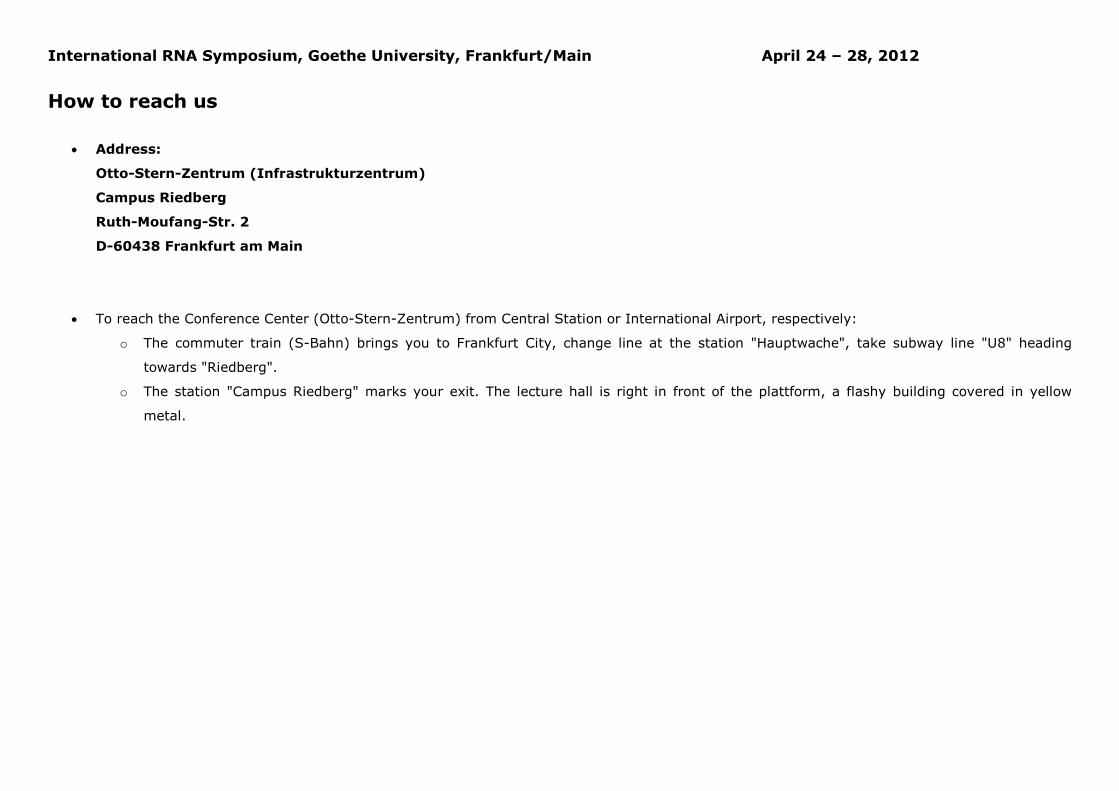
International RNA Symposium, Goethe University, Frankfurt/Main April 24 – 28, 2012
How to reach us
• Address:
Otto-Stern-Zentrum (Infrastrukturzentrum)
Campus Riedberg
Ruth-Moufang-Str. 2
D-60438 Frankfurt am Main
• To reach the Conference Center (Otto-Stern-Zentrum) from Central Station or International Airport, respectively:
o The commuter train (S-Bahn) brings you to Frankfurt City, change line at the station "Hauptwache", take subway line "U8" heading
towards "Riedberg".
o The station "Campus Riedberg" marks your exit. The lecture hall is right in front of the plattform, a flashy building covered in yellow
metal.
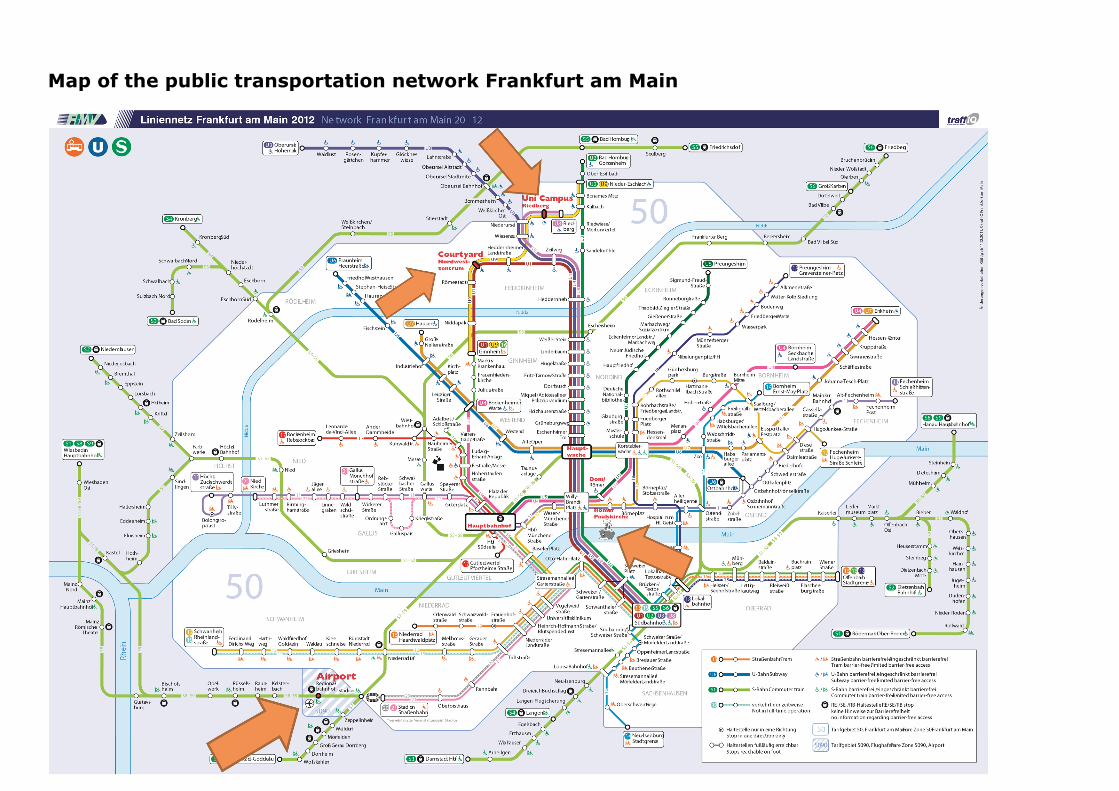
Map of the public transportation network Frankfurt am Main
International RNA Symposium, Goethe University, Frankfurt/Main April 24 – 28, 2012
Impressum Speaker of the SFB 902: Harald Schwalbe E-Mail [email protected]
Organisation: Marco Betz E-Mail [email protected]
Assistants: Rainer Berg and Anna Paulus Organisation Committee: Harald Schwalbe, Beatrix Suess, Alexander Heckel, Enrico Schleiff, Thomas Prisner, Jens Wöhnert
Correspondence to: Marco Betz N100 / 2.OG / Raum Nr. 2.14 Goethe-University Frankfurt am Main Max-von-Laue-Str. 9 60438 Frankfurt am Main, Germany
Next page: Announcement for an upcoming event
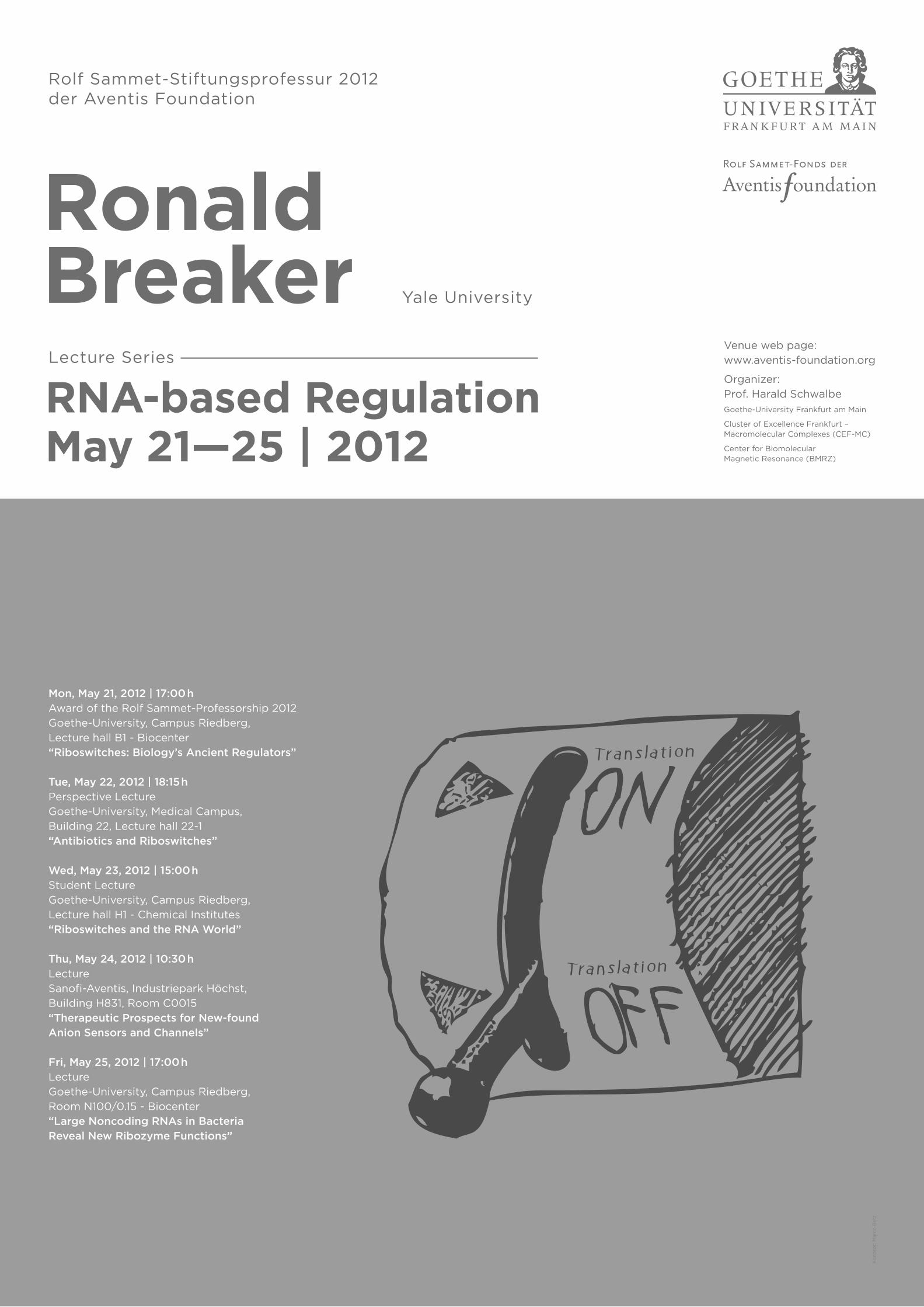
Ronald BreakerRNA-based RegulationMay 21—25 | 2012
Rolf Sammet-Stiftungsprofessur 2012 der Aventis Foundation
Yale University
Lecture Series
Mon, May 21, 2012 | 17:00 hAward of the Rolf Sammet-Professorship 2012
Goethe-University, Campus Riedberg,
Lecture hall B1 - Biocenter
“Riboswitches: Biology’s Ancient Regulators”
Tue, May 22, 2012 | 18:15 hPerspective Lecture
Goethe-University, Medical Campus,
Building 22, Lecture hall 22-1
“Antibiotics and Riboswitches”
Wed, May 23, 2012 | 15:00 hStudent Lecture
Goethe-University, Campus Riedberg,
Lecture hall H1 - Chemical Institutes
“Riboswitches and the RNA World”
Thu, May 24, 2012 | 10:30 hLecture
Sanofi-Aventis, Industriepark Höchst,
Building H831, Room C0015
“Therapeutic Prospects for New-found Anion Sensors and Channels”
Fri, May 25, 2012 | 17:00 hLecture
Goethe-University, Campus Riedberg,
Room N100/0.15 - Biocenter
“Large Noncoding RNAs in Bacteria Reveal New Ribozyme Functions”
Venue web page:
www.aventis-foundation.org
Organizer:
Prof. Harald SchwalbeGoethe-University Frankfurt am Main
Cluster of Excellence Frankfurt –Macromolecular Complexes (CEF-MC)
Center for BiomolecularMagnetic Resonance (BMRZ)
Ko
nzep
t: M
arc
o B
etz