interstitial lung disease final - handout
TRANSCRIPT

9/9/2020
1
Interstitial Lung Disease
James Allen, MDMedical Director, The Ohio State University Wexner Medical Center East
Professor of Internal MedicineDivision of Pulmonary and Critical Care MedicineThe Ohio State University Wexner Medical Center
ILD: The 10,000 Foot View
Which of the following are the leading causes of death?
1. Lung cancer
2. Breast cancer
3. Colon cancer
4. Prostate cancer
5. Ovarian cancer
6. Leukemia
7. Pancreatic cancer
8. Idiopathic pulmonary fibrosis
Which of the following are the leading causes of death?
1. Lung cancer: 158,080 deaths per year
2. Breast cancer
3. Colon cancer
4. Prostate cancer
5. Ovarian cancer
6. Leukemia
7. Pancreatic cancer
8. Idiopathic pulmonary fibrosis

9/9/2020
2
0
20000
40000
60000
80000
100000
120000
140000
160000
180000
LungCancer
ColonCancer
PancreaticCancer
BreastCancer
IdiopathicPulmonary
Fibrosis
ProstateCancer
Leukemia OvarianCancer
Deaths Per YearInterstitial Lung Disease
Accumulation of scar and/or inflammation in the lung
PFTs show restriction and low diffusing capacity
High resolution chest CT– Ground glass infiltrates: inflammation– Honeycomb infiltrates: scar– Reticular markings: inflammation or scar
Hemosiderosis
Granulomatosis with polyangiitis
Drug-Induced Fibrosis
Systemic Sclerosis
Systemic Lupus Erythematosus
Sjogren’s Syndrome
Mycobacterial Infection
Histoplasmosis
Aspiration
Lipoid Pneumonia
Diffuse pulmonary neuroendocrine hyperplasia
Polymyositis
Mixed Connective Tissue Disease
Microlithiasis
Churg-Strauss Syndrome
Pneumocystis carinii
Oxygen Toxicity
Cryptogenic Organizing Pneumonia
Non-Specific Interstitial Pneumonitis
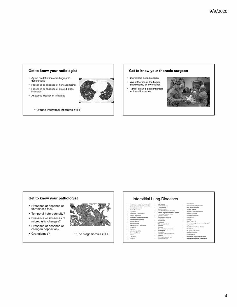
Interstitial Lung Diseases Desquamative Interstitial Pneumonitis
Lymphocytic Interstitial Pneumonitis
Eosinophilic pneumonia
Alveolar Proteinosis
Amyloidosis
Lymphangitic Carcinomatosis
Radiation Pneumonitis
Langerhan’s Cell Granulomatosis
Lymphangioleiomyomatosis
Tuberous Sclerosis
Neurofibromatosis
Hypersensitivity Pneumonitis
Sarcoidosis
Berylliosis
Ankylosing Spondylitis
Rheumatoid Arthritis
Silicosis
Asbestosis
Lymphoma
IgG4 disease
Hard metal disease
Crohn’s disease
Ulcerative collitis
Idiopathic inflammatory myopathy
Familial idiopathic pulmonary fibrosis
Hermansky-Pudlak syndrome
Gaucher’s disease
Goodpasture’s syndrome
Nitrofurantoin
Methotrexate
Amiodarone
Talc granulomatosis
Siderosis
Tannosis
Coal worker’s pneumoconiosis
Sulfasalazine
Minocycline
Idiopathic pulmonary fibrosis
Bleomycin
Mixed dust pneumoconiosis
Hard metal disease
Initial work-up of interstitial lung disease:
Thorough history and physical exam
Autoimmune screen Serologies
Pulmonary function tests
High resolution chest CT

9/9/2020
3
Physical Examination:
Crackles present: IPF Asbestosis
Crackles absent: Sarcoidosis Langerhan’s cell histiocytosis Chronic eosinophilic pneumonia
Digital clubbing:
Mechanic’s Hands
Clues From Infiltrate Locations:
Upper lobes: Silicosis Sarcoidosis Hypersensitivity pneumonitis
Lower lobes: IPF Asbestosis
Peripheral infiltrates Chronic eosinophilic pneumonia
Second-line tests in interstitial lung disease:
Bronchoalveolar lavage Primarily useful when chronic eosinophilic pneumonia, infection, or
malignancy is suspected
Transbronchial biopsy Primarily useful when infection, malignancy, or sarcoidosis is
suspected
Bronchoscopic cryobiopsy Role still not defined
Surgical lung biopsy When ground glass infiltrates predominate When the CT scan is not typical for UIP When there remains diagnostic uncertainty after initial work-up
Interstitial Lung Disease Diagnosis Requires A Multi-Disciplinary Approach
9/9/2020
4
Get to know your radiologist
Agree on definition of radiographic descriptions
Presence or absence of honeycombing
Presence or absence of ground glass infiltrates
Anatomic location of infiltrates
**Diffuse interstitial infiltrates ≠ IPF
Get to know your thoracic surgeon
2 or 3 lobe deep biopsies
Avoid the tips of the lingula, middle lobe, or lower lobes
Target ground glass infiltrates or transition zones
Get to know your pathologist
Presence or absence of fibroblastic foci?
Temporal heterogeneity?
Presence or absences of microcystic changes?
Presence or absence of collagen deposition?
Granulomas? **End stage fibrosis ≠ IPF
Hemosiderosis
Granulomatosis with polyangiitis
Drug-Induced Fibrosis
Systemic Sclerosis
Systemic Lupus Erythematosus
Sjogren’s Syndrome
Mycobacterial Infection
Histoplasmosis
Aspiration
Lipoid Pneumonia
Diffuse pulmonary neuroendocrine hyperplasia
Polymyositis
Mixed Connective Tissue Disease
Microlithiasis
Churg-Strauss Syndrome
Pneumocystis carinii
Oxygen Toxicity
Cryptogenic Organizing Pneumonia
Non-Specific Interstitial Pneumonitis
Interstitial Lung Diseases Desquamative Interstitial Pneumonitis
Lymphocytic Interstitial Pneumonitis
Eosinophilic pneumonia
Alveolar Proteinosis
Amyloidosis
Lymphangitic Carcinomatosis
Radiation Pneumonitis
Langerhan’s Cell Granulomatosis
Lymphangioleiomyomatosis
Tuberous Sclerosis
Neurofibromatosis
Hypersensitivity Pneumonitis
Sarcoidosis
Berylliosis
Ankylosing Spondylitis
Rheumatoid Arthritis
Silicosis
Asbestosis
Lymphoma
IgG4 disease
Hard metal disease
Crohn’s disease
Ulcerative collitis
Idiopathic inflammatory myopathy
Familial idiopathic pulmonary fibrosis
Hermansky-Pudlak syndrome
Gaucher’s disease
Goodpasture’s syndrome
Nitrofurantoin
Methotrexate
Amiodarone
Talc granulomatosis
Siderosis
Tannosis
Coal worker’s pneumoconiosis
Sulfasalazine
Minocycline
Idiopathic pulmonary fibrosis
Bleomycin
Mixed dust pneumoconiosis
Hard metal disease

9/9/2020
5
Idiopathic Pulmonary Fibrosis
Most common ILD of unknown etiology
Mainly affects people > 50 years, most are over the age of 60 years
Incidence is estimated at 7-16 cases per 100,000 per year
The incidence is increasing
Possible risk factors for developing IPF include cigarette smoking, occupational/environmental exposures
Idiopathic Pulmonary Fibrosis
History/Exam Gradual onset and progressive dyspnea and/or a
nonproductive cough Bibasilar inspiratory crackles (Velcro crackles) Clubbing is common
PFTs show restriction, low diffusing capacity and desaturation with exertion
Idiopathic Pulmonary Fibrosis
Diagnosis confirmed by imaging, lung biopsy
CT findings: usual interstitial pneumonitis (UIP) Subpleural, basal predominance interstitial/reticular infiltrates Honeycombing with or without traction bronchiectasis Minimal ground glass infiltrates
Biopsy findings: usual interstitial pneumonitis (UIP) pathologic pattern Temporal & geographic heterogeneity Collagen deposition
Fibroblastic foci
UIP: Peripheral reticular infiltrates

9/9/2020
6
UIP: Traction bronchiectasis UIP: Basilar honeycomb infiltrates
Normal lung alveoli: medium powerThe histologic finding in IPF is usual interstitial pneumonitis (UIP):
1. Temporal & geographic heterogeneity2. Fibroblast foci3. Prominent collagen fibrosis4. Microcysts

9/9/2020
7
Temporal & Geographic Heterogeneity in UIP
A
BC
Fibroblastic foci in UIP
Collagen deposition in UIP Microcysts in UIP

9/9/2020
8
IPF: Honeycombing
Usual Interstitial Pneumonitis ≠ IPF
Causes Of Usual Interstitial Pneumonitis*
Rheumatoid-associated
Scleroderma-associated
Chronic hypersensitivity pneumonitis
Radiation-induced
Drug-induced
“Post-inflammatory pulmonary fibrosis”
Idiopathic pulmonary fibrosis
*Different causes of UIP have very different prognoses and treatments
What causes IPF?
#2 Epithelial Injury
Dusty environment
Tobacco smoke
Viruses
Acid reflux/aspiration
#1 Genetic Predisposition
Surfactant protein C
Surfactant protein A2
TERT
TERC
MUC5B

9/9/2020
9
Familial pulmonary fibrosis:
Accounts for 5-10% of patients with IPF
Patients look just like IPF
Typically ages 50-70
Definition: first degree relative with IPF
Probably autosomal dominant with variable penetrance
Genetic cause found in about 10% of familial pulmonary fibrosis
Treatment is the same as IPF
IPF Treatment: What Works?
Oxygen
Pulmonary rehabilitation
Anti-fibrotic drugs: Pirfenidone Nintedanib
Lung transplant
Effect of pulmonary rehabilitation on interstitial lung disease
300310320330340350360370380390400
6MWT Distance (m)
Pre
Post
0
0.5
1
1.5
2
2.5
3
3.5
4
Borg Scale
Pre
Post
Collard et al. Chest 2009; 135:442-7
What medications do work for IPF?
Anti-fibrotic TGFβ inhibitor
Slows rate of progression by about half
3 capsules three times daily
Side effects: Sun sensitivity Nausea, weight loss Increased liver enzymes
$90-100,000 per year
Nintedanib (Ofev)
Tyrosine kinase inhibitor
Slows rate of progression by about half
150 mg twice daily
Side effects: Diarrhea Nausea, weight loss Increased liver enzymes
$90-100,000 per year
Pirfenidone (Esbriet)

9/9/2020
10
Pirfenidone versus Placebo
-500
-400
-300
-200
-100
0
Pirfenidone
Placebo
Weeks0 12 24 36 52
Cha
nge
in F
VC
(m
l)
N Engl J Med 2014; 370:2083-2092
Nintedanib versus Placebo
-300
-250
-200
-150
-100
-50
0
Nintedanib
Placebo
Weeks0 12 24 36 52
Cha
nge
in F
VC
(m
l)
N Engl J Med 2014; 370:2071-2082
Adult Lung Transplants Major Diagnoses by Year
0
500
1,000
1,500
2,000
2,500
3,000
3,500
4,000
Nu
mb
er o
f T
ran
spla
nts
Transplant Year
COPD A1ATD CF IPF ILD-not IPF Retransplant
2019JHLT. 2019 Oct; 38(10): 1015-1066
0
25
50
75
100
0 1 2 3 4 5 6 7 8 9 10 11 12 13 14 15 16 17 18 19 20
Su
rviv
al
(%)
Years
A1ATD (N=3,257) CF (N=9,428) COPD (N=19,159)
IPF (N=15,574) ILD-not IPF (N=3,336) IPAH (N=1,944)
Adult Lung Transplants Survival by Major Diagnosis
2019JHLT. 2019 Oct; 38(10): 1015-1066
Median IPF survival = 5.2 years

9/9/2020
11
Typical Clinical Course
Disability
Time (years)
Death
1 2 3 4 5
“Stair-Step” Clinical Course (exacerbations of IPF)
Disability
Time (years)
Death
1 2 3 4 5
When patients with IPF are worse:
Progression of IPF
Acute interstitial pneumonitis
Anemia
Heart failure
Pulmonary embolism
Lung cancer
Infection
Pneumothorax
Clinical approach to IPF:
1. Establish a confident diagnosis
2. Search for non-IPF contributors of dyspnea Heart failure Anemia Infection Thyroid disease
3. Assess candidacy for transplant early

9/9/2020
12
Clinical approach to IPF (continued):
4. Stop smoking
5. Assess for esophageal reflux GERD Hiatal hernias
6. Monitor disease progression FVC TLC DLCO Oxygen saturation with exercise
7. Start home oxygen early
Clinical approach to IPF (continued):
8. Pulmonary rehabilitation
9. Assess for & treat depression
10. Regular vaccinations
11. End of life discussions early
And some other interstitial lung diseases…
Non-Specific Interstitial Pneumonitis
Second most common idiopathic interstitial pneumonia
Affects men and women equally with an average age about 10 years younger than IPF
Almost always seen in the setting of rheumatologic disease
Shortness of breath and dry cough
Physical findings include inspiratory crackles, ± clubbing

9/9/2020
13
Non-Specific Interstitial Pneumonitis
Important to differentiate from IPF NSIP 5 year mortality <10% Survival > 6-10 years
Treatment Prednisone Mycophenolate Azathioprine Cyclophosphamide
“Mechanic’s Hands”
Non-specific interstitial pneumonitis: Ground Glass Infiltrates
Non-specific interstitial pneumonitis (NSIP) pathology:
– Alveolar wall inflammatory cells
– Temporal homogeneity
– No fibroblast foci
– Minimal collagen fibrosis
Hypersensitivity Pneumonitis
Etiology often hard to identify– Birds, feathers, down– Hot tubs– Occupation– Drugs
Pathology:– T-suppressor cell alveolitis– Poorly formed granulomas
Treatment:– Remove offending
antigen– Prednisone
Outcome:– Complete resolution– Chronic fibrosis

9/9/2020
14
Hypersensitivity Pneumonitis Radiology
Frequently upper lobe dominant infiltrates
Ground glass infiltrates
Nodular infiltrates
Lymphocytic infiltratesMultinucleated Giant CellPoorly-formed granuloma

9/9/2020
15
Lettuce farm worker with dyspnea Hot Tub Lung
41 year old woman referred for suspected idiopathic pulmonary fibrosis
And then she said…
“And oh by the way, did I tell you that we raise goats and donkeys in the barn in our back yard?”
“And oh by the way, did I tell you we have a Quacker Parrot? And Cockatiels? And Parakeets?”
“And oh by the way, did I tell you we have birds living in our attic and there’s a hole in my closet ceiling so that my clothes are covered with bird feathers and bird poop?”

9/9/2020
16
Common Occupational Lung Diseases
Asbestosis: boilermakers, plumbers, pipefitters
Silicosis: miners, quarry workers, sandblasters, foundry workers, many others
Asbestosis
CT appearance resembles UIP
Pathology resembles UIP May see ferruginous bodies
Diagnosis usually made based on occupational history
Bilateral calcified pleural plaques strongly supports exposure However plaques ≠ asbestosis

9/9/2020
17
60 year old woman whose father was an pipe-fitter; her mother laundered his dusty work clothes. Both her mother and father died of asbestosis
78 year old man with dyspnea for 10 years. Formerly worked in construction
Silicosis
Upper lobe predominant pulmonary nodules
Upper lobe progressive massive fibrosis
Calcified mediastinal lymph nodes

9/9/2020
18
Foundry Worker With Dyspnea
Lymphocytic Interstitial Pneumonitis
Most commonly seen with Sjogren’s Presents with multiple bilateral thin-walled cysts
Can be seen with HIV, lupus, autoimmune myositis, rheumatoid arthritis Presents with diffuse interstitial/alveolar infiltrates
Treatment = steroids, mycophenolate, or azathioprine
Need to watch for evolution into lymphoma
Sjogren’s-associated LIP

9/9/2020
19
Desquamative Interstitial Pneumonitis
> 90% are smokers– Rarely associated with collagen vascular disease
Typical age = 30-50
Chest CT: – Ground glass infiltrates– CXR may be normal
Pathology:– Abundant smoker’s macrophages– Little alveolar wall inflammation– Little fibrosis
Desquamative Interstitial Pneumonitis
Desquamative Interstitial Pneumonitis Desquamative Interstitial Pneumonitis

9/9/2020
20
Desquamative Interstitial Pneumonitis Desquamative Interstitial Pneumonitis:
Treatment: Smoking cessation!!!! Steroids Occasionally immunosuppressive medications
Prognosis: Excellent if treated early
Sarcoidosis
Multi-organ disease
High incidence in young African Americans & Scandinavians
Exam = normal lung auscultation; +/-erythema nodosum
Chest x-ray = adenopathy and/or nodular infiltrates
BAL = increased lymphocytes (T helper) Erythema Nodosum
Sarcoidosis

9/9/2020
21
Sarcoidosis Sarcoidosis
Diagnosis based on finding granulomatous inflammation in a patient with a compatible clinical history
Rule out other cause of granulomas Infections such as mycobacterial and fungal infections Beryllium exposure Foreign body granulomas
Differentiate from granulomas related to hypersensitivity pneumonitis
Sarcoidosis
Evaluation:• Pulmonary function tests• Eye exam• EKG• Calcium level• Liver function tests
Treatment:• Prednisone
• Methotrexate second line• Mild cases may resolve without
treatment• Prognosis for recovery is
generally good
Watch the May 2020 MedNet webcast on Sarcoidosis!
Acute interstitial pneumonitis
Diagnosis of exclusion
Sudden-onset of worsened oxygenation and ground glass infiltrates
Can occur as: With underlying IPF Idiopathic With underlying rheumatologic disease
Lung biopsy = diffuse alveolar damage (identical to ARDS)
Steroids may help

9/9/2020
22
Acute interstitial pneumonitis Acute Interstitial Pneumonitis
Acute lung injury
Pathology = diffuse alveolar damage Indistinguishable histologically from ARDS
Acute interstitial pneumonitis is a diagnosis of exclusion
Heart failure Consider BNP Consider cardiac echo
Pulmonary embolism Consider CT-PA
Infection Consider BAL
Langerhan’s Cell Histiocytosis
Adult form: generally limited to lung involvement Childhood form is a multisystem malignancy of Langerhan’s cells
Primarily occurs in smokers
Chest CT shows multiple small thin-walled cysts plus nodules
Biopsy shows stellate nodules staining for Langerhan’s cells CD1a stain S-100 stain

9/9/2020
23
Langerhan’s Cell Histiocytosis: 65 year old woman with dyspnea for 6 months
Langerhan’s Cell Histiocytosis: 57 year old woman with dyspnea for 1 year
Cryptogenic Organizing Pneumonia
Initially present with a subacute flu-like syndrome that lasts for a few weeks
Physical examination, laboratory testing is nonspecific
Lung biopsy is diagnostic
Usually responds to corticosteroids
Good prognosis is caught early
Can be idiopathic or can be the initial presentation of underlying rheumatologic or autoimmune disease
64year old woman with “inflammatory arthritis”
9/9/2020
24
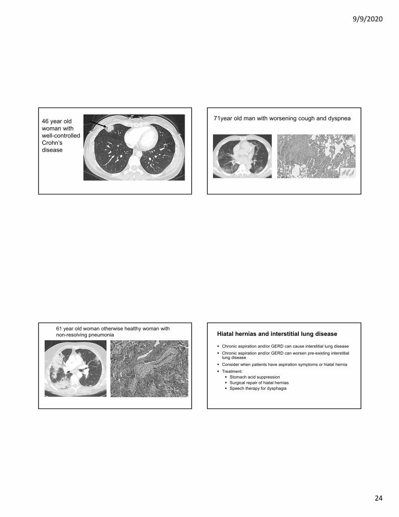
46 year old woman with well-controlled Crohn’sdisease
71year old man with worsening cough and dyspnea
61 year old woman otherwise healthy woman with non-resolving pneumonia Hiatal hernias and interstitial lung disease
Chronic aspiration and/or GERD can cause interstitial lung disease
Chronic aspiration and/or GERD can worsen pre-existing interstitial lung disease
Consider when patients have aspiration symptoms or hiatal hernia
Treatment: Stomach acid suppression Surgical repair of hiatal hernias Speech therapy for dysphagia
9/9/2020
25
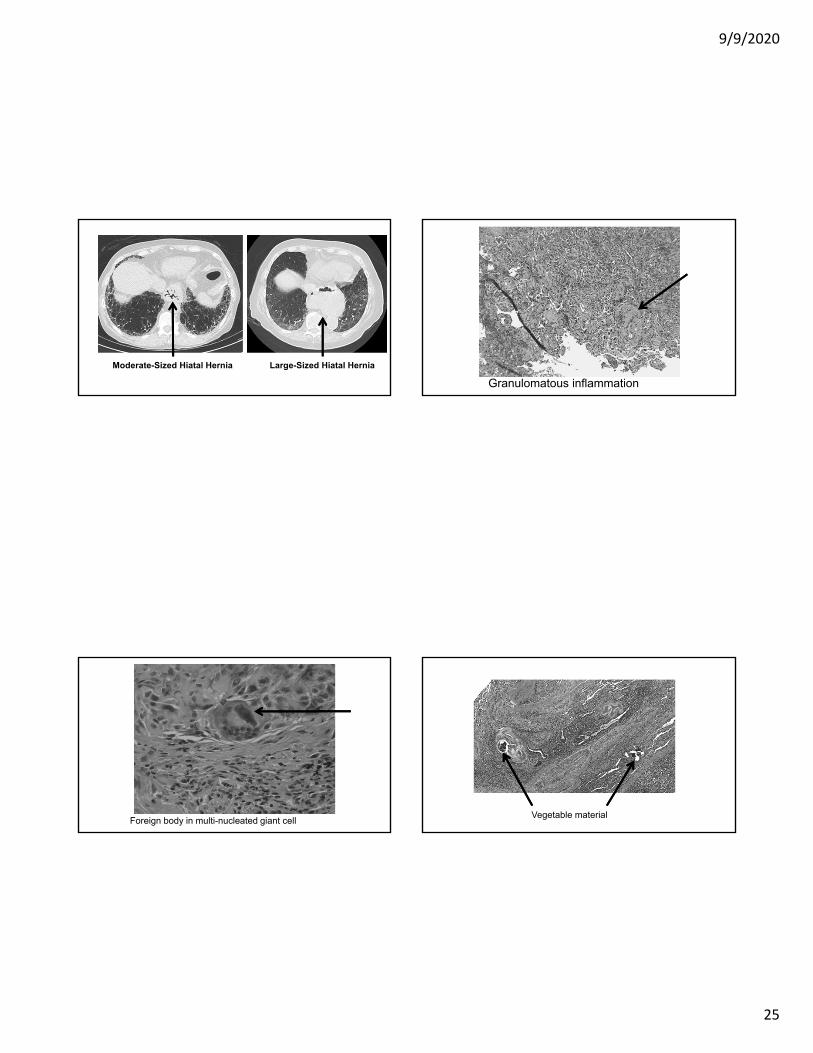
Moderate-Sized Hiatal Hernia Large-Sized Hiatal Hernia
Granulomatous inflammation
Foreign body in multi-nucleated giant cellVegetable material

9/9/2020
26
Drug-induced lung disease
Difficult to diagnose
No reliable clinical, imaging, bronchoalveolar lavage(BAL), or histopathologic feature that is specific of, or diagnostic for drug-induced ILD
Establish a definite temporal relationship between exposure to the agent and the onset of the lung disease
Stop the drug, consider corticosteroids
Drug-induced lung disease
Dozens of drugs implicated
Common drugs: Minocycline Nitrofurantoin (macrodantin) Amiodarone Methotrexate Chemotherapy drugs
www.pneumotox.com is a great reference
Macrodantin-induced lung disease 70 year old woman who was told she had IPF12 years ago. She took macrodantin daily from 1996 - 1999

9/9/2020
27
Sulfasalazine-induced lung diseaseFebruary 2017
Pre-AmiodaroneMay 2017
During AmiodaroneJune 2017
Post-Amiodarone
Talc Granulomatosis
History = remote IV drug use (especially Ritalin)
Exam = soft basilar crackles
PFTs = resemble emphysema
HRCT = may be normal
Biopsy = polarizable foreign body material
Treatment = none

9/9/2020
28
Interstitial Lung Disease: Summary
Your history is the most important diagnostic tool
A confident diagnosis requires a multidisciplinary approach
“UIP” is a CT pattern and a histologic pattern, it is not a disease